Segmental Liver Anatomy SEGMENTAL LIVER ANATOMY · 2018-09-10 · Segmental Liver Anatomy SEGMENTAL...
1
Segmental Liver Anatomy The Couinaud classification divides the liver into 8 functional segments. The hepatic veins are found at the periphery of each segment, whereas the center has branches of the portal veins, hepatic arteries, and bile ducts. The middle hepatic vein divides the liver into left and right lobes. The left hepatic vein divides the left lobe into lateral (2, 3) and medial (4a, 4b) segments. The right hepatic vein divides the right lobe into anterior (5, 8) and posterior (6, 7) segments. The caudate lobe (1) has hepatic veins that often drain directly into the IVC. The portal vein divides the liver into upper (2, 4a, 8, 7) and lower (3, 4b, 5, 6) segments, and can usually be identified without IV contrast. A line drawn from the middle of the gallbladder fossa to the IVC (green) roughly divides the liver into left and right lobes. The falciform ligament (red) roughly divides the left lobe into lateral and medial segments (the left hepatic vein usually is located slightly to the left of the falciform ligament. More detailed information can be obtained at Radiology Assistant. Radiological Society of the Netherlands: http://www.radiologyassistant.nl/en/4375bb8dc241d , and Virtual Liver. Toronto General Hospital Department of Surgery http://pie.med.utoronto.ca/VLiver/ 3 4b 5 6 3 4b 5 6 3 4b 5 6 RL LL RL LL RL LL C C C 2 4a 8 7 8 7 8 7 2 4a 2 4a 1 1 1 Off-the-Wall Guide
Transcript of Segmental Liver Anatomy SEGMENTAL LIVER ANATOMY · 2018-09-10 · Segmental Liver Anatomy SEGMENTAL...
Segmental Liver Anatomy
SEGMENTAL LIVER ANATOMY
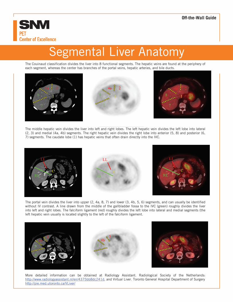
The Couinaud classification divides the liver into 8 functional segments. The hepatic veins are found at the periphery of each segment, whereas the center has branches of the portal veins, hepatic arteries, and bile ducts.
The middle hepatic vein divides the liver into left and right lobes. The left hepatic vein divides the left lobe into lateral (2, 3) and medial (4a, 4b) segments. The right hepatic vein divides the right lobe into anterior (5, 8) and posterior (6, 7) segments. The caudate lobe (1) has hepatic veins that often drain directly into the IVC.
The portal vein divides the liver into upper (2, 4a, 8, 7) and lower (3, 4b, 5, 6) segments, and can usually be identified without IV contrast. A line drawn from the middle of the gallbladder fossa to the IVC (green) roughly divides the liver into left and right lobes. The falciform ligament (red) roughly divides the left lobe into lateral and medial segments (the left hepatic vein usually is located slightly to the left of the falciform ligament.
More detailed information can be obtained at Radiology Assistant. Radiological Society of the Netherlands: http://www.radiologyassistant.nl/en/4375bb8dc241d, and Virtual Liver. Toronto General Hospital Department of Surgery http://pie.med.utoronto.ca/VLiver/
34b
5
6
34b
5
6
34b
5
6
RL
LL
RL
LL
RL
LL
C C C
24a
8
7
8
7
8
7
24a 24a
1 1 1
Off-the-Wall Guide