SECTION ONE CORE UNIT TWO CELL AND CELL ORGANELLES/CELL ...
37
MUGWE MARTIN AUTHOR OF NEW BIOLOGY EXTRA//0708028584 1 A NOVEL DISSECTON OF CYTOLOGY/NEW BIOLOGY EXTRA//MUGWE.MARTIN//0780280584 SECTION ONE CORE UNIT TWO CELL AND CELL ORGANELLES/CELL CHEMISTRY AND FUNCTION Major Concept To study the molecular and functional organisation of a cell and its subcellular organelles Specific Objectives 1. To know importance of cell, and the types: Prokaryotic and eukaryotic cell. 2. Learn the essential differences of a prokaryotic cell and eukaryotic cell. 3. Draw a diagram of an eukaryotic cell showing different cell organelles. 4. Study the following cellular organelles: • Nucleus—its structure and functions. • Mitochondrion, the power house of a cell. Learn its structure and functions. • Study endoplasmic reticulum, its types, structure and functions. • Learn structure and functions of Golgi complexes. • Study about lysosomes, • Learn about peroxisomes: Their structure and functions. • Study the structure and functions of cytoskeleton. COMMON TERMS ENCOUNTERED IN CYTOLOGY TERM(S) LITERAL MEANINGS/UNDERSTANDING Cytology Study of cells by means of light and electron microscopes; Cell theory Protoplasm Organelle Tissue Cytogenetic Primary lysosome Glycosylation *S/Svedberg unit Eukaryotes Prokaryotes Polyribosomes (polysomes) Cytosol Cell inclusions Compartmentation Glycocalyx All living matter composed of cells; all cell arise from pre-existing cells; all metabolism occurs in cells; Living contents of the cell consisting of the cytoplasm and one or more nuclei Structurally and functionally discrete component of a living cell frequently membrane bound A collection of cells physically linked and associated with intercellular substance specialized to perform a particular function; Linking cytology with genetics; relating structure and behaviour of chromosomes during cell division; Refers to the vesicle that has just budded off from the Golgi body containing processed enzymes Process of combining carbohydrates with proteins forming glycol- proteins; Related to the rate of sedimentation in a centrifuge/greater the number the greater the rate of sedimentation (70S/80S) Organisms with DNA enclosed by the nuclear envelope Organism with DNA not enclosed by the nuclear membrane Chains of ribosomes Fluid part of the cytoplasm Structures suspended in the cytoplasm but donot perform any metabolic role Refers to the development of separate organelles performing specific roles in a cell Long carbohydrate molecule attached to membrane lipids and proteins
Transcript of SECTION ONE CORE UNIT TWO CELL AND CELL ORGANELLES/CELL ...

MUGWE MARTIN AUTHOR OF NEW BIOLOGY EXTRA//0708028584
1 A NOVEL DISSECTON OF CYTOLOGY/NEW BIOLOGY EXTRA//MUGWE.MARTIN//0780280584
SECTION ONE CORE UNIT TWO
CELL AND CELL ORGANELLES/CELL CHEMISTRY AND FUNCTION Major Concept
To study the molecular and functional organisation of a cell and its subcellular organelles
Specific Objectives
1. To know importance of cell, and the types: Prokaryotic and eukaryotic cell.
2. Learn the essential differences of a prokaryotic cell and eukaryotic cell.
3. Draw a diagram of an eukaryotic cell showing different cell organelles.
4. Study the following cellular organelles:
• Nucleus—its structure and functions.
• Mitochondrion, the power house of a cell. Learn its structure and functions.
• Study endoplasmic reticulum, its types, structure and functions.
• Learn structure and functions of Golgi complexes.
• Study about lysosomes,
• Learn about peroxisomes: Their structure and functions.
• Study the structure and functions of cytoskeleton.
COMMON TERMS ENCOUNTERED IN CYTOLOGY
TERM(S) LITERAL MEANINGS/UNDERSTANDING
Cytology Study of cells by means of light and electron microscopes;
Cell theory
Protoplasm
Organelle
Tissue
Cytogenetic
Primary lysosome
Glycosylation
*S/Svedberg unit
Eukaryotes
Prokaryotes
Polyribosomes
(polysomes)
Cytosol
Cell inclusions
Compartmentation
Glycocalyx
All living matter composed of cells; all cell arise from pre-existing cells;
all metabolism occurs in cells;
Living contents of the cell consisting of the cytoplasm and one or more
nuclei
Structurally and functionally discrete component of a living cell
frequently membrane bound
A collection of cells physically linked and associated with intercellular
substance specialized to perform a particular function;
Linking cytology with genetics; relating structure and behaviour of
chromosomes during cell division;
Refers to the vesicle that has just budded off from the Golgi body
containing processed enzymes
Process of combining carbohydrates with proteins forming glycol-
proteins;
Related to the rate of sedimentation in a centrifuge/greater the
number the greater the rate of sedimentation (70S/80S)
Organisms with DNA enclosed by the nuclear envelope
Organism with DNA not enclosed by the nuclear membrane
Chains of ribosomes
Fluid part of the cytoplasm
Structures suspended in the cytoplasm but donot perform any metabolic
role
Refers to the development of separate organelles performing specific
roles in a cell
Long carbohydrate molecule attached to membrane lipids and proteins

MUGWE MARTIN AUTHOR OF NEW BIOLOGY EXTRA//0708028584
2 A NOVEL DISSECTON OF CYTOLOGY/NEW BIOLOGY EXTRA//MUGWE.MARTIN//0780280584
CELL Cell refers to the basic unit of structure and function in living organism.
The concept of homeostasis at cellular level Cell is physically separated from its environment by cell membrane/plasma membrane;yet
capable of exchange with its environment;chemicals which are raw materials are acquired
and waste products removed;in doing work, the system can maintain stability;control of
enzyme activity allosterically in cells;
Thus cell is the fundamental unit of life.
Modern cell theory can be divided into the following fundamental statements: Cells make up all living matter
All cells arise from other cells
The genetic information required during the maintenance of existing cells and the
production of new cells passes from one generation to the Other next generation
The chemical reaction of an organism that is its Metabolism, both anabolism and catabolism,
takes place in the cells.
ORGANISMAL THEORY (REACTIONS TOWARDS THE PROPONENTS OF THE CELL
THEORY)
The whole organism is the basic entity and cells are merely incidental sub-units
Looks at an organism as a Utopia of interdependent cells whose function are dictated by the
needs of the whole organism.
EXCEPTIONS TO THE CELL THEORY/DISCREPANCIES
Mitochondria and chloroplast have their own genetic material and hence are self-replicating
Viruses are considered alive by some but not made up of cells. Viruses have very many
features of living things but by definition of cell theory they are not alive
The first cell did not originate from pre-existing cells.
UNITS OF LIFE BEYOND THE CELL
UNIT(S) NOTES
Tissues
Organs
Organ-system
Organisms
Populations
communities
Group of cells, usually of the same type specialized to perform
specific functions eg xylem/bone/muscle/epithelium
Made up of tissues coordinated to perform certain functions eg
eye/leaf/kidney
Group of organs which combine to perform a specific function eg
endocrine system
Depending on their complexity, may each be just one cell eg bacterium
or Amoeba or millions of cells with a variety of functional units as
above eg oak tree/man.
Group of organism of the same species occupying a given area at a
particular time eg a small herd of cattle
Population of different species living in balance in nature. They form
part of the biosphere

MUGWE MARTIN AUTHOR OF NEW BIOLOGY EXTRA//0708028584
3 A NOVEL DISSECTON OF CYTOLOGY/NEW BIOLOGY EXTRA//MUGWE.MARTIN//0780280584
SCHEMATA SHOWING DIFFERENT UNITS OF LIFE
TYPES OF CELLS
In general two types of cells exist in nature. They are:
1. Prokaryotic cells
2. Eukaryotic cells
PROKARYOTIC CELLS
Typical prokaryotic cells (Greek: Pro-before and karyon- nucleus) include the bacteria and
cyanobacteria. Most studied prokaryotic cell is Escherichia coli (E. coli).
CHARACTERISTICS
• Minimum of internal organisation and smaller in size
• Does not have any membrane bound organelles.
• Genetic material is not enclosed by a nuclear membrane
• DNA is not complexed with histones. Histones are not found in prokaryotic cells
• Respiratory system is closely associated with its plasma membrane and
• Sexual reproduction does not involve mitosis or meiosis.
Morphology of bacterial cells
Size
o Small in size/0.1 to 10 microns/µm;
Shape
o Cocci; spherical/oval; eg streptococcus pyogenes;
o Bacillus; rods; e.g. bacillus anthrancis;
o Filamentous; branched/thin filaments; eg actinomycetes;
o Spirillum; curved/spiral; single flagellum; eg treponoma pallidum
o Vibrios; comma shaped; eg vibrio cholerae;
Arrangement
Cocci
o Streptococci; chains of bacterial cells; eg Streptococcus pneumoniae;
o Staphylococci; bunches; eg staphylococcus aureus;

MUGWE MARTIN AUTHOR OF NEW BIOLOGY EXTRA//0708028584
4 A NOVEL DISSECTON OF CYTOLOGY/NEW BIOLOGY EXTRA//MUGWE.MARTIN//0780280584
o Diplococci; pairs; eg diplococcus pneumoniae;
o Tetrad; packets of four; eg pediococcus;
o Sarcinae; packets of eight; eg sarcina ureae;
Bacillus
o Single bacillus; eg E.coli;
o Streptobacillus; chains; e.g. streptobacillus moniliform;
o Coccobacilli; short and ovoid; eg haemophilus influenza;
Drawing of a prokaryotic cell
KINGDOM PROKARYOTAE/MONERA
Contains two major phyla i.e. eubacteria and cyanobacteria
The Archea-bacteria are not true bacteria/pseudo-bacteria/primitive bacteria and they fall in
three categories’.
Methanogens
Thermoacidophils and halophiles
FUNCTONS OF THE PARTS OF A BACTERIUM
PARTS ROLE(S)
Cell wall
Capsule
Cellsurface
membrane
Physical barrier which protects against mechanical damage and exudes certain
substances
Protects bacterium from other cells eg white blood cells, also helps groups of
bacteria to stick together for further protection;
Differentially permeable layer which controls entry and exit of chemicals;

MUGWE MARTIN AUTHOR OF NEW BIOLOGY EXTRA//0708028584
5 A NOVEL DISSECTON OF CYTOLOGY/NEW BIOLOGY EXTRA//MUGWE.MARTIN//0780280584
Mesosomes
Flagellum
Pili
Circular DNA
Plasmids
Ribosomes
Glycogen granule
Lipid droplet
Photosynthetic
lamellae
Provide large surface area for attachment of respiratory enzymes;
For movement of bacterium because its rigid, corkscrew shape and rotating base
helps the cell spin through fluids;
Attachment on surfaces;
Genetic information for replication of bacteria.
Genes for survival in harsh conditions eg in presence of antibiotics
70S type for protein synthesis
Stores carbohydrate for break down during respiration to provide energy
Store lipids as a more concentrated, longer-term, store for conversion to
carbohydrate and use in respiration;
Contain enzymes and bacterio- chlorophyll and therefore carry out
photosynthesis; GRAM POSITIVE AND GRAM NEGATIVE BACTERIA
GRAM POSITIVE GRAM NEGATIVE
Lack lipid layer along their murein;
Affected
by antibacterial enzymes/lysozyme;
high concentration of murein
Stain purple with Gram stain/crystal violet
solution;
Cell wall is thicker;
Have lipid layer along their murein;
Not affected by antibacterial enzymes;
Low concentration of murein
Becomes pink on decolourisation with ethanol;
Cell wall is thinner;
ADAPTATIONS OF BACTERIA
Capsules and slimy layer/gummy secretions; make them less susceptible to phagocytosis by
white blood cells;
Cell wall rigid due to peptidoglycan/murein for support;/ prevent osmotic shocks/ Gram
negative bacteria have lipid-rich layer covering murein which confers resistance to antibiotics;
Flagellum;long to propel the bacteria along;
Pili/Fimbriae; for cell to surface attachment providing support/cell to cell attachment
forming colonies;
Sex pilus;sexual reproduction/conjugation;
Mesosomes;in folding of the plasma membrane, housing enzymes for respiration/aid in the
formation of the new cross wall/facilitates the separation of the two daughter molecules of
DNA;
Photosynthetic sacs; tubular/sheet-like containing photosynthetic pigments; for
photosynthesis;
Circular DNA;containing genes which control the activities of the cell;
Ribosomes; sites for protein synthesis;
Production of large quantities of spores; which results into rapid multiplications;
Endospores;thick walled extremely resistant to harsh condition;
Plasmid;extra- chromosomal DNA which confers ability to antibiotics resistance/ use of
hydrocarbons/sexual reproduction/ conjugation;
Small in size; decreases nutrient requirements/increasing surface area for diffusion of
nutrients into the cell;

MUGWE MARTIN AUTHOR OF NEW BIOLOGY EXTRA//0708028584
6 A NOVEL DISSECTON OF CYTOLOGY/NEW BIOLOGY EXTRA//MUGWE.MARTIN//0780280584
ENDOSYMBIOTIC THEORY/ENDOSYMBIOGENESIS
Evolution of membrane bound organelles is explained by Endosymbiotic theory or
endosymbiogenesis;Mitochondria and chloroplast were initially independent prokaryotic cells
bacteria like organisms which gained access, by accident, to the host cell (larger prokaryotic
cell) and entered symbiotic relationshipPresumably conditions in the cell host were favorable
for prokaryote whilst in return the prokaryotes provided increased capacity of cell to form
ATP (mitochondria) and chloroplast increased cells capacity to form food.
OR
Alternately the organelles may have risen by invaginations of the plasma membrane which
became “pinched off” to give separate membrane bound structure within the main cell. EVIDENCES FOR THE ENDOSYMBIOTIC THEORY
Both mitochondria and chloroplast have naked and circular DNA
Both mitochondria and chloroplast have 70S ribosome
Both mitochondrial and chloroplast protein synthesis are sensitive to antibiotics for example
chloramphenical and streptomycin
The ability of the mitochondria and chloroplast to divide themselves/self-replication;
Ribosomes of mitochondria and chloroplasts resemble those of bacteria and cyanobacteria,
with respect to size and nucleotide sequence
Mitochondria and chloroplasts reproduce independently of their eukaryotic host cell by a
Process similar to the binary fission of bacteria.
The thylakoid membranes of chloroplasts resemble the photosynthetic membranes of
cyanobacteria.
Advantages conferred due to the presence of membrane organelles
Potentially harmful reactants or enzymes can be isolated inside organelle so they won’t
damage the rest of the cell;
The rate of any metabolic pathway inside an organelle can be controlled by regulating the
rate at which the membrane surrounding the organelle allows the first reactant to enter.
Containing enzymes for particular metabolic pathway within organelles means product of one
reaction will always be at a close proximity to the next enzyme in the sequence.
The organelles increase surface area to volume ratio because as the cells become larger, the
proportion of membrane area to cell volume reduced.
Specialization resulting into orderliness and efficiency
EUKARYOTIC CELLS
The eukaryotic cells (Greek: Eu-true and karyon-nucleus) include the protists, fungi, plants
and animals including humans. Cells are larger in size. CHARACTERISTICS
Considerable degree of internal structure with a large number of distinctive membrane
enclosed having specific functions
Nuc l eus is the site for informational components collectively called chromatin
Sexual reproduction involves both mitosis and meiosis
Respiratory site is the mitochondria
In the plant cells, the site of the conversion of radiant energy to chemical energy is the
highly structural chloroplasts.

MUGWE MARTIN AUTHOR OF NEW BIOLOGY EXTRA//0708028584
7 A NOVEL DISSECTON OF CYTOLOGY/NEW BIOLOGY EXTRA//MUGWE.MARTIN//0780280584
ANIMAL CELL/ULTRASTRUCTURE
PLANT CELL
These are found in plants Cell membrane
organelles
Inclusions Cell organelles (i) Nucleus
Ribosomes
Endoplasmic reticulum
(SER/RER)
Mitochondria (v) Dictyosomes (vi) Microfilaments (vii) Microtubules (viii) Vacuole
Chloroplasts Cytoplasmic inclusions (i) Starch grains
Fat droplets
PLANT AND ANIMAL CELL SEEN UNDER LIGHT MICROSCOPE

MUGWE MARTIN AUTHOR OF NEW BIOLOGY EXTRA//0708028584
8 A NOVEL DISSECTON OF CYTOLOGY/NEW BIOLOGY EXTRA//MUGWE.MARTIN//0780280584
COMPARISONS
FEATURE ANIMAL CELL PLANT CELL
Cell wall
Plastids
Centrioles
Shape
Vacuole
Food stored
Vesicles
Cholesterol
Cilia/flagella
plasmodesmata
Irregular
small/many
Glycogen
/regular
One large
Starch
ULTRA-STRUCTURE OF A PLANT CELL
Comparisons of prokaryotes and eukaryotes SIMILARITIES
o Both have plasma membrane;
o Both have ribosomes;
o Both have cytoplasm;
o Both are composed of cells;
o Both have genetic material, DNA;
o Both have cilia and flagella;
o Both carry out life processes such as reproduction and photosynthesis;
o

MUGWE MARTIN AUTHOR OF NEW BIOLOGY EXTRA//0708028584
9 A NOVEL DISSECTON OF CYTOLOGY/NEW BIOLOGY EXTRA//MUGWE.MARTIN//0780280584
DIFFERENCES
Prokaryotic cell Eukaryotic cell 1. Smaller in size 1 to 10 μm 1. Larger in size 10 to 100 μm or more 2. Mainly unicellular 2. Mainly multicellular (with few exceptions). Several
different types present 3. Single membrane, surrounded by rigid cell wall 3. Lipid bilayer membrane with proteins 4. Anaerobic or aerobic 4. Aerobic 5. Not well defined nucleus, only a nuclear zone with
DNA 5. Nucleus well defined, 4 to 6 μm in diameter, contains DNA
and surrounded by a perinuclear membrane Histones absent Histones present
6. No nuclei 6. Nucleolus present, rich in RNA 7. Cytoplasm contains no cell organelles 7. Membrane bound cell organelles are present 8. Ribosomes present free in cytoplasm 8. Ribosomes studded on outer surface of endoplasmic
reticulum present 9. Mitochondria absent. Enzymes of energy 9. Mitochondria present Power house of the cell.
metabolism bound to membrane Enzymes of energy metabolism are located in Mitochondria 10. Golgi apparatus absent. Storage granules 10. Golgi apparatus present—flattened single membrane
with polysaccharides Vesicles 11. Lysosomes—absent 11. Lysosomes present—single membrane vesicle containing
packets of hydrolytic enzymes 12. Cell division usually by fission, no mitosis 12. Cell division—by mitosis 13. Cytoskeleton—absent 13. Cytoskeleton—present 14. RNA and protein synthesis in same compartment 14. RNA synthesised and processed in nucleus. Proteins
synthesised in cytoplasm 15. Examples are bacteria, cyanobacteria, rickettsia 15. Examples: Protists, fungi, plants and animal cells
CELL ORGANELLES
NUCLEUS:
The nucleus contains more than 95 per cent of the cell’s DNA and is the control centre of
the eukaryotic cell.
Nuclear envelope: A double membrane structure called the nuclear envelope separates the
nucleus from the cytosol.
Nuclear pore complexes: These are embedded in the nuclear envelope. These complex
structures control the movement of proteins and the nucleic acid ribonucleic acids (RNAs)
across the nuclear envelope.
Chromatin: DNA in the nucleus is coiled into a dense mass called chromatin, so named because
it is stained darkly with certain dyes. (Euchromatin and heterochromatin)
Nucleolus: A second dense mass closely associated with the inner nuclear envelope is called
nucleolus.
Nucleoplasm: Nucleoplasm of nucleus contains various enzymes such as DNA polymerases, and
RNA polymerases, for m-RNA and t-RNA synthesis.
Functions
DNA replication and RNA transcription of DNA occur in the nucleus. Transcription is the first
step in the expression of genetic information and is the major metabolic activity of the nucleus.
The nucleolus is non-membranous and contains RNA polymerase, RNAase, ATPase and other
enzymes but no DNA polymerase. Nucleolus is the site of synthesis of ribosomal RNA (r-RNA)
Nucleolus is also the major site where ribosome subunits are assembled.
Control of the cell division through genes
Storage of hereditary information (in genes) and transformation of this information from one
generation of the species to the next.

MUGWE MARTIN AUTHOR OF NEW BIOLOGY EXTRA//0708028584
10 A NOVEL DISSECTON OF CYTOLOGY/NEW BIOLOGY EXTRA//MUGWE.MARTIN//0780280584
Structure of the nucleus
ADAPTATIONS OF THE NUCLEUS
Nuclear envelope; double membrane to regulate materials that enter or leave the nucleus
Nuclear envelope perforated/with pores to allow exchange of materials between the cytoplasm
and the nucleus; inner-membrane of the nuclear envelope anchors chromosomes during
interphase;
Nucleoplasm; jelly-like material contains chromatins/coils of DNA forming chromosomes that
are genetic material/perpetuation of genes;
Nucleolus; spherical bodies; manufacture ribosomal RNA used in assembling of ribosomes
MITOCHONDRION:
Mitochondrion is the power house of cell VITAL INFORMATION
Number: The number of mitochondria in a cell varies dramatically. Some algae contain only
one mitochondrion, whereas the protozoan Chaos contains half a million. A mammalian liver
cell contains from 800 to 2500 mitochondria.
Size: They vary greatly in size. A typical mammalian mitochondrion has a diameter of 0.2
to 0.8 μ and a length of 0.5 to 1.0 μm.
Shape: The shape of mitochondrion is not static.
Mitochondria assume many different shapes under different metabolic conditions.
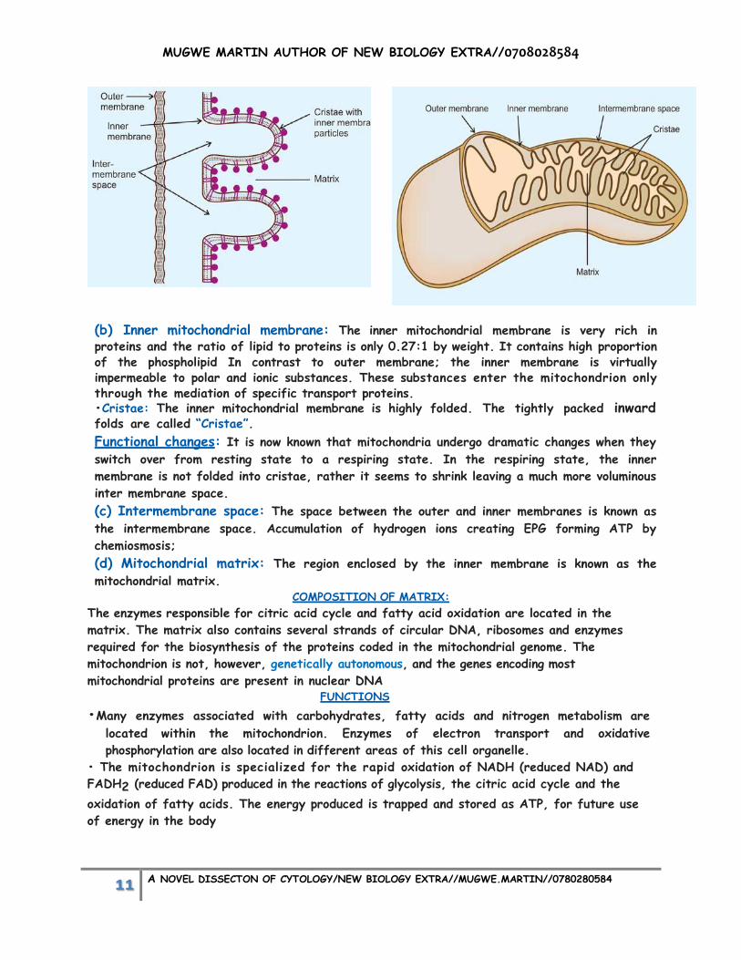
Structure and Functions The mitochondrion is bounded by two concentric membranes that have markedly different
properties and biological functions
mitochondrial membranes (a) Outer mitochondrial membrane: The outer mitochondrial membrane consists mostly of
phospholipids and contains a considerable amount of cholesterol. The outer membrane also
contains many copies of the protein called Porin.
functions of Porin and other proteins
(i) These proteins form channels that permit substances with LOW molecular weights of
diffuse freely across the outer mitochondrial membrane.
(ii) Other proteins in the outer membrane carry out various reactions in fatty acid and
phospholipid biosynthesis and are responsible for some oxidation reactions.
MUGWE MARTIN AUTHOR OF NEW BIOLOGY EXTRA//0708028584
11 A NOVEL DISSECTON OF CYTOLOGY/NEW BIOLOGY EXTRA//MUGWE.MARTIN//0780280584
(b) Inner mitochondrial membrane: The inner mitochondrial membrane is very rich in proteins and the ratio of lipid to proteins is only 0.27:1 by weight. It contains high proportion of the phospholipid In contrast to outer membrane; the inner membrane is virtually impermeable to polar and ionic substances. These substances enter the mitochondrion only through the mediation of specific transport proteins. •Cristae: The inner mitochondrial membrane is highly folded. The tightly packed inward folds are called “Cristae”. Functional changes: It is now known that mitochondria undergo dramatic changes when they
switch over from resting state to a respiring state. In the respiring state, the inner
membrane is not folded into cristae, rather it seems to shrink leaving a much more voluminous
inter membrane space.
(c) Intermembrane space: The space between the outer and inner membranes is known as
the intermembrane space. Accumulation of hydrogen ions creating EPG forming ATP by
chemiosmosis;
(d) Mitochondrial matrix: The region enclosed by the inner membrane is known as the
mitochondrial matrix. COMPOSITION OF MATRIX:
The enzymes responsible for citric acid cycle and fatty acid oxidation are located in the
matrix. The matrix also contains several strands of circular DNA, ribosomes and enzymes
required for the biosynthesis of the proteins coded in the mitochondrial genome. The
mitochondrion is not, however, genetically autonomous, and the genes encoding most
mitochondrial proteins are present in nuclear DNA FUNCTIONS
•Many enzymes associated with carbohydrates, fatty acids and nitrogen metabolism are
located within the mitochondrion. Enzymes of electron transport and oxidative
phosphorylation are also located in different areas of this cell organelle.
• The mitochondrion is specialized for the rapid oxidation of NADH (reduced NAD) and
FADH2 (reduced FAD) produced in the reactions of glycolysis, the citric acid cycle and the
oxidation of fatty acids. The energy produced is trapped and stored as ATP, for future use
of energy in the body
MUGWE MARTIN AUTHOR OF NEW BIOLOGY EXTRA//0708028584
12 A NOVEL DISSECTON OF CYTOLOGY/NEW BIOLOGY EXTRA//MUGWE.MARTIN//0780280584
Vital role in Apoptosis; Cytochrome C and second mitochondria-derived activator of caspases
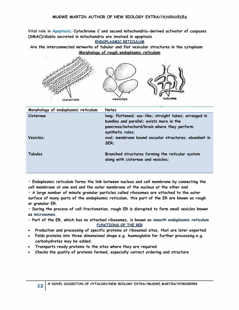
(SMAC)/diablo secreted in mitochondria are involved in apoptosis ENDOPLASMIC RETICULUM
Are the interconnected networks of tubular and flat vesicular structures in the cytoplasm Morphology of rough endoplasmic reticulum
Morphology of endoplasmic reticulum Notes
Cisternae
Vesicles;
Tubules
long; flattened; sac-like; straight tubes; arranged in
bundles and parallel; exists more in the
pancreas/notochord/brain where they perform
synthetic roles;
oval; membrane bound vacuolar structures; abundant in
SER;
Branched structures forming the reticular system
along with cisternae and vesicles;
• Endoplasmic reticulum forms the link between nucleus and cell membrane by connecting the
cell membrane at one end and the outer membrane of the nucleus at the other end
• A large number of minute granular particles called ribosomes are attached to the outer
surface of many parts of the endoplasmic reticulum, this part of the ER are known as rough
or granular ER.
• During the process of cell fractionation, rough ER is disrupted to form small vesicles known
as microsomes.
• Part of the ER, which has no attached ribosomes, is known as smooth endoplasmic reticulum FUNCTIONS OF THE RER
Production and processing of specific proteins at ribosomal sites, that are later exported
Folds proteins into three dimensional shape e.g. haemoglobin for further processing e.g.
carbohydrates may be added.
Transports ready proteins to the sites where they are required.
Checks the quality of proteins formed, especially correct ordering and structure

MUGWE MARTIN AUTHOR OF NEW BIOLOGY EXTRA//0708028584
13 A NOVEL DISSECTON OF CYTOLOGY/NEW BIOLOGY EXTRA//MUGWE.MARTIN//0780280584
SER
Synthesis of lipids and other steroids like cholesterol, progesterone and testosterone.
Synthesis and repair of membranes by producing cholesterol and phospholipids,
For metabolism of glycogen in the liver e.g. glucose-6-phosphatase enzyme in SER
converts glucose-6-phosphate to glucose.
Contains enzymes that detoxicate lipid- soluble drugs, alcohol and metabolic
wastes from the liver.
SER attaches receptors to cell membrane proteins in plant cells
Sarcoplasmic reticulum regulates muscle contraction through storage and release of
calcium ions.
GOLGI COMPLEXES
They are also called Dictyosomes. Each eukaryotic cell contains a unique stack of smooth
surfaced compartments or cisternae that make up the Golgi complex. The ER is usually
closely associated with the Golgi complexes, which contain flattened, fluid filled Golgi sacs.
The Golgi complex has a Proximal or Cis compartment, a medial compartment and a distal or
Trans compartment.
FUNCTIONS
(i) On the proximal or cis side, the Golgi complexes receive the newly synthesised proteins
by ER via transfer vesicles. (ii) The post-translational modifications take place in the Golgi lumen (median part) where
the carbo hydrates and lipid precursors are added to proteins to form glycoproteins and lipoproteins respectively.
(iii) On the distal or Trans side they release proteins via modified membranes called secretory vesicles. These secretory vesicles move to and fuse with the plasma membrane where the contents may be expelled by a process called exocytosis.
Drawing of the Golgi body

MUGWE MARTIN AUTHOR OF NEW BIOLOGY EXTRA//0708028584
14 A NOVEL DISSECTON OF CYTOLOGY/NEW BIOLOGY EXTRA//MUGWE.MARTIN//0780280584
General functions of Golgi body o To modify, sort and package proteins that are made at the rough o Endoplasmic reticulum for secretion (export) or for use within the cell. o To form carbohydrates e.g. polysaccharides are attached to a protein to form proteoglycans
present in the extracellular matrix of the animal cell. o Transport of lipid molecules around the cell. o Formation of lysosomes containing hydrolytic enzymes. o Formation of peroxisomes. o In plant cells, Golgi produces vesicles that join to form cell plates during cell division. o Secretory vesicles produced by Golgi contain a variety of important substances e.g.
neurotransmitters, hormones, mucin, zymogen e.g. pepsinogen, etc. o Fusion of Golgi vesicles with cell membrane maintains the membrane which is used to form
phagocytic vacuoles and Pinocytic vesicles
CELL MEMBRANE
Cell membrane is composed of three types of substances:
1. Proteins (55%)
2. Lipids (40%)
3. Carbohydrates (5%).
MAJOR PROPONENTS OF THE MEMBRANE STRUCTURE
1. Danielli-Davson model
‘Danielli-Davson model’ was the first proposed basic model of membrane structure. It was
proposed by James F Danielli and Hugh Davson in 1935. This model was basically a‘sandwich of
lipids’ covered by proteins on both sides.
2. Unit membrane model
In 1957, JD Robertson replaced ‘Danielli-Davson Model’ by ‘Unit membrane model’ on the basis
of electron microscopic studies
3. Fluid mosaic model Later in 1972, SJ Singer and GL Nicholson proposed ‘The fluid mosaic model’. According to
them, the membrane is a fluid with mosaic of proteins.

MUGWE MARTIN AUTHOR OF NEW BIOLOGY EXTRA//0708028584
15 A NOVEL DISSECTON OF CYTOLOGY/NEW BIOLOGY EXTRA//MUGWE.MARTIN//0780280584
Structure
Bimolecular layer of phospholipids; with inwardly directed hydrophobic tails; and outwardly
directed hydrophilic heads; variety of protein molecules with irregular arrangement/mosaic
arrangement; some proteins occur on the surface of the phospholipid
layer/peripheral/extrinsic; while some extend into it/integral/intrinsic; some extend
completely across/transmembrane proteins; present between phospholipids is cholesterol;
glycocalyx, glycoproteins and glycolipids; which are antennae like structures at the surface;
MEANING OF THE PHRASE “FLUID MOSAIC MODEL “
Fluid because molecules within membrane can move around within their own layer;
Mosaic because protein molecules are dotted around within the membrane;
Model because no one has seen membrane looking as a diagram and hence it’s a
representation; EVIDENCES FOR THE FLUID MOSAIC MODEL
Water soluble molecules enter the cell less readily than compounds that dissolve in
lipidsplasma membrane has a higher proportion of lipids;
freeze- etching has shown when a membrane is fractured along its mid line globular proteins
occur some occur on, some buried in the lipid bilayer, rather than forming sheets of proteins
on the surface;
Some reactions of proteins in the membrane with enzymes and antibodies have shown that
some membrane proteins are exposed on the surface, some are exposed on both surfaces and
some inaccessible;
Tagging of membrane components with marker substance/fluorescent dye has shown that the
molecules of the plasma membrane are on the move membrane is fluid, ever changing;
At outer surface of the cell, antennae like carbohydrate molecule form complex with many
membrane proteins and lipids and their presence can be shown by addition of non-self-
antigen to the cell;
DISTRIBUTION AND FUNCTION OF MEMBRANES IN ORGANISMS
o Plasma membrane; Selective transport in and out of cell/Cell to cell recognition/Antigen
presentation/Hormonal signaling/Separating cytoplasm from the rest of the cell
o Nuclear membrane; Limits DNA/Allows outward diffusion of RNA/Entry of ATP into the
nucleoplasm;
o Mitochondria outer membrane; allows entry of products of glycolysis;

MUGWE MARTIN AUTHOR OF NEW BIOLOGY EXTRA//0708028584
16 A NOVEL DISSECTON OF CYTOLOGY/NEW BIOLOGY EXTRA//MUGWE.MARTIN//0780280584
o Mitochondrial inner membrane; Attachment of respiratory enzymes; Attachments of
respiratory pigments for electron transport;
o Endoplasmic reticulum membranes; Intracellular transport/Attachment of
ribosomes/Synthesis of steroids
o Chloroplast outer membrane; allows photosynthetic products out and substrates in
o Chloroplast lamellae; Reservoir for photosynthetic pigments;
o Golgi membranes; Sorting of ER synthesized material/Synthesis of glycoprotein/Synthesis
of polysaccharides e.g. cellulose in plants
o Lysosomes; Limiting escape of lytic enzymes
o Tonoplast; enclosing cell sap/Selective entry of water and solutes
o Phagocytic/ micropinocytic vesicles; Uptake of materials into cells
o Root hairs plasma membrane; increasing surface area of epidermal cells
o Autolytic/autophagic vacuoles; intracellular digestion
o Neurilemma; diffusion of Na+ and K+ allowing electrical polarity
FUNCTIONS OF THE CELL MEMBRANE
o Protective function: Cell membrane protects the cytoplasm and the organelles present in
the cytoplasm
o Selective permeability: Cell membrane acts as a semipermeable membrane, which allows
only some substances to pass through it and acts as a barrier for other substances
o Absorptive function: Nutrients are absorbed into the cell through the cell membrane
o Excretory function: Metabolites and other waste products from the cell are excreted out
through the cell membrane
o Exchange of gases: Oxygen enters the cell from the blood and carbon dioxide leaves the
cell and enters the blood through the cell membrane
o Maintenance of shape and size of the cell: Cell membrane is responsible for the
maintenance of shape and size of the cell.
o Has receptor sites for chemical messengers such as hormones;
o Contain enzymes catalyzing specific reactions;
o Transport proteins carry out transport of molecules such as glucose;
ROLE OF CELL MEMBRANE IN MOVEMENT OF MATERIALS ACROSS IT
o Selective barrier; allowing passage of specific molecules into cells;
o Establishment of concentration gradient; determining direction of movement of
materials/substances;
o Carrier/channel proteins; for movement of materials into and outside cells;
o Receptors for binding of materials which have to be moved inside cells;
o Non-polar to allow movement of non- polar materials eg steroid hormones;
o fluidity/flexibility to allow movement of transport proteins spanning the membrane
easily/phagocytosis possible;
o Stability to avoid collapse of membrane;
o membranes budded off forming vesicles during endocytosis to move materials inside cells;
o flexible and fluidity to fuse with other membranes to allow transport of materials
o Microvilli; increasing surface area for absorption of materials to be transported;

MUGWE MARTIN AUTHOR OF NEW BIOLOGY EXTRA//0708028584
17 A NOVEL DISSECTON OF CYTOLOGY/NEW BIOLOGY EXTRA//MUGWE.MARTIN//0780280584
MEMBRANE LIPIDS The major classes of membrane lipids are:
– Phospholipids
– Glycolipids
– Cholesterol.
They all are amphipathic molecules, i.e. they have both hydrophobic and hydrophilic ends.
Membrane lipids spontaneously form bilayer in aqueous medium, burying their hydrophobic tails
and leaving their hydrophilic ends exposed to the water
Summary of the functions of the lipids in the plasma membrane
glycolipids Are involved in cell-to-cell recognition
cholesterol
Stabilizes membrane structure by preventing phospholipids from closely packing together
Lipid bilayer Being semi-permeable, it controls movement of substances in and out of the cell
Membrane proteins
Channel proteins; provide passageways through the membrane for certain hydrophilic (water-
soluble) substances such as polar/charged molecules;
Transport proteins/PUMPs; spend energy (ATP) to transfer materials across the membrane
active transport/passive transport;
Recognition proteins; distinguish the identity of neighboring cells
Adhesion proteins; attach cells to neighboring cells/provide anchors for the internal filaments
and tubules that give stability to the cell;
Receptor proteins; provide binding sites for hormones or other trigger molecules
Electron transfer proteins;are involved in transferring electrons from one molecule to
another during chemical reactions;
Integral proteins provide the structural integrity of the cell membrane
Enzymes catalyzing specific chemical reactions in membranes
Carrier proteins; having receptor sites for binding of molecules to be transported eg glucose;
DATA CONCERNING MEMBRANE COMPONENTS
Membrane Percentage mass
Protein Lipid Carbohydrate
A 18 79 3
B 51 49 0
C 52 44 4
D 76 24 0
OBSERVATION
Plasma membrane D has much higher protein content than plasma A
Membrane D is more metabolically active compared to A/membrane A is metabolically inert;
MUGWE MARTIN AUTHOR OF NEW BIOLOGY EXTRA//0708028584
18 A NOVEL DISSECTON OF CYTOLOGY/NEW BIOLOGY EXTRA//MUGWE.MARTIN//0780280584
COMPARE CELL WALL AND CELL MEMBRANE
Similarities
o Both surround cells;
o Both are permeable;
o Both are made up of proteins and sugars;
Differences
Cell wall Cell membrane
Thickness normally measured in µm; Usually measured in nm;
Surrounds plant cells/not animal cells; Surrounds animal cells;
Found only outside the cells; Found either inside or outside cells;
Contain cellulose/peptidoglycan/murein in
prokaryotes/contain chitin in fungi;
Phospholipids/proteins/cholesterol;
Freely permeable; Partially permeable;
Mechanical strength; Selective barrier;
Rigid; Fluid;
Plasmodesmata present Plasmodesmata absent;
Secondary thickening can occur No secondary thickening
GLYCOCALYX
Long carbohydrate molecule attached to membrane lipids and proteins
o They play roles such as cell to cell recognition;
o Functioning as receptor sites for chemical signals such as hormones;
o Binding cells together into tissues;
o carbohydrates are negatively charged so limit entry and exit of negatively charged
substances; FACTORS AFFECTING FLUIDITY OF THE PLASMA MEMBRANE
o Temperature; high temperature increases fluidity low temperature decreases fluidity;
o Length of fatty acids; fluidity increases with decreasing length; long chain decreases
fluidity;longer chain increases surface area for interactions between adjacent chains
increasing vandarwaals forces of attraction
o Unsaturated fatty acids; have kinks in the fatty acid tails which prevents close packing
hence increasing fluidity saturated fats lack kinks ;close packing decreasing fluidity;
o Cholesterol; between phospholipids inhibits close packing/ at high temperature decreases
fluidity low temperature increases fluidity;
How cell membrane cholesterol maintains fluidity
Cholesterol molecules orient themselves in the lipid bilayer in such a way that their hydroxide
groups remain close to polar head groups of the phospholipids; their rigid plate-like steroid
rings interact with and partly immobilize those regions of hydrocarbon chains that are closet
to the polar head groups, leaving chains flexible; cholesterol inhibits phase transition by
preventing hydrocarbon chains from coming together and crystallizing.
Cholesterol also tends to decrease the permeability of lipid bilayer to small water-soluble
molecules and is thought to enhance both the flexibility and the mechanical stability of bilayer

MUGWE MARTIN AUTHOR OF NEW BIOLOGY EXTRA//0708028584
19 A NOVEL DISSECTON OF CYTOLOGY/NEW BIOLOGY EXTRA//MUGWE.MARTIN//0780280584
(Schematic) cholesterol molecules interacting with two phospholipids molecules in a monolayer
(after Albert’s etal 1989)
SYNOPTIC LINK OF MEMBRANE FLUIDITY USING A GRAPH
When phospholipid bilayers are heated, the tail become more mobile at critical transition
temperature, they absorb a great deal of heat and become so mobile that they behave like a
liquid. The graph below shows the effect of temperature on the heat absorption of a pure
phospholipid bilayer and one to which 20% cholesterol has been added.
a) (I) Compare the two graphs one with phospholipids while the other lacking
CLUE: This requires both differences and similarities
Areas of differences and similarities on the graph include Peak /maximum reached.
o The peak /maximum (do not quote figures)
o How have they reached the peak eg both rise/increase to reach the
o Peak.
o What happens after the peak eg both decline /decrease after the peak?
Trends/patterns which are similar: - Range needed
o Start with the range and units and end with the statement.
Differences:
o Use the words while/ whereas when answering.

MUGWE MARTIN AUTHOR OF NEW BIOLOGY EXTRA//0708028584
20 A NOVEL DISSECTON OF CYTOLOGY/NEW BIOLOGY EXTRA//MUGWE.MARTIN//0780280584
o Where range is required write the range and units , then the statement
o Areas of observation recorded as difference include:
The peak/maximum reached
o The levels of the peaks, lower maximum reached and higher maximum reached
o When peaks are reached. (Quote the figure of X-axis and unit) start with statement and then the
fig in X-axis/units
o What happens after the peak/maximum reached?
Trends/patterns which appear different (range needed)
o The statement must come first and then the range/unit.
o Levels within a certain range
b (I) how does the phospholipid tail behaving like a liquid affect the permeability of the
plasma membrane? (3 marks)
Permeability increases; membrane becomes leaky;selectivity of membrane lost;
(II) How is this different for the bilayer with 20% cholesterol added?(2 marks)
Permeability reduces with increasing temperature; cholesterol prevents melting of lipids in
the membrane;
(Iv) Suggest function for cholesterol molecules in plasma membranes. Explain your answer.
Regulating fluidity of the plasma membrane within certain limits; making membrane less
fluid at higher temperature and but more fluid at lower ones
(V) Importance of fluidity of plasma membrane.
Affects membrane activity such as ease of membranes to fuse; activity of membrane-
bound enzymes; and transport proteins
PHOSPHOLIPIDS
Phospholipids are the lipid substances containing phosphorus and fatty acids.
Phospholipid molecules are arranged in two layers
Each phospholipid molecule resembles the headed pin in shape. The outer part of the
phospholipid molecule is called the head portion and the inner portion
Is called the tail portion, Head portion is the polar end and it is soluble in
Water and has strong affinity for water (hydrophilic). Tail portion is the non-polar end. It is
insoluble in water and repelled by water (hydrophobic).
Two layers of phospholipids are arranged in such a way that the hydrophobic tail portions
meet in the center of the membrane. Hydrophilic head portions of outer layer face the ECF
and those of the inner layer face ICF
PHOSPHOLIPID BI-LAYER STRUCTURE

MUGWE MARTIN AUTHOR OF NEW BIOLOGY EXTRA//0708028584
21 A NOVEL DISSECTON OF CYTOLOGY/NEW BIOLOGY EXTRA//MUGWE.MARTIN//0780280584
Explain what happens when a thin layer of phospholipids is spread over
Small water quantity The phospholipid layers orientate themselves in a single monomolecular layer with non-polar
hydrophobic tails projecting out of water; whilst the polar hydrophilic heads lie in the
surface of water;
Large quantities of water and shaken Micelles are formed; in which two layers of lipids occurs/bimolecular layers of
phospholipids; with hydrophobic tail pointing inwards away from water and hydrophilic tails
projects in water;
DESCRIBE THE DIFFERENT TYPES OF MOVEMENTS THAT LIPIDS EXHIBIT IN THE PLASMA
MEMBRANE
Flip- flop/transbilayer movement; lipids migrating from one monolayer to another monolayer
of lipid bimolecular layer;
Lateral diffusion; lipids change places with their neighbors within monolayer;
Rotation; rotate rapidly along their longitudinal axes in a monolayer;
Flexion; high degree of rotation of lipid hydrocarbons/tails;
Schemata showing the movements

MUGWE MARTIN AUTHOR OF NEW BIOLOGY EXTRA//0708028584
22 A NOVEL DISSECTON OF CYTOLOGY/NEW BIOLOGY EXTRA//MUGWE.MARTIN//0780280584
BACKGROUND OF THE GRAPH
Beetroot tissues contain a carotenoid called Betalain, which escapes on heating due to
increased permeability of the cell membrane on heating
RANGE REASON(S)
From 00c to 600c; gradual
decrease in percentage
transmission;
From 600c to 70oc; rapid
decrease in transmission
From 70oc to 80oc;slight/gradual
decrease in transmission
Increased temperature increases KINETIC energy of the
proteins and lipids hence move faster and with more energy;
leave gaps in the Tonoplast/vacuolar membrane; more permeable
hence Betalain is lost making water coloured/red; reducing
transmission.
Proteins lose their shape due to breakage of hydrogen bond
making them; denatured making protein channels bigger; much
Betalain is lost; high temperature increases rate of diffusion of
Betalain/melting of phospholipids creating big gaps;
Cell integrity completely lost/structure lost due to very high
temperature hence less Betalain lost
Effects of the membrane fluidity
• As membrane fluidity increases, its permeability to water and other small hydrophilic
molecules also increases.
• As fluidity increases, the lateral mobility of integral proteins also increases.
Peroxisomes/Microbodies
• These organelles resemble the lysosomes in their appearance, but they differ both in
function and in their synthesis.
Functions of peroxisomes • Peroxisomes contain enzymes peroxidases and catalase which are concerned with the
metabolism of peroxide. Thus, the peroxisomes are involved in the detoxification of peroxide.
• Peroxisomes are also capable of carrying out β-oxidation of fatty acid.
Glyoxysomes contain enzymes that degrade lipids into sugars during seed germination.

MUGWE MARTIN AUTHOR OF NEW BIOLOGY EXTRA//0708028584
23 A NOVEL DISSECTON OF CYTOLOGY/NEW BIOLOGY EXTRA//MUGWE.MARTIN//0780280584
CILIA AND FLAGELLA
structure of cilium Microtubules; a pair of central singlets; surrounded by nine peripheral doublets/ring of nine
paired ones; with arm-like process; 9+2 array/pattern; surrounded by cell surface
membrane; each peripheral filaments consists of an A and B microtubule; A microtubule
has protein dynein/ATpase; central filaments connected to microtubule A by radial spokes;
(CROSS-SECTION)
(LONGITUDINAL-SECTION)
Compare cilia and flagella
Similarities
o Both have 9+2 array/pattern;
o Both are used for locomotion;
o Both are surrounded by cell membrane;
o Both are microscopic;
o Both are contractile;
o Both are filamentous;
Differences
Cilia Flagella
More Numerous; Fewer/less in number;
May occur throughout the surface of the cell; Occur at one end of the cell;
Shorter processes; Longer processes;
Beat in coordinated rhythm; Beat independently;
Sweeping/pendular motion; Undulatory motion;
Other functions such as
feeding/circulation/sensory;
Locomotory functions only;
FUNCTIONS OF CILIA AND FLAGELLA Ciliary movement enables paramecium to drive food into their gullet. In certain mulluscs Ciliary movement facilitates gaseous exchange by passing water currents
over the gills
In echinoderms Ciliary movement enables locomotion by driving water through the water

MUGWE MARTIN AUTHOR OF NEW BIOLOGY EXTRA//0708028584
24 A NOVEL DISSECTON OF CYTOLOGY/NEW BIOLOGY EXTRA//MUGWE.MARTIN//0780280584
vascular system.
Cilia lining the respiratory tract of humans’ drives away the microbes and dust particles
towards the nose or mouth.
Cilia in the oviduct or fallopian tubes of human female moves ova towards the uterus. Cilia in nephridia of annelids e.g. earthworms moves wastes
Flagellum of sperms enables their swimming movement.
Flagellum enables the movement in certain protozoans like euglena
CYTOSKELETONS
The cytoplasm of most eukaryotic cells contains network of protein filaments that interact
extensively with each other and with the component of the plasma membrane.
The plasma membrane is anchored to the cytoskeleton. The cytoskeleton is not a rigid
permanent framework of the cell but is a dynamic, changing structure.
• The cytoskeleton consists of three primary protein filaments:
1. Microfilaments
2. Microtubules
3. Intermediate filaments. MICROFILAMENTS
Are about 5 nm in diameter. They are made up of protein actin. Actin filaments form a
meshwork just underlying the plasma membrane of cells and are referred to as cell cortex,
which is labile. They disappear as cell motility increases or upon malignant transformation of
cells. The function of microfilaments to help muscle contraction MICROTUBULES
are cylindrical tubes,20 to 25 nm in diameter, made up of protein tubulin.
Microtubules are necessary for the formation and function of mitotic spindle.
They provide stability to the cell.
They prevent tubules of ER from collapsing.
These are the major components of axons and dendrites. INTERMEDIATE
Filaments are so called as their diameter (10 nm) is intermediate between that of
microfilaments (5 nm) and of microtubules (25 nm).
Intermediate filaments are formed from fibrous protein which varies with different tissue
type.
They play role in cell-to-cell attachment and help to stabilize the epithelium. They provide
strength and rigidity to axons/connect adjacent cells through desmosomes
Subclasses of intermediate filaments
Intermediate filaments are divided into five subclasses:
i. Keratins (in epithelial cells)
ii. Glial filaments (in astrocytes)
iii. Neurofilaments (in nerve cells)
iv. Vimentin (in many types of cells)
v. Desmin (in muscle fibers).
Functions of the cytoskeletons
o The cytoskeleton gives cells their characteristic shape and form, provides attachment
points for organelles, fixing their location in cells and also makes communication between
parts of the cell possible.
o It is also responsible for the separation of chromosomes during cell division.

MUGWE MARTIN AUTHOR OF NEW BIOLOGY EXTRA//0708028584
25 A NOVEL DISSECTON OF CYTOLOGY/NEW BIOLOGY EXTRA//MUGWE.MARTIN//0780280584
o The internal movement of the cell organelles as well as cell locomotion and muscle fiber
contraction could not take place without the cytoskeleton. It acts as “track” on which cells
can move organelles, chromosomes
Drawing of cytoskeletons
Microtubes intermediate filaments
((Microfilament) CELL WALL (not an organelle)
The cell wall is differentiated into three layers
o Primary cell wall
o Secondary cell wall
o Tertiary cell wall COMPARISONS
Primary cell wall Secondary cell wall Tertiary cell wall
Outermost layer of cell
wall;
Found below primary cell
wall;
Found below secondary cell
wall;
Comparatively thin and
elastic, delicate and
permeable;
Comparatively thick, and
impermeable;
Thicker than secondary;
Found in meristemic, young
and paranchymatous cells;
Found in mature and
permanent cells;
Found in tracheids and mature
gymnosperms;
Made up of cellulose; Cellulose, pectin, lignin, and
other substances;
Made up of Xylan;
Simple and smooth; Simple; Forms fingerlike process by
evagination of the cell wall;

MUGWE MARTIN AUTHOR OF NEW BIOLOGY EXTRA//0708028584
26 A NOVEL DISSECTON OF CYTOLOGY/NEW BIOLOGY EXTRA//MUGWE.MARTIN//0780280584
DESCRIPTION OF THE CELL WALL STRUCTURE
The cell wall is made up of primary cell wall, middle lamellae, secondary cell wall and tertiary
cell wall; primary cell wall consists of cellulose microfibrils running through matrix of
polysaccharide; pectates and hemicellulose; deposited on either sides of the middle lamellae;
microfibrils run in all directions; portion of the endoplasmic reticulum is trapped across the
middle lamellae and forms the plasmodesma; outermost layer; comparatively thin; elastic;
delicate and permeable; simple and smooth; secondary cell wall; formed by further deposition
of cellulose on the inside of the primary cell wall; microfibrils tightly packed; thicker;
impermeable; found below primary cell wall; cellulose cell walls oriented in the direction of the
cell elongation; impregnated with cutin/suberin.
Tertiary cell wall; found below secondary cell wall; thicker than secondary and primary; found
in tracheids and gymnosperms; impregnated with XYLAN. CHEMICAL CHANGES THAT OCCUR DURING CELL WALL GROWTH
o Lignifications; deposition of lignin on cell wall;
o cutinization; conversion of cellulose into cutin in the cell wall;
o suberisation;deposition of suberin on the cork cell walls;
o Mineralization;deposition of mineral substances such as silica, calcium carbonate and
calcium oxalate;
o mucilaginous changes;cellulose cell wall is converted into mucilage; Eg bacteria; FUNCTIONS OF THE CELL WALL
Mechanical strength and skeletal support provided for individual cells and for the plant as
a whole; due to extensive lignification;
Cell walls resistant to expansion and allow development of turgidity as a result of osmotic
influx of water molecules;
Orientation of the cellulose microfibrils limits cell growth and shape;
System of interconnected cellulose cell walls/apoplast; functions as a major pathway of
transport of water and dissolved salts; plasmodesmata in cell walls for symplast pathway;
Develop coating of waxy cutin/cuticle; epidermal cells reducing water loss and risk of
infections; cork cells are impregnated by suberin to prevent desiccation;
Walls of the xylem vessels/tracheids and sieve tubes are adapted for long distance
transport of materials through cells;
Cell walls of endodermal cells are impregnated by suberin forming barrier to water
movement;
Cell walls modified for as food reserves; as in storage of hemicellulose in seeds;
Cell walls of transfer cells develop an increased surface area for active loading of solutes
into sieve tubes; STAGES OF FORMATION OF THE CELLULOSE CELL WALL
in plant cells the spindle fibres begin to disappear during telophase everywhere except in the
region of the equatorial plane; move outwards in diameter and increase in number to form a
barrel-shaped phragmoplast; microtubes, ribosomes, mitochondria, endoplasmic reticulum and
Golgi body which produce number of fluid filled vesicles, appear in the centre of the cell;
coalescence to form a cell plate which grows across equatorial plane; the contents of the
vesicles contribute to the new middle lamella and the cell walls of the new daughter cells;
membranes form new cell membrane; the spreading cell plate eventually fuses with parent cell
walls. The new cell wall formed called primary cell wall; thickened at later stage further with

MUGWE MARTIN AUTHOR OF NEW BIOLOGY EXTRA//0708028584
27 A NOVEL DISSECTON OF CYTOLOGY/NEW BIOLOGY EXTRA//MUGWE.MARTIN//0780280584
cellulose and other substances such as lignin and suberin to form secondary cell wall; in certain
areas the vesicles fail to fuse and cytoplasmic contact remain between daughter cells. These
cytoplasmic channels are lined by the cell surface membrane and form plasmodesmata; Structure of the plant cell wall formed as a result of cytokinesis
ADAPTATIONS OF THE CELL WALL
Cellulose fibres running parallel allowing maximum formation of the hydrogen bonds giving the cell wall high tensile strength/effect is cumulative Thick cell wall made up of three layers making it strong/tough to resists bursting due to osmotic influx of water molecules Secondary walls may be cutinized / suberinised for preventing water loss/desiccation as a result of high temperature The cellulose microfibrils group forming macrofibrils which allow them to give much support Primary cell wall is flexible to allow expansion as water enters by osmosis
LYSOSOMES
Lysosomes are the membrane-bound vesicular organelles found throughout the cytoplasm. The
lysosomes are formed by Golgi apparatus. The enzymes synthesized in rough endoplasmic
reticulum are processed and packed in the form of small vesicles in the Golgi apparatus. Then,
these vesicles are pinched off from Golgi apparatus and become the lysosomes. Among the
organelles of the cytoplasm, the lysosomes have the thickest covering membrane. The
membrane is formed by a bilayered lipid material. It has many small granules which contain
hydrolytic enzymes.
Types of Lysosomes
Lysosomes are of two types:
1. Primary lysosome, which is pinched off from Golgi apparatus. It is inactive in spite of
having hydrolytic enzymes
2. Secondary lysosome, which is the active lysosome. It is formed by the fusion of a primary
lysosome with phagosome or endosome.
Important lysosomal enzymes
1. Proteases, which hydrolyze the proteins into amino acids
2. Lipases, which hydrolyze the lipids into fatty acids and glycerides
3. Amylases, which hydrolyze the polysaccharides into glucose
4. Nucleases, which hydrolyze the nucleic acids into mononucleotides.

MUGWE MARTIN AUTHOR OF NEW BIOLOGY EXTRA//0708028584
28 A NOVEL DISSECTON OF CYTOLOGY/NEW BIOLOGY EXTRA//MUGWE.MARTIN//0780280584
Mechanism of lysosomal function
Lysosomal functions involve two mechanisms:
1. Heterophagy: Digestion of extracellular materials engulfed by the cell via endocytosis
2. Autophagy: Digestion of intracellular materials such as worn-out cytoplasmic organelles.
Specific functions of lysosomes 1. Degradation of macromolecules
Macromolecules are engulfed by the cell by means of endocytosis (phagocytosis, pinocytosis or
receptor mediated endocytosis. The macromolecules such as bacteria, engulfed by the cell via
phagocytosis are called phagosome or vacuoles. The other macromolecules taken inside via
pinocytosis or receptor-mediated endocytosis are called endosomes.
The primary lysosome fuses with the phagosome or endosome to form the secondary lysosome.
The pH in the secondary lysosome becomes acidic and the lysosomal enzymes are activated.
The bacteria and the other macromolecules are digested and degraded by these enzymes. The
secondary lysosome containing these degraded waste products moves through cytoplasm and
fuses with cell membrane. Now the waste products are eliminated by exocytosis.
2. Degradation of worn-out organelles
The rough endoplasmic reticulum wraps itself around the worn-out organelles like mitochondria
and forms the vacuoles called autophagosomes. One primary lysosome fuses with one
autophagosome to form the secondary lysosome. The enzymes in the secondary lysosome are
activated. Now, these enzymes digest the contents of autophagosome.
3. Removal of excess secretory products in the cells
Lysosomes in the cells of the secretory glands remove the excess secretory products by
degrading the secretory granules.
4. Secretory function – secretory lysosomes having secretory function called secretory
lysosomes are found in some of the cells, particularly in the cells of immune system. The
conventional lysosomes are modified into secretory
Lysosomes by combining with secretory granules (which contain the particular secretory
product of the cell)
Examples of secretory lysosomes:
i. Lysosomes in the cytotoxic T lymphocytes and natural killer (NK) cells secrete perforin and
granzymes, which destroy both viral-infected cells and tumor cells. Perforin is a pore-forming
protein that initiates cell death. Granzymes belong to the family of serine proteases (enzymes
that dislodge the peptide bonds of the proteins) and cause the cell death by apoptosis
ii. Secretory lysosomes of melanocytes secrete melanin
iii. Secretory lysosomes of mast cells secrete serotonin, which is a vasoconstrictor substance
and inflammatory mediator. 5. Role in fertilisation; Acrosome in spermatozoa releases enzymes which digest the limiting membrane of the ovum to enable sperm entry and start fertilization. The lysosome in cytoplasm of Ova enables digestion of stored food 6. Role in development; Tadpole metamorphosis (regression of tail) and regression of Wolffian ducts involve shedding of tissues with removal of whole cells and extracellular material by lysosome enzymes during bone development, osteoclasts release lysosomal enzymes that remodel bones. 7. Autolysis. Primary lysosome releases hydrolytic enzymes within a dead cell to digest the whole cell.
MUGWE MARTIN AUTHOR OF NEW BIOLOGY EXTRA//0708028584
29 A NOVEL DISSECTON OF CYTOLOGY/NEW BIOLOGY EXTRA//MUGWE.MARTIN//0780280584
ROLE OF LYSOSOMES IN SECRETION OF THYROXINE
The cells making up the thyroid glands are stimulated by the thyroid stimulating hormone to take up thyroglobulin by pinocytosis forming Pinocytic vesicles which fuse with primary lysosome and thyroglobulin is hydrolysed to produce active enzyme thyroxine before lysosomes fuse with the cell surface membrane thus secreting the hormone into the blood
FACTS CONCERNING LYSOSOMES
Size: Mean diameter is approximately 0.4 (varies in between that of microsomes and
mitochondria). They are surrounded by a lipoprotein membrane.
• Lysosomes are found in all animal cells, except erythrocytes, in varying numbers and types.
• PH: pH inside the lysosomes is lower than that of cytosol. The lysosomal enzymes have an
optimal pH around 5. Acid phosphatase is used as a marker enzyme for this organelle
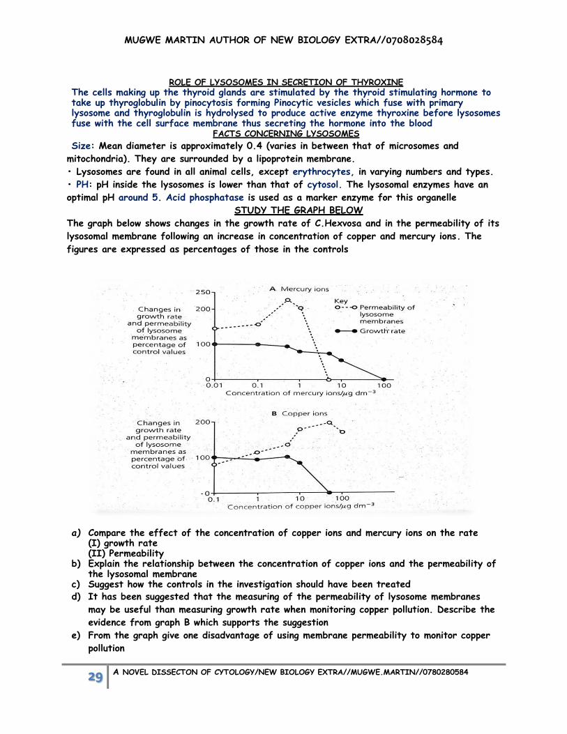
STUDY THE GRAPH BELOW
The graph below shows changes in the growth rate of C.Hexvosa and in the permeability of its
lysosomal membrane following an increase in concentration of copper and mercury ions. The
figures are expressed as percentages of those in the controls
a) Compare the effect of the concentration of copper ions and mercury ions on the rate
(I) growth rate (II) Permeability
b) Explain the relationship between the concentration of copper ions and the permeability of the lysosomal membrane
c) Suggest how the controls in the investigation should have been treated d) It has been suggested that the measuring of the permeability of lysosome membranes
may be useful than measuring growth rate when monitoring copper pollution. Describe the
evidence from graph B which supports the suggestion
e) From the graph give one disadvantage of using membrane permeability to monitor copper
pollution

MUGWE MARTIN AUTHOR OF NEW BIOLOGY EXTRA//0708028584
30 A NOVEL DISSECTON OF CYTOLOGY/NEW BIOLOGY EXTRA//MUGWE.MARTIN//0780280584
f)
CHLOROPLAST
Chloroplast envelope; double membrane; controls entry and exit of materials in and out of the
chloroplast;
Stroma; colourless gelatinous matrix containing enzymes responsible for the light independent
stage of photosynthesis; Contains 70S ribosome, circular DNA and oil droplets;
Grana; structures look like a stack of coins; 50 grana make up a chloroplast; have flattened
discs, thylakoid discs; where chlorophyll molecules are attached; grana carry out light
dependent stage of photosynthesis in which energy inform of ATP is made; Starch granules are temporary store of carbohydrates made during photosynthesis
Structure of the chloroplast
Similarities between the chloroplast and mitochondria
o Both are enclosed by double membranes;
o Both contain circular DNA;
o Both contain 70S ribosomes;
o Both produce ATP by chemiosmosis;
o Both lack nuclear envelope;
o Both their inner membrane is folded;
o Both occur in eukaryotic cells;
Differences CHLOROPLAST MITOCHONDRION
Site of photosynthesis;
Contains thylakoid membranes;
ATP production from light;
Cristae absent;
Contains photosynthetic pigments;
Has starch granules;
Matrix is gelatinous;
Found only higher plants;
Site for respiration;
Lacks thylakoid membranes;
ATP production from oxidation of organic
compounds;
Cristae present;
Lacks photosynthetic pigments;
Has phosphate granules;
Matrix is semi-rigid;
Found in both plants and animals;

MUGWE MARTIN AUTHOR OF NEW BIOLOGY EXTRA//0708028584
31 A NOVEL DISSECTON OF CYTOLOGY/NEW BIOLOGY EXTRA//MUGWE.MARTIN//0780280584
ADAPTATIONS OF THE CHLOPLASTS TO CARRY OUT LIGHT DEPEDENT AND INDEPENDENT STAGE
Granal membranes provide a large surface area for the attachment of photosynthetic
pigments (chlorophyll and carotenoids)/electron carriers and enzymes that carry out
dark reaction;
A network of proteins in the grana that hold the photosynthetic pigments in a precise
manner forming photosystems allowing maximum absorption of light;
Grana membranes have ATP synthase enzyme which manufacture ATP by
chemiosmosis;
The fluid of stroma houses all enzymes needed for Calvin cycle;
The stroma fluid of surrounds the grana so the products of light-dependent reactions
in the grana can easily reach/pass into the stroma;
Chloroplasts contain DNA and ribosomes so they can quickly manufacture some
proteins needed for photosynthesis;
PLASTIDS
Plastids are organelles in plant cells that develop from small bodies, proplastids; found in the
meristemic regions;
PLASTIDS NOTES
Chloroplasts;
Chromoplasts;
Leucoplasts;
Contain chlorophyll and carotenoid pigments; carry out
photosynthesis; found mainly in leaves;
Non- photosynthetic colored plastids containing red, green
pigments; usually associated with fruits and flowers in which
bright colours serve to attract insects/birds and other
mammals;
Colorless plastids/ lacking pigments; modified for food storage
such as roots/seeds and young leaves; can be further classified
basing on stored food eg amyloplasts stores starch;
lipdoplast/elaioplasts/oleoplasts store lipids; and proteoplasts
store proteins;
VACUOLES Plant vacuoles are large, sac-like structures in which a single membrane called Tonoplast encloses a fluid called cell sap, containing water and various dissolved substances.
Functions of vacuoles The Tonoplast isolates the vacuolar sap from the cytosol, enabling vacuolar pathway of
water. Vacuoles in some flowers have coloured pigments that give petals bright
coloured for attracting pollinators. Serve as stores of reserve food, secretory products or waste products. It stores salts, nutrients, minerals, pigments, proteins etc.
It maintains cell turgor by osmotic uptake of water since vacuolar sap has a higher
solute concentration than cytosol.
In meristemic cells, vacuoles bring about growth by initiating cell elongation.
Serve as stores of waste products like tannins, which are excreted when leaves fall.

MUGWE MARTIN AUTHOR OF NEW BIOLOGY EXTRA//0708028584
32 A NOVEL DISSECTON OF CYTOLOGY/NEW BIOLOGY EXTRA//MUGWE.MARTIN//0780280584
In fresh water protozoans like amoeba and paramecium, contractile vacuoles regulate the water content of cells.
Food vacuoles formed by phagocytosis (endosomes/phagosome) enable bulk intake of food.
RIBOSOMES
DISTRIBUTION AND OCCURANCE
Ribosomes occur in both prokaryotic and eukaryotic cells; in prokaryotic cells the ribosomes
are freely in the cytoplasm and the eukaryotic ribosomes are freely attached on the outer
surface of the endoplasmic reticulum; yeast cells/reticulocytes/lymphocytes/meristemic
tissues/embryonic nerve cells have large numbers of ribosomes in their cytoplasm;
TYPES OF RIBOSOMES
70S ribosomes; smaller in size and have low sedimentation coefficient; and molecular weight of
2.7x10⁶ Daltons/unit of molecular weight
80S ribosome; have sedimentation coefficient of 80S and molecular weight of 40x106daltons
Structure of ribosomes
Oblate spheroid structures (hydrated/porous); two subunits; one larger subunit is larger and
has dome shape-like shape; while other is smaller with cap-like shape;70S consists of 50S and
30S subunits and 80S consist of60S and 40S subunits;
ROLE: sites for protein synthesis
The diagram below shows the chemical composition of the ribosomes
CYTOPLASM
Jellylike material formed by 80% of water. It contains a clear liquid portion called cytosol and
proteins, carbohydrates, lipids or electrolytes in nature. Cytoplasm also contains many
organelles with distinct structure
Cytoplasm is made up of two zones:
1. Ectoplasm: Peripheral part of cytoplasm, situated just beneath the cell membrane
2. Endoplasm: Inner part of cytoplasm, interposed between the ectoplasm and the nucleus. ROLES OF THE CYTOPLASM
Store of vital materials such as salts/sugars/ions/vitamins
Ground substance is a site for metabolic pathway; eg glycolysis/synthesis of fatty
acids/nucleotides/aminocids
Movement of organelles by cytoplasmic streaming;
Cytosol also contains free ribosomes often in the polysomes form. They contain many different
types of proteins and ribosomal RNA or r-RNA. ORGANELLES INVOLVED IN THE PRODUCTION AND SYNTHESIS OF ENZYMES
Nucleus;contains DNA;template for different mRNA coding for different enzymes;
Mitochondria;supplies energy inform of ATP;

MUGWE MARTIN AUTHOR OF NEW BIOLOGY EXTRA//0708028584
33 A NOVEL DISSECTON OF CYTOLOGY/NEW BIOLOGY EXTRA//MUGWE.MARTIN//0780280584
Rough endoplasmic reticulum; supports ribosomes;transports proteins made by ribosomes;
proteins fold into 3-D structure inside membranes;
Golgi body;modifies proteins ;( remolding the carbohydrate antennae to become markers);
packages proteins; budded off as vesicles; fuse with plasma membrane to discharge the
content to the outside;
MICRO-VILLI
Finger-like projections/extensions of the cell membrane of some animal cells; found on the
epithelia/simple brush boarded columnar epithelia; increase surface area for absorption of
substances such as nutrients. They are lining the intestinal epithelium and kidney tubule
epithelium; consists of the actin and myosin forming terminal web; which ensures upright
posture and shape; and the interaction between myosin and actin allow movements/backward
and forward movement; they can also be associated with enzymes.
Plant cells lack microvilli because their rigid cell walls impose restrictions on extensions of the
cell surface membrane but the cell surface membrane area is increased by the intuckings of
the transfer cells for transport of materials CENTROSOME AND CENTRIOLES
Centrosome is the membrane-bound cellular organelle situated almost in the center of cell, close to nucleus. It consists of two cylindrical; hollow; long paired structures called centrioles which are made up of proteins. Centrioles are responsible for the movement of chromosomes during cell division..
Cellular organization - centrosomes are involved in organizing microtubules, whose position
determines position of organelles e.g. nucleus (M.T.O.C)
Blepharoplasts/Kinetosomes originate due to replication of the centrioles; and hence vital in
formation of cilia and flagella.
Structure: Two cylinders, held at right angle to each other, each about 0.3µm-0.5µm long and
0.24µm in diameter, made of nine triplets of microtubules arranged in a ring in a 9+0 pattern.
PROSIBLE QUESTIONS IN CYTOLOGY AND THEIR RESPONSES
1) Explain how the plasma membrane permits interaction with the outside environment.
plasma membrane is a selectively permeable membrane; Small molecules, like H2, O2, and
CO2 readily diffuse through the membraneChannel proteins provide passage for certain
dissolved substancesTransport proteins actively transport substances against a
concentration gradient;glycocalyx/glycolipids/recognition proteins/glycoproteins for cell-
to-cell interactions; Receptor proteins recognize hormones and transmit their signals to
the interior of the cell; substances exported into the external environment by
exocytosis; substances are packaged in vesicles that merge with the plasma membrane
contents are released to the outside; food and other substances are imported by
endocytosis; plasma membrane encircles the substance and encloses it in a vesicle;
2) Explain the different types of vesicular movements
What do we already know?
Vesicular transport uses vesicles or other bodies in the cytoplasm to move macromolecules or
large particles across the plasma membrane. Types of vesicular transport are described
below.

MUGWE MARTIN AUTHOR OF NEW BIOLOGY EXTRA//0708028584
34 A NOVEL DISSECTON OF CYTOLOGY/NEW BIOLOGY EXTRA//MUGWE.MARTIN//0780280584
Solution
o Exocytosis; describes the process of vesicles fusing with the plasma membranereleasing
their contents to the outside of the cell eg enzyme secretion;
o Endocytosis; describes the capture of a substance outside the cell when the plasma
membrane emerges to engulf it;Phagocytosis (“cellular eating”) occurs when un dissolved
material enters the cell; plasma membrane wraps around the solid material and engulfs it,
forming a Phagocytic vesicle; Phagocytic cells (such as certain white blood cells) attack and
engulf bacteria in this manner; Pinocytosis (“cellular drinking”) plasma membrane folds
inward to form a channel allowing the liquid to enter cell forming vacoulesReceptor-mediated
endocytosis Molecules such as cholesterol; from extracellular environment bind with
specific receptor molecules on the cell surface; receptor sites become filled; the surface
folds inwards; until a coated vesicle finally separates from the cell surface membrane into
the cytoplasm;
3) Explain the importance of vesicles and vacuoles in organisms.
What do we already know?
Vacuoles and vesicles are fluid-filled, membrane-bound bodies.
o Transport vesicles; move materials between organelles/ between organelles and the plasma
membrane;
o Food vacuoles;are temporary receptacles of nutrients;
o Storage vacuoles; in plants store starch/pigments/toxic substances; (nicotine, for
example).
o Central vacuoles;When fully filled, exert turgor/pressure, on the cell walls, thus
maintaining rigidity in the cell;/also store nutrients/function as lysosomes
o Contractile vacuoles; are specialized organelles in single-celled organisms that collect and
pump excess water out of the cell;
4) Explain the role of different types of special junctions between animal cells.
What do we already know?
Cell junctions serve to anchor cells to one another or to provide a passageway for cellular
exchange.
Solution
o Desmosomes protein attachments between adjacent animal cells;(such as skin or
heart muscle).
o Tight junctions are tightly stitched seams between animal cells preventing the
movement of material between the cells
o Gap junctions are narrow tunnels between animal cells that consist of proteins,
connexons The proteins prevent the cytoplasm of each cell from mixing, but allow the
passage of ions and small molecules;
o Plasmodesmata (singular, plasmodesma) are narrow channels between plant cellsallows
exchange of materials between two plant cells

MUGWE MARTIN AUTHOR OF NEW BIOLOGY EXTRA//0708028584
35 A NOVEL DISSECTON OF CYTOLOGY/NEW BIOLOGY EXTRA//MUGWE.MARTIN//0780280584
5) The graph below shows the changes in concentration of substance A on the inside and
outside of a partially permeable membrane, during a 50 minute period.
a) Explain the relationship between concentration of A inside and outside the membrane
At 0 minute the concentration of substance A inside is 0 A/mgdm-3 while outside is
very high; no net movement of substance A by diffusion had occuredfrom 0 minute
to about 10 minutes concentration of substance A increases rapidly in the inside while
decreases rapidly on the outside because of the high concentration gradient
substance A diffuses very rapidly to the inside; from 10 minute to about 35 minutes
concentration inside increases gradually while outside decreases gradually because the
steepness of the concentration gradient decreases as more materials enter the
plasma membrane so few materials outside the membrane from 40 minute to 50
minute concentration remains constant because materials inside equals to
outside/equilibrium established hence no diffusion
b) Name the process being investigated and evidence
Diffusion;rate of movement of substance A depends on the concentration gradient
when the concentration outside is very high and inside low/no substance A; uptake
occurs very fast

MUGWE MARTIN AUTHOR OF NEW BIOLOGY EXTRA//0708028584
36 A NOVEL DISSECTON OF CYTOLOGY/NEW BIOLOGY EXTRA//MUGWE.MARTIN//0780280584
Multiple Choice Questions (MCQs)
1. The following is the metabolic function of ER:
a) RNA processing
b) Fatty acid oxidation
c) Synthesis of plasma protein
d) ATP-synthesis
2. In biologic membranes, integral proteins and lipids interact mainly by:
a) Covalent bond
b) Both hydrophobic and covalent bond
c) Hydrogen and electrostatic bond
d) None of the above
3. Plasma membrane is made up of:
a) Lipid bilayer
b) Protein bilayer
c) Carbohydrate bilayer
d) Lipid single layer
4. Select the subcellular component involved in the formation of ATP:
a) Nucleus
b) Plasma membrane
c) Mitochondria
d) Golgi apparatus
5. Mitochondrial DNA is:
a) Maternal inherited
b) Paternal inherited
c) Maternal and paternal inherited
d) None of the above
6. All of the following statements about the nucleus are true, except:
a) Outer nuclear membrane is connected to ER
b) It is the site of storage of genetic material
c) Nucleolus is surrounded by a bilayer membrane
d) Outer and inner membranes of nucleus are connected at nuclear pores
7. Golgi apparatus is present in all of the following except:
a) RBC
b) Parenchymal cells
c) Skeletal muscle cells
d) Pancreatic cell
8. Peroxisomes arise from:
a) Golgi membrane
b) Lysosomes
c) Mitochondria
d) Pre-existing peroxisomes and budding off from the smooth ER
9. Na+ - K+ ATPase is the marker enzyme of:
a) Nucleus
b) Plasma membrane
c) Golgi bodies
d) Cytosol

MUGWE MARTIN AUTHOR OF NEW BIOLOGY EXTRA//0708028584
37 A NOVEL DISSECTON OF CYTOLOGY/NEW BIOLOGY EXTRA//MUGWE.MARTIN//0780280584