SCIENCE TRANS MED Therapeutic targeting of the MYC signal by inhibition of histone chaperone FACT in...
14
CANCER Therapeutic targeting of the MYC signal by inhibition of histone chaperone FACT in neuroblastoma Daniel R. Carter, 1,2 * Jayne Murray, 1 * Belamy B. Cheung, 1,2 Laura Gamble, 1 Jessica Koach, 1 Joanna Tsang, 1 Selina Sutton, 1 Heyam Kalla, 1 Sarah Syed, 1 Andrew J. Gifford, 1,3 Natalia Issaeva, 4 Asel Biktasova, 4 Bernard Atmadibrata, 1 Yuting Sun, 1 Nicolas Sokolowski, 1 Dora Ling, 1 Patrick Y. Kim, 1 Hannah Webber, 1 Ashleigh Clark, 1 Michelle Ruhle, 1 Bing Liu, 1 André Oberthuer, 5,6 Matthias Fischer, 5,7 Jennifer Byrne, 8,9 Federica Saletta, 8 Le Myo Thwe, 8,9 Andrei Purmal, 10 Gary Haderski, 11 Catherine Burkhart, 11 Frank Speleman, 12 Katleen De Preter, 12 Anneleen Beckers, 12 David S. Ziegler, 1,2,13 Tao Liu, 12 Katerina V. Gurova, 10,14 Andrei V. Gudkov, 10,14 Murray D. Norris, 1,15 Michelle Haber, 1† Glenn M. Marshall 1,13† Amplification of the MYCN oncogene predicts treatment resistance in childhood neuroblastoma. We used a MYC target gene signature that predicts poor neuroblastoma prognosis to identify the histone chaperone FACT (facilitates chromatin transcription) as a crucial mediator of the MYC signal and a therapeutic target in the disease. FACT and MYCN expression created a forward feedback loop in neuroblastoma cells that was essential for maintaining mutual high expression. FACT inhibition by the small-molecule curaxin compound CBL0137 markedly reduced tumor initiation and progression in vivo. CBL0137 exhibited strong synergy with standard chemotherapy by blocking repair of DNA damage caused by genotoxic drugs, thus creating a synthetic lethal environment in MYCN-amplified neuroblastoma cells and suggesting a treatment strategy for MYCN-driven neuroblastoma. INTRODUCTION Neuroblastoma is a pediatric cancer of the sympathetic nervous system, which often presents at a clinically advanced stage with primary or acquired resistance to conventional chemotherapy (1, 2). Amplification of the c-MYC homolog MYCN is observed in ~15 to 20% of patients (3, 4), and this feature strongly correlates with poor clinical outcome (1, 2). Direct evidence of MYCN as an oncogene is highlighted by the TH-MYCN mouse model, in which a MYCN transgene driven by the tyrosine hydroxylase (TH) promoter is sufficient to recapitulate many of the features of human neuroblastoma (5). In addition to MYCN ampli- fication being an effective biomarker for poor prognosis in high-risk neuroblastoma patients, it has also been reported that increased activa- tion of MYC transcriptional target genes predicts poor prognosis in clin- ical stage 4 patients, independently of MYCN amplification (6). Paradoxically, these patients had very low expression of MYCN, suggest- ing that c-MYC, the expression of which is inversely correlated to MYCN, acts in a functionally redundant manner for MYC-directed transcription (6). Thus, targeting genes downstream of MYC proteins may be an alternative therapeutic strategy for high-risk neuroblastoma patients. Here, we used a prognostic MYC target gene signature to identify the histone chaperone FACT (facilitates chromatin transcription) as a promising therapeutic target in neuroblastoma. We found that FACT and MYCN expression act in a positive feedback regulatory loop in neu- roblastoma cells, are strongly correlated during tumor initiation, and result in a remarkable susceptibility of neuroblastoma cells to the FACT inhibitor CBL0137. Moreover, because of the role of FACT in DNA re- pair, CBL0137 created a synthetic lethal environment when combined with genotoxic chemotherapy, suggesting a promising treatment ap- proach for this aggressive childhood malignancy. RESULTS A MYC target gene signature identifies the histone chaperone FACT as a prognostic marker in neuroblastoma To identify MYC target genes as therapeutic targets in neuroblastoma, we examined the expression of a previously reported MYC core target gene signature (7) in neuroblastoma tumors derived from patients at primary diagnosis. We conducted an unsupervised hierarchical clustering on 649 neuroblastoma patients (4), and as expected, this iden- tified not only marked up-regulation in a cluster that corresponded to MYCN-amplified tumors (MYCN-amplified cluster) but also high ex- pression in some MYCN-nonamplified tumors (MYC activation cluster) (Fig. 1A). The MYC activation cluster was associated with in- dicators of poor prognosis, including older patient age (>18 months) or 1 Children’s Cancer Institute Australia, Lowy Cancer Research Centre, University of New South Wales, Randwick, New South Wales 2031, Australia. 2 School of Women’s and Children’s Health, UNSW Australia, Randwick, New South Wales 2031, Australia. 3 Department of Anatomical Pathology (SEALS), Prince of Wales Hospital, Randwick, New South Wales 2031, Australia. 4 Department of Surgery, Otolaryngology, and Yale Cancer Center, Yale University, New Haven, CT 06511, USA. 5 Department of Pediatric Oncology and Hematology, Children’s Hospital, University of Cologne, 50931 Cologne, Germany. 6 Department of Neonatology and Pediatric Intensive Care Medicine, Children’s Hospital, University of Cologne, 50931 Cologne, Germany. 7 Max Planck Institute for Metabolism Research, 50931 Cologne, Germany. 8 Children’s Cancer Research Unit, Kids Research Institute, The Children’s Hospital at Westmead, Locked Bag 4001, Westmead, New South Wales 2145, Australia. 9 University of Sydney Discipline of Paediatrics and Child Health, The Children’s Hospital at Westmead, Locked Bag 4001, Westmead, New South Wales 2145, Australia. 10 Incuron, LLC, Buffalo, NY 14203, USA. 11 Buffalo BioLabs, LLC, Buffalo, NY 14203, USA. 12 Center for Medical Genetics (CMGG), Ghent University, Medical Research Building (MRB1), De Pintelaan 185, 9000 Ghent, Belgium. 13 Kids Cancer Centre, Sydney Children’ s Hospital, Randwick, New South Wales 2031, Australia. 14 Department of Cell Stress Biology, Roswell Park Cancer Institute, Elm and Carlton Streets, Buffalo, NY 14263, USA. 15 University of New South Wales Centre for Childhood Cancer Research, Randwick, New South Wales 2031, Australia. *These authors are co-first authors. †Corresponding author. E-mail: [email protected] (G.M.M.); [email protected]. edu.au (M.H.) RESEARCH ARTICLE www.ScienceTranslationalMedicine.org 4 November 2015 Vol 7 Issue 312 312ra176 1 on November 11, 2015 http://stm.sciencemag.org/ Downloaded from
-
Upload
selina-sutton -
Category
Documents
-
view
78 -
download
0
Transcript of SCIENCE TRANS MED Therapeutic targeting of the MYC signal by inhibition of histone chaperone FACT in...

R E S EARCH ART I C L E
CANCER
http:D
ownloaded from
Therapeutic targeting of the MYC signal by inhibitionof histone chaperone FACT in neuroblastomaDaniel R. Carter,1,2* Jayne Murray,1* Belamy B. Cheung,1,2 Laura Gamble,1 Jessica Koach,1
Joanna Tsang,1 Selina Sutton,1 Heyam Kalla,1 Sarah Syed,1 Andrew J. Gifford,1,3 Natalia Issaeva,4
Asel Biktasova,4 Bernard Atmadibrata,1 Yuting Sun,1 Nicolas Sokolowski,1 Dora Ling,1
Patrick Y. Kim,1 Hannah Webber,1 Ashleigh Clark,1 Michelle Ruhle,1 Bing Liu,1
André Oberthuer,5,6 Matthias Fischer,5,7 Jennifer Byrne,8,9 Federica Saletta,8 Le Myo Thwe,8,9
Andrei Purmal,10 Gary Haderski,11 Catherine Burkhart,11 Frank Speleman,12 Katleen De Preter,12
Anneleen Beckers,12 David S. Ziegler,1,2,13 Tao Liu,12 Katerina V. Gurova,10,14
Andrei V. Gudkov,10,14 Murray D. Norris,1,15 Michelle Haber,1† Glenn M. Marshall1,13†
Amplification of the MYCN oncogene predicts treatment resistance in childhood neuroblastoma. We used aMYC target gene signature that predicts poor neuroblastoma prognosis to identify the histone chaperone FACT(facilitates chromatin transcription) as a crucial mediator of the MYC signal and a therapeutic target in thedisease. FACT and MYCN expression created a forward feedback loop in neuroblastoma cells that was essentialfor maintaining mutual high expression. FACT inhibition by the small-molecule curaxin compound CBL0137markedly reduced tumor initiation and progression in vivo. CBL0137 exhibited strong synergy with standardchemotherapy by blocking repair of DNA damage caused by genotoxic drugs, thus creating a synthetic lethalenvironment in MYCN-amplified neuroblastoma cells and suggesting a treatment strategy for MYCN-drivenneuroblastoma.
//stm
on November 11,
.sciencemag.org/
INTRODUCTION
Neuroblastoma is a pediatric cancer of the sympathetic nervous system,which often presents at a clinically advanced stage with primary oracquired resistance to conventional chemotherapy (1, 2). Amplificationof the c-MYC homolog MYCN is observed in ~15 to 20% of patients(3, 4), and this feature strongly correlates with poor clinical outcome(1, 2). Direct evidence of MYCN as an oncogene is highlighted by theTH-MYCN mouse model, in which a MYCN transgene driven by thetyrosine hydroxylase (TH) promoter is sufficient to recapitulatemany ofthe features of human neuroblastoma (5). In addition toMYCN ampli-fication being an effective biomarker for poor prognosis in high-riskneuroblastoma patients, it has also been reported that increased activa-tion ofMYC transcriptional target genes predicts poor prognosis in clin-
1Children’s Cancer Institute Australia, Lowy Cancer Research Centre, University of NewSouth Wales, Randwick, New South Wales 2031, Australia. 2School of Women’s andChildren’s Health, UNSW Australia, Randwick, New South Wales 2031, Australia.3Department of Anatomical Pathology (SEALS), Prince of Wales Hospital, Randwick,New South Wales 2031, Australia. 4Department of Surgery, Otolaryngology, and YaleCancer Center, Yale University, New Haven, CT 06511, USA. 5Department of PediatricOncology and Hematology, Children’s Hospital, University of Cologne, 50931Cologne, Germany. 6Department of Neonatology and Pediatric Intensive CareMedicine, Children’s Hospital, University of Cologne, 50931 Cologne, Germany. 7MaxPlanck Institute for Metabolism Research, 50931 Cologne, Germany. 8Children’sCancer Research Unit, Kids Research Institute, The Children’s Hospital at Westmead,Locked Bag 4001, Westmead, New South Wales 2145, Australia. 9University of SydneyDiscipline of Paediatrics and Child Health, The Children’s Hospital at Westmead,Locked Bag 4001, Westmead, New South Wales 2145, Australia. 10Incuron, LLC, Buffalo,NY 14203, USA. 11Buffalo BioLabs, LLC, Buffalo, NY 14203, USA. 12Center for MedicalGenetics (CMGG), Ghent University, Medical Research Building (MRB1), De Pintelaan185, 9000 Ghent, Belgium. 13Kids Cancer Centre, Sydney Children’s Hospital, Randwick,New South Wales 2031, Australia. 14Department of Cell Stress Biology, Roswell Park CancerInstitute, Elm and Carlton Streets, Buffalo, NY 14263, USA. 15University of New South WalesCentre for Childhood Cancer Research, Randwick, New South Wales 2031, Australia.*These authors are co-first authors.†Corresponding author. E-mail: [email protected] (G.M.M.); [email protected] (M.H.)
www.ScienceT
2015
ical stage 4 patients, independently of MYCN amplification (6).Paradoxically, these patients had very low expression ofMYCN, suggest-ing that c-MYC, the expressionofwhich is inversely correlated toMYCN,acts in a functionally redundant manner forMYC-directed transcription(6). Thus, targeting genes downstream of MYC proteins may be analternative therapeutic strategy for high-risk neuroblastoma patients.
Here, we used a prognosticMYC target gene signature to identify thehistone chaperone FACT (facilitates chromatin transcription) as apromising therapeutic target in neuroblastoma. We found that FACTandMYCNexpression act in a positive feedback regulatory loop in neu-roblastoma cells, are strongly correlated during tumor initiation, andresult in a remarkable susceptibility of neuroblastoma cells to the FACTinhibitor CBL0137. Moreover, because of the role of FACT in DNA re-pair, CBL0137 created a synthetic lethal environment when combinedwith genotoxic chemotherapy, suggesting a promising treatment ap-proach for this aggressive childhood malignancy.
RESULTS
A MYC target gene signature identifies the histonechaperone FACT as a prognostic marker in neuroblastomaTo identify MYC target genes as therapeutic targets in neuroblastoma,we examined the expression of a previously reported MYC core targetgene signature (7) in neuroblastoma tumors derived from patients atprimary diagnosis. We conducted an unsupervised hierarchicalclustering on 649 neuroblastomapatients (4), and as expected, this iden-tified not only marked up-regulation in a cluster that corresponded toMYCN-amplified tumors (MYCN-amplified cluster) but also high ex-pression in some MYCN-nonamplified tumors (MYC activationcluster) (Fig. 1A). The MYC activation cluster was associated with in-dicators of poor prognosis, including older patient age (>18 months) or
ranslationalMedicine.org 4 November 2015 Vol 7 Issue 312 312ra176 1

R E S EARCH ART I C L E
on Novem
ber 11, 2015http://stm
.sciencemag.org/
Dow
nloaded from
Fig. 1. FACT predicts poor prognosis in neuroblastoma patients and isassociated with MYC signaling. (A) Unsupervised hierarchical clustering on
649 neuroblastoma patients stratified by expression quartiles of theMYC targetsignature. MYC target signature expression quartiles were generated by aver-
649 neuroblastoma patients according to a 51-gene MYC target signature (7).Clinical parameters (top) were included as follows: patient age (>18 months:red; <18months: green), MYCN amplification status (amplified: red; nonampli-fied: green), and International Neuroblastoma Staging System (stage 1 + 2:green; stage 3: orange; stage 4: red; stage 4S: blue). Clustering was performedaccording to the Euclidean distance using the R2 microarray analysis and visu-alization platform (http://r2.amc.nl). (B) Kaplan-Meier plots for overall survival of
www.ScienceT
aging the expression of all 51 genemembers of the MYC signature per patientand ranking from lowest to highest: Q1, 0 to 25th patient percentile; Q2, 25th to50th patient percentile; Q3, 50th to 75th patient percentile; and Q4, 75th to100th patient percentile. P value, pairwise log-rank tests on Q1 versus Q4, Q2versusQ4, orQ3 versusQ4. See table S7 for individual P values. (C) Kaplan-Meierplots for overall survival of 649 neuroblastoma patients stratified by expressionquartiles of SPT16 (left) and SSRP1 (right) as for (B) above.
ranslationalMedicine.org 4 November 2015 Vol 7 Issue 312 312ra176 2

R E S EARCH ART I C L E
Dow
nloaded
advanced clinical stage 3 or 4, and the MYC target signature as a wholestrongly predicted poor overall and event-free survival (Fig. 1, A and B,and fig. S1A). Next, we examined each gene member of the MYC acti-vation signature by Cox proportional hazards modeling for prognosticsignificance using either (i) univariate analysis, (ii) multivariate analysisconsidering MYCN amplification status, or (iii) multivariate analysisconsidering MYCN amplification status, patient age, and stage (tableS1). Eleven of 51 genes were found to be significant predictors of poorprognosis in all three analyses (table S1). Among these 11 candidates, wesought a MYC-regulated target with a clinically relevant and strongchemical inhibitor and preclinical/clinical evidence for a role in cancer(table S2). Suppressor of Ty 16 (SUPT16H; hereafter referred to asSPT16) was the only gene that conformed to these criteria. Togetherwith structure-specific recognition protein 1 (SSRP1), SPT16 comprisesthe histone chaperone heterodimer complex FACT (8, 9), which hasbeen identified as a potential therapeutic target in a number of cancermodels (10–12). Curaxin class compounds, including the lead com-pound CBL0137, are potent inhibitors of FACT that deplete free FACTfrom the soluble fraction of the nucleus, functionally reducing FACTregulation of chromatin (12). CBL0137 exhibits potent antitumor effi-
on Novem
ber 11, 2015http://stm
.sciencemag.org/
from
cacy and minimal toxicity in experimentalcancer models (12) and is currently beingtested in early-phase clinical trials (http://clinicaltrials.gov/show/NCT01905228).
SPT16 and SSRP1 are highly dependenton each other for regulation of expressionand function (13), so we next examinedthe expression of SSRP1 and SPT16 inneuroblastoma tumors. Both FACT sub-units were associated with poor overalland event-free survival in two cohorts ofprimary neuroblastoma patients (Fig. 1Cand fig. S1, B andC) (4, 14). UsingCox pro-portional hazards modeling, we found highmRNA expression of SSRP1 and SPT16 tobe strong and independent predictors ofpoor prognosis by univariate analysis orwhen consideringMYCN amplification sta-tus alone or combined with patient age andstage (table S3, A and B). Moreover, SPT16and SSRP1 expression in the 649 patient tu-mor cohort showed a strong associationwith clinical parameters of poor prognosis(fig. S1, D and E), as well as a correlationwith MYCN expression or average MYCtarget gene expression (fig. S1, F and G).
Next, we examinedwhether high SSRP1protein expression was similarly associatedwith poor clinical outcome. We used atissue microarray of 47 primary untreatedneuroblastomas with representative prog-nostic indicators for neuroblastoma (fig.S1H), and we found that high SSRP1 pro-tein expression was a strong predictor ofpoor prognosis (fig. S1I). With semi-quantitative scoring, high SSRP1 expressionwas associated with MYCN amplification,but this comparison did not reach statistical
www.ScienceT
significance (fig. S1, J andK). To confirm that SSRP1 protein expressionwas higher in MYCN-amplified tumors, we used quantitative Westernblotting on 12 primary neuroblastoma tumors and showed markedlyhigher amounts of SSRP1 in the MYCN-amplified tumors (fig. S1, Land M). Moreover, high amounts of SSRP1 and SPT16 proteincorrelated with high MYCN or c-MYC protein expression in culturedneuroblastoma cell lines (fig. S1, N to Q).
FACT and MYCN expression are controlled in a forwardfeedback loopBecause SPT16 is a direct transcriptional target of c-MYC in fibro-sarcoma cell lines (7), we hypothesized thatMYCNtranscriptionally reg-ulates FACT expression in neuroblastoma cells. Consistent with thisprediction, small interfering RNA (siRNA) knockdown of MYCN de-creased SPT16 and SSRP1 mRNA and protein expression in MYCN-amplified BE(2)C and KELLY neuroblastoma cell lines (Fig. 2, A andB). Both SPT16 and SSRP1 gene promoters contain aMYCE-box trans-activation motif ~500 base pairs upstream of their transcriptional startsites. We used chromatin immunoprecipitation assays to demonstrateenrichment of MYCN at the E-box motif of both the SPT16 and SSRP1
60 kD
80 kD
150 kD
37 kD
60 kD
37 kD
siCo
ntro
l
siCo
ntro
lSPT16
SSRP1
Fig. 2. FACT and MYCN act in a transcriptional positive feedback loop. (A) Mean mRNA expres-sion for SPT16, SSRP1, or MYCN in BE(2)C and KELLY cells treated with control, MYCN-1, or MYCN-2
siRNA. Data displayed were obtained 48 hours after transfection and were normalized to mRNA ex-pression in a control siRNA sample. b2-Microglobulin (b2M) was used as a reference gene. *P < 0.05,**P < 0.01, ***P < 0.001. See table S7 for individual P values. (B) Western blots for SPT16, SSRP1, andMYCN protein expression in BE(2)C and KELLY cells treated with control, MYCN-1, or MYCN-2 siRNA.Data displayed were obtained 48 hours after transfection. Glyceraldehyde-3-phosphate de-hydrogenase (GAPDH) was used as a loading control. (C) Mean mRNA expression for SPT16, SSRP1,or MYCN in BE(2)C and KELLY cells treated with control or combined SSRP1 and SPT16 siRNA. Datadisplayed were obtained 72 hours after transfection and normalized to mRNA expression in a controlsiRNA sample. b2-Microglobulin was used as a reference gene. ***P < 0.001. See table S7 for individualP values. (D) Western blots for SPT16, SSRP1, and MYCN protein expression in BE(2)C and KELLY cellstreated with control or combined SSRP1 and SPT16 siRNA. Data displayed were obtained 72 hoursafter transfection. GAPDH was used as a loading control.ranslationalMedicine.org 4 November 2015 Vol 7 Issue 312 312ra176 3

R E S EARCH ART I C L E
on Novem
ber 11, 2015http://stm
.sciencemag.org/
Dow
nloaded from
core promoters (fig. S2, A to C). Moreover, siRNA knockdown ofMYCN reduced binding at these sites, suggesting thatMYCN can tran-scriptionally up-regulate both FACT components. Additionally, wefound that this was a general feature of MYC proteins because c-MYC siRNAs also resulted in markedly reduced protein expressionof both FACT subunits in MYCN nonamplified neuroblastoma cells(fig. S2D).
FACT regulates nucleosome structure to promote transcriptionalinitiation and elongation (8, 9). Because FACT is enriched at the c-MYCpromoter in HT1080 fibrocarcinoma cells (10), we hypothesized thatFACT may also enhance the transcription ofMYCN in neuroblastomacells, completing a forward feedback loop. Indeed, we found that siRNAknockdown of SSRP1 and SPT16 decreasedMYCNmRNA and proteinexpression (Fig. 2, C and D, and fig. S2E), whereas overexpression ofSSRP1 and/or SPT16 increased MYCNmRNA and protein concentra-tions in neuroblastoma cells (fig. S2, F and G). In addition, cyclohexi-mide assays showed that SSRP1 or SPT16 knockdown markedlyreducedMYCNprotein half-life, whereas SPT16/SSRP1 overexpressionprolonged the half-life of MYCN (fig. S2, H and I), suggesting thatFACT regulates MYCN protein expression by two discrete mechan-isms, one at the transcriptional level and the other posttranslational.This was further supported by gene set enrichment analysis, whichshowed a strong correlation between the pattern of altered gene expres-sion after SSRP1 knockdown and MYCN target gene signatures (fig.S2J). Together, our data indicate a positive regulatory expression loopinvolving FACT and MYCN, which drives MYCN transcription andprotein stability by independent mechanisms.
FACT is highly expressed in neuroblastoma precursor cells inTH-MYCN+/+ miceWe have previously shown that highMYCN expression caused post-natal persistence and hyperplasia of neuroblasts as precancerous le-sions in the developing sympathetic ganglia of TH-MYCNmice (15).Immunohistochemical analyses showed that in wild-type ganglia,SSRP1 protein expression was rapidly down-regulated after birth,whereas TH-MYCN+/+ ganglia displayed high SSRP1 expression upuntil tumor formation at 6 weeks of age (Fig. 3A and fig. S3A). SSRP1expression was highly correlated withMYCN expression (Fig. 3A andfig. S3, A to C), and the degree of MYCN and SSRP1 expression washighly correlated with the extent of neuroblast hyperplasia in ganglia(Fig. 3B and fig. S3, A and D to F). These results suggest that FACT isup-regulated in precancerous TH-MYCN+/+neuroblasts.
FACT inhibitor CBL0137 restores developmental signals inpostnatally persistent neuroblasts from TH-MYCN miceTo evaluate whether FACT is required for TH-MYCN neuroblast per-sistence and hyperplasia, we treated perinatal TH-MYCN mice withthe FACT inhibitor CBL0137. Low-dose intraperitoneal administra-tion of CBL0137 from 6 days of age for a total of 5 days reduced theproportion of ganglia with neuroblast hyperplasia in both hemizygote(TH-MYCN+/−) and homozygote (TH-MYCN+/+) mice at 2 weeks ofage (Fig. 3, C and D). Furthermore, low-dose CBL0137 prophylaxisfrom 6 days of age until 4 weeks of age delayed subsequent tumorgrowth in TH-MYCN+/+ mice (Fig. 3E). Histological analysis of tu-mors from mice given CBL0137 prophylaxis revealed an increase inganglion-like cells within tumors (fig. S3G), suggesting that CBL0137promoted the differentiation of neuroblasts that had pathologicallypersisted postnatally. Consistent with this observation, we also demon-
www.ScienceT
strated that all-trans retinoic acid treatment of the human neuroblas-toma cell line SH-SY5Y rapidly down-regulated expression of bothSSRP1 and SPT16 (fig. S3H) at a time that coincided with terminalneuritic differentiation in vitro (16, 17).
We have previously shown that MYCN blocked developmental celldeletion signals in premalignant TH-MYCN+/+ ganglia cells after nervegrowth factor or serum withdrawal in vitro, which mirrored postnatalneuroblast persistence andhyperplasia in vivo (15, 18,19). To evaluate therole of FACT in neuroblast death responses, we cultured primary gangliafrom 2-week-old TH-MYCN+/+ mice with CBL0137. TH-MYCN+/+
ganglia were more sensitive to the cytopathic effects of CBL0137 incultures deprived of serum compared with wild-type ganglia (Fig. 3Fand fig. S3I). Moreover, when 1-week-old TH-MYCN+/+ mice weretreated with low-dose CBL0137 for 5 days, primary ganglia culturesderived from treated mice also exhibited sensitivity to trophic factorwithdrawal comparedwith vehicle-treatedmice (fig. S3J). Together, thissuggested that CBL0137 restored normal cell deletion responses totrophic factor withdrawal, despite high MYCN expression, blockingthe earliest steps of this MYCN-driven embryonal cancer.
We have previously shown that p53 stress responses to trophicfactor withdrawal are impaired in a MYCN-dependent manner inTH-MYCN+/+ ganglia compared with wild-type ganglia (19).Consistentwith this observation, gene set enrichment analysis identifiedsuppression of gene sets related to p53 target genes in TH-MYCN+/+
mice compared with age-matched wild-type mice (table S4).CBL0137 has previously been shown to activate p53; thus, we evaluatedwhether CBL0137 could restore p53 stress responses in serum-deprivedTH-MYCN+/+ ganglia. Rescue experiments with the p53 inhibitorpifithrin-a (PFTa) (20) and the pan-caspase inhibitor OPH-Q-VDdemonstrated that CBL0137 sensitizedTH-MYCN+/+ ganglia to trophicfactor withdrawal in a p53-dependent manner (fig. S3K) and requiredactivation of caspase signaling (fig. S3L). Inactivation of p53 as themechanismof resistance to trophicwithdrawal inTH-MYCN+/+ gangliawas further supported by the finding that the p53 signal activatorNutlin-3a (21) also restored the death responses to trophic factorwithdrawal in a p53-dependent, proapoptotic manner (fig. S3, M to P).
FACT inhibition is an effective therapeutic strategy forMYCN-driven neuroblastomaNext, we examined the impact of FACT inhibition on neuroblastomacell viability. FACT siRNAs reduced cell viability of BE(2)C and SH-SY5Y cells, neuroblastoma cells with high expression of MYCN andc-MYC, respectively (figs. S1N and S4, A and B). Moreover, FACTsiRNAs inhibitedMYCN protein expression, as well as colony numberincreases in SHEP.MYCN3 cells, a doxycycline-inducible neuroblastomacell model of MYCN overexpression (fig. S4, C and D). CBL0137 washighly toxic to a panel of seven human neuroblastoma cell lines com-pared with normal fibroblasts and was particularly potent against neuro-blastoma cell lines with high c-MYC or MYCN protein expression(Fig. 4A and figs. S1N and S4E). Consistent with this observation,CBL0137 markedly decreased colony-forming capacity of doxycycline-treated SHEP.MYCN3 cells, as well as MYCN mRNA, MYCN protein,and MYCN binding to the promoter of FACT subunits in BE(2)C andKELLY cells (Fig. 4, B to D, and fig. S4F).
We have previously shown that CBL0137 exhibited promising anti-tumor efficacy in multiple preclinical cancer models (12), so we nextexamined CBL0137 in vivo against established neuroblastoma inTH-MYCN+/+mice. Intravenous administration of CBL0137 (60 mg/kg;
ranslationalMedicine.org 4 November 2015 Vol 7 Issue 312 312ra176 4

R E S EARCH ART I C L E
on Novem
ber 11, 2015http://stm
.sciencemag.org/
Dow
nloaded from
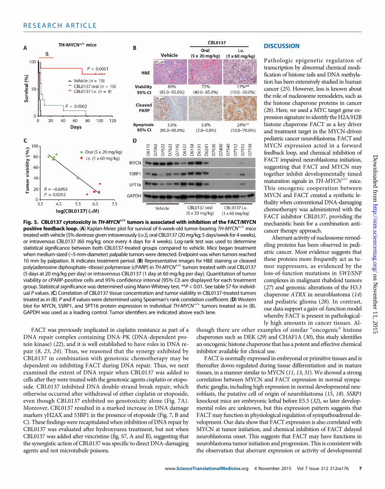
every 4 days for eight doses) to 6-week-old TH-MYCN+/+ mice withadvanced neuroblastoma delayed tumor growth, with most mice ex-hibiting long-term tumor regression (Fig. 5A and fig. S5A). Oral CBL0137
www.ScienceT
(20 mg/kg; five doses weekly for 4 weeks) and low-dose intravenousCBL0137 (40 mg/kg; every 4 days for eight doses) were less effectivebut still prolonged time until maximum tumor burden (Fig. 5A and
Fig. 3. CBL0137 overcomes developmental signals that drive neuro-blast persistence in TH-MYCNmice. (A) Semiquantitative histology scoring
in (C). (E) Six-day-old TH-MYCN+/+ mice were treated with intraperitoneal ve-hicle control (0.2% methylcellulose × 5 days/week) or CBL0137 for 3 weeks
was used to determine the relative expression of SSRP1 in wild-type and TH-MYCN+/+ mice through perinatal time points as indicated. P value was calcu-lated using analysis of variance (ANOVA), testing the significance of genotypeas the source of variation. At least three independentmice had one ormoreganglia present for analysis for each time point/genotype. (B) Represent-ative immunohistochemistry images comparing serial ganglion sections of2-week-old wild-type and TH-MYCN+/+ mice. Hematoxylin and eosin (H&E)staining and immunoblotting for SSRP1 andMYCN are shown. Arrowheadsindicate areas of neuroblast hyperplasia. (C) Six-day-old wild-type, TH-MYCN+/−,and TH-MYCN+/+ mice were treated with intraperitoneal vehicle control (0.2%methylcellulose) or CBL0137 for 5 days (10 mg/kg per day), and the propor-tion of ganglia with hyperplastic neuroblasts was evaluated by histologicalexamination. Ganglia were considered to be hyperplastic if >30 focal neu-roblasts were present per ganglion. Data displayed indicate the averageproportion of hyperplastic ganglia per mouse ± SE. Sample sizes were asfollows: wild type/vehicle, 6; wild type/CBL0137, 4; TH-MYCN+/−/vehicle, 13;TH-MYCN+/−/CBL0137, 13; TH-MYCN+/+/vehicle, 9; and TH-MYCN+/+/CBL0137, 9.(D) Representative H&E images of ganglion histology frommice treated as
(10mg/kg per day × 5 days/week) and thereafter assessed for tumor growth.A Kaplan-Meier plot is displayed for vehicle- or CBL0137-treated mice consid-ering time tomaximumtumorburden (when tumorswere 10mm indiameterby palpation). ℞ indicates treatment period. Log-rank test was used to cal-culate statistical significance between CBL0137 and vehicle-treated tumors.(F) Primary sympathetic ganglia were isolated and cultured from 2-week-old TH-MYCN+/+ or wild-type age-matched controls. Ganglia cultures weretreatedwith serum-rich or serum-deprivedmedium (1 of 30 normal concentra-tion of serum) for 48 hours, aswell aswith various concentrations of CBL0137for 24 hours, and viabilitywas calculated by cell counts on bIII-tubulin–positivecells for each of the TH-MYCN+/+ or wild-type cultures. Plot shows the non-linear regression line of percentage cell viability as determined by counts forbIII-tubulin–positive immunofluorescence normalized to untreated/serum-richsamples for each of the TH-MYCN+/+ or wild-type cultures. Data displayedrepresent average cell viability ± SE from the combined data of three in-dependent biological replicates. Red circle indicates the concentration ofCBL0137 that causes serum-deprived TH-MYCN+/+ cultures to have equal cellviability to serum-deprived wild-type cultures.
ranslationalMedicine.org 4 November 2015 Vol 7 Issue 312 312ra176 5

R E S EARCH ART I C L E
on Novem
ber 11, 2015http://stm
.sciencemag.org/
Dow
nloaded from
fig. S5A). We next examined the histology of tumors derived fromTH-MYCN+/+ mice after short-term CBL0137 treatment as either anoral (20 mg/kg for 5 days) or an intravenous (20, 40, or 60 mg/kg for1 day) formulation. We found that the tumor tissue demonstrated amarked treatment effect, particularly for the intravenously treated(60 mg/kg) group, characterized by extensive necrosis, hemorrhage,and apoptosis compared to controls (Fig. 5B, fig. S5B, and tables S5and S6). Moreover, intravenous CBL0137 (60 mg/kg)–treated mice ex-hibited evidence of metastatic regression with no mice demonstratingpulmonary metastases, whereas vehicle- and low-dose CBL0137–treated mice demonstrated marked metastatic spread to the lungs(fig. S5C). There was a substantial accumulation of CBL0137 in treatedtumors compared to other organs/body fluids, with intravenousCBL0137 (60 mg/kg)–treated animals showing the highest concentra-tions of intratumoral CBL0137 (fig. S5D). Tumor concentrations of
www.ScienceTranslationalMedicine.org 4 No
CBL0137 also showed a strong inversecorrelation with tumor viability (Fig. 5Cand fig. S5E). SSRP1, SPT16, and MYCNprotein expression was decreased in in-travenous CBL0137 (60 mg/kg)–treatedtumors compared to both oral CBL0137and vehicle treatment (Fig. 5D).Moreover,intravenous administration of CBL0137to BALB/c nude mice bearing flank xeno-grafts of the humanneuroblastoma cell lineBE(2)C resulted in delayed tumor growthand decreased MYCN protein expression(fig. S5, F andG). Together, this suggeststhat the inhibitory effect of CBL0137 onFACT has a downstream negative impacton MYCN expression and results in amarked therapeutic selectivity for MYCN-driven neuroblastoma tissues in vitro andin vivo.
CBL0137 exhibits synergywith DNA-damagingcytotoxic chemotherapyKnockdown of SSRP1 has been previouslyidentified to sensitize ovarian and breastcancer cells to cytotoxic chemotherapy(22), suggesting that CBL0137 may en-hance neuroblastoma chemotherapy. Weused colony formation and cell viability as-says in neuroblastoma cells to demonstratethat CBL0137 synergizes with mafosfa-mide (the active metabolite of cyclophos-phamide) and that this synergy wasexclusive to neuroblastoma cells in com-parison with normal primary fibroblasts(Fig. 6A and fig. S6A). The combinationof CBL0137 andmafosfamide was farmoretoxic in neuroblastoma cells compared tonormal controls, indicating that awide ther-apeutic index exists for this combinationtreatment (fig. S6B). CBL0137 combina-tion synergy extended to several otherchemotherapeutics used in neuroblasto-
ma therapy, albeit with some exceptions (fig. S6, C and D). Consistentwith these findings, cyclophosphamide, etoposide, cisplatin, and vin-cristine each potently increased CBL0137 efficacy in vivo againstestablished TH-MYCN+/+ tumors (Fig. 6B). Moreover, when intra-venous CBL0137 was combined with clinically relevant chemotherapyregimens used to treat high-risk/refractory neuroblastoma patients,namely, cyclophosphamide/topotecan and irinotecan/temozolomide,strong tumor delay was observed in both the TH-MYCN+/+transgenicmice (Fig. 6C) and BALB/c nude mice harboring BE(2)C xenografts(fig. S6E). Combination therapies were mostly well tolerated in reci-pient mice, with CBL0137/vincristine a notable exception. Some oc-casions of weight loss were observed, and mice were removed fromthe study as follows: cisplatin (2 of 10 mice), cisplatin/CBL0137 (2 of10mice), vincristine/CBL0137 (5 of 10mice), and CBL0137/irinotecan/temozolomide (2 of 10 mice).
Fig. 4. High MYCN expression sensitizes neuroblastoma cells to CBL0137. (A) CBL0137 IC50 (50%inhibitory concentration) was determined from resazurin reduction assays on a panel of neuroblastoma
cell lines and primary lung fibroblast controls. Cells were classified as high and low MYC-expressing celllines on the basis of their MYCN or c-MYC expression as displayed in fig. S1N. Data displayed are 72 hoursafter treatment. IC50 was determined using nonlinear regression analysis from at least threeindependent biological replicates. Average IC50 ± SE is shown. (B) Colony assays were conducted fordoxycycline (dox)–inducible MYCN overexpressing SHEP.MYCN3 cells (42) treated with an escalatingdose of CBL0137. The graph shows the average number of colonies at each concentration of drug fromthree independent biological replicates + SE. Extra sum of squares F test was used to determine statis-tical significance between nonlinear regression of +dox (+MYCN) versus −dox (−MYCN) cells. (C)MYCNmRNA expression in BE(2)C and KELLY cells treated with increasing concentrations of CBL0137. Data aredisplayed as mean-normalized mRNA expression from three replicate experiments ± SE. Data obtainedare 48 hours after treatment. b2-Microglobulin was used as a reference gene. (D) Western blot for MYCNprotein expression in BE(2)C and KELLY cells treated with increasing concentrations of CBL0137. Datadisplayed are 48 hours after treatment. GAPDH was used as a loading control.vember 2015 Vol 7 Issue 312 312ra176 6
R E S EARCH ART I C L E
on Novem
ber 11, 2015http://stm
.sciencemag.org/
Dow
nloaded from
FACT was previously implicated in cisplatin resistance as part of aDNA repair complex containing DNA-PK (DNA-dependent pro-tein kinase) (22), and it is well established to have roles in DNA re-pair (8, 23, 24). Thus, we reasoned that the synergy exhibited byCBL0137 in combination with genotoxic chemotherapy may bedependent on inhibiting FACT during DNA repair. Thus, we nextexamined the extent of DNA repair when CBL0137 was added tocells after they were treatedwith the genotoxic agents cisplatin or etopo-side. CBL0137 inhibited DNA double-strand break repair, whichotherwise occurred after withdrawal of either cisplatin or etoposide,even though CBL0137 exhibited no genotoxicity alone (Fig. 7A).Moreover, CBL0137 resulted in a marked increase in DNA damagemarkers gH2AX and 53BP1 in the presence of etoposide (Fig. 7, B andC). These findingswere recapitulatedwhen inhibition ofDNA repair byCBL0137 was evaluated after hydroxyurea treatment, but not whenCBL0137 was added after vincristine (fig. S7, A and B), suggesting thatthe synergistic action of CBL0137was specific to direct DNA-damagingagents and not microtubule poisons.
www.ScienceTranslationalMedicine.org 4 No
DISCUSSION
Pathologic epigenetic regulation oftranscription by abnormal chemical modi-fication of histone tails and DNAmethyla-tion has been extensively studied in humancancer (25). However, less is known aboutthe role of nucleosome remodelers, such asthe histone chaperone proteins in cancer(26). Here, we used a MYC target gene ex-pression signature to identify theH2A/H2Bhistone chaperone FACT as a key driverand treatment target in the MYCN-drivenpediatric cancer neuroblastoma. FACT andMYCN expression acted in a forwardfeedback loop, and chemical inhibition ofFACT impaired neuroblastoma initiation,suggesting that FACT and MYCN maytogether inhibit developmentally timedmaturation signals in TH-MYCN+/+ mice.This oncogenic cooperation betweenMYCN and FACT created a synthetic le-thality when conventional DNA-damagingchemotherapy was administered with theFACT inhibitor CBL0137, providing themechanistic basis for a combination anti-cancer therapy approach.
Aberrant activity of nucleosome remod-eling proteins has been observed in pedi-atric cancer. Most evidence suggests thatthese proteins more frequently act as tu-mor suppressors, as evidenced by theloss-of-function mutations in SWI/SNFcomplexes in malignant rhabdoid tumors(27) and genomic alterations of the H3.3chaperone ATRX in neuroblastoma (14)and pediatric glioma (28). In contrast,our data support a gain-of-functionmodelwhereby FACT is present in pathological-ly high amounts in cancer tissues. Al-
though there are other examples of similar “oncogenic” histonechaperones such as DEK (29) and CHAF1A (30), this study identifiesan oncogenic histone chaperone that has a potent and effective chemicalinhibitor available for clinical use.
FACT is normally expressed in embryonal or primitive tissues and isthereafter down-regulated during tissue differentiation and in maturetissues, in a manner similar to MYCN (11, 13, 31). We showed a strongcorrelation between MYCN and FACT expression in normal sympa-thetic ganglia, including high expression in normal developmental neu-roblasts, the putative cell of origin of neuroblastoma (15, 18). SSRP1knockout mice are embryonic lethal before E5.5 (32), so later develop-mental roles are unknown, but this expression pattern suggests thatFACTmay function in physiological regulation of sympathoadrenal de-velopment. Our data show that FACT expression is also correlated withMYCN at tumor initiation, and chemical inhibition of FACT delayedneuroblastoma onset. This suggests that FACT may have functions inneuroblastoma tumor initiation and progression. This is consistentwiththe observation that aberrant expression or activity of developmental
Fig. 5. CBL0137 cytotoxicity in TH-MYCN+/+ tumors is associated with inhibition of the FACT/MYCNpositive feedback loop. (A) Kaplan-Meier plot for survival of 6-week-old tumor-bearing TH-MYCN+/+ mice
treatedwith vehicle [5%dextrose given intravenously (i.v.)], oral CBL0137 (20mg/kg; 5 days/week for 4weeks),or intravenous CBL0137 (60 mg/kg; once every 4 days for 4 weeks). Log-rank test was used to determinestatistical significance between both CBL0137-treated groups compared to vehicle. Mice began treatmentwhenmedium-sized (~5-mm-diameter) palpable tumors were detected. Endpoint was when tumors reached10 mm by palpation. ℞ indicates treatment period. (B) Representative images for H&E staining or cleavedpoly(adenosine diphosphate–ribose) polymerase (cPARP) in TH-MYCN+/+ tumors treated with oral CBL0137(5 days at 20mg/kg per day) or intravenous CBL0137 (1 day at 60mg/kg per day). Quantitation of tumorviability or cPARP-positive cells and 95% confidence interval (95% CI) are displayed for each treatmentgroup. Statistical significance was determined usingMann-Whitney test. **P < 0.01. See table S7 for individ-ual P values. (C) Correlation of CBL0137 tissue concentration and tumor viability in CBL0137-treated tumorstreated as in (B). P and R values were determined using Spearman’s rank correlation coefficient. (D) Westernblot for MYCN, SSRP1, and SPT16 protein expression in individual TH-MYCN+/+ tumors treated as in (B).GAPDH was used as a loading control. Tumor identifiers are indicated above each lane.vember 2015 Vol 7 Issue 312 312ra176 7

R E S EARCH ART I C L E
on Novem
ber 11, 2015http://stm
.sciencemag.org/
Dow
nloaded from
Fig. 6. CBL0137 synergizes with neuroblastoma chemotherapy. (A)Colony-forming assays were performed for BE(2)C and KELLY cells treated
days when treatment began. P value was determined using pairwiselog-rank test for CBL0137/chemotherapy combinations versus their re-
with CBL0137 (CBL) in combination with mafosfamide (MAF), the activemetabolite of cyclophosphamide. Combination index (CI) at a fractionalcell kill of 0.80 is shown using the Chou-Talalay method (43). CI <0.9, syn-ergistic. Data are displayed as the average number of colonies at eachconcentration of drugs from three independent biological replicates ± SE.(B) Kaplan-Meier plots of tumor-bearing TH-MYCN+/+ mice treated with ve-hicle (5% dextrose) or intravenous CBL0137 (40 mg/kg; once every 4 daysfor 4 weeks) combined with cyclophosphamide (CPM; 15 mg/kg per day),etoposide (VP16; 6 mg/kg per day), cisplatin (2 mg/kg per day), or vincris-tine (VCR; 2 mg/kg per day). Mice were treated starting at 6 weeks of age,when 5-mm palpable tumors were detected. Endpoint was at 10-mm tu-mor diameter by palpation. Cyclophosphamide, etoposide, cisplatin, andvincristine were all administered intraperitoneally for five consecutive
www.ScienceT
spective single treatment controls. ℞ indicates treatment period. (C) Kaplan-Meier plots of tumor-bearing TH-MYCN+/+ mice treated with vehicle (5%dextrose) or intravenous CBL0137 (40mg/kg; once every 4 days for 4 weeks)combined with cyclophosphamide (10 mg/kg per day) and topotecan(TOPO; 5 mg/kg per day) (left) or irinotecan (IRINO; 2 mg/kg per day)and temozolomide (TEMOZ; 5 mg/kg per day) (right). Mice were treatedat 6 weeks of age, starting when 5-mm palpable tumors were detected.Endpoint was at 10-mm tumor diameter by palpation. Cyclophospha-mide, topotecan, irinotecan, and temozolomide were all administered in-traperitoneally for five consecutive days when treatment began. P valuewas determined using pairwise log-rank test for CBL0137/chemotherapycombinations versus their respective treatment controls. ℞ indicatestreatment period.
ranslationalMedicine.org 4 November 2015 Vol 7 Issue 312 312ra176 8

R E S EARCH ART I C L E
on Novem
ber 11, 2015http://stm
.sciencemag.org/
Dow
nloaded from
regulators are key determinants of neuroblastoma tumorigenesis (18).Animal models with conditional knockout of SSRP1 or SPT16 will re-vealmore specific roles of FACT in sympathoadrenal development and,by association, in MYCN-driven tumorigenesis.
FACT controls nucleosome “eviction” and “reassembly” of histones,thus promoting the smooth passage of RNA polymerase II during genetranscription (8, 9, 33). Our previous data have demonstrated that SSRP1is enriched at the promoter regions of oncogene transcription factors andtheir respective transcriptional targets (10). We show here that FACTmaintains high concentrations of MYCN protein in neuroblastoma cellsby drivingMYCN transcription andprotein stability.Moreover, FACT is
www.ScienceTranslationalMedicine.org 4 No
a functional cofactor to oncogenic mutantH-RasV12 in malignant transformation ofmammary epithelial cells (10). These ob-servations support a model wherebyFACTdrives bothmalignant transcriptionand oncogenic transcription factor activi-ty, yet the mechanism that governs FACTtranscriptional specificity in cancer cellsremains unclear. It may simply be that in-appropriate expression of FACT innormalcell types is sufficient to collaborate withother oncogenic transcription factors todrive a gene program that favors malig-nant transformation and transcription.
Our results demonstrate that FACTcan enhance MYCN transcription andprotein stability in neuroblastoma cells,even those with multiple copies ofMYCNandanalreadyhighMYCNexpression.Al-thoughMYCN amplification is a commonfeature of human neuroblastoma, recentstudies have indicated that mechanismsin addition to gene amplification are re-quired to maintain the very high MYCNprotein concentrations necessary for thefully malignant phenotype (34–37). Col-lectively, these findings are consistent withobservations made in tumorigenic murineMycmodels showing that different thresh-olds of MYC expression result in differentcomponents of the malignant phenotype,and suggest that MYCN expression andprotein stability are important treatmenttargets (38).
For neuroblastoma patients with ad-vanced disease, prognosis is still poor (1, 2).Many of these patients’ tumors are char-acterized byMYCN amplificationor over-activation of MYC target genes (6). Weshow here that CBL0137was highly effec-tive against established primary tumorsand pulmonary metastases in TH-MYCN+/+
mice and that CBL0137 treatment was ac-companied by marked MYCN depletionin tumors after only a single intravenousdose, suggesting that targeting FACTmaybean important strategy todecreaseMYCN
expression and overcome primary and metastatic disease (37, 39–41).This treatment approach is particularly promising because the ex-pression of MYCN and FACT is markedly higher in malignant com-paredwith normal tissues (10, 11, 15). Accordingly, our data showed thatCBL0137 inhibition of FACT andMYCNwas associated with exten-sive cytotoxicity in advanced TH-MYCN+/+ tumors but exhibitedminimal toxicity, consistent with findings in other animal models ofcancer (12). Moreover, the previously described inhibitory activityof CBL0137 against nuclear factor kB (NFkB)transcriptional targetingand activation of p53 by casein kinase–dependent Ser32 phosphoryl-ation (12) makes CBL0137 a potential multimodal cancer therapeutic
Fig. 7. CBL0137 prevents DNA damage repair after genotoxic chemotherapy. (A) BE(2)C cellswere treated for 24 hours with or without the following chemotherapeutics: 2 mM cisplatin (left) or
5 mM etoposide (right). Chemotherapeutics were subsequently removed, and medium was supple-mented with or without 100 nM CBL0137 for 22 hours (refer to schematic above gel for the admin-istration schedule). The repair of chemotherapy-induced DNA double-strand breaks (DSBs) in thepresence or absence of CBL0137 was determined using pulsed-field gel electrophoresis. Datadisplayed are representative of two independent biological replicates. Dashed white lines indicateareas where gel was cropped for presentation. (B) BE(2)C cells were treated with or without 5 mMetoposide for 5 hours. Etoposide was subsequently removed, and medium was supplemented withor without 200 nM CBL0137 for 3 hours (refer to schematic above the images for the administrationschedule). Cells were then fixed and processed for immunofluorescence with anti-gH2AX (red) andanti-53BP1 (green), as well as 4′,6-diamidino-2-phenylindole (DAPI) DNA stain (blue). (C) From (B),quantitation of the percentage of cells with >10 positive immunofluorescent foci for DNA damagemarkers gH2AX (top) or 53BP1 (bottom). Data are displayed as the average percentage of positivecells ± SE from two independent biological replicates. Mbp, mega base pairs.vember 2015 Vol 7 Issue 312 312ra176 9

R E S EARCH ART I C L E
on Novem
ber 11, 2015http://stm
.sciencemag.org/
Dow
nloaded from
for a variety of malignancies characterized by defects within thesepathways.
FACT has been shown to promote DNA damage repair in the faceof DNA-damaging chemotherapy (22), suggesting that it may act as achemoresistance factor. Here, we showed that CBL0137 was particu-larly effective as a combination therapywith chemotherapeutic agents.In addition, combiningCBL0137with chemotherapy regimens current-ly used after relapse potentiated its activity, highlighting a potentialtherapeutic strategy for high-risk neuroblastoma patients. Althoughour data and those of others have shown that CBL0137 alone is notgenotoxic (12), inhibition of FACT created a synthetic lethal environ-ment in the presence of cisplatin and etoposide but not vincristine.Moreover, combinations of CBL0137 withDNA-damaging agents suchas cisplatin, cyclophosphamide, and etoposide were better tolerated inour mouse models than the nongenotoxic microtubule inhibitor vin-cristine, suggesting that the most effective role for CBL0137 in anti-cancer therapywill be coadministrationwith primaryDNA-damagingagents. We also observed that p53 status was not a determinant ofCBL0137 combination therapy sensitivity, consistent with previousfindings for CBL0137 as a single agent in adult cancer types (12). Con-sidering that genotoxic chemotherapy is associated with marked non-specific toxicity in patients, targeting FACT with the nongenotoxicCBL0137 promises to enhance cancer cell specificity of these agents,which are the current mainstay of neuroblastoma therapy.
Future studies will need to address some of the limitations of thisstudy. First, it will be necessary to unveil the specific mechanism bywhich FACT modifies MYCN expression at both the transcriptionaland posttranslational level. Second, we still lack in-depth understandingof the mechanism and consequences of CBL0137-induced changes inchromatin, in particular how this relates to inhibition of FACT-dependent transcriptional programs and to specific DNA repair path-ways that are affected. Detailed analyses of CBL0137 effects on DNArepair pathways such as homologous repair and nonhomologous endjoining will be required. Third, the mechanism by which CBL0137 ac-cumulates at high concentration in neuroblastoma tumors and whetherthis occurs in other cancer types are unknown. In particular, mecha-nisms of CBL0137 uptake, metabolism, and efflux will need to be elu-cidated. Finally, it remains to be exploredwhether CBL0137 can be usedas a therapy for other cancers and whetherMYC deregulation can serveas a biomarker for CBL0137 sensitivity. With these additional studies,CBL0137 or newer FACT inhibitors may serve as a therapy for neuro-blastoma and potentially other MYC-driven cancers.
MATERIALS AND METHODS
Detailed methods are available in the Supplementary Materials.
SUPPLEMENTARY MATERIALS
www.sciencetranslationalmedicine.org/cgi/content/full/7/312/312ra176/DC1Materials and MethodsFig. S1. Data supporting Fig. 1.Fig. S2. Data supporting Fig. 2.Fig. S3. Data supporting Fig. 3.Fig. S4. Data supporting Fig. 4.Fig. S5. Data supporting Fig. 5.Fig. S6. Data supporting Fig. 6.Fig. S7. Data supporting Fig. 7.
www.ScienceTra
Fig. S8. Fluorescence in situ hybridization for MYCN amplification in tissue microarray.Table S1. Cox proportional hazards modeling of MYC target signature members in 649 neu-roblastoma patients.Table S2. Literature analysis of lead candidates that favor poor outcome in neuroblastoma.Table S3. Cox proportional hazards modeling of SSRP1 and SPT16 in 649 neuroblastoma patients.Table S4. Gene set enrichment analysis of 2-week-old TH-MYCN+/+ ganglia compared with age-matched wild-type mice.Table S5. Histology of TH-MYCN+/+ mice treated with oral or intravenous CBL0137.Table S6. Histology of TH-MYCN+/+ mice treated with a dose-escalating regimen of intravenousCBL0137.Table S7. P values not shown in figures.References (44–59)
REFERENCES AND NOTES
1. G. M. Brodeur, Neuroblastoma: Biological insights into a clinical enigma. Nat. Rev. Cancer 3,203–216 (2003).
2. J. M. Maris, Recent advances in neuroblastoma. N. Engl. J. Med. 362, 2202–2211 (2010).3. M. Schwab, K. Alitalo, K.-H. Klempnauer, H. E. Varmus, J. M. Bishop, F. Gilbert, G. Brodeur,
M. Goldstein, J. Trent, Amplified DNA with limited homology to myc cellular oncogene isshared by human neuroblastoma cell lines and a neuroblastoma tumour. Nature 305,245–248 (1983).
4. H. Kocak, S. Ackermann, B. Hero, Y. Kahlert, A. Oberthuer, D. Juraeva, F. Roels, J. Theissen,F. Westermann, H. Deubzer, V. Ehemann, B. Brors, M. Odenthal, F. Berthold, M. Fischer,Hox-C9 activates the intrinsic pathway of apoptosis and is associated with spontaneousregression in neuroblastoma. Cell Death Dis. 4, e586 (2013).
5. W. A. Weiss, K. Aldape, G. Mohapatra, B. G. Feuerstein, J. M. Bishop, Targeted expression ofMYCN causes neuroblastoma in transgenic mice. EMBO J. 16, 2985–2995 (1997).
6. F.Westermann, D.Muth, A. Benner, T. Bauer, K.-O. Henrich, A. Oberthuer, B. Brors, T. Beissbarth,J. Vandesompele, F. Pattyn, B. Hero, R. König, M. Fischer, M. Schwab, Distinct transcriptionalMYCN/c-MYC activities are associated with spontaneous regression or malignant progressionin neuroblastomas. Genome Biol. 9, R150 (2008).
7. H. Ji, G. Wu, X. Zhan, A. Nolan, C. Koh, A. De Marzo, H. M. Doan, J. Fan, C. Cheadle, M. Fallahi,J. L. Cleveland, C. V. Dang, K. I. Zeller, Cell-type independent MYC target genes reveal aprimordial signature involved in biomass accumulation. PLOS One 6, e26057 (2011).
8. T. Formosa, The role of FACT in making and breaking nucleosomes. Biochim. Biophys. Acta1819, 247–255 (2012).
9. D. Reinberg, R. J. Sims III, de FACTo nucleosome dynamics. J. Biol. Chem. 281, 23297–23301(2006).
10. H. Garcia, J. C. Miecznikowski, A. Safina, M. Commane, A. Ruusulehto, S. Kilpinen, R. W. Leach,K. Attwood, Y. Li, S. Degan, A. R. Omilian, O. Guryanova, O. Papantonopoulou, J. Wang,M. Buck, S. Liu, C. Morrison, K. V. Gurova, Facilitates chromatin transcription complex isan “accelerator” of tumor transformation and potential marker and target of aggressivecancers. Cell Rep. 4, 159–173 (2013).
11. H. Garcia, D. Fleyshman, K. Kolesnikova, A. Safina, M. Commane, G. Paszkiewicz, A. Omelian,C. Morrison, K. Gurova, Expression of FACT in mammalian tissues suggests its role in main-taining of undifferentiated state of cells. Oncotarget 2, 783–796 (2011).
12. A. V. Gasparian, C. A. Burkhart, A. A. Purmal, L. Brodsky, M. Pal, M. Saranadasa, D. A. Bosykh,M. Commane, O. A. Guryanova, S. Pal, A. Safina, S. Sviridov, I. E. Koman, J. Veith, A. A. Komar,A. V. Gudkov, K. V. Gurova, Curaxins: Anticancer compounds that simultaneously suppressNF-kB and activate p53 by targeting FACT. Sci. Transl. Med. 3, 95ra74 (2011).
13. A. Safina, H. Garcia, M. Commane, O. Guryanova, S. Degan, K. Kolesnikova, K. V. Gurova,Complex mutual regulation of facilitates chromatin transcription (FACT) subunits on bothmRNA and protein levels in human cells. Cell Cycle 12, 2423–2434 (2013).
14. J. J. Molenaar, J. Koster, D. A. Zwijnenburg, P. van Sluis, L. J. Valentijn, I. van der Ploeg,M. Hamdi, J. van Nes, B. A. Westerman, J. van Arkel, M. E. Ebus, F. Haneveld, A. Lakeman,L. Schild, P. Molenaar, P. Stroeken, M. M. van Noesel, I. Øra, E. E. Santo, H. N. Caron,E. M. Westerhout, R. Versteeg, Sequencing of neuroblastoma identifies chromothripsisand defects in neuritogenesis genes. Nature 483, 589–593 (2012).
15. L. M. Hansford, W. D. Thomas, J. M. Keating, C. A. Burkhart, A. E. Peaston, M. D. Norris,M. Haber, P. J. Armati, W. A.Weiss, G. M.Marshall, Mechanisms of embryonal tumor initiation:Distinct roles for MycN expression and MYCN amplification. Proc. Natl. Acad. Sci. U.S.A. 101,12664–12669 (2004).
16. Y. Nishida, N. Adati, R. Ozawa, A. Maeda, Y. Sakaki, T. Takeda, Identification and classifica-tion of genes regulated by phosphatidylinositol 3-kinase- and TRKB-mediated signallingpathways during neuronal differentiation in two subtypes of the human neuroblastomacell line SH-SY5Y. BMC Res. Notes 1, 95 (2008).
17. G. M. Marshall, J. L. Bell, J. Koach, O. Tan, P. Kim, A. Malyukova, W. Thomas, E. O. Sekyere,T. Liu, A. M. Cunningham, V. Tobias, M. D. Norris, M. Haber, M. Kavallaris, B. B. Cheung,
nslationalMedicine.org 4 November 2015 Vol 7 Issue 312 312ra176 10

R E S EARCH ART I C L E
on Novem
ber 11, 2015http://stm
.sciencemag.org/
Dow
nloaded from
TRIM16 acts as a tumour suppressor by inhibitory effects on cytoplasmic vimentin andnuclear E2F1 in neuroblastoma cells. Oncogene 29, 6172–6183 (2010).
18. G. M. Marshall, D. R. Carter, B. B. Cheung, T. Liu, M. K. Mateos, J. G. Meyerowitz, W. A. Weiss,The prenatal origins of cancer. Nat. Rev. Cancer 14, 277–289 (2014).
19. M. Calao, E. O. Sekyere, H. J. Cui, B. B. Cheung, W. D. Thomas, J. Keating, J. B. Chen, A. Raif,K. Jankowski, N. P. Davies, M. V. Bekkum, B. Chen, O. Tan, T. Ellis, M. D. Norris, M. Haber,E. S. Kim, J. M. Shohet, T. N. Trahair, T. Liu, B. J. Wainwright, H. F. Ding, G. M. Marshall,Direct effects of Bmi1 on p53 protein stability inactivates oncoprotein stress responsesin embryonal cancer precursor cells at tumor initiation. Oncogene 32, 3616–3626 (2013).
20. P. G. Komarov, E. A. Komarova, R. V. Kondratov, K. Christov-Tselkov, J. S. Coon, M. V. Chernov,A. V. Gudkov, A chemical inhibitor of p53 that protects mice from the side effects of cancertherapy. Science 285, 1733–1737 (1999).
21. L. T. Vassilev, B. T. Vu, B. Graves, D. Carvajal, F. Podlaski, Z. Filipovic, N. Kong, U. Kammlott,C. Lukacs, C. Klein, N. Fotouhi, E. A. Liu, In vivo activation of the p53 pathway by small-molecule antagonists of MDM2. Science 303, 844–848 (2004).
22. J. Sand-Dejmek, G. Adelmant, B. Sobhian, A. S. Calkins, J. Marto, D. J. Iglehart, J.-B. Lazaro,Concordant and opposite roles of DNA-PK and the “facilitator of chromatin transcription”(FACT) in DNA repair, apoptosis and necrosis after cisplatin. Mol. Cancer 10, 74 (2011).
23. V. Kari, A. Shchebet, H. Neumann, S. A. Johnsen, The H2B ubiquitin ligase RNF40 coop-erates with SUPT16H to induce dynamic changes in chromatin structure during DNA double-strand break repair. Cell Cycle 10, 3495–3504 (2011).
24. D. M. Keller, X. Zeng, Y. Wang, Q. H. Zhang, M. Kapoor, H. Shu, R. Goodman, G. Lozano,Y. Zhao, H. Lu, A DNA damage–induced p53 serine 392 kinase complex contains CK2,hSpt16, and SSRP1. Mol. Cell 7, 283–292 (2001).
25. S. B. Baylin, P. A. Jones, A decade of exploring the cancer epigenome—Biological andtranslational implications. Nat. Rev. Cancer 11, 726–734 (2011).
26. R. J. Burgess, Z. Zhang, Histone chaperones in nucleosome assembly and human disease.Nat. Struct. Mol. Biol. 20, 14–22 (2013).
27. B. G. Wilson, C. W. Roberts, SWI/SNF nucleosome remodellers and cancer. Nat. Rev. Cancer11, 481–492 (2011).
28. J. Schwartzentruber, A. Korshunov, X.-Y. Liu, D. T. W. Jones, E. Pfaff, K. Jacob, D. Sturm,A. M. Fontebasso, D.-A. Quang, M. Tönjes, V. Hovestadt, S. Albrecht, M. Kool, A. Nantel,C. Konermann, A. Lindroth, N. Jäger, T. Rausch, M. Ryzhova, J. O. Korbel, T. Hielscher,P. Hauser, M. Garami, A. Klekner, L. Bognar, M. Ebinger, M. U. Schuhmann, W. Scheurlen,A. Pekrun, M. C. Frühwald, W. Roggendorf, C. Kramm, M. Dürken, J. Atkinson, P. Lepage,A. Montpetit, M. Zakrzewska, K. Zakrzewski, P. P. Liberski, Z. Dong, P. Siegel, A. E. Kulozik,M. Zapatka, A. Guha, D. Malkin, J. Felsberg, G. Reifenberger, A. von Deimling, K. Ichimura,V. P. Collins, H. Witt, T. Milde, O. Witt, C. Zhang, P. Castelo-Branco, P. Lichter, D. Faury, U. Tabori,C. Plass, J. Majewski, S. M. Pfister, N. Jabado, Driver mutations in histone H3.3 and chromatinremodelling genes in paediatric glioblastoma. Nature 482, 226–231 (2012).
29. T. M. Wise-Draper, R. A. Mintz-Cole, T. A. Morris, D. S. Simpson, K. A. Wikenheiser-Brokamp,M. A. Currier, T. P. Cripe, G. C. Grosveld, S. I. Wells, Overexpression of the cellular DEK proteinpromotes epithelial transformation in vitro and in vivo. Cancer Res. 69, 1792–1799 (2009).
30. E. Barbieri, K. De Preter, M. Capasso, Z. Chen, D. M. Hsu, G. P. Tonini, S. Lefever, J. Hicks,R. Versteeg, A. Pession, F. Speleman, E. S. Kim, J. M. Shohet, Histone chaperone CHAF1Ainhibits differentiation and promotes aggressive neuroblastoma. Cancer Res. 74, 765–774(2014).
31. K. A. Zimmerman, G. D. Yancopoulos, R. G. Collum, R. K. Smith, N. E. Kohl, K. A. Denis, M. M. Nau,O. N. Witte, D. Toran-Allerand, C. E. Gee, J. D. Minna, F. W. Alt, Differential expression of mycfamily genes during murine development. Nature 319, 780–783 (1986).
32. S. Cao, H. Bendall, G. G. Hicks, A. Nashabi, H. Sakano, Y. Shinkai, M. Gariglio, E. M. Oltz,H. E. Ruley, The high-mobility-group box protein SSRP1/T160 is essential for cell viability inday 3.5 mouse embryos. Mol. Cell. Biol. 23, 5301–5307 (2003).
33. G. Orphanides, G. LeRoy, C.-H. Chang, D. S. Luse, D. Reinberg, FACT, a factor that facilitatestranscript elongation through nucleosomes. Cell 92, 105–116 (1998).
34. G. M. Marshall, P. Y. Liu, S. Gherardi, C. J. Scarlett, A. Bedalov, N. Xu, N. Iraci, E. Valli, D. Ling,W. Thomas, M. van Bekkum, E. Sekyere, K. Jankowski, T. Trahair, K. L. Mackenzie, M. Haber,M. D. Norris, A. V. Biankin, G. Perini, T. Liu, SIRT1 promotes N-Myc oncogenesis through apositive feedback loop involving the effects of MKP3 and ERK on N-Myc protein stability.PLOS Genet. 7, e1002135 (2011).
35. P. Y. Liu, N. Xu, A. Malyukova, C. J. Scarlett, Y. T. Sun, X. D. Zhang, D. Ling, S.-P. Su, C. Nelson,D. K. Chang, J. Koach, A. E. Tee, M. Haber, M. D. Norris, C. Toon, I. Rooman, C. Xue, B. B. Cheung,S. Kumar, G. M. Marshall, A. V. Biankin, T. Liu, The histone deacetylase SIRT2 stabilizes Myconcoproteins. Cell Death Differ. 20, 503–514 (2013).
36. T. Otto, S. Horn, M. Brockmann, U. Eilers, L. Schüttrumpf, N. Popov, A. M. Kenney, J. H. Schulte,R. Beijersbergen, H. Christiansen, B. Berwanger, M. Eilers, Stabilization of N-Myc is a criticalfunction of Aurora A in human neuroblastoma. Cancer Cell 15, 67–78 (2009).
37. L. Chesler, C. Schlieve, D. D. Goldenberg, A. Kenney, G. Kim, A. McMillan, K. K. Matthay,D. Rowitch, W. A. Weiss, Inhibition of phosphatidylinositol 3-kinase destabilizes Mycnprotein and blocks malignant progression in neuroblastoma. Cancer Res. 66, 8139–8146(2006).
www.ScienceTra
38. D. J. Murphy, M. R. Junttila, L. Pouyet, A. Karnezis, K. Shchors, D. A. Bui, L. Brown-Swigart,L. Johnson, G. I. Evan, Distinct thresholds govern Myc’s biological output in vivo. CancerCell 14, 447–457 (2008).
39. Y. H. Chanthery, W. C. Gustafson, M. Itsara, A. Persson, C. S. Hackett, M. Grimmer, E. Charron,S. Yakovenko, G. Kim, K. K. Matthay, W. A. Weiss, Paracrine signaling through MYCN en-hances tumor-vascular interactions in neuroblastoma. Sci. Transl. Med. 4, 115ra113 (2012).
40. J. E. Delmore, G. C. Issa, M. E. Lemieux, P. B. Rahl, J. Shi, H. M. Jacobs, E. Kastritis, T. Gilpatrick,R. M. Paranal, J. Qi, M. Chesi, A. C. Schinzel, M. R. McKeown, T. P. Heffernan, C. R. Vakoc,P. L. Bergsagel, I. M. Ghobrial, P. G. Richardson, R. A. Young, W. C. Hahn, K. C. Anderson,A. L. Kung, J. E. Bradner, C. S. Mitsiades, BET bromodomain inhibition as a therapeuticstrategy to target c-Myc. Cell 146, 904–917 (2011).
41. A. Puissant, S. M. Frumm, G. Alexe, C. F. Bassil, J. Qi, Y. H. Chanthery, E. A. Nekritz, R. Zeid,W. C. Gustafson, P. Greninger, M. J. Garnett, U. McDermott, C. H. Benes, A. L. Kung, W. A. Weiss,J. E. Bradner, K. Stegmaier, Targeting MYCN in neuroblastoma by BET bromodomain inhibition.Cancer Discov. 3, 308–323 (2013).
42. A. Slack, Z. Chen, R. Tonelli, M. Pule, L. Hunt, A. Pession, J. M. Shohet, The p53 regulatorygene MDM2 is a direct transcriptional target of MYCN in neuroblastoma. Proc. Natl. Acad.Sci. U.S.A. 102, 731–736 (2005).
43. T. C. Chou, P. Talalay, Quantitative analysis of dose-effect relationships: The combinedeffects of multiple drugs or enzyme inhibitors. Adv. Enzyme Regul. 22, 27–55 (1984).
44. L. Chesler, W. A. Weiss, Genetically engineered murine models—Contribution to ourunderstanding of the genetics, molecular pathology and therapeutic targeting of neuro-blastoma. Semin. Cancer Biol. 21, 245–255 (2011).
45. M. E. H. Hammond, D. F. Hayes, M. Dowsett, D. C. Allred, K. L. Hagerty, S. Badve, P. L. Fitzgibbons,G. Francis, N. S. Goldstein, M. Hayes, D. G. Hicks, S. Lester, R. Love, P. B. Mangu, L. McShane,K. Miller, C. K. Osborne, S. Paik, J. Perlmutter, A. Rhodes, H. Sasano, J. N. Schwartz, F. C. G. Sweep,S. Taube, E. E. Torlakovic, P. Valenstein, G. Viale, D. Visscher, T. Wheeler, R. B. Williams, J. L. Wittliff,A. C. Wolff, American Society of Clinical Oncology/College of American Pathologists guidelinerecommendations for immunohistochemical testing of estrogen and progesterone receptors inbreast cancer. J. Clin. Oncol. 28, 2784–2795 (2010).
46. V. K. Mootha, C. M. Lindgren, K.-F. Eriksson, A. Subramanian, S. Sihag, J. Lehar, P. Puigserver,E. Carlsson, M. Ridderstråle, E. Laurila, N. Houstis, M. J. Daly, N. Patterson, J. P. Mesirov,T. R. Golub, P. Tamayo, B. Spiegelman, E. S. Lander, J. N. Hirschhorn, D. Altshuler, L. C. Groop,PGC-1a-responsive genes involved in oxidative phosphorylation are coordinately downre-gulated in human diabetes. Nat. Genet. 34, 267–273 (2003).
47. A. Subramanian, P. Tamayo, V. K. Mootha, S. Mukherjee, B. L. Ebert, M. A. Gillette, A. Paulovich,S. L. Pomeroy, T. R. Golub, E. S. Lander, J. P. Mesirov, Gene set enrichment analysis: A knowledge-based approach for interpreting genome-wide expression profiles. Proc. Natl. Acad. Sci. U.S.A.102, 15545–15550 (2005).
48. C. A. Burkhart, A. J. Cheng, J. Madafiglio, M. Kavallaris, M. Mili, G. M. Marshall, W. A. Weiss,L. M. Khachigian,M. D. Norris, M. Haber, Effects of MYCN antisense oligonucleotide administrationon tumorigenesis in amurine model of neuroblastoma. J. Natl. Cancer Inst. 95, 1394–1403 (2003).
49. M. J. Henderson, M. Haber, A. Porro, M. A. Munoz, N. Iraci, C. Xue, J. Murray, C. L. Flemming,J. Smith, J. I. Fletcher, S. Gherardi, C.-K. Kwek, A. J. Russell, E. Valli, W. B. London, A. B. Buxton,L. J. Ashton, A. C. Sartorelli, S. L. Cohn, M. Schwab, G. M. Marshall, G. Perini, M. D. Norris, ABCCmultidrug transporters in childhood neuroblastoma: Clinical and biological effectsindependent of cytotoxic drug efflux. J. Natl. Cancer Inst. 103, 1236–1251 (2011).
50. H. Shimada, I. M. Ambros, L. P. Dehner, J.-i. Hata, V. V. Joshi, B. Roald, Terminology andmorphologic criteria of neuroblastic tumors: Recommendations by the International Neu-roblastoma Pathology Committee. Cancer 86, 349–363 (1999).
51. H. Shimada, I. M. Ambros, L. P. Dehner, J.-i. Hata, V. V. Joshi, B. Roald, D. O. Stram, R. B. Gerbing,J. N. Lukens, K. K. Matthay, R. P. Castleberry, The International Neuroblastoma PathologyClassification (the Shimada system). Cancer 86, 364–372 (1999).
52. R. I. Geran, N. H. Greenberg, M. M. Macdonald, A. M. Schumacher, B. J. Abbott, Protocols forscreening chemical agents and natural products against animal tumors and otherbiological systems. Cancer Chemother. Rep. 3, 51–61 (1972).
53. T.-C. Chou, Drug combination studies and their synergy quantification using the Chou-Talalay method. Cancer Res. 70, 440–446 (2010).
54. N. Issaeva, H. D. Thomas, T. Djureinovic, J. E. Jaspers, I. Stoimenov, S. Kyle, N. Pedley,P. Gottipati, R. Zur, K. Sleeth, V. Chatzakos, E. A. Mulligan, C. Lundin, E. Gubanova, A. Kersbergen,A. L. Harris, R. A. Sharma, S. Rottenberg, N. J. Curtin, T. Helleday, 6-Thioguanine selectively killsBRCA2-defective tumors and overcomes PARP inhibitor resistance. Cancer Res. 70, 6268–6276(2010).
55. V. Marcel, S. E. Ghayad, S. Belin, G. Therizols, A.-P. Morel, E. Solano-Gonzàlez, J. A. Vendrell,S. Hacot, H. C. Mertani, M. A. Albaret, J.-C. Bourdon, L. Jordan, A. Thompson, Y. Tafer, R. Cong,P. Bouvet, J.-C. Saurin, F. Catez, A.-C. Prats, A. Puisieux, J.-J. Diaz, p53 acts as a safeguard oftranslational control by regulating fibrillarin and rRNAmethylation in cancer. Cancer Cell 24,318–330 (2013).
56. Y.-C. Hwang, T.-Y. Lu, D.-Y. Huang, Y.-S. Kuo, C.-F. Kao, N.-H. Yeh, H.-C. Wu, C.-T. Lin, NOLC1,an enhancer of nasopharyngeal carcinoma progression, is essential for TP53 to regulateMDM2 expression. Am. J. Pathol. 175, 342–354 (2009).
nslationalMedicine.org 4 November 2015 Vol 7 Issue 312 312ra176 11
R E S EARCH ART I C L E
Dow
nloa
57. C. Zhang, C. Yin, L. Wang, S. Zhang, Y. Qian, J. Ma, Z. Zhang, Y. Xu, S. Liu, HSPC111 governsbreast cancer growth by regulating ribosomal biogenesis.Mol. Cancer Res. 12, 583–594 (2014).
58. R. A. Shamanna, M. Hoque, T. Pe’ery, M. B. Mathews, Induction of p53, p21 and apoptosisby silencing the NF90/NF45 complex in human papilloma virus-transformed cervical car-cinoma cells. Oncogene 32, 5176–5185 (2013).
59. X. Z. Zhou, K. P. Lu, The Pin2/TRF1-interacting protein PinX1 is a potent telomerase inhib-itor. Cell 107, 347–359 (2001).
Acknowledgments: We thank R. Lukeis andM. Suter at the Department of Cytogenetics (SydPath),St. Vincent’s Hospital, Darlinghurst, New South Wales, Australia, for histological assistance withthe tissue microarray. We also thank J. Shohet (Baylor College of Medicine) for providing theSHEP.MYCN3 cells used in this study. Funding: Thisworkwas supported by programgrants fromthe National Health andMedical Research Council, Australia (grants APP1016699 and APP1085411),Cancer Institute NSW (grant 10/TPG/1-03), Cancer Council NSW (grant PG-11-06), Australian RotaryHealth/Rotary Club of Adelaide, Fund for Scientific Research Flanders (FWO), German Cancer Aid(grant 110122), German Ministry of Science and Education (BMBF) as part of the e:Med initiative(grants 01ZX1303A and 01ZX1307D), and Fördergesellschaft Kinderkrebs-Neuroblastom-Forschung e.V. Author contributions: M.H. and G.M.M. conceived the project. D.R.C., M.H.,and G.M.M. supervised the project. B.B.C., C.B., D.S.Z., T.L., K.V.G., and A.V.G. were advisors forthe project. A.O., M.F., J.B., F. Speleman, K.D.P., and A.B. supplied tissues or patient/mouse datafor the experiments. D.R.C., J.M., A.J.G., N.I., A.P., M.D.N., M.H., and G.M.M. designed theexperiments. D.R.C., J.M., H.K., L.G., J.K., J.T., S. Sutton, S. Syed, A.J.G., N.I., A.B., B.A., Y.S., N.S., D.L.,P.Y.K., H.W., A.C., M.R., B.L., F. Salleta, L.M.T., A.P., and G.H. performed the experiments. D.R.C.,
www.ScienceTra
J.M., L.G., A.J.G., N.I., B.L., and A.P. analyzed the experiments. D.R.C. and G.M.M. wrote the man-uscript. J.M., L.G., A.J.G., K.V.G., A.V.G., M.D.N., and M.H. reviewed the manuscript. Competinginterests: This work was funded in part by grants to K.V.G., A.V.G., M.D.N., and M.H. fromIncuron, LLC. K.V.G. and A.V.G. are co-inventors on patents describing CBL0137. M.D.N.and M.H. own some stock in Cleveland BioLabs, which is entitled to a portion of the royaltiesfor sale of Incuron’s product, CBL0137. The other authors declare that they have no competinginterests. Data and materials availability: Gene expression data for neuroblastoma tumors(n = 649) are available at the Gene Expression Omnibus under accession no. GSE45547. Allother gene expression data are available at the Gene Expression Omnibus under accessionno. GSE71062. CBL0137 is available under a material transfer agreement from Incuron, LLC.
Submitted 23 March 2015Accepted 26 August 2015Published 4 November 201510.1126/scitranslmed.aab1803
Citation: D. R. Carter, J. Murray, B. B. Cheung, L. Gamble, J. Koach, J. Tsang, S. Sutton, H. Kalla,S. Syed, A. J. Gifford, N. Issaeva, A. Biktasova, B. Atmadibrata, Y. Sun, N. Sokolowski, D. Ling,P. Y. Kim, H. Webber, A. Clark, M. Ruhle, B. Liu, A. Oberthuer, M. Fischer, J. Byrne, F. Saletta,L. M. Thwe, A. Purmal, G. Haderski, C. Burkhart, F. Speleman, K. De Preter, A. Beckers,D. S. Ziegler, T. Liu, K. V. Gurova, A. V. Gudkov, M. D. Norris, M. Haber, G. M. Marshall,Therapeutic targeting of the MYC signal by inhibition of histone chaperone FACT inneuroblastoma. Sci. Transl. Med. 7, 312ra176 (2015).
de
nslationalMedicine.org 4 November 2015 Vol 7 Issue 312 312ra176 12
on Novem
ber 11, 2015http://stm
.sciencemag.org/
d from
10.1126/scitranslmed.aab1803] (312), 312ra176. [doi:7Science Translational Medicine
(November 4, 2015) Murray D. Norris, Michelle Haber and Glenn M. Marshall David S. Ziegler, Tao Liu, Katerina V. Gurova, Andrei V. Gudkov,Burkhart, Frank Speleman, Katleen De Preter, Anneleen Beckers, Saletta, Le Myo Thwe, Andrei Purmal, Gary Haderski, CatherineLiu, André Oberthuer, Matthias Fischer, Jennifer Byrne, Federica Y. Kim, Hannah Webber, Ashleigh Clark, Michelle Ruhle, BingAtmadibrata, Yuting Sun, Nicolas Sokolowski, Dora Ling, Patrick Syed, Andrew J. Gifford, Natalia Issaeva, Asel Biktasova, BernardJessica Koach, Joanna Tsang, Selina Sutton, Heyam Kalla, Sarah Daniel R. Carter, Jayne Murray, Belamy B. Cheung, Laura Gamble,chaperone FACT in neuroblastomaTherapeutic targeting of the MYC signal by inhibition of histone
Editor's Summary
to kill cancer cells and treat neuroblastoma in mouse models.vicious cycle. They demonstrated that curaxins work in synergy with standard genotoxic chemotherapyMYC and FACT. The authors then used curaxins, which are drugs that inhibit FACT, to break the demonstrated its role in a feedback loop that allows tumor cells to maintain a high expression of bothidentified a histone chaperone called FACT as a mediator of MYC signaling in neuroblastoma and
. haveet al oncogene are particularly aggressive. Carter MYCand tumors with amplifications of the Neuroblastoma is a common pediatric cancer of the nervous system. It is often difficult to treat,
Uncovering the FACTs in neuroblastoma
This information is current as of November 11, 2015. The following resources related to this article are available online at http://stm.sciencemag.org.
Article Tools
http://stm.sciencemag.org/content/7/312/312ra176article tools: Visit the online version of this article to access the personalization and
MaterialsSupplemental
http://stm.sciencemag.org/content/suppl/2015/11/02/7.312.312ra176.DC1"Supplementary Materials"
Related Content
http://stke.sciencemag.org/content/sigtrans/4/166/pe15.fullhttp://stke.sciencemag.org/content/sigtrans/7/349/ra102.fullhttp://stm.sciencemag.org/content/scitransmed/3/95/95ra74.fullhttp://stm.sciencemag.org/content/scitransmed/4/115/115ra3.fullhttp://stm.sciencemag.org/content/scitransmed/4/141/141ra91.fullhttp://stm.sciencemag.org/content/scitransmed/7/283/283ra55.full
's sites:ScienceThe editors suggest related resources on
is a registered trademark of AAAS.MedicineScience TranslationalAssociation for the Advancement of Science; all rights reserved. The title
Science, 1200 New York Avenue, NW, Washington, DC 20005. Copyright 2015 by the Americanweekly, except the last week in December, by the American Association for the Advancement of
(print ISSN 1946-6234; online ISSN 1946-6242) is publishedScience Translational Medicine
on Novem
ber 11, 2015http://stm
.sciencemag.org/
Dow
nloaded from

Permissionshttp://www.sciencemag.org/about/permissions.dtlObtain information about reproducing this article:
is a registered trademark of AAAS.MedicineScience TranslationalAssociation for the Advancement of Science; all rights reserved. The title
Science, 1200 New York Avenue, NW, Washington, DC 20005. Copyright 2015 by the Americanweekly, except the last week in December, by the American Association for the Advancement of
(print ISSN 1946-6234; online ISSN 1946-6242) is publishedScience Translational Medicine
on Novem
ber 11, 2015http://stm
.sciencemag.org/
Dow
nloaded from