Schreibersite: Synthesis, Characterization and Corrosion ...
56
University of South Florida Scholar Commons Graduate eses and Dissertations Graduate School January 2015 Schreibersite: Synthesis, Characterization and Corrosion and Possible Implications for Origin of Life Nikita Latesha La Cruz University of South Florida, [email protected] Follow this and additional works at: hp://scholarcommons.usf.edu/etd Part of the Geochemistry Commons , and the Geology Commons is esis is brought to you for free and open access by the Graduate School at Scholar Commons. It has been accepted for inclusion in Graduate eses and Dissertations by an authorized administrator of Scholar Commons. For more information, please contact [email protected]. Scholar Commons Citation La Cruz, Nikita Latesha, "Schreibersite: Synthesis, Characterization and Corrosion and Possible Implications for Origin of Life" (2015). Graduate eses and Dissertations. hp://scholarcommons.usf.edu/etd/5724
Transcript of Schreibersite: Synthesis, Characterization and Corrosion ...
University of South FloridaScholar Commons
Graduate Theses and Dissertations Graduate School
January 2015
Schreibersite: Synthesis, Characterization andCorrosion and Possible Implications for Origin ofLifeNikita Latesha La CruzUniversity of South Florida, [email protected]
Follow this and additional works at: http://scholarcommons.usf.edu/etd
Part of the Geochemistry Commons, and the Geology Commons
This Thesis is brought to you for free and open access by the Graduate School at Scholar Commons. It has been accepted for inclusion in GraduateTheses and Dissertations by an authorized administrator of Scholar Commons. For more information, please contact [email protected].
Scholar Commons CitationLa Cruz, Nikita Latesha, "Schreibersite: Synthesis, Characterization and Corrosion and Possible Implications for Origin of Life"(2015). Graduate Theses and Dissertations.http://scholarcommons.usf.edu/etd/5724
Schreibersite: Synthesis, Characterization and Corrosion and Possible Implications for
Origin of Life
by
Nikita L. La Cruz
A thesis submitted in partial fulfillment of the requirements for the degree of
Master of Science in Geology with a concentration in Geochemistry
School of Geosciences College of Arts and Sciences University of South Florida
Major Professor: Matthew Pasek, Ph.D. Jeffrey Ryan, Ph.D. Zachary Atlas, Ph.D.
Date of Approval: May 21, 2015
Keywords: phosphides, meteorite minerals, chemical evolution of life
Copyright © 2015, Nikita L. La Cruz

DEDICATION
I dedicate this work to my mother (Shellon Murray), my father (Lloyd La Cruz), my
younger sister, (Nelanie La Cruz), my uncles (Pierre Murray and Ron Campbell) and the
members of my extended family who have always been supportive of me in my educational
pursuits. Your help has been invaluable and I would not have been able to do this without you.

ACKNOWLEDGMENTS
First, I thank God for granting me the health, strength, wisdom and understanding
necessary to complete this work.
Second, I thank my advisor, Dr. Matthew Pasek for taking a leap of faith and hiring me as
his research assistant to work on this project. Your faith in my ability to work on this project
motivated me to work diligently to complete the necessary tasks. I am also very grateful for the
many discussions we had where you provided useful insight and suggestions, all of which made
this work a success. I also thank you for introducing me to your family. Spending time with them
helped me miss my family less and for that I will be forever grateful.
Third, I thank NASA and NSF for providing funding to the Center for Chemical
Evolution, which in turn provided the funding that allowed me to complete this project and by
extension my masters degree.
Fourth, I would like to thank my amazing lab mates, the members of Team Phosphorus.
To Danny, Jackie, Kristyn, Maheen and Tian, I say thank you very much for your
encouragement and for putting up with me during times of stress. I also thank you for all the time
you spent listening to my presentations and providing comments which helped me to improve
them.
Fifth, I thank my collaborators and co-authors. The completion of this project would have
been very difficult without your input.

Sixth, I thank my friends: Noelle DeFreitas-Adams, Sharleen vanAms and Karissa Jansen
for being there for me and ensuring that I remain mentally stable. Your words of encouragement
and conversations filled of joy and laughter helped me remain grounded and allowed me to focus
when it was necessary.
Seventh, I thank the faculty and staff of the School of Geosciences at University of South
Florida. I felt at home in this department and I learned a lot about my area of interest as well as
the field of geology at large. I especially thank Ms. Carolyn Rivera and Ms. Mandy Stuck for all
that they helped me with.
Eighth, I thank Pandora, Spotify and SoundCloud for allowing me to listen to my favorite
reggae artists (Bob Marley, Damian Marley and K’Naan) during the many hours I spent working
in lab and writing.
Last but by no means least, I thank the members of my immediate and extended family.
Your phone calls, messages, prayers, words of encouragement were very much appreciated.
Including me in your lives also helped me feel less lonely and very connected to life at home and
this meant a lot to me. I will continue to work hard to make you guys proud of me.
i
TABLE OF CONTENTS
List of Tables ................................................................................................................................. iii
List of Figures ................................................................................................................................ iv
Abstract ............................................................................................................................................v
Chapter One: Introduction ...............................................................................................................1 Phosphides ...........................................................................................................................1 Schreibersite .........................................................................................................................1
Schreibersite and origins of life ...........................................................................................3 Why synthesize schreibersite? .............................................................................................4
Chapter Two: Methods ....................................................................................................................5 Materials ..............................................................................................................................5 Synthesis ..............................................................................................................................5
Synthesis of schreibersite .........................................................................................5 Synthesis of nickel-phosphide .................................................................................6
Characterization ...................................................................................................................7 X-ray Diffractometry ...............................................................................................7 Electron Microprobe Point Analysis ........................................................................7 Micro-Raman Spectroscopy.....................................................................................7 X-ray Photoelectron Spectroscopy ..........................................................................8
Corrosion experiments .........................................................................................................8 Corrosion experiments without a chelating agent ....................................................8 Corrosion experiments with chelating agent............................................................9 Surface analyses of corroded schreibersite ..............................................................9 P-31 NMR Analysis .................................................................................................9 Speciation Analysis ................................................................................................10 ICP-MS ..................................................................................................................11 ICP-OES ................................................................................................................11
Phosphorylation experiments .............................................................................................12 Phosphorylation of choline by synthetic schreibersite...........................................12
Chapter Three: Results ...................................................................................................................13 Synthesis ............................................................................................................................13
Characterization ................................................................................................................13 EMPA ....................................................................................................................13 XRD .......................................................................................................................15 Micro-Raman Spectroscopy...................................................................................15

ii
XPS ........................................................................................................................16 Corrosion............................................................................................................................19
P-31 NMR Spectroscopy .......................................................................................20 Speciation Analysis ................................................................................................20 ICP-MS ..................................................................................................................22 ICP-OES ................................................................................................................22
Phosphorylation ................................................................................................................23 Phosphorylation of choline ....................................................................................23
Chapter Four: Discussion ...............................................................................................................24 Synthesis ............................................................................................................................24 Characterization .................................................................................................................25
XPS ........................................................................................................................25 Corrosion............................................................................................................................26
P-31 NMR Analysis ...............................................................................................26 Speciation Analysis ................................................................................................27 ICP-MS and ICP-OES ...........................................................................................27 Speciation Analysis and ICP-OES Comparison .....................................................28
Phosphorylation .................................................................................................................28 Phosphorylation of choline ....................................................................................28
Chapter Five: Implications .............................................................................................................30
Chapter Six: Conclusions ...............................................................................................................31
References ......................................................................................................................................33
Appendix: Copyright Permissions .................................................................................................36
About the Author .............................................................................................................. End Page
iii
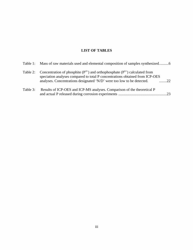
LIST OF TABLES
Table 1: Mass of raw materials used and elemental composition of samples synthesized. .........6
Table 2: Concentration of phosphite (P3+) and orthophosphate (P5+) calculated from speciation analyses compared to total P concentrations obtained from ICP-OES analyses. Concentrations designated ‘N/D’ were too low to be detected. ........22
Table 3: Results of ICP-OES and ICP-MS analyses. Comparison of the theoretical P and actual P released during corrosion experiments ...................................................23

iv
LIST OF FIGURES
Figure 1: Synthesized schreibersite samples. (A) Vesicular schreibersite obtained by rapid cooling. (B) Schreibersite sample obtained by slow cooling. (C) Dense schreibersite samples obtained when specimen were heated to the eutectic melting point after 235 hrs. .......................................................................14
Figure 2. Synthesized schreibersite samples with discolorations. ...............................................14
Figure 3. SEM images of schreibersite. (A) 50X magnification. (B) 100X magnification ........15
Figure 4. XRD of synthesized schreibersite with comparison of characteristic d values for schreibersite. ..........................................................................................................16
Figure 5. Raman spectra of uncorroded samples compared to those from Pirim et al. (2014). (A) Raman spectrum of synthesized schreibersite. (B) Raman spectra of schreibersite inclusion from Sikhote-Alin (a), Seymchan (b), Odessa (c) meteorites; synthetic Fe3P (d) and synthetic Fe2NiP (e). Reprinted from “Investigation of schreibersite and intrinsic oxidation products from Sikhote-Alin, Seymchan, and Odessa meteorites and Fe3P and Fe2NiP synthetic surrogates”, Pirim, C., et al., Figure 15, pg. 271, 2014, with permission from Elsevier .................17
Figure 6. XPS spectra obtained for schreibersite synthesized in this study. Spectrum (A) is at a depth of 15 nm, (B) is at a depth of 3.8 nm and (C) is before etching.....................................................................................................18
Figure 7. XPS spectra showing binding energies of Fe (A) and P (B). .......................................18
Figure 8. XPS Spectra of corroded schreibersite. .......................................................................19
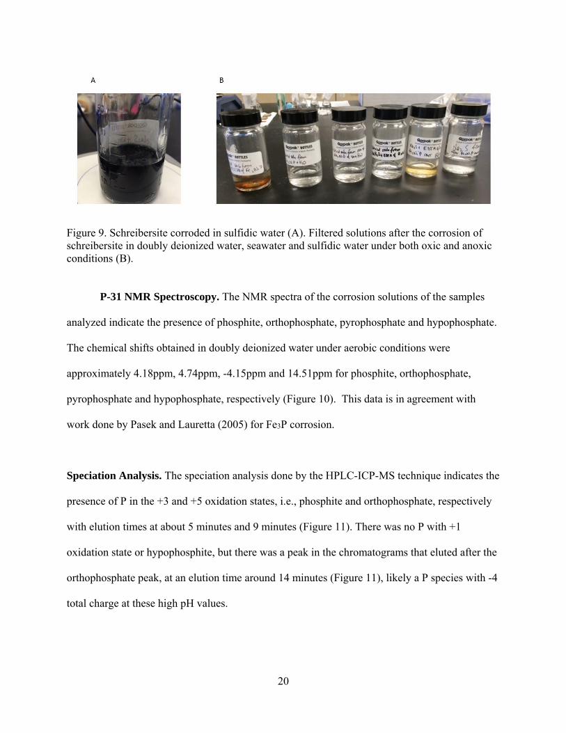
Figure 9. Schreibersite corroded in sulfidic water (A). Filtered solutions after the corrosion of schreibersite in doubly deionized water, seawater and sulfidic water under both oxic and anoxic conditions (B). ..........................................20
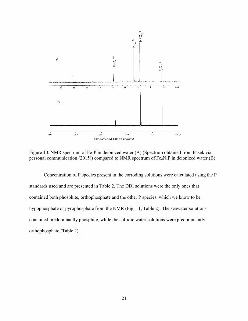
Figure 10. NMR spectrum of Fe3P in deionized water (A) (original spectrum from M. A. Pasek via personal communication, 2015) compared to NMR spectrum of Fe2NiP in deionized water (B). ................................................................21
Figure 11 Chromatogram of standards used in speciation experiments (A) compared to the chromatogram for Fe2NiP in deionized water. ..................................................22

v
Figure 12 NMR spectrum indicating the formation of phosphocholine. .....................................23

v
ABSTRACT
We present study of the synthesis and reactions of an analog of the meteoritic mineral
schreibersite with formula (Fe,Ni)3P, believed to be a prebiotic source of reactive phosphorus
that may have prompted the formation of phosphorylated biomolecules near the time of the
origin of life (Pasek and Lauretta, 2005). The mineral was synthesized by mixing stoichiometric
proportions of elemental iron, nickel and phosphorus and heating in a tube furnace at 820°C for
approximately 235 hours under argon or under vacuum, a modification of the method of Skála
and Drábek (2002). The mineral was characterized using X-ray diffractometry (XRD), X-ray
photoelectron spectroscopy (XPS), micro-raman spectroscopy and electron microprobe analysis
(EMPA). Characterization indicates that both schreibersite, with approximate formula Fe2NiP
and the mineral nickel-phosphide, FeNi2P were synthesized.
In addition to characterization of the solid product, the reactions of the synthetic
schreibersite were investigated to determine the similarity between these and prior work done
with Fe3P. Synthetic schreibersite was corroded in several solutions: seawater and sulfidic water
under both oxic and anoxic conditions. After corrosion, the solutions were analyzed using
phosphorus nuclear magnetic resonance spectroscopy (31P NMR) and high performance liquid
chromatography attached to an inductively coupled plasma mass spectrometer (HPLC-ICP-MS)
to determine phosphorus speciation as well as concentrations of phosphorus present in solution.
As expected from previous studies, the NMR and HPLC-ICP-MS results indicated the presence
of orthophosphate, phosphite, pyrophosphate and hypophosphate in the corrosion solutions
(Pasek and Lauretta, 2005). The HPLC-ICP-MS results indicate that the extent of corrosion of

vi
the mineral—measured by the concentration of phosphorus released—depends on the ionic
strength of the solution, as well as the presence or absence of the chelating agent. Finally, we
report the successful phosphorylation of a potentially prebiotic molecule—choline—using
synthesized schreibersite.
1
CHAPTER ONE: INTRODUCTION
Phosphides
Phosphides are an intriguing group of minerals because they contain phosphorus (P) in a
reduced oxidation state as opposed to the oxidized (P5+) state that are ubiquitous in minerals on
the earth. Phosphide species include schreibersite ((Fe,Ni)3P), perryite ((Ni,Fe)8(Si,P)3),
barringerite ((Fe,Ni)2P) (hexagonal)), nickel-phosphide ((Ni,Fe)3P)), florenskyite (Fe(Ti,Ni)P),
allabogdanite ((Fe,Ni)2P (orthorhombic)), melliniite ((Ni,Fe)4P), andreyivanovite (FeCrP),
monipite (MoNiP) and negevite (NiP2), halamishite (Ni5P4), transjordanite (Ni2P), murashkoite
(FeP) (Pasek, 2014) and zuktamrurite (FeP2) which were recently discovered (Britvin et al.,
2015). Since most environments on the earth have an oxidizing character, phosphide minerals
are rarely found on the earth. Notable exceptions are the phosphides recently discovered near the
Dead Sea (Britvin et al., 2015), trace amounts of schreibersite found in native iron from Disko
Island, Greenland (Pedersen, 1981), an occurrence of barringerite in a garnet peridotite in China
(Yang et al., 2005) and iron phosphide in fulgurites (Pasek et al., 2012; Essene and Fisher, 1986).
Schreibersite
Schreibersite is an important minor mineral phase in iron meteorites (Geist et al. 2005)
and is the most phosphorus rich species present in iron meteorites (Bartoschewitz, 2003). Based
on the chemical formula of this mineral, there are three compositional variations of schreibersite

2
that are of interest to this study: iron phosphide (Fe3P), nickel-phosphide (FeNi2P) and
schreibersite (Fe2NiP). The three constituents of schreibersite exist in a solid solution. The
mineral has a metallic luster, a high density (an approximate average of 7.4 g/cm3) and a
hardness of approximately 6.5-7 on the Mohs hardness scale. It is brittle, is attracted to a magnet
and produces a dark gray streak in streak tests. Schreibersite has a tetragonal crystal system and
is a member of the I4 space group (Skála and Císařová, 2005). The oxidation state of the P in
schreibersite is approximately -1 based on binding energies obtained from XPS studies (Pirim et
al., 2014; Bryant et al., 2013; Pelavin et al., 1970).
Meteorites have long been used by scientists to gain insight into the formation of
planetary bodies (McDonough and Sun, 1995; Ganapathy and Anders, 1974). The earth’s core is
known to be predominantly iron and nickel but many studies propose the presence of light
elements in the core (Poirier, 1994; Washington, 1925; Birch, 1952, Ringwood, 1979; Williams
and Knittle, 1997; Scott et.al., 2007; Li and Fei, 2003). P in schreibersite is siderophilic, and
because schreibersite is one of the phases that is ubiquitous in iron meteorites, a class of bodies
used to approximate the non-volatile composition of the earth’s core (Scott et al., 2007),
schreibersite may be of interest to scientists trying to determine the composition of the core. Wu
et al (2011) showed that schreibersite is not stable at the temperatures and pressures present in
the earth’s core, therefore, it is unlikely to be the dominant phosphide phase present in the core.
However, they showed that melliniite could be present in the core, therefore a study like this one,
which seeks to synthesize a similar phosphide mineral could potentially be of interest to groups
seeking to synthesize melliniite for future research.
3
Schreibersite and origins of life
We study schreibersite because we are interested in the role that P played in the chemical
evolution of life on planet earth. Phosphorus is one of the six important biogenic elements, the
others being: carbon, hydrogen, nitrogen, oxygen and sulfur. P is an important element in
modern biochemistry as it is present in biomolecules that are important for cellular structure,
storage and transfer of genetic information, signal transduction and for energetics (Westheimer
1987, Maciá et.al. 1997). Although phosphorus is ubiquitous in biological systems, phosphate,
the P species most likely present on the early earth, is poorly reactive towards organic molecules.
Hence, the facile synthesis of prebiotic phosphorus compounds using terrestrial phosphate
minerals has been hindered by the low reactivity and solubility of phosphates such as apatite.
How then were the P-O-C and P-C bonds, so ubiquitous in biochemistry made?
Gulick (1955) suggested that low oxidation state P species would facilitate the formation
of phosphorylated organic molecules and he suggested that the corrosion of schreibersite should
produce the necessary reactive P species. Yamagata et al. (1991) suggested that polyphosphates,
resulting from volcanic activity on the early earth, may have been responsible for the
phosphorylation of organic molecules on the early. Pasek and Lauretta (2005) partially
confirmed Gulick’s hypothesis when they corroded iron phosphide, an analog of schreibersite,
and observed the formation of low oxidation state P species. Interestingly enough, their corrosion
also resulted in the formation of pyrophosphate, which is one of the polyphosphate species
suggested to have been important for phosphorylation on the early earth by Yamagata et al.
(Pasek and Lauretta, 2005; Yamagata et al., 1991). Recently, Pasek et al. (2013) demonstrated
the formation of glycerol phosphate when iron phosphide, as a schreibersite simulant, was placed
in a solution of glycerol dissolved water and heated. Furthermore, some of the earliest carbonate

4
rocks appear to bear small quantities of phosphite, suggesting a meteoritic component to P on the
early earth (Pasek et. al., 2013). These data hint to a role for schreibersite in the origin of life on
the earth.
Why synthesize schreibersite?
The synthesis of schreibersite is important because the only commercially available
analog of schreibersite is Fe3P and while it is similar to meteoritic schreibersite, it contains no
nickel. Nickel acts as a noble metal in schreibersite and therefore, has important effects on the
mineral’s reactivity (Bryant et al., 2013). Meteorites are rare and therefore expensive.
Schreibersite occurs as very small inclusions in the matrices of iron meteorites. Therefore, it is
expensive to acquire a sufficient quantity of schreibersite to conduct experiments and it is also
difficult to isolate the schreibersite grains from the meteorite matrices. Meteoritic schreibersite
is, therefore, very expensive and difficult to attain. Another issue with the use of meteoritic
schreibersite is that the samples are not usually chemically homogeneous (Pirim et. al., 2014).
This heterogeneity, therefore, introduces uncertainty into experiments being conducted. The
aforementioned reasons motivated this work. Here, we present the synthesis, characterization and
corrosion of schreibersite and nickel-phosphide. The synthesis was accomplished by modifying
the method presented by Skála and Drábek (2005) and the synthesized product was characterized
using petrographic as well as surface chemistry techniques and then used in experiments seeking
to answer questions relating to the origin of life on earth.

5
CHAPTER TWO: METHODS
Materials
Iron (Fe) powder (98+% purity), nickel (Ni) powder (99.9% metals basis), phosphorus
(P) powder (red amorphous, 98.9% metals basis), sodium hydroxide (NaOH) pellets (98%) and
phosphorous acid (98% purity) were acquired from Alfa Aesar. Doubly deionized water was
generated in house using a Barnstead (Dubuque, IA) Nanopure® Diamond analytical combined
reverse osmosis-deionization system. Seawater solutions were made using an aquarium sea salt
mixture produced by Instant Ocean®. Sulfidic water solutions were made using sodium sulfide
(Na2S) (98%) obtained from EMD Chemicals. Ethylenediamminetetraacetic acid powder
(99.4%), hypophosphorous acid (50% by volume) solution were obtained from Sigma Aldrich.
The isopropyl alcohol (laboratory grade) used to clean samples after cutting, the hydrochloric
acid (12.1 N) and the phosphoric acid solution (85% by volume, ACS Grade) were obtained
from Fischer Scientific. Deuterium oxide used (99.8%) was obtained from ARCOS.
Synthesis
Synthesis of schreibersite. The mineral was synthesized using a modified version of the
method outlined by Skála and Drábek in 2002.
The Fe, Ni and P powders were weighed using an Ohaus Precision Advanced balance so
that the molar ratio of the elements was 2:1:1 (actual masses reported in Table 1). The powders
were then thoroughly mixed in a mortar with a pestle and then transferred to a ceramic boat. The
boat was then placed in a ThermoScientific Lindberg/Blue M 1200°C split hinge tube furnace.

6
Four purges were done using argon gas and a vacuum pump. When an argon atmosphere was
established in the tube, the temperature was raised to 820°C and this temperature was maintained
for approximately 235 hours.
Table 1. Mass of raw materials used and elemental composition of samples synthesized. Sample Mass of Mass
of product (g)
Yield (%)
Cooling technique
Chemical formula
Time heated (hr.)
P (g) Ni (g)
Fe (g)
1 0.610 1.175 2.252 3.899 98.6 Rapid cooling
Fe2.06Ni0.93P1.05 234
2 0.618 1.181 2.243 3.916 97.6 Rapid cooling
Fe1.88Ni1.07P1.03 232
3 0.614 1.175 2.224 3.896 98.1 Slow cooling
Fe2.07Ni0.89P1.04 237.5
4 0.622 1.181 2.261 3.948 97.9 Slow cooling
Fe1.04Ni1.9P0.97 234
Vesicular ‘bread loaf’ type samples of schreibersite were obtained by decreasing the
temperature either rapidly (temperature dropped from 820°C to 25°C in approximately half hour)
or slowly (temperature dropped from 820°C to 25°C in approximately 1 day). The product
obtained was then weighed and the yield was determined.
Dense schreibersite beads were obtained by raising the furnace temperature to 1048°C
(the eutectic melting point of schreibersite) and maintaining that temperature for approximately 1
hour. After this time, the temperature was reduced to room temperature slowly and the product
obtained was then weighed and the yield was determined.
Synthesis of nickel-phosphide. Nickel-phosphide was synthesized using the method
outlined for schreibersite, however, the Fe, Ni and P powders were weighed so that the molar
ratio of Fe, Ni and P was 1:2:1.
7
Characterization
X-ray Diffractometry (XRD). Synthesized samples were analyzed using X-ray
diffractometry. XRD is a technique used to determine the crystal structures of minerals. The
measurements were made using an XRD apparatus with a cobalt anode at Olympus Corporation
in Massachusetts. Each sample was crushed and sieved using a 150 micrometer (µm) mesh and
about 20 milligrams (mg) were placed in the apparatus and both XRD diffractograms and XRF
spectra were obtained. Data pertaining to the crystal structures of schreibersite and nickel-
phosphide exist in the literature. It was, therefore, hypothesized that if the synthesized samples
were good analogs of schreibersite and nickel-phosphide, the crystal structure data obtained from
these experiments would be similar to those present in the literature. The diffractograms obtained
were compared to literature data for schreibersite and nickel-phosphide to determine if the
synthesized samples are good analogs of the meteoritic material.
Electron Microprobe Point Analysis (EMPA). EMPA analyses were done to determine
the chemical composition, and by extension the empirical formulas of the samples synthesized.
The analyses were done remotely at Florida International University’s (FIU) EMPA facility. The
instrument used was an Electron Probe MicroAnalyzer JXA-8900-R. An accelerating voltage of
20kV and a tube current of 20nA were used. Pieces of each sample were mounted in epoxy and
polished prior to analysis at FIU. The percentage of Fe, Ni, and P in each sample were
determined and these values were used to determine empirical formulas of the samples. SEM
images of the samples were obtained using the JEOL JSM-5910-LV at FIU at magnifications of
50X and 100X.
Micro-Raman Spectroscopy (Raman) Raman spectra of uncorroded schreibersite
samples were collected using a Bruker Senterra dispersive micro-Raman spectrometer. The
8
excitation wavelength used in the experiments was 532nm at a power of 20mW and the spectra
were collected in static mode over a spectral range from 70 to 3500 cm-1 The accumulation time
was 10 seconds and 10 scans were done during each acquisition. The spectra obtained were
compared to Raman spectra of iron phosphide, synthetic schreibersite and meteoritic
schreibersite.
X-ray Photoelectron Spectroscopy (XPS). This technique was used to determine the
elements present, and the chemical states of those elements present in the product. XPS data
were obtained using a Thermo Scientific K-Alpha analytical XPS system and the vacuum
pressure during the experiments was maintained at approximately 7 10 Torr. The spot size of
the X-ray source was set to 400µm and the beam provided radiation with an energy of 1486.7
eV. The crushed corroded and uncorroded schreibersite samples were introduced into an XPS
system in copper mounts, whereas dense samples of schreibersite were mounted using carbon
tape so that XPS surveys of the pristine and cut surfaces could be carried out. The spectra were
collected after etching with for 10 seconds to a depth of approximately 3.0nm using a
current of 3000V.
Corrosion experiments
These experiments were done to determine whether the synthetic schreibersite behaves
similarly to meteoritic schreibersite, as well as, the iron phosphide analog in aqueous solutions.
Corrosion experiments without a chelating agent. The samples were cut using a saw
and then cleaned with isopropyl alcohol. The corrosion experiments were done under oxic (in the
9
presence of oxygen) and anoxic conditions (in the absence of oxygen). Three experiments were
carried out under oxic conditions and three were carried out under anoxic conditions.
For the reactions done under oxic conditions, the samples were heated to approximately
40°C and stirred in a flask containing about 25mL of either: deionized water, seawater or a
sulfidic water solution (made by acidifying a 0.1M sodium sulfide solution with 12M
hydrochloric acid). The experiments done under anoxic conditions, were done in a glove box in
an argon atmosphere. The samples were heated to approximately 40°C and stirred in sealed
flasks containing about 25mL of deionized water, seawater or sulfidic water. These reactions
were allowed to stir for approximately one week. After this time, the pieces of schreibersite were
analyzed using XPS and the reaction solutions were retained to be analyzed via P-31 NMR,
HPLC-ICP-MS and ICP-MS.
Corrosion experiments with chelating agent. The same experiments were also
conducted using powdered samples of schreibersite and nickel-phosphide. The samples were
crushed and then about 1g of each sample were heated and stirred in a flask containing 10mL
0.05M EDTA and 10mL of either deionized water, seawater or sulfidic water and these
experiments were done both under oxic and anoxic conditions. After approximately one week,
the solutions were collected for P-31 NMR, HPLC-ICP-MS and ICP-OES and the corroded
powders were analyzed via XPS.
Surface analyses of corroded schreibersite. The samples were analyzed using XPS
utilizing the guidelines outlined in the characterization section.
P-31 NMR Analysis. This technique was done to determine the phosphorus species
produced during corrosion experiments. The supernatant from the reaction vessels were decanted
10
into flask and the pH of solution was adjusted to 14 using NaOH pellets. The solutions were then
filtered using Whatmann No. 5 filter paper and about 10mL of each solution were placed in a
watch glass and left to evaporate to dryness. The residues were then rehydrated using
approximately 0.5.mL deuterium oxide. The solutions were then filtered using inline 0.45µm
Puradisc filters and they were transferred to NMR tubes. P-31 NMR analyses with hydrogen
both coupled and decoupled to phosphorus were done for each sample using a Unity INOVA 400
spectrometer operating at a frequency of 161.9Hz for P-31. The spectra obtained were compared
to spectra for iron phosphide and meteoritic schreibersite corroded under similar conditions.
Speciation Analysis. This technique was also used to determine the phosphorus species
formed during the corrosion of synthetic schreibersite. Phosphorus speciation analyses were
conducted using a Perkin-Elmer S200 High Performance Liquid Chromatograph (HPLC) fitted
with a Dionex ionPac® AS17C Chromatographic column with an AG17 Guard Column coupled
to a Perkin Elmer Elan DRC II Inductively Coupled Plasma Mass Spectrometer (ICP-MS).
Detection of P speciation was accomplished using an in-line Perkin-Elmer dynamic reaction cell
in DRC mode for phosphorus oxide at mass 47. Individual settings for speciation peaks were set
as the following: P(+1) retention time of 1.325 min with the peak search time from 0.100 min to
5.000 min, P(+3) retention time of 6.500 min with a peak search time from 4.000 min to 8.000
min, and P(+5) retention time of 9.500 min with the peak search time from 8.646 min to 20.000
min. Optimum speciation results were obtained by using a KOH mobile phase solution with a
linear gradient ranging from concentrations of 3.5 mM for the first two minutes and then
increasing linearly to 35 mM for the last 10 min at a flow rate of 1.5 mL/min.
11
The standards used in the analysis were made by diluting hypophosphorous acid (P1+),
phosphorous acid (P3+), and phosphoric acid to concentrations of 1 10 , 5 10 ,
1 10 , 5 10 .
ICP-MS. These analyses were done to determine the total amount of soluble P released
during corrosion experiments done without a chelating agent. The instrument used was a Perkin
Elmer Elan DRC II Inductively Coupled Plasma Mass Spectrometer (ICP-MS). In order to
determine the concentrations, calibration curves were made using standard solutions that were
run prior to the samples. These standard solutions comprised 0.1mL of the corroding solution,
0.1mL nitric acid and a sufficient amount of the 2ppm Fe-Ni-P standard so that the concentration
of P was 10ppb, 40ppb, 100ppb and 500ppb when diluted to the final volume of 10mL. Blanks of
the corroding solutions were made by dissolving 0.1mL in 10mL DDI and the samples to be
analyzed were diluted 10 times (DDI), 500 times (Seawater) and 100 times and 500 times
(sulfidic water).
ICP-OES. The ICP-OES analyses were done to determine the total amount of P released
during the corrosion experiments done with a chelating agent. The instrument used was a Perkin
Elmer Optima 2000 DV. The standard solutions used in this analyses was very similar to those
used in the ICP-MS analyses. The major differences were that 0.02mL EDTA and 2 drops 1M
NaOH were added to the standard solutions and blanks for the corroding solutions. The samples
were diluted 10 times (DDI) and 100 times (seawater and sulfidic water).
12
Phosphorylation experiments
Schreibersite is proposed as a solution to the phosphorus problem on the early earth.
Therefore, one of the ultimate experiments to test this hypothesis is to make the C-O-P bond in
an organic molecule present on the early earth using the corrosion products of schreibersite.
Phosphorylation of choline by synthetic schreibersite. Approximately 5g Fe2NiP were
added to 15mL DI water and this mixture was constantly stirred and heated at 85-90oC in a
tightly sealed glass tube. The reaction mixture was allowed to corrode for 1 month and after this,
the glass tube was opened and 2g choline chloride and 1g urea were added to the reaction
mixture. The reaction vessel was then sealed and stirred and heated at the same temperature for
another 2 weeks. The reaction mixture was then treated with a 1M Na2S solution to raise the pH
to 11 or 12. The mixture was then filtered and prepared for NMR using the previously outlined
technique.
13
CHAPTER THREE: RESULTS
Synthesis
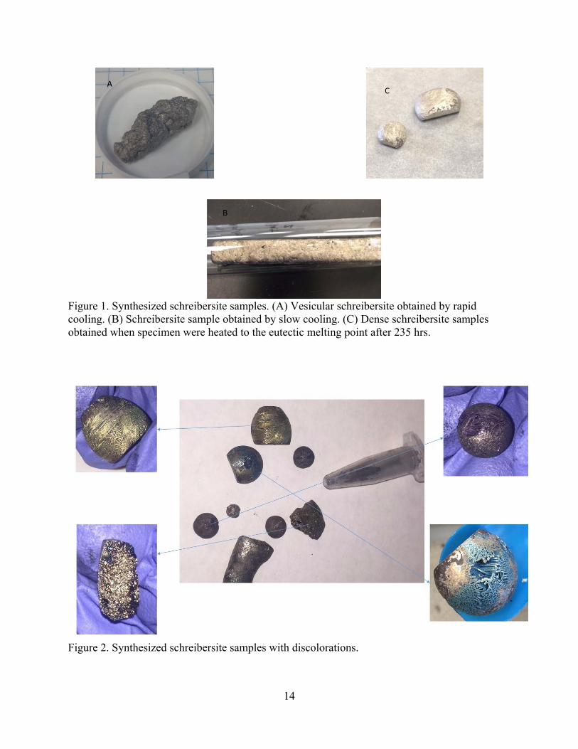
The samples synthesized had a metallic luster and were attracted to magnet. The
appearance of the samples varied depending on the synthesis techniques used (Fig. 1). Samples
that were cooled rapidly had a more glassy texture than those cooled more slowly. Samples
heated to the melting point were dense beads that were smooth on one side and had needle-like
crystal habit on the other side; this crystal habit was not observed in the other samples which had
a massive crystal habit. The dense variety produced ‘smooth surfaces’ when cut, whereas the
other variety was more vesicular with jagged edges. The products were synthesized with yields
above 90% in all instances (Table 1).
In one instance, the synthesis resulted in samples that were different in appearance than
those that were previously synthesized. Unlike the other samples that had a uniform metallic
luster, these samples had a metallic luster and blue, yellow and pink discolorations (Figure 2).
Characterization
EMPA. The empirical formulas obtained from the EMPA analyses were on average:
Fe2.06Ni0.93P1.05, Fe1.92Ni1.07P1.02, Fe2.07Ni0.89P1.04 and Fe1.04Ni1.9P0.97. These formulas are very
similar to either Fe2NiP or FeNi2P and thus confirmed that both schreibersite and nickel-
phosphide were synthesized, respectively.
14
Figure 1. Synthesized schreibersite samples. (A) Vesicular schreibersite obtained by rapid cooling. (B) Schreibersite sample obtained by slow cooling. (C) Dense schreibersite samples obtained when specimen were heated to the eutectic melting point after 235 hrs.
Figure 2. Synthesized schreibersite samples with discolorations.

15
The SEM images indicate the presence of what appears to be two phases (Fig. 3). There
is a darker phase which is present in a light gray matrix. EMPA analyses of these different
phases indicate that both phases were schreibersite as they had formulas similar to Fe2NiP. The
light phase had formulas that were, on average, Fe2.02Ni1.09P0.97 and the dark phase had formulas
that were, on average, Fe1.96Ni0.72P1.32.
Figure 3. SEM images of schreibersite. (A) 50X magnification. (B) 100X magnification.
XRD. X-ray diffractograms of the synthesized material are provided as Figure 4. The d
values from the samples were similar to those in the literature for schreibersite, and was therefore
a very good indication that the products were successfully synthesized.
Micro-Raman Spectroscopy. The Raman spectra obtained for the synthesized
schreibersite are similar to those obtained for natural as well as synthetic samples obtained by
Pirim et al. (2014) and natural samples obtained by Kaliwoda et al. (2013) (Figure 5).

16
Figure 4. XRD of synthesized schreibersite with comparison of characteristic d values for schreibersite.
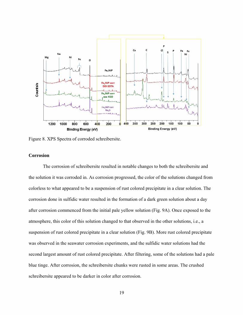
XPS. XPS spectra again agree with those obtained for natural samples by Pirim et al.
(2014) (Figure 6). They all contained a peak indicating the presence of a carbon impurity at ~286
eV. The binding energy for a phosphorus 2p electron was measured to be approximately 129.84
eV (Figure 7). The spectra of the corroded samples were interesting as they indicate the presence
of species from the solutions used in corrosion. For instance, Na, Mg, Ca and Cl were identified
with binding energies of 1074.93 eV, 1306.26 eV, 353.68 eV and 202.8 eV, respectively on the
surface of the schreibersite corroded in seawater. S, Cl and Na were identified on the sample
corroded in sulfidic water with binding energies of 169.69 eV, 200.21 eV and 1072.45 eV,
respectively. The average binding energies of the 2p electron of P, the 2p3 electron of Ni and the
2p electron of Fe in the spectra of these corroded samples were about 135 eV, 857 eV and 713
eV, respectively (Figure 8). The average binding energy of the 2p electron of P in the seawater
and sulfidic water samples was approximately 170 eV.

17
Figure 5. Raman spectra of uncorroded samples compared to those from Pirim et al. (2014). (A) Raman spectrum of synthesized schreibersite. (B) Raman spectra of schreibersite inclusion from Sikhote-Alin (a), Seymchan (b), Odessa (c) meteorites; synthetic Fe3P (d) and synthetic Fe2NiP (e). Reprinted from “Investigation of schreibersite and intrinsic oxidation products from Sikhote-Alin, Seymchan, and Odessa meteorites and Fe3P and Fe2NiP synthetic surrogates”, Pirim, C., et al., Figure 15, pg. 271, 2014, with permission from Elsevier.

18
Figure 6. XPS spectra obtained for schreibersite synthesized in this study. Spectrum (A) is at a depth of 15 nm, (B) is at a depth of 3.8 nm and (C) is before etching.
Figure 7. XPS spectra showing binding energies of Fe (A) and P (B).
19
Figure 8. XPS Spectra of corroded schreibersite.
Corrosion
The corrosion of schreibersite resulted in notable changes to both the schreibersite and
the solution it was corroded in. As corrosion progressed, the color of the solutions changed from
colorless to what appeared to be a suspension of rust colored precipitate in a clear solution. The
corrosion done in sulfidic water resulted in the formation of a dark green solution about a day
after corrosion commenced from the initial pale yellow solution (Fig. 9A). Once exposed to the
atmosphere, this color of this solution changed to that observed in the other solutions, i.e., a
suspension of rust colored precipitate in a clear solution (Fig. 9B). More rust colored precipitate
was observed in the seawater corrosion experiments, and the sulfidic water solutions had the
second largest amount of rust colored precipitate. After filtering, some of the solutions had a pale
blue tinge. After corrosion, the schreibersite chunks were rusted in some areas. The crushed
schreibersite appeared to be darker in color after corrosion.
20
Figure 9. Schreibersite corroded in sulfidic water (A). Filtered solutions after the corrosion of schreibersite in doubly deionized water, seawater and sulfidic water under both oxic and anoxic conditions (B).
P-31 NMR Spectroscopy. The NMR spectra of the corrosion solutions of the samples
analyzed indicate the presence of phosphite, orthophosphate, pyrophosphate and hypophosphate.
The chemical shifts obtained in doubly deionized water under aerobic conditions were
approximately 4.18ppm, 4.74ppm, -4.15ppm and 14.51ppm for phosphite, orthophosphate,
pyrophosphate and hypophosphate, respectively (Figure 10). This data is in agreement with
work done by Pasek and Lauretta (2005) for Fe3P corrosion.
Speciation Analysis. The speciation analysis done by the HPLC-ICP-MS technique indicates the
presence of P in the +3 and +5 oxidation states, i.e., phosphite and orthophosphate, respectively
with elution times at about 5 minutes and 9 minutes (Figure 11). There was no P with +1
oxidation state or hypophosphite, but there was a peak in the chromatograms that eluted after the
orthophosphate peak, at an elution time around 14 minutes (Figure 11), likely a P species with -4
total charge at these high pH values.
21
Figure 10. NMR spectrum of Fe3P in deionized water (A) (Spectrum obtained from Pasek via personal communication (2015)) compared to NMR spectrum of Fe2NiP in deionized water (B).
Concentration of P species present in the corroding solutions were calculated using the P
standards used and are presented in Table 2. The DDI solutions were the only ones that
contained both phosphite, orthophosphate and the other P species, which we know to be
hypophosphate or pyrophosphate from the NMR (Fig. 11, Table 2). The seawater solutions
contained predominantly phosphite, while the sulfidic water solutions were predominantly
orthophosphate (Table 2).

22
Figure 11. Chromatogram of standards used in speciation experiments (A) compared to the chromatogram for Fe2NiP in deionized water.
Table 2. Concentration of phosphite (P3+) and orthophosphate (P5+) calculated from speciation analyses compared to total P concentrations obtained from ICP-OES analyses. Concentrations designated ‘N/D’ were too low to be detected.
Solution [P3+] (mM) [P5+] (mM) [Pother] [P total]OES (mM) DDI 0.13 0.13 present 0.49DDI EDTA 0.14 0.12 present 1.8Seawater Present but N/D N/D Not observed 0.14Seawater EDTA 0.88 N/D Not observed 0.23Sulfidic water N/D 0.77 Not observed 0.56Sulfidic water EDTA N/D 0.75
Not observed 0.94
ICP-MS. The ICP-MS analysis indicated the release of P of concentrations ranging from
0.016mM to 0.39mM in DDI and seawater, respectively (Table 2).
ICP-OES This analysis indicated the release of P of concentrations ranging from
0.14mM to 1.8mM in seawater and DDI with EDTA, respectively (Table 2). In general, more P
was released in the experiments where the corroding solutions contained EDTA.

23
Table 3. Results of ICP-OES and ICP-MS analyses. Comparison of the theoretical P and actual P released during corrosion experiments.
Solution Mass of Fe2NiP (g)
No. moles P (mmol)
Theoretical [P] (mM)
Actual [P] (mM)
% P released Technique
DDI 1.052 5.096 254.8 0.49 0.19
ICP-OES
DDI EDTA 1.178 5.707 285.3 1.8 0.63 SeaH2O 1.112 5.386 269.3 0.14 0.053 SeaH2O EDTA 1.022 4.950 247.5 0.23 0.091Sulfidic H2O 1.062 5.144 257.2 0.56 0.23 Sulfidic H2O EDTA 1.113 5.391 269.5 0.94 0.35 DDI A 0.621 3.008 120.3 0.016 0.0133
ICP-MS
DDI AN 0.858 4.156 166.2 0.024 0.0144
SeaH2O A 0.673 3.260 130.4 0.28 0.215
SeaH2O AN 0.763 3.696 147.8 0.39 0.264
Sulfidic H2O 0.707 3.424 137.0 0.25 0.183
Phosphorylation
Phosphorylation of choline. The NMR analysis of the solution obtained after the
phosphorylation experiment indicated the presence of phosphocholine based on the presence of
the triplet at chemical shift of approximately 3.89 ppm (Figure 12).
Figure 12. NMR spectrum indicating the formation of phosphocholine.

24
CHAPTER FOUR: DISCUSSION
Synthesis
Schreibersite is readily synthesized by mixing stoichiometric ratios of iron, nickel, and
phosphorus and heating for the aforementioned time at the specified temperature. Further, the
characterization of the mineral using XRD, microprobe, Raman, and XPS all confirm this
material to be identical to meteoritic schreibersite. The best schreibersite was made when the
product was heated to the melting point after 235 hours. These samples were dense and contain
the characteristic needle-like crystal pattern observed in meteoritic schreibersite samples. While
the appearance of these samples were different from others synthesized without heating to the
melting point afterwards, the characterization analyses indicated that both types of samples were
schreibersite.
The synthesized samples with the discolorations (Fig. 2) were synthesized using the
method used to synthesize the other samples. However, after the synthesis it was noted that the
valves on the tube furnace were not closed correctly. This, therefore, means that the samples
were synthesized in an atmosphere containing oxygen and not the argon atmosphere that was
intended. The samples were more than likely oxidized and the discolorations observed are
possibly evidence of oxidation.
Close examination of the SEM images may evoke thoughts of the Widmanstäten pattern
ubiquitous in iron meteorites. This pattern is usually the result of the presence of kamacite and
taenite (Goldstein et al., 2009). However, the analysis indicates that neither of these minerals are
present in the synthesized samples. Nonetheless, the difference in chemical composition of the
25
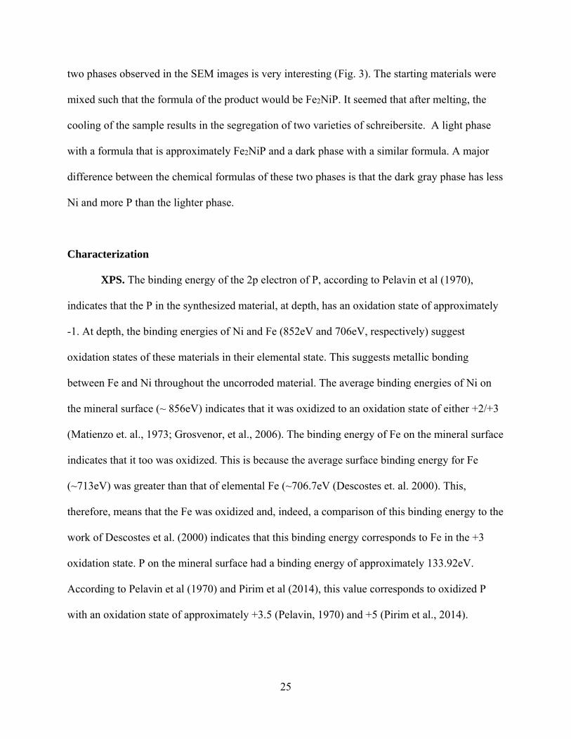
two phases observed in the SEM images is very interesting (Fig. 3). The starting materials were
mixed such that the formula of the product would be Fe2NiP. It seemed that after melting, the
cooling of the sample results in the segregation of two varieties of schreibersite. A light phase
with a formula that is approximately Fe2NiP and a dark phase with a similar formula. A major
difference between the chemical formulas of these two phases is that the dark gray phase has less
Ni and more P than the lighter phase.
Characterization
XPS. The binding energy of the 2p electron of P, according to Pelavin et al (1970),
indicates that the P in the synthesized material, at depth, has an oxidation state of approximately
-1. At depth, the binding energies of Ni and Fe (852eV and 706eV, respectively) suggest
oxidation states of these materials in their elemental state. This suggests metallic bonding
between Fe and Ni throughout the uncorroded material. The average binding energies of Ni on
the mineral surface (~ 856eV) indicates that it was oxidized to an oxidation state of either +2/+3
(Matienzo et. al., 1973; Grosvenor, et al., 2006). The binding energy of Fe on the mineral surface
indicates that it too was oxidized. This is because the average surface binding energy for Fe
(~713eV) was greater than that of elemental Fe (~706.7eV (Descostes et. al. 2000). This,
therefore, means that the Fe was oxidized and, indeed, a comparison of this binding energy to the
work of Descostes et al. (2000) indicates that this binding energy corresponds to Fe in the +3
oxidation state. P on the mineral surface had a binding energy of approximately 133.92eV.
According to Pelavin et al (1970) and Pirim et al (2014), this value corresponds to oxidized P
with an oxidation state of approximately +3.5 (Pelavin, 1970) and +5 (Pirim et al., 2014).

26
XPS data indicates that S was also oxidized during the sulfide corrosion experiments. The
average binding energy of about 170eV is similar to that reported for sulfate (169eV) by
Descostes et al. (2000) and Klauber et al. (2001). Since all these species are being oxidized in
solution, it suggests that these oxidations may be facilitated by reduction of hydrogen in water
(Pasek and Lauretta 2005).
Corrosion
During corrosion experiments the colors of the solutions changed from green to brown,
which suggest the oxidation of Fe (ii) to Fe (iii) and the binding energies obtained from the XPS
analyses of the corroded samples indicated that the Fe on the surface was indeed oxidized. Some
of the solutions had a light blue color suggesting the presence of oxidized Ni. Binding energies
for Ni obtained from XPS analyses confirmed that the Ni in the corroded samples were indeed
oxidized. The solution chemistry, therefore, confirms the surface chemistry.
P-31 NMR Analysis. The NMR spectra of the filtered solutions after corrosion
experiments provide further evidence that this synthetic method produces a useful proxy for
natural schreibersite. Corrosion of the prepared samples replicate prior work on corrosion of both
natural schreibersite and Fe3P (Pasek and Lauretta, 2005; Pasek et al., 2007 and Bryant et al.,
2013) and the chemical shifts in our spectra agree, within reason, to those obtained by the
aforementioned workers. The use of EDTA in corrosion experiments definitely improved the
quality of NMR spectra obtained. Also, as expected, there was more corrosion in solutions with
higher ionic strengths, i.e., seawater and sulfidic water, as compared to those with lower, i.e.,
doubly deionized water.
27
Speciation Analysis. These results support those obtained by the P-31 NMR analysis.
The peak ratios and chemical shifts in the NMR spectra were not identical to those obtained by
previous workers so these results could have been questioned. This analysis, however, confirms
the presence of phosphite, orthophosphate and then another species with elution time greater than
orthophosphate. For the chromatography technique being utilized in this study, ions with higher
total charge stay on the column for longer periods of time and therefore have longer elution times
(Pech et al. 2011). This, therefore, means that the peak eluting after orthophosphate has a higher
charge than that of orthophosphate. Based on our NMR results, we know that pyrophosphate and
hypophosphate (both having a charge of negative four) were present in these samples, therefore,
the last peak in the chromatogram represents one or both of these species.
ICP-MS and ICP-OES. The results obtained from these analyses were both interesting
and confusing. They indicate that the amount of P released depends on the amount of
schreibersite corroded, the presence or absence of a chelating agent and the ionic strength of the
corroding solution. The amount of schreibersite used is definitely important because schreibersite
is the source of P, so corroding a larger amount of schreibersite should result in the release of
more P. The chelating agent is important because it bonds to cations in solution thereby allowing
the P oxyanions formed to be detected by analyses such as these. Intuitively, we would expect
that more corrosion would occur in a solution with higher ionic strength due to the high
concentration of charge carriers. This was observed, for the most part, in both the ICP-MS and
ICP-OES analyses. However, a very peculiar phenomenon was observed in the ICP-OES
analyses. The highest concentrations of P were observed in the corroding solution with the
lowest ionic strength, DDI. More P was, however, released in the DDI experiment with EDTA.

28
Speciation Analysis and ICP-OES Comparison. For the speciation analyses,
concentrations could have only been calculated for phosphite and orthophosphate as those were
the species for which we had standards. Since the solutions analyzed via the speciation technique
were the same ones analyzed by the ICP-OES technique, the concentrations were compared. In
some instances, there are some discrepancies between the concentrations obtained by the two
techniques, however, in some, the concentrations agree with each other. For instance, the
concentration of P measured in seawater with EDTA was 0.23mM, but the speciation analysis
measured 0.88mM of phosphite in that sample, suggesting that the ICP-OES technique
underestimated the P concentrations. This was also the case for the sulfidic water samples. For
one sample, the amount of phosphite present was less than the total P obtained from the ICP-
OES analysis, while it was greater for the other. For the DDI samples, on the other hand, the
ICP-OES results seemed to be an overestimation. The chromatogram for the DDI with EDTA
(Fig. 11) had larger peaks, with larger areas than all the other samples, so it was expected that the
concentrations would be large. However, the speciation analyses indicated that the
concentrations obtained, were in fact less than those obtained for the other solutions. The
concentrations obtained from the speciation analyses makes sense intuitively, because there
should be more corrosion in solutions with higher ionic strengths, and therefore, greater release
of P. These results indicate that more work needs to be done to better ascertain which of the
techniques used is best suited to determine the amount of P released after corrosion.
Phosphorylation
Phosphorylation of choline. Finally, the synthesized schreibersite has now been
demonstrated to phosphorylate choline, making phosphocholine. Synthesis of this molecule

29
demonstrates that schreibersite will react with organic compounds to generate prebiotic
molecules. Moreover, we have also previously demonstrated that the synthetic analog of
schreibersite can easily phosphorylate the membrane making compound, glycerol, to form
glycerol phosphates under mild conditions (Pasek et al. 2013). This phosphorylation is, therefore,
more proof that these synthetic samples are a good analog of the commercially available
schreibersite.
Urea was added to the reaction mixture because it has been known to facilitate the
phosphorylation reactions (Osterberg, et al., 1973). Furthermore, our group has previously shown
that when urea is added to choline chloride and heated, it forms eutectic mixtures which can then
be phosphorylated in the presence of phosphorus minerals (Gull et al. 2014). The phosphate ester
of choline can also be formed when struvite is used as a P source but the prebiotic relevance of
struvite is questionable (Gull and Pasek 2013).
30
CHAPTER FIVE: IMPLICATIONS
In addition to highlighting that schreibersite was successfully synthesized, this work
indicates that synthetic schreibersite is a useful analog for meteoritic schreibersite. It is the
closest available analog to meteoritic schreibersite, and, unlike the commercially available
analog, it contains nickel. It is also easier and less expensive to acquire than meteoritic samples
making it ideal for studies requiring meteoritic schreibersite. On that note, these samples are well
suited for origin of life studies in which researchers are trying to determine the role that
phosphorus may have played in the origin of life on the early earth. The samples are chemically
homogeneous, for the most part, and contain only the constituents present in schreibersite (Fe, Ni
and P), thus uncertainties resulting from impurities are not an issue. Finally, this synthesis may
be useful to scientists interesting in synthesizing other phosphide minerals that may be
interesting in other types of studies.
31
CHAPTER SIX: CONCLUSIONS
All of the analyses done indicate that schreibersite and nickel-phosphide were
successfully synthesized with good yields. The corrosion of schreibersite leads to the formation
of phosphorus species with a variety of oxidation states: +3, +4 and +5 (phosphite,
hyposphosphate, orthophosphate and pyrophosphate). The products have also been shown to be
able to phosphorylate important prebiotic organic compounds.
The synthesis is straightforward and reproducible. The use of multiple characterization
techniques all indicate production of the target product. XPS data obtained for these samples
allow us to answer questions about the oxidation state of the elements in schreibersite. They
indicate that below the surface, the phosphorus is reduced while the Fe and Ni are in their
elemental states. On the surface, as we would expect, all three components are oxidized. The
extent of oxidation of the Fe and Ni is unclear and this lack of clarity may be because these two
elements have high affinity for each other, thus their chemistry gets very complicated when they
are bonded to each other.
The corrosion of schreibersite provided further confirmation that this synthetic analog
behaves similarly to commercially available iron phosphide and naturally occurring schreibersite
samples. Further, the observations made in the solution chemistry were confirmed by the surface
analyses.
Finally, the phosphorylation of choline using these synthetic schreibersite samples opens
new possibilities for the origin of life community. Schreibersite is believed to be a prebiotic

32
source of reactive phosphorus on the early earth. The fact that choline was phosphorylated using
schreibersite gives support to the theory that extraterrestrial phosphorus provided an impetus for
the chemical evolution of life on our planet.

33
REFERENCES
Bartoschewitz, R., et al. "NWA 1500: The first basaltic ureilite?" Meteoritics and Planetary Science Supplement38 (2003): 5114.
Birch, Francis. "Density and composition of mantle and core." Journal of geophysical research 69.20 (1964): 4377-4388.
Bryant, David E., et al. "Hydrothermal modification of the Sikhote-Alin iron meteorite under low pH geothermal environments. A plausibly prebiotic route to activated phosphorus on the early Earth." Geochimica et Cosmochimica Acta109 (2013): 90-112.
Britvin, Sergey N., et al. "Earth's Phosphides in Levant and insights into the source of Archean prebiotic phosphorus." Scientific reports 5 (2015).
Descostes, M., et al. "Use of XPS in the determination of chemical environment and oxidation state of iron and sulfur samples: constitution of a data basis in binding energies for Fe and S reference compounds and applications to the evidence of surface species of an oxidized pyrite in a carbonate medium." Applied Surface Science 165.4 (2000): 288-302.
Essene, E. J., and D. C. Fisher. "Lightning strike fusion: extreme reduction and metal-silicate liquid immiscibility." Science 234.4773 (1986): 189-193.
Ganapathy, Ramachandran, and Edward Anders. "Bulk compositions of the moon and earth, estimated from meteorites." Lunar and Planetary Science Conference Proceedings. Vol. 5. 1974.
Geist, V., et al. "Investigations of the meteoritic mineral (Fe, Ni) 3P." Crystal Research and Technology 40.1‐2 (2005): 52-64.
Goldstein, J. I., E. R. D. Scott, and N. L. Chabot. "Iron meteorites: Crystallization, thermal history, parent bodies, and origin." Chemie der Erde-Geochemistry 69.4 (2009): 293-325.
Grosvenor, Andrew P., et al. "New interpretations of XPS spectra of nickel metal and oxides." Surface Science 600.9 (2006): 1771-1779.
Gull, Maheen, et al. "Prebiotic phosphate ester syntheses in a deep eutectic solvent." Journal of molecular evolution 78.2 (2014): 109-117.
Gull, Maheen, and Matthew A. Pasek. "Is struvite a prebiotic mineral?" Life 3.2 (2013): 321-330.
Kaliwoda, Melanie, et al. "New Raman spectroscopic data of the almahata sitta meteorite." Spectroscopy Letters 46.2 (2013): 141-146.
34
Klauber, Craig, et al. "Sulphur speciation of leached chalcopyrite surfaces as determined by X-ray photoelectron spectroscopy." International Journal of Mineral Processing 62.1 (2001): 65-94.
Li, J., and Y. Fei. "Experimental constraints on core composition." Treatise on Geochemistry 2 (2003): 521-546.
Maciá, E., M. V. Hernández, and J. Oró. "Primary sources of phosphorus and phosphates in chemical evolution." Origins of Life and Evolution of the Biosphere 27.5-6 (1997): 459-480.15.
Matienzo, J., et al. "X-ray photoelectron spectroscopy of nickel compounds. “Inorganic Chemistry 12.12 (1973): 2762-2769.
McDonough, William F., and S-S. Sun. "The composition of the Earth.” Chemical geology 120.3 (1995): 223-253.
Österberg, R., L. E. Orgel, and R. Lohrmann. "Further studies of urea-catalyzed phosphorylation reactions." Journal of molecular evolution 2.2-3 (1973): 231-234.
Pasek, Matthew A. "Phosphorus as a lunar volatile." Icarus (2014).
Pasek, Matthew A. “Personal communication.” (May 13, 2015). TS.
Pasek, Matthew A., Kristin Block, and Virginia Pasek. "Fulgurite morphology: a classification scheme and clues to formation." Contributions to Mineralogy and Petrology 164.3 (2012): 477-492.
Pasek, Matthew A., Jason P. Dworkin, and Dante S. Lauretta. "A radical pathway for organic phosphorylation during schreibersite corrosion with implications for the origin of life." Geochimica et Cosmochimica Acta 71.7 (2007): 1721-1736.
Pasek, Matthew A., and Dante S. Lauretta. "Aqueous corrosion of phosphide minerals from iron meteorites: a highly reactive source of prebiotic phosphorus on the surface of the early Earth." Astrobiology 5.4 (2005): 515-535.
Pasek, Matthew A., et al. "Evidence for reactive reduced phosphorus species in the early Archean ocean." Proceedings of the National Academy of Sciences 110.25 (2013): 10089-10094.
Pech, Herbe, et al. "Elucidating the redox cycle of environmental phosphorus using ion chromatography." Journal of chromatographic science 49.8 (2011): 573-581.
Pedersen, A. K. "Armalcolite-bearing Fe-Ti oxide assemblages in graphite-equilibrated salic volcanic rocks with native iron from Disko, central West Greenland." Contributions to Mineralogy and Petrology 77.4 (1981): 307-324.
Pelavin, M. D. N. J. M., et al. "Phosphorus 2p electron binding energies. Correlation with extended Hueckel charges." The Journal of Physical Chemistry 74.5 (1970): 1116-1121.
35
Pirim, C., et al. "Investigation of schreibersite and intrinsic oxidation products from Sikhote-Alin, Seymchan, and Odessa meteorites and Fe3P and Fe2NiP synthetic surrogates." Geochimica et Cosmochimica Acta 140 (2014): 259-274.
Poirier, Jean-Paul. "Light elements in the Earth's outer core: a critical review. “Physics of the earth and planetary interiors 85.3 (1994): 319-337.
Ringwood, Alfred Edward. "Origin of the Earth and Moon." New York, Springer-Verlag New York, Inc., 1979. 307 p. 1 (1979).
Scott, Henry P., et al. "Equation of state and high‐pressure stability of Fe3P‐schreibersite: Implications for phosphorus storage in planetary cores. “Geophysical research letters 34.6 (2007).
Skála, Roman, and Ivana Císařová. "Crystal structure of meteoritic schreibersites: determination of absolute structure." Physics and chemistry of minerals 31.10 (2005): 721-732.
Skála, Roman, and Milan Drábek. "Powder data for synthetic analogue of a mineral nickelphosphide." Powder Diffraction 17.04 (2002): 322-325.
Washington, Henry Stephens. "The chemical composition of the earth. “American Journal of Science 53 (1925): 351-378.
Williams, Q., and E. Knittle. "Constraints on core chemistry from the pressure dependence of the bulk modulus." Physics of the earth and planetary interiors 100.1 (1997): 49-59.
Wu, Xiang, et al. "Elasticity and anisotropy of iron‐nickel phosphides at high pressures." Geophysical Research Letters 38.20 (2011).
Yamagata, Y., et al. "Volcanic production of polyphosphates and its relevance to prebiotic evolution." (1991): 516-519.
Yang, Jingsui, et al. “Discovery of Fe2P alloy in garnet peridotite from the Chinese Continental Scientific Drilling Project (CCSD) main hole.” Acta Petrolodica Sinica 21. 2 (2005): 271-276.

36
APPENDIX: COPYRIGHT PERMISSIONS

37

38
39

40
41
42

43

ABOUT THE AUTHOR
Nikita L. La Cruz was born in the South American/ Caribbean nation: Guyana. Although
she was born in the capital, she calls Matthew’s Ridge, a small, rural village in the north west of
Guyana home. Nikita was sent to live in the city with her aunts because her parents wanted her to
be able to attain the best education possible in Guyana. Her success at the examinations at the
end of elementary school earned her a place at Queen’s College, arguably the best high school in
the country. While at Queen’s College, she developed a liking for the sciences, especially
Chemistry. After graduating with Ordinary Level as well as Advanced Level certificates, Nikita
decided that she would pursue a bachelor’s degree in Chemistry abroad.
She started Gettysburg College in PA, USA in the fall of 2009 as a chemistry major. By
the end of her first year, she declared Environmental Studies as a second major. The first
environmental studies class that she took was Earth Systems Science and it was this class that
made her realize that she wanted to be an economic geologist.
She moved to Tampa, FL to attend University of South Florida. Here, she worked as a
research assistant for Dr. Matthew Pasek on this project as she also worked towards her masters
in geology. Nikita has had the opportunity to present both her undergraduate and graduate
research on many occasions at conferences and symposiums both in the U.S. and abroad.
After graduation, Nikita will move to the University of Michigan in Ann Arbor to purse a
PhD in economic geology. This degree will allow Nikita to move back to Guyana and work in
the mining industry established there. While working in Guyana, she will be able to participate in
activities that will allow her to fulfill her dream of effecting positive change in her country.