Scedosporium and Lomentospora: an updated overview of ...
25
HAL Id: hal-01789215 https://hal.archives-ouvertes.fr/hal-01789215 Submitted on 10 Apr 2019 HAL is a multi-disciplinary open access archive for the deposit and dissemination of sci- entific research documents, whether they are pub- lished or not. The documents may come from teaching and research institutions in France or abroad, or from public or private research centers. L’archive ouverte pluridisciplinaire HAL, est destinée au dépôt et à la diffusion de documents scientifiques de niveau recherche, publiés ou non, émanant des établissements d’enseignement et de recherche français ou étrangers, des laboratoires publics ou privés. Scedosporium and Lomentospora: an updated overview of underrated opportunists Andoni Ramirez-Garcia, Aize Pellon, Aitor Rementeria, Idoia Buldain, Eliana Barreto-Bergter, Rodrigo Rollin-Pinheiro, Jardel Vieira Meirelles, Mariana Ingrid D. S. Xisto, Stephane Ranque, Vladimir Havlicek, et al. To cite this version: Andoni Ramirez-Garcia, Aize Pellon, Aitor Rementeria, Idoia Buldain, Eliana Barreto-Bergter, et al.. Scedosporium and Lomentospora: an updated overview of underrated opportunists. Medical Mycol- ogy, Oxford University Press, 2018, 56 (1), pp.S102-S125. 10.1093/mmy/myx113. hal-01789215
Transcript of Scedosporium and Lomentospora: an updated overview of ...

HAL Id: hal-01789215https://hal.archives-ouvertes.fr/hal-01789215
Submitted on 10 Apr 2019
HAL is a multi-disciplinary open accessarchive for the deposit and dissemination of sci-entific research documents, whether they are pub-lished or not. The documents may come fromteaching and research institutions in France orabroad, or from public or private research centers.
L’archive ouverte pluridisciplinaire HAL, estdestinée au dépôt et à la diffusion de documentsscientifiques de niveau recherche, publiés ou non,émanant des établissements d’enseignement et derecherche français ou étrangers, des laboratoirespublics ou privés.
Scedosporium and Lomentospora: an updated overviewof underrated opportunists
Andoni Ramirez-Garcia, Aize Pellon, Aitor Rementeria, Idoia Buldain, ElianaBarreto-Bergter, Rodrigo Rollin-Pinheiro, Jardel Vieira Meirelles, Mariana
Ingrid D. S. Xisto, Stephane Ranque, Vladimir Havlicek, et al.
To cite this version:Andoni Ramirez-Garcia, Aize Pellon, Aitor Rementeria, Idoia Buldain, Eliana Barreto-Bergter, et al..Scedosporium and Lomentospora: an updated overview of underrated opportunists. Medical Mycol-ogy, Oxford University Press, 2018, 56 (1), pp.S102-S125. �10.1093/mmy/myx113�. �hal-01789215�

Medical Mycology, 2018, 56, S102–S125doi: 10.1093/mmy/myx113
Review Article
Review Article
Scedosporium and Lomentospora: an updated
overview of underrated opportunists
Andoni Ramirez-Garcia1,∗, Aize Pellon1, Aitor Rementeria1,
Idoia Buldain1, Eliana Barreto-Bergter2, Rodrigo Rollin-Pinheiro2,
Jardel Vieira de Meirelles2, Mariana Ingrid D. S. Xisto2,
Stephane Ranque3, Vladimir Havlicek4, Patrick Vandeputte5,6,
Yohann Le Govic5,6, Jean-Philippe Bouchara5,6, Sandrine Giraud6,
Sharon Chen7, Johannes Rainer8, Ana Alastruey-Izquierdo9,
Maria Teresa Martin-Gomez10, Leyre M. Lopez-Soria11, Javier Peman12,
Carsten Schwarz13, Anne Bernhardt14, Kathrin Tintelnot14,
Javier Capilla15, Adela Martin-Vicente15,16, Jose Cano-Lira15,
Markus Nagl17, Michaela Lackner17, Laszlo Irinyi18, Wieland Meyer18,
Sybren de Hoog19 and Fernando L. Hernando1
1Fungal and Bacterial Biomics Research Group, Department of Immunology, Microbiology and Para-sitology, Faculty of Science and Technology, University of the Basque Country (UPV/EHU), Leioa, Spain,2Instituto de Microbiologia, UFRJ, Rio de Janeiro, RJ, Brazil, 3Laboratoire de Parasitologie-Mycologie,AP-HM / CHU Timone, Marseille, France, 4Institute of Microbiology, Academy of Sciences of the CzechRepublic, Prague, Czech Republic, 5Laboratoire de Parasitologie-Mycologie, CHU, Angers, France, 6Host-Pathogen Interaction Study Group (EA 3142), UNIV Angers, UNIV Brest, Angers, France, 7Centre forInfectious Diseases and Microbiology Laboratory Services, ICPMR, Westmead Hospital, The Univer-sity of Sydney, New South Wales, Australia, 8Institute of Microbiology, Leopold-Franzens UniversityInnsbruck, Austria, 9Mycology Reference Laboratory, National Centre for Microbiology. Instituto deSalud Carlos III. Majadahonda, Madrid, Spain, 10Microbiology Department. Hospital Universitari Valld’Hebron, Barcelona, Spain, 11Microbiology Department. Hospital Universitario Cruces, Barakaldo, Spain,12Microbiology Department, Hospital Universitario y Politecnico La Fe, Valencia, Spain, 13Cystic FibrosisCentre Berlin/Charite-Universitatsmedizin Berlin, Germany, 14Mycotic and Parasitic Agents and My-cobacteria, Robert Koch Institute, Berlin, Germany, 15Mycology Unit, Medical School and IISPV, Universi-tat Rovira i Virgili, Reus, Spain, 16Department of Clinical Pharmacy and Translational Science, Universityof Tennessee Health Science Center, Memphis, TN USA, 17Division of Hygiene and Medical Microbiology,Medical University of Innsbruck, Innsbruck, Austria, 18Molecular Mycology Research Laboratory, Centrefor Infectious Diseases and Microbiology, Westmead Clinical School, Sydney Medical School – West-mead Hospital, Marie Bashir Institute for Infectious Diseases and Biosecurity, The University of Sydney,Westmead Institute for Medical Research, Sydney, New South Wales, Australia and 19Westerdijk FungalBiodiversity Institute, Utrecht, The Netherlands∗To whom correspondence should be addressed. Dr. Andoni Ramirez-Garcia, Department of Immunology, Microbiologyand Parasitology, Faculty of Science and Technology, University of the Basque Country (UPV/EHU), Leioa 48940, Bizkaia,Spain. Tel: +34-946-015090; Fax: +34-946-013500; E-mail: [email protected]
Received 1 June 2017; Revised 17 August 2017; Accepted 10 October 2017; Editorial Decision 8 September 2017
S102 C© The Author(s) 2017. Published by Oxford University Press on behalf of The International Society for Human and AnimalMycology. All rights reserved. For permissions, please e-mail: [email protected]
Dow
nloaded from https://academ
ic.oup.com/m
my/article-abstract/56/suppl_1/S102/4925971 by SC
DU
Mediterranee user on 10 April 2019

Ramirez-Garcia et al. S103
Abstract
Species of Scedosporium and Lomentospora are considered as emerging opportunists,affecting immunosuppressed and otherwise debilitated patients, although classicallythey are known from causing trauma-associated infections in healthy individuals. Clinicalmanifestations range from local infection to pulmonary colonization and severe invasivedisease, in which mortality rates may be over 80%. These unacceptably high rates aredue to the clinical status of patients, diagnostic difficulties, and to intrinsic antifungal re-sistance of these fungi. In consequence, several consortia have been founded to increaseresearch efforts on these orphan fungi. The current review presents recent findings andsummarizes the most relevant points, including the Scedosporium/Lomentospora taxon-omy, environmental distribution, epidemiology, pathology, virulence factors, immunol-ogy, diagnostic methods, and therapeutic strategies.
Key words: fungi, pathogen, emergent, infection.
Introduction
Nearly all pathogenic fungi are present in the environ-ment adapted to very different habitats where they playvarying roles in recycling of organic matter. With some oftheir causative agents being either opportunistic or primarypathogens, fungal infections show an increasing incidenceworldwide, affecting millions of individuals, with mortalityrates that may be higher than 50% in susceptible patientpopulations.1
Among pathogenic fungi, Scedosporium species, includ-ing Lomentospora prolificans (formerly Scedosporium pro-lificans),2 can cause infections in both immunocompetentand immunocompromised hosts, where they can act as pri-mary or opportunistic pathogens.3,4 These species cause abroad range of clinical manifestations, from colonization ofthe respiratory tract, superficial infections and allergic reac-tions, to severe invasive localized or disseminated mycoses.Patients at risk are particularly those immunocompromisedand with hematological malignancies.3,5 Individuals suf-fering from near-drowning events in water polluted withfungal propagules are also at risk of infections with centralnervous system (CNS) involvement.5
Moreover, Scedosporium/Lomentospora are among themost commonly recovered fungi from respiratory se-cretions of patients suffering from chronic pulmonaryconditions such as cystic fibrosis (CF).6 Although theyare mostly asymptomatic colonizers,7,8 this may bethe first step toward pathology. L. prolificans typi-cally causes disseminated infections in immunocompro-mised patients, where it is associated with high mortal-ity.3,8–11 Scedosporium boydii and S. apiospermum arethe most frequently isolated species, but in some regionsS. aurantiacum is more common. The high degrees of in-trinsic antifungal resistance make these infections difficultto manage.12
The high mortality rates of deep and disseminated infec-tions necessitate focusing resources and efforts to cope withthe challenges posed by Scedosporium and Lomentosporaspecies, such as improving diagnostic methods, or designingnew effective therapies.
Therefore, the members of the Scedosporium workinggroup of the International Society for Human and AnimalMycology (ISHAM), present at their 5th Workshop in Bil-bao in 2016, decided to prepare a detailed review describ-ing the taxonomy, environmental distribution, epidemiol-ogy, pathology, virulence factors, immunology, diagnosticmethods, and available therapeutic strategies.
Taxonomy, DNA barcoding, and new species
The nomenclature of the genus Scedosporium/Pseudalle-scheria has undergone numerous changes over the lastdecade following the introduction of molecular phyloge-netics, which led to an increasing resolution at and belowthe species level. In addition, the fundamental change infungal taxonomy allowing only a single name per fun-gal species, effectively abolishing the dual nomenclaturebased on the anamorph/teleomorph concept,13 resulted inthe adoption of the name Scedosporium at the expense ofPseudallescheria.2
The first comprehensive revision of the genus conductedin 2005 by Gilgado et al.14 using four genetic loci (β-tubulin(BT2 (= exon 2–4) and TUB (= exon 5–6)), calmodulinand the internal transcribed spacer regions (ITS1/2) of therDNA gene cluster) recognized S. apiospermum (incl. P.boydii) as a species complex, in addition to S. auranti-acum and S. minutisporum. Within the S. apiospermum/P.boydii complex, three existing species were recognized:P. angusta, P. ellipsoidea, and P. fusoidea.14 A secondrevision further recognised a new species S. dehoogii andmaintained S. apiospermum and P. boydii as distinct species
Dow
nloaded from https://academ
ic.oup.com/m
my/article-abstract/56/suppl_1/S102/4925971 by SC
DU
Mediterranee user on 10 April 2019

S104 Medical Mycology, 2018, Vol. 56, No. S1
based on TUB sequences together with morphologicaland physiological criteria.15 A significant genetic diversitywithin the S. apiospermum/P. boydii complex was notedin sequence analysis of the D1/D2 region of the LSU ofrDNA, ITS1/2 and elongation factor 1-alpha;16 ITS1/2 andBT217,18 and the actin, BT2 and small ribosomal protein60S L10 (RP60S) sequences in combination with AFLPanalysis.19 While the use of some loci, such as BT2, showbetter discriminatory resolution, barcoding of the ITS1/2regions is sufficient for distinction of all relevant entitiesin clinical practice.19 Rainer and Kaltseis (2010) describeda new species S. deficiens,20 closely related to S. dehoogiibased on ITS1/2 and BT2 corresponding with growth differ-ences on polyvinyl alcohol agar supplemented with dieseland rapeseed oil, and growth at 41◦C, but no referencesequences were submitted to any public database, and in-sufficient proof of novelty was provided. Recently anothernew species phylogenetically related to S. aurantiacum wasdescribed, based on ITS, BT2 and calmodulin, named S.cereisporum.21 In summary, after the One Fungus = OneName movement22 and sequencing studies, the genus Sce-dosporium now contains the following 10 species: S. au-rantiacum, S. minutisporum, S. desertorum, S. cereisporum,and S. dehoogii, in addition to the S. apiospermum complexthat comprises S. angustum, S. apiospermum, S. boydii, S.ellipsoideum, and S. fusarium (Fig. 1).
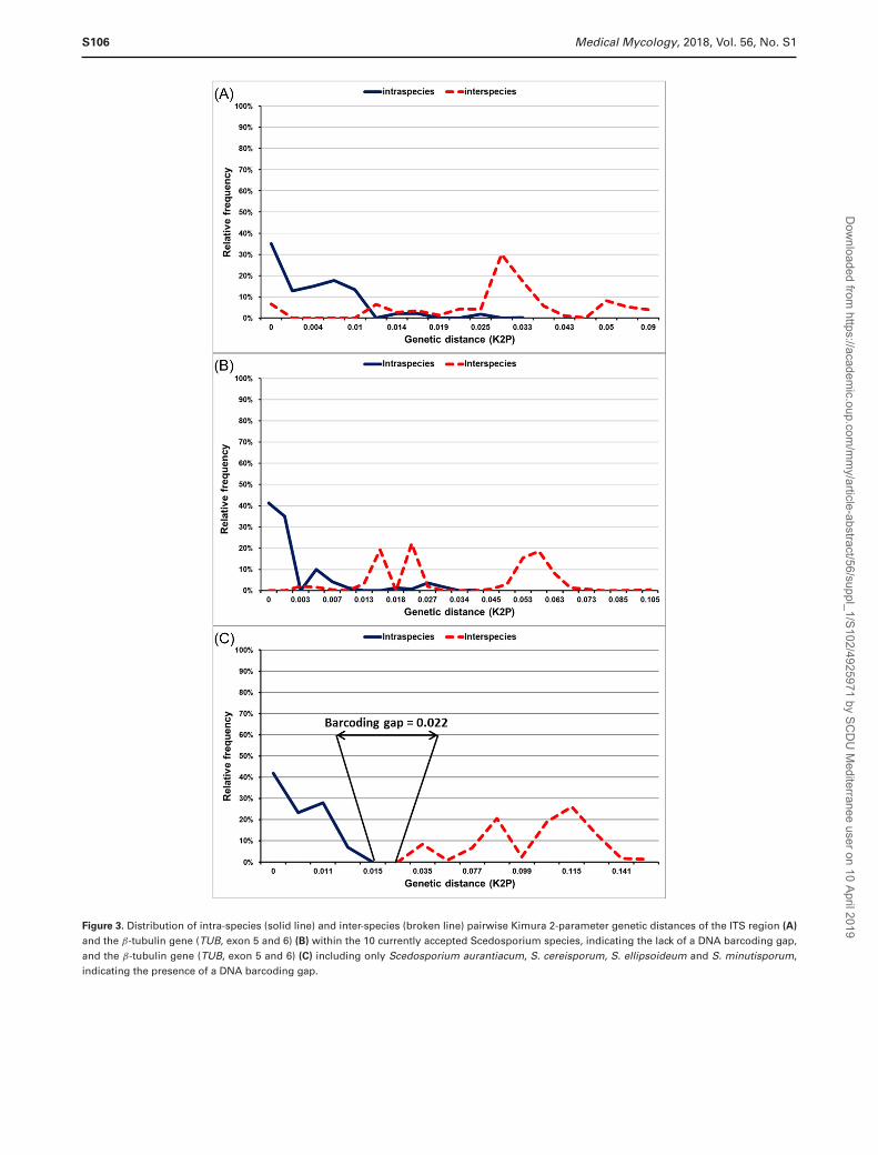
A phylogenetic analysis of 104 TUB sequences (Fig. 1),representative of all subgroups found among 407 analyzedTUB sequences, as well as an analysis of the intra-speciesvariation of all 10 currently accepted Scedosporium speciesrevealed high genetic variation within S. dehoogii, S. boydii,and S. apiospermum (Fig. 2), indicating that those shouldbe treated as species complexes, and the identified sub-clades may indicate cryptic species. This was also confirmedby DNA barcoding gap analysis carried out on 538 ITS(Fig. 3A) and 407 TUB sequences (Fig. 3B), showing thatthere is no barcoding gap within the genus Scedosporiumif all current ten species are included. The loss of the bar-coding gap is due to the high genetic variation found withinS. dehoogii, S. boydii, and S. apiospermum. However, thedescription of those subclades as separate species needs fur-ther study, including molecular data in association withmorphological, physiological, and clinical relevant data.There are clear barcoding gaps between S. minutisporum, S.desertorum, S. aurantiacum, and S. cereisporum (Fig. 3C)indicating that they are well-defined species. The separa-tion of S. angustum and S. fusoideum needs to be furtherinvestigated, taking into account the low genetic diversitywithin and between those two species, when compared tothe genetic variation found in S. dehoogii, S. boydii, andS. apiospermum (Fig. 1 and 2). Finally, L. prolificans wasshown to be unrelated to Scedosporium and therefore was
Figure 1. Phylogenetic tree of Scedosporium species based on 104 tubu-lin sequences (TUB, exon 5 and 6) representing the currently knowngenetic variation, using Maximum Likelihood analysis (GTR+G model).Bootstrap values above 80 are indicated at the nodes. Type strains arein bold italics. Petriellopsis africana and Lomentospora prolificans areused as outgroups.
Dow
nloaded from https://academ
ic.oup.com/m
my/article-abstract/56/suppl_1/S102/4925971 by SC
DU
Mediterranee user on 10 April 2019

Ramirez-Garcia et al. S105
Figure 2. Nucleotide diversity (π) in % and number of polymorphic sites (S) in the ITS1/2 regions (dark blue bar and light blue line, respectively) andβ-tubulin gene (TUB, exon 5 and 6) (red bar and green line, respectively) of the nine of the ten currently accepted Scedosporium species for whichsequences from more than one strain were available.
reclassified as Lomentospora prolificans,23 and the genusLomentospora was reinstated for this species.2
Environmental distribution
and epidemiology
Knowledge of the ecological niches of Scedosporium/Lomentospora species is essential for a better understand-ing of the dispersal of these fungi and for the potentialidentification of a source of an infection.
Ecological aspects
Scedosporium and Lomentospora species have been iso-lated from a wide range of environments, including an-thropogenic influenced habitats,24,25 oil-soaked soils, cattledung, and sewage.26 In addition, polluted waters have beendescribed as reservoirs specific for these fungi, and thesewere identified as sources of infection after near-drowningevents.27 However, adjacent agricultural soils were foundto be colonized in a greater magnitude than water or sedi-ment, suggesting the former is a main habitat of these fungi.
Subsequent investigations concerning the ecology of Sce-dosporium species confirmed the correlation between theirabundance and human impact on environments.25,28–31
Agricultural areas30 as well as playgrounds and soils inurban surroundings25,32 were consistently found to be heav-
ily colonized. Scedosporium spp. are described to degradealkanes,20,26 and therefore it is not surprising that they areresponsible for 10% of the fungi found in leachate fromsoil remediation.31 The impact of alkanes and elevated tem-perature on the soil mycobiota was studied in laboratorymodels. It was shown that the abundance of Scedosporiumspp. (mainly S. apiospermum and S. dehoogii) correlateswith diesel fuel concentration and elevated temperatures(10% w/v and 25◦C were tested, respectively). The num-ber of Aspergillus and Penicillium isolates decreased in thesame system (Eggertsberger M, unpublished results). In thiscontext it should be mentioned that the temperature in ur-ban soils, that is, in traffic islands can reach more than 30◦Ceven in temperate climates.33
The occurrence of Scedosporium spp. is also influencedby the pH of the substrate, with an optimum of 6–8. Onlyfew colonies were recovered from acidic (like most of theforest soils) or basic (as French seashores) soils. Anotherslight but positive correlation was postulated by Kaltseiset al.25 concerning fungal density and nitrate concen-tration in soil. In industrially fertilized crop-fields lessScedosporium colonies were isolated than in biologicallymanaged fields without mineral fertilizing regimes (Mall B,unpublished results). Concerning nitrogen usage, it shouldbe pointed out that Scedosporium spp. can use comple-ment compounds of the innate immune system in liquor asnitrogen source.34 As an additional ecophysiological
Dow
nloaded from https://academ
ic.oup.com/m
my/article-abstract/56/suppl_1/S102/4925971 by SC
DU
Mediterranee user on 10 April 2019
S106 Medical Mycology, 2018, Vol. 56, No. S1
Figure 3. Distribution of intra-species (solid line) and inter-species (broken line) pairwise Kimura 2-parameter genetic distances of the ITS region (A)
and the β-tubulin gene (TUB, exon 5 and 6) (B) within the 10 currently accepted Scedosporium species, indicating the lack of a DNA barcoding gap,and the β-tubulin gene (TUB, exon 5 and 6) (C) including only Scedosporium aurantiacum, S. cereisporum, S. ellipsoideum and S. minutisporum,indicating the presence of a DNA barcoding gap.
Dow
nloaded from https://academ
ic.oup.com/m
my/article-abstract/56/suppl_1/S102/4925971 by SC
DU
Mediterranee user on 10 April 2019

Ramirez-Garcia et al. S107
Table 1. Major epidemiological differences according to major groups of Scedosporium/Lomentospora species.
Lomentospora prolificans
Scedosporium apiospermumspecies complex (other thanScedosporium aurantiacum) S. aurantiacum
Geographical distribution Australia, European regions,particularly Spain, Southern USA
Worldwide Australia, European regions
Ecology Soil, decaying matter Sewerage, polluted environmentsof high human activity
Sewerage, polluted environmentsof high human activity
Host risk groups Largely immunocompromisedpatients, in particular those withmalignancy, and organ and stemcell transplant recipients
Chronic lung disease includingcystic fibrosis, bronchiectasis; neardrowning; immunocompetent andimmunocompromised
Chronic lung disease includingcystic fibrosis, bronchiectasis; neardrowning; immunocompetent andimmunocompromised
Case clusters Reported Reported Not defined
feature that helps to survive in the human host,the siderophore production of Scedosporium spp. inslightly acidic substrates could be of interest.35 Fur-thermore, S. apiospermum, S. aurantiacum, and L.prolificans were identified by molecular analyses inmesophilic bagasse composts in 3.8%, but it seems tobe unclear whether the identification method excludedS. boydii.36
Distribution patterns of the Scedosporium species showregional differences.25,28,30 In Australia, S. aurantiacum ac-counted for more than 50% of all environmental isolatesstudied, whereas S. apiospermum and S. dehoogii are pre-dominant in Austria and France, respectively. Ecologicalpreferences were observed, for example, in the abundanceof S. dehoogii in the presence of high levels of human ac-tivity.25,30 For its part, S. aurantiacum is characteristic ofagricultural areas in the west of France.30
Clinical epidemiology
Species-specific patterns, host risk groups, organ-specificpredilection, and in vitro antifungal susceptibili-ties,8,10,18,37–39 underline that understanding of theepidemiology is essential to clinical management. Sce-dosporium apiospermum and S. boydii have a worldwidedistribution; by contrast, L. prolificans is rarely encoun-tered in environmental samples and appears more com-monly in the arid climates of Australia and Spain.8,9,39,40
More recently, L. prolificans has been recognized inother European countries, the USA and Korea.11,38,41–43
Many S. aurantiacum infections have been reportedfrom Australia,8,39 the Netherlands,44 and Japan.45 Theepidemiological features between the three main groupsof pathogens within Scedosporium and Lomentospora aresummarized in Table 1.
Immunocompromised hostsSolid organ transplant (SOT) and hematopoietic stem celltransplant (HSCT) patients account for a large propor-
tion of patients at high risk for invasive Scedosporium/Lomentospora infections. However, individuals with can-cer and other immunodeficiencies are also at risk for thesemycoses. For SOT and HSCT patients, the risk of dissem-ination varies with the type of transplant and immuno-suppressive regimen, degree and duration of neutropenia,environmental exposure, and type of antifungal prophy-laxis.8,38,42,46,47 Comparison of infection incidence in thesepatients across studies is difficult due to the use of differentdenominators. In a population-based survey, Heath et al. 8
reported an incidence of 1/100 000 population, of whichtwo-thirds of cases occurred in SOT patients.
Regarding two studies in the USA series, Scedospo-rium/Lomentospora infections accounted for 25% of allnon-Aspergillus mould infections in transplant recipients(SOT, 29%; HSCT 71%),38 while in another study of aHSCT cohort a frequency of 1.11 cases/100 000 patient-inpatient days was reported.48 In the first report, Husainet al.38 found that disseminated disease occurred more of-ten in HSCT (69%) than in SOT recipients (53%), par-ticularly by L. prolificans (39% vs. 17%; P = .05), withinfections in HSCT recipients having an earlier median on-set (1.3 months vs. 4 months, P = .007), being more fun-gaemic (33% vs. 11%, P = .04), and strongly related toneutropenia (67% vs. 9%, P < .001). Additionally, HSCTrecipients were more likely to have received prior antifungalprophylaxis (64% vs. 17%), and those that received anti-fungal prophylaxis tended to have later onset of Scedospo-rium/Lomentospora infections compared to those who didnot (median time to onset, 4 vs. 2.3 months).38 The ear-lier occurrence of disease after HSCT, generally during thepre-engraftment period has been noted.3,49
According to this, predictors of invasive disease have in-cluded HSCT and leukemia, with acute leukemia and L.prolificans infection predicting death.8 Doligalski et al.50
describe Scedosporium infections in 3.5% of the patientsafter lung transplantation, and the 3-month all-cause mor-tality was 21.7%. In a single center, 16 out of 27 SOTpatients were considered colonized with Scedosporium,
Dow
nloaded from https://academ
ic.oup.com/m
my/article-abstract/56/suppl_1/S102/4925971 by SC
DU
Mediterranee user on 10 April 2019

S108 Medical Mycology, 2018, Vol. 56, No. S1
colonization being relatively common in lung transplantrecipients (73%).42 Invasive disease occurred in 11 patients(41%) with L. prolificans and S. apiospermum species com-plex causing 41% and 55% of cases, respectively. The6-month mortality was 55%, similar to other studies.8,38
Over two–thirds of patients who developed Scedosporiuminfections had received immunosuppression with alem-tuzumab or anti-thymocyte globulin, which may accountfor the higher mortality given their profound immunosup-pression. Regarding clinical manifestations of Scedospo-rium/Lomentospora infections in SOT and HSCT patients,they may range from sinopulmonary disease and brain ab-scess to disseminated infection and aneurysms, which areoften fatal.51–54
Infections caused by Scedosporium/Lomentospora un-commonly occur in patients with hematological malig-nancy,43,55,56 advanced human immunodeficiency virus(HIV) infection,57 and primary immunodeficiency disor-ders.58,59 These mycoses have attributable mortality of upto 77% in patients with acute leukemia.55 As with HSCT re-cipients, patients with hematological malignancy are morelikely to be neutropenic at the time of diagnosis of Scedospo-rium/Lomentospora infections and to have disseminateddisease.8,49,56 On the other hand, Tammer et al.57 reviewed22 HIV-infected patients with detection of Scedosporiumspecies in clinical specimens; invasive scedosporiosis wasproven in 54.5% of patients, among them disseminationoccurred in 66.7% with a mortality rate of 75%. Patientswith invasive scedosporiosis were more likely to have CD4cell counts <100/μl. Cases of Scedosporium/Lomentosporainfections in patients with chronic granulomatous disease(CGD) have been described.58–60 Most of these infectionsinvolved the lung or soft tissue although disseminated in-fection has been reported, with S. apiospermum accountingfor most of them. Moreover, breakthrough infections havebeen described in patients who were on long-term antifun-gal treatment or prophylaxis.59
Non-immunosuppressed hostsScedosporium species are classically known from traumaticinfections, leading to arthritis of eumycetoma, and frompulmonary colonization, often in preformed cavities, even-tually leading to allergic bronchopulmonary mycosis.
Colonization of lungs of patients with CF by Scedospo-rium/Lomentospora species is well established and the rateranges between 0 and 21%,61–64 being the second mostfrequent species after A. fumigatus.7 Species prevalence inthese patients varies within the region studied: S. boydiiwas the most frequent species (62%) in a French cohort,followed by S. apiospermum (24%), S. aurantiacum (10%),and S. minutisporum (4%).65 In a study performed in Ger-man CF patients, S. apiospermum was the most frequent
species (49%) followed by S. boydii (29%), L. prolificans(12%), S. aurantiacum (5%), and S. minutisporum (5%).66
In contrast, L. prolificans was the most frequent speciesisolated in patients with CF in Northern Spain.67 In Aus-tralia, the most frequent species seems to be S. aurantiacumfollowed by L. prolificans and S. apiospermum.68 Scedospo-rium dehoogii has rarely been isolated in human infectionsand to our knowledge never causing colonization in theairways of CF patients.
Numerous cases of S. apiospermum eumycetoma havebeen described in the literature, mostly affecting the lowerlimbs. These infections are found worldwide including tem-perate regions. Case reports on eumycetoma from Europe,United States, and Brazil were ascribed to S. apiospermum/S. boydii69–72 but mostly identified with classical methodsso that it cannot be ascertained whether S. aurantiacum orS. dehoogii were involved in any of these cases.
A special category is formed by cerebral infection afternear-drowning. The etiologic agents are reportedly mem-bers of the S. apiospermum complex, but most data werepublished prior to molecular species distinction. Tintelnotet al.73 re-identified 11 isolates and showed that most of theisolates belong to S. apiospermum sensu stricto, althoughS. boydii and S. aurantiacum were also identified.73,74 Fur-thermore, S. aurantiacum has been reported from a survivorof a tsunami in Japan.45 To date, L. prolificans has not beenreported in this clinical context.
Human pathology
The patients’ immune status and fungal portal of entry seemto play an important role in the clinical course of Scedospo-rium / Lomentospora infections. Patients with fully compe-tent immune systems may be asymptomatically colonizedor locally infected. On the other hand, in patients withtrauma involving major vessels, with severe injuries in thevicinity of the CNS, or with immune dysfunction, invasiveinfections are frequently found.
Colonization
Scedosporium colonization of the airways in patients withCF usually starts during adolescence, becoming chronic inup to 54% of patients having Scedosporium positive cul-tures (unpublished data), with one predominant strain thatcan be identified over several years.67,75,76 Bronchial col-onization may lead to chronic inflammation or even tolife-threatening invasive disease in cases of severe immuno-suppression, such as lung transplant or hematological ma-lignancies.3,5,77,78
Of interest, Scedosporium conidia are rarely found inthe air79 so that the exact mechanism leading to airway
Dow
nloaded from https://academ
ic.oup.com/m
my/article-abstract/56/suppl_1/S102/4925971 by SC
DU
Mediterranee user on 10 April 2019

Ramirez-Garcia et al. S109
colonization remains to be ascertained. Moreover, the pres-ence of Scedosporium/Lomentospora in respiratory secre-tions of patients suffering from non-CF bronchiectasis isscant and tends to be associated with preexisting cavities,leading to eumycetomas and pulmonary fungus balls.78
ABPA and mucoid Pseudomonas aeruginosa colonizationare positively correlated with Scedosporium/Lomentosporacolonization.80 In this sense, it is worth highlighting that arecent study has shown that P. aeruginosa is able to inhibitS. aurantiacum and L. prolificans growth, with this inhi-bition being associated but not limited to the non-mucoidphenotype of the bacterium.81
Revealing the epidemiology of human colonization byScedosporium/Lomentospora is further hampered by thefact that they are slow growing moulds. Molecular strate-gies of detection have been proposed,82,83 revealing rates ofcolonization higher than those assessed by culture. Unfor-tunately, there are no molecular techniques commerciallyavailable for this purpose, making the general implementa-tion of this approach into the clinical laboratories difficult.
Allergic bronchopulmonary mycoses
Scedosporium, but not Lomentospora, has been linkedto clinical cases of allergic bronchopulmonary mycoses(ABPM),7 with 3% of the ABPM cases reported in the lit-erature being related to Scedosporium species. While it isnot clear to what extent colonization drives long-term de-cline of pulmonary function, cases of Scedosporium-relatedABPM have been linked to a clear respiratory deteriora-tion of patients.84 The clinical picture of ABPM caused bynon-Aspergillus species tends to differ from classical allergicbronchopulmonary aspergillosis (ABPA), with asthma be-ing less frequent and with higher immunoglobulin E (IgE)levels. Promising serological methods aimed at the specificdetection of antibodies against Scedosporium are under de-velopment85 but still not available.
Localized infections
Localized infections by Scedosporium/Lomentosporaspecies include different organs and clinical manifestations:(1) cutaneous infections; (2) eumycetoma; (3) muscle, jointand bone infections; and (4) ocular infections.
Cutaneous infectionsSkin manifestations may be the initial presentation of a sub-cutaneous scedosporiosis after traumatic inoculation, or asign of hematogenous dissemination (Fig. 4A). They canmimic those caused by other fungi, such as species of As-pergillus or Fusarium with ecchymosis, necrotic papules,and hemorrhagic bullae, but they may also present solitary
ulcers, infiltrative erythematous plaques and nodules, orsuppurative nodules and ulcers. Both S. apiospermum andL. prolificans have been reported to cause soft tissue in-fections in immunocompromised hosts, including patientsreceiving chronic steroid therapy for chronic obstructivepulmonary disease or receiving immunosuppressive ther-apy for rheumatoid arthritis.3,86,87
EumycetomaThis is a chronic progressive granulomatous infectionof the subcutaneous tissue. It may affect muscles, bones,cartilage, and joints, most often involving the lower extrem-ities, usually the foot. Like other subcutaneous mycoses,the fungi enter through a penetrating trauma. The lesionis painless and grows slowly with well-defined margins, re-maining localized for long periods. Multiple nodules canappear and spontaneously drain purulent material mixedwith soft, <2 mm size, and white to yellowish, grains re-sembling fig seeds. Interconnected sinus tracts are usuallypresent by the end of the first year and may close and healcompletely, while new ones may open. Involvement of liga-ments, joint cartilage, and even bone may occur with time.Eumycetoma can produce profound disability and defor-mity but constitutional symptoms rarely appear. Clinicallyand radiologically, eumycetomata caused by S. apiosper-mum species complex or L. prolificans are similar to thosecaused by other fungi.3,71
Muscle, joint, and bone infectionsWound infections, arthritis, and osteomyelitis usually oc-cur when anatomic barriers are ruptured by trauma orsurgery. Osteomyelitis is described in lung transplanted re-cipients88,89 as a severe complication of immunosuppres-sion. Joint or bone infection by S. apiospermum or L. pro-lificans results in acute septic arthritis and acute or subacuteosteomyelitis, respectively. Plain radiography may be nor-mal in earlier stages, but magnetic resonance imaging helpsto confirm clinical diagnosis. However, the etiological or-ganism cannot be identified without culture or moleculardetection from articular fluid or a bone biopsy.3,90
Ocular infectionsScedosporium species can cause keratitis among immuno-competent hosts and usually following a corneal trauma.Clinical presentation resembles other types of keratitis (lo-cal pain, photophobia, decrease visual acuity, lacrimation)and the cornea examination reveals gray to white lesionswith irregular margins and elevated borders, ring infiltrate,hypopyon and keratitic precipitates. Endophthalmitis in im-munocompetent individuals may be caused by S. apiosper-mum. S. boydii or L. prolificans are secondary to surgery,traumatic inoculation, intravenous drug addiction, and
Dow
nloaded from https://academ
ic.oup.com/m
my/article-abstract/56/suppl_1/S102/4925971 by SC
DU
Mediterranee user on 10 April 2019

S110 Medical Mycology, 2018, Vol. 56, No. S1
Figure 4. (A) Disseminated subcutaneous scedosporiosis manifesting as cellulitis in a kidney tranplant recipient. Courtesy of Dr. Oscar Len (Valld’Hebron Hospital, Barcelona, Spain). (B) Grocott-Gomori staining of brain section showing abundant irregular hyphae from a case of invasivescedosporiosis. (C) Gramstaining of positive blood culture showing septated hyphae and adventitious conidia from a patient with disseminatedscedosporiosis. (D) Pure culture of Scedosporium apiospermum complex isolated from a wound infection in a lung transplant patient.
contiguous spread from an adjacent site. However, inimmunocompromised patients, endophthalmitis is usuallypart of disease dissemination, secondary to parenteral nu-trition or chemotherapy. Endophthalmitis curses with oc-ular pain, photophobia, and blurred vision, these symp-toms not being specific for scedosporiosis. Fundoscopicexamination shows creamy-white, well-circumscribed le-sions of the choroids and retina, vitreous infiltrates andhypopyon.3,91,92
Disseminated Infections
Scedosporium/Lomentospora disseminated infection (SDI)usually takes place in severely immunocompromised hosts,such as patients with cancer and hematological malignan-cies, hematopoietic stem cells or solid organ transplantrecipients, patients with immunodeficiency, and those re-ceiving immunosuppressive therapy.3,5,50,93–95 It happens
following hematogenous spread from lungs, skin, or anysource of localized infection. Recently, a disseminated in-fection in three patients after transplantation of a nearly-drowned donor has been reported.96 As well as in other in-vasive fungal infections, SDI may result in a wide spectrumof syndromes, depending on the primary focus, patient´simmune status, and time of evolution of the disease.
Central nervous system (CNS) infectionsThis is a severe manifestation of disseminated infection(Fig. 4B). In the literature, neurotropism of Scedospo-rium/Lomentospora is often mentioned. In immunocom-promised patients, CNS infection may appear as a manifes-tation of systemic disease in the absence of a clear spreadingfocus,38,51 while in immunocompetent hosts it mostly re-sults from a near-drowning episode with aspiration ofconidia from contaminated water and further hematoge-nous dissemination from lungs.97,98 CNS infection has been
Dow
nloaded from https://academ
ic.oup.com/m
my/article-abstract/56/suppl_1/S102/4925971 by SC
DU
Mediterranee user on 10 April 2019

Ramirez-Garcia et al. S111
occasionally reported following trauma and iatrogenic pro-cedures, and after contiguous spread from infected para-nasal sinuses.99,100 Clinical manifestations include single ormultiple brain abscesses, meningitis and ventriculitis.98,99
Endocarditis and other intravascular infectionsThese uncommon manifestations of disseminated Sce-dosporium infections are associated with high mortalityrates. Mycotic aneurysms, especially those involving theaorta and vertebrobasilar circulatory system, have been de-scribed in both immunocompromised and immunocompe-tent hosts.53 Endocarditis evolves in severely immunocom-promised patients and in those enduring risk factors, sucha valve replacement or an intravascular or intracavitary de-vice insertion.92 Twelve cases of L. prolificans endocarditiswere reported in the literature.101,102 Most patients wereimmunocompromised and developed left-side infectionswith large vegetations and systemic embolism. S. apiosper-mum complex endocarditis has been frequently associatedwith cardioverter-defibrillators or pacemaker insertion. Inthis setting, patients often tend to suffer from right-sideendocarditis and large artery thromboembolism.103,104
Systemic infectionThis is the most catastrophic expression of disseminatedinfection (Fig. 4C), fostered by the ability of Scedospo-rium species to invade blood vessels and to sporulate intissue. In patients with acute leukemia or with allogeneichematopoietic stem cell transplant Scedosporium producesfatal massive infections in the context of aplasia or severeneutropenia. Many reports of systemic infection due to L.prolificans in this group of patients have been published,with a higher incidence in Australia and Spain,105,106 andnosocomial outbreaks during hospital reconstruction havebeen also reported.56,107 Clinical features include fever, dys-pnea, lung infiltrates, signs and symptoms of meningoen-cephalitis, skin lesions and other manifestations resultingfrom multiple organ involvement. In this setting, L. pro-lificans and S. apiospermum complex are isolated fromblood cultures in a high percentage of patients.9,11,38,48,106
In solid organ transplant recipients, systemic infection isfavored by immunosuppression in the setting of graft ver-sus host disease51 and previous colonization by Scedospo-rium.52,108 Other risk groups for developing disseminatedinfection with multiple organ involvement are HIV patientswith CD4 < 50/μl57 and those receiving immunosuppres-sive therapy.109
Host-pathogen interactions: immune
response and fungal virulence factors
The host immune response is a complex network of cellu-lar and molecular mechanisms that can determine patient
survival but, on the other hand, fungal cells have also de-veloped strategies to evade immune responses and to over-come stressful conditions encountered inside the host110
(see Fig. 5).
Host immune response
As the infectious propagules of Scedosporium/Lomento-spora species are able to invade the host through a range ofdifferent sites (including: airways, puncture wounds, etc.),the immune responses also vary, with different immunecells and pathways being challenged to clear them.3 Thus,general barriers as epithelia with the mucociliary system,tissue-resident immune cells, and the secretion of defensemolecules play essential roles in the immune response tothese infections.111,112 In these first stages of fungal in-vasion, recognition of fungal cells is mediated by patternrecognition receptors (PRRs),113,114 but only dectin-1 andTLRs have been studied and proved to be determinantin the recognition of Scedosporium cells.115–117 Althoughthere are structural and compositional differences amongspecies of the S. apiospermum complex, peptidorhamno-mannans, rhamnomannans, and α-glucans from the fungalcell wall seem to be relevant pathogen associated molecularpatterns.116,118–120
After recognition by PRRs, phagocytes, includingmacrophages, neutrophils, and dendritic cells (DC),121 andother cells with phagocytic capacity promote fungal death,growth delay or inhibition and recruit polymorphonuclearleukocytes (PMNs) by synthesis of pro-inflammatory cy-tokines.122,123 Conidia of L. prolificans seem to be phago-cytized in a manner comparable to Aspergillus, at least bymonocyte-derived macrophages,124 despite the larger sizeof its conidia.105 In contrast, germination of L. prolificansconidia is inhibited less efficiently than that of A. fumigatusconidia.124
Although the cytokines locally expressed during Sce-dosporium infection have been poorly studied, inter-feron γ (IFN-γ ) and GM-CSF have been described toenhance the activity of phagocytes against Scedospo-rium species.125–127 It is also known that interleukin(IL) 15 increases IL-8 release from PMNs and en-hances PMN-induced hyphal damage and oxidative burstagainst L. prolificans.128 Additionally, compared to As-pergillus species, L. prolificans has been shown in vitroto induce higher synthesis of tumor necrosis factor α
(TNF-α) and IL-6 by human monocytes,129 in relationwith differences in the cell wall composition. In gen-eral, these cytokines are important to resist invasive in-fections by promoting respiratory burst and monocyteand neutrophil migration.130,131 Some cytokines thus havean immunomodulatory function against Scedosporium
Dow
nloaded from https://academ
ic.oup.com/m
my/article-abstract/56/suppl_1/S102/4925971 by SC
DU
Mediterranee user on 10 April 2019
S112 Medical Mycology, 2018, Vol. 56, No. S1
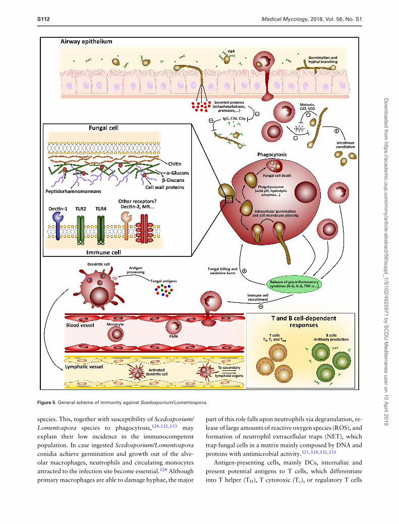
Figure 5. General scheme of immunity against Scedosporium/Lomentospora.
species. This, together with susceptibility of Scedosporium/Lomentospora species to phagocytosis,124,132,133 mayexplain their low incidence in the immunocompetentpopulation. In case ingested Scedosporium/Lomentosporaconidia achieve germination and growth out of the alve-olar macrophages, neutrophils and circulating monocytesattracted to the infection site become essential.124 Althoughprimary macrophages are able to damage hyphae, the major
part of this role falls upon neutrophils via degranulation, re-lease of large amounts of reactive oxygen species (ROS), andformation of neutrophil extracellular traps (NET), whichtrap fungal cells in a matrix mainly composed by DNA andproteins with antimicrobial activity.121,124,132,133
Antigen-presenting cells, mainly DCs, internalize andpresent potential antigens to T cells, which differentiateinto T helper (TH), T cytotoxic (Tc), or regulatory T cells
Dow
nloaded from https://academ
ic.oup.com/m
my/article-abstract/56/suppl_1/S102/4925971 by SC
DU
Mediterranee user on 10 April 2019

Ramirez-Garcia et al. S113
(Treg), depending on the stimulus and PRR involved.114
In this way, “innate” is connected with “adaptive” orlong-term immunity in which mainly TH1, TH2, and TH17cells114,134,135 conform the best known antifungal response,but little is known about their specific role against Sce-dosporium/Lomentospora species. On the other hand, Bcells are usually activated through TH cells to produce anti-bodies whose role in immunity has long time remained un-clear.136 Many antigenic proteins have been recently iden-tified in S. boydii85,137 and L. prolificans,138–140 and someof the antibodies recognizing them might be protective.141
Interestingly, L. prolificans conidia are more strongly rec-ognized by salivary immunoglobulin A (IgA) than hyphae,while sera recognize both forms similarly. This observationis consistent with a fungal airway invasion in which conidiarather than hyphae are inhaled by the host.
Virulence factors
The ability of Scedosporium/Lomentospora species to ger-minate is remarkable, which in the case of S. boydii has beendescribed to be enhanced by contact with human cells.142
L. prolificans is capable of conidiation in host tissue, whichpromotes dissemination and explains the rapid progressionof the disease.143
Among the specific molecules, some peptidopolysaccha-rides are immunologically active, able to regulate pathogen-esis and host immune response.144 Of these, peptidorham-nomannan (PRM), which is expressed on both conidia andhyphal cell walls and has been related to fungal adhesionand endocytosis by epithelial cells and macrophages, de-serves special attention.142,145–147 PRM may facilitate col-onization, virulence, and dissemination by the fungus asconsequence of an exacerbation of the infection process thatreduces the inflammatory response.148 Moreover, PRM isrecognized by antibodies, which is useful for developmentof diagnostics.149 S. boydii–derived rhamnomannans re-quire TLR-4 signaling for cytokine release by macrophages,as well as MAPKs phosphorylation and IκBα degrada-tion.120
Glucans have widely been reported as ligands for TLRsand activators of the immune response. S. boydii surfaceα-glucan, a glycogen-like polysaccharide consisting of linear4-linked α-D-Glcp residues substituted at position 6 withα-D-Glcp branches, is essential to phagocytosis of conidiaand induces cytokine secretion by cells of the innate immunesystem involving TLR2, CD14, and MyD88.116 β-glucansare used as a diagnostic strategy for several fungal infec-tions, but Scedosporium species release low levels of thispolysaccharide.150
Glucosylceramides (GlcCer) or CMHs are the mainneutral glycosphingolipids expressed by almost all fun-
gal species studied so far, including species of the S.apiospermum species complex.151,152 These molecules areassociated with fungal growth and differentiation and con-sequently play a role in the infectivity of fungal cells.153–155
Structural differences between fungal and mammalian (orplant) CMHs make these molecules potential targets for thedevelopment of new antifungal drugs, to be used alone orin conjunction with conventional antifungals.156
Host invasion-related enzymes are further virulence fac-tors of strategic relevance for Scedosporium species.144
Among these are proteolytic enzymes, which are key com-ponents to invade tissues, eliminate defense mechanismsand assist in nutrient acquisition. A serine protease able todegrade fibrinogen was described in S. apiospermum, whichmight act as mediator of severe chronic inflammation inpatients suffering from cystic fibrosis.157 Moreover, somemetalloproteases with ability to hydrolyze different sub-strates as IgG, laminin, fibronectin, or mucin have been de-scribed in S. boydii and S. apiospermum.158–160 Scedospo-rium species are also able to degrade complement systemcompounds of the innate immune system.34
Acid and alkaline ecto-phosphatase activities were alsoin mycelia of S. boydii.161 In Candida spp. these have beenrelated to adhesion and endocytosis,162,163 but limited in-formation is available on their relevance to pathogenesis inScedosporium. Enzymes such as Cu/Zn cytosolic superox-ide dismutase164 and a monofunctional catalase165 from S.boydii have been described to be important for evasion ofthe fungus to the host immune response, the latter beingalso useful for diagnostic purposes.85 Two siderophores,dimerumic acid and N(α)-methyl coprogen B, were identi-fied in S. boydii and the latter was used as a marker of theairway colonization by this species.35,166
The pigment melanin might contribute to virulence sinceit is a general protective component UV radiation and otherkind of environmental stress. Lomentospora prolificans andS. boydii produce melanin through the dihydroxynaphtha-lene (DHN) biosynthetic pathway.167,168 While melaninplays a protective role in the survival of the opportunistto oxidative killing, it does not contribute to resistance toamphotericin B.169
Diagnostics
Timely recognition of Scedosporium/Lomentospora infec-tions remains challenging, particularly in patients with CFwhere airway infections still are a major cause of mortal-ity.170–172 Distinction of colonization from infection can becrucial for adequate patient management. The definition ofpulmonary infection in CF includes the following criteria:(1) increased sputum production, (2) repeated isolation ofthe same species from sputum or BAL (≥2x in 6 months),
Dow
nloaded from https://academ
ic.oup.com/m
my/article-abstract/56/suppl_1/S102/4925971 by SC
DU
Mediterranee user on 10 April 2019

S114 Medical Mycology, 2018, Vol. 56, No. S1
(3) pulmonary infiltrate(s) on chest CT-scan or X-ray, (4)treatment failure with antibiotic therapy, (5) unclear lungfunction decline, (6) exclusion of new/other bacteria (e.g.,nontuberculous mycobacteria), and (7) exclusion of ABPA.
Diagnosis classically relies on the detection of fungifrom clinical samples by direct microscopic examinationof the clinical specimen, or histological analysis, and cul-ture on appropriate culture media (Fig. 4B–D). Histopatho-logical examination of biopsies can be performed to diag-nose these mycoses, for example, using KOH treatment.Unfortunately, it is difficult to distinguish Scedosporium/Lomentospora-infected tissues from those infected by As-pergillus or Fusarium, as all of them present hyaline hy-phae (excluding L. prolificans that may exhibit highlymelanised hyphae), regular hyphal septation, and dichoto-mous branching. However, several unique features mayhelp pathologists to diagnose Scedosporium/Lomentosporamycoses, such as irregular branching patterns or intravas-cular and intratissue conidiation 3,173
For isolation, semi-selective culture media are useful forthe detection of Scedosporium and Lomentospora amidstcompeting and more rapidly growing microbes, particu-larly A. fumigatus. Sce-Sel+ media, containing dichloranand benomyl,174 greatly facilitate recovery of Scedosporiumspecies (N.B. benomyl inhibits growth of L. prolificans)from polymicrobial clinical samples.68,175,176 Direct detec-tion and identification from clinical samples by molecular-based techniques may also constitute a valuable alternative.In this way, a species-specific multiplex PCR assay has beendeveloped to detect the clinically most important Scedospo-rium/Lomentospora species from respiratory secretions.177
Morphologically and physiologically L. prolificans iseasily differentiated from Scedosporium species based onits susceptibility to cycloheximide, the black color of itscolonies, and its characteristic flask-shaped and annellatedconidiogenous cells. However, species distinction withinthe S. apiospermum species complex is often impossible.Growth characteristics and utilization of carbohydratesor enzymatic activities, assist in main species differentia-tion but are inadequate for separation of lineages withinthe S. apiospermum complex, as demonstrated using theTaxa Profile MicronautTM (Merlin Diagnostika GmbH,Germany) system, which analyzes 570 physiological reac-tions.178 In S. aurantiacum, Biolog Phenotype analysis usingGEN III MicroPlateTM (Biolog Inc., Hayward, CA, USA)containing 94 assorted substrates, reveals metabolic differ-ences between high and low virulence strains, suggesting alink between virulence and ability to utilize D-turanose.179
Nucleotide sequence-based analysis is the current goldstandard for fungal identification.17 rDNA ITS sequenc-ing appropriately identifies the main species in Scedospo-rium/Lomentospora,180 but the partial β-tubulin gene
(BT2) is needed to differentiate closely related species. Ofnote, the status of some species like S. ellipsoidea, whichis very close to S. boydii is still debated (see above).2 Like-wise, reversed line blot hybridization has been successfullyapplied in sputum samples from patients with CF.82 Multi-locus sequence typing (MLST) was used to analyze isolatesfrom patients with CF, with three MLST schemes for S.apiospermum, S. boydii, and S. aurantiacum are now onlineat http://mlst.mycologylab.org.76 Recently the analysis ofsome repetitive DNA sequences using the semi-automatedDiversilabTM system from bioMerieux allowed the identi-fication and genotyping within pathogenic Scedosporiumspecies.181
Matrix-laser desorption/ionization mass spectrometry(MALDI-TOF/MS) has become available for the first-lineidentification. It is more economical and its identificationaccuracy is comparable to that of DNA sequencing.182–185
The quality of the reference spectra is decisive for reliableidentification (Fig. 6A). The current commercially availableMALDI-TOF/MS identification solutions are inadequatefor Scedosporium/Lomentospora and it would be necessarythe development of an online reference MALDI-TOF massspectra library database, specialized in fungal identification,and curated by expert mycologists.
Among the novel assays is PCR-ElectroSpray Ionization-Time of Flight/Mass Spectrometry (ESI-TOF/MS), whichinvolves 16 singleplex polymerase chain reaction (PCR) as-says using broad-range primers targeting nuclear or mi-tochondrial genes, and T2 magnetic resonance (T2MR).PCR-ESI-TOF/MS allows rapid determination of molecu-lar weight and base composition in the amplicons afterelectrospray ionization and chromatographic separation,and resulting profiles are compared with a database pro-vided by the manufacturer.186–188 This technique has beenused to determine the distribution of fungal communitiesdirectly from bronchoalveolar lavage fluid specimens.189
T2MR technology rapidly and accurately detects the pres-ence of molecular targets within a sample without the needfor purification or extraction,190,191 but designing primersis challenging.192
Specific monoclonal antibodies (MAbs) have been de-veloped allowing for species distinction.167,193 Two MAbstargeting respectively an immunodominant carbohydrateepitope on an extracellular 120-kDa antigen present inthe spore and hyphal cell walls of S. apiospermum andS. boydii or the tetrahydroxynaphtalene reductase of thedihydroxynaphtalene-melanin pathway in L. prolificans,may be used in immunofluorescence assay to differentiatethese fungi from other septate fungal pathogens on histo-logical sections.
Recently some Scedosporium proteins, including amonofunctional cytosolic catalase, proved to be interesting
Dow
nloaded from https://academ
ic.oup.com/m
my/article-abstract/56/suppl_1/S102/4925971 by SC
DU
Mediterranee user on 10 April 2019

Ramirez-Garcia et al. S115
Figure 6. (A) Reference spectra for Scedosporium apiospermum, S. boydii, S. aurantiacum and Lomentospora prolificans identification by matrix-laser desorption/ionization mass spectrometry (MALDITOF/MS). (B) Example of matrix-assisted laser desorption/ionization with Fourier transform ioncyclotron resonance (MALDI-FTICR) mass spectrum annotation. Ferricrocin-like molecules (C28H47N9O13) were observed in protonated, sodiated,or potassiated forms represented by signals at m/z 718.3358, 740.3184 and 756.2921, respectively. This intracellular siderophore was annotatedin a sample of S. boydii (IHEM 15155) and was released from intact fungal spores by microwave-enhanced extraction to methanol. Note that allcompounds annotated by Cyclobranch in red were tentatively assigned according to library accurate mass matching with 1 ppm accuracy.
Dow
nloaded from https://academ
ic.oup.com/m
my/article-abstract/56/suppl_1/S102/4925971 by SC
DU
Mediterranee user on 10 April 2019

S116 Medical Mycology, 2018, Vol. 56, No. S1
markers of a Scedosporium infection, and works are cur-rently being performed in order to develop standardizedserological tests.85
In addition to proteomic approaches with MALDI-TOFor LC-MS/MS identification of Scedosporium/Lomento-spora ribosomal equipment,139,182 mass spectrometry canbe used in metabolomics to gain access to specific low-molecular weight biomarkers. Melanin and its degrada-tion products represent the first target in L. prolificans.Diverse lipids were also detected on intact spores of L.prolificans and S. apiospermum.194 The metabolite AS-183was detected in fermentation broth of Scedosporium spp.SPC-15549.195
Siderophores have gained attention as disease biomark-ers as well as virulence factors.196,197 Two siderophore rep-resentatives have been rigorously described in Scedospo-rium genus, dimerumic acid and N(α)-methyl coprogen B,35
the former possibly being a degradation product of the lat-ter. Siderophores may occur in various ionic forms in massspectra. Generally, they are observed as ferri- or desferri-forms, but combinations with sodium or potassium ions arepossible depending on the sample type.197 For example, inhost tissue the generation of [M+Na]+, [M+K]+, [M+Fe-2H]+, or [M+Fe+Na-3H]+ ions is quite common. Recentlya new dereplication tool called Cyclobranch has been devel-oped for the rediscovery of above described compounds.198
It is based on an integrated library of hundreds of microbialsiderophores and secondary metabolites including toxinsand nonribosomal peptides. Dereplication (the process ofclassifying already known compounds) can be performedon conventional mass spectra generated by any ionizationtechnique as well as on liquid chromatography/mass spec-trometry or imaging mass spectrometry datasets. These dataformats are batch-processed and incorporation of impor-tant biometals (including iron) can be supported in calcula-tions and data presentations. An example of a siderophoreannotated in a sample of S. boydii by matrix-assisted laserdesorption/ionization with Fourier transform ion cyclotronresonance (MALDI-FTICR) mass spectrum is illustrated inFigure 6B. It is worth mentioning that Cyclobranch is afree tool (available at http://ms.biomed.cas.cz/cyclobranch/)dedicated to exact mass data. In addition to dereplica-tion, the de novo sequencing of new microbial structuresis also possible. The calculator works with approximately520 nonisobaric building blocks arising from ribosomal,nonribosomal or polyketide syntheses making the charac-terization of new siderophores198 or cyclic, branched, orbranched cyclic peptides199 feasible.
Therapeutic strategies
Treatment of deep-seated Scedosporium or Lomentosporainfections still remains challenging because of the limited
susceptibility of these fungi to all current antifungal drugs.Scedosporium species are resistant to 5-flucytosine and am-photericin B, as well as to the first generation triazole drugs,fluconazole and itraconazole. In addition, they have a re-duced susceptibility to echinocandins, particularly caspo-fungin and anidulafungin, and exhibit resistance to the mostrecent triazole drug, isavuconazole, S. aurantiacum beingthe least susceptible to antifungal drugs.12,66,200 Likewise,L. prolificans is a pan-antifungal resistant species.3,12,201 Inthis connection, it is also relevant to highlight that the avail-able antifungal spectrum is quite limited, and as such moreefforts need to focus on the development of novel effectivedrugs.202,203
For treatment of Scedosporium/Lomentospora infec-tions, the European guidelines recommend voriconazole asfirst-line treatment200 together with surgical debridementwhen possible. Although favorable results have been ob-served following such recommendations, the outcome re-mains poor with mortality rates of >65% and nearly 100%when CNS affectation or dissemination occurs.204,205 Aminimum inhibitory concentration (MIC) of less than2 μg/ml could be predictive of a favorable outcome forScedosporium species.206 Despite the differences on in vitrosusceptibility among genera, the outcome remains similarespecially when dissemination occurs. For this reason, it isof crucial interest to find therapeutic alternatives for thesechallenging and difficult-to-treat infections.
Antifungal combination therapy has emerged as apromising strategy since therapeutic effect can be achievedat lower concentrations and thus reducing toxic side ef-fects, improving safety and tolerability, shortening the ther-apeutic effect and preventing treatment failure when an-timicrobial resistance is suspected. Few studies have eval-uated the in vitro activity of double combinations againstScedosporium spp. and L. prolificans. Among them, com-bined voriconazole and amphotericin B or echinocandinshave shown synergistic effects against both S. apiosper-mum and L. prolificans,207–209 as well as terbinafine plusitraconazole, miconazole or voriconazole against L. prolif-icans.3,210,211 However, the combination of voriconazoleplus terbinafine or liposomal amphotericin B has demon-strated variable outcome in the treatment of these infec-tions.212–221 Limited data are available on combinations ofmore than two antifungals. Two triple combinations (am-photericin B plus voriconazole plus anidulafungin or mica-fungin) have been tested against L. prolificans and showedsynergy222,223.
The in vitro activity of combinations of antifungalswith miltefosine, antipsychotic drugs or cysteine deriva-tives is being investigated as a potential treatment al-ternative.224–226 It is also highlighting the capacity ofinhibitors of Heat shock proteins, calcineurin and deacety-lases against fungal species.227–233 However, their effect
Dow
nloaded from https://academ
ic.oup.com/m
my/article-abstract/56/suppl_1/S102/4925971 by SC
DU
Mediterranee user on 10 April 2019

Ramirez-Garcia et al. S117
on Scedosporium/Lomentospora species should be furtherresearched.
Murine studies have also shown promising results forcombinations of antifungals with granulocyte-colony stim-ulating factor,234–236 and clinical experience suggests thatreversion of neutropenia is a key factor in the outcome of afungal infection.218,237
Reviewing recent clinical cases reported in the literature,four CF patients treated with antifungal drugs because of asuspected pulmonary Scedosporium/Lomentospora infec-tion have been reported since 2013.80,238–240 Moreover,in Germany 36 cases of antifungal treatment of Scedospo-rium/Lomentospora infections in patients with CF were an-alyzed (Schwarz C et al. unpublished results). In 20/36 anti-fungal courses a therapeutic response was achieved (regressin radiology or symptoms, or increase in FEV1). These re-sults demonstrated a significant superiority of the use of acombination of three drugs versus two and two drugs ver-sus one drug. Among the antifungal drugs, voriconazole re-mains the first therapeutic choice,200 potentially combinedwith an echinocandin for Scedosporium infections or withterbinafine for Lomentospora infections.
Prospects in susceptibility to antifungals and
resistance mechanisms
Among the drugs that are currently in the pipelines,one might be promising for treatment of Scedosporium/Lomentospora infections. The Japanese company Eisai Co.discovered E1210, a new first-in-class broad spectrum an-tifungal drug acting in vitro against clinically importantyeasts and molds,241 and in vivo in experimental models ofcandidiasis, aspergillosis, and fusariosis.242 This drug tar-gets the inositol acylation step in the biosynthesis pathwayof the glycosyl phosphatidyl inositol (GPI) anchor. GPI-anchored cell wall proteins play a key role in fungal biologyand virulence, and blockage of this metabolic pathway re-sults in defects in cell wall biosynthesis, hyphal elongationand adherence of fungal cells to biological substrates. Invitro susceptibility testing using a large set of S. apiosper-mum (n = 28), S. aurantiacum (n = 7) and L. prolificans (n= 28) isolates revealed that MICs using E1210 were at least10 fold lower than found in currently used drugs, includ-ing voriconazole.243 This compound, which is licensed since2015 by Amplyx (San Diego, USA–APX001) was approvedon June 2016 by the FDA for treatment of candidiasis, in-vasive aspergillosis and coccidioidomycosis.
Mutations in the “hot spot” regions of the Fks1gene, encoding the catalytic subunit of the β-1,3-glucansynthase (the target of echinocandins), have been de-scribed, which may explain the reduced susceptibility ofScedosporium species and L. prolificans to echinocan-
dins.244 The low in vitro susceptibility (or primary re-sistance) of Scedosporium/Lomentospora species to azoledrugs may result from resistance mechanisms similarto those extensively studied for A. fumigatus245–249
such as point mutations in the coding sequence ofCYP51A orthologues leading to a reduced affinity ofazole drugs for their target, or constitutive overex-pression of some efflux pumps. Specifically L. prolif-icans showed alterations in of shorter and wider hy-phae and structural and compositional changes in theCW, possibly mediating L. prolificans resistance toVRC.250
Future trends in antifungal drugs
There are nowadays some very promising novel antifungalcompounds, such as F901318 (Chen S, unpublished results)and N-chlorotaurine (NCT). The F901318 compound rep-resents a novel class of antifungal drug that inhibits di-hydroorotate dehydrogenase, a key enzyme in pyrimidinebiosynthesis.251 The compound has been recently investi-gated for 50 clinical Scedosporium and Lomentospora iso-lates (Biswas et al. In vitro susceptibility testing of thenovel orotomide antifungal agent F901318 against Aus-tralian Scedosporium and Lomentospora pathogens, EC-CMID, Vienna, Austria, 22–25 April 2017, P1704), andit was active against all isolates of L. prolificans as wellas S. apiospermum, S. boydii, and S. aurantiacum, withMICs falling ranging from 0.125 to 0.5 mg/l. Similar re-sults have been found in another study (Alastruey-Izquierdoet al. unpublished data) testing 123 clinical isolates of S.apiospermum, S. boydii, S. aurantiacum, S. dehoogii, S. el-lipsoideus, and L. prolificans with MIC range for all isolatesof 0.007–0.5, and by Wiederhold and coworkers against S.apiospermum, S. aurantiacum, S. dehoogii, S. boydii, andL. prolificans, with MIC ranging from ≤0.008 to 0.25, withthe last species being the most resistant ones.252
The N-chloro derivative of the amino acid taurine isa long-lived oxidant generated by activated granulocytesand monocytes during inflammation and oxidative burst inphagolysosomes.253 Moreover, it is more stable and muchless toxic in vivo than HOCl.254
In the 90s, the chemical synthesis of NCT as a crys-talline sodium salt (Cl-HN-CH2-CH2-SO3Na) could be es-tablished, demonstrating broad-spectrum killing activityagainst microbes.255,256 Due to its unspecific mechanismof action, development of resistance is extremely improba-ble. Three key features of NCT contribute to its successfulclinical application: (1) transhalogenation:257 which makesthe net microbicidal activity of NCT markedly enhancedin vivo, above all against fungi; (2) chlorine cover:258
which avoids regrowth (postantifungal effect) and induces
Dow
nloaded from https://academ
ic.oup.com/m
my/article-abstract/56/suppl_1/S102/4925971 by SC
DU
Mediterranee user on 10 April 2019

S118 Medical Mycology, 2018, Vol. 56, No. S1
loss of virulence; (3) inactivation of virulence factors ofpathogens.257
Clinical phase I and II studies demonstrated very goodtolerability of topical 1% (55 mM) NCT in aqueous solu-tion for skin ulcers, conjunctivitis, external otitis, and oralinfections.256 Recently, inhaled 1% NCT was well toleratedin pigs, mice, and humans (pilot tests and a phase I study),respectively.259–261
At this concentration, NCT was able to kill all Scedospo-rium species tested, that is, both hyphae and conidia of S.apiospermum, S. boydii, and L. prolificans, within severalhours at pH 7.1 and 37◦C.262 As expected, addition ofammonium chloride (NH4Cl) reduced the killing times toapproximately 5 min because of transhalogenation. Indeed,LIVE/DEAD staining of conidia disclosed increased perme-ability of the cell membrane and wall, which is decisivefor killing. However, short, sublethal incubation times of10–60 min in plain NCT significantly increased germinationtime and decreased germination rate of conidia. Moreover,such sublethally treated conidia lost their virulence in vivoafter injection into larvae of G. mellonella, so that the larvaesurvived similar to mock-injected controls.262
A second study was done to investigate NCT on itsmicrobicidal activity in vitro in artificial sputum medium(ASM) mimicking the composition of cystic fibrosis mu-cus at 37◦C and pH 6.9.263 Under these conditions, 1%NCT killed bacteria and spores already within 10 min and15 min, respectively, to the detection limit of 102 CFU/ml(reduction by 5–6 log10). A reduction by 2 log10 was stillachieved by 0.1% (bacteria) and 0.3% (fungi) NCT largelywithin 10–30 min. This markedly more rapid killing (par-ticularly of fungi) in ASM compared to phosphate buffercan be explained by transhalogenation.
In this review, the state-of-the-art of the emerging op-portunistic fungal pathogens Scedosporium/Lomentosporais discussed, mainly focusing on the scientific knowledgeacquired in the last decade. Summarizing, in taxonomythe genus Lomentospora is clearly independent from Sce-dosporium, which currently contains ten species. Thesefungi are found in environments of high human activity,polluted waters and soils/composts, while their prevalencevaries with geography, environmental pH and chemicalcontent, especially aliphatic hydrocarbons. They infect im-munosuppressed and immunocompetent individuals wherenear-drowning events pose a special risk. Furthermore, col-onization of the respiratory tract is common in patientswith chronic lung diseases such as CF.
The main virulence factors described are PRM andother cell-wall peptidopolysaccharides, proteolytic en-zymes, superoxide dismutase, catalase, siderophores, andmelanin. The immune status of the patient seems vi-tal to control infections, being TLRs and Dectin-1 cru-cial for fungal recognition and phagocytosis. Specific
response, including humoral, might also be of impor-tance. The difficulty to detect and identify these fungifrom nonsterile samples results in the fact that thereal epidemiology remains to be undetermined, warrant-ing future efforts on the improvement of conventionalmethods, molecular tools, detection of serological mark-ers and secondary metabolites. A rapid and specificdetection of the etiologic agent remains to be very impor-tant for the initiation of appropriate treatment. Regardingtherapy, although several new strategies are being testedwith promising results, nowadays a combination of twoor even three anti-fungal drugs is recommended. Amongthe future perspectives, in addition to immunotherapy,NCT deserves to be mentioned because its broad-spectrummicrobicidal activity, tolerability, and anti-inflammatoryproperties.
In conclusion, although great advances in Scedosporium/Lomentospora have been made, much remains to be ascer-tained, including (1) the identification of definitive mark-ers for the definition of species in Scedosporium that al-low a better knowledge of its distribution and impact inhuman pathology, (2) a deeper understanding of its sur-vival strategies and interaction with hosts, (3) the devel-opment of faster, accurate and easy-to-implement clinicaltools for diagnosis, and (4) the finding of in vivo activecompounds to treat the wide range of infections, many ofthe life-threatening, caused by these fungi.
Acknowledgments
The authors gratefully acknowledge the support to V.H. fromCzech Science Foundation (16-20229S), to W.M. and C.S. fromthe National Health and Medical Research Council of Australia(APP1031952 and APP1121936), to A.R., A.R.G ., and F.L.H. fromthe University of the Basque Country (UPV/EHU) (GIU15/36), andto E.B.B. from the Conselho Nacional de Desenvolvimento Cientıficoe Tecnologico (CNPq) and Fundacao de Amparo a Pesquisa no Es-tado do Rio de Janeiro (FAPERJ).
Declaration of interest
The authors report no conflicts of interest. The authors alone areresponsible for the content and the writing of the paper.
References
1. Brown GD, Denning DW, Gow NAR, Levitz SM, Netea MG, WhiteTC. Hidden killers: human fungal infections. Sci Transl Med. 2012; 4:165rv13.
2. Lackner M, de Hoog GS, Yang L et al. Proposed nomenclature for Pseu-dallescheria, Scedosporium and related genera. Fungal Divers. 2014; 67:1–10.
3. Cortez KJ, Roilides E, Quiroz-Telles F et al. Infections caused by Sce-dosporium spp. Clin Microbiol Rev. 2008; 21: 157–197.
4. Richardson M, Lass-Florl C. Changing epidemiology of systemic fungalinfections. Clin Microbiol Infect. 2008; 14: 5–24.
Dow
nloaded from https://academ
ic.oup.com/m
my/article-abstract/56/suppl_1/S102/4925971 by SC
DU
Mediterranee user on 10 April 2019

Ramirez-Garcia et al. S119
5. Guarro J, Kantarcioglu AS, Horre R et al. Scedosporium apiospermum:changing clinical spectrum of a therapy-refractory opportunist. Med My-col. 2006; 44: 295–327.
6. Paugam A, Baixench MT, Demazes-Dufeu N et al. Characteristics andconsequences of airway colonization by filamentous fungi in 201 adultpatients with cystic fibrosis in France. Med Mycol. 2010; 48: S32–S36.
7. Cimon B, Carrere J, Vinatier JF, Chazalette JP, Chabasse D, BoucharaJP. Clinical significance of Scedosporium apiospermum in patients withcystic fibrosis. Eur J Clin Microbiol Infect Dis. 2000; 19: 53–56.
8. Heath CH, Slavin MA, Sorrell TC et al. Population-based surveillancefor scedosporiosis in Australia: epidemiology, disease manifestations andemergence of Scedosporium aurantiacum infection. Clin Microbiol In-fect. 2009; 15: 689–693.
9. Rodriguez-Tudela JL, Berenguer J, Guarro J et al. Epidemiology andoutcome of Scedosporium prolificans infection, a review of 162 cases.Med Mycol. 2009; 47: 359–370.
10. Grenouillet F, Botterel F, Crouzet J et al. Scedosporium prolificans: anemerging pathogen in France? Med Mycol. 2009; 47: 343–350.
11. Tintelnot K, Just-Nubling G, Horre R et al. A review of German Sce-dosporium prolificans cases from 1993 to 2007. Med Mycol. 2009; 47:351–358.
12. Lackner M, De Hoog GS Verweij PE et al. Species-specific antifungalsusceptibility patterns of Scedosporium and Pseudallescheria species. An-timicrob Agents Chemother. 2012; 56: 2635–2642.
13. Mcneill J, Barrie FR, Buck WR et al. (eds.) International Code of Nomen-clature for Algae, Fungi, and Plants (Melbourne Code). Regnum Veg-etabile 154. Oberreifenberg, Germany: Koeltz Sci Books, 2012.
14. Gilgado F, Cano J, Gene J, Guarro J. Molecular phylogeny of the Pseu-dallescheria boydii species complex: Proposal of two new species. J ClinMicrobiol. 2005; 43: 4930–4942.
15. Gilgado F, Cano J, Gene J, Sutton DA, Guarro J. Molecular and pheno-typic data supporting distinct species statuses for Scedosporium apiosper-mum and Pseudallescheria boydii and the proposed new species Sce-dosporium dehoogii. J Clin Microbiol. 2008; 46: 766–771.
16. Zeng JS, Fukushima K, Takizawa K et al. Intraspecific diversity of speciesof the Pseudallescheria boydii complex. Med Mycol. 2007; 45: 547–558.
17. Lackner M, Klaassen CH, Meis JF, van den Ende AHGG, de Hoog GS.Molecular identification tools for sibling species of Scedosporium andPseudallescheria. Med Mycol. 2012; 50: 497–508.
18. Lackner M, Hagen F, Meis JF et al. Susceptibility and diversity in thetherapy-refractory genus Scedosporium. Antimicrob Agents Chemother.2014; 58: 5877–5885.
19. Chen M, Zeng J, de Hoog GS et al. The “species complex” issue inclinically relevant fungi: A case study in Scedosporium apiospermum.Fungal Biol. 2016; 120: 137–146.
20. Rainer J, Kaltseis J. Diversity in Scedosporium dehoogii (Microascaceae):S. deficiens sp. nov. Sydowia. 2010; 62: 137–147.
21. Crous PW, Wingfield MJ, Burgess TI et al. Fungal planet descriptionsheets: 469–557. Persoonia. 2016; 37: 218–403.
22. Hawksworth DL, Crous PW, Redhead SA et al. The Amsterdam decla-ration on fungal nomenclature. IMA Fungus. 2011; 2: 105–112.
23. Hennebert GL, Desai BG. Lomentospora prolificans, a new hyphomycetefrom greenhouse soil. Mycotaxon. 1974; 1: 45–50.
24. de Hoog GS Marvin-Sikkema FD, Lahpoor GA, Gottschall JC, Prins RA,Guicho E. Ecology and physiology of the emerging opportunistic fungiPseudallescheria boydii and Scedosporium prolificans. Mycoses. 1994;37: 71–78.
25. Kaltseis J, Rainer J, de Hoog GS. Ecology of Pseudallescheria and Sce-dosporium species in human-dominated and natural environments andtheir distribution in clinical samples. Med Mycol. 2009; 47: 398–405.
26. April TM, Abbott SP, Foght JM, Currah RS. Degradation of hydrocar-bons in crude oil by the ascomycete Pseudallescheria boydii (Microas-caceae). Can J Microbiol. 1998; 44: 270–278.
27. Horre R, Lackner M, Buzina W, Tintelnot K. Near-drowning and cere-bral infections caused by fungi belonging to the Pseudallescheria/Scedosporium-complex. Anasthesiol Intensivmed NotfallmedSchmerzther. 2010; 45: 152–158 [in German].
28. Harun A, Gilgado F, Chen SCA, Meyer W. Abundance of Pseu-dallescheria/Scedosporium species in the Australian urban environmentsuggests a possible source for scedosporiosis including the colonizationof airways in cystic fibrosis. Med Mycol. 2010; 48: S70–S76.
29. Janda-Ulfig K, Ulfig K, Cano J, Guarro J. A study of the growth ofPseudallescheria boydii isolates from sewage sludge and clinical sourceson tributyrin, rapeseed oil, biodiesel oil and diesel oil. Ann Agric EnvironMed. 2008; 15: 45–49.
30. Rougeron A, Schuliar G, Leto J et al. Human-impacted areas ofFrance are environmental reservoirs of the Pseudallescheria boy-dii/Scedosporium apiospermum species complex. Environ Microbiol.2015; 17: 1039–1048.
31. Selbmann L, Egidi E, Isola D et al. Biodiversity, evolution and adaptationof fungi in extreme environments. Plant Biosystems. 2013; 147: 237–246.
32. Romao D, Sabino R, Verıssimo C et al. Children and Sand Play: Screeningof potential harmful microorganisms in sandboxes, parks, and beaches.Curr Fungal Infect Rep. 2015; 9: 155–163.
33. Kaltseis J. Opportunistic human pathogenic fungi of the generaPseudallescheria/Scedosporium. PhD Thesis. University of Innsbruck,2013.
34. Rainer J, Rambach G, Kaltseis J, Hagleitner M, Heiss S, Speth C. Phy-logeny and immune evasion: a putative correlation for cerebral Pseu-dallescheria/Scedosporium infections. Mycoses. 2011; 54: 48–55.
35. Bertrand S, Larcher G, Landreau A, Richomme P, Duval O, Bouchara JP.Hydroxamate siderophores of Scedosporium apiospermum. BioMetals.2009; 22: 1019–1029.
36. De Gannes V, Eudoxie G, Hickey WJ. Insights into fungal communitiesin composts revealed by 454-pyrosequencing: Implications for humanhealth and safety. Front Microbiol. 2013; 4: 164.
37. Rodriguez-Tudela JL, Berenguer J, Guarro J et al. Epidemiology andoutcome of Scedosporium prolificans infection, a review of 162 cases.Med Mycol. 2009; 47: 359–370.
38. Husain S, Munoz P, Forrest G et al. Infections due to Scedosporiumapiospermum and Scedosporium prolificans in transplant recipients: clin-ical characteristics and impact of antifungal agent therapy on outcome.Clin Infect Dis. 2005; 40: 89–99.
39. Delhaes L, Harun A, Chen SCA et al. Molecular typing of AustralianScedosporium isolates showing genetic variability and numerous S. au-rantiacum. Emerg Infect Dis. 2008; 14: 282–290.
40. Idigoras P, Perez-Trallero E, Pineiro L et al. Disseminated infection andcolonization by Scedosporium prolificans: a review of 18 cases, 1990–1999. Clin Infect Dis. 2001; 32: e158–e165.
41. Bhagavatula S, Vale L, Evans J, Carpenter C, Barnes RA. Scedosporiumprolificans osteomyelitis following penetrating injury: A case report. MedMycol Case Rep. 2014; 4: 26–29.
42. Johnson LS, Shields RK, Clancy CJ. Epidemiology, clinical manifesta-tions, and outcomes of Scedosporium infections among solid organ trans-plant recipients. Transpl Infect Dis. 2014; 16: 578–587.
43. Song MJ, Lee JH, Lee NY. Fatal Scedosporium prolificans infection in apaediatric patient with acute lymphoblastic leukaemia. Mycoses. 2011;54: 81–83.
44. Kooijman CM, Kampinga GA, de Hoog GS, Goudswaard WB, ReijnenMM. Successful treatment of Scedosporium aurantiacum osteomyelitisin an immunocompetent patient. Surg Infect. 2007; 8: 605–610.
45. Nakamura Y, Suzuki N, Nakajima Y et al. Scedosporium aurantiacumbrain abscess after near-drowning in a survivor of a tsunami in Japan.Respir Investig. 2013; 51: 207–211.
46. Pappas PG, Alexander BD, Andes DR et al. Invasive fungal infectionsamong organ transplant recipients: results of the Transplant-AssociatedInfection Surveillance Network (TRANSNET). Clin Infect Dis. 2010;50: 1101–1111.
47. Shoham S, Marr KA. Invasive fungal infections in solid organ transplantrecipients. Future Microbiol. 2012; 7: 639–655.
48. Lamaris GA, Chamilos G, Lewis RE, Safdar A, Raad II, KontoyiannisDP. Scedosporium infection in a tertiary care cancer center: a review of25 cases from 1989–2006. Clin Infect Dis. 2006; 43: 1580–1584.
Dow
nloaded from https://academ
ic.oup.com/m
my/article-abstract/56/suppl_1/S102/4925971 by SC
DU
Mediterranee user on 10 April 2019

S120 Medical Mycology, 2018, Vol. 56, No. S1
49. Baddley JW, Stroud TP, Salzman D, Pappas PG. Invasive mold infectionsin allogeneic bone marrow transplant recipients. Clin Infect Dis. 2001;32: 1319–1324.
50. Doligalski CT, Benedict K, Cleveland AA et al. Epidemiology of invasivemold infections in lung transplant recipients. Am J Transplant. 2014; 14:1328–1333.
51. Castiglioni B, Sutton DA, Rinaldi MG, Fung J, Kusne S. Pseudallescheriaboydii (Anamorph Scedosporium apiospermum). Infection in solid organtransplant recipients in a tertiary medical center and review of the liter-ature. Medicine. 2002; 81: 333–348.
52. Morio F, Horeau-Langlard D, Gay-Andrieu F et al. Disseminated Sce-dosporium/Pseudallescheria infection after double-lung transplantationin patients with cystic fibrosis. J Clin Microbiol. 2010; 48: 1978–1982.
53. Ong A, Blyth CC, Bency R et al. Fatal mycotic aneurysms due to Sce-dosporium and Pseudallescheria infection. J Clin Microbiol. 2011; 49:2067–2071.
54. Sayah DM, Schwartz BS, Kukreja J, Singer JP, Golden JA, Leard LE.Scedosporium prolificans pericarditis and mycotic aortic aneurysm ina lung transplant recipient receiving voriconazole prophylaxis. TransplInfect Dis. 2013; 15: E70–E74.
55. Caira M, Girmenia C, Valentini CG et al. Scedosporiosis in patients withacute leukemia: a retrospective multicenter report. Haematologica. 2008;93: 104–110.
56. Cooley L, Spelman D, Thursky K, Slavin M. Infection with Scedosporiumapiospermum and S. prolificans, Australia. Emerg Infect Dis. 2007; 13:1170–1177.
57. Tammer I, Tintelnot K, Braun-Dullaeus RC et al. Infections due toPseudallescheria/Scedosporium species in patients with advanced HIVdisease - a diagnostic and therapeutic challenge. Int J Infect Dis. 2011;15: e422–e429.
58. Gompels MM, Bethune CA, Jackson G, Spickett GP. Scedosporiumapiospermum in chronic granulomatous disease treated with an HLAmatched bone marrow transplant. J Clin Pathol. 2002; 55: 784–786.
59. Jabado N, Casanova JL, Haddad E et al. Invasive pulmonary infectiondue to Scedosporium apiospermum in two children with chronic granu-lomatous disease. Clin Infect Dis. 1998; 27: 1437–1441.
60. Santos PE, Oleastro M, Galicchio M, Zelazko M. Fungal infections inpaediatric patients with chronic granulomatous disease. Rev IberoamMicol. 2000; 17: 6–9 [in Spanish].
61. Pihet M, Carrere J, Cimon B et al. Occurrence and relevance of filamen-tous fungi in respiratory secretions of patients with cystic fibrosis - areview. Med Mycol. 2009; 47: 387–397.
62. Montagna MT, Barbuti G, Paglionico F et al. Retrospective analysis ofmicroorganisms isolated from cystic fibrosis patients in Southern Italy,2002–2010. J Prev Med Hyg. 2011; 52: 209–214.
63. Gungor O, Tamay Z, Guler N, Erturan Z. Frequency of fungi in respi-ratory samples from Turkish cystic fibrosis patients. Mycoses. 2013; 56:123–129.
64. Ziesing S, Suerbaum S, Sedlacek L. Fungal epidemiology and diversity incystic fibrosis patients over a 5-year period in a national reference center.Med Mycol. 2016; 54: 781–786.
65. Zouhair R, Rougeron A, Razafimandimby B, Kobi A, Bouchara JP, Gi-raud S. Distribution of the different species of the Pseudallescheria boy-dii/Scedosporium apiospermum complex in French patients with cysticfibrosis. Med Mycol. 2013; 51: 603–613.
66. Sedlacek L, Graf B, Schwarz C et al. Prevalence of Scedosporium speciesand Lomentospora prolificans in patients with cystic fibrosis in a mul-ticenter trial by use of a selective medium. J Cyst Fibros. 2015; 14:237–241.
67. Lackner M, Rezusta A, Villuendas MC, Palacian MP, Meis JF, KlaassenCH. Infection and colonisation due to Scedosporium in Northern Spain.An in vitro antifungal susceptibility and molecular epidemiology studyof 60 isolates. Mycoses. 2011; 54: 12–21.
68. Blyth CC, Harun A, Middleton PG et al. Detection of occult Scedospo-rium species in respiratory tract specimens from patients with cysticfibrosis by use of selective media. J Clin Microbiol. 2010; 48: 314–316.
69. Buonfrate D, Gobbi F, Angheben A et al. Autochthonous cases of myce-toma in Europe: report of two cases and review of literature. PLoS One.2014; 9: e100590.
70. Queiroz-Telles F, McGinnis MR, Salkin I, Graybill JR. Subcutaneousmycoses. Infect Dis Clin North Am. 2003; 17: 59–85.
71. Oliveira FM, Unis G, Hochhegger B, Severo LC. Scedosporium apiosper-mum eumycetoma successfully treated with oral voriconazole: report ofa case and review of the Brazilian reports on scedosporiosis. Rev InstMed Trop Sao Paulo. 2013; 55: 121–123.
72. Green WO, Adams TE. Mycetoma in the United States: a review andreport of seven additional cases. Am J Clin Pathol. 1964; 42: 75–91.
73. Tintelnot K, Wagner N, Seibold M, de Hoog GS, Horre R. Re-identification of clinical isolates of the Pseudallescheria boydii-complexinvolved in near-drowning. Mycoses. 2008; 51: 11–16.
74. Garzoni C, Emonet S, Legout L et al. Atypical infections in tsunamisurvivors. Emerg Infect Dis. 2005; 11: 1591–1593.
75. Defontaine A, Zouhair R, Cimon B et al. Genotyping study of Scedospo-rium apiospermum isolates from patients with cystic fibrosis. J Clin Mi-crobiol. 2002; 40: 2108–2114.
76. Bernhardt A, Sedlacek L, Wagner S, Schwarz C, Wurstl B, Tintelnot K.Multilocus sequence typing of Scedosporium apiospermum and Pseu-dallescheria boydii isolates from cystic fibrosis patients. J Cyst Fibros.2013; 12: 592–598.
77. Blyth CC, Middleton PG, Harun A, Sorrell TC, Meyer W, Chen SC-A.Clinical associations and prevalence of Scedosporium spp. in Australiancystic fibrosis patients: identification of novel risk factors? Med Mycol.2010; 48: S37–S44.
78. Kantarcioglu AS, de Hoog SG, Guarro J. Clinical characteristics and epi-demiology of pulmonary pseudallescheriasis. Rev Iberoam Micol. 2012;29: 1–13.
79. Yamamoto N, Bibby K, Qian J et al. Particle-size distributions and sea-sonal diversity of allergenic and pathogenic fungi in outdoor air. ISMEJ. 2012; 6: 1801–1811.
80. Schwarz C, Thronicke A, Staab D, Tintelnot K. Scedosporium apiosper-mum: a fungal pathogen causing pneumonia in a patient with cysticfibrosis. JMM Case Reports. 2015; 2. doi: 10.1099/jmmcr.0.000061
81. Chen SC-A, Patel S, Meyer W et al. Pseudomonas aeruginosa inhibits thegrowth of Scedosporium and Lomentospora in vitro. Mycopathologia.2017. doi:10.1007/s11046-017-0140-x.
82. Lu Q, van den Ende AHGG, de Hoog GS et al. Reverse line blot hybridis-ation screening of Pseudallescheria/Scedosporium species in patients withcystic fibrosis. Mycoses. 2011; 54: 5–11.
83. Gu Z, Buelow DR, Petraitiene R, Petraitis V, Walsh TJ, HaydenRT. Quantitative multiplexed detection of common pulmonary fungalpathogens by labeled primer polymerase chain reaction. Arch PatholLab Med. 2014; 138: 1474–1480.
84. Russell GK, Gadhok R, Simmonds NJ. The destructive combination ofScediosporium apiosperum lung disease and exuberant inflammation incystic fibrosis. Paediatr Respir Rev. 2013; 14: 22–25.
85. Mina S, Marot-Leblond A, Cimon B et al. Purification and characteriza-tion of a mycelial catalase from Scedosporium boydii, a useful tool forspecific antibody detection in patients with cystic fibrosis. Clin VaccineImmunol. 2015; 22: 37–45.
86. Montejo M, Muniz M, Zarraga S et al. Case Report. Infection due toScedosporium apiospermum in renal transplant recipients: a report oftwo cases and literature review of central nervous system and cutaneousinfections by Pseudallescheria boydii/Sc. apiospermum. Mycoses. 2002;45: 418–427.
87. Uenotsuchi T, Moroi Y, Urabe K et al. Cutaneous Scedosporiumapiospermum infection in an immunocompromised patient and a reviewof the literature. Acta Derm Venereol. 2005; 85: 156–159.
88. Thomson S, Alibhai K, Winkelaar G et al. Case report of vertebral os-teomyelitis and mycotic abdominal aortic aneurysm caused by Scedospo-rium apiospermum in a lung transplant patient with cystic fibrosis. In:Transplantation Proceedings. 2015; 47: 204–209.
89. Denton EJ, Smibert O, Gooi J et al. Invasive Scedosporium sternal os-teomyelitis following lung transplant: Cured. Med Mycol Case Rep.2016; 12: 14–16.
Dow
nloaded from https://academ
ic.oup.com/m
my/article-abstract/56/suppl_1/S102/4925971 by SC
DU
Mediterranee user on 10 April 2019

Ramirez-Garcia et al. S121
90. Taj-Aldeen SJ, Rammaert B, Gamaletsou M et al. Osteoarticular infec-tions caused by non-Aspergillus filamentous fungi in adult and pediatricpatients: a systematic review. Medicine. 2015; 94: e2078.
91. Thomas PA, Kaliamurthy J. Mycotic keratitis: epidemiology, diagnosisand management. Clin Microbiol Infect. 2013; 19: 210–220.
92. Bernhardt A, Seibold M, Rickerts V, Tintelnot K. Cluster analysis ofScedosporium boydii infections in a single hospital. Int J Med Microbiol.2015; 305: 724–728.
93. Panackal AA, Marr KA. Scedosporium/Pseudallescheria Infections.Semin Respir Crit Care Med. 2004; 25: 171–181.
94. Neofytos D, Fishman JA, Horn D et al. Epidemiology and outcome ofinvasive fungal infections in solid organ transplant recipients. TransplInfect Dis. 2010; 12: 220–229.
95. Musk M, Chambers D, Chin W, Murray R, Gabbay E. Successful treat-ment of disseminated Scedosporium infection in 2 lung transplant recip-ients: review of the literature and recommendations for management. JHear Lung Transplant. 2006; 25: 1268–1272.
96. Kim SH, Ha YE, Youn JC et al. Fatal scedosporiosis in multiple solidorgan allografts transmitted from a nearly-drowned donor. Am J Trans-plant. 2015; 15: 833–840.
97. Katragkou A, Dotis J, Kotsiou M, Tamiolaki M, Roilides E. Scedospo-rium apiospermum infection after near-drowning. Mycoses. 2007; 50:412–421.
98. Gelabert-Gonzalez M, Llovo-Taboada J, Reyes-Santıas R et al. Scedospo-rium apiospermum brain abscess. Report of one case with literature re-view. Neurocirugia. 2010; 21: 125–131 [in Spanish].
99. Berenguer J, Diaz-Mediavilia J, Urra D, Munoz P. Central nervous systeminfection caused by Pseudallescheria Boydii: case report and review. RevInfect Dis. 1989; 11: 890–896.
100. Bryan C, DiSalvo A, Kaufman L, Kaplan W, Brill AH, Abbott DC. Petriel-lidium boydii infection of the sphenoid sinus. Am J Clin Pathol. 1980;74: 846–851.
101. Kelly M, Stevens R, Konecny P. Lomentospora prolificans endocarditis -case report and literature review. BMC Infect Dis. 2016; 16: 36.
102. Wakabayashi Y, Okugawa S, Tatsuno K et al. Scedosporium prolificansendocarditis: case report and literature review. Intern Med. 2016; 55:79–82.
103. Sarvat B, Sarria JC. Implantable cardioverter-defibrillator infection dueto Scedosporium apiospermum. J Infect. 2007; 55: e109–e113.
104. Fernandez Guerrero ML, Askari E, Prieto E, Gadea I, Roman A. Emerg-ing infectious endocarditis due to Scedosporium prolificans: a model oftherapeutic complexity. Eur J Clin Microbiol Infect Dis. 2011; 30: 1321–1324.
105. Wood GM, McCormack JG, Muir DB et al. Clinical features of humaninfection with Scedosporium inflatum. Clin Infect Dis. 1992; 14: 1027–1033.
106. Berenguer J, Rodriguez-Tudela JL, Richard C et al. Deep infectionscaused by Scedosporium prolificans. A report on 16 cases in Spain and areview of the literature. Scedosporium Prolificans Spanish Study Group.Medicine. 1997; 76: 256–265.
107. Alvarez M, Lopez-Ponga B, Rayon C et al. Nosocomial outbreak causedby Scedosporium prolificans (inflatum): four fatal cases in leukemic pa-tients. J Clin Microbiol. 1995; 33: 3290–3295.
108. Sahi H, Avery RK, Minai OA et al. Scedosporium apiospermum (Pseu-doallescheria boydii) infection in lung transplant recipients. J Hear LungTransplant. 2007; 26: 350–356.
109. Kennedy B, Larcombe R, Chaptini C, Gordon DL. Interaction betweenvoriconazole and flucloxacilin during treatment of disseminated Sce-dosporium apiospermum infection. J Antimicrob Chemother. 2015; 70:2171–2173.
110. Cooney NM, Klein BS. Fungal adaptation to the mammalian host: it is anew world, after all. Curr Opin Microbiol. 2008; 11: 511–516.
111. Roilides E, Simitsopoulou M. Local innate host response and filamentousfungi in patients with cystic fibrosis. Med Mycol. 2010; 48: S22–S31.
112. Walsh TJ, Roilides E, Cortez K, Kottilil S, Bailey J, Lyman CA. Control,immunoregulation, and expression of innate pulmonary host defensesagainst Aspergillus fumigatus. Med Mycol. 2005; 43: S165–S172.
113. Netea MG, Brown GD, Kullberg BJ, Gow NAR. An integrated model ofthe recognition of Candida albicans by the innate immune system. NatRev Microbiol. 2008; 6: 67–78.
114. Romani L. Immunity to fungal infections. Nat Rev Immunol. 2011; 11:275–288.
115. Lamaris GA, Chamilos G, Lewis RE, Kontoyiannis DP. Virulence studiesof Scedosporium and Fusarium species in Drosophila melanogaster. JInfect Dis. 2007; 196: 1860–1864.
116. Bittencourt VCB, Figueiredo RT, da Silva RB et al. An alpha-glucan ofPseudallescheria boydii is involved in fungal phagocytosis and toll-likereceptor activation. J Biol Chem. 2006; 281: 22614–22623.
117. Lamaris GA, Lewis RE, Chamilos G et al. Caspofungin-mediated beta-glucan unmasking and enhancement of human polymorphonuclear neu-trophil activity against Aspergillus and non-Aspergillus hyphae. J InfectDis. 2008; 198: 186–192.
118. Lopes LCL, da Silva MID, Bittencourt VCB et al. Glycoconjugates andpolysaccharides from the Scedosporium/Pseudallescheria boydii com-plex: structural characterisation, involvement in cell differentiation, cellrecognition and virulence. Mycoses. 2011; 54: 28–36.
119. Figueiredo RT, Bittencourt VC, Lopes LCL, Sassaki G, Barreto-BergterE. Toll-like receptors (TLR2 and TLR4) recognize polysaccharides ofPseudallescheria boydii cell wall. Carbohydr Res. 2012; 356: 260–264.
120. Figueiredo RT, Fernandez PL, Dutra FF et al. TLR4 recognizes Pseu-dallescheria boydii conidia and purified rhamnomannans. J Biol Chem.2010; 285: 40714–40723.
121. Brakhage AA, Bruns S, Thywissen A, Zipfel PF, Behnsen J. Interactionof phagocytes with filamentous fungi. Curr Opin Microbiol. 2010; 13:409–415.
122. Waldorf AR, Levitz SM, Diamond RD. In vivo bronchoalveolarmacrophage defense against Rhizopus oryzae and Aspergillus fumiga-tus. J Infect Dis. 1984; 150: 752–760.
123. Waldorf AR. Pulmonary defense mechanisms against opportunistic fun-gal pathogens. Immunol Ser. 1989; 47: 243–271.
124. Gil-Lamaignere C, Maloukou A, Rodriguez-Tudela JL, Roilides E. Hu-man phagocytic cell responses to Scedosporium prolificans. Med Mycol.2001; 39: 169–175.
125. Roilides E, Holmes A, Blake C, Venzon D, Pizzo PA, Walsh TJ. Antifun-gal activity of elutriated human monocytes against Aspergillus fumigatushyphae: enhancement by granulocyte-macrophage colony-stimulatingfactor and interferon-gamma. J Infect Dis. 1994; 170: 894–899.
126. Gil-Lamaignere C, Winn RM, Simitsopoulou M, Maloukou A, WalshTJ, Roilides E. Interferon gamma and granulocyte–macrophage colony-stimulating factor augment the antifungal activity of human polymor-phonuclear leukocytes against Scedosporium spp.: comparison withAspergillus spp. Med Mycol. 2005; 43: 253–260.
127. Simitsopoulou M, Gil-Lamaignere C, Avramidis N et al. Antifungal ac-tivities of posaconazole and granulocyte-macrophage colony-stimulatingfactor ex vivo and in mice with disseminated infection due to Scedospo-rium prolificans. Antimicrob Agents Chemother. 2004; 48: 3801–3805.
128. Winn RM, Gil-Lamaignere C, Roilides E et al. Effects of interleukin-15on antifungal responses of human polymorphonuclear leukocytes againstFusarium spp. and Scedosporium spp. Cytokine. 2005; 31: 1–8.
129. Warris A, Netea MG, Verweij PE et al. Cytokine responses and regulationof interferon-gamma release by human mononuclear cells to Aspergillusfumigatus and other filamentous fungi. Med Mycol. 2005; 43: 613–621.
130. Figari IS, Mori NA, Palladino MA. Regulation of neutrophil migra-tion and superoxide production by recombinant tumor necrosis factors-alpha and -beta: comparison to recombinant interferon-gamma andinterleukin-1 alpha. Blood. 1987; 70: 979–984.
131. Borish L, Rosenbaum R, Albury L, Clark S. Activation of neutrophils byrecombinant interleukin 6. Cell Immunol. 1989; 121: 280–289.
132. Gil-Lamaignere C, Roilides E, Mosquera J, Maloukou A, Walsh TJ. An-tifungal triazoles and polymorphonuclear leukocytes synergize to causeincreased hyphal damage to Scedosporium prolificans and Scedosporiumapiospermum. Antimicrob Agents Chemother. 2002; 46: 2234–2237.
133. Gil-Lamaignere C, Roilides E, Maloukou A, Georgopoulou I, PetrikkosG, Walsh TJ. Amphotericin B lipid complex exerts additive antifungal
Dow
nloaded from https://academ
ic.oup.com/m
my/article-abstract/56/suppl_1/S102/4925971 by SC
DU
Mediterranee user on 10 April 2019
S122 Medical Mycology, 2018, Vol. 56, No. S1
activity in combination with polymorphonuclear leucocytes against Sce-dosporium prolificans and Scedosporium apiospermum. J AntimicrobChemother. 2002; 50: 1027–1030.
134. Wuthrich M, Deepe G, Klein B. Adaptive Immunity to Fungi. Annu RevImmunol. 2012; 30: 115–148.
135. Borghi M, Renga G, Puccetti M et al. Antifungal Th immunity: growingup in family. Front Immunol. 2014; 5: 506.
136. Casadevall A. Antibody immunity and invasive fungal infections. InfectImmun. 1995; 63: 4211–4218.
137. Da Silva BA, Sodre CL, Souza-Goncalves AL et al. Proteomic analysis ofthe secretions of Pseudallescheria boydii, a human fungal pathogen withunknown genome. J Proteome Res. 2012; 11: 172–188.
138. Pellon A, Ramirez-Garcia A, Antoran A et al. Scedosporium prolificansimmunomes against human salivary immunoglobulin A. Fungal Biol.2014; 118: 94–105.
139. Pellon A, Ramirez-Garcia A, Buldain I, Antoran A, Rementeria A, Her-nando FL. Immunoproteomics-based analysis of the immunocompetentserological response to Lomentospora prolificans. J Proteome Res. 2016;15: 595–607.
140. Buldain I, Ramirez-Garcia A, Pellon A et al. Cyclophilin and enolase arethe most prevalent conidial antigens of Lomentospora prolificans rec-ognized by healthy human salivary IgA and cross-react with Aspergillusfumigatus. Proteomics Clin Appl. 2016; 10: 1058–1067.
141. Casadevall A, Pirofski LA. Immunoglobulins in defense, pathogenesis,and therapy of fungal diseases. Cell Host Microbe. 2012; 11: 447–456.
142. Pinto MR, de Sa ACM, Limongi CL, Rozental S, Santos ALS, Barreto-Bergter E. Involvement of peptidorhamnomannan in the interaction ofPseudallescheria boydii and HEp2 cells. Microbes Infect. 2004; 6: 1259–1267.
143. Ortoneda M, Pastor FJ, Mayayo E, Guarro J. Comparison of the vir-ulence of Scedosporium prolificans strains from different origins in amurine model. J Med Microbiol. 2002; 51: 924–928.
144. Santos AL, Bittencourt VC, Pinto MR, Silva BA, Barreto-Bergter E. Bio-chemical characterization of potential virulence markers in the humanfungal pathogen Pseudallescheria boydii. Med Mycol. 2009; 47: 375–386.
145. Barreto-Bergter E, Sassaki GL, Wagner R et al. The opportunistic fungalpathogen Scedosporium prolificans: carbohydrate epitopes of its glyco-proteins. Int J Biol Macromol. 2008; 42: 93–102.
146. Gorin PAJ, da Silva MID, Sassaki GL et al. Carbohydrates present in theglycoprotein from conidia of the opportunistic pathogen Scedosporiumprolificans. Carbohyd Polym. 2010; 79: 927–932.
147. Xisto MI, Bittencourt VC, Liporagi-Lopes LC et al. O-Glycosylation incell wall proteins in Scedosporium prolificans is critical for phagocytosisand inflammatory cytokines production by macrophages. PLoS One.2015; 10: e0123189.
148. Xisto MI, Liporagi-Lopes LC, Munoz JE et al. Peptidorhamnomannannegatively modulates the immune response in a scedosporiosis murinemodel. Med Mycol. 2016; 54: 846–855.
149. Pinto MR, Mulloy B, Haido RM, Travassos LR, Barreto Bergter E. Apeptidorhamnomannan from the mycelium of Pseudallescheria boydii isa potential diagnostic antigen of this emerging human pathogen. Micro-biology. 2001; 147: 1499–1506.
150. Odabasi Z, Paetznick VL, Rodriguez JR, Chen E, McGinnis MR,Ostrosky-Zeichner L. Differences in beta-glucan levels in culture super-natants of a variety of fungi. Med Mycol. 2006; 44: 267–272.
151. Barreto-Bergter E, Pinto MR, Rodrigues ML. Structure and biologicalfunctions of fungal cerebrosides. An Acad Bras Cienc. 2004; 76: 67–84.
152. Barreto-Bergter E, Sassaki GL, Souza LM et al. Carbohydrate epitopesin glycoprotein from the opportunistic fungal pathogen Scedosporiumapiospermum. Carbohyd Polym. 2011; 85: 349–355.
153. Rodrigues ML, Travassos LR, Miranda KR et al. Human antibodiesagainst a purified glucosylceramide from Cryptococcus neoformans in-hibit cell budding and fungal growth. Infect Immun. 2000; 68: 7049–7060.
154. Pinto MR, Rodrigues ML, Travassos LR, Haido RM, Wait R, Barreto-Bergter E. Characterization of glucosylceramides in Pseudallescheria
boydii and their involvement in fungal differentiation. Glycobiology.2002; 12: 251–260.
155. da Silva AF, Rodrigues ML, Farias SE, Almeida IC, Pinto MR, Barreto-Bergter E. Glucosylceramides in Colletotrichum gloeosporioides are in-volved in the differentiation of conidia into mycelial cells. FEBS Lett.2004; 561: 137–143.
156. Rollin-Pinheiro R, Liporagi-Lopes LC, de Meirelles JV, De SouzaLM, Barreto-Bergter E. Characterization of Scedosporium apiospermumglucosylceramides and their involvement in fungal development andmacrophage functions. PLoS One. 2014; 9: e98149.
157. Larcher G, Cimon B, Symoens F, Tronchin G, Chabasse D, Bouchara JP.A 33 kDa serine proteinase from Scedosporium apiospermum. BiochemJ. 1996; 315: 119–126.
158. Silva BA., Pinto MR, Soares RM, Barreto-Bergter E, Santos AL.Pseudallescheria boydii releases metallopeptidases capable of cleavingseveral proteinaceous compounds. Res Microbiol. 2006; 157: 425–432.
159. Silva BA, Souza-Goncalves AL, Pinto MR, Barreto-Bergter E, SantosALS. Metallopeptidase inhibitors arrest vital biological processes in thefungal pathogen Scedosporium apiospermum. Mycoses. 2011; 54: 105–112.
160. Pereira MM, Silva BA, Pinto MR, Barreto-Bergter E, dos Santos AL. Pro-teins and peptidases from conidia and mycelia of Scedosporium apiosper-mum strain HLPB. Mycopathologia. 2009; 167: 25–30.
161. Kiffer-Moreira T, Pinheiro AA, Pinto MR et al. Mycelial forms of Pseu-dallescheria boydii present ectophosphatase activities. Arch Microbiol.2007; 188: 159–166.
162. Fernanado PH, Panagoda GJ, Samaranayake LP. The relationship be-tween the acid and alkaline phosphatase activity and the adherence ofclinical isolates of Candida parapsilosis to human buccal epithelial cells.APMIS. 1999; 107: 1034–1042.
163. Belanger PH, Johnston DA, Fratti RA, Zhang M, Filler SG. Endocytosisof Candida albicans by vascular endothelial cells is associated with tyro-sine phosphorylation of specific host cell proteins. Cell Microbiol. 2002;4: 805–812.
164. Lima OC, Larcher G, Vandeputte P et al. Molecular cloning and bio-chemical characterization of a Cu,Zn-superoxide dismutase from Sce-dosporium apiospermum. Microbes Infect. 2007; 9: 558–565.
165. Mina S, Staerck C, d’Almeida SM et al. Identification of Scedosporiumboydii catalase A1 gene, a reactive oxygen species detoxification factorhighly expressed in response to oxidative stress and phagocytic cells.Fungal Biol. 2015; 119: 1322–1333.
166. Bertrand S, Bouchara JP, Venier MC, Richomme P, Duval O, LarcherG. N(α)-methyl coprogen B, a potential marker of the airway coloniza-tion by Scedosporium apiospermum in patients with cystic fibrosis. MedMycol. 2010; 48: S98–107.
167. Thornton CR, Ryder LS, Le Cocq K Soanes DM. Identifying the emerg-ing human pathogen Scedosporium prolificans by using a species-specificmonoclonal antibody that binds to the melanin biosynthetic enzymetetrahydroxynaphthalene reductase. Environ Microbiol. 2015; 17: 1023–1038.
168. Ghamrawi S, Renier G, Saulnier P et al. Cell wall modifications duringconidial maturation of the human pathogenic fungus Pseudallescheriaboydii. PLoS One. 2014; 9: e100290.
169. Al-Laaeiby A, Kershaw MJ, Penn TJ, Thornton CR. Targeted Disruptionof melanin biosynthesis genes in the human pathogenic fungus Lomen-tospora prolificans and Its Consequences for Pathogen Survival. Int JMol Sci. 2016; 17: 444
170. Elizur A, Cannon CL, Ferkol TW. Airway inflammation in cystic fibrosis.Chest. 2008; 133: 489–495.
171. Regamey N, Tsartsali L, Hilliard TN et al. Distinct patterns of inflam-mation in the airway lumen and bronchial mucosa of children with cysticfibrosis. Thorax. 2012; 67: 164–170.
172. Accurso F. Update in cystic fibrosis 2005. Am J Respir Crit Care Med.2006; 173: 944–947.
173. Kimura M, Maenishi O, Ito H, Ohkusu K. Unique histological charac-teristics of Scedosporium that could aid in its identification: Case Report.Pathol Int. 2010; 60: 131–136.
Dow
nloaded from https://academ
ic.oup.com/m
my/article-abstract/56/suppl_1/S102/4925971 by SC
DU
Mediterranee user on 10 April 2019
Ramirez-Garcia et al. S123
174. Rainer J, Kaltseis J, de Hoog SG, Summerbell RC. Efficacy of a selectiveisolation procedure for members of the Pseudallescheria boydii complex.Antonie Van Leeuwenhoek. 2008; 93: 315–322.
175. Horre R, Marklein G, Siekmeier R, Nidermajer S, Reiffert SM. Selectiveisolation of Pseudallescheria and Scedosporium species from respiratorytract specimens of cystic fibrosis patients. Respiration. 2009; 77: 320–324.
176. Sedlacek L, Graf B, Schwarz C et al. Prevalence of Scedosporium speciesand Lomentospora prolificans in patients with cystic fibrosis in a mul-ticenter trial by use of a selective medium. J Cyst Fibros. 2015; 14:237–241.
177. Harun A, Blyth CC, Gilgado F, Middleton P, Chen SC, Meyer W. Devel-opment and validation of a multiplex PCR for detection of Scedosporiumspp. in respiratory tract specimens from patients with cystic fibrosis. JClin Microbiol. 2011; 49: 1508–1512.
178. Horre R, Schaal KP, Marklein G, de Hoog GS, Reiffert SM. Physiologicaltyping of Pseudallescheria and Scedosporium strains using Taxa Profile, asemi-automated, 384-well microtitre system. Mycoses. 2011; 54: 56–65.
179. Kaur J, Duan SY, Vaas LAI et al. Phenotypic profiling of Scedosporiumaurantiacum, an opportunistic pathogen colonizing human lungs. PLoSOne. 2015; 10: e0122354.
180. Irinyi L, Serena C, Garcia-Hermoso D et al. International Society ofHuman and Animal Mycology (ISHAM)-ITS reference DNA barcodingdatabase: the quality controlled standard tool for routine identificationof human and animal pathogenic fungi. Med Mycol. 2015; 53: 313–337.
181. Matray O, Mouhajir A, Giraud S et al. Semi-automated repetitivesequence-based PCR amplification for species of the Scedosporiumapiospermum complex. Med Mycol. 2016; 54: 409–419.
182. Bernhard M, Zautner AE, Steinmann J, Weig M, Groß U, Bader O.Towards proteomic species barcoding of fungi - An example using Sce-dosporium/Pseudallescheria complex isolates. Fungal Biol. 2016; 120:162–165.
183. Coulibaly O, Marinach-Patrice C, Cassagne C, Piarroux R, Mazier D,Ranque S. Pseudallescheria/Scedosporium complex species identificationby Matrix-Assisted Laser Desorption Ionization Time-Of-Flight MassSpectrometry. Med Mycol. 2011; 49: 621–626.
184. Sitterle E, Giraud S, Leto J et al. Matrix-assisted laser desorptionionization-time of flight mass spectrometry for fast and accurate identifi-cation of Pseudallescheria/Scedosporium species. Clin Microbiol Infect.2014; 20: 929–935.
185. Sleiman S, Halliday CL, Chapman B et al. Performance of Matrix-Assisted Laser Desorption Ionization-Time of Flight Mass Spectrome-try for Identification of Aspergillus, Scedosporium, and Fusarium spp.in the Australian Clinical Setting. J Clin Microbiol. 2016; 54: 2182–2186.
186. Bacconi A, Richmond GS, Baroldi MA et al. Improved sensitivity formolecular detection of bacterial and Candida infections in blood. J ClinMicrobiol. 2014; 52: 3164–3174.
187. Metzgar D, Frinder M, Lovari R et al. Broad-spectrum biosensor capableof detecting and identifying diverse bacterial and Candida species inblood. J Clin Microbiol. 2013; 51: 2670–2678.
188. Alanio A, Garcia-hermoso D, Mercier-delarue S et al. Molecular identifi-cation of Mucorales in human tissues: contribution of PCR electrosprayionization mass spectrometry. Clin Microbiol Infect. 2015; 21: e1–5.
189. Shin JH, Ranken R, Sefers SE et al. Detection, identification, and distri-bution of fungi in bronchoalveolar lavage specimens by use of multilocusPCR coupled with electrospray ionization/mass spectrometry. J Clin Mi-crobiol. 2013; 51: 136–141.
190. Mylonakis E, Clancy CJ, Ostrosky-Zeichner L et al. T2 magnetic res-onance assay for the rapid diagnosis of candidemia in whole blood: Aclinical trial. Clin Infect Dis. 2015; 60: 892–899.
191. Pfaller MA, Wolk DM, Lowery TJ. T2MR and T2Candida: novel tech-nology for the rapid diagnosis of candidemia and invasive candidiasis.Future Microbiol. 2016; 11: 103–117.
192. Alanio A, Bretagne S. Performance evaluation of multiplex PCR includ-ing Aspergillus-not so simple! Med Mycol. 2017; 55: 56–62.
193. Thornton CR. Tracking the emerging human pathogen Pseudallescheriaboydii by using highly specific monoclonal antibodies. Clin Vaccine Im-munol. 2009; 16: 756–764.
194. Krasny L, Strohalm M, Bouchara JP et al. Scedosporium and Pseu-dallescheria low molecular weight metabolites revealed by databasesearch. Mycoses. 2011; 54: 37–42.
195. Kuroda K, Yoshida M, Uosaki Y et al. AS-183, A novel inhibitor ofAcyl-CoA: Cholesterol acyltransferase produced by Scedosporium sp.SPC-15549. J Antibiot (Tokyo). 1993; 46: 1196–1202.
196. Prichystal J, Schug KA, Lemr K, Novak J, Havlicek V. Structural analysisof natural products. Anal Chem. 2016; 88: 10338–10346.
197. Pluhacek T, Lemr K, Ghosh D, Milde D, Novak J, Havlicek V. Character-ization of microbial siderophores by mass spectrometry. Mass SpectromRev. 2016; 35: 35–47.
198. Novak J, Sokolova L, Lemr K et al. Batch-processing of imaging or liquid-chromatography mass spectrometry datasets and de novo sequencing ofpolyketide siderophores. Biochim Biophys Acta - Proteins Proteomics.doi.org/10.1016/j.bbapap.2016.12.003.
199. Novak J, Lemr K, Schug KA, Havlıcek V. CycloBranch: De novo sequenc-ing of nonribosomal peptides from accurate product ion mass spectra. JAm Soc Mass Spectrom. 2015; 26: 1780–1786.
200. Tortorano AM, Richardson M, Roilides E et al. ESCMID and ECMMjoint guidelines on diagnosis and management of hyalohyphomycosis:Fusarium spp., Scedosporium spp. and others. Clin Microbiol Infect.2014; 20: 27–46.
201. Pellon A, Ramirez-Garcia A, Buldain I et al. Pathobiology of Lomen-tospora prolificans: could this species serve as a model of primaryantifungal resistance? Int J Antimicrob Agents. 2017. doi:10.1016/j.ijantimicag.2017.06.009.
202. Osherov N, Kontoyiannis DP. The anti-Aspergillus drug pipeline: Is theglass half full or empty? In: Medical Mycology. 2017; 55: 118–124.
203. Perfect JR. The antifungal pipeline: a reality check. Nat Rev Drug Discov.2017. doi:10.1038/nrd.2017.46.
204. Troke P, Aguirrebengoa K, Arteaga C et al. Treatment of scedosporio-sis with voriconazole: clinical experience with 107 patients. AntimicrobAgents Chemother. 2008; 52: 1743–1750.
205. Buzina W, Feierl G, Haas D et al. Lethal brain abscess due to the fungusScedosporium apiospermum (teleomorph Pseudallescheria boydii) aftera near-drowning incident: case report and review of the literature. MedMycol. 2006; 44: 473–477.
206. Martin-Vicente A, Guarro J, Gonzalez GM, Lass-Florl C, Lackner M,Capilla J. Voriconazole MICs are predictive for the outcome of experi-mental disseminated scedosporiosis. J Antimicrob Chemother. 2017; 72:1118–1122.
207. Cuenca-Estrella M, Alastruey-Izquierdo A, Alcazar-Fuoli L et al. Invitro activities of 35 double combinations of antifungal agents againstScedosporium apiospermum and Scedosporium prolificans. AntimicrobAgents Chemother. 2008; 52: 1136–1139.
208. Yustes C, Guarro J. In vitro synergistic interaction between ampho-tericin B and micafungin against Scedosporium spp. Antimicrob AgentsChemother. 2005; 49: 3498–3500.
209. Steinbach WJ, Schell WA, Miller JL, Perfect JR. Scedosporium prolificansosteomyelitis in an immunocompetent child treated with voriconazoleand caspofungin, as well as locally applied polyhexamethylene biguanide.J Clin Microbiol. 2003; 41: 3981–3985.
210. Meletiadis J, Mouton JW, Meis JFGM, Verweij PE. In vitro drug interac-tion modeling of combinations of azoles with terbinafine against clinicalScedosporium prolificans isolates. Antimicrob Agents Chemother. 2003;47: 106–117.
211. Meletiadis J, Mouton JW, Rodriguez-Tudela JL, Meis JFGM, VerweijPE. In vitro interaction of terbinafine with itraconazole against clini-cal isolates of Scedosporium prolificans. Antimicrob Agents Chemother.2000; 44: 470–472.
212. Ochi Y, Hiramoto N, Takegawa H et al. Infective endocarditis causedby Scedosporium prolificans infection in a patient with acute myeloidleukemia undergoing induction chemotherapy. Int J Hematol. 2015; 101:620–625.
Dow
nloaded from https://academ
ic.oup.com/m
my/article-abstract/56/suppl_1/S102/4925971 by SC
DU
Mediterranee user on 10 April 2019

S124 Medical Mycology, 2018, Vol. 56, No. S1
213. Ananda-Rajah MR, Grigg A, Slavin MA. Breakthrough disseminatedScedosporium prolificans infection in a patient with relapsed leukaemiaon prolonged voriconazole followed by posaconazole prophylaxis. My-copathologia. 2008; 166: 83–86.
214. Uno K, Kasahara K, Kutsuna S et al. Infective endocarditis and meningitisdue to Scedosporium prolificans in a renal transplant recipient. J InfectChemother. 2014; 20: 131–133.
215. Nishimori M, Takahashi T, Suzuki E et al. Fatal fungemia with Scedospo-rium prolificans in a patient with acute myeloid leukemia. Japanese J MedMycol. 2014; 55: E63–E70.
216. Bhat S V, Paterson DL, Rinaldi MG, Veldkamp PJ. Scedosporium pro-lificans brain abscess in a patient with chronic granulomatous disease:successful combination therapy with voriconazole and terbinafine. ScandJ Infect Dis. 2007; 39: 87–90.
217. Gosbell IB, Toumasatos V, Yong J, Kuo RS, Ellis DH, Perrie RC. Cure oforthopaedic infection with Scedosporium prolificans, using voriconazoleplus terbinafine, without the need for radical surgery. Mycoses. 2003;46: 233–236.
218. Howden BP, Slavin MA, Schwarer AP, Mijch AM. Successful control ofdisseminated Scedosporium prolificans infection with a combination ofvoriconazole and terbinafine. Eur J Clin Microbiol Infect Dis. 2003; 22:111–113.
219. Li JYZ, Yong TY, Grove DI, Coates PTH. Successful control of Sce-dosporium prolificans septic arthritis and probable osteomyelitis withoutradical surgery in a long-term renal transplant recipient. Transpl InfectDis. 2008; 10: 63–65.
220. Studahl M, Backteman T, Stalhammar F, Chryssanthou E, Petrini B.Bone and joint infection after traumatic implantation of Scedosporiumprolificans treated with voriconazole and surgery. Acta Paediatr. 2003;92: 980–982.
221. Tong SYC, Peleg AY, Yoong J, Handke R, Szer J, Slavin M. Break-through Scedosporium prolificans infection while receiving voriconazoleprophylaxis in an allogeneic stem cell transplant recipient. Transpl InfectDis. 2007; 9: 241–243.
222. Rodrıguez MM, Calvo E, Serena C, Marine M, Pastor FJ, Guarro J.Effects of double and triple combinations of antifungal drugs in a murinemodel of disseminated infection by Scedosporium prolificans. AntimicrobAgents Chemother. 2009; 53: 2153–2155.
223. Martin-Vicente A, Guarro J, Capilla J. Does a triple combination havebetter activity than double combinations against multiresistant fungi?Experimental in vitro evaluation. Int J Antimicrob Agents. 2017; 49:422–426.
224. Galgoczy L, Homa M, Papp T, Manikandan P, Vagvolgyi C. In vitroantifungal activity of cysteine derivatives and their combinations withantifungal agents against clinically relevant Scedosporium species. Int JClin Med Microbiol. 2016; 1: 111.
225. Homa M, Galgoczy L, Toth E et al. In vitro antifungal activity of an-tipsychotic drugs and their combinations with conventional antifungalsagainst Scedosporium and Pseudallescheria isolates. Med Mycol. 2015;53: 890–895.
226. Biswas C, Sorrell TC, Djordjevic JT, Zuo X, Jolliffe KA, Chen SCA.In vitro activity of miltefosine as a single agent and in combinationwith voriconazole or posaconazole against uncommon filamentous fun-gal pathogens. J Antimicrob Chemother. 2013; 68: 2842–2846.
227. Blatzer M, Blum G, Jukic E et al. Blocking Hsp70 enhances the efficiencyof amphotericin B treatment against resistant Aspergillus terreus strains.Antimicrob Agents Chemother. 2015; 59: 3778–3788.
228. Cowen LE. The fungal Achilles’ heel: Targeting Hsp90 to cripple fungalpathogens. Curr Opin Microbiol. 2013; 16: 377–384.
229. Cowen LE, Singh SD, Koehler JR et al. Harnessing Hsp90 function asa powerful, broadly effective therapeutic strategy for fungal infectiousdisease. Proc Natl Acad Sci U S A. 2009; 106: 2818–2823.
230. Steinbach WJ, Reedy JL, Cramer RA, Perfect JR, Heitman J. Harnessingcalcineurin as a novel anti-infective agent against invasive fungal infec-tions. Nat Rev Microbiol. 2007; 5: 418–430.
231. Pfaller MA, Messer SA, Georgopapadakou N, Martell LA, BestermanJM, Diekema DJ. Activity of MGCD290, a Hos2 histone deacetylase
inhibitor, in combination with azole antifungals against opportunisticfungal pathogens. J Clin Microbiol. 2009; 47: 3797–3804.
232. Pfaller MA, Rhomberg PR, Messer SA, Castanheira M. In vitro activ-ity of a Hos2 deacetylase inhibitor, MGCD290, in combination withechinocandins against echinocandin-resistant Candida species. DiagnMicrobiol Infect Dis. 2015; 81: 259–263.
233. Robbins N, Spitzer M, Yu T et al. An antifungal combination matrixidentifies a rich pool of adjuvant molecules that enhance drug activityagainst diverse fungal pathogens. Cell Rep. 2015; 13: 1481–1492.
234. Ortoneda M, Capilla J, Pujol I et al. Liposomal amphotericin B and gran-ulocyte colony-stimulating factor therapy in a murine model of invasiveinfection by Scedosporium prolificans. J Antimicrob Chemother. 2002;49: 525–529.
235. Ortoneda M, Capilla J, Pastor FJ, Serena C, Guarro J. Interaction ofgranulocyte colony-stimulating factor and high doses of liposomal am-photericin B in the treatment of systemic murine scedosporiosis. DiagnMicrobiol Infect Dis. 2004; 50: 247–251.
236. Martinez M, Chen V, Tong AJ, Hamilton K, Clemons K V., Stevens DA.Experimental evidence that granulocyte transfusions are efficacious intreatment of neutropenic hosts with pulmonary aspergillosis. AntimicrobAgents Chemother. 2013; 57: 1882–1887.
237. Ravikumar S, Win MS, Chai LYA. Optimizing outcomes in immunocom-promised hosts: Understanding the role of immunotherapy in invasivefungal diseases. Front Microbiol. 2015; 6: 1322.
238. Holle J, Leichsenring M, Meissner PE. Nebulized voriconazole in infec-tions with Scedosporium apiospermum–case report and review of theliterature. J Cyst Fibros. 2014; 13: 400–402.
239. Padoan R, Poli P, Colombrita D, Borghi E, Timpano S, Berlucchi M.Acute Scedosporium apiospermum endobronchial infection in cystic fi-brosis. Pediatr Infect Dis J. 2016; 35: 701–702.
240. Rolfe NE, Haddad TJ, Wills TS. Management of Scedosporium apiosper-mum in a pre- and post-lung transplant patient with cystic fibrosis. MedMycol Case Rep. 2013; 2: 37–39.
241. Miyazaki M, Horii T, Hata K et al. In vitro activity of E1210, a novelantifungal, against clinically important yeasts and molds. AntimicrobAgents Chemother. 2011; 55: 4652–4658.
242. Hata K, Horii T, Miyazaki M et al. Efficacy of oral E1210, a newbroad-spectrum antifungal with a novel mechanism of action, in murinemodels of candidiasis, aspergillosis, and fusariosis. Antimicrob AgentsChemother. 2011; 55: 4543–4551.
243. Castanheira M, Duncanson FP, Diekema DJ, Guarro J, Jones RN, PfallerMA. Activities of E1210 and comparator agents tested by CLSI andEUCAST broth microdilution methods against Fusarium and Scedospo-rium species identified using molecular methods. Antimicrob AgentsChemother. 2012; 56: 352–357.
244. Johnson ME, Katiyar SK, Edlind TD. New Fks hot spot for acquiredechinocandin resistance in Saccharomyces cerevisiae and its contribu-tion to intrinsic resistance of Scedosporium species. Antimicrob AgentsChemother. 2011; 55: 3774–3781.
245. Debeljak N, Fink M, Rozman D. Many facets of mammalian lanosterol14α-demethylase from the evolutionarily conserved cytochrome P450family CYP51. Arch Biochem Biophys. 2003; 409: 159–171.
246. Albarrag AM, Anderson MJ, Howard SJ et al. Interrogation of re-lated clinical pan-azole-resistant Aspergillus fumigatus strains: G138C,Y431C, and G434C single nucleotide polymorphisms in cyp51A, upreg-ulation of cyp51A, and integration and activation of transposon Atf1 inthe cyp51A promoter. Antimicrob Agents Chemother. 2011; 55: 5133–5121.
247. Stensvold C, Jørgensen L, Arendrup M. Azole-resistant invasive as-pergillosis: relationship to agriculture. Curr Fungal Infect Rep. 2012;6: 178–191.
248. Fraczek MG, Bromley M, Buied A et al. The cdr1B efflux transporteris associated with non-cyp51a-mediated itraconazole resistance in As-pergillus fumigatus. J Antimicrob Chemother. 2013; 68: 1486–1496.
249. Verweij PE, Chowdhary A, Melchers WJG, Meis JF. Azole resistancein Aspergillus fumigatus: Can we retain the clinical use of mold-activeantifungal azoles? Clin Infect Dis. 2016; 62: 362–368.
Dow
nloaded from https://academ
ic.oup.com/m
my/article-abstract/56/suppl_1/S102/4925971 by SC
DU
Mediterranee user on 10 April 2019
Ramirez-Garcia et al. S125
250. Pellon A, Ramirez-Garcia A, Buldain I, Antoran A, Rementeria A, Her-nando FL. Molecular and cellular responses of the pathogenic fungus Lo-mentospora prolificans to the antifungal drug voriconazole. PLoS One.2017; 12: e0174885.
251. Oliver JD, Sibley GE, Beckmann N et al. F901318 represents a novel classof antifungal drug that inhibits dihydroorotate dehydrogenase. Proc NatlAcad Sci U S A. 2016; pii: 201608304.
252. Wiederhold NP, Law D, Birch M. Dihydroorotate dehydrogenase in-hibitor F901318 has potent in vitro activity against Scedosporiumspecies and Lomentospora prolificans. J Antimicrob Chemother. 2017.doi:10.1093/jac/dkx065.
253. Grisham MB, Jefferson MM, Melton DF, Thomas EL. Chlorination ofendogenous amines by isolated neutrophils. Ammonia-dependent bacte-ricidal, cytotoxic, and cytolytic activities of the chloramines. J Biol Chem.1984; 259: 10404–10413.
254. Marcinkiewicz J, Kontny E. Taurine and inflammatory diseases. AminoAcids. 2014; 46: 7–20.
255. Gottardi W, Nagl M. Chemical properties of N-chlorotaurine sodium, akey compound in the human defence system. Arch Pharm (Weinheim).2002; 335: 411–421.
256. Gottardi W, Nagl M. N-chlorotaurine, a natural antiseptic with out-standing tolerability. J Antimicrob Chemother. 2010; 65: 399–409.
257. Gottardi W, Debabov D, Nagl M. N-chloramines, A promising classof well-tolerated topical anti-infectives. Antimicrob Agents Chemother.2013; 57: 1107–1114.
258. Gottardi W, Nagl M. Chlorine covers on living bacteria: The initialstep in antimicrobial action of active chlorine compounds. J AntimicrobChemother. 2005; 55: 475–482.
259. Geiger R, Treml B, Pinna A et al. Tolerability of inhaled N-chlorotaurinein the pig model. BMC Pulm Med. 2009; 9: 33.
260. Nagl M, Mair C, Dietrich H et al. Tolerability of inhaled N-chlorotaurineversus sodium chloride in the mouse. J Med Res Pract. 2013; 2: 163–170.
261. Schwienbacher M, Treml B, Pinna A et al. Tolerability of inhaled N-chlorotaurine in an acute pig streptococcal lower airway inflammationmodel. BMC Infect Dis. 2011; 11: 231.
262. Lackner M, Binder U, Reindl M et al. N-Chlorotaurine exhibits fungi-cidal activity against therapy-refractory Scedosporium species and Lo-mentospora prolificans. Antimicrob Agents Chemother. 2015; 59: 6454–6462.
263. Gruber M, Moser I, Nagl M, Lackner M. Bactericidal and fungi-cidal activity of N-chlorotaurine is enhanced in cystic fibrosis spu-tum medium. Antimicrob Agents Chemother. 2017; 61: e02527–16.
Dow
nloaded from https://academ
ic.oup.com/m
my/article-abstract/56/suppl_1/S102/4925971 by SC
DU
Mediterranee user on 10 April 2019