SCBM341 - Cell InjuryHand-out/L01-Cell Injury and... · SCBM341 - Cell Injury 1 ... Common Causes...
47
SCBM341 - Cell Injury 1 Associate Professor Dr. Wannee Jiraungkoorskul Department of Pathobiology, Faculty of Science, Mahidol University Tel: 02-201-5563, E-mail: [email protected]
Transcript of SCBM341 - Cell InjuryHand-out/L01-Cell Injury and... · SCBM341 - Cell Injury 1 ... Common Causes...

SCBM341 - Cell Injury
1
Associate Professor Dr. Wannee Jiraungkoorskul
Department of Pathobiology, Faculty of Science, Mahidol University
Tel: 02-201-5563, E-mail: [email protected]

2
• 1. Discuss reversible and irreversible cellular injury in terms
of different etiology, type, pathogenesis and morphology.
• 2. Discuss cause of cell injury (ischemia, hypoxia)
• 3. Compare type of necrosis (coagulation, liquefactive,
caseous, fat and gangrenous) in terms of etiology, common
tissues involved, pathogenesis, and morphology.
Objectives

3
• Injury : Any physical or chemical stimulus that
perturbs cellular homeostasis.
– Lethal injury : is followed by the death of cell.
– Sublethal injury : is not followed by cell death and
the cell is able to adjust by reaching some altered
steady states (-----> Cell Adaptation).
Basic Concept and Definition

44

5

6
Vulnerable intracellular systems
1.ATP generation - Mitochondrial
aerobic respiration
2. Cell membrane integrity - Ionic and
osmotic homeostasis
3. Protein synthesis - Cell structure and
function
4. Integrity of the genetic
apparatus - Interactions with DNA
Injured Cells: Mechanisms
1 3
2
4

7
Immunology
Cellular
Injury
Microbiology
GeneticPhysical
Chemical
Ischemic/
hypoxicNutrition
Common Causes of Cell Injury Malnutrition, Vitamin deficiency,Obesity
Autoimmune disease,Hypersensitivity
Virus, bacteria, fungi, parasite
Organic and inorganic agents
Temperature, Radiation
Free radical

8
• Oxygen deprivation:
– Ischemia is a decrease or loss of blood supply, a
consequence of which is hypoxia, and also loss of
nutrients such as glucose.
– Hypoxia or decreased O2 in the blood, interrupts
aerobic respiration in cells e.g.,
• carbon monoxide (CO) poisoning (hemoglobin
can’t carry O2)
Common Causes of Cell Injury

9

10
• 1. Cellular (Hydropic) swelling
• Reduced activity of sodium pump
• Increase in cell size secondary to water influx
• Acute and reversible, Pale cytoplasm
Morphology of Reversible Cell Injury

11http://clinicalscienceblogyaseen.files.wordpress.com/2013/02/hydropic-swelling2.jpg

12
Mitochondria swelling
http://classconnection.s3.amazonaws.com/770/flashcards/998770/jpg/picture41326418189241.jpg

Cellular Swelling• Cellular swelling (synonyms: hydropic change, vacuolar degeneration,
cellular edema) is an acute reversible change resulting as a response to
nonlethal injuries. It is an intracytoplasmic accumulation of water due to
incapacity of the cells to maintain the ionic and fluid homeostasis. It is
easy to be observed in parenchymal organs : liver (hepatitis, hypoxia),
kidney (shock), myocardium (hypoxia). It may be local or diffuse,
affecting the whole organ.
• Gross examination, the affected organ is enlarged, pale and soft.
• Microscopically, the cells are enlarged, with a clear cytoplasm (due to the
presence of small clear or pale vacuoles, with indistinct shape and limits)
and a normal nucleus in central position; blood capillaries are compressed,
explaining the organ's pallor. 13
14
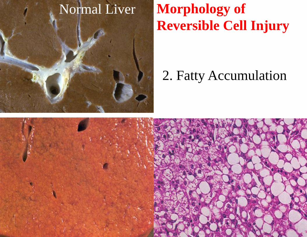
2. Fatty Accumulation
Normal Liver Morphology of
Reversible Cell Injury

15http://clinicalscienceblogyaseen.files.wordpress.com/2013/02/fatty-change-liver_steatosis.jpg

16

17

18

Fatty Change
• Fatty change or Steatosis represents the intracytoplasmic accumulation
of triglyceride (neutral fats) of parenchimal organs, such as: liver,
myocardium and kidney.
• Mechanisms : increase of free fatty acids (starvation, diabetes and
chronic ethylism/alcoholism), reduction of free fatty acids oxidation
(hypoxia, toxins, chronic ethylism/alcoholism), increase of esterification
of free fatty acids into triglycerides (due to increased free fatty acids or
reduction of their oxidation, chronic ethylism/alcoholism) and reduced
export of triglycerides due to deficiency of lipid binding apoprotein
(starvation/malnutrition, toxins). Initially, fatty change does not impair
the cells function, being reversible.19

2020
1. excess entry of free
fatty acid into the liver
2. Enhanced fatty acid
synthesis
3. Decreased fatty acid
oxidation
4. Increased esterification
of free fatty acid to
triglycerides
5. Decreased apoprotein
synthesis
6. Impaired secretion
from the liver

21
Necrosis
• A sequence of morphological changes that follow
cell death in a tissue
• There are 2 major processes
• 1. Enzymatic digestion of the cell
– From dead cells (autolysis)
– From invading inflammatory cells (heterolysis)
• 2. Denaturation of proteins

22
Types of Necrosis
Enzymatic necrosis vs. protein denaturation
Liquefactive necrosis Coagulation necrosis
Other patterns:
Gangrenous necrosis
Caseous necrosis
Fat necrosis

23
Coagulation Necrosis
• Most common
• Preserves the structural outline of tissues for days
• Acids denature structural proteins and enzymes
• Characteristic of hypoxic death of cells in all
tissues except brain

24
• Myocardial infarct
• Histology – 12-18 hours
– Eosinophilic, anuclear
fibers
– White blood cell infiltrate
• Gross – days 4-5
– Pale infarct
– Hyperemic border
Normal cardiac muscle
Coagulation Necrosis

25
Coagulation Necrosis

26
Coagulation Necrosis

27
Coagulation Necrosis
http://library.med.utah.edu/WebPath/jpeg4/ENDO051.jpg
The contrast between normal adrenal cortex and the small pale infarct

28
Liquefactive Necrosis
• Characteristic of focal
bacterial or fungal infections
• Attraction of inflammatory
cells generate the enzymatic
digestion pus
• Process obliterates structure
of the tissue
• Characteristic of necrosis in
CNS

29
Liquefactive Necrosis

30
Liquefactive Necrosis
Kidney:
• Focal fungal infection
• Site is filled with
white blood cells and
debris
• The abscess destroys
structural detail
31
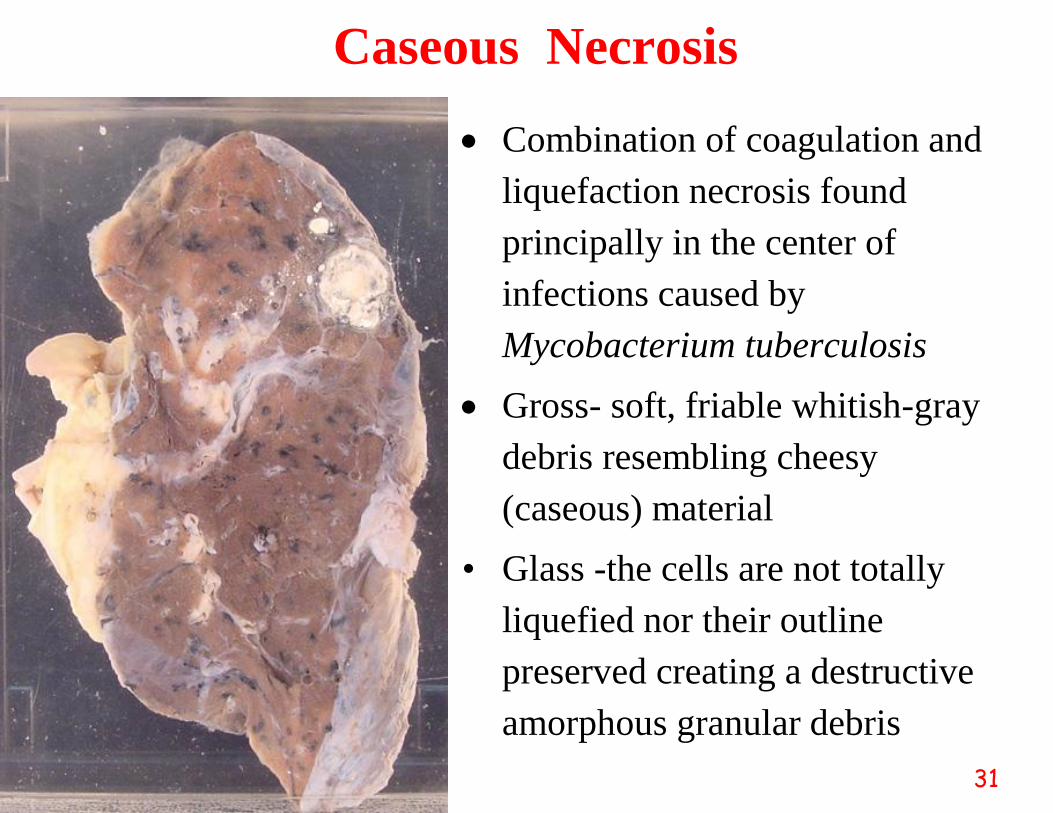
Caseous Necrosis
Combination of coagulation and
liquefaction necrosis found
principally in the center of
infections caused by
Mycobacterium tuberculosis
Gross- soft, friable whitish-gray
debris resembling cheesy
(caseous) material
• Glass -the cells are not totally
liquefied nor their outline
preserved creating a destructive
amorphous granular debris

32
In Granuloma (Tubercle) of Tuberculosis
Multinucleated Giant Cell (Langhan)
Caseous Necrosis
Johann Ferdinand Friedrich
Theodor Langhans
(1839-1915)
German pathologist

33https://secure.health.utas.edu.au/intranet/cds/pathprac/Files/Cases/Respiratory/Case36/Pictures36/G2.jpg

34https://secure.health.utas.edu.au/intranet/cds/pathprac/Files/Cases/Respiratory/Case36/Pictures36/G2.jpg

35https://secure.health.utas.edu.au/intranet/cds/pathprac/Files/Cases/Respiratory/Case36/Pictures36/G2.jpg

36https://secure.health.utas.edu.au/intranet/cds/pathprac/Files/Cases/Respiratory/Case36/Pictures36/G2.jpg

37https://secure.health.utas.edu.au/intranet/cds/pathprac/Files/Cases/Respiratory/Case36/Pictures36/G2.jpg

38https://secure.health.utas.edu.au/intranet/cds/pathprac/Files/Cases/Respiratory/Case36/Pictures36/G2.jpg

39
Fat Necrosis
Specific morphological pattern of cell death
encountered, when lipases escape into fatty depot
e.g., acute pancreatic necrosis
lipase
Fat (triglyceride) Glycerol + Fatty acid + cations
saponification
cation soaps (Ca++, Na+, K+ etc.)
(Amorphous granular basophilic deposits)

40
• Focal areas of fat necrosis in peritoneal cavity
– Caused by release of pancreatic enzymes
• Lipases
– Liquefy fat
– Hydrolyze triglyceride esters to release fatty acid
• Fatty acids combine with cations, e.g. calcium
– Fat saponification / chalky white areas
Fat Necrosis

41
Fat necrosis, acute pancreatitis - Inflammation necrosis causes the
release of lipases from the pancreatic acini. Acute pancreatitis is a
result of alcoholism or gall stones.
http://www.cuhk.edu.hk/med/paf/slides/necrosis/images/I3.jpg

42

43
Gangrenous Necrosis
* Not a distinctive pattern
* Usually refers to a limb which has lost its blood supply and has
subsequently been infected by bacteria
* When coagulative pattern is dominant the process may be termed
“dry gangrene” and when the liquefactive action is more
pronounced it may be designated as “wet gangrene”

44

45
How do You Tell a
Cell is Dead?
• Cytoplasm becomes
hypereosinophilic,
• Nuclear pyknosis,
karyorrhexis, and
karyolysis are the easiest
ways to tell that cells are
dead.

46
karyolysis

References
Kumar V, Abbas AK, Aster JC. Robbins Basic Pathology. 10th ed. Elsevier 2017.
Vinay Kumar
Stanley RobbinsAmerican Pathologist (1915–2003)1st ed Textbook of Pathology 1957
Abul K. Abbas
Jon C. Aster
The Internet Pathology Laboratory for Medical Education,
Mercer University School of Medicine
http://library.med.utah.edu/WebPath/


![Cell Injury[1]](https://static.fdocuments.net/doc/165x107/563dba79550346aa9aa5f218/cell-injury1-56a51a5ef0c98.jpg)