RX Herculink Elite® Renal and Biliary Stent System ...RX Herculink Elite® Renal and Biliary Stent...
37
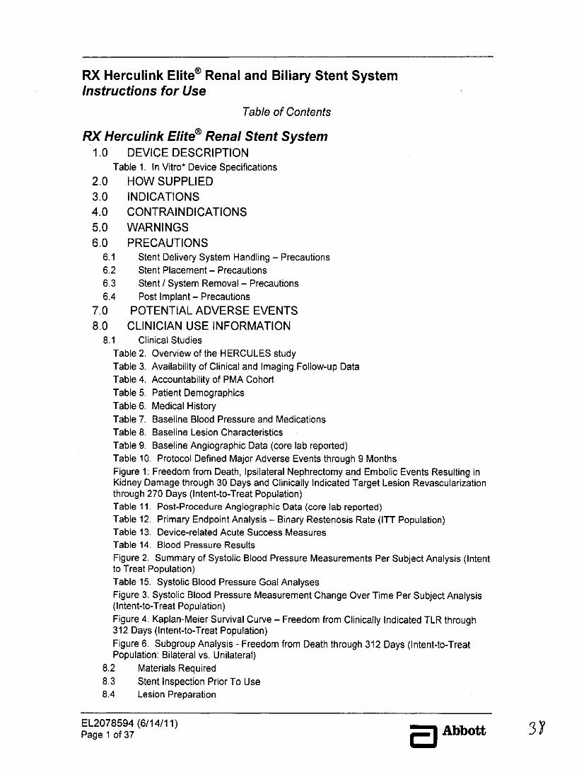
RX Herculink Elite® Renal and Biliary Stent System Instructions for Use Table of Contents RX Herculink Elite® Renal Stent System 1.0 DEVICE DESCRIPTION Table 1. In Vitro* Device Specifications 2.0 HOW SUPPLIED 3.0 INDICATIONS 4.0 CONTRAINDICATIONS 5.0 WARNINGS 6.0 PRECAUTIONS 6.1 Stent Delivery System Handling - Precautions 6.2 Stent Placement - Precautions 6.3 Stent / System Removal - Precautions 6.4 Post Implant - Precautions 7.0 POTENTIAL ADVERSE EVENTS 8.0 CLINICIAN USE INFORMATION 8.1 Clinical Studies Table 2. Overview of the HERCULES study Table 3. Availability of Clinical and Imaging Follow-up Data Table 4. Accountability of PMA Cohort Table 5. Patient Demographics Table 6. Medical History Table 7. Baseline Blood Pressure and Medications Table 8. Baseline Lesion Characteristics Table 9. Baseline Angiographic Data (core lab reported) Table 10. Protocol Defined Major Adverse Events through 9 Months Figure 1: Freedom from Death, Ipsilateral Nephrectomy and Embolic Events Resulting in Kidney Damage through 30 Days and Clinically Indicated Target Lesion Revascularization through 270 Days (Intent-to-Treat Population) Table 11. Post-Procedure Angiographic Data (core lab reported) Table 12. Primary Endpoint Analysis - Binary Restenosis Rate (ITT Population) Table 13. Device-related Acute Success Measures Table 14. Blood Pressure Results Figure 2. Summary of Systolic Blood Pressure Measurements Per Subject Analysis (Intent to Treat Population) Table 15. Systolic Blood Pressure Goal Analyses Figure 3. Systolic Blood Pressure Measurement Change Over Time Per Subject Analysis (Intent-to-Treat Population) Figure 4. Kaplan-Meier Survival Curve - Freedom from Clinically Indicated TLR through 312 Days (Intent-to-Treat Population) Figure 6. Subgroup Analysis - Freedom from Death through 312 Days (Intent-to-Treat Population: Bilateral vs. Unilateral) 8.2 Materials Required 8.3 Stent Inspection Prior To Use 8.4 Lesion Preparation EL2078594 (6/14/11) Page 1 of 37 5 Abbott
Transcript of RX Herculink Elite® Renal and Biliary Stent System ...RX Herculink Elite® Renal and Biliary Stent...
RX Herculink Elite® Renal and Biliary Stent SystemInstructions for Use
Table of Contents
RX Herculink Elite® Renal Stent System1.0 DEVICE DESCRIPTION
Table 1. In Vitro* Device Specifications2.0 HOW SUPPLIED3.0 INDICATIONS4.0 CONTRAINDICATIONS5.0 WARNINGS6.0 PRECAUTIONS
6.1 Stent Delivery System Handling - Precautions6.2 Stent Placement - Precautions6.3 Stent / System Removal - Precautions6.4 Post Implant - Precautions
7.0 POTENTIAL ADVERSE EVENTS8.0 CLINICIAN USE INFORMATION
8.1 Clinical StudiesTable 2. Overview of the HERCULES studyTable 3. Availability of Clinical and Imaging Follow-up DataTable 4. Accountability of PMA CohortTable 5. Patient DemographicsTable 6. Medical HistoryTable 7. Baseline Blood Pressure and MedicationsTable 8. Baseline Lesion CharacteristicsTable 9. Baseline Angiographic Data (core lab reported)Table 10. Protocol Defined Major Adverse Events through 9 MonthsFigure 1: Freedom from Death, Ipsilateral Nephrectomy and Embolic Events Resulting inKidney Damage through 30 Days and Clinically Indicated Target Lesion Revascularizationthrough 270 Days (Intent-to-Treat Population)Table 11. Post-Procedure Angiographic Data (core lab reported)Table 12. Primary Endpoint Analysis - Binary Restenosis Rate (ITT Population)Table 13. Device-related Acute Success MeasuresTable 14. Blood Pressure ResultsFigure 2. Summary of Systolic Blood Pressure Measurements Per Subject Analysis (Intentto Treat Population)Table 15. Systolic Blood Pressure Goal AnalysesFigure 3. Systolic Blood Pressure Measurement Change Over Time Per Subject Analysis(Intent-to-Treat Population)Figure 4. Kaplan-Meier Survival Curve - Freedom from Clinically Indicated TLR through312 Days (Intent-to-Treat Population)Figure 6. Subgroup Analysis - Freedom from Death through 312 Days (Intent-to-TreatPopulation: Bilateral vs. Unilateral)
8.2 Materials Required8.3 Stent Inspection Prior To Use8.4 Lesion Preparation
EL2078594 (6/14/11)Page 1 of 37 5 Abbott

8.5 Guide Wire Lumen Flush8.6 Stent Delivery System Preparation8.7 Stent Delivery Procedure8.8 Stent Deployment Procedure
9.0 REFERENCES10.0 PATENTS AND TRADEMARKS
RX Herculink Elite® Biliary Stent System1.0 DEVICE DESCRIPTION
Table 1. In Vitro* Device Specifications2.0 HOW SUPPLIED3.0 INDICATIONS4.0 CONTRAINDICATIONS5.0 WARNINGS6.0 PRECAUTIONS
6.1 Stent Delivery System Handling - Precautions6.2 Stent Placement - Precautions6.3 Stent / System Removal - Precautions6.4 Post Implant - Precautions
7.0 POTENTIAL ADVERSE EVENTS8.0 CLINICIAN USE INFORMATION
8.1 Materials Required8.2 Stent Inspection Prior To Use8.3 Stricture Evaluation / Biliary Drainage8.4 Stricture Pre-dilatation8.5 Guide Wire Lumen Flush8.6 Stent Delivery System Preparation8.7 Stent Delivery Procedure8.8 Stent Deployment Procedure
9.0 PATENTS AND TRADEMARKS
EL2078594 (6/14/11)Page 2 of 37 Abbott

RX Herculink Elitee Renal Stent System
CAUTION: Federal (USA) law restricts this device to sale by or on the orderof a physician.
1.0 DEVICE DESCRIPTION2.0 HOW SUPPLIED3.0 INDICATIONS4.0 CONTRAINDICATIONS5.0 WARNINGS6.0 PRECAUTIONS7.0 POTENTIAL ADVERSE EVENTS8.0 CLINICIAN USE INFORMATION9.0 REFERENCES10.0 PATENTS AND TRADEMARKS
1.0 DEVICE DESCRIPTION
The RX Herculink Elite Renal Stent System includes:
* A balloon expandable L605 cobalt chromium alloy stent pre-mounted on theballoon of a rapid exchange (RX) stent delivery system
* Two radiopaque markers located underneath the balloon which identify the stentposition and fluoroscopically mark the working length of the balloon
* Proximal shaft markers to aid with delivery catheter position, relative to a renalguiding catheter tip
* A third marker located approximately 30 cm from the center of the balloon thataids in locating the guide wire exit lumen and facilitating catheter removal andexchange
The delivery system can be utilized to optimize the stent wall apposition post stentdeployment.Table 1. In Vitro* Device Specifications
In Vitro* Rated RecommendedExnd Stent Stent Burst Recomm Minimum.Ster Lengths Deployment Pressure SheathDiameter GuiiCaheerI(mm) (mm) Pressure RBP uidinCheter ID Introducer"
(atm) (atm) (F)l(inches)(mm)4.0 12, 15, 18 11 14 6/0.067/1.70 5/0.071/1.804.5 12, 15, 18 11 14 6/0.067/1.70 5/0.071/1.805.0 12, 15, 18 11 14 6/0.067/1.70 5/0.071/1.805.5 12, 15, 18 11 14 6/0.067/1.70 5/0.071/1.806.0 12, 15, 18 11 14 6/0.067/1.70 5/0.071/1.806.5 12, 15, 18 11 14 6/0.067/1.70 5/0.071/1.807.0 15, 18 11 14 6/0.067/1.70 5/0.071/1.80
* All data provided is based on in vitro testing. Assure full deployment of the stent (see Clinician UseInformation section 8.8 Stent Deployment Procedure). Deployment pressures should be based on lesioncharacteristics." See individual manufacturer specifications for (F) equivalent.
EL2078594 (6/14/11)Page 3 of 37 Abbott

2.0 HOW SUPPLIED
Sterile. This device is sterilized with electron beam radiation. Non-pyrogenic. Do not useif the package is open or damaged.Storage. Store in a dry, dark, cool place.
Contents. One (1) RX Herculink Elite Renal Stent System, one (1) Protective/Regrooming sheath, one (1) flush tool
3.0 INDICATIONS
The RX Herculink Elite Renal Stent System is indicated for use in patients withatherosclerotic disease of the renal arteries following sub-optimal percutaneoustransluminal renal angioplasty (PTRA) of a de novo or restenotic atherosclerotic lesion (515 mm in length) located within 10 mm of the renal ostium and with a reference vesseldiameter of 4.0 - 7.0 mm. Suboptimal PTRA is defined as 50% residual stenosis, 20mmHg peak systolic or 10 mmHg mean translesional pressure gradient, flow-limitingdissection, or TIMI [Thrombolysis In Myocardial Infarction] flow < 3.
4.0 CONTRAINDICATIONS
The RX Herculink Elite Renal Stent System is contraindicated for use in:* Patients with a contra indication for antiplatelet/anticoagulant therapy.* Patients who have a lesion that cannot be crossed with a wire or a balloon
angioplasty catheter.* Patients with bleeding disorders.* Patients with a known hypersensitivity to cobalt or chrome.* Target lesions that are resistant to complete balloon inflation.
* Stenting of an arterial vessel where leakage from the artery could be exacerbatedby placement of a stent.
* Patients with a target lesion with a large amount of adjacent acute or subacutethrombus
6.0 WARNINGS
The long term safety and effectiveness of this device for use in the renal arterial systemhave not been established.
Should unusual resistance be felt at any time during lesion access or Delivery Systemremoval, the introducer sheath I guiding catheter and stent system should be removed asa single unit. Applying excessive force to the Stent Delivery System can potentiallyresult in loss or damage to the Stent and Delivery System components. (SeeStent / System Removal - Precautions.)
Since the use of this device carries the associated risk of subacute thrombosis, vascularcomplications and / or bleeding events, judicious selection of patients is necessary.
Stenting across a major bifurcation may hinder or prevent future diagnostic or therapeuticprocedures.
Once fully deployed, the stent cannot be repositioned.
EL2078594 (6/14/11)Page 4 of 37 Abbott

Persons allergic to L605 cobalt chromium alloy may suffer an allergic reaction to thisimplant.
Only physicians familiar with the complications, side effects and hazards commonlyassociated with renal stent placement should use this device.
The RX Herculink Elite Renal Stent System is intended to perform as a system. Thestent should not be removed for use in conjunction with other dilatation catheters, norshould the RX Herculink Elite Renal Stent System be used in conjunction with otherstents.
The safety and effectiveness of multiple overlapping stents have not beenestablished. However, when multiple stents are required, stent materials shouldbe of similar composition.
6.0 PRECAUTIONS
6.1 Stent Delivery System Handling - Precautions
* For single use only. Do not resterilize or reuse. Note product "Use by" date.
* Do not remove stent from its delivery balloon, as removal may damage thestent and I or lead to stent embolization.
* Carefully inspect the RX Herculink Elite Renal Stent System prior to use to verifythat the stent has not been damaged in shipment and that the device dimensionsare suitable for the specific procedure. Take care to avoid unnecessary handling.
* Refer to the instructions for use supplied with any interventional devices to beused in conjunction with the RX Herculink Elite Renal Stent System, for theirintended uses, contraindications, and potential complications.
* Special care must be taken not to handle or in any way disrupt the stent on theballoon. This is most important during stent system removal from the packaging,placement over a guide wire and advancement through a guiding catheter orintroducer sheath.
* Do not "roll" the mounted stent with your fingers as this action may loosen thestent from the delivery balloon.
* Use only the appropriate balloon inflation media. Do not use air or any gaseousmedium to inflate the balloon as this may cause uneven expansion and difficultyin deployment of the stent.
6.2 Stent Placement - Precautions
* Do not prepare or pre-inflate balloon prior to stent deployment other than asdirected. Use balloon purging technique described in the Clinician UseInformation section.
* The inflated balloon diameter of the system used to deploy the stent shouldapproximate the diameter of the vessel. Oversizing of the stent can result in aruptured vessel. To ensure full expansion of the stent, the balloon should beinflated to a minimum of nominal pressure.
EL2078594 (6/14/11)Page 5 of 37 Abbott

* Implanting a stent may lead to dissection of the vessel distal and I or proximal tothe stent and may cause acute closure of the vessel requiring additionalintervention (surgical intervention, further dilatation, placement of additionalstents, or other).
* Do not expand the stent if it is not properly positioned in the vessel. (See StentISystem Removal - Precautions.)
* Stenting across a major bifurcation may hinder or prevent future side branchaccess.
* Balloon pressures should be monitored during inflation. Do not exceed RatedBurst Pressure (RBP) as indicated on product label. Use of pressures higherthan specified on product label may result in a ruptured balloon with possiblevessel damage or perforation.
* Stent retrieval methods (use of additional wires, snares and I or forceps) mayresult in additional trauma to the vasculature and I or the vascular access site.Complications may include bleeding, hematoma or pseudoaneurysm.
* The RX Herculink Elite Renal Stent System is intended for deployment and post-deployment dilatation of the stent only and should not be used to dilate otherlesions.
* Do not attempt to pull an unexpanded stent back through the introducersheath I guiding catheter; dislodgment of the stent from the balloon mayoccur.
6.3 Stent / System Removal - Precautions
Should unusual resistance be felt at any time during either lesion access or removal ofthe Delivery System post-stent implantation, the entire system should be removed as asingle unit.
When removing the Delivery System as a single unit:
* DO NOT retract the Delivery System into the introducer sheath / guiding catheter.
* Position the proximal balloon marker just distal to the tip of the introducersheath I guiding catheter.
* Advance the guide wire in the anatomy as far distally as safely possible.
* Secure the Delivery System to the introducer sheath / guiding catheter; thenremove the introducer sheath / guiding catheter, guide wire and Delivery Systemas a single unit.
Failure to follow these steps and I or applying excessive force to the Delivery System canpotentially result in loss or damage to the stent and / or Delivery System components.
If it is necessary to retain guide wire position for subsequent vessel access, leave theguide wire in place and remove all other system components.
6.4 Post Implant - Precautions
Great care must be exercised when crossing a newly deployed stent with a guide wireor balloon catheter to avoid disrupting the stent geometry.
EL2078594 (6/14/11) A o2Pag 6m.f37t Ahhott I0

Magnetic Resonance Imaging (MRI)
Non-clinical testing has demonstrated that the Herculink Elite stent, in single and inoverlapped configurations up to 33 mm in length, is MR Conditional. It can be scannedsafely under the following conditions:
* Static magnetic field of 1.5 or 3 Tesla
* Spatial gradient field of 2500 Gauss/cm or less
* Maximum whole-body-averaged specific absorption rate (SAR) of 2.0 W/kg(normal operating mode) for any duration of MRI scan that would otherwise besafe for the patient without implant.
MRI at 1.5 or 3 Tesla may be performed immediately following the implantation of theHerculink Elite stent.
The Herculink Elite stent should not migrate in this MRI environment. Magnetic force onthe Herculink Elite state was tested according to ASTM F2052-06e. Non-clinical testingat field strengths greater than 3 Tesla has not been performed to evaluate stent migrationor heating.
Stent heating was derived by using the measured non-clinical, in vitro temperature riseaccording to ASTM F2182-09 in a GE Signa HDx 3 Tesla scanner and in a GE 1.5 Teslacoil in combination with the local specific absorption rates (SARs) in a digitized human 'heart model. The temperature rise was derived by a validated calculation. At overlappedlengths up to 33 mm, the Herculink Elite stent produced a non-clinical maximum localtemperature rise of less than 30C at a maximum whole body averaged SAR of 2.0 W/kg(normal operating mode) for an MRI sequence of 15 minutes. These calculations do nottake into consideration the cooling effects of blood flow.
The effects of MRI on overlapped stents greater than 33 mm in length or stents withfractured struts are unknown.Image artifact may be present when scanning the Herculink Elite stent as demonstratedin non-clinical testing performed according to ASTM F2119-07 in a GE Signa HDx 3Tesla scanner. The image artifact (both inside and outside the device lumen) extendsapproximately 7 mm from the device using the spin echo sequence (TR = 500 ms; TE =20 ms; flip angle = 900) and 13 mm from the device using the gradient echo sequence(TR = 100 ms; TE = 15 ms; flip angle = 30*). MR image quality may be compromised ifthe area of interest is in the exact same area, or relatively close to, the position of theHerculink Elite stent. Therefore, it may be necessary to optimize the MR imagingparameters in the presence of Herculink Elite stents.
EL2078594 (6/14/11) 5bPage 7 of 37 Abbot

7.0 POTENTIAL ADVERSE EVENTS
Potential complications associated with percutaneous renal artery treatment including theuse of a renal stent may include, but are not limited to, the following:
* Abscess* Allergic reaction to Cobalt Chromium or contrast agents* Arrhythmias (ventricular fibrillation, ventricular tachycardia, other)* Arteriovenous fistula* Bowel infarct* Death* Dialysis* Dissection* Drug reaction to antiplatelet agents* Drug reaction, allergic reaction to contrast media* Emboli (air, tissue, or thrombotic emboli) resulting in tissue ischemia/infarction* Emergency surgery to correct vascular complications* Emergent renal artery bypass surgery* Extremity ischemia/amputation* Fever* Gastrointestinal symptoms from anticoagulation/antiplatelet medication* Hematoma at vascular access site* Hemorrhage requiring transfusion* Hypersensitivity reactions* Hypotension/hypertension* Infection and pain at vascular access site* Intimal tear* Kidney infarct* Myocardial infarction* Myocardial ischemia* Nephrectomy* Peripheral neuropathy* Pseudoaneurysm at vascular access site* Pseudoaneurysm formation* Renal artery thrombosis, aneurysm, rupture, perforation, occlusion, spasm, or
restenosis* Renal insufficiency or failure* Stent migration or embolization* Stent misplacement* Stroke/cerebral vascular accident* Tissue necrosis or ulceration
EL2078594 (6/14/11) AbtPage 8 of 37 Abbott

8.0 CLINICIAN USE INFORMATION
8.1 Clinical Studies
Results from a multi-center clinical study (IDE G060067, also known as the HERCULESstudy) demonstrate the safety and effectiveness of the RX Herculink Elite Renal StentSystem. Specifically, the 9-month binary restenosis rate was 10.5%, meeting theperformance goal of 28.6%. In addition, use of the RX Herculink Elite Renal StentSystem was associated with a low rate of major adverse events (MAEs), high technicaland procedural success rates, improvement in hypertension, and maintenance of renalfunction. An overview of the HERCULES study is presented in Table 2.
Table 2. Overview of the HERCULES study
Device RX Herculink Elite Renal Stent System
Study Design Non-randomized, prospective, single-arm, multi-center clinicalstudy
Patients Enrolled 202 (76 male and 126 female)
Number of Sites 37 investigational sites
Primary Endpoint Binary restenosis rate at 9 months, defined as 60% diameterstenosis, measured by duplex ultrasound (or quantitativeangiography, if necessary) by an independent core laboratory.Analysis was done on a per-lesion basis.
Secondary MAEs: A composite safety endpoint of the following indices: deathEndpoints for any reason at 30 days; ipsilateral nephrectomy at 30 days;
embolic events resulting in kidney damage at 30 days; andclinically indicated target lesion revascularization (TLR) up to 9months (270 days).
Change in blood pressure: Systolic and diastolic blood pressureswere measured at baseline, post-procedure, 1, 6 and 9 months andthe group average at each time point reported. The change at 9months from baseline, for systolic and diastolic pressure, wascalculated for each enrolled patient. Confidence intervals (95%) forthe average changes were reported. In addition, systolic bloodpressure was reported using categories of < 140 mmHg, a 140mmHg and < 160 mmHg, a 160 mmHg and < 180 mmHg, and a180 mmHg.Change in Antihypertensive Medications: Number and type ofantihypertensive medications at baseline, 1, 6, and 9 months werecollected. The group average number of antihypertensivemedications at each time point was reported. In addition, data wasreported as a percentage of patients on 1, 2, 3, or 4 or moremedications at baseline, 1, 6, and 9 months.
Change in Renal Function: Defined as change in serum creatinine(sCr), it was measured at baseline, post-procedure, 1, 6 and 9months, with the group average reported at each time point. Thechange at 9 months from baseline was calculated for each enrolledpatient.
EL2078594 (6/14/11) AbtPage 9 of 37 Abbott

Acute Device Success: The achievement of successful deliveryand deployment of the assigned device(s) and successful removalof the delivery system as intended to the designated location. Itwas reported as a percentage of devices that were attempted to beimplanted.
Acute Clinical Success: Procedure success without major adverseevents (MAE) or access site events requiring surgical orpercutaneous intervention prior to hospital discharge. It ispresented on a per subject basis.Clinically Indicated Target Lesion Revascularization: Defined asdiameter stenosis 60%, as determined by the angiographic orultrasound core laboratory, and any revascularization (including butnot limited to atherectomy, embolectomy, endarterectomy, bypasssurgery, repeat angioplasty, or stent implantation) to the targetlesion. Freedom from clinically indicated TLR was evaluated at 9months and is presented as time to first event on a per-lesionbasis.
Acute Procedural Success: The attainment of a final result of <30% residual stenosis as determined by the angiographic corelaboratory, reported as a percentage of treated lesions.
Study Hypothesis Renal arteries treated with the RX Herculink Elite Stent will have abinary restenosis rate at 9 months that meets a performance goalof 28.6%.
Patient Follow-up 1-month clinic visit: Blood pressure measurement, anti-hypertensive medication review, laboratory (sCr, BNP assay),duplex ultrasound, laboratory, adverse events.
6-month clinic visit: Blood pressure measurement, anti-hypertensive medication review, adverse events, laboratory (sCr).
9-month clinic visit with ultrasound: Blood pressure measurement,anti-hypertensive medication review, adverse events, laboratory(sCr), duplex ultrasound, angiogram (only if duplex ultrasound doneat nine-month follow up visit is determined to be "not evaluable", orat the discretion of the Investigator).
12-month clinic visit: Blood pressure measurement, anti-hypertensive medication review, adverse events, laboratory (sCr).24-month clinic visit: Blood pressure measurement, anti-hypertensive medication review, adverse events, laboratory (sCr),duplex ultrasound.
36-month clinic visit: Blood pressure measurement, anti-hypertensive medication review, adverse events, laboratory (sCr),duplex ultrasound.
EL2078594 (6/14/11)Page 10 of 37 Abbott

Study DesignThe primary objective of the clinical study was to assess the binary restenosis rate at 9months after stenting, with binary restenosis determined by duplex ultrasound orangiogram. The lesion was considered restenotic if the lesion was ? 60% diameter priorto the 9-month primary endpoint evaluation. Specifically, the primary hypothesis was thatthe binary restenosis rate at 9 months on a lesion basis meets the performance goal of28.6% (i.e., < 28.6%). Secondary analyses included MAEs (defined as death for anyreason at 30 days, ipsilateral nephrectomy at 30 days, clinically-indicated target lesionrevascularization up to 9 months, and embolic events resulting in kidney damage at 30days), acute device success, acute procedural success, acute clinical success, changesin blood pressures at 9 months, change in anti-hypertensive medication in-take at 9months, and renal function outcomes (measured by serum creatinine).
The HERCULES study was a prospective, non-randomized, multi-center, single-armclinical study to demonstrate efficacy and safety of the RX Herculink Elite Renal StentSystem (Herculink Elite) in the treatment of suboptimal post-procedural percutaneoustransluminal renal angioplasty (PTRA) of atherosclerotic de novo or restenotic renalartery stenoses in patients with uncontrolled hypertension. Subjects were eligible toenroll in the study if they had a suboptimal angioplasty (defined as ? 50% residualstenosis, 20 mmHg systolic or 10 mmHg mean translesional pressure gradient, flow-limiting dissection or TIMI [Thrombolysis In Myocardial Infarction] flow < 3) for de novo orrestenotic renal artery lesions (5 15 mm length) due to atherosclerosis originating within10 mm of the renal ostium, a reference vessel diameter 4 mm and 7 mm, anduncontrolled hypertension (defined as systolic blood pressure [SBP] a 140 mmHg ordiastolic blood pressure [DBP] 2 90 mmHg on at least two anti-hypertensivemedications). Treatment of two lesions, one per side, was allowed per patient. Patientswere excluded from the study if they met any of the following conditions: only onefunctioning kidney, kidney transplant, had a Q-wave myocardial infarction (MI) within 30-days of the index procedure, or serum creatinine a 2.5 mg/dl. Angiography wasperformed post stent implantation. Follow-up included ultrasound imaging at 1 month,and ultrasound (angiogram, if ultrasound was not interpretable) at 9 months. Clinicalassessments (e.g., blood chemistry, blood pressure, assessment of anti-hypertensivemedications) were scheduled for 1, 6, 9, 12, 24, and 36 months.
Independent core laboratories analyzed angiographic and ultrasonic imaging. The studywas overseen by an independent data safety monitoring board (DSMB) comprised ofphysicians and a biostatistician. The committee was set up and convened at the pre-specified interim time points of the trial to evaluate the safety of the trial. A ClinicalEndpoint Adjudication Committee (CEAC) adjudicated the suspected cases of peri-procedural death, ipsilateral nephrectomy and embolic events that result in kidneydamage.
The principal effectiveness of the Herculink Elite was evaluated to determine if the binaryrestenosis rate at 9 months was less than the performance goal (PG) of 28.6% that wasderived from the contemporary literature of PTRA and stenting for the atheroscleroticlesions in the aortorenal ostium. The sample size and power calculation were based onthe primary endpoint of binary restenosis rate at 9 months. The PG for the binaryrestenosis rate at 9 months was determined to be 28.6%, based on pre-market studies ofother renal artery stents used in a similar manner and which are now PMA-approved.
EL2078594 (6/14/11) L#Page 11 of 37 Abbott
The primary and secondary endpoints measuring the safety and effectiveness of thedevice and procedure are described below.
Null Hypothesis: the 9-month binary restenosis rate, y, is greater than or equal to 28.6%.(Interpretation: the binary restenosis rate does not meet a performance goal of 28.6%.)
Ho: y a 28.6%
Alternative Hypothesis: the 9-month binary restenosis rate, y, is less than 28.6%.(Interpretation: the binary restenosis rate meets a performance goal of 28.6%.)
Ha: y < 28.6%.
The null hypothesis was tested using one-sided exact binomial test at significance levelof 0.05.
For binary and categorical variables, event counts, rates and their confidence intervalswere analyzed. Continuous variables were analyzed using the mean, standard deviation,and 95% confidence intervals (CI). Kaplan-Meier methodology was used to estimate thedistributions of time-to-event variables.
Clinical Inclusion and Exclusion Criteria
The inclusion criteria for the HERCULES study were as follows:* Subject is 2 18 years of age.* Subject and subject's physician agree to have the subject return for all required
contact following study enrollment.* Subject has been informed of the nature of the study, and has provided written
informed consent, approved by the appropriate Institutional Review Board (IRB) ofthe respective clinical site.
* Subject is a candidate for renal artery stenting.* Subject has uncontrolled systolic hypertension (systolic BP & 140 mmHg), or
uncontrolled diastolic hypertension (diastolic BP 90 mmHg), or a combination ofboth in the presence of at least two (2) or more antihypertensive medications.
* Baseline sCr of 5 2.5 mg/dl.* Subject has unilateral or bilateral de novo or restenotic after PTA (in-stent
restenosis excluded) atherosclerotic lesion(s). If bilateral lesions are to be treated,the most severe lesion must be successfully treated without complications beforeprogressing to treat the second lesion. Treatment of bilateral lesions is to occur inthe same procedural event.
* Renal stenosis must be visually estimated to be 60% by angiography.* Subject has a suboptimal PTA result, defined as one of the following:
- a 50% residual stenosis,- 10 mm Hg mean gradient or 20 mm Hg peak systolic gradient across the
target lesion, or- A flow-limiting dissection (NHLBI grade D) or TIMI flow < 3.
* Renal stenosis must be visually estimated to be within 10 mm of the aortic renalborder by angiography.
* Target vessel reference diameter must be visually estimated to be 4 mm and 57 mm by angiography.
EL2078594 (6/14/11)Page 12 of 37 m Abbott

* Target lesion length must be visually estimated to be 5 15 mm (includingdissection) by angiography.
* Expected ability to deliver the stent to the lesion (absence of excessive tortuosityor calcification).
* Expected ability to fully expand the stent.
The exclusion criteria for the HERCULES study were as follows:* Known hypersensitivity or contraindication to cobalt chromium or standard
intraprocedure anticoagulant(s); sensitivity to contrast which cannot beadequately pre-treated with medication.
* Known allergy or contraindication to clopidogrel (Plavixo) or aspirin.* Bleeding disorder or hypercoagulable disorder, or will refuse blood transfusions.* Gastrointestinal (GI) bleeding within 30 days before the index procedure that
would interfere with antiplatelet therapy.* Renal insufficiency, defined as serum Creatinine > 2.5 mg/dl.* Any immunosuppressive disorder, access site infection, or acute systemic
infection due to any cause.* Medical illnesses (e.g., cancer, end-stage congestive heart failure) that may
cause the subject to be non-compliant with protocol requirements, confound thedata interpretation, or is associated with a life expectancy of less than three years.
* Medical illnesses that would make them unlikely to respond to treatment (e.g.,sickle cell nephropathy/sickle cell disease, scleroderma, arteriolarnephrosclerosis, hemolytic-uremic syndrome and vasculitis).
* Q-wave MI within 30 days before index procedure.* Stroke or transient ischemic attack (TIA) within 30 days before index procedure.* History of congestive heart failure and has a previously documented left
ventricular ejection fraction (LVEF) < 25%.* Subject is normotensive or has adequate control of hypertension (SBP <140 mm
Hg and DBP <90 mm Hg) utilizing diet control and/or medication regimeninvolving only one antihypertensive medication.
* Acute thrombophlebitis or deep vein thrombosis.* Actively participation in another drug or device trial and did not complete the
required protocol follow-up period. Subject may be enrolled only once in this studyand may not participate in any other clinical trial during the follow-up period.
* Unable to understand and cooperate with study procedures or provide informedconsent.
* Unable to return for follow-up visits.* Subject is pregnant.* Subject has undergone vascular surgery, such as coronary artery bypass grafting,
abdominal aortic aneurysm repair, or aorto-femoral bypass, and has not fullyrecovered from the effects of surgery (<3 months).
* Subject has planned staged treatment of bilateral renal artery stenosis.* Subject has had prior surgical intervention to the target artery, or has undergone
previous stent placement in the target lesion.* Target lesion is located in a transplanted kidney.* Kidney to be treated is < 8 cm as determined by duplex ultrasound report,
computed tomography angiography (CTA) report, or magnetic resonanceangiography (MRA) report within 180 days before procedure. If kidney size is
EL2078594 (6/14/11) AbtPage 13 of 37 Abbott

documented by more than one method, e.g. CTA and ultrasound, and one of themethods is duplex ultrasound, the kidney size as documented by duplexultrasound shall be used to determine study eligibility.
* Subject has planned additional ancillary procedure(s) during renal stentingprocedure.
Angiographic Exclusion Criteria:* Subject has a lesion segment, including dissection, >15 mm in length.* Requirement for more than 1 stent to treat full length of lesion and dissection.* Target lesion has a total (100%) occlusion.* Evidence of thrombus or mobile filling defect in the target lesion or vessel.* Co-existing aneurysmal or occlusive disease of the abdominal aorta requiring
surgical reconstruction during the follow-up period.* Fibromuscular dysplasia.* Subject artery has patent bifurcation within 10 mm of ostium that might be
covered by placement of a stent.* The target lesion is within the artery of a solitary functioning kidney or, the subject
has a contralateral totally occluded renal artery.* For planned treatment of bilateral lesions: the more critical lesion, i.e. lesion with
the greater stenosis (which should be treated first), is either treatedunsuccessfully or requires a bailout procedure. (NOTE: Less critical lesion isexcluded at this point.) .
Follow-up Schedule
Clinical assessments occurred at baseline, operative/discharge and postoperativeintervals at 1, 6, 9 months and 1, 2, and 3 years, as seen in Table 3.
Table 3. Availability of Clinical and Imaging Follow-up DataFollow-up Period Clinical Evaluations1-month clinic visit Blood pressure measurement, anti-hypertensive medication review,
laboratory (sCr, BNP assay), duplex ultrasound, laboratory, adverseevents.
6-month clinic visit Blood pressure measurement, anti-hypertensive medication review,adverse events, laboratory (sCr).
9-month clinic visit Blood pressure measurement, anti-hypertensive medication review,with ultrasound adverse events, laboratory (sCr), duplex ultrasound, angiogram (only if
duplex ultrasound done at nine-month follow up visit is determined to be"not evaluable", or at the discretion of the Investigator).
12-month clinic visit Blood pressure measurement, anti-hypertensive medication review,adverse events, laboratory (sCr)
24-month clinic visit Blood pressure measurement, anti-hypertensive medication review,adverse events, laboratory (sCr), duplex ultrasound.
36-month clinic visit Blood pressure measurement, anti-hypertensive medication review,adverse events, laboratory (sCr), duplex ultrasound.
EL2078594 (6/14/11)Page 14 of 37 m Abbott

Clinical Endpoints
With regards to safety, there were no hypothesis-driven primary or secondary safetyendpoints.
With regards to effectiveness, the primary effectiveness endpoint was evaluatedaccording to the hypothesis that renal arteries treated with the RX Herculink Elite RenalStent will have a binary restenosis rate at 9 months on a lesion basis that meets aperformance goal of 28.6%. The formulation of the hypothesis can be found in the studydesign section. The study was considered a success if the primary effectivenesshypothesis was met.
Accountability of PMA Cohort
Patient availability for study follow-up through 9 months is summarized in Table 4. Thefirst subject was enrolled into the HERCULES study on August 31, 2007 and the lastsubject was enrolled on October 2, 2009. Over this period, 202 male and femalesubjects, who met the eligibility criteria and agreed to participate in the study and signedan informed consent, were enrolled at 37 study sites in the United States (US). Thirty-nine (39) subjects had bilateral lesions treated. Therefore, the intention-to-treat (ITT)population consists of 202 subjects and 241 lesions.
The last 9-month follow-up visit was completed on June 23, 2010. One subject did nothave any post-procedure follow-up, a second subject was confirmed as lost-to-follow upat 182 days, and 4 subjects were withdrawn from the study. Through the 9 month follow-up 5 subjects expired. Three subjects had a study stent attempted without success, anda non-study stent implanted; they completed the study at the 30 day follow-up timepoint.Therefore, there were a total of 189 subjects eligible for the 9 month follow-up visit.
Table 4. Accountability of PMA Cohort1-month visit 6-month visit 9-month visit
Death 0 4 1
Withdrawn 0 0 4Lost to follow-up 0 1 030-day follow-up complete for non-study stent 0 3 0Eligible 202 194 189
Study Population Demographics and Baseline Parameters
Patient demographics (Table 5), medical history (Table 6), baseline blood pressure andmedications (Table 7) and baseline lesion characteristics (Table 8) and baselineangiographic data (Table 9) were consistent with patient populations described inpublished literature of renal stent intervention. The mean age of the study populationswas 72.1 ± 9.4, with 62.4% female gender. The key risk factors include diabetes mellitus45.0%, hypercholesterolemia 86.1%, coronary artery disease 67.3% and current orformer tobacco use 56.9%. The baseline mean creatinine was 1.2 ± 0.4 mg/dl and61.5% of subjects had an estimated baseline glomerular filtration rate (eGFR) of < 60ml/min per 1.73 m2 (Table 6).
EL2078594 (6/14/11)Page 15 of 37

Table 5. Patient Demographics
Demographic Value (N = 202 patients)Sex
Male 37.6% ( 76/202)Female 62.4% (126/202)
Age (years, mean ± SD (range)) 72.1 t 9.4 (44 - 89)Ethnicity
Caucasian or White 83.7% (169/202)Black or African American 7.9% (16/202)
Hispanic or Latino 7.4% (15/202)Other 1.0% (2/202)
Native Hawaiian or other Pacific Islander 0.5% (1/202)Asian 0.5% (1/202)
Table 6. Medical History
Past or Current Medical Condition Percent Patients(numberltotal number)
DiabetesTotal 45.0% (91/202)
Type I 3.5% (7/202)Type II 41.6% (84/202)
HypercholesterolemiaTotal 86.1% (174/202)
Requiring medication 82.2% (166/202)Not Requiring medication 4.0% (8/202)
Coronary Artery Disease 67.3% (136/202)Brain Natriuretic Peptide (BNP) Level (pg/ml) 181.2 ± 297.0 (192)Renal Function
Serum Creatinine (mg/dl) 1.2 ± 0.4 (202)eGFR < 60 mlmin per 1.73 m 61.5% (123/200)
Former Smoker 44.1% (89/202)Current Smoker 12.9% (26/202)
Table 7. Baseline Blood Pressure and Medications
Past or Current Medical Condition Percent Patients(numberltotal number)
Blood PressueMean Systolic 162.3 ± 18.5 (202)
Mean Diastolic 77.7 ± 11.5 (202)Anti-Hypertensive Medications
1 Medication 0.5% (1/202)2 Medications 29.2% (59/202)3 Medications 30.7% (62/202)4 Medications 39.6% (80/202)
EL2078594 (6/14/11)Page 16 of 37 9 Abbott 53

Table 8. Baseline Lesion Characteristics
Characteristic Percent Lesions(numbertotal number)
Lesion LocationRight Renal Artery 52.3% (126/241)
Left Renal Artery 47.7% (115/241)Type of Lesion
Restenotic . 0% (0/115)de novo 100% (241/241)
Target Lesion LocationOstial 67.6% (163/241)
Within 10 mm of Ostium 32.4% (78/241)Renal Arteries Treated
Unilateral 67.6% (163/241)Bilateral 32.4% (78/241)
Suboptimal PTA OutcomeResidual Stenosis 2 50% 90.7% (214/236)
10 mmHg mean gradient or 20 mmHg peak systolic gradier 25.0% ( 59/236)Flow-limiting dissection (NHLBI grade D) or TIMI flow < 3 4.7% (11/236)
Target Lesion Stenosis 81.3 ± 10.0 (241)
Table 9. Baseline Angiographic Data (core lab reported)Measure Mean ± SD (range, total lesions)
Lesion Length (mm) 8.5 ± 3.1 (1.8 - 22.1, n = 241)
Pre-procedureRVD (mm) 5.34 ± 1.1 (2.9 - 7.9, n = 241)MLD (mm) 1.8 ± 0.7 (0.3 - 3.8, n = 241)
Percent Diameter Stenosis (%) 65.9 ± 11.4 (37.8 - 92.9, n = 241)
Safety and Effectiveness Results
Safety Results
The safety endpoint for the HERCULES study is the composite rate of freedom frommajor adverse events (MAEs), including all cause death, ipsilateral nephrectomy, andembolic events resulting in kidney damage, all through 30 days, and clinically indicatedtarget lesion revascularization (TLR) up to 270 days. Through the first 30 days, therewas 1 death and 2 embolic events resulting in kidney damage. The freedom from MAEsand clinically indicated TLRs at 9 months was 94.8%, as shown in Table 10 and theKaplan-Meier analysis (Figure 1).
EL2078594 (6/14/11)Page 17 of 37 2 Abbott

Adverse effects that occurred in the PMA clinical study
Table 10. Protocol Defined Major Adverse Events through 9 Months
MAEs Through 30-Days (N=202 Subjects) [95% Cl](N= 241 Lesions)All-cause death 0.5% [0.0%, 2.7%]
Device-related death 0.0% [0.0%, 1.8%]
Embolic events resulting in kidney damage 1.0% [0.1%, 3.5%]
Nephrectomy 0.0% [0.0%, 1.8%]
Clinically indicated TLR 0.0% [0.0%, 1.8%]
Stent thrombosis 0.0% [0.0%, 1.8%]
Retroperitoneal bleed 0.5% (0.0%, 2.7%]
Myocardial Infarction 0.0% [0.0%, 1.8%]
Stroke 0.0% [0.0%, 1.8%]
Events Through 9 Months (312 Days) (N=202 Subjects) [95% CI](N= 241 Lesions
Renal Failure 1.0% [0.1%, 3.5%]
Renal Insufficiency 2.0% [0 5%, 5 0%]
2 (N-_202 Subjects)Events Through 9 Months (312 Days) [95% C](N= 241 Lesions
All TLR 7.4% [3.9%, 10.9%]
Clinically Indicated TLR 5.9% [2.6%, 9.2%]
Death 2.6% [0.3%, 5.0%]'By Clopper Pearson exact confidence interval2Based on Kaplan Meier estimate
EL2078594 (6/14/11)Page 18 of 37 Abbott

Figure 1: Freedom from Death, Ipsilateral Nephrectomy and Embolic EventsResulting in Kidney Damage through 30 Days and Clinically Indicated TargetLesion Revascularization through 270 Days (Intent-to-Treat Population)
5a%-
0 20 40 60 80 10 120 140 160 1W 200 220 240 260 280
Days Pbst Index Procdure
Days Post Index Procedure 0 (0, 30] (30, 180] (180, 270]
Subjects at Risk 202 201 198 191
Subjects Censored 0 1 5 186
Number of Events 1 2 2 5Event Free (%) 99.5% 98.5% 97.5% 94.8%
Standard Error () 0.5% 0.9% 1.1% 1.6%
Effectiveness Results
Two hundred forty-two (242) RX Herculink stents were placed to treat 241 renal arterylesions. By core lab assessment, the mean post-procedure percent diameter stenosiswas 3.4% (Table 11). Therefore, the RX Herculink Elite Renal Stent System waseffective in establishing patency at the conclusion of the procedure.
Table 11. Post-Procedure Angiographic Data (core lab reported)
Measure Mean ± SD (range, total lesions)Post-procedure
MLD (mm) 5.1 ± 0.8 (3.4 - 7.3, n=240)Percent Diameter Stenosis () 3.4 ± 12.5 (-43.1 - 29.8, n=240)
EL2078594 (6/14/11)Page 19 of 37 Abbott

Primary Endpoint
The 9-month binary restenosis rate met the performance goal (Table 12). The 9-monthbinary restenosis rate was 10.5% and the upper limit of the 95% one-sided confidenceinterval was 14.7%, which is below the pre-specified performance goal of 28.6% (p <0.0001). These results demonstrate the effectiveness of the RX Herculink Elite RenalStent System in treating atherosclerotic lesions of the renal arteries following suboptimalangioplasty.
Table 12. Primary Endpoint Analysis - Binary Restenosis Rate (ITT Population)Measure Mean ± SD (range, total lesions)9-Month In-Stent Binary Restenosis (? 60%) Rate 10.5% (22/209)[One-Sided 95% Conf. Interval] 14.7%]One-Sided Exact Binomial Test p-value2 < 0.0001
By Clopper-Pearson exact confidence interval2 One-sided p-value is computed using the exact binomial test with the objective performance criterion (OPC) of 28.6%
Secondary Endpoints
Secondary endpoint analyses included MAEs, device-related success measures (i.e.,acute device success, acute procedural success, and acute clinical success), bloodpressure-related outcomes (i.e., systolic and diastolic blood pressure, use of anti-hypertensive medications), and renal function (as measured by serum creatinine).
Device-related success measures ranged from 96.0% to 99.2%. Device success(successful delivery and deployment of a RX Herculink Elite stent), acute proceduralsuccess (< 30% residual stenosis post-procedure by core lab analysis), and acute clinicalsuccess (procedure success without MAE or access site event requiring surgical orpercutaneous intervention prior to hospital discharge) outcomes are summarized in Table13. These results support the safety and effectiveness of the RX Herculink Elite RenalStent System in establishing renal artery patency.
Table 13. Device-related Acute Success Measures
Effectiveness (N =202 Subjects) [(M = 241 Lesions)
Study Device Success 96.0% (237/247) [92.7%, 98.0%]Procedural Success 99.2% (238/240) [97.0%, 99.9%]
Clinical Success 98.0% (197/201) [95.0%, 99.5%]'By Clopper-Pearson exact confidence interval
Blood pressure-related outcomes demonstrated decreased systolic blood pressure frompre-procedure to 9-month follow-up. The baseline mean systolic and diastolic bloodpressures were 162.3 ± 18.5 mmHg and 77.7 ± 11.5 mmHg, respectively. At 9 monthsmean systolic and diastolic blood pressures were 145.3 ± 21.3 mmHg and 75.4 ± 11.5mmHg, respectively. When compared to baseline, the mean SBP was reduced at all timepoints (Table 14 and Figure 2). The blood pressure data indicate that over 40% ofsubjects achieved the SBP <140 mmHg at 9 months after renal stenting. The proportionof patients with SBP < 160 mmHg was increased to 76.3% at 9 months from the 56.4% at
EL2078594 (6/14/11)Page 20 of 37 5 Abbott 57
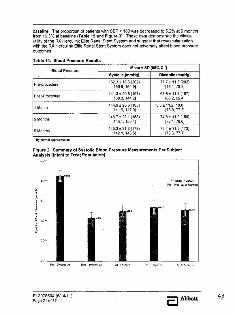
baseline. The proportion of patients with SBP 180 was decreased to 5.2% at 9 monthsfrom 19.3% at baseline (Table 15 and Figure 3). These data demonstrate the clinicalutility of the RX Herculink Elite Renal Stent System and suggest that revascularizationwith the RX Herculink Elite Renal Stent System does not adversely affect blood pressureoutcomes.
Table 14. Blood Pressure Results
Blood Pressure Mean ± SD (95% Cl')Systolic (mmHg) Diastolic (mmHg)
Pre-procedure 162.3 ± 18.5 (202) 77.7 ± 11.5 (202)Pre-procedure [159.8, 164:9] [76.1, 79.3]
Post-Procedure 141.3 ± 20.8 (197) 67.8 ± 11.4 (197)[138.3, 144.2] [66.2, 69.4]
1 Month 144.9 ± 20.6 (183) 75.5 ± 11.2 (183)[141.9, 147.9] [73.9, 77.21
6 Months 146.7 ± 23.1 (158) 74.8 ± 11.2 (158)[143.1, 150.4] [73.1, 76.6]
9 M145.3 ± 21.3 (173) 75.4 ± 11.5 (173)[142.1, 148.5] [73.6, 77.1)
By normal approximation
Figure 2. Summary of Systolic Blood Pressure Measurements Per SubjectAnalysis (Intent to Treat Population)
170-
162,3
160- P-value: <00001(Pre-Proc vs. 9 Mont)
$467449 45.3
140 1413
130-
no-
120-Pre-Pocoedum Pbst-Procedurm At 1 Month At 6 Months At 9 Months
EL2078594 (6/14/11)Page 21 of 37 5 Abbott

Table 15. Systolic Blood Pressure Goal AnalysesTime Period Subjects (%) (N) 95% Cl'
Pre-Procedure< 140 (mmHg) 0.5% (1/202) [0.0%, 2.7%]
140 and < 160 (mmHg) 55.9% (113/202) (48.8%, 62.9%]160 and < 180 (mmHg) 24.3% (49/202) [18.5%, 30.8%]
180 (mmHg) 19.3% (39/202) [14.1%, 25.4%]
Post-Procedure< 140 (mmHg) 46.2% (91/197) [39.1%, 53.4%]
140 and < 160 (mmHg) 37.1% (73/197) [30.3%, 44.2%]160 and < 180 (mmHg) 12.2% (24/197) [8.0%, 17.6%]
180 (mmHg) 4.6% (9/197) [2.1%, 8.5%]
30-Day< 140 (mmHg) 42.1% (77/183) [34.8%, 49.6%]
140 and < 160 (mmHg) 33.9% (62/183) [27.1%, 41.2%]160 and < 180 (mmHg) 17.5%, (32/183) [12.3%, 23.8%]
180 (mmHg) 6.6% (12/183) [3.4%, 11.2%]
6-Month< 140 (mmHg) 41.8% (66/158) [34.0%, 49.9%]
a 140 and < 160 (mmHg) 33.5% (53/158) [26.2%, 41.5%]160 and < 180 (mmHg) 15.2% (24/158) [10.0%, 21.8%]
? 180 (mmHg) 9.5% (15/158) [5.4%, 15.2%]
9-Month< 140 (mmHg) 40.5% (70/173) [33.1%, 48.2%]
140 and < 160 (mmHg) 35.8% (62/173) [28.7%, 43.5%]160 and < 180 (mmHg) 18.5% (32/173) 13.0%, 25.1%
a 180 (mmHg) 5.2% (9/173) [2.4%, 9.6%]By Clopper-Pearson exact confidence interval
EL2078594 (6/14/11) AbbottPage 22 of 37

Figure 3. Systolic Blood Pressure Measurement Change Over Time Per SubjectAnalysis (Intent-to-Treat Population)
* < 140 0 a 140 and < 160 kO 160 and < 180 m 2180 SBP a 180 (pre-procvs9 mo)p< 0.0001
SBP 2 160 (pre-poc vs 9 mo) p < 0.0001
SBP <140 (pre-proc vs 9 mo) p < 0 0001100% __------
90% 12217.5 15218.5
80%
60%0% -- W.
-6 50%
40%
30% 5.
20%
10%
0%Pre-Proc Post-Proc 1-Month 6-Month 9-Month
Note: The p-value is not pre-specified and is for descriptive purpose only.
Change in renal function at 9 months
In addition, renal function was maintained (i.e., did not worsen) from pre-procedure to 9-month follow-up based on serum creatinine levels and eGFR, further demonstrating theclinical utility of the Herculink Elite stent and suggesting that revascularization with theHerculink Elite stent does not adversely affect renal function.
Primary and Secondary Patency
Primary and secondary patency at 9 months were 88.0% (184/209) and 95.2% (198/208),respectively.
Target Lesion Revascularization
Kaplan-Meier Survival Analysis yields a 92.6% freedom from all Target LesionRevascularization (TLR) through 9 months. Clinically indicated TLR through 9 monthswas determined based on the angiographic core laboratory reported percent diameterstenosis of a 60%. Kaplan-Meier Survival Analysis yields a 94.1% freedom from clinicallyindicated TLR through 9 months (Figure 4).
EL2078594 (6/14/11)Page 23 of 37 Abbott

Figure 4. Kaplan-Meier Survival Curve - Freedom from Clinically Indicated TLRthrough 312 Days (Intent-to-Treat Population)
58- I
0 20 40 60 80 100 120 140 S0 N0 200 220 Q4 250 2 W0 300 320Days lbsxt Index Procedure
Days Post Index Procedure 0 (0, 30] (30, 180] (180, 270] (270, 312]Lesions at Risk 241 241 238 231 194
Lesions Censored 0 3 5 32 189
Number of Events 0 0 2 5 5
Event Free 100% 100% 99.1% 96.9% 94.1%
Standard Error () 0.0% 0.0% 0.6% 1.2% 1.7%
Note: Lesions at risk gives the number of lesions at risk of an event at the start of the interval, whilelesions censored and number of events are the incremental counts of lesions censored or with eventsduring the interval. The intervals are denoted as half-open bracket expression, where the start of interval'(is exclusive and the end of the interval ']' is inclusive.
EL2078594 (6/14/11)Page 24 of 37 E )Abbott

Subgroup Analyses
A subgroup analysis has been performed on subjects who were treated for unilateralversus bilateral lesions. Analyses at follow-up visits showed similar results between theunilateral and bilateral treatment groups. However, the Kaplan-Meier analysis showed asignificant increase of mortality over the 9 months period in the bilateral group: 5.2% inbilateral vs. 1.3% in unilateral group (log rank p value = 0.0214) (Figure 5).Figure 5. Subgroup Analysis - Freedom from Death through 312 Days (Intent-to-Treat Population: Bilateral vs. Unilateral)
--------------------------. - - -
50%-
0 20 40 60 8 1 12 1o 160 1 200 220 240 260 280 300 320
Days Post Index Procedure
Solid line: Bilateral Lesion Subjects (n= 39).Dashed lie: Unilaerl Lesia subjects (53)
Day Pot Idex0 (0, 30] (30, 180] (180, 270] (270, 312]Procedure
Subjects at Ris-ki __39 39 38 38 32Subjects Censored 0 0 0 5 31Number of Events 0 1 0 1 1
Event Free () 100% 97.4%/ 97.4% 94.8% 91.8%Standard Error () 0.0% 2.5% 2.5% 3.6% 4.5%
UnilateralSubjects at Risk | 163 163 162 17135
Subjects Censored I 0 1 t 3 22 135Number of Events 0 02 0 0
EetFe(% 10% 100% 98.7% 9.% 98.7%Standlard Error (% 0.0% 0.0% 0.9%00/9 0.9%
Tests Between Groups Test iiChi-Square I DF p-valueLog-Rank 15.293 1 1 0.0214Wilcoxon 4.932 1 0.0264
EL2078594 (6/14/11) AbtPage 25 of 37 m Abt

8.2 Materials Required* Introducer sheath I guiding catheter in the appropriate size and configuration for
the selected Stent Delivery System (refer to Table 1).* 2 - 3 syringes (10 - 20 cc)* 1,000 u / 500 cc normal saline* 0.014" (0.36 mm) diameter guide wire of appropriate length* 60% contrast diluted 1:1 with normal saline* Inflation device* Three-way stopcock* Torque device (if applicable)* Guide wire introducer
8.3 Stent Inspection Prior To Use
Prior to using the RX Herculink Elite Renal Stent System, carefully remove the systemfrom the package and inspect for bends, kinks, and other damage. Verify that the stent islocated between the radiopaque balloon markers. Do not use if any defects are noted.
8.4 Lesion Preparation
1. Standard percutaneous technique should be used to place the introducer sheath /guiding catheter in the vessel. An appropriate sized (0.014") guide wire should beadvanced across the lesion and into the common vessel.
2. Pre-dilate the lesion with an appropriate size balloon dilatation catheter to closelymatch the lumen diameter proximal and distal to the lesion.
3. Withdraw the balloon dilatation catheter leaving the guide wire in place.
8.5 Guide Wire Lumen Flush
1. Remove the protective cover from the tip.
2. Using the flush tool, flush the guide wire lumen with HepNS until fluid exits the guidewire exit notch.
8.6 Stent Delivery System Preparation
1. Prepare an inflation device / syringe with diluted contrast medium.
2. Attach the inflation device / syringe to the stopcock; attach to the inflation port.
3. With the tip down, orient the Delivery System vertically.
4. Open the stopcock to the Delivery System; pull negative for 30 seconds; release toneutral for contrast fill.
5. Close the stopcock to the Delivery System; purge the inflation device / syringe of allair.
EL2078594 (6/14/11)Page 26 of 37 Abbott

6. Repeat steps 3 through 5 until all air is expelled. Note: If air is seen in the shaft,repeat Delivery System Preparation steps 3 through 5 to prevent uneven stentexpansion.
7. If a syringe was used, attach a prepared inflation device to stopcock.
8. Open the stopcock to the Delivery System, leave on neutral.
8.7 Stent Delivery Procedure
1. Wipe the exposed guide wire with Heparinized saline to remove residual blood orcontrast medium.
2. Fully open the hemostatic valve. Maintain neutral pressure on the inflation device.
3. Backload the Delivery System onto the proximal portion of the guide wire whilemaintaining guide wire position across the target lesion.
4. Advance the Delivery System over the guide wire to target lesion. Utilize radiopaqueballoon markers to position the stent across the lesion; perform angiography toconfirm stent position. If applicable tighten the hemostatic valve.
Note: If during the process of moving the Delivery System into position you notice thestent has moved on the balloon, do not deploy the stent. The entire system should beremoved as a single unit. See Stent /System Removal- Precautions section forspecific removal instructions.
5. The stent is now ready to be deployed.
8.8 Stent Deployment Procedure
CAUTION: Refer to product label for in vitro stent outer diameter, deployment pressure,and RBP.
1. Slowly inflate the delivery balloon to low pressure; hold until balloon inflation isobserved both proximally and distally to the stent. Continue balloon expansion to thespecified stent deployment pressure. Confirm complete expansion of the stent /balloon fluoroscopically. If necessary, the delivery balloon can be used to post dilatethe stent to optimize stent apposition.
Do not exceed RBP: A larger PTA catheter may be used to dilate the stent. Do notexpand the 4.0 mm - 6.0 mm stents beyond 7.0 mm. Do not expand the 6.5 mm - 7.0mm stents beyond 8.0 mm.
2. After stent deployment, draw negative pressure on the inflation device for 30 secondsor until the delivery balloon is fully deflated.
3. Return the inflation device to neutral pressure to allow the balloon to refold duringremoval through the guide catheter.
4. With the inflation device on neutral pressure, carefully withdraw the delivery catheterwith the guide wire remaining across the lesion.
EL2078594 (6/14/11)Page 27of 37 E Abbott

Note: Should unusual resistance be felt at any time during either lesion access, orremoval of an undeployed stent, the Stent System, wire, and guiding catheter shouldbe removed as a single unit. See Stent/System Removal - Precautions section forspecific removal instructions.
5. Confirm optimal stent apposition using standard angiographic techniques. Ifnecessary, post dilate within stent. Post dilatation balloon diameters should closelymatch vessel reference diameter
9.0 REFERENCES
The physician should consult current literature on current medical practice on balloondilatation and placement of balloon expandable stents.
10.0 PATENTS AND TRADEMARKSThis product and I or its use are covered by one or more of the following United StatesPatents: 5,242,396; 5,421,955; 5,514,154; 5,546,646; 5,569,295; 5,603,721;5,649,952; 5,728,158; 5,735,893; 5,738,674; 5,759,192; 5,766,238; 5,780,807;5,916,234; 6,056,776; 6,066,167; 6,066,168; 6,131,266; 6,296,655; 6,309,412;6,369,355; 6,419,693; 6,428,568; 6,432,133; 6,482,166; 6,485,511; 6,511,504;6,568,235; 6,589,207; 6,596,022; 6,620,193; 6,629,991; 6,651,478; 6,689,159;6,736,843; 6,827,734; 6,835,059; 6,840,081; 6,908,479; 7,060,218.Additional patents pending.
Herculink Elite is a registered trademark of the Abbott Group of Companies.Plavix is a registered trademark of Sanofi-Aventis.
Abbott VascularSanta Clara, CA 95054-2807 USA
Customer ServiceTEL: (800) 227-9902FAX: (800) 601-8874Outside USA TEL: (951) 914 -4669Outside USA FAX: (951) 914 -2531
EL2078594 (6/14/11)Page 28 of 37

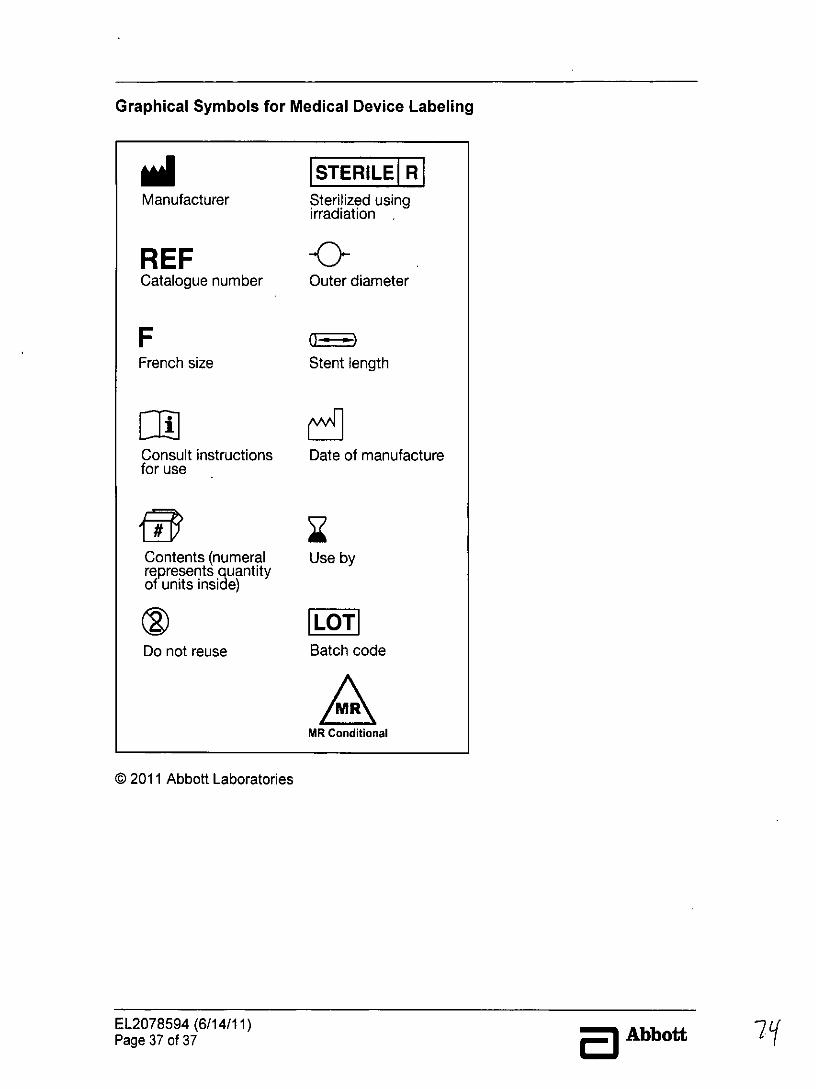
Graphical Symbols for Medical Device Labeling
STERILERManufacturer Sterilized using
irradiation
REF -0-Catalogue number Outer diameter
F sFrench size Stent length
Consult instructions Date of manufacturefor use
Contents (numeral Use byrepresents quantityof units inside)
LOTDo not reuse Batch code
AMR Conditional
@ 2011 Abbott Laboratories
EL2078594 (6/14/11)Page 29 of 37 Abbott

RX Herculink Elite® Biliary Stent SystemCAUTION: Federal (USA) law restricts this device to sale by or on the orderof a physician.1.0 DEVICE DESCRIPTION2.0 HOW SUPPLIED3.0 INDICATIONS4.0 CONTRAINDICATIONS5.0 WARNINGS6.0 PRECAUTIONS7.0 POTENTIAL ADVERSE EVENTS8.0 CLINICIAN USE INFORMATION9.0 PATENTS AND TRADEMARKS
1.0 DEVICE DESCRIPTION
The RX Herculink Elite Biliary Stent System includes:
* A balloon expandable L605 cobalt chromium alloy stent pre-mounted on theballoon of a rapid exchange (RX) stent delivery system
* Two radiopaque markers located underneath the balloon which identify the stentposition and fluoroscopically mark the working length of the balloon
* Proximal shaft markers to aid with delivery catheter position, relative to a biliaryguiding catheter tip
* A third marker located approximately 30 cm from the center of the balloon thataids in locating the guide wire exit lumen and facilitating catheter removal andexchange
The delivery system can be utilized to optimize the stent wall apposition post stentdeployment.
Table 1. In Vitro* Device Specificationsin Vitro* Rated Recommended
EStent Stent Burst MinimumStentMimuDiamet Lengths Deployment Pressure Minimum Sheath
(mm) Pressure RBP Guiding Catheter ID Introducer"(mm) (atm) (atm) (F)1(inches)(mm) (F)/(inches)l(mm)4.0 12, 15, 18 11 14 6/0.067/1.70 5/0.071/1.804.5 12, 15, 18 11 14 6/0.067/1.70 5/0.071/1.805.0 12, 15, 18 11 14 6/0.067/1.70 5/0.071/1.805.5 12, 15, 18 11 14 6/0.067/1.70 5/0.071/1.806.0 12, 15, 18 11 14 6/0.067/1.70 5/0.071/1.806.5 12,15,18 11 14 6/0.067/1.70 5/0.071/1.807.0 15, 18 11 14 6/0.067/1.70 5/0.071/1.80
All data provided is based on in vitro testing. Assure full deployment of the stent (see Clinician UseInformation section 8.8 Stent Deployment Procedure). Deployment pressures should be based on stricturecharacteristics.** See individual manufacturer specifications for (F) equivalent.
EL2078594 (6/14/11)Page 30 of 37 E Abbott

2.0 HOW SUPPLIED
Sterile. This device is sterilized with electron beam radiation. Non-pyrogenic. Do not useif the package is open or damaged.
Storage. Store in a dry, dark, cool place.
Contents. One (1) RX Herculink Elite Biliary Stent System, one (1) Protective /Regrooming sheath, one (1) flush tool
3.0 INDICATIONS
The RX Herculink Elite Biliary Stent System is intended for palliation of malignantstrictures in the biliary tree.
4.0 CONTRAINDICATIONS
The RX Herculink Elite Biliary Stent System is contraindicated for use in:
* Stenting a perforated duct where the leakage from the duct can be enhanced bythe prosthesis
* Patients with bleeding disorders
* Severe ascites
5.0 WARNINGS
The long term safety and effectiveness of this device in the biliary system have not beenestablished.
Should unusual resistance be felt at any time during stricture access or DeliverySystem removal, the introducer sheath / guiding catheter and stent system should beremoved as a single unit. Applying excessive force to the Stent Delivery System canpotentially result in loss or damage to the Stent and Delivery System components. (SeeStent / System Removal - Precautions.)
Stenting across a major bifurcation may hinder or prevent future diagnostic or therapeuticprocedures.
Once fully deployed, the stent cannot be repositioned.
Persons allergic to L605 cobalt chromium alloy may suffer an allergic reaction to thisimplant.
Only physicians familiar with the complications, side effects and hazards commonlyassociated with biliary stent placement should use this device.
The RX Herculink Elite Biliary Stent System is intended to perform as a system. Thestent should not be removed for use in conjunction with other dilatation catheters, norshould the RX Herculink Elite Biliary Stent System be used in conjunction with otherstents.
The safety and effectiveness of multiple overlapping stents have not beenestablished. However, when multiple stents are required, stent materials should beof similar composition.
EL2078594 (6/14/11)Page 31 of 37 E Abbott

6.0 PRECAUTIONS
6.1 Stent Delivery System Handling - Precautions
* For single use only. Do not resterilize or reuse. Note product "Use by" date.
* Do not remove stent from its delivery balloon.
* Special care must be taken not to handle or in any way disrupt the stent on theballoon. This is most important during stent system removal from the packaging,placement over a guide wire and advancement through a guiding catheter orintroducer sheath.
* Do not "roll" the mounted stent with your fingers as this action may loosen thestent from the delivery balloon.
* Use only the appropriate balloon inflation media. Do not use air or any gaseousmedium to inflate the balloon as this may cause uneven expansion and difficultyin deployment of the stent.
6.2 Stent Placement - Precautions
* Do not prepare or pre-inflate balloon prior to stent deployment other than asdirected. Use balloon purging technique described in the Clinician UseInformation section.
* The inflated balloon diameter of the system used to deploy the stent shouldapproximate the diameter of the bile duct. Oversizing of the stent can result in aruptured bile duct. To ensure full expansion of the stent, the balloon should beinflated to a minimum of nominal pressure.
* Implanting a stent may lead to dissection of the duct distal and / or proximal to thestent and may cause acute closure of the duct requiring additional intervention(surgical intervention, further dilatation, placement of additional stents, or other).
* Do not expand the stent if it is not properly positioned in the bile duct. (See Stent/System Removal - Precautions.)
* Balloon pressures should be monitored during inflation. Do not exceed RatedBurst Pressure (RBP) as indicated on product label. Use of pressures higherthan specified on product label may result in a ruptured balloon with possible bileduct damage or perforation.
Do not attempt to pull an unexpanded stent back through the introducersheath I guiding catheter; dislodgment of the stent from the balloon may occur.
6.3 Stent I System Removal - Precautions
Should unusual resistance be felt at any time during either stricture access or removalof the Delivery System post-stent implantation, the entire system should be removed as asingle unit.
EL2078594 (6/14/11)Page 32 of 37 Abbott

When removing the Delivery System as a single unit:
* DO NOT retract the Delivery System into the introducer sheath / guiding catheter.
* Position the proximal balloon marker just distal to the tip of the introducersheath / guiding catheter.
* Advance the guide wire in the anatomy as far distally as safely possible.
* Secure the Delivery System to the introducer sheath / guiding catheter; thenremove the introducer sheath / guiding catheter, guide wire and Delivery Systemas a single unit.
Failure to follow these steps and / or applying excessive force to the Delivery System canpotentially result in loss or damage to the stent and / or Delivery System components.
If it is necessary to retain guide wire position for subsequent biliary access, leave theguide wire in place and remove all other system components.
6.4 Post Implant - Precautions
Great care must be exercised when crossing a newly deployed stent with a guide wireor balloon catheter to avoid disrupting the stent geometry.
Magnetic Resonance Imaging (MRI)
Non-clinical testing has demonstrated that the Herculink Elite stent, in single and inoverlapped configurations up to 33 mm in length, is MR Conditional. It can be scannedsafely under the following conditions:
* Static magnetic field of 1.5 or 3 Tesla
* Spatial gradient field of 2500 Gauss/cm or less
* Maximum whole-body-averaged specific absorption rate (SAR) of 2.0 W/kg(normal operating mode) for any duration of MRI scan that would otherwise besafe for the patient without implant.
MRI at 1.5 or 3 Tesla may be performed immediately following the implantation of theHerculink Elite stent.
The Herculink Elite stent should not migrate in this MRI environment. Magnetic force onthe Herculink Elite state was tested according to ASTM F2052-06e. Non-clinical testingat field strengths greater than 3 Tesla has not been performed to evaluate stent migrationor heating.
Stent heating was derived by using the measured non-clinical, in vitro temperature riseaccording to ASTM F2182-09 in a GE Signa HDx 3 Tesla scanner and in a GE 1.5 Teslacoil in combination with the local specific absorption rates (SARs) in a digitized humanheart model. The temperature rise was derived by a validated calculation. At overlappedlengths up to 33 mm, the Herculink Elite stent produced a non-clinical maximum localtemperature rise of less than 30C at a maximum whole body averaged SAR of 2.0 W/kg(normal operating mode) for an MRI sequence of 15 minutes. These calculations do nottake into consideration the cooling effects of blood flow.
The effects of MRI on overlapped stents greater than 33 mm in length or stents withfractured struts are unknown.
EL2078594 (6/14/11) t0Page 33 of 37 Abbott

Image artifact may be present when scanning the Herculink Elite stent as demonstratedin non-clinical testing performed according to ASTM F2119-07 in a GE Signa HDx 3Tesla scanner. The image artifact (both inside and outside the device lumen) extendsapproximately 7 mm from the device using the spin echo sequence (TR = 500 ms; TE =20 ms; flip angle = 900) and 13 mm from the device using the gradient echo sequence(TR = 100 ms; TE = 15 ms; flip angle = 300). MR image quality may be compromised ifthe area of interest is in the exact same area, or relatively close to, the position of theHerculink Elite stent. Therefore, it may be necessary to optimize the MR imagingparameters in the presence of Herculink Elite stents.
7.0 POTENTIAL ADVERSE EVENTS
Potential complications associated with the use of a biliary stent may include, but are notlimited to, the following:
* Abscess* Bile duct injury, including rupture and perforation* Bile duct occlusion / obstruction* Cholangitis* Death* Hypersensitivity or allergic reaction to drugs* Infection* Pancreatitis* Parenchymal hemorrhage* Peritonitis* Sepsis* Tumor overgrowth at the stent ends
8.0 CLINICIAN USE INFORMATION
8.1 Materials Required
* Introducer sheath / guiding catheter in the appropriate size and configuration forthe selected Stent Delivery System (refer to Table 1).
* 2 - 3 syringes (10-20 cc)* 1,000 u / 500 cc normal saline* 0.014" (0.36 mm) diameter guide wire of appropriate length* 60% contrast diluted 1:1 with normal saline* Inflation device* Three-way stopcock* Torque device (if applicable)* Guide wire introducer
8.2 Stent Inspection Prior To UsePrior to using the RX Herculink Elite Biliary Stent System, carefully remove the systemfrom the package and inspect for bends, kinks, and other damage. Verify that the stent islocated between the radiopaque balloon markers. Do not use if any defects are noted.
EL2078594 (6/14/11) AbbottPage 34 of 37 A o

8.3 Stricture Evaluation / Biliary DrainageStandard percutaneous transhepatic cholangiography should be performed to assess thebiliary tree followed by the passage of a guide wire through the stricture and theplacement of an internal / external biliary drainage catheter.8.4 Stricture Pre-dilatation
1. Standard percutaneous technique should be used to place an introducer sheath /guiding catheter in the biliary tree. A 0.014" (0.36 mm) diameter guide wire should beadvanced across the stricture and into the common bile duct.
2. Stricture and bile ducts may need to be pre-dilated with balloon dilatation. Pre-dilatation catheter diameters should closely match the duct diameter proximal anddistal to the stricture to be treated.
8.5 Guide Wire Lumen Flush
1. Remove the protective cover from tip.
2. Using the flush tool, flush the guide wire lumen with normal saline until fluid exits theguide wire exit notch.
8.6 Stent Delivery System Preparation
1. Prepare an inflation device / syringe with diluted contrast medium.
2. Attach the inflation device / syringe to the stopcock; attach to the inflation port.3. With the tip down, orient the Delivery System vertically.
4. Open the stopcock to the Delivery System; pull negative for 30 seconds; release toneutral for contrast fill.
5. Close the stopcock to the Delivery System; purge the inflation device / syringe of allair.
6. Repeat steps 3 through 5 until all air is expelled. Note: If air is seen in the shaft,repeat Balloon Preparation steps 3 through 5 to prevent uneven stent expansion.
7. If a syringe was used, attach a prepared inflation device to stopcock.8. Open the stopcock to the Delivery System, leave on neutral.
8.7 Stent Delivery Procedure1. Wipe the exposed guide wire with normal saline.2. Maintain neutral pressure on inflation device.3. Backload the Delivery System onto the proximal portion of the guide wire while
maintaining guide wire position across the stricture.4. Advance the Delivery System over the guide wire to target stricture. Utilize
radiopaque balloon markers to position the stent across stricture; performcholangiography to confirm stent position.
Note: If during the process of moving the Delivery System into position you notice thestent has moved on the balloon, do not deploy the stent. The entire system should beremoved as a single unit. See Stent/System Removal - Precautions section forspecific Delivery System removal instructions.
5. The stent is now ready to be deployed.
EL2078594 (6/14/11) A72/Page 35 of 37 E Abbott

8.8 Stent Deployment Procedure
CAUTION. Refer to product label for in vitro stent outer diameter, deploymentpressure, and RBP.1. Slowly inflate the delivery balloon to low pressure; hold until balloon inflation is
observed both proximally and distally to the stent. Continue balloon expansion to thespecified stent deployment pressure. Confirm complete expansion of the stent /balloon fluoroscopically. If necessary, the delivery balloon can be used to post dilatethe stent to optimize stent apposition.
Do not exceed RBP: A larger PTA catheter may be used to dilate the stent. Do notexpand the 4.0 mm - 6.0 mm stent beyond 7.0 mm. Do not expand the 6.5 mm - 7.0mm diameter stent beyond 8.0 mm.
2. After stent deployment, draw negative pressure on the inflation device for 30 secondsor until the delivery balloon is fully deflated.
3. Return the inflation device to neutral pressure to allow the balloon to refold duringremoval through the guide catheter.
4. With the inflation device on neutral pressure, carefully withdraw the delivery catheterwith the guide wire remaining across the stricture.
Note: Should unusual resistance be felt at any time during either stricture accessor removal of an undeployed stent, the Stent System, guide wire and guiding cathetershould be removed as a single unit. See Stent / System Removal - Precautionssection for specific removal instructions.
5. Confirm optimal stent apposition using standard cholangiographic techniques. Ifnecessary, post dilate within the stent. Post dilatation balloon diameters shouldclosely match bile duct reference diameter.
9.0 PATENTS AND TRADEMARKSThis product and / or its use are covered by one or more of the following United StatesPatents: 5,242,396; 5,421,955; 5,514,154; 5,546,646; 5,569,295; 5,603,721;5,649,952; 5,728,158; 5,735,893; 5,738,674; 5,759,192; 5,766,238; 5,780,807;5,916,234; 6,056,776; 6,066,167; 6,066,168; 6,131,266; 6,296,655; 6,309,412;6,369,355; 6,419,693; 6,428,568; 6,432,133; 6,482,166; 6,485,511; 6,511,504;6,568,235; 6,589,207; 6,596,022; 6,620,193; 6,629,991; 6,651,478; 6,689,159;6,736,843; 6,827,734; 6,835,059; 6,840,081; 6,908,479; 7,060,218.Additional patents pending.
Herculink Elite is a registered trademark of the Abbott Group of Companies.
Abbott VascularSanta Clara, CA 95054-2807 USA
CUSTOMER SERVICETEL: (800) 227-9902FAX: (800) 601-8874Outside USA TEL: (951) 914 -4669Outside USA FAX: (951) 914 -2531
EL2078594 (6/14/11)Page 36 of.37 Abbott
Graphical Symbols for Medical Device Labeling
STERILERManufacturer Sterilized using
irradiation
REF -0Catalogue number Outer diameter
FFrench size Stent length
Consult instructions Date of manufacturefor use
Contents (numeral Use byrepresents quantityof units inside)
LOTDo not reuse Batch code
AMR Conditional
@ 2011 Abbott Laboratories
EL2078594 (6/14/11)Page 37 of 37 5 Abbott