Ruxolitinib Suppresses Liver Fibrosis Progression and ...
36
Ruxolitinib Suppresses Liver Fibrosis Progression and Accelerates Fibrosis Reversal via Selectively Targeting Janus Kinase 1/2 Zhenghui Song Southern Medical University Xinhui Liu Southern Medical University Wan Zhang Southern Medical University Yue Luo Southern Medical University Hua Xiao Michigan State University Yun Liu XiangNan University Guanqi Dai Southern Medical University Jian Hong Jinan University Aimin Li ( [email protected] ) Southern Medical University https://orcid.org/0000-0001-9459-5540 Research Article Keywords: Janus Kinase 1, Janus Kinase 2, Liver Fibrosis, HSCs, Ruxolitinib Posted Date: November 10th, 2021 DOI: https://doi.org/10.21203/rs.3.rs-1050686/v1 License: This work is licensed under a Creative Commons Attribution 4.0 International License. Read Full License
Transcript of Ruxolitinib Suppresses Liver Fibrosis Progression and ...

Ruxolitinib Suppresses Liver Fibrosis Progressionand Accelerates Fibrosis Reversal via SelectivelyTargeting Janus Kinase 1/2Zhenghui Song
Southern Medical UniversityXinhui Liu
Southern Medical UniversityWan Zhang
Southern Medical UniversityYue Luo
Southern Medical UniversityHua Xiao
Michigan State UniversityYun Liu
XiangNan UniversityGuanqi Dai
Southern Medical UniversityJian Hong
Jinan UniversityAimin Li ( [email protected] )
Southern Medical University https://orcid.org/0000-0001-9459-5540
Research Article
Keywords: Janus Kinase 1, Janus Kinase 2, Liver Fibrosis, HSCs, Ruxolitinib
Posted Date: November 10th, 2021
DOI: https://doi.org/10.21203/rs.3.rs-1050686/v1
License: This work is licensed under a Creative Commons Attribution 4.0 International License. Read Full License
1
Ruxolitinib suppresses liver fibrosis progression and accelerates
fibrosis reversal via selectively targeting Janus kinase 1/2
Zhenghui Song1,2,#, Xinhui Liu1,2,3,#, Wan Zhang1,2, Yue Luo1,2,3, Hua Xiao3, Yun
Liu4, Guanqi Dai1,2, Jian Hong5,*, Aimin Li1,2,3,*
1 Department of Hepatology, Cancer Center, Integrated Hospital of Traditional
Chinese Medicine, Southern Medical University, Guangzhou, 510315, China
2 School of Traditional Chinese Medicine, Southern Medical University,
Guangzhou, Guangdong, 510515, China
3 Department of Physiology, Michigan State University, East Lansing, MI, 48824,
USA
4 Department of Endocrinology and Metabolic Diseases, Affiliated Hospital
(Clinical College) of Xiangnan University, Chenzhou, 423000, China
5 School of Medicine, Jinan University, Guangzhou 510632, China
* Corresponding author
Email: [email protected] (A. Li) and [email protected] (H. Jian)
Address: No.13 Shiliugang Road, Guangzhou, Guangdong, 510315, China.
# These authors contributed equally to this work.

2
Abstract
Background: JAK1 and JAK2 have been implicated in fibrosis and cancer as
a fibroblast-related marker; however, their role in liver fibrosis has not been
elucidated. Here, we aim to determine the effect and underlying mechanism of
JAK1/2 inhibition on liver fibrosis and hepatic stellate cells (HSCs) and further
explore the therapeutic efficacy of Ruxolitinib, a JAK1/2 selective inhibitor, on
preventing and reversing liver fibrosis in mice.
Methods: Immunohistochemistry staining of JAK1 and JAK2 were performed
on liver tissue in mice with hepatic fibrosis and human liver tissue microarray of
liver cirrhosis and liver cancer. LX-2 cells treated with specific siRNA of JAK1
and JAK2 were used to analysis activation, proliferation and migration of HSCs
regulated by JAK1/2. The effects of Ruxolitinib (JAK1/2 inhibitor) on liver
fibrosis were studied in LX-2 cells and two progressive and reversible fibrosis
animal models (carbon tetrachloride (CCl4), Thioacetamide (TAA)).
Results: We found that JAK1/2 expression was positively correlated with the
progression of HCC in humans and the levels of liver fibrosis in mice. Silencing
of JAK1/2 down-regulated their downstream signaling and inhibited proliferation,
migration, and activation of HSCs in vitro, while Ruxolitinib had similar effects
on HSCs. Importantly, Ruxolitinib significantly attenuated fibrosis progression,
improved cell damage, and accelerated fibrosis reversal in the liver of mice
treated with CCl4 or TAA.
Conclusions: JAK1/2 regulates the function of HSCs and plays an essential

3
role in liver fibrosis and HCC development. Its inhibitor, Ruxolitinib, may be an
effective drug for preventing and treating liver fibrosis.
Keywords
Janus Kinase 1; Janus Kinase 2; Liver Fibrosis; HSCs; Ruxolitinib
Background
Liver fibrosis is a chronic liver injury mainly caused by hepatitis virus B and C
infection, alcohol abuse and nonalcoholic steatohepatitis (NASH) [1]. Liver
fibrosis eventually progresses to liver cirrhosis, a common cause of death
worldwide [2], resulting in serious complications, including portal hypertension,
liver failure, and hepatocellular carcinoma (HCC), leading to failure of liver
function and destruction of liver structures, and ultimately organ dysfunction
and death [3,4]. Currently, liver transplantation is the only effective cure, which
brings a tremendous economic burden to the patients [5]. Liver fibrosis is due
to excessive accumulation of extracellular matrix (ECM) in response to chronic
liver damage. Hepatic stellate cells (HSCs) are considered the most critical cell
type for the production of collagens [6-8]. When stimulated by liver damage,
HSCs will be activated, and then differentiate into myofibroblasts acquire
fibrogenic property for producing ECM, resulting in fibrotic scar [9,10]. Many
drug companies try to find methods to halting scarring or even remove existing
scars, but no drug is yet approved for treating cirrhosis [11]. Thus, the

4
development of effective anti-fibrotic drugs to stop progression to cirrhosis or
even reverse advanced fibrosis is urgently needed [12].
Several signaling pathways are found to be related to liver fibrosis, and may be
potential targets for treatment. The Janus kinase/Signal Transducer and
Activator of Transcription protein (JAK/STAT) signaling pathway is a chain of
interactions between proteins and involved in processes such as immunity, cell
division, cell death, and tumor formation [13-19]. Recent studies have reported
that STATs are associated with tissue fibrosis, including skin, lung, liver,
systemic sclerosis (SSc), and STAT3 inhibitors have been shown to be effective
in the Carbon tetrachloride (CCl4)-induced liver fibrosis model [20,21]. JAKs act
as the upstream of STATs, and JAK/STAT signaling pathway plays a role in
promoting the fibrosis, and selective JAK2 inhibitor can effectively block
fibroblast activation improve fibrosis [22]. Besides, recent studies suggested
that JAK1 might also play an indirect role in promoting fibrosis through the
transphosphorylation of JAK2 [23]. However, the role of JAK1 and JAK2 in liver
fibrosis remains unclear.
Ruxolitinib is the only effective small-molecule JAK1/2 selective inhibitor
approved by the US Food and Drug Administration (FDA) for myelofibrosis
treatment in 2011. It is mainly used for the treatment of intermediate or high-
risk bone marrow fibers, which is well tolerated in a multicenter study and can
effectively alleviate splenomegaly, improve spleen hyperfunction and systemic
symptoms of patients [24-26]. Recently, it has been reported that Ruxolitinib is

5
effective and safe for the treatment of hematological diseases such as
polycythemia vera, lymphoma, etc.[27,28], and can benefit some patients with
pancreatic cancer [28]. Currently, a series of clinical studies of Ruxolitinib in
malignant glioma, multiple myeloma, lung cancer, breast cancer, colorectal
cancer, head, and neck squamous cell carcinoma, and prostate cancer are
underway (https://clinicaltrials.gov/ct2/results?cond=ruxolitinib). However, the
efficacy of Ruxolitinib on liver fibrosis and liver cancer has not been explored.
In the present study, we found up-regulated JAK1 and JAK2 were associated
with liver disease in humans and mice, while knockdown of JAK1 and JAK2
inhibited activation, proliferation, and migration of HSCs. Also, Ruxolitinib, the
JAK1/2 selective inhibitor, blocked HSCs activation, both in cell culture and in
animal fibrosis models, included by CCl4 and Thioacetamide (TAA).
Furthermore, we found that Ruxolitinib attenuated liver fibrosis progression,
accelerated its reversal, and protected from liver damage, which provides a new
direction and clinical transformation basis for the treatment of liver fibrosis and
even early intervention of HCC.
Methods
Cell culture
Immortalized human HSC cell line (LX-2) was purchased from Chinese
Academy of Sciences Cell Bank (Shanghai, China) and cultured in Dulbecco’s
modified Eagle’s medium (DMEM) (Gibco, China), supplemented with 10% fetal

6
bovine serum. (FBS) (Gibco, USA) and 1% Penicillin/Streptomycin at 5% CO2
and 37°C. All cell experiments were performed in strict accordance with cell
culture protocols.
Chemicals
For in vitro experiments, Ruxolitinib (INCB018424, Selleck chemicals, USA)
was dissolved in DMSO and further diluted to the required concentration. For
in vivo experiments, Ruxolitinib suspension was prepared in 0.5%
carboxymethyl cellulose sodium normal saline solution.
Small interfering RNA (siRNA) Transfection
siRNA for JAK1 and JAK2 were designed and synthesized by RiboBio
(Guangzhou, China). siRNAs were transfected into cells using Lipofectamine
3000 (Invitrogen) according to the manufacturer’s protocol. Cells were collected
after 48-72h for further experiments.
Human samples
Human liver tissue microarray was purchased from Biomax, including 5 liver
samples with normal liver tissue, 22 liver samples with cirrhosis, and 53 liver
samples with liver cancer which were collected in US.
Histology and immunohistochemistry
Formalin-fixed, paraffin-embedded liver tissue samples were cut into 4 µm-thick
sections and stained with hematoxylin eosin (H&E), Sirius red, and
immunohistochemistry (IHC) according to standard procedures. Fibrosis was
scored according to the Ishak scoring system [29]. The amount of Sirius red

7
staining was quantified with ImageJ. For IHC, liver sections were stained with
the following antibodies: JAK1 (sc-1677; Santa Cruz Biotechnology), JAK2
(#3230; Cell signaling Technology), Alpha-Smooth muscle actin (α-SMA,1A4,
ab7817; Abcam), Both the intensity and extent of immunostaining were taken
into consideration when analyzing the data. The intensity of staining was
determined by the following rules: 0 for negative; 1 for weak staining; 2 for
moderate staining; 3 for strong staining. The staining extent was determined by
the following rules: 0 for no staining; 1 for less than 10%; 2 for 10% to 50%; 3
for 51% to 75%; 4 for more than 75%. We randomly selected 5 areas from each
area to count the intensity and extent of staining and to calculate the mean
staining extent. The score was obtained by plus these two values (intensity
score + extent score).
Liver function assay
Serum levels of several biochemical markers were measured to assess liver
function and liver injury. The measured biochemical markers included alkaline
phosphatase (ALP), alanine aminotransferase (ALT), aspartate
aminotransferase (AST), total bilirubin (TBIL) and albumin (Alb) were measured
using standard laboratory assays.
Western blotting
Western blotting was performed as previously described [30]. The primary
antibodies were purchased from Santa Cruz Biotechnology [JAK1 (sc-1677)],
Cell signaling Technology [JAK2 (#3230), phosho-JAK1 (Tyr1022/1023, #3331),

8
phosho-JAK2 (Tyr1007/1008, #3771), STAT3(#9139), phosho-STAT3
(Tyr705,#9145), phosho-STAT3 (Ser705, #9134), STAT5 (#94205), phosho-
STAT5 (Tyr694, #9359)], Abcam [α-SMA (1A4, ab7817)], Bimake [PDGFRβ
(A5541), PAI-1 (A5396)], Bioworld [β-actin (#64132)]. β-actin was used as a
loading control for all blots.
Quantitative Real-time RT-PCR
Total RNA was extracted from cells and tissues using Trizol (Takara, Japan),
and was reverse transcribed into cDNA according to the manufacturer’s
instructions, and the GAPDH gene was used as gene control. The relative gene
expression ratio was calculated by the ΔΔCt method. Experiments were
performed according to the manufacturer’s instructions (Takara, Japan).
Independent experiments were done in triplicate.
Cell proliferation assay
LX-2 cells were seeded at a density of 3000 cells/well in 96-well microplates.
The cells were treated with siRNA transfection or varying concentrations of
Ruxolitinib as designed (0.1-100 μM). Cell viability was measured using the Cell
Counting Assay Kit-8 (CCK-8; Dojindo, Japan) according to the manufacturer’s
instructions. For each experimental condition, five parallel wells were assigned
to each group. Experiments were performed in triplicate.
Cell migration assay
LX-2 cells were seeded in a 6-well plate treated with siRNA transfection or drug
for 24h. Migration assays were conducted using 24-well Boyden chambers

9
containing inserts (8 μm pores; BD Biosciences,USA). The lower chamber was
filled with medium containing 10% serum, whereas the top chamber contained
1 × 10⁵ cells without serum. The plates were incubated at 37 °C in 5% CO2 for
10h. After migration, the cells that had migrated to the underside of the
membrane were fixed with paraformaldehyde and stained with 0.1% crystal
violet. Migrated cells on each insert were counted in five randomly selected
fields and quantified using the ImageJ software.
Cell apoptosis assay
LX-2 cells were seeded in 6-well plates and treated with different concentrations
of Ruxolitinib for 24 hours. For cells were then harvested and stained with
Annexin V-FITC Apoptosis Detection Kit (BD Pharmingen, CA) according to the
manufacture’s protocol. Experiments were performed in triplicate.
Mouse models
All animal experiments were conducted in accordance with the National
Institutes of Health guide for the care and use of Laboratory animals and
approved by the Animal Care and Use Committee of Southern Medical
University.
Liver fibrosis progression and reversal model induced by CCl4
For the Liver fibrosis progression model, 4-6 weeks old male C57BL/6 mice
(Vital River, Beijing, China) were treated three times a week with or without
0.1ml of a 40% CCl4 in olive oil by oral gavage for 8 weeks and mice were
treated with or without Ruxolitinib (30mg/kg, oral gavage, each day) from 5 to

10
8 weeks. For Liver fibrosis reversal model, 4-6 weeks old male C57BL/6 mice
(Vital River, Beijing, China) were treated three times a week with or without
0.1ml of a 40% CCl4 in olive oil by oral gavage for 6 weeks, and then mice
allowed to recover form 6 to 8 weeks, with or without treatment with Ruxoltinib
(30mg/kg, oral gavage, each day).
Panlobular liver fibrosis model induced by TAA
6-8 weeks old male C57BL/6 mice were accepted an optimistic dose-escalating
TAA as described [31]. And then mice were allowed to recover from 6 to 10
weeks, with or without treatment with Ruxolitinib (30mg/kg, oral gavage, each
day).
Statistical analysis
All statistical analyses were performed with GraphPad Prism V.5.00. Data are
expressed as mean ± standard deviation (SD). Differences between two groups
were compared using a two-tailed unpaired Student’s t-test. One-way ANOVA
was used for sample comparison among multiple groups. Statistical
significance was defined as P<0.05.
Results
Up-regulated JAK1 and JAK2 are associated with liver fibrosis and liver
disease in human and mice
To evaluate whether JAK1 and JAK2 expression are associated with liver
fibrosis/cirrhosis and liver cancer, IHC staining was used to examine JAK1 and

11
JAK2 expression in human liver tissue array with confirmed cirrhosis and cancer.
JAK1 and JAK2 expression were markedly higher in liver cirrhosis tissues
(n=22), while highest in liver cancer tissues (n=53) compared to normal liver
tissues (n=5) (Fig.1A and B). We further analyzed the relationship between the
expression of JAK1 and JAK2 and the degree of liver fibrosis in mice. Sirius
Red staining was used to confirm constructed liver fibrosis mouse model
induced by CCl4 for 6 weeks or 8 weeks respectively, and IHC staining showed
expression of JAK1 and JAK2 were a significant increase in fibrosis liver
compared with controls (Fig.1C and D), which is constant to human liver
disease. Our results suggest that the expression of JAK1 and JAK2 was
associated with liver cancer progression and was also positively correlated with
the severity of liver fibrosis.
Knockdown of JAK1 and JAK2 inhibits activation, proliferation and
migration of HSCs
Since the activation and proliferation of HSCs in the main trigger for liver fibrosis
and subsequent liver disease, to investigate the role of JAK1 and JAK2 on
HSCs, LX-2 cells were transfected with siJAK1, siJAK2, or both of them. The
transfected efficiency was confirmed by qRT-PCR and Western blotting
(Fig.2A). As α-SMA and platelet-derived growth factor receptor beta (PDGFRβ)
is the common marker for HSCs activation, the mRNA and protein level were
detected to investigate the role of JAK1 and JAK2 on the activation of HSCs.
As shown in Figure 2B, α-SMA and PDGFRβ were significantly decreased in

12
LX-2 cells transfected with siJAK1 or siJAK2. Furthermore, the lowest
expression was found in cells co-transfected with siJAK1 and siJAK2.
Next, we analyzed the effect of JAK1 and JAK2 on the proliferation and
migration of the HSCs. Knockdown of JAK1 or JAK2 had a similar impact on
inhibiting proliferation, while the proliferation of HSCs was further decreased in
the co-transfected group (Fig.2C). Likewise, the number of migrated HSCs was
significantly reduced in cells with siJAK1 or siJAK2 and lowest in the co-
transfected group compared to the control group (Fig.2D). Furthermore, we
investigated the effect of JAK1 and JAK2 on the downstream signaling and
revealed that expression of p-JAK1, p-JAK2, STAT3, and p-STAT3 was
significantly decreased (Fig.2E). These results indicate that knockdown JAK1
and JAK2 may inhibit the activation, proliferation and migration of HSCs.
JAK1 and JAK2 antagonism has anti-fibrotic activity in vitro
Ruxolitinib is a selective small-molecule oral inhibitor of JAK1/2, and it was
verified to effectively inhibit JAK1 phosphorylation at Tyr1022/1023 and JAK2
phosphorylation at Tyr1007/1008, the critical activation site at the kinase
domain of JAK1 and JAK2, respectively (Supporting Fig.S1). Moreover, we
revealed that Ruxolitinib significantly decreased STAT3 phosphorylation at
Tyr705 and Ser727, STAT5 phosphorylation at Tyr694, while no significant
changes were observed in STAT3 and STAT5 (Fig.3A). Then we utilized
Ruxolitinib to investigate whether JAK1 and JAK2 antagonism has anti-fibrotic
activity.
13
By using Ruxolitinib on LX-2 cells, we found Ruxolitinib inhibits cell proliferation
in a dose and time-dependent manner in LX-2 cells (Fig.3B), and the half-
maximal inhibitory concentration (IC50) of Ruxolitinib on LX-2 cell was 14.01 μM
(Fig.3C). Furthermore, Ruxolitinib significantly inhibited the expression of HSCs
activation marker (α-SMA and PDGFRβ) and collagen-associated marker
(COL1A1 and PAI-1) (Fig.3D). Besides, Ruxolitinib also significantly inhibited
the number of migrations (Fig.3E) and induced the level of apoptosis (Fig.3F)
in LX-2 cells in a dose-dependent manner. These results demonstrate that
Ruxolitinib, as the JAK1 and JAK2 antagonism, inhibits proliferation, activation,
migration and promoted cell apoptosis of HSCs in vitro and may exert an anti-
fibrotic activity by inhibiting phosphorylation site of JAK1/2 downstream
signaling.
Ruxolitinib attenuates the progression of liver pan-lobular fibrosis
induced by CCl4
To determine the appropriate dose of Ruxolitinib in vivo, we explored and
conformed the appropriate drug concentration (30mg/kg) in the preliminary
experiment and verified it had no side effects on normal mice, which was used
in the subsequent animal experiments (Supporting Fig.S2).
Progressive liver pan-lobular fibrosis was induced in C57BL/6 mice by repeated
CCl4 oral gavage for up to 8 weeks, as described above. To evaluate the anti-
fibrotic efficacy of Ruxolitinib on advanced liver fibrosis, we administered
Ruxolitinib (30mg/kg, oral gavage, each day) concurrently with fibrosis

14
induction, from 4 weeks to 8 weeks while vehicle control were administered in
parallel (Fig.4A). Gross morphology and HE staining analysis revealed that
Ruxolitinib significantly reduced liver fibrosis and further confirmed by Sirius red
staining. Also, IHC staining showed expression of α-SMA was increased in mice
with CCl4 while significantly decreased in mice treated with Ruxolitinib
highlighting the inhibition of Ruxolitinib on the activation of HSCs (Fig.4B and
C). Moreover, Western blotting showed that Ruxolitinib effectively inhibits the
expression of p-JAK1, p-JAK2, and α-SMA (Fig.4D). To explore that the effect
of Ruxolitinib on hepatic function, serum level of ALT, AST, TBIL, ALP, and Alb
were examined. We found that Ruxolitinib treatment partially decreased the
level of ALT, AST, and TBIL, which is increased by CCl4, indicating that it may
partially protect the liver from damage (Fig.4E). These results suggest that
Ruxolitinib attenuates CCl4-induced liver fibrosis by targeting JAK1/2 and
activation of HSCs.
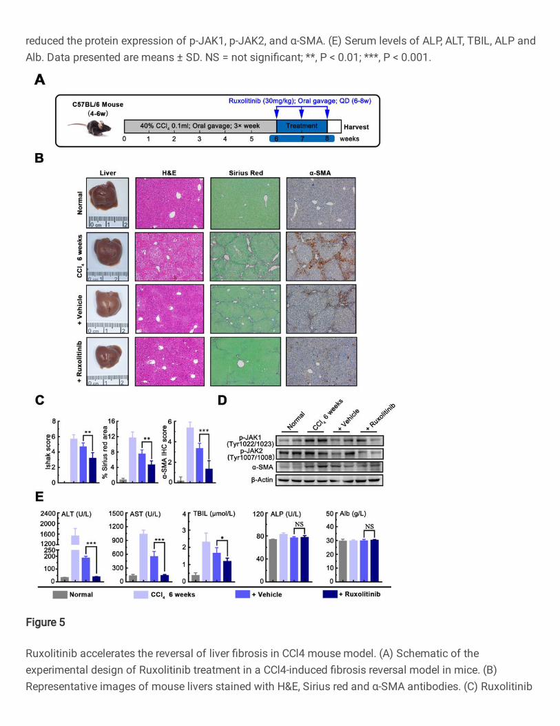
Ruxolitinib accelerates the reversal of liver fibrosis in mice
Since exploring effective intervention for reversing liver fibrosis in meaning for
liver disease, we sought to test whether Ruxolitinib may promote liver fibrosis
reversal in different mice models induced by CCl4 or TAA, which is widely used
as a reversible model for study drug intervention [31]. As shown in Figure 5A,
the liver fibrosis mice were induced by CCl4 for 6 weeks, and then mice were
allowed to recover from 6 weeks to 8 weeks, with or without treatment with
Ruxolitinib. Morphological analysis and tissue staining showed that the

15
development of liver fibrosis and activation of HSCs in the mice treated with
CCl4 for 6 weeks, while partially spontaneous reversion was found in 8-week-
old mice without Ruxolitinib. Interestingly, the degree of liver fibrosis and the
activation of HSCs were significantly reversed in 8-week-old mice treated with
Ruxolitinib for 2 weeks which is confirmed by western blot (Fig.5B-D).
Furthermore, the serum markers of hepatic function were examined, and as
shown in Figure 5E, the levels of ALT, AST, and TBIL were significantly reduced
in mice with Ruxolitinib compared that those without Ruxolitinib, indicating
Ruxolitinib may promote the improvement of liver function in mice.
To confirm the reversal effect of Ruxolitinib, another liver fibrosis mice model
induced by TAA was used and consistent results were found. As shown in
Figure 6A, the mice were administered with TAA for 6 weeks, and then allowed
to recover from 6 weeks to 10 weeks, with or without treatment with Ruxolitinib.
Similarly, Ruxolitinib was found to significantly reversed the degree of liver
fibrosis and the activation of HSCs (Fig.6B-D) and decreased the levels of ALT,
AST and ALP in 10 weeks mice. In conclusion, Ruxolitinib accelerates reversal
of liver fibrosis and improve the liver damage in different mice model induced
by CCl4 or TAA.
Discussion
Liver fibrosis and cirrhosis are major health problems worldwide, causing more
than 1 million deaths per year [32], for which there are currently no approved

16
effective drugs [33]. Activated HSCs play a crucial role in liver fibrosis. However,
the molecular mechanisms by which HSCs are activated and converted to a
fibroblast phenotype are not fully understood. In this study, we found that
enhanced JAK1 and JAK2 expression were associated with liver
fibrosis/cirrhosis and liver cancer. IHC staining further demonstrated that the
upregulation of JAK1 and JAK2 was mainly localized in hepatocytes and HSCs.
More interestingly, we found that expression of JAK1 and JAK2 was positively
correlated with the progression of liver cancer and the severity of liver fibrosis.
Further, silencing of JAK1 and JAK2 down-regulated its downstream signaling
and inhibited proliferation, activation, migration of HSCs. JAK1/2 inhibition had
similar actions on HSCs in a concentration-dependent model in vitro and
obviously attenuated the progress of liver fibrosis, promoted its reversal, and
improved the liver damage in different liver fibrosis mice model induced by CCl4
or TAA. Therefore, JAK1/2 may be considered as a potential marker of activated
HSCs and therapeutic targets in the treatment of liver fibrosis.
The JAK family is a class of non-receptor tyrosine kinases, play an important
role in the development of many diseases, especially JAK2, which has been
treated as a target of myeloproliferative diseases [34]. The existing reports
suggests that STAT3 plays an important role in the development of liver fibrosis
and demonstrates that STAT3 selective inhibitors can effectively attenuate the
progression of liver fibrosis [21,35-37]. However, JAK1 and JAK2, which are
upstream of STAT3, have been mainly demonstrated to play a role in blood

17
system diseases such as myelofibrosis and lymphoma [22,23]. Interestingly,
this study showed that JAK1/2 expression was up-regulated in liver fibrosis and
HCC, and further positively associated with liver cancer progression and the
severity of liver fibrosis, indicating that JAK1/2 may take an action in liver
fibrosis. Activated HSCs have been demonstrated to play a central role in liver
fibrogenesis by producing most of the ECM. HSCs are quiescent, and the
underlying mechanism of activation is still not clear. We found We found JAK1/2
promoted the proliferation and activation of HSCs in vitro. Take together, these
results suggest that JAK1/2 promotes activation of HSCs and may be useful
markers to monitor liver fibrosis and HCC development.
Subsequently evidence has demonstrated that Ruxolitinib had significant
antifibrotic activity both in vitro and in vivo. Ruxolitinib is the most potent JAK1/2
inhibitor for blocking JAK1 phosphorylation at Tyr1022/1023 and JAK2
phosphorylation at Tyr1007/1008. A variety of chronic liver diseases lead to
cirrhosis and HCC associated with high morbidity and mortality, while the
treatments for advanced liver fibrosis and cirrhosis are still unsatisfactory.
Therefore, understanding the mechanism and finding the effective drug to treat
and reverse liver fibrosis are urgently needed [38]. Our date showed that
Ruxolitinib significantly inhibited the activation of HSCs in vitro and suppressed
liver fibrosis progression and accelerated reversal of liver fibrosis in
independent murine models. We speculate that these phenomenon are mainly
due to the anti-fibrotic and hepatoprotective effects of Ruxolitinib. However, the

18
specific mechanism underlying these phenomenon are not fully elucidated.
Further studies to explore the mechanism by primary HSCs and to investigate
whether JAK1/2 inhibition prevents recurrence of HCC with the background of
liver fibrosis in needed.
More interestingly, we observed that Ruxolitinib has a good effect on improving
liver function in different liver fibrosis models, especially improving acute liver
injury such as transaminase and bilirubin, which caused our concern. Patients
with advanced cirrhosis and liver cancer miss effective treatment opportunities
due to the accompanying liver function damage. Ruxolitinib has been shown to
significantly improve acute liver injury, reduce transaminase and bilirubin, and
achieve the purpose of improving liver function, which may win the opportunity
for HCC patients to get anti-tumor treatments.
To date, there are no efficient anti-fibrotic therapies available for chronic liver
disease and HCC. Innovative medical treatments to stop or even reverse
fibrosis are urgently needed. Therefore, JAK1 and JAK2 might play a key role
in the process of liver fibrogenesis, and its potential inhibitor, Ruxolitinib, may
be clinically useful in preventing or treating liver fibrosis (Fig.7).
Conclusions
In conclusion, JAK1/2 regulate the biological function of HSCs, highlighting its
role in liver fibrosis and early prevention of HCC development and its inhibitor,
Ruxolitinib, may be an effective drug for preventing and reversing liver fibrosis.

19
Abbreviations: Alb, albumin; ALP, Alkaline phosphatase; ALT, Alanine
aminotransferase; α-SMA, Alpha-Smooth muscle actin; AST, Aspartate
aminotransferase; CCl4, Carbon tetrachloride; COL1A1, Collagen type I alpha
1; ECM, Extracellular matrix; FDA, Food and Drug Administration; HCC,
Hepatocellular carcinoma; H&E, Hematoxylin-eosin; HSCs, Hepatic stellate
cells; IC50, half maximal inhibitory concentration; IHC, immunohistochemistry;
JAK, Janus kinase; JAK/STAT, Janus kinase/Signal Transducer and Activator
of Transcription; NASH, nonalcoholic steatohepatitis; PAI-1, Plasminogen
activator Inhibitor-1; PDGFRβ, Platelet-derived growth factor receptor beta;
siRNA, small interfering RNA; SSc, Systemic sclerosis; TAA, Thioacetamide;
TBIL, total bilirubin
Declarations
Ethics approval and consent to participate
All animal experiments were conducted in accordance with the National
Institutes of Health guide for the care and use of Laboratory animals and
approved by the Animal Care and Use Committee of Southern Medical
University.
Availability of data and materials
The datasets generated and/or analyzed during the current study are not
publicly available but are available from the corresponding author upon

20
reasonable request.
Competing interests
All authors declare no conflicts of interest.
Funding
This work was supported by the Project of Administration of Traditional Chinese
Medicine of Guangdong Province of China (NO. 20203006); Science and
Technology Program of Guangzhou, China (NO. 202002030075); President's
fund of Integrated hospital of traditional Chinese Medicine (NO. 1201902004).
Authors’ contributions
Zhenghui Song and Xinhui Liu designed the experiments, wrote the manuscript,
and participated in most of the experiments. Yue Luo, Yun Liu assisted in cell
biological experiments. Wan Zhang and Guanqi Dai assisted in animal studies.
Hua Xiao revised the manuscript. Aimin Li and Jian Hong conceived and
supervised the project.
References
1. Higashi T, Friedman SL, Hoshida Y. Hepatic stellate cells as key target in
liver fibrosis. Adv Drug Deliv Rev 2017; 121: 27-42.
2. Qu C, Zheng D, Li S, Liu Y, Lidofsky A, Holmes JA, et al. Tyrosine kinase
SYK is a potential therapeutic target for liver fibrosis. Hepatology 2018; 68(3):
1125-39.
3. Tsochatzis EA, Bosch J, Burroughs AK. Liver cirrhosis. Lancet 2014;
383(9930): 1749-61.
4. Wynn TA, Ramalingam TR. Mechanisms of fibrosis: therapeutic translation

21
for fibrotic disease. Nature medicine 2012; 18(7): 1028-40.
5. Cohen-Naftaly M, Friedman SL. Current status of novel antifibrotic
therapies in patients with chronic liver disease. Therap Adv Gastroenterol 2011;
4(6): 391-417.
6. Bataller R, Brenner DA. Liver fibrosis. J Clin Invest 2005; 115(2): 209-18.
7. Fuchs BC, Hoshida Y, Fujii T, Wei L, Yamada S, Lauwers GY, et al.
Epidermal growth factor receptor inhibition attenuates liver fibrosis and
development of hepatocellular carcinoma. Hepatology (Baltimore, Md) 2014;
59(4): 1577-90.
8. Chen L, Li J, Zhang J, Dai C, Liu X, Wang J, et al. S100A4 promotes liver
fibrosis via activation of hepatic stellate cells. J Hepatol 2015; 62(1): 156-64.
9. Friedman SL. Hepatic stellate cells: protean, multifunctional, and enigmatic
cells of the liver. Physiological reviews 2008; 88(1): 125-72.
10. Krizhanovsky V, Yon M, Dickins RA, Hearn S, Simon J, Miething C, et al.
Senescence of activated stellate cells limits liver fibrosis. Cell 2008; 134(4):
657-67.
11. Drew L. Liver cirrhosis: scar wars. Nature 2018; 564(7736): S73.
12. Ikenaga N, Peng ZW, Vaid KA, Liu SB, Yoshida S, Sverdlov DY, et al.
Selective targeting of lysyl oxidase-like 2 (LOXL2) suppresses hepatic fibrosis
progression and accelerates its reversal. 2017; 66(9): 1697-708.
13. Priceman SJ, Kujawski M, Shen S, Cherryholmes GA, Lee H, Zhang C, et
al. Regulation of adipose tissue T cell subsets by Stat3 is crucial for diet-
induced obesity and insulin resistance. Proceedings of the National Academy
of Sciences of the United States of America 2013; 110(32): 13079-84.
14. Deng J, Liu Y, Lee H, Herrmann A, Zhang W, Zhang C, et al. S1PR1-STAT3
signaling is crucial for myeloid cell colonization at future metastatic sites.
Cancer cell 2012; 21(5): 642-54.
15. Park EJ, Lee JH, Yu GY, He G, Ali SR, Holzer RG, et al. Dietary and genetic
obesity promote liver inflammation and tumorigenesis by enhancing IL-6 and
22
TNF expression. Cell 2010; 140(2): 197-208.
16. Carro MS, Lim WK, Alvarez MJ, Bollo RJ, Zhao X, Snyder EY, et al. The
transcriptional network for mesenchymal transformation of brain tumours.
Nature 2010; 463(7279): 318-25.
17. Marotta LL, Almendro V, Marusyk A, Shipitsin M, Schemme J, Walker SR,
et al. The JAK2/STAT3 signaling pathway is required for growth of
CD44(+)CD24(-) stem cell-like breast cancer cells in human tumors. The
Journal of clinical investigation 2011; 121(7): 2723-35.
18. Schroeder A, Herrmann A, Cherryholmes G, Kowolik C, Buettner R, Pal S,
et al. Loss of androgen receptor expression promotes a stem-like cell
phenotype in prostate cancer through STAT3 signaling. Cancer Res 2014; 74(4):
1227-37.
19. Yu H, Lee H, Herrmann A, Buettner R, Jove R. Revisiting STAT3 signalling
in cancer: new and unexpected biological functions. Nat Rev Cancer 2014;
14(11): 736-46.
20. Kong X, Horiguchi N, Mori M, Gao B. Cytokines and STATs in Liver Fibrosis.
Frontiers in physiology 2012; 3: 69.
21. Deng YR, Ma HD, Tsuneyama K, Yang W, Wang YH, Lu FT, et al. STAT3-
mediated attenuation of CCl4-induced mouse liver fibrosis by the protein kinase
inhibitor sorafenib. Journal of autoimmunity 2013; 46: 25-34.
22. Zhang Y, Liang R, Chen CW, Mallano T, Dees C, Distler A, et al. JAK1-
dependent transphosphorylation of JAK2 limits the antifibrotic effects of
selective JAK2 inhibitors on long-term treatment. Annals of the rheumatic
diseases 2017; 76(8): 1467-75.
23. Chakraborty D, Šumová B, Mallano T, Chen CW, Distler A, Bergmann C, et
al. Activation of STAT3 integrates common profibrotic pathways to promote
fibroblast activation and tissue fibrosis. Nat Commun 2017; 8(1): 1130.
24. Verstovsek S, Mesa RA, Gotlib J, Levy RS, Gupta V, DiPersio JF, et al. A
double-blind, placebo-controlled trial of ruxolitinib for myelofibrosis. N Engl J

23
Med 2012; 366(9): 799-807.
25. Harrison C, Kiladjian JJ, Al-Ali HK, Gisslinger H, Waltzman R, Stalbovskaya
V, et al. JAK inhibition with ruxolitinib versus best available therapy for
myelofibrosis. N Engl J Med 2012; 366(9): 787-98.
26. Quintás-Cardama A, Vaddi K, Liu P, Manshouri T, Li J, Scherle PA, et al.
Preclinical characterization of the selective JAK1/2 inhibitor INCB018424:
therapeutic implications for the treatment of myeloproliferative neoplasms.
Blood 2010; 115(15): 3109-17.
27. Vannucchi AM, Kiladjian JJ, Griesshammer M, Masszi T, Durrant S,
Passamonti F, et al. Ruxolitinib versus standard therapy for the treatment of
polycythemia vera. The New England journal of medicine 2015; 372(5): 426-35.
28. Scott LM, Gandhi MK. Deregulated JAK/STAT signalling in
lymphomagenesis, and its implications for the development of new targeted
therapies. Blood Rev 2015; 29(6): 405-15.
29. Ding N, Yu RT, Subramaniam N, Sherman MH, Wilson C, Rao R, et al. A
vitamin D receptor/SMAD genomic circuit gates hepatic fibrotic response. Cell
2013; 153(3): 601-13.
30. Hong J, Hu K, Yuan Y, Sang Y, Bu Q, Chen G, et al. CHK1 targets spleen
tyrosine kinase (L) for proteolysis in hepatocellular carcinoma. The Journal of
clinical investigation 2012; 122(6): 2165-75.
31. Popov Y, Sverdlov DY, Sharma AK, Bhaskar KR, Li S, Freitag TL, et al.
Tissue transglutaminase does not affect fibrotic matrix stability or regression of
liver fibrosis in mice. Gastroenterology 2011; 140(5): 1642-52.
32. Mokdad AA, Lopez AD, Shahraz S, Lozano R, Mokdad AH, Stanaway J, et
al. Liver cirrhosis mortality in 187 countries between 1980 and 2010: a
systematic analysis. BMC Med 2014; 12: 145.
33. Troeger JS, Mederacke I, Gwak GY, Dapito DH, Mu X, Hsu CC, et al.
Deactivation of hepatic stellate cells during liver fibrosis resolution in mice.
Gastroenterology 2012; 143(4): 1073-83.e22.

24
34. Rumi E, Cazzola M. Diagnosis, risk stratification, and response evaluation
in classical myeloproliferative neoplasms. 2017; 129(6): 680-92.
35. Choi S, Jung HJ, Kim MW, Kang JH, Shin D, Jang YS, et al. A novel STAT3
inhibitor, STX-0119, attenuates liver fibrosis by inactivating hepatic stellate cells
in mice. Biochemical and biophysical research communications 2019.
36. Nunez Lopez O, Bohanon FJ, Wang X, Ye N, Corsello T, Rojas-Khalil Y, et
al. STAT3 Inhibition Suppresses Hepatic Stellate Cell Fibrogenesis: HJC0123,
a Potential Therapeutic Agent for Liver Fibrosis. Laboratory investigation; a
journal of technical methods and pathology 2016; 6(102): 100652-63.
37. Wang Z, Li J, Xiao W, Long J. The STAT3 inhibitor S3I-201 suppresses
fibrogenesis and angiogenesis in liver fibrosis. 2018; 98(12): 1600-13.
38. Lee UE, Friedman SL. Mechanisms of hepatic fibrogenesis. Best practice
& research Clinical gastroenterology 2011; 25(2): 195-206.
Figure legends
Fig.1. Up-regulated JAK1 and JAK2 are associated with liver fibrosis and
liver disease in human and mice. (A) JAK1 and JAK2 immunohistochemistry
(IHC) was performed in human liver tissues microarray of normal, cirrhosis, and
cancer (magnification 100X). (B) JAK1 and JAK2 scores quantified by IHC in
normal liver, liver cirrhosis, and liver cancer. (C) The IHC and Sirius red staining
of JAK1 and JAK2 were performed in mouse normal and fibrosis for 6 weeks
CCl4-induced or 8 weeks CCl4-induced liver tissues (magnification 100X and
200X). (D) JAK1 and JAK2 scores quantified by IHC in normal liver, 6 weeks
CCl4-induced liver fibrosis and 8 weeks CCl4-induced liver fibrosis. Data
presented are means ± SD. **, P < 0.01; ***, P < 0.001.
Fig.2. Knockdown of JAK1 and JAK2 inhibits activation, proliferation and

25
migration of HSCs. (A) The left panel shows LX-2 cells co-interfered with
siJAK1 and siJAK2 reduced JAK1 and JAK2 mRNA expression. The right panel
shows that siJAK2 reduced JAK1 and JAK2 protein expression in LX-2 cells co-
interfered with siJAK1. (B) The left panel shows that knockdown JAK1 and
JAK2 reduced mRNA expression of ACTA2, PDGFRβ in LX-2 cells. The right
panel shows knockdown JAK1 and JAK2 reduced protein expression of α-SMA,
PDGFRβ. (C) Knockdown expression of JAK1 and JAK2 inhibited LX-2 cell
proliferation. (D) The left panel shows knockdown JAK1 and JAK2 inhibited
migration (magnification 100X). The right shows the number of migrations
counted after interfered with JAK1 and JAK2 in LX-2 cells. (E) LX-2 cells
knockdown JAK1 and JAK2 reduced protein expression of p-JAK1
(Tyr1022/1023), p-JAK2 (Tyr1007/1008), STAT3, p-STAT3 (Tyr705), p-STAT3
(Ser727). Date presented are means ± SD. *, P <0.05; **, P < 0.01; ***, P <
0.001.
Fig.3. JAK1 and JAK2 antagonism had anti-fibrotic activity in vitro. (A) The
protein levels of JAK downstream in LX-2 cells treated with different
concentrations of Ruxolitinib. (B) Ruxolitinib inhibited cell proliferative effect in
LX-2 cells in a dose and time-dependent manner. (C) The half-maximal
inhibitory concentration (IC50) value of Ruxolitinib on LX-2 cells. (D) The upper
panel shows relative mRNA expression of ACTA2, PDGFRβ, COL1A1, and PAI-
1 in LX-2 cells treated with different concentrations of Ruxolitinib. The below
shows protein expression of α-SMA, PDGFRβ, and PAI-1. (E) The left panel

26
shows that treatment with Ruxolitinib inhibited LX-2 cell proliferation
(magnification 100X). The right panel shows the number of migration counted
in LX-2 cells treated with different concentrations of Ruxolitinib. (F) The left
panel shows that treatment with Ruxolitinib promoted LX-2 cells apoptosis. The
right panel shows the apoptosis rate of LX-2 cells treated with different
concentrations of Ruxolitinib. Data presented are means ± SD. NS = not
significant; *, P < 0.05; **, P < 0.01; ***, P < 0.001.
Fig.4. Ruxolitinib attenuates the progression of liver pan-lobular fibrosis
induced by CCl4. (A) Schematic of the experimental design of Ruxolitinib
treatment in a CCl4-induced fibrosis progression model in mice. (B)
Representative images of mouse livers stained with H&E, Sirius red, and α-
SMA antibodies. (C) Ruxolitinib reduced Ishak fibrosis score, and Sirius red, α-
SMA IHC staining (magnification 100X). (D) Ruxolitinib reduced the protein
expression of p-JAK1, p-JAK2, and α-SMA. (E) Serum levels of ALP, ALT, TBIL,
ALP and Alb. Data presented are means ± SD. NS = not significant; **, P < 0.01;
***, P < 0.001.
Fig.5. Ruxolitinib accelerates the reversal of liver fibrosis in CCl4 mouse
model. (A) Schematic of the experimental design of Ruxolitinib treatment in a
CCl4-induced fibrosis reversal model in mice. (B) Representative images of
mouse livers stained with H&E, Sirius red and α-SMA antibodies. (C) Ruxolitinib
reduced Ishak fibrosis score, and Sirius red, α-SMA IHC staining (magnification
100X). (D) Ruxolitinib reduced the protein expression of p-JAK1, p-JAK2, and

27
α-SMA. (E) Serum levels of ALP, ALT, TBIL, ALP, and Alb. Date presented are
means ± SD. NS = not significant; *, P < 0.05; **, P < 0.01; ***, P < 0.001.
Fig.6. Ruxolitinib accelerates the reversal of liver fibrosis in the TAA
mouse model. (A) Schematic of the experimental design of Ruxolitinib
treatment in a TAA-induced fibrosis reversal model in mice. (B) Representative
images of mouse livers stained with H&E, Sirius red and α-SMA antibodies. (C)
Ruxolitinib reduced Ishak fibrosis score, and Sirius red, α-SMA IHC staining
(magnification 100X). (D) Ruxolitinib reduced the protein expression of p-JAK1,
p-JAK2, and α-SMA. (E) Serum levels of ALP, ALT, TBIL, ALP and Alb. Data
presented are means ± SD. NS = not significant; *, P < 0.05; **, P < 0.01; ***, P
< 0.001.
Fig.7. Proposed model of Ruxolitinib suppresses liver fibrosis
progression and accelerates fibrosis reversal via selectively targeting
JAK 1/2.

Figures
Figure 1
Up-regulated JAK1 and JAK2 are associated with liver �brosis and liver disease in human and mice. (A)JAK1 and JAK2 immunohistochemistry (IHC) was performed in human liver tissues microarray of normal,cirrhosis, and cancer (magni�cation 100X). (B) JAK1 and JAK2 scores quanti�ed by IHC in normal liver,liver cirrhosis, and liver cancer. (C) The IHC and Sirius red staining of JAK1 and JAK2 were performed inmouse normal and �brosis for 6 weeks CCl4-induced or 8 weeks CCl4-induced liver tissues (magni�cation100X and 200X). (D) JAK1 and JAK2 scores quanti�ed by IHC in normal liver, 6 weeks CCl4-induced liver�brosis and 8 weeks CCl4-induced liver �brosis. Data presented are means ± SD. **, P < 0.01; ***, P <0.001.

Figure 2
Knockdown of JAK1 and JAK2 inhibits activation, proliferation and migration of HSCs. (A) The left panelshows LX-2 cells co-interfered with siJAK1 and siJAK2 reduced JAK1 and JAK2 mRNA expression. Theright panel shows that siJAK2 reduced JAK1 and JAK2 protein expression in LX-2 cells co-interfered withsiJAK1. (B) The left panel shows that knockdown JAK1 and JAK2 reduced mRNA expression of ACTA2,PDGFRβ in LX-2 cells. The right panel shows knockdown JAK1 and JAK2 reduced protein expression of α-

SMA, PDGFRβ. (C) Knockdown expression of JAK1 and JAK2 inhibited LX-2 cell proliferation. (D) The leftpanel shows knockdown JAK1 and JAK2 inhibited migration (magni�cation 100X). The right shows thenumber of migrations counted after interfered with JAK1 and JAK2 in LX-2 cells. (E) LX-2 cellsknockdown JAK1 and JAK2 reduced protein expression of p-JAK1 (Tyr1022/1023), p-JAK2(Tyr1007/1008), STAT3, p-STAT3 (Tyr705), p-STAT3 (Ser727). Date presented are means ± SD. *, P <0.05;**, P < 0.01; ***, P < 0.001.
Figure 3

JAK1 and JAK2 antagonism had anti-�brotic activity in vitro. (A) The protein levels of JAK downstream inLX-2 cells treated with different concentrations of Ruxolitinib. (B) Ruxolitinib inhibited cell proliferativeeffect in LX-2 cells in a dose and time-dependent manner. (C) The half-maximal inhibitory concentration(IC50) value of Ruxolitinib on LX-2 cells. (D) The upper panel shows relative mRNA expression of ACTA2,PDGFRβ, COL1A1, and PAI-1 in LX-2 cells treated with different concentrations of Ruxolitinib. The belowshows protein expression of α-SMA, PDGFRβ, and PAI-1. (E) The left panel shows that treatment withRuxolitinib inhibited LX-2 cell proliferation (magni�cation 100X). The right panel shows the number ofmigration counted in LX-2 cells treated with different concentrations of Ruxolitinib. (F) The left panelshows that treatment with Ruxolitinib promoted LX-2 cells apoptosis. The right panel shows theapoptosis rate of LX-2 cells treated with different concentrations of Ruxolitinib. Data presented aremeans ± SD. NS = not signi�cant; *, P < 0.05; **, P < 0.01; ***, P < 0.001.

Figure 4
Ruxolitinib attenuates the progression of liver pan-lobular �brosis induced by CCl4. (A) Schematic of theexperimental design of Ruxolitinib treatment in a CCl4-induced �brosis progression model in mice. (B)Representative images of mouse livers stained with H&E, Sirius red, and α-SMA antibodies. (C) Ruxolitinibreduced Ishak �brosis score, and Sirius red, α-SMA IHC staining (magni�cation 100X). (D) Ruxolitinib
reduced the protein expression of p-JAK1, p-JAK2, and α-SMA. (E) Serum levels of ALP, ALT, TBIL, ALP andAlb. Data presented are means ± SD. NS = not signi�cant; **, P < 0.01; ***, P < 0.001.
Figure 5
Ruxolitinib accelerates the reversal of liver �brosis in CCl4 mouse model. (A) Schematic of theexperimental design of Ruxolitinib treatment in a CCl4-induced �brosis reversal model in mice. (B)Representative images of mouse livers stained with H&E, Sirius red and α-SMA antibodies. (C) Ruxolitinib

reduced Ishak �brosis score, and Sirius red, α-SMA IHC staining (magni�cation 100X). (D) Ruxolitinibreduced the protein expression of p-JAK1, p-JAK2, and α-SMA. (E) Serum levels of ALP, ALT, TBIL, ALP, andAlb. Date presented are means ± SD. NS = not signi�cant; *, P < 0.05; **, P < 0.01; ***, P < 0.001.
Figure 6
Ruxolitinib accelerates the reversal of liver �brosis in the TAA mouse model. (A) Schematic of theexperimental design of Ruxolitinib treatment in a TAA-induced �brosis reversal model in mice. (B)

Representative images of mouse livers stained with H&E, Sirius red and α-SMA antibodies. (C) Ruxolitinibreduced Ishak �brosis score, and Sirius red, α-SMA IHC staining (magni�cation 100X). (D) Ruxolitinibreduced the protein expression of p-JAK1, p-JAK2, and α-SMA. (E) Serum levels of ALP, ALT, TBIL, ALP andAlb. Data presented are means ± SD. NS = not signi�cant; *, P < 0.05; **, P < 0.01; ***, P < 0.001.
Figure 7
Proposed model of Ruxolitinib suppresses liver �brosis progression and accelerates �brosis reversal viaselectively targeting JAK 1/2.
Supplementary Files
This is a list of supplementary �les associated with this preprint. Click to download.
SupplementalMaterial.pdf