Ruxolitinib reverses dysregulated T helper cell responses ...
14
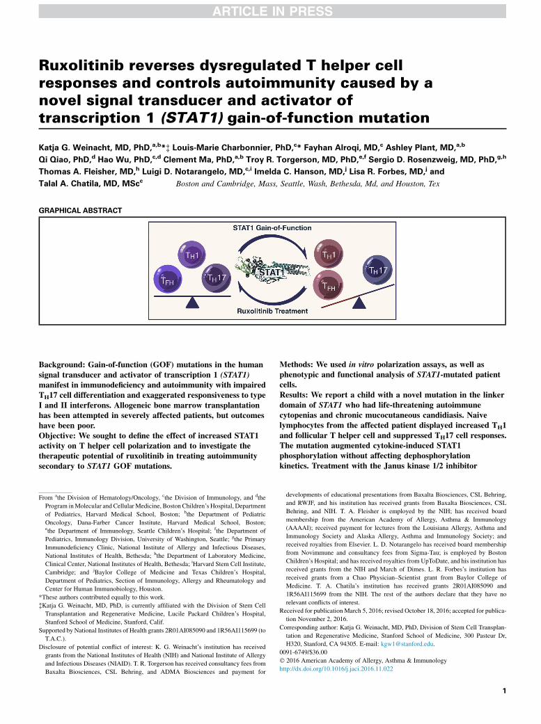
Ruxolitinib reverses dysregulated T helper cell responses and controls autoimmunity caused by a novel signal transducer and activator of transcription 1 (STAT1) gain-of-function mutation Katja G. Weinacht, MD, PhD, a,b *à Louis-Marie Charbonnier, PhD, c * Fayhan Alroqi, MD, c Ashley Plant, MD, a,b Qi Qiao, PhD, d Hao Wu, PhD, c,d Clement Ma, PhD, a,b Troy R. Torgerson, MD, PhD, e,f Sergio D. Rosenzweig, MD, PhD, g,h Thomas A. Fleisher, MD, h Luigi D. Notarangelo, MD, c,i Imelda C. Hanson, MD, j Lisa R. Forbes, MD, j and Talal A. Chatila, MD, MSc c Boston and Cambridge, Mass, Seattle, Wash, Bethesda, Md, and Houston, Tex GRAPHICAL ABSTRACT Background: Gain-of-function (GOF) mutations in the human signal transducer and activator of transcription 1 (STAT1) manifest in immunodeficiency and autoimmunity with impaired T H 17 cell differentiation and exaggerated responsiveness to type I and II interferons. Allogeneic bone marrow transplantation has been attempted in severely affected patients, but outcomes have been poor. Objective: We sought to define the effect of increased STAT1 activity on T helper cell polarization and to investigate the therapeutic potential of ruxolitinib in treating autoimmunity secondary to STAT1 GOF mutations. Methods: We used in vitro polarization assays, as well as phenotypic and functional analysis of STAT1-mutated patient cells. Results: We report a child with a novel mutation in the linker domain of STAT1 who had life-threatening autoimmune cytopenias and chronic mucocutaneous candidiasis. Naive lymphocytes from the affected patient displayed increased T H 1 and follicular T helper cell and suppressed T H 17 cell responses. The mutation augmented cytokine-induced STAT1 phosphorylation without affecting dephosphorylation kinetics. Treatment with the Janus kinase 1/2 inhibitor From a the Division of Hematology/Oncology, c the Division of Immunology, and d the Program in Molecular and Cellular Medicine, Boston Children’s Hospital, Department of Pediatrics, Harvard Medical School, Boston; b the Department of Pediatric Oncology, Dana-Farber Cancer Institute, Harvard Medical School, Boston; e the Department of Immunology, Seattle Children’s Hospital; f the Department of Pediatrics, Immunology Division, University of Washington, Seattle; g the Primary Immunodeficiency Clinic, National Institute of Allergy and Infectious Diseases, National Institutes of Health, Bethesda; h the Department of Laboratory Medicine, Clinical Center, National Institutes of Health, Bethesda; i Harvard Stem Cell Institute, Cambridge; and j Baylor College of Medicine and Texas Children’s Hospital, Department of Pediatrics, Section of Immunology, Allergy and Rheumatology and Center for Human Immunobiology, Houston. *These authors contributed equally to this work. àKatja G. Weinacht, MD, PhD, is currently affiliated with the Division of Stem Cell Transplantation and Regenerative Medicine, Lucile Packard Children’s Hospital, Stanford School of Medicine, Stanford, Calif. Supported by National Institutes of Health grants 2R01AI085090 and 1R56AI115699 (to T.A.C.). Disclosure of potential conflict of interest: K. G. Weinacht’s institution has received grants from the National Institutes of Health (NIH) and National Institute of Allergy and Infectious Diseases (NIAID). T. R. Torgerson has received consultancy fees from Baxalta Biosciences, CSL Behring, and ADMA Biosciences and payment for developments of educational presentations from Baxalta Biosciences, CSL Behring, and RWJF, and his institution has received grants from Baxalta Biosciences, CSL Behring, and NIH. T. A. Fleisher is employed by the NIH; has received board membership from the American Academy of Allergy, Asthma & Immunology (AAAAI); received payment for lectures from the Louisiana Allergy, Asthma and Immunology Society and Alaska Allergy, Asthma and Immunology Society; and received royalties from Elsevier. L. D. Notarangelo has received board membership from Novimmune and consultancy fees from Sigma-Tau; is employed by Boston Children’s Hospital; and has received royalties from UpToDate, and his institution has received grants from the NIH and March of Dimes. L. R. Forbes’s institution has received grants from a Chao Physician–Scientist grant from Baylor College of Medicine. T. A. Chatila’s institution has received grants 2R01AI085090 and 1R56AI115699 from the NIH. The rest of the authors declare that they have no relevant conflicts of interest. Received for publication March 5, 2016; revised October 18, 2016; accepted for publica- tion November 2, 2016. Corresponding author: Katja G. Weinacht, MD, PhD, Division of Stem Cell Transplan- tation and Regenerative Medicine, Stanford School of Medicine, 300 Pasteur Dr, H320, Stanford, CA 94305. E-mail: [email protected]. 0091-6749/$36.00 Ó 2016 American Academy of Allergy, Asthma & Immunology http://dx.doi.org/10.1016/j.jaci.2016.11.022 1
Transcript of Ruxolitinib reverses dysregulated T helper cell responses ...
Ruxolitinib reverses dysregulated T helper cellresponses and controls autoimmunity caused by anovel signal transducer and activator oftranscription 1 (STAT1) gain-of-function mutation
Katja G. Weinacht, MD, PhD,a,b*� Louis-Marie Charbonnier, PhD,c* Fayhan Alroqi, MD,c Ashley Plant, MD,a,b
Qi Qiao, PhD,d Hao Wu, PhD,c,d Clement Ma, PhD,a,b Troy R. Torgerson, MD, PhD,e,f Sergio D. Rosenzweig, MD, PhD,g,h
Thomas A. Fleisher, MD,h Luigi D. Notarangelo, MD,c,i Imelda C. Hanson, MD,j Lisa R. Forbes, MD,j and
Talal A. Chatila, MD, MScc Boston and Cambridge, Mass, Seattle, Wash, Bethesda, Md, and Houston, Tex
GRAPHICAL ABSTRACT
Background: Gain-of-function (GOF) mutations in the humansignal transducer and activator of transcription 1 (STAT1)manifest in immunodeficiency and autoimmunity with impairedTH17 cell differentiation and exaggerated responsiveness to typeI and II interferons. Allogeneic bone marrow transplantationhas been attempted in severely affected patients, but outcomeshave been poor.Objective: We sought to define the effect of increased STAT1activity on T helper cell polarization and to investigate thetherapeutic potential of ruxolitinib in treating autoimmunitysecondary to STAT1 GOF mutations.
From athe Division of Hematology/Oncology, cthe Division of Immunology, and dthe
Program inMolecular and CellularMedicine, Boston Children’s Hospital, Department
of Pediatrics, Harvard Medical School, Boston; bthe Department of Pediatric
Oncology, Dana-Farber Cancer Institute, Harvard Medical School, Boston;ethe Department of Immunology, Seattle Children’s Hospital; fthe Department of
Pediatrics, Immunology Division, University of Washington, Seattle; gthe Primary
Immunodeficiency Clinic, National Institute of Allergy and Infectious Diseases,
National Institutes of Health, Bethesda; hthe Department of Laboratory Medicine,
Clinical Center, National Institutes of Health, Bethesda; iHarvard Stem Cell Institute,
Cambridge; and jBaylor College of Medicine and Texas Children’s Hospital,
Department of Pediatrics, Section of Immunology, Allergy and Rheumatology and
Center for Human Immunobiology, Houston.
*These authors contributed equally to this work.
�Katja G. Weinacht, MD, PhD, is currently affiliated with the Division of Stem Cell
Transplantation and Regenerative Medicine, Lucile Packard Children’s Hospital,
Stanford School of Medicine, Stanford, Calif.
Supported by National Institutes of Health grants 2R01AI085090 and 1R56AI115699 (to
T.A.C.).
Disclosure of potential conflict of interest: K. G. Weinacht’s institution has received
grants from the National Institutes of Health (NIH) and National Institute of Allergy
and Infectious Diseases (NIAID). T. R. Torgerson has received consultancy fees from
Baxalta Biosciences, CSL Behring, and ADMA Biosciences and payment for
Methods: We used in vitro polarization assays, as well asphenotypic and functional analysis of STAT1-mutated patientcells.Results: We report a child with a novel mutation in the linkerdomain of STAT1 who had life-threatening autoimmunecytopenias and chronic mucocutaneous candidiasis. Naivelymphocytes from the affected patient displayed increased TH1and follicular T helper cell and suppressed TH17 cell responses.The mutation augmented cytokine-induced STAT1phosphorylation without affecting dephosphorylationkinetics. Treatment with the Janus kinase 1/2 inhibitor
developments of educational presentations from Baxalta Biosciences, CSL Behring,
and RWJF, and his institution has received grants from Baxalta Biosciences, CSL
Behring, and NIH. T. A. Fleisher is employed by the NIH; has received board
membership from the American Academy of Allergy, Asthma & Immunology
(AAAAI); received payment for lectures from the Louisiana Allergy, Asthma and
Immunology Society and Alaska Allergy, Asthma and Immunology Society; and
received royalties from Elsevier. L. D. Notarangelo has received board membership
from Novimmune and consultancy fees from Sigma-Tau; is employed by Boston
Children’s Hospital; and has received royalties from UpToDate, and his institution has
received grants from the NIH and March of Dimes. L. R. Forbes’s institution has
received grants from a Chao Physician–Scientist grant from Baylor College of
Medicine. T. A. Chatila’s institution has received grants 2R01AI085090 and
1R56AI115699 from the NIH. The rest of the authors declare that they have no
relevant conflicts of interest.
Received for publication March 5, 2016; revised October 18, 2016; accepted for publica-
tion November 2, 2016.
Corresponding author: Katja G. Weinacht, MD, PhD, Division of Stem Cell Transplan-
tation and Regenerative Medicine, Stanford School of Medicine, 300 Pasteur Dr,
H320, Stanford, CA 94305. E-mail: [email protected].
0091-6749/$36.00
� 2016 American Academy of Allergy, Asthma & Immunology
http://dx.doi.org/10.1016/j.jaci.2016.11.022
1
Abbreviations used
ATG: Anti-thymocyte globulin
BSA: Body surface area
GOF: Gain of function
IC50: Half-maximal inhibitory concentration
ICOS: Inducible costimulator
JAK: Janus kinase
PD: Programmed death
STAT: Signal transducer and activator of transcription
TC1: Cytotoxic T type 1
TFH: Follicular T helper
J ALLERGY CLIN IMMUNOL
nnn 2017
2 WEINACHT ET AL
ruxolitinib reduced hyperresponsiveness to type I and IIinterferons, normalized TH1 and follicular T helper cellresponses, improved TH17 differentiation, cured mucocutaneouscandidiasis, and maintained remission of immune-mediatedcytopenias.Conclusions: Autoimmunity and infection caused by STAT1GOF mutations are the result of dysregulated T helper cellresponses. Janus kinase inhibitor therapy could represent aneffective targeted treatment for long-term disease control inseverely affected patients for whom hematopoietic stem celltransplantation is not available. (J Allergy Clin Immunol2017;nnn:nnn-nnn.)
Key words: STAT1 gain of function, IFN-g, ruxolitinib, autoimmu-nity, TH1 cell, TH17 cell, follicular T helper cell, T helper cellpolarization
Signal transducer and activator of transcription 1 (STAT1) is amember of the STAT family of transcription factors which play akey role in the cellular response to interferons and is a centralcomponent in many other signaling pathways, including in-terleukins, growth factors, and hormones. In response to extra-cellular receptor stimulation, Janus kinase (JAK) activation leadsto phosphorylation of cytoplasmic STAT1, followed by homo-dimerization or heterodimerization with other phosphorylatedSTAT family members. The dimers translocate into the nucleusand bind designated promoter elements to activate transcriptionof their respective target genes.1-3
STAT1 is the target of both loss-of-function and gain-of-function (GOF) mutations. Whereas the former are associatedwith susceptibility to mycobacterial and/or viral infections, thelatter give rise to a mixed phenotype of autoimmunity, mucocu-taneous candidiasis, and invasive fungal infections related toaugmented TH1 and diminished TH17 cell responses.4-9 STAT1GOF mutations prompt a signal-induced increase in levels ofphosphorylated STAT1 (phospho-STAT) and amplifiedtranscription of interferon-responsive genes, which lead toautoimmunity.5,6,10 Delayed dephosphorylation with ensuingaccumulation of phospho-STAT1 in the nucleus has beenproposed as a mechanistic basis in most reportedcases.1,6,11-15 How increased STAT1 activity compromises TH17immunity to result in chronic mucocutaneous candidiasis andother invasive fungal and viral infections is less wellunderstood.12,16,17 Excessive production of interferons, IL-27,and programmed death (PD) 1 ligand can directly impairTH17 cell differentiation.18,19 Alternatively, predominance ofSTAT1 signaling over STAT3 signaling might deviate theresponse to IL-6, IL-21, and IL-23 away from STAT3, whichnormally mediates TH17 cell development.1,18
The clinicalmanagement of patientswithSTAT1GOFmutationsremains challenging.20-22 In particular, controlling autoimmunityis difficult because conventional immunosuppression adds to thealready increased risk of infections. Therapy-refractory or life-threatening disease is considered a noncanonical indication forallogeneic hematopoietic stem cell transplantation; however, theimmune phenotype of STAT1 GOF mutations amplifies thetransplant-related risk for uncontrolled infections and graft-versus-host disease, contributing further to the poor prognosis.21,23
Liu et al24 were the first to provide proof of principle that JAKinhibitors can successfully treat STAT1-mediated hyperrespon-siveness to interferons in patients with vascular and pulmonary
syndrome caused by mutations in TMEM173, which encodesthe stimulator of interferon genes. Higgins et al10 reported hairregrowth in a patient with alopecia areata secondary to a STAT1GOF mutation after treatment with ruxolitinib. Most recently,M€ossner et al25 observed improvement of chronicmucocutaneouscandidiasis with ruxolitinib and a reactive increase in IL-17A/Flevels.
Here we describe the immunophenotypic analysis of a patientwith life-threatening autoimmune cytopenias and a novel GOFmutation in the linker domain of STAT1. Importantly, in additionto increasing TH1 and suppressing TH17 cell differentiation, theaugmented STAT1 activity dysregulated follicular T helper(TFH) cell responses. This finding was corroborated in adifferent patient with known STAT1T385M GOF mutation in theDNA-binding domain who presented solely with chronic muco-cutaneous candidiasis and opportunistic infections but withoutclinical evidence of autoimmunity.13,26,27 Long-term treatmentwith the JAK inhibitor ruxolitinib decreased the increasedSTAT1 phosphorylation, reversed the dysregulated TH1 and TFH
cell development, improved the previously impaired TH17response, and enabled effective control of the autoimmune cyto-penias. This is the first report demonstratingmechanistic evidencethat pharmacologic manipulation of the JAK-STAT pathway inpatients with STAT1 GOF mutations leads to reversal of the im-mune dysregulation phenotype and provides proof of principlethat JAK inhibitors are not only effective in treating activeautoimmune disease and immunodeficiency secondary tohyperresponsiveness of STAT1 but also in reversing the aberrantpriming of naive cells, thereby maintaining long-term diseasecontrol and sustained remission.
METHODS
Patients and healthy subjectsAll study participants were recruited after obtaining written informed
consent approved by the Boston Children’s Hospital Institutional Review
Board.
PharmacotherapyThe IL-1 receptor antagonist anakinra (Kineret; Sobi, Stockholm, Sweden)
was administered intravenously twice daily at a dose of 100 mg.
Four infusions with equine anti-thymocyte globulin (ATG; Atgam; Pfizer,
New York, NY) were administered intravenously at a dose of 40 mg/kg body
weight per infusion 24 hours apart. Supportive therapy during the infusions
consisted of acetaminophen, diphenhydramine, and methylpredinisolone.
Treatment with intravenous cyclosporine (SandIMMUNE; Novartis, East
Hanover, NJ) was initiated on day 1 of ATG therapy at a dose of 4 mg/kg body
J ALLERGY CLIN IMMUNOL
VOLUME nnn, NUMBER nn
WEINACHT ET AL 3
weight per day and titrated to a serum level of 175 to 250 mg/L. The route of
administration was converted to oral after 4 weeks, maintaining the same
serum target level.
Eculizumab (Soliris; Alexion Pharmaceuticals, Cheshire, Conn) was
administered intravenously at a dose of 600 mg per infusion. Only 1 infusion
was administered because of a lack of efficacy. Supportive therapy during
the infusion consisted of acetaminophen, diphenhydramine, and
methylprednisolone. The patient received a meningococcal vaccination prior
to treatment with eculizumab, as well as meningococcal prophylaxis with
azithromycin for 6 months after infusion.
Rituximab (Rituxan; Genentech, South San Francisco, Calif) was admin-
istered intravenously at a dose of 375 mg/m2 body surface area (BSA) once
weekly for 4 consecutive weeks. Supportive therapy during the infusions
consisted of acetaminophen, diphenhydramine, and methylprednisolone.
Treatment with ruxolitinib (Jakafi; Incyte, Wilmington, Del) was initiated
at a low dose of 5 mg/m2 BSA once daily because of concomitant use of other
CYP3A4-inhibiting medications. The ruxolitinib dose was escalated until the
amount of phospo-STAT1 induced in the patient’s CD41 T cells was equal
to that in healthy control cells. The final therapeutic ruxolitinib dose was
10 mg/m2 BSA per day administered orally in 2 divided doses.
STAT1 sequencingExons 3 to 23 of STAT1, including exon/intron boundaries, were amplified
from genomic DNA by means of PCR and sequenced bidirectionally with
dye-terminator chemistry. PCR amplification of exon 20 was carried out
with the following primers: STAT1E20_F (GATAAGAGCGGGGAGGG)
and STAT1E20_R (TGAAGCTGGACTCAGGC). The mutation was pre-
dicted to be deleterious by using SIFT and PolyPhen-2.28,29
Protein modelingThe STAT1E545K mutant structure was generated by using SWISS-MODEL
with STAT1 structures (PDB code: 1YVL and 1BF5).30-33 Structural alignment
was performed inCoot, andmolecular representationwas displayed in PyMOL.34
Antibodies and flow cytometryMonoclonal antibodies to the following human proteins were used for
staining: CD3 (UCHT1), CD4 (RPA-T4), CD8 (RPA-T8), CD45RA (HI100),
CCR7 (G043H7), inducible costimulator (ICOS; C398.4A), PD1 (eBioJ105),
CXCR5 (MU5UBEE), phospho-STAT1 (KIKSI0803), and phospho-STAT3
(LUVNKLA; all from eBioscience, San Diego, Calif); IL-17 (BL168), IFN-g
(4S.B3), CCR6 (G034E3), and CXCR3 (G025H7; all from BioLegend, San
Diego, Calif); and STAT1 (246523; R&D Systems, Minneapolis, Minn).
Appropriate isotype controls were used in parallel. PBMCs were incubated
with mAbs against surface proteins for 30 minutes on ice.
Intracellular STAT1 stainingSTAT1 staining was performed with an eBioscience Fixation/Permeabili-
zation kit, according to the manufacturer’s instructions.
Phospho-STAT1 and phospho-STAT3 stainingPBMCs were stimulated in complete medium for 20 minutes with
appropriate cytokines: human IFN-b (20 ng/mL; Miltenyi Biotec, Bergisch
Gladbach, Germany), IFN-g, or IL-21 (20 ng/mL; PeproTech, Rocky Hills,
NJ). Subsequently, PBMCs were fixed with 2% paraformaldehyde for
20 minutes on ice, permeabilized with 90% methanol for 30 minutes on ice,
and stained with CD3, CD4, phospho-STAT1, and phospho-STAT3 mAbs in
PBS for 30 minutes.
Ex vivo cytokine detectionPBMCs were isolated from whole blood by means of centrifugation over
a Ficoll gradient and stimulated in complete medium in the presence of
anti-CD2/CD3/CD28 beads (Miltenyi Biotec) and 100 U/mL recombinant
human IL-2 (PeproTech) for 2 days. Subsequently, cell suspensions were
incubatedwith phorbol 12-myristate 13-acetate (50 ng/mL; Sigma-Aldrich, St
Louis, Mo), ionomycin (500 ng/mL; Sigma-Aldrich), and GolgiPlug (BD
Biosciences, San Jose, Calif), according to the manufacturer’s instructions, in
complete medium for 4 hours before surface staining. Permeabilization and
intracellular IFN-g and IL-17 staining were carried out with an eBioscience
Fixation/Permeabilization kit, as described above. Datawere collectedwith an
LSRFortessa cytometer (BD Biosciences) and analyzed with FlowJo software
(TreeStar, Ashland, Ore).
JAK inhibitor treatment in vitroPBMCs were incubated for 4 hours in the presence of different
concentrations of ruxolitinib, which is primarily a JAK1/2 inhibitor (10 or
100 nmol/L; Selleckchem, Houston, Tex), and tofacitinib, which is predom-
inantly a JAK3 inhibitor (10 and 100 nmol/L; Sigma-Aldrich), or vehicle
(dimethyl sulfoxide) alone before stimulation with recombinant human IFN-b
(20 ng/mL; Miltenyi Biotec), IFN-g, or IL-21 (20 ng/mL, PeproTech).
T helper cell subset differentiationCD41 T cells were enriched from PBMCs by means of negative selection
with magnetic beads (Miltenyi Biotec), and naive CD45RA1CCR71CD41 T
cells were then isolated by means of cell sorting with a BD FACSAria cytom-
eter. Naive CD41 T cells were seeded at a concentration of 53 105 cells per
well in a 96-well plate in complete medium and stimulated with anti-CD2/
CD3/CD28 beads (Miltenyi Biotec) alone (TH0 condition) or in the presence
of recombinant human cytokines: IL-12 (20 ng/mL) for TH1 conditions
(BioLegend); IL-6 (20 ng/mL), IL-23 (10 ng/mL; both from BioLegend),
and TGF-b1 (5 ng/mL; R&D Systems) for TH17 conditions; and IL-12
(2 ng/mL), IL-23 (10 ng/mL), and TGF-b1 (5 ng/mL) for TFH conditions.35
Statistical analysisComparisons between the patient and healthy control subjects were
analyzed by using the unpaired Student t test and 1- or 2-way ANOVAwith
posttest analysis. Two-sided P values of less than .05 were considered
statistically significant.
RESULTS
Refractory autoimmune cytopenias associated with
a novel STAT1 GOF mutationA 10-year-old girl with a longstanding history of Evans
syndrome manifesting in autoimmune hemolytic anemia andimmune thrombocytopenia presented to our institution with anacute exacerbation of her disease (in the following referred to as‘‘the’’ patient or patient 1). She required daily packed redblood cell transfusions to keep her hemoglobin level at greaterthan 6 g/dL and experienced systemic bleeding symptomsrefractory to platelet transfusion. At presentation, the patienthad already been treated with a prolonged course of steroids andmultiple doses of intravenous immunoglobulins. Although thesetherapies did not induce remission, withdrawal of steroidsspurred the rate of hemolysis further, necessitating continuedglucocorticoid therapy.
The patient had a history of chronicmucocutaneous candidiasisinvolving her nails, oral mucosa, and vaginal tract. She also hadchronic diarrhea and severe chronic lung disease with respiratoryinsufficiency requiring supplemental oxygen. She was a poorresponder to vaccines and had been treated with immunoglobulinreplacement over extended periods of her life. The parentswere nonconsanguineous, and there was no family history of
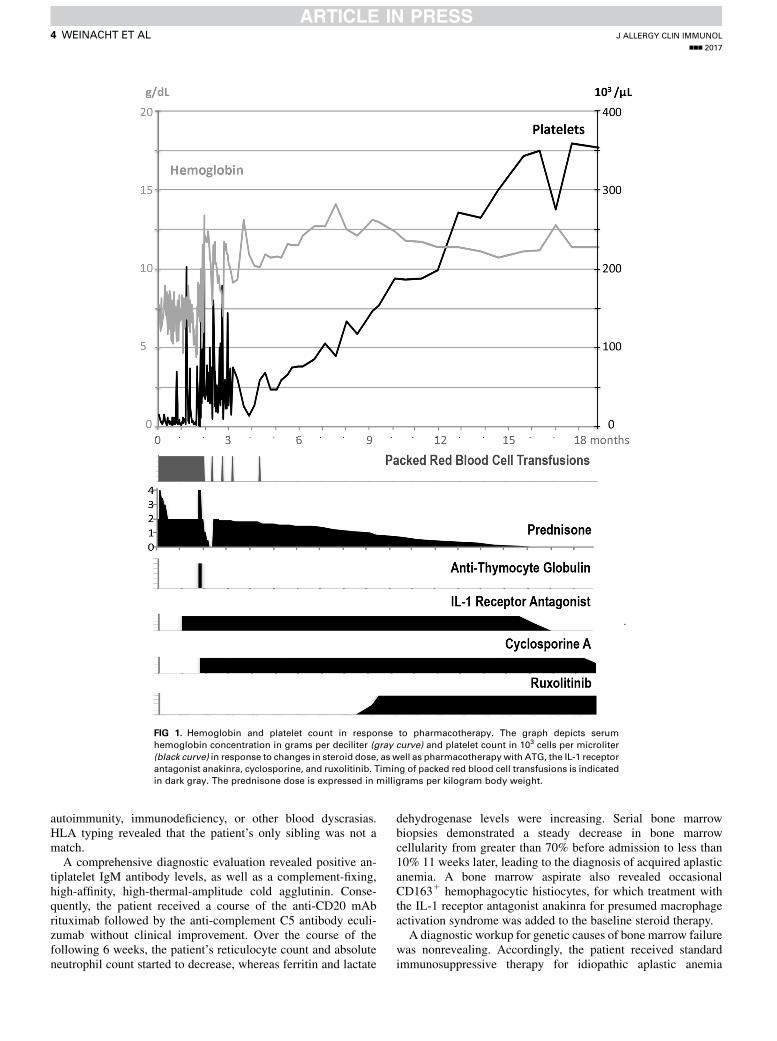
FIG 1. Hemoglobin and platelet count in response to pharmacotherapy. The graph depicts serum
hemoglobin concentration in grams per deciliter (gray curve) and platelet count in 103 cells per microliter
(black curve) in response to changes in steroid dose, as well as pharmacotherapywith ATG, the IL-1 receptor
antagonist anakinra, cyclosporine, and ruxolitinib. Timing of packed red blood cell transfusions is indicated
in dark gray. The prednisone dose is expressed in milligrams per kilogram body weight.
J ALLERGY CLIN IMMUNOL
nnn 2017
4 WEINACHT ET AL
autoimmunity, immunodeficiency, or other blood dyscrasias.HLA typing revealed that the patient’s only sibling was not amatch.
A comprehensive diagnostic evaluation revealed positive an-tiplatelet IgM antibody levels, as well as a complement-fixing,high-affinity, high-thermal-amplitude cold agglutinin. Conse-quently, the patient received a course of the anti-CD20 mAbrituximab followed by the anti-complement C5 antibody eculi-zumab without clinical improvement. Over the course of thefollowing 6 weeks, the patient’s reticulocyte count and absoluteneutrophil count started to decrease, whereas ferritin and lactate
dehydrogenase levels were increasing. Serial bone marrowbiopsies demonstrated a steady decrease in bone marrowcellularity from greater than 70% before admission to less than10% 11 weeks later, leading to the diagnosis of acquired aplasticanemia. A bone marrow aspirate also revealed occasionalCD1631 hemophagocytic histiocytes, for which treatment withthe IL-1 receptor antagonist anakinra for presumed macrophageactivation syndrome was added to the baseline steroid therapy.
A diagnostic workup for genetic causes of bone marrow failurewas nonrevealing. Accordingly, the patient received standardimmunosuppressive therapy for idiopathic aplastic anemia
FIG 2. STAT1E545K mutation leads to hyperphosphorylation GOF. A, Sanger sequencing revealed a
monoallelic c.1633G>A substitution in STAT1. B, Structural alignment between STAT1 structure and
modeled E545K mutant structure (white). WT domains are colored (SH2 in green, dimerization domain in
cyan, and IFN-g peptide in orange) to highlight their position relative to residue 545 (WT structure in yellow
and mutant in magenta). C, Total STAT1 expression in CD41 T cells determined by means of flow
cytometry in patient 1 and a control subject. D, Phospho-STAT1 (P-STAT1) expression in CD41 T cells
stimulated with IFN-b (20 ng/mL) and IFN-g (20 ng/mL) in the patient and a control subject (top) and
dose-response curve with increasing interferon concentrations (bottom). E, Dephosphorylation kinetics
of phospho-STAT1 in response to deprivation of IFN-b and IFN-g in CD41 T cells represented as absolute
mean fluorescence intensity (MFI; top) and normalized to maximum expression before deprivation
(bottom). ***P < .001, 2-way ANOVA.
J ALLERGY CLIN IMMUNOL
VOLUME nnn, NUMBER nn
WEINACHT ET AL 5
without an available matched sibling donor consisting of 4 dosesof equine ATG and cyclosporine. Although the initial posttreat-ment course was complicated by intraventricular hemorrhage andacute respiratory decompensation, over the following 8-weekperiod, the patient’s clinical course stabilized. The absoluteneutrophil and reticulocyte counts began to increase, which isconsistent with bone marrow recovery in response to treatmentwith ATG and cyclosporine. However, hemolysis started to flare,and hemoglobin levels rapidly decreased again as soon as steroidswere weaned, indicating that ATG and cyclosporine successfullytreated the patient’s aplastic anemia but did not bring theautoimmune cytopenias into complete remission (Fig 1 and seeFig E1 in this article’s Online Repository at www.jacionline.org). Together with the development of acute steroid-induced dia-betes, the need for an alternate therapy to control the persistingautoimmune cytopenias became critical.
Unbiased genetic testing for 200 known/selected inborn errorsof immunity identified a monoallelic de novo missense mutation
in the coding region of STAT1 that resulted in an amino acid sub-stitution in the linker domain of the protein and was predicted tobe deleterious by using SIFT and PolyPhen-2 (c.1633G>A;p.E545K; Fig 2, A and B). Structure modeling revealed that theE545 residue is far away from the previously proposed STAT1dimerization interface but close to the SH2 domain, which bindsthe activating IFN-g receptor peptide, suggesting that thismutation might affect STAT dimer binding to cytokine receptorsor kinases.32 This mutation did not affect expression of totalSTAT1 protein in CD41 and CD81T cells or in B cells at baseline(Fig 2, C, and data not shown). However, on stimulation ofPBMCswith IFN-b (20 ng/mL) or IFN-g (20 ng/mL) the patient’sCD41 T cells demonstrated a significant increase in STAT1phosphorylation compared with that seen in control cells(Fig 2, D). Unlike other reported STAT1 GOF mutations, theE545K mutation did not affect the dephosphorylation kineticson cytokine deprivation, which led to normalization ofphospho-STAT1 expression down to baseline levels in cells
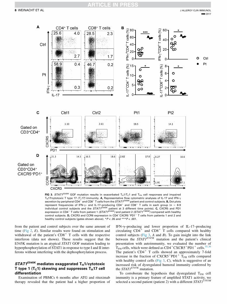
FIG 3. STAT1E545K GOF mutation results in exacerbated TH1/TC1 and TFH cell responses and impaired
TH17/cytotoxic T type 17 (Tc17) immunity. A, Representative flow cytometric analyses of IL-17 and IFN-g
secretion by peripheral CD41 and CD81 T cells from the STAT1E545K patient and control subjects. B,Dot plots
represent frequencies of IFN-g– and IL-17–producing CD41 and CD81 T cells in each group (n 5 8-9
individual control subjects and the STAT1E545K patient at 3 different time points). C, CXCR5 and PD1
expression in CD41 T cells from patient 1 (STAT1E545K) and patient 2 (STAT1T385M) compared with healthy
control subjects. D, CXCR3 and CCR6 expression in CD41CXCR51PD11 T cells from patients 1 and 2 and
healthy control subjects (gates shown above). *P < .05 and ***P < .001.
J ALLERGY CLIN IMMUNOL
nnn 2017
6 WEINACHT ET AL
from the patient and control subjects over the same amount oftime (Fig 2, E). Similar results were found on stimulation andwithdrawal of the patient’s CD81 T cells with the respectiveinterferon (data not shown). These results suggest that theE545K mutation is an atypical STAT1 GOF mutation leading tohyperphosphorylation of STAT1 in response to type I and II inter-ferons without interfering with the dephosphorylation process.
STAT1E545K mediates exaggerated TH1/cytotoxic
T type 1 (TC1) skewing and suppresses TH17 cell
differentiationExamination of PBMCs 6 months after ATG and rituximab
therapy revealed that the patient had a higher proportion of
IFN-g–producing and lower proportion of IL-17–producingcirculating CD41 and CD81 T cells compared with healthycontrol subjects (Fig 3, A and B). To gain insight into the linkbetween the STAT1E545K mutation and the patient’s clinicalpresentation with autoimmunity, we evaluated the number ofTFH cells, which were defined as CD41CXCR51PD11 cells.35,36
The patient’s CD41 T cells showed an approximately 7-foldincrease in the fraction of CXCR51PD11 TFH cells comparedwith healthy control cells (Fig 3, C), which is suggestive of anincreased risk of dysregulated humoral immunity conferred bythe STAT1E545K mutation.
To corroborate the hypothesis that dysregulated TFH cellimmunity is a primary feature of amplified STAT1 activity, weselected a second patient (patient 2) with a different STAT1T385M
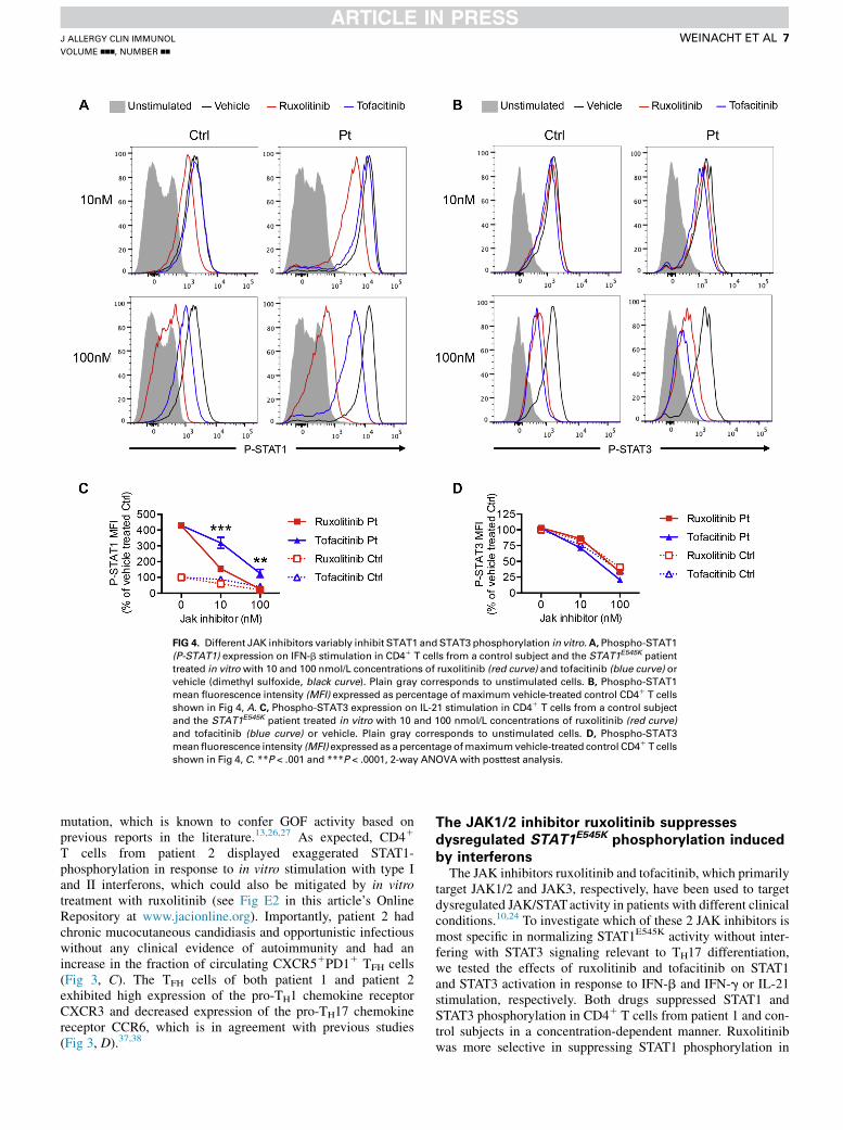
FIG 4. Different JAK inhibitors variably inhibit STAT1 andSTAT3 phosphorylation in vitro.A, Phospho-STAT1
(P-STAT1) expression on IFN-b stimulation in CD41 T cells from a control subject and the STAT1E545K patient
treated in vitrowith 10 and 100 nmol/L concentrations of ruxolitinib (red curve) and tofacitinib (blue curve) or
vehicle (dimethyl sulfoxide, black curve). Plain gray corresponds to unstimulated cells. B, Phospho-STAT1
mean fluorescence intensity (MFI) expressed as percentage of maximum vehicle-treated control CD41 T cells
shown in Fig 4, A. C, Phospho-STAT3 expression on IL-21 stimulation in CD41 T cells from a control subject
and the STAT1E545K patient treated in vitro with 10 and 100 nmol/L concentrations of ruxolitinib (red curve)
and tofacitinib (blue curve) or vehicle. Plain gray corresponds to unstimulated cells. D, Phospho-STAT3
meanfluorescence intensity (MFI) expressed as a percentage ofmaximumvehicle-treated control CD41 T cells
shown in Fig 4, C. **P < .001 and ***P < .0001, 2-way ANOVA with posttest analysis.
J ALLERGY CLIN IMMUNOL
VOLUME nnn, NUMBER nn
WEINACHT ET AL 7
mutation, which is known to confer GOF activity based onprevious reports in the literature.13,26,27 As expected, CD41
T cells from patient 2 displayed exaggerated STAT1-phosphorylation in response to in vitro stimulation with type Iand II interferons, which could also be mitigated by in vitrotreatment with ruxolitinib (see Fig E2 in this article’s OnlineRepository at www.jacionline.org). Importantly, patient 2 hadchronic mucocutaneous candidiasis and opportunistic infectiouswithout any clinical evidence of autoimmunity and had anincrease in the fraction of circulating CXCR51PD11 TFH cells(Fig 3, C). The TFH cells of both patient 1 and patient 2exhibited high expression of the pro-TH1 chemokine receptorCXCR3 and decreased expression of the pro-TH17 chemokinereceptor CCR6, which is in agreement with previous studies(Fig 3, D).37,38
The JAK1/2 inhibitor ruxolitinib suppresses
dysregulated STAT1E545K phosphorylation induced
by interferonsThe JAK inhibitors ruxolitinib and tofacitinib, which primarily
target JAK1/2 and JAK3, respectively, have been used to targetdysregulated JAK/STATactivity in patients with different clinicalconditions.10,24 To investigate which of these 2 JAK inhibitors ismost specific in normalizing STAT1E545K activity without inter-fering with STAT3 signaling relevant to TH17 differentiation,we tested the effects of ruxolitinib and tofacitinib on STAT1and STAT3 activation in response to IFN-b and IFN-g or IL-21stimulation, respectively. Both drugs suppressed STAT1 andSTAT3 phosphorylation in CD41 T cells from patient 1 and con-trol subjects in a concentration-dependent manner. Ruxolitinibwas more selective in suppressing STAT1 phosphorylation in
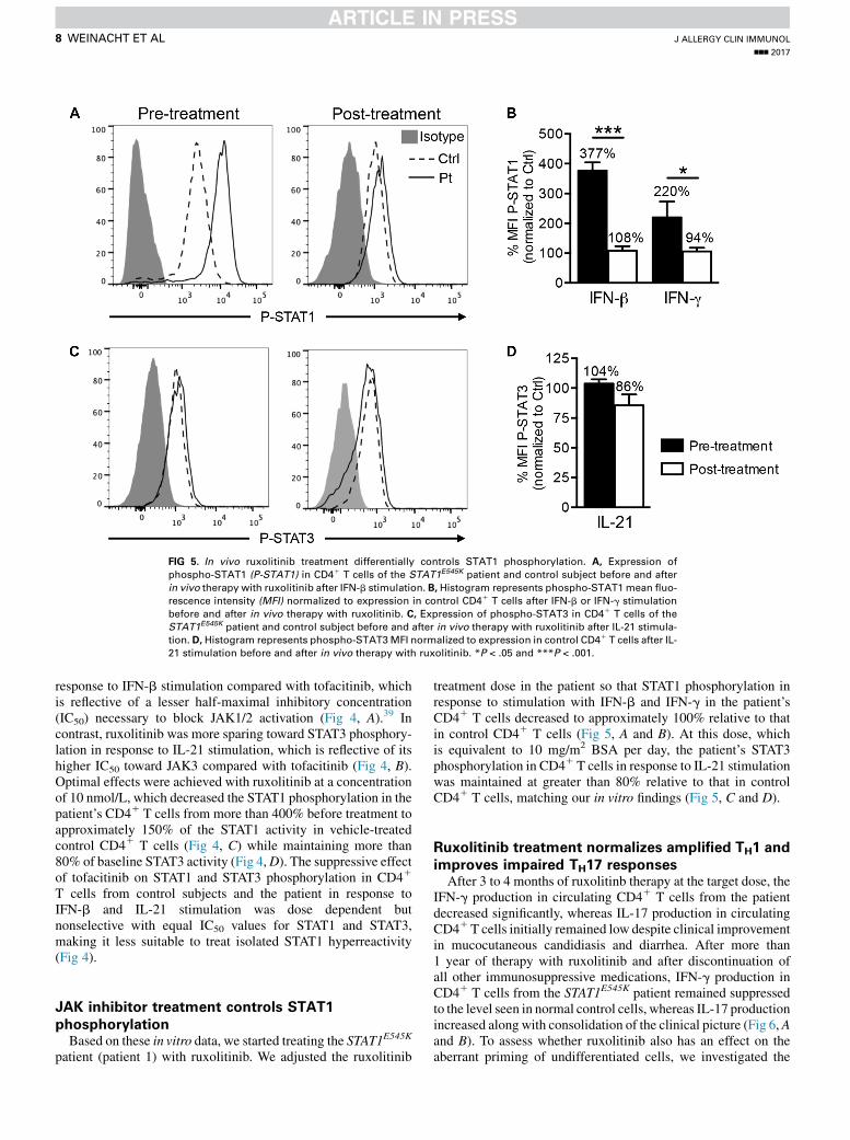
FIG 5. In vivo ruxolitinib treatment differentially controls STAT1 phosphorylation. A, Expression of
phospho-STAT1 (P-STAT1) in CD41 T cells of the STAT1E545K patient and control subject before and after
in vivo therapy with ruxolitinib after IFN-b stimulation. B, Histogram represents phospho-STAT1mean fluo-
rescence intensity (MFI) normalized to expression in control CD41 T cells after IFN-b or IFN-g stimulation
before and after in vivo therapy with ruxolitinib. C, Expression of phospho-STAT3 in CD41 T cells of the
STAT1E545K patient and control subject before and after in vivo therapy with ruxolitinib after IL-21 stimula-
tion. D, Histogram represents phospho-STAT3MFI normalized to expression in control CD41 T cells after IL-
21 stimulation before and after in vivo therapy with ruxolitinib. *P < .05 and ***P < .001.
J ALLERGY CLIN IMMUNOL
nnn 2017
8 WEINACHT ET AL
response to IFN-b stimulation compared with tofacitinib, whichis reflective of a lesser half-maximal inhibitory concentration(IC50) necessary to block JAK1/2 activation (Fig 4, A).39 Incontrast, ruxolitinib was more sparing toward STAT3 phosphory-lation in response to IL-21 stimulation, which is reflective of itshigher IC50 toward JAK3 compared with tofacitinib (Fig 4, B).Optimal effects were achieved with ruxolitinib at a concentrationof 10 nmol/L, which decreased the STAT1 phosphorylation in thepatient’s CD41 T cells from more than 400% before treatment toapproximately 150% of the STAT1 activity in vehicle-treatedcontrol CD41 T cells (Fig 4, C) while maintaining more than80% of baseline STAT3 activity (Fig 4,D). The suppressive effectof tofacitinib on STAT1 and STAT3 phosphorylation in CD41
T cells from control subjects and the patient in response toIFN-b and IL-21 stimulation was dose dependent butnonselective with equal IC50 values for STAT1 and STAT3,making it less suitable to treat isolated STAT1 hyperreactivity(Fig 4).
JAK inhibitor treatment controls STAT1
phosphorylationBased on these in vitro data, we started treating the STAT1E545K
patient (patient 1) with ruxolitinib. We adjusted the ruxolitinib
treatment dose in the patient so that STAT1 phosphorylation inresponse to stimulation with IFN-b and IFN-g in the patient’sCD41 T cells decreased to approximately 100% relative to thatin control CD41 T cells (Fig 5, A and B). At this dose, whichis equivalent to 10 mg/m2 BSA per day, the patient’s STAT3phosphorylation in CD41 T cells in response to IL-21 stimulationwas maintained at greater than 80% relative to that in controlCD41 T cells, matching our in vitro findings (Fig 5, C and D).
Ruxolitinib treatment normalizes amplified TH1 and
improves impaired TH17 responsesAfter 3 to 4 months of ruxolitinb therapy at the target dose, the
IFN-g production in circulating CD41 T cells from the patientdecreased significantly, whereas IL-17 production in circulatingCD41T cells initially remained low despite clinical improvementin mucocutaneous candidiasis and diarrhea. After more than1 year of therapy with ruxolitinib and after discontinuation ofall other immunosuppressive medications, IFN-g production inCD41 T cells from the STAT1E545K patient remained suppressedto the level seen in normal control cells, whereas IL-17 productionincreased along with consolidation of the clinical picture (Fig 6,Aand B). To assess whether ruxolitinib also has an effect on theaberrant priming of undifferentiated cells, we investigated the
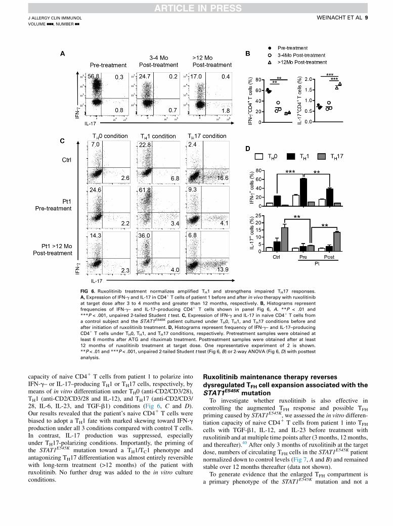
FIG 6. Ruxolitinib treatment normalizes amplified TH1 and strengthens impaired TH17 responses.
A, Expression of IFN-g and IL-17 in CD41 T cells of patient 1 before and after in vivo therapy with ruxolitinib
at target dose after 3 to 4 months and greater than 12 months, respectively. B, Histograms represent
frequencies of IFN-g– and IL-17–producing CD41 T cells shown in panel Fig 6, A. **P < .01 and
***P < .001, unpaired 2-tailed Student t test. C, Expression of IFN-g and IL-17 in naive CD41 T cells from
a control subject and the STAT1E545K patient cultured under TH0, TH1, and TH17 conditions before and
after initiation of ruxolitinib treatment. D, Histograms represent frequency of IFN-g– and IL-17–producing
CD41 T cells under TH0, TH1, and TH17 conditions, respectively. Pretreatment samples were obtained at
least 6 months after ATG and rituximab treatment. Posttreatment samples were obtained after at least
12 months of ruxolitinib treatment at target dose. One representative experiment of 2 is shown.
**P < .01 and ***P < .001, unpaired 2-tailed Student t test (Fig 6, B) or 2-way ANOVA (Fig 6, D) with posttest
analysis.
J ALLERGY CLIN IMMUNOL
VOLUME nnn, NUMBER nn
WEINACHT ET AL 9
capacity of naive CD41 T cells from patient 1 to polarize intoIFN-g– or IL-17–producing TH1 or TH17 cells, respectively, bymeans of in vitro differentiation under TH0 (anti-CD2/CD3/28),TH1 (anti-CD2/CD3/28 and IL-12), and TH17 (anti-CD2/CD3/28, IL-6, IL-23, and TGF-b1) conditions (Fig 6, C and D).Our results revealed that the patient’s naive CD41 T cells werebiased to adopt a TH1 fate with marked skewing toward IFN-gproduction under all 3 conditions compared with control T cells.In contrast, IL-17 production was suppressed, especiallyunder TH17-polarizing conditions. Importantly, the priming ofthe STAT1E545K mutation toward a TH1/TC1 phenotype andantagonizing TH17 differentiation was almost entirely reversiblewith long-term treatment (>12 months) of the patient withruxolitinib. No further drug was added to the in vitro cultureconditions.
Ruxolitinib maintenance therapy reverses
dysregulated TFH cell expansion associated with the
STAT1E545K mutationTo investigate whether ruxolitinib is also effective in
controlling the augmented TFH response and possible TFH
priming caused by STAT1E545K, we assessed the in vitro differen-tiation capacity of naive CD41 T cells from patient 1 into TFH
cells with TGF-b1, IL-12, and IL-23 before treatment withruxolitinib and at multiple time points after (3 months, 12months,and thereafter).40 After only 3 months of ruxolitinib at the targetdose, numbers of circulating TFH cells in the STAT1E545K patientnormalized down to control levels (Fig 7, A and B) and remainedstable over 12 months thereafter (data not shown).
To generate evidence that the enlarged TFH compartment isa primary phenotype of the STAT1E545K mutation and not a
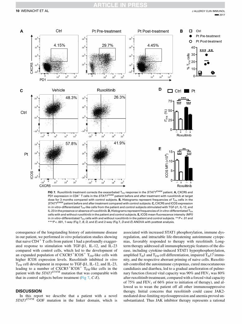
FIG 7. Ruxolitinib treatment corrects the exacerbated TFH response in the STAT1E545K patient. A, CXCR5 and
PD1 expression in CD41 T cells in the STAT1E545K patient before and after treatment with ruxolitinib at target
dose for 3 months compared with control subjects. B, Histograms represent frequencies of TFH cells in the
STAT1E545K patient before and after treatment comparedwith control subjects.C,CXCR5 and ICOS expression
in in vitro–differentiated TFH-like cells from the patient and control subjects stimulatedwith TGF-b1, IL-12, and
IL-23 in thepresenceorabsenceof ruxolitinib.D,Histogramsrepresent frequenciesof invitro–differentiatedTFH
cells with andwithout ruxolitinib in the patient and control subjects.E, ICOSmeanfluorescence intensity (MFI)
in in vitro–differentiated TFH cells with andwithout ruxolitinib in the patient and control subjects. **P < .01 and
***P < .001, 1-way (Fig 7, B, D, and E) and 2-way (Fig 7, D and E) ANOVA with posttest analysis.
J ALLERGY CLIN IMMUNOL
nnn 2017
10 WEINACHT ET AL
consequence of the longstanding history of autoimmune diseasein our patient, we performed in vitro polarization studies showingthat naive CD41 T cells from patient 1 had a profoundly exagger-ated response to stimulation with TGF-b1, IL-12, and IL-23compared with control cells, which led to the development ofan expanded population of CXCR51ICOS1 TFH-like cells withhigher ICOS expression levels. Ruxolitinib inhibited in vitroTFH cell development in response to TGF-b1, IL-12, and IL-23,leading to a number of CXCR51ICOS1 TFH-like cells in thepatient with the STAT1E545K mutation that was comparable withthat in control subjects before treatment (Fig 7, C-E).
DISCUSSIONIn this report we describe that a patient with a novel
STAT1E545K GOF mutation in the linker domain, which is
associated with increased STAT1 phosphorylation, immune dys-regulation, and intractable life-threatening autoimmune cytope-nias, favorably responded to therapy with ruxolitinib. Long-term therapy addressed all immunophenotypic features of the dis-ease, including cytokine-induced STAT1 hyperphosphorylation,amplified TH1 and TFH cell differentiation, impaired TH17 immu-nity, and the respective aberrant priming of naive cells. Ruxoliti-nib controlled the autoimmune cytopenias, cured mucocutaneouscandidiasis and diarrhea, led to a gradual amelioration of pulmo-nary function (forced vital capacity was 90% and FEV1 was 80%after ruxolitinib treatmeant, compared with a forced vital capacityof 75% and FEV1 of 66% prior to initiation of therapy), and al-lowed us to wean the patient off all other immunosuppressivetherapy. Initial concerns that ruxolitinib could cause JAK2-mediated dose-limiting myelosuppression and anemia proved un-substantiated. Thus JAK inhibitor therapy represents a rational
J ALLERGY CLIN IMMUNOL
VOLUME nnn, NUMBER nn
WEINACHT ET AL 11
and effective therapy in patients with this disease. As with allimmunomodulatory and immunosuppressive medications, care-ful clinical surveillance for side effects, including serious infec-tions and malignancy, is warranted.
Like other STAT1 GOF mutations, the STAT1E545K mutationaugmented STAT1 phosphorylation in response to cytokinesignaling. However, unlike other common STAT1GOFmutations,it potentiated STAT1 phosphorylation without affectingdephosphorylation after withdrawal of the activating cytokine.14
Also, it did not perturb the basal levels of phospho-STAT1 absentcytokine stimulation, suggesting that it promoted increasedrecruitment of STAT1 at the respective cytokine receptor. Ofnote, STAT3 phosphorylation was normal.
The STAT1E545K mutation augmented the in vitro polarizationof naive CD41 T cells into TH1 and TFH cell subsets whilerendering them resistant to TH17 polarization. Similar skewingof helper T cell subsets was also observed in vivo, with the patientshowing increased TH1 and TFH but decreased TH17 cell counts inthe circulation. Suppressed TH17 differentiation was noteddespite normal STAT3 phosphorylation. Our findings areconsistent with those of recent studies demonstrating thatSTAT1 GOF mutations act distally to suppress STAT3 activationof components of the TH17 transcriptional program, includingRORC, without affecting cytokine-mediated STAT3 phosphoryla-tion.41 Surprisingly, the STAT1E545Kmutation was associatedwitha markedly expanded pool of circulating TFH cells. Similarobservations were made in a patient (patient 2) with a differentSTAT1 GOF mutation affecting the DNA-binding domain whowas clinically free from any signs of autoimmunity. Thesefindings suggest that a dysregulated TFH cell response is a primaryfeature of increased STAT1 activity independent of and notsecondary to clinical autoimmunity. The molecular mechanismsleading to this enlarged TFH cell compartment are not clear. TFH
cell expansion has been associated with humoral autoimmunity,and several monogenic immune dysregulatory diseases with hu-moral autoimmunity are associated with an expanded TFH cellpool, including cytotoxic T lymphocyte–associated antigen 4and LPS-responsive beige-like anchor protein deficiency.36,42
By decreasing STAT1 hyperphosphorylation, JAK inhibitorscombat the sequelae of unopposed interferon release, normalizeTH1 andTFH cell responses, and treat autoimmunity. In our studieslong-term JAK inhibitor treatment also promoted TH17 differenti-ation of naive CD41 T cells in vitro and rescued IL-17 productionin vivo, which was clinically reflected by a significant improve-ment in mucosal immunity under ruxolitinib therapy. This is inline with the observations of Higgins et al10 and M€ossner et al25
who reported improvement of oral candidiasis in response to rux-olitinib and relapse as the drug was withdrawn. Our concerns thatJAK inhibition could compromise mucosal immunity further bydecreasing STAT3 activity proved unsubstantiated because ruxo-litinib only had a minor effect on STAT3 signaling.
The observation that ruxolitinib therapy reversed theexaggerated TFH phenotype and priming caused by STAT1E545K
in its entirety is concordant with its ability to control autoimmunemanifestations in our patient over a period of more than18 months. These observations suggest a role for JAK inhibitorsas a maintenance treatment to prevent the development of furtherautoimmune disease, for which patients with STAT1 GOFmutations are at extremely high risk over their lifetime. Theavailability of a targeted small-molecule therapy is particularlyrelevant in severely affected patients for whom a donor for
allogeneic hematopoietic stem cell transplantation is notavailable. Even with a suitable donor, the extent of autosensitiza-tion and allosensitization in patients with a longstanding historyof autoimmunity together with their inherent underlyingcomorbidities often call the indication to proceed with anallogeneic hematopoietic stem cell transplantation into question.
The findings described above have implications for a broadpatient population. Because STAT1 GOF mutations can presentwith a wide range of clinical phenotypes, heightened clinicalvigilance should prompt the physician to consider this diagnosisin any patient with autoimmune disease associated withmucocutaneous candidiasis or other opportunistic infections.Finally, this case illustrates how profoundly knowledge aboutthemolecular underpinnings of autoimmune conditions can affecttherapy and outcome.
Key messages
d A STAT1E545K GOF mutation mediated TH1/TC1 skewing,TH17 cell suppression, and an exaggerated TFH cellresponse.
d The JAK inhibitor ruxolitinib mitigated STAT1 hyper-phosphorylation, normalized TH1 and TFH cell differenti-ation, and improved TH17 cell development both in vitroand in vivo.
REFERENCES
1. Boisson-Dupuis S, Kong XF, Okada S, Cypowyj S, Puel A, Abel L, et al. Inborn
errors of human STAT1: allelic heterogeneity governs the diversity of immunolog-
ical and infectious phenotypes. Curr Opin Immunol 2012;24:364-78.
2. Subramaniam PS, Torres BA, Johnson HM. So many ligands, so few transcription
factors: a new paradigm for signaling through the STAT transcription factors.
Cytokine 2001;15:175-87.
3. O’Shea JJ, Holland SM, Staudt LM. JAKs and STATs in immunity,
immunodeficiency, and cancer. N Engl J Med 2013;368:161-70.
4. van de Veerdonk FL, Plantinga TS, Hoischen A, Smeekens SP, Joosten LA, Gilis-
sen C, et al. STAT1 mutations in autosomal dominant chronic mucocutaneous
candidiasis. N Engl J Med 2011;365:54-61.
5. Hori T, Ohnishi H, Teramoto T, Tsubouchi K, Naiki T, Hirose Y, et al. Autosomal-
dominant chronic mucocutaneous candidiasis with STAT1-mutation can be
complicated with chronic active hepatitis and hypothyroidism. J Clin Immunol
2012;32:1213-20.
6. Liu L, Okada S, Kong XF, Kreins AY, Cypowyj S, Abhyankar A, et al. Gain-of-
function human STAT1 mutations impair IL-17 immunity and underlie chronic
mucocutaneous candidiasis. J Exp Med 2011;208:1635-48.
7. Nahum A. Dala lI. Clinical manifestations associated with novel mutations in the
coiled-coil domain of STAT1. LymphoSign J 2014;1:97-103.
8. Roifman C. Monoallelic STAT1 mutations and disease patterns. LymphoSign J
2014;1:57-9.
9. Sharfe N, Nahum A, Newell A, Dadi H, Ngan B, Pereira SL, et al. Fatal combined
immunodeficiency associated with heterozygous mutation in STAT1. J Allergy
Clin Immunol 2014;133:807-17.
10. Higgins E, Al Shehri T, McAleer MA, Conlon N, Feighery C, Lilic D, et al. Use of
ruxolitinib to successfully treat chronic mucocutaneous candidiasis caused by gain-
of-function signal transducer and activator of transcription 1 (STAT1) mutation.
J Allergy Clin Immunol 2015;135:551-3.e3.
11. Yamazaki Y, Yamada M, Kawai T, Morio T, Onodera M, Ueki M, et al. Two novel
gain-of-function mutations of STAT1 responsible for chronic mucocutaneous
candidiasis disease: impaired production of IL-17A and IL-22, and the presence
of anti-IL-17F autoantibody. J Immunol 2014;193:4880-7.
12. Sampaio EP, Hsu AP, Pechacek J, Bax HI, Dias DL, Paulson ML, et al. Signal
transducer and activator of transcription 1 (STAT1) gain-of-function mutations
and disseminated coccidioidomycosis and histoplasmosis. J Allergy Clin Immunol
2013;131:1624-34.
13. Frans G, Moens L, Schaballie H, Van Eyck L, Borgers H, Wuyts M, et al. Gain-of-
function mutations in signal transducer and activator of transcription 1 (STAT1):
J ALLERGY CLIN IMMUNOL
nnn 2017
12 WEINACHT ET AL
chronic mucocutaneous candidiasis accompanied by enamel defects and delayed
dental shedding. J Allergy Clin Immunol 2014;134:1209-13.e6.
14. Mizoguchi Y, Tsumura M, Okada S, Hirata O, Minegishi S, Imai K, et al. Simple
diagnosis of STAT1 gain-of-function alleles in patients with chronic
mucocutaneous candidiasis. J Leukoc Biol 2014;95:667-76.
15. Bohmer FD, Friedrich K. Protein tyrosine phosphatases as wardens of STAT
signaling. JAKSTAT 2014;3:e28087.
16. Kumar N, Hanks ME, Chandrasekaran P, Davis BC, Hsu AP, Van Wagoner NJ,
et al. Gain-of-function signal transducer and activator of transcription 1 (STAT1)
mutation-related primary immunodeficiency is associated with disseminated mu-
cormycosis. J Allergy Clin Immunol 2014;134:236-9.
17. T�oth B, M�ehes L, Task�o S, Szalai Z, Tulassay Z, Cypowyj S, et al. Herpes in
STAT1 gain-of-function mutation. Lancet 2012;379:2500.
18. Puel A, Cypowyj S, Marodi L, Abel L, Picard C, Casanova JL. Inborn errors of
human IL-17 immunity underlie chronic mucocutaneous candidiasis. Curr Opin
Allergy Clin Immunol 2012;12:616-22.
19. Romberg N, Morbach H, Lawrence MG, Kim S, Kang I, Holland SM, et al. Gain-
of-function STAT1 mutations are associated with PD-L1 overexpression and a
defect in B-cell survival. J Allergy Clin Immunol 2013;131:1691-3.
20. O’Shea JJ, Schwartz DM, Villarino AV, Gadina M, McInnes IB, Laurence A. The
JAK-STAT Pathway: Impact on Human Disease and Therapeutic Intervention.
Annu Rev Med 2015;66:311-28.
21. Aldave JC, Cachay E, Nunez L, Chunga A, Murillo S, Cypowyj S, et al. A 1-year-
old girl with a gain-of-function STAT1 mutation treated with hematopoietic stem
cell transplantation. J Clin Immunol 2013;33:1273-5.
22. Wildbaum G, Shahar E, Katz R, Karin N, Etzioni A, Pollack S. Continuous G-CSF
therapy for isolated chronic mucocutaneous candidiasis: complete clinical
remission with restoration of IL-17 secretion. J Allergy Clin Immunol 2013;132:
761-4.
23. Faitelson Y, Bates A, Shroff M, Grunebaum E, Roifman CM, Naqvi A. A mutation
in the STAT1 DNA-binding domain associated with hemophagocytic lymphohis-
tiocytosis. LymphoSign J 2014;1:87-95.
24. Liu Y, Jesus AA, Marrero B, Yang D, Ramsey SE, Montealegre Sanchez GA, et al.
Activated STING in a vascular and pulmonary syndrome. N Engl J Med 2014;371:
507-18.
25. M€ossner R, Diering N, Bader O, Forkel S, Overbeck T, Gross U, et al. Ruxolitinib
induces interleukin 17 and ameliorates chronic mucocutaneous candidiasis caused
by STAT1 gain-of-function mutation. Clin Infect Dis 2016;62:951-3.
26. Takezaki S, Yamada M, Kato M, Park MJ, Maruyama K, Yamazaki Y, et al.
Chronic mucocutaneous candidiasis caused by a gain-of-function mutation in the
STAT1 DNA-binding domain. J Immunol 2012;189:1521-6.
27. Soltesz B, Toth B, Shabashova B, Bondarenko A, Okada S, Cypowyj S, et al. New
and recurrent gain-of-function STAT1 mutations in patients with chronic mucocu-
taneous candidiasis. J Med Genet 2013;50:567-78.
28. Ng PC, Henikoff S. Accounting for human polymorphisms predicted to affect
protein function. Genome Res 2002;12:436-46.
29. Adzhubei IA, Schmidt S, Peshkin L, Ramensky VE, Gerasimova A, Bork P, et al. A
method and server for predicting damaging missense mutations. Nat Methods
2010;7:248-9.
30. Emsley P, Cowtan K. Coot: model-building tools for molecular graphics. Acta
Crystallogr D Biol Crystallogr 2004;60:2126-32.
31. Chen X, Vinkemeier U, Zhao Y, Jeruzalmi D, Darnell JE Jr, Kuriyan J. Crystal
structure of a tyrosine phosphorylated STAT-1 dimer bound to DNA. Cell 1998;
93:827-39.
32. Mao X, Ren Z, Parker GN, Sondermann H, Pastorello MA, Wang W, et al. Struc-
tural bases of unphosphorylated STAT1 association and receptor binding. Mol Cell
2005;17:761-71.
33. Biasini M, Bienert S, Waterhouse A, Arnold K, Studer G, Schmidt T, et al.
SWISS-MODEL: modelling protein tertiary and quaternary structure using
evolutionary information. Nucleic Acids Res 2014;42:W252-8.
34. Delano WL. The PyMol molecular graphics system. Palo Alto (Calif): DeLano Sci-
entific; 2002.
35. Schmitt N, Liu Y, Bentebibel SE, Munagala I, Bourdery L, Venuprasad K, et al.
The cytokine TGF-beta co-opts signaling via STAT3-STAT4 to promote the differ-
entiation of human TFH cells. Nat Immunol 2014;15:856-65.
36. Charbonnier LM, Janssen E, Chou J, Ohsumi TK, Keles S, Hsu JT, et al.
Regulatory T-cell deficiency and immune dysregulation, polyendocrinopathy,
enteropathy, X-linked-like disorder caused by loss-of-function mutations in
LRBA. J Allergy Clin Immunol 2015;135:217-27.
37. Ma CS, Wong N, Rao G, Avery DT, Torpy J, Hambridge T, et al. Monogenic
mutations differentially affect the quantity and quality of T follicular helper cells
in patients with human primary immunodeficiencies. J Allergy Clin Immunol
2015;136:993-1006.e1.
38. Ma CS, Wong N, Rao G, Nguyen A, Avery DT, Payne K, et al. Unique and shared
signaling pathways cooperate to regulate the differentiation of human CD41 T
cells into distinct effector subsets. J Exp Med 2016;213:1589-608.
39. Quintas-Cardama A, Kantarjian H, Cortes J, Verstovsek S. Janus kinase inhibitors
for the treatment of myeloproliferative neoplasias and beyond. Nat Rev Drug
Discov 2011;10:127-40.
40. Kinnunen T, Chamberlain N, Morbach H, Choi J, Kim S, Craft J, et al.
Accumulation of peripheral autoreactive B cells in the absence of functional human
regulatory T cells. Blood 2013;121:1595-603.
41. Zheng J, van de Veerdonk FL, Crossland KL, Smeekens SP, Chan CM, Al Shehri T,
et al. Gain-of-function STAT1 mutations impair STAT3 activity in patients with
chronic mucocutaneous candidiasis (CMC). Eur J Immunol 2015;45:2834-46.
42. Kuehn HS, Ouyang W, Lo B, Deenick EK, Niemela JE, Avery DT, et al. Immune
dysregulation in human subjects with heterozygous germline mutations in CTLA4.
Science 2014;345:1623-7.
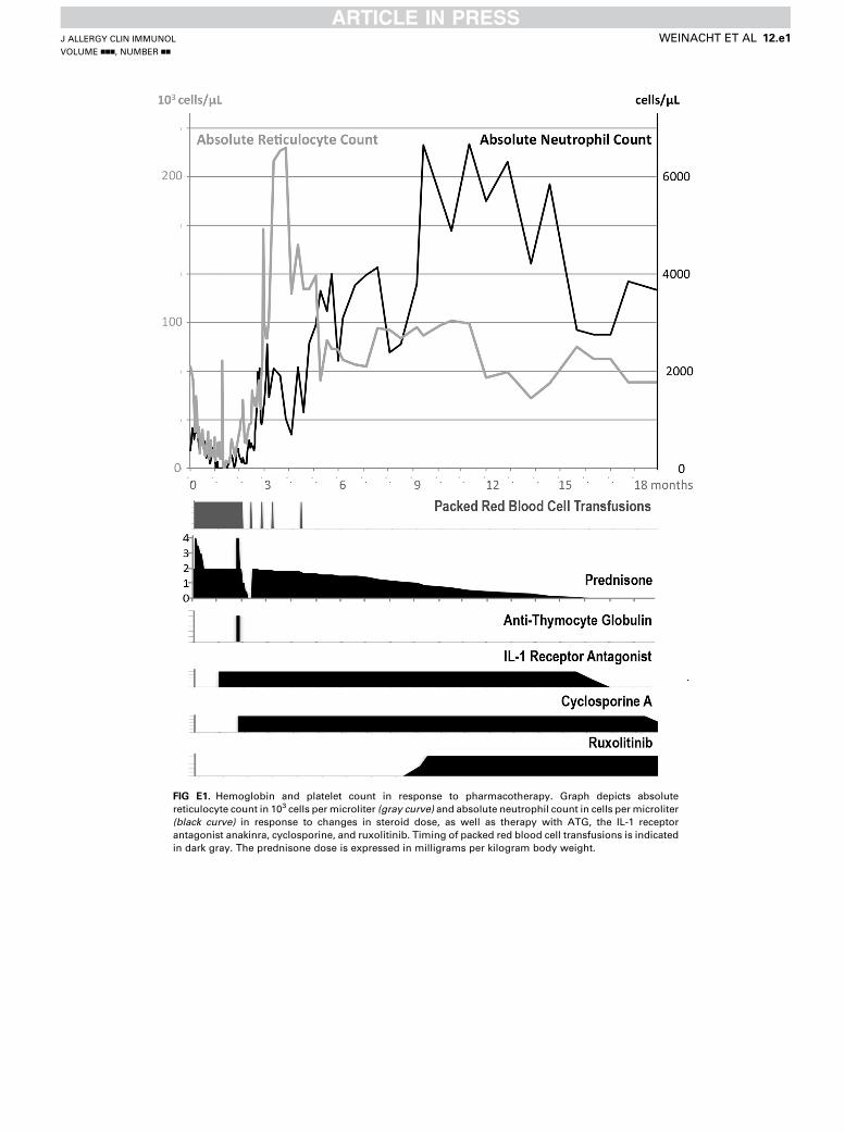
FIG E1. Hemoglobin and platelet count in response to pharmacotherapy. Graph depicts absolute
reticulocyte count in 103 cells per microliter (gray curve) and absolute neutrophil count in cells per microliter
(black curve) in response to changes in steroid dose, as well as therapy with ATG, the IL-1 receptor
antagonist anakinra, cyclosporine, and ruxolitinib. Timing of packed red blood cell transfusions is indicated
in dark gray. The prednisone dose is expressed in milligrams per kilogram body weight.
J ALLERGY CLIN IMMUNOL
VOLUME nnn, NUMBER nn
WEINACHT ET AL 12.e1
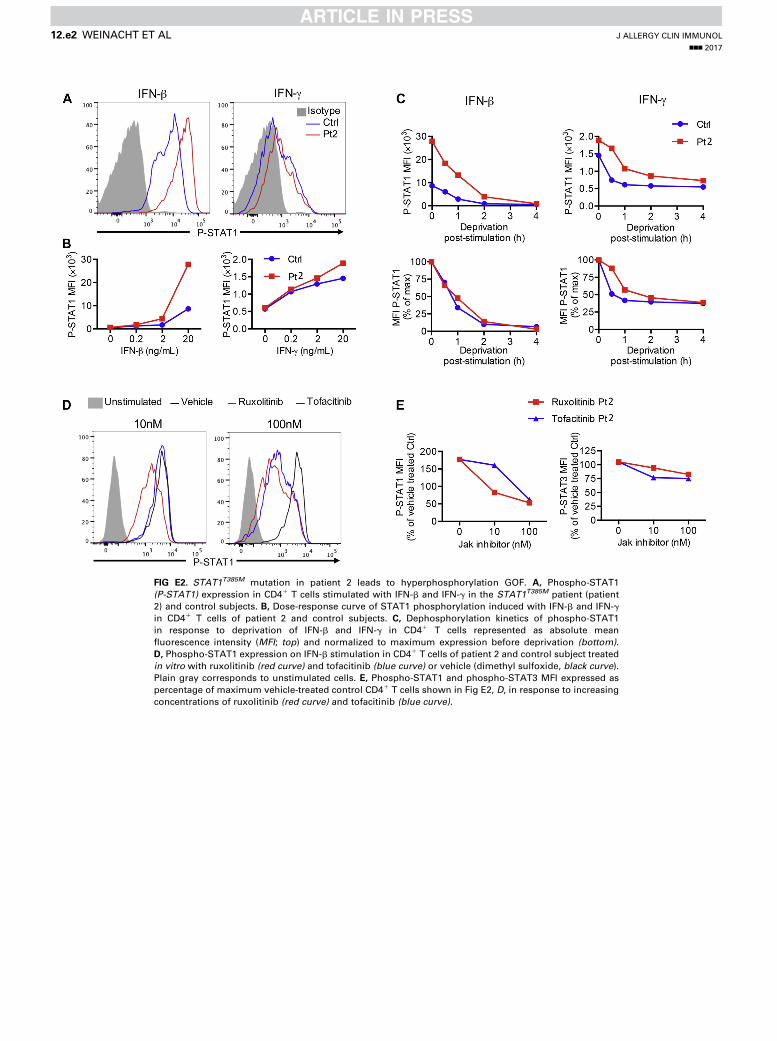
FIG E2. STAT1T385M mutation in patient 2 leads to hyperphosphorylation GOF. A, Phospho-STAT1
(P-STAT1) expression in CD41 T cells stimulated with IFN-b and IFN-g in the STAT1T385M patient (patient
2) and control subjects. B, Dose-response curve of STAT1 phosphorylation induced with IFN-b and IFN-g
in CD41 T cells of patient 2 and control subjects. C, Dephosphorylation kinetics of phospho-STAT1
in response to deprivation of IFN-b and IFN-g in CD41 T cells represented as absolute mean
fluorescence intensity (MFI; top) and normalized to maximum expression before deprivation (bottom).
D, Phospho-STAT1 expression on IFN-b stimulation in CD41 T cells of patient 2 and control subject treated
in vitro with ruxolitinib (red curve) and tofacitinib (blue curve) or vehicle (dimethyl sulfoxide, black curve).
Plain gray corresponds to unstimulated cells. E, Phospho-STAT1 and phospho-STAT3 MFI expressed as
percentage of maximum vehicle-treated control CD41 T cells shown in Fig E2, D, in response to increasing
concentrations of ruxolitinib (red curve) and tofacitinib (blue curve).
J ALLERGY CLIN IMMUNOL
nnn 2017
12.e2 WEINACHT ET AL