Evaluation of Prosthetic Heart Valves by Transesophageal Echocardiography

J A C C : C A R D I O V A S C U L A R I M A G I N G VO L . 1 1 , N O . 4 , 2 0 1 8
ª 2 0 1 8 B Y T H E A M E R I C A N C O L L E G E O F C A R D I O L O G Y F O U N D A T I O N
P U B L I S H E D B Y E L S E V I E R
STATE-OF-THE-ART PAPERS
Roles of TransesophagealEchocardiography and CardiacComputed Tomography forEvaluation of Left Atrial Thrombusand Associated Pathology
A Review and Critical AnalysisFaraz Pathan, MBBS,a Harvey Hecht, MD,b Jagat Narula, MD, PHD,b Thomas H. Marwick, MBBS, PHD, MPHc
ABSTRACT
ISS
Fro
Yo
rel
are
Ma
Evaluation of the left atrium and left atrial appendage for the presence of thrombus prior to cardioversion and pulmonary
vein isolation, and of the entire heart for embolic sources in the setting of cryptogenic stroke, has long been standard
medical care. Guidelines have uniformly recommended transesophageal echocardiography (TEE) to accomplish these
goals. In recent years, computed tomographic angiography has demonstrated diagnostic accuracy similar to that of TEE
for the detection of thrombus. Analysis of the pertinent data and relative merits of the 2 technologies leads to the
conclusions that: 1) both modalities have some unique, nonoverlapping capabilities that may dictate their use in specific
situations; 2) computed tomographic angiography is a reasonable alternative to TEE when the primary aim is to
exclude left atrial and left atrial appendage thrombus and in patients in whom the risks associated with TEE outweigh
the benefits; and 3) both options should be discussed with the patient in the setting of shared decision making.
(J Am Coll Cardiol Img 2018;11:616–27) © 2018 by the American College of Cardiology Foundation.
A fter a steady increase over the past 50 years,the prevalence of atrial fibrillation (AF) hasreached 3% (1). The burden of AF is projected
to more than double between 2010 and 2030 (2),driven particularly by the aging population. The pri-mary driver of the association of AF with significantmorbidity and mortality is thromboembolism, andAF ablation and cardioversion have an inherent riskrelated to left atrial (LA) appendage thrombi. Transe-sophageal echocardiography (TEE) has underpinnedthe exclusion of thromboembolic sources before
N 1936-878X/$36.00
m the aMenzies Institute for Medical Research, Hobart, Australia; bIcahn
rk; and the cBaker Heart and Diabetes Institute, Melbourne, Australi
ationships relevant to the contents of this paper to disclose. Drs. Patha
joint first authors.
nuscript received November 26, 2017; accepted December 21, 2017.
cardioversion, following cryptogenic cerebrovascularaccidents and before pulmonary vein isolation (PVI)and other procedures requiring LA and left atrialappendage (LAA) instrumentation. The Heart RhythmSociety has mandated that in the absence of 3 weeksof therapeutic systemic anticoagulation, TEE shouldbe performed to screen for thrombus prior to theseprocedures if the patient has been in AF for >48 hor for an unknown duration (3). Cardiac computedtomographic angiography (CTA) is also able to fillthis role and as a low-risk noninvasive test would
https://doi.org/10.1016/j.jcmg.2017.12.019
School of Medicine at Mount Sinai, New York, New
a. The authors have reported that they have no
n and Hecht contributed equally to this paper and
AB BR E V I A T I O N S
AND ACRONYM S
AF = atrial fibrillation
CTA = computed tomographic
angiography
LA = left atrial
LAA = left atrial appendage
LV = left ventricular
PVI = pulmonary vein isolation
SEC = spontaneous
echocardiographic contrast
TEE = transesophageal
echocardiography
J A C C : C A R D I O V A S C U L A R I M A G I N G , V O L . 1 1 , N O . 4 , 2 0 1 8 Pathan et al.A P R I L 2 0 1 8 : 6 1 6 – 2 7 TEE, CTA, and LAA Thrombus
617
need to be considered. However, the Heart RhythmSociety task force did not recommend that CTA beused to screen for LA thrombi in patients at highrisk for stroke. Likewise, the evaluation of embolicsources for stroke is routinely performed using TEErather than CTA. A review of the data pertinent tothe capabilities of the 2 technologies may facilitatethe development of new recommendations for a vari-ety of clinical scenarios. In comparing the relativemerits of TEE and CTA, we evaluated the role ofeach modality with reference to patient populationsundergoing cardioversion and PVI, as well as patientstested for a cardioembolic source in the context ofcryptogenic stroke. These recommendations are ouropinions and interpretations of the data and are notto be construed as societal guidelines.
TARGETS OF IMAGING
LAATHROMBUSANDSPONTANEOUSECHOCARDIOGRAPHIC
CONTRAST (SEC). The presence of LAA thrombus is anadverse prognostic marker for future cardiovascularevents (4). Absence of a thrombus confirms that it issafe to proceed with cardioversion, with a lowthromboembolic event rate (0% to 0.8%) in patientswho are appropriately anticoagulated (5,6). Thepresence of an LAA thrombus mandates deferringcardioversion and prolonged anticoagulation.
The complex interplay of atrial endothelium, bloodconstituents, and stasis of blood flow (i.e., the com-ponents of Virchow’s triad) results in erythrocyterouleau formation, manifesting as SEC or “smoke.”These red cell aggregates can progress to sludge andeventually to thrombus (Figure 1) (7,8). The long-termrisk for thromboembolic events is greater in thepresence of SEC than in the patients without (12% vs3% per year; p ¼ 0.002), even in the absence of LAthrombi. SEC and warfarin use have been reported asthe significant predictors of future thromboembolicevents, independent of moderate to severe left ven-tricular (LV) dysfunction, previous thromboembo-lism, and complex arch atheroma (9). The density ofSEC determines the risk for thromboembolism, andpatients with AF and dense SEC have a higher likeli-hood of cerebral embolism (22%) and death (10). Pa-tients with dense SEC have a stroke rate of 18.2% peryear if not treated with warfarin compared with 4.5%per year when on warfarin (11). The detection ofstroke risk in this setting is dependent not just on thedetection of thrombus; 3% of patients with CHADSscores of 0 or 1 have LA thrombi, and 8% have denseSEC (12). Assessment of SEC is a critical albeit un-derused determinant of duration, intensity, and typeof anticoagulant therapy.
LAA MORPHOLOGY. CTA has been used suc-cessfully to categorize LAA morphology. TheLAA can broadly be classified into 4morphological categories: “chicken wing”(most common), “cactus,” “windsock,” andcauliflower (Figure 2), the latter most oftenassociated with embolic events (8).
Morphological evaluation for the purposeof atrial appendage occluder devices has beenreviewed (13), but it merits discussionbecause it elegantly demonstrates the inter-section of the 2 modalities. Comprehensiveevaluation of LAA morphology is essentialprior to and during deployment of closure
devices. Important parameters include assessmentof the ostium, landing zone, depth of the mainlobe, relationship of the main lobe to additionallobes, and proximity to the circumflex coronaryartery (13).TEE and CTA can be used for sizing purposes,though recent evidence suggests that CTA is moreaccurate and results in change to device size in up toone-half of patients undergoing LAA closure (14).Nevertheless, TEE is indispensable, as it enablesassessment and planning, as well as monitoring of theprocedure from septal puncture to instantaneousassessment of complications. Furthermore,3-dimensional TEE improves sizing compared with2-dimensional multiplanar imaging and has betterreproducibility (15,16). An ideal strategy to facilitatethe safe deployment of atrial appendage occludersinvolves both multiplanar CTA and TEE (17).
LAA FUNCTIONAL EVALUATION. LAA function canbe assessed using ejection fraction, pulse-wavevelocities, and deformation imaging. LAA flowvelocities have been the most robustly studiedfunctional parameter; there are 4 separate phases,including emptying (range 50 � 6 cm/s to 83 �25 cm/s), filling (46 � 12 cm/s to 60 � 19 cm/s), abiphasic systolic reflection wave, and an early dia-stolic emptying waveform (8). An emptyingvelocity <20 cm/s is associated with thrombus, SEC,and subsequent thromboembolic risk (18). Lowemptying velocities should serve as a reminder toreassess the LAA for thrombus and clarify any un-certainty with contrast (8).
EVALUATION OF COEXISTENT AND INTERDEPENDENT
PATHOLOGY. AF is often associated with multiplecardiac pathologies. Recent multicenter trials docu-mented that between 14% and 26% of patients withAF have significant valvular disease (19), and registrydata show valvular abnormalities in >60% (20).The prevalence of coronary artery disease in AF

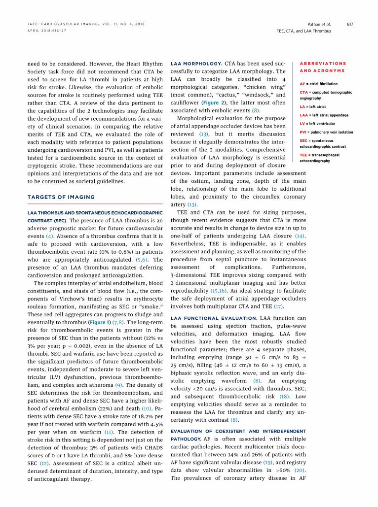
FIGURE 1 Abnormal Transesophageal Echocardiographic Findings Within the Left Atrial Appendage
A B C D
E F G
90
PAT T: 37.0CTEE T: 40.4C
(A) The presence of a large pectinate muscle can sometimes be confused for a left atrial appendage (LAA) thrombus. (B) In this case, the pectinate muscle is better
defined by 3-dimensional (3D) transesophageal echocardiography. (C) Spontaneous echocardiographic contrast is seen in the LAA. (D) A more echo-dense, amorphous
finding consistent with sludge is seen within the LAA (arrowheads). Zero-degree (E) and 95� (F) views and 3D imaging (G) show a thrombus within the LAA (arrows).
Reprinted with permission from Beigel et al. (8).
Pathan et al. J A C C : C A R D I O V A S C U L A R I M A G I N G , V O L . 1 1 , N O . 4 , 2 0 1 8
TEE, CTA, and LAA Thrombus A P R I L 2 0 1 8 : 6 1 6 – 2 7
618
is between 17.0% and 46.5% and has prognostic im-plications (21). Finally, diastolic dysfunction is asso-ciated with AF in up to 27% of cases (22).
The unique, nonoverlapping qualities of TEEand CTA are critical to evaluating certain cardiacpathologies and may dictate physician and or patientchoice.
TEE is ideal for determining the presence, severity,and mechanism of coexistent valvular pathology,particularly mitral regurgitation, and identifying apotentially reversible phenotype (23–26).
Diastolic function assessed using TEE is a predictorof success of ablation and maintenance of sinusrhythm after AF ablation (27). Pulmonary venous flowpatterns may predict AF recurrence post-cardioversion (28).
CTA provides delineation of the coronary anatomywithout additional contrast or radiation, because it isavailable for analysis in all gated studies. Withappropriate phase selection, the coronary arteries canbe accurately interpreted in the setting of AF and
provide important prognostic information that canlead to therapeutic intervention.
INDICATIONS FOR TEE
TEE has been the reference standard for assessingthrombus, SEC, and sludge in the LAA and is rec-ommended by clinical guidelines (3,7). The sensi-tivity of TEE for LA thrombus against asurgicopathological reference standard is 93% to100% (29,30); false-negative findings can result inthromboembolic sequelae (6). Despite 99% to 100%specificity, false-positive results (e.g., due to pecti-nate muscles, dense SEC, or sludge), which alsoincrease the probability of underlying thrombus,may lead to an inappropriate cancellation ofcardioversion or ablation. The use of echocardio-graphic contrast is associated with a lowerincidence of artifacts compared with routine TEE(2% vs. 29%; p < 0.001) (31), with increased abilityto exclude thrombus (Figure 3) (32).

FIGURE 2 Left Atrial Appendage Morphology by Multiple Modalities
A
B
C
D
E
F
G
H
I
J
K
L
The 4 different left atrial appendage morphologies as shown by transesophageal echocardiography (top), cine angiography (middle), and 3-dimensional computed
tomography (bottom): cauliflower (A to C), windsock (D to F), cactus (G to I), and chicken wing (J to L). Reprinted with permission from Beigel et al. (8).
J A C C : C A R D I O V A S C U L A R I M A G I N G , V O L . 1 1 , N O . 4 , 2 0 1 8 Pathan et al.A P R I L 2 0 1 8 : 6 1 6 – 2 7 TEE, CTA, and LAA Thrombus
619
CARDIOVERSION. In the ACUTE (Assessment ofCardioversion Using Transesophageal Echocardiog-raphy) trial, 13.8% of patients in the TEE arm werefound to have thrombi, 88% of which were in the LAA(6). This contrasts with 4.4% and 2.9% of patientsanticoagulated for >4 weeks on novel oral anticoag-ulant agents or warfarin (33). TEE-guided early car-dioversion is equivalent to prolonged anticoagulation
prior to cardioversion, with fewer bleeding compli-cations and greater success of cardioversion.
LAA velocity can also be used to predict success ofcardioversion and long-term maintenance of sinusrhythm; a velocity <33.9 cm/s was independentlyassociated with AF recurrence and stroke after suc-cessful electric cardioversion and a velocity <20.2cm/s with mortality (34). Conversely, an LAA

FIGURE 3 Microbubble Contrast-Enhanced Transesophageal Echocardiography
10 10
5 5
V V
A B
Example of native (A) and contrast-enhanced (B) transesophageal echocardiography in a patient with atrial fibrillation (arrows: left atrial
appendage with sludge-like material in the tip of the left atrial appendage during native imaging and homogenous contrast distribution after
application of the transpulmonary contrast agent). Reprinted with permission from Jung et al. (32).
Pathan et al. J A C C : C A R D I O V A S C U L A R I M A G I N G , V O L . 1 1 , N O . 4 , 2 0 1 8
TEE, CTA, and LAA Thrombus A P R I L 2 0 1 8 : 6 1 6 – 2 7
620
emptying velocity >40 cm/s is significantly associ-ated with maintenance of sinus rhythm followingcardioversion at 1 year, independent of SEC, LAdimension, LV ejection fraction, and duration ofAF <1 week (35).
When the duration of AF is >48 h or uncertain orwhen compliance with anticoagulation is uncertain,TEE-guided cardioversion is recommended (3).
PVI. LAA thrombus is reported in up to 5.4% ofappropriately anticoagulated patients prior to abla-tion (33), so exclusion of LAA thrombus is imperative.Particularly high rates of LAA thrombus (21%) andcerebrovascular accident (6%) have been observedafter electric isolation of the LAA, despite anti-coagulation. Silent cerebral lesions following AFablation are associated with cognitive decline and areidentified in 4% to 39% of cases (36,37). Silent cere-bral lesions are related to procedural and patient-related factors (age, low LV ejection fraction, andthe presence of SEC) (38), although SEC lacks an
TABLE 1 Accuracy of Computed Tomographic Angiography for Left A
Transesophageal Echocardiography as a Gold Standard
n % Thrombus Sensitivi
All 2,955 8.9 � 7.0 96%
Delayed phase 753 14 � 8 100%
All PVI 1,836 5.8 � 5.0 91%
Delayed-phase PVI 101 2.9–19.3 100%
NPV ¼ negative predictive value; PPV ¼ positive predictive value; PVI ¼ pulmonary vein
association with an increase in early or late occur-rence of stroke (39). Nonetheless, more intense anti-coagulation is associated with a lower incidence of LAthrombus in patients with SEC (40).
In patients undergoing ablation, an emptyingvelocity <28 cm/s is an independent predictor of therisk for AF recurrence (41).
CRYPTOGENIC STROKE. Potential cardiac embolicsources in addition to LA thrombus include LVthrombi, vegetation, tumor, and intracardiac shunt,and all are identified by TEE with good to excellentsensitivity and specificity (7). Imaging followingstroke can identify cardioembolic source(s) anddetermine the plausibility that the identified sour-ce(s) are the cause of embolism. Imaging is of valuewhen these coexist (e.g., patent foramen ovale inthe setting of SEC and arch atheroma) or are inter-dependent (mitral stenosis causing AF, SEC, andLA thrombus). Real-time assessment (mobility,attachment, echo density, Valsalva maneuver
trial and Left Atrial Appendage Thrombus With
ty Specificity PPV NPV Accuracy
92% 41% 99% 94%
99% 92% 100% 99%
95% 33% 100% 95%
100% 100% 100% 100%
isolation.

FIGURE 4 Examples of Left Atrial Appendage Filling Patterns by Computed Tomographic Angiography
A
C D
E F
B
LAA LAA
LAALAA
LAALAA
(Top) Left atrial appendage (LAA) filling defect (A) remaining essentially unchanged (B) after 90-s delayed imaging, consistent with thrombus.
(Middle) LAA filling defect (C)with moderate resolution (D) after 90-s delayed imaging, consistent with a combination of thrombus and “slow flow.”
(Bottom) LAA filling defect (E) with total resolution (F) after 90-s delayed imaging, consistent with “slow flow” without thrombus.
J A C C : C A R D I O V A S C U L A R I M A G I N G , V O L . 1 1 , N O . 4 , 2 0 1 8 Pathan et al.A P R I L 2 0 1 8 : 6 1 6 – 2 7 TEE, CTA, and LAA Thrombus
621

FIGURE 5 Demonstration of Non–Left Atrial Appendage Embolic Sources by Computed Tomographic Angiography
LV apicalthrombus
A B
C D
A B
C D
E F G
Ulceratedplaque
Ao
SVC
ASD
PFO
RA
LA
LA
(Left) (A) Apical thrombus post–myocardial infarction (arrow). (B) Ulcerated ascending aortic plaque (arrows). (C) Sinus venosus atrial septal defect (ASD) (arrow). (D)
patent foramen ovale (PFO) (arrow). (Bottom) Pericardial aortic valve subacute bacterial endocarditis: vegetation (white arrows) and abscess (orange arrow) on
computed tomographic angiography. Sagittal (E), axial (F), and transverse (G) views. (Right) (A) Aortic valve fibroelastoma on 3-dimensional transesophageal
echocardiography. Fibroelastoma on axial (B), sagittal (C), and transverse (D) views on computed tomographic angiography. LA ¼ left atrium; LV ¼ left ventricular;
RA ¼ right atrium; SVC ¼ superior vena cava.
Pathan et al. J A C C : C A R D I O V A S C U L A R I M A G I N G , V O L . 1 1 , N O . 4 , 2 0 1 8
TEE, CTA, and LAA Thrombus A P R I L 2 0 1 8 : 6 1 6 – 2 7
622
demonstrating reversal of shunting) can help makedecisions about anticoagulation, device closure, andrhythm monitoring by detecting the likely culprit(7). Nonetheless, emboli originate from the LAA in75% of patients with cardioembolic strokes (7), andTEE has the benefit of detecting not only aorticplaque and LAA thrombus but also LAA peak flowvelocity (<27 cm/s), as associations of thromboem-bolic events. Low LAA emptying velocities may be asign of paroxysmal AF (42). Conversely, in thestroke population, an emptying velocity >55 cm/shas excellent negative predictive value forthrombus and SEC (43).
Impaired LAA ejection fraction (<40%) predictsparoxysmal AF after cryptogenic stroke (44). Patientswith LA thrombus tend to have lower speckletracking–derived LAA ejection fractions than thosewithout (45), which may be of value in predictingshort- and long-term outcomes post-stroke (46).
Indeed, LAA dysfunction could represent a newtherapeutic target for anticoagulation irrespective ofrhythm following a cryptogenic stroke.
INDICATIONS FOR CTA
CTA provides accuracy similar to that of TEE for thedetection of LA thrombus (47). Unlike TEE, for whichthe reference standards were postmortem and oper-ative findings, CTA has been validated only by com-parison with TEE. Online Table 1 summarizes ameta-analysis of 19 studies with 2,955 patients inwhom both CTA and TEE were performed within 7days to rule out LA and LAA thrombi before PVI orcardioversion for AF and likelihood of stroke (47). Forthe entire population, the sensitivity, specificity, andaccuracy for detection of thrombus by TEE were 96%,92%, and 99%, respectively, with positive predictivevalue of 41% and negative predictive value of 94%. In

TABLE 2 Relative Utility of Transesophageal Echocardiography Versus
Computed Tomographic Angiography for Underlying Pathologies
Thrombotic Substrate TEE CTA
LAA thrombus þþþ þþþ (delayed protocol)
SEC þþþ þþAlternative thromboembolic sources
Aortic arch atheroma þþ þþþPatent foramen ovale þþþ þþVegetations þþþ þþLV thrombus þþ þþþ
LA function/morphology
LAA emptying velocity þþþ ND
LA/LAA EF þþþ þ (retrospective gating)
LAA morphology/device sizing þþ (3D TEE) þþþCoexistent/interdependent pathology
Systolic/diastolic function þþ þ (retrospective gating)
Valvular pathology þþþ þCoronary artery disease — þþþ
CTA¼ computed tomographic angiography; EF¼ ejection fraction; LA¼ left atrial; LAA¼ left atrial appendage; LV¼ leftventricular; ND¼ no data; SEC¼ spontaneous echocardiographic contrast; TEE¼ transesophageal echocardiography.
J A C C : C A R D I O V A S C U L A R I M A G I N G , V O L . 1 1 , N O . 4 , 2 0 1 8 Pathan et al.A P R I L 2 0 1 8 : 6 1 6 – 2 7 TEE, CTA, and LAA Thrombus
623
753 patients with delayed imaging from 30 to 180 safter contrast injection, the sensitivity, specificity,and accuracy were nearly 100%, and the positivepredictive value increased to 92% (Online Table 2).The results were very similar for the 1,836 PVI pa-tients (Table 1). There were no significant differencesbetween studies with retrospective data analysis andprospectively designed studies and between studieswith and without electrocardiographic gating. Thosecases that demonstrate complete resolution ondelayed imaging are routinely reported as “slow flow”
and are thought to represent the equivalent of SEC,with partial resolution on delayed imaging consistentwith a combination of SEC and thrombus. However,there are no published formal comparisons of SEC andslow flow. Computed tomographic angiographic ex-amples of LAA filling patterns are shown in (Figure 4).PVI AND CARDIOVERSION. CTA is an essentialcomponent for the work-up of PVI, describing thenumber of pulmonary veins, determining the locationand geometric relationships of the ostia, and evalu-ating the presence and severity of stenosis (48). Theadditional utility of CTA prior to PVI and cardiover-sion is its ability to accurately identify LA and LAAthrombus as discussed earlier and to decrease thelikelihood of embolic strokes. The pertinent infor-mation has already been acquired because the leftatrium and LAA are always in the field of view andavailable for analysis. The addition of delayed-phaseimaging increases the absorbed radiation dose,which is minimized by restricting the acquisition toencompass just the LAA. When CTA has already beenperformed for PVI planning, delayed-phase imaging isthe only additional radiation exposure.
The effectiveness of delayed imaging in the clinicalpractice of ablation for AF and atrial flutter wasstudied by Bilchick et al. (49) in 320 ablation patientswho underwent nongated CTA with delayed imaging40 s after contrast injection, with TEE only afterabnormal or equivocal findings on CTA but not afternormal findings. As part of their routine ablationprotocol, intracardiac echocardiography was per-formed in all patients. Using intracardiac echocardi-ography as the reference standard, the sensitivity andnegative predictive value of CTA were 100%. Withequivocal computed tomographic angiographic re-sults classified as negative, the specificity and posi-tive predictive value were also 100%; when classifiedas positive, specificity was 98%. Patients with normalresults on CTA had neither thrombus on intracardiacechocardiography nor procedure-related stroke ortransient ischemic attack. TEE was performed in57.5% prior to implementation of the protocol. Afterimplementation, 24% underwent post-CTA TEE at the
request of the referring physician despite normalfindings on CTA; none had thrombus on TEE. Theseexcellent results persisted across all levels of strokerisk and CHA2DS2-VASc score.
Similarly, Sawit et al. (50) demonstrated the in-cremental diagnostic value of delayed imaging in acohort of 176 patients, acquiring both early anddelayed images. They demonstrated sensitivity andspecificity of 100% with good to excellent intraclasscorrelation using the delayed imaging protocol.
CRYPTOGENIC STROKE. There has not been aseparate meta-analysis for cryptogenic stroke alone,but individual study results are presented in OnlineTable 1 and are similar to PVI/cardioversion(sensitivity of 100% and specificity of 98% for delayedimaging protocols). Hur et al. (51) evaluated therelative diagnostic yield of CTA compared with TEEfollowing cryptogenic strokes with an expandedscope including high-risk (cardiac thrombi, cardiactumor, valvular vegetations and endocarditis, andaortic arch atheroma) and medium-risk sources (SEC,patent foramen ovale, atrial septal aneurysm, andmitral annular calcification).
The overall sensitivity and specificity of CTA forthe detection of thrombus by TEE were 93% and 100%respectively and were 96 and 100% for high-risksources (51). In the setting of cryptogenic stroke, asfor TEE, the evaluation of embolic sources other thanthe left atrium or LAA is critical and can be easilyaccomplished by CTA, which represents a robustalternative for patients with absolute or relativecontraindications to TEE (Figure 5).

TABLE 3 Prevalence of Thrombus and Other Embolic Sources and Recommendations for
Evaluation
Indication Incidence of LA/LAA Thrombus Recommendation
Cardioversion 2.9%/4.4%*† (33) to 13.8%‡ (6) TEE if secondary objectives(except CAD) required
CTA or TEE if secondaryobjectives not required
CTA if CAD analysis required
Pulmonary vein isolation 1.9% (56) to 5.4%† (33) CTA
Stroke evaluation Thrombus: 1.1%–8.3%PFO: 36.3%–50.4%Arch atheroma: 27.4%–74.5% (57)
TEE
Atrial appendage occlusion 6.3% (58) CTA þ TEE
*Prevalence of LA thrombus in this group is extrapolated from prevalence in pre-ablation cohort.†Anticoagulated. ‡Not anticoagulated.
PFO ¼ patent foramen ovale; TTE ¼ transthoracic echocardiography.
TABLE 4
Transesop
A
Esophageatumor,diverti
Active upp
Recent up
Esophagec
Esophago
Data from H
GI ¼ gast
Pathan et al. J A C C : C A R D I O V A S C U L A R I M A G I N G , V O L . 1 1 , N O . 4 , 2 0 1 8
TEE, CTA, and LAA Thrombus A P R I L 2 0 1 8 : 6 1 6 – 2 7
624
ANALYSIS AND POTENTIAL
CLINICAL IMPLICATIONS
WHEN TO DO CTA? In patients who have alreadyundergone CTA for PVI planning, the equivalency ofthe data presented here suggests that TEE is neededonly if the LAA evaluation is indeterminate or otherinformation is required (Tables 2 and 3). Despite theexcellent safety profile of TEE (major morbidity ormortality <0.01% in >27,000 conscious or sedatedpatients [52]), major complications can occur,including esophageal perforation or laceration,bleeding, and airway compromise. Certain patientpopulations have been identified as higher risk forcomplications and represent absolute or relativecontraindications to TEE (Table 4) (52). In patientswith oral, esophageal, or gastric disease, theAmerican Society of Echocardiography recommendsconsidering other imaging modalities, including CTA(52). Other populations who present a challengeinclude frail elderly patients, those requiring signifi-cant sedation, and patients with severely reduced LV
Absolute and Relative Contraindications to
hageal Echocardiography
bsolute Contraindications Relative Contraindications
l pathology (stricture, trauma,scleroderma, Mallory-Weiss tear,culum)
Symptomatic hiatal hernia
er GI bleeding History of GI surgery
per GI surgery Recent upper GI bleed
tomy Esophagitis, peptic ulcer disease
gastrectomy Thoracoabdominal aneurysm
Barrett’s esophagus
History of dysphagia
Coagulopathy, thrombocytopenia
ilberath et al. (52).
rointestinal.
ejection fractions, severe valvular stenosis, severepulmonary hypertension, obstructive sleep apnea,and significant pulmonary disease, in whom anadvanced airway or anesthetic skill set would bemandated. In these groups, the risk-benefit profilemay favor CTA over TEE (52,53). Similarly, despitestudies demonstrating the safety of TEE in patientswith varices, the selection bias in such retrospectivedatasets means that CTA, particularly for stage 3varices, is preferable (54).
In 1.9% of cases, TEE cannot be performed becauseof difficulty advancing the probe, and in 0.88% ofcases, the procedure might need to be abandonedbecause of patient intolerance. In both situations,persisting with attempts at intubation or continuingthe procedure risks complications. In such situations,CTA offers a robust alternative (55). The lack ofavailability of personnel to perform TEE may lead to adedicated CTA protocol in emergent settings that mayenable exclusion of thrombus and discharge from theemergency department, reducing both the length ofstay and time spent in AF. Clearly, this does notobviate the need for a comprehensive cardiac evalu-ation including echocardiographic assessment.
WHEN TO DO TEE? In the context of cardioversion orcryptogenic stroke work-up, the questions that shouldbe asked of imaging exceed the exclusion of thrombusand include prognostication and assessment of coex-istent or interdependent pathology. The supplementalSEC, LA, LAA, LV, and valvular function informationgained from TEE can influence downstream manage-ment decisions, should be provided in all patientsundergoing TEE, and provide value not afforded byCTA (Tables 2 and 3).
TEEshouldbeusedwhentherearecontraindicationsto CTA. CTA should not be performed in the settingof renal dysfunction (creatinine >1.5 to 1.8 mg/dl),except in dialysis patients. Contrast anaphylaxis is acontraindication to CTA; less severe contrast reactionsare routinely addressed by pre-procedural steroid andantihistamine administration and are not a contrain-dication. Subcutaneous contrast extravasation mayoccur but rarely has long-term sequelae. AF or otherarrhythmias arenot a contraindication, because,unlikein coronary CTA in which they may raise problems ofinterpretation and require retrospective imaging, thenoncontractile LAA is easily imaged without motionartifact, and some centers do not even gate their scansto the electrocardiogram.
SHARED DECISION MAKING. Patient involvement indecision making has become an integral part of patientcare. However, the average patient’s expected pref-erence for noninvasive CTA rather than more invasive

CENTRAL ILLUSTRATION Targets of Imaging and Clinical Scenario–Based Recommendations
Pathan, F. et al. J Am Coll Cardiol Img. 2018;11(4):616–27.
Thrombus on computed tomographic angiography (CTA) and spontaneous echocardiographic contrast (SEC) on transesophageal echocardiography (TEE).
Imaging targets for morphological evaluation of the left atrium. Left atrial (LA) function assessed using left atrial appendage (LAA) emptying pulse-wave velocity.
Coexistent and interdependent pathology (TEE demonstrating severe mitral regurgitation and CTA demonstrating significant >50% stenosis of left anterior
descending coronary artery (LAD) due to mixed plaque. Alternative thromboembolic sources: left ventricular thrombus on CTA and complex aortic arch atheroma
on TEE. 3D ¼ 3-dimensional; CAD ¼ coronary artery disease; LM ¼ left main coronary artery.
J A C C : C A R D I O V A S C U L A R I M A G I N G , V O L . 1 1 , N O . 4 , 2 0 1 8 Pathan et al.A P R I L 2 0 1 8 : 6 1 6 – 2 7 TEE, CTA, and LAA Thrombus
625
TEE should not cloud the appropriate choice of pro-cedure. Both procedures should be explained, andpatient should be informed that TEE is the procedurecurrently recommended by the professional societies.The physician recommendation should be for the bestprocedure to obtain the necessary data with thehighest benefit/harm ratio for an individual patientbased on an integration of the nuances of each case.When TEE and CTA are equally viable alternatives,patient preference should be the deciding factor.
CONCLUSIONS
TEE has been the standard of care, but CTA isnoninvasive, and the delayed imaging protocols have
excellent sensitivity and specificity comparable towith those of TEE. CTA is a reasonable alternative toTEE when the primary aim is to exclude LA and LAAthrombus and in patients in whom the risks associ-ated with TEE outweigh the benefits; this may be anincreasingly important consideration as the targetpopulation ages (Central Illustration). The low eventrates in trials such as ACUTE make a head-to-headcomparison against CTA a difficult task.
ADDRESS FOR CORRESPONDENCE: Dr. Harvey S.Hecht, Mount Sinai Saint Luke’s Hospital, 1111Amsterdam Avenue, New York, New York 10025.E-mail: [email protected].

Pathan et al. J A C C : C A R D I O V A S C U L A R I M A G I N G , V O L . 1 1 , N O . 4 , 2 0 1 8
TEE, CTA, and LAA Thrombus A P R I L 2 0 1 8 : 6 1 6 – 2 7
626
RE F E RENCE S
1. Chugh SS, Havmoeller R, Narayanan K, et al.Worldwide epidemiology of atrial fibrillation: aGlobal Burden of Disease 2010 Study. Circulation2014;129:837–47.
2. Colilla S, Crow A, Petkun W, Singer DE, Simon T,Liu X. Estimates of current and future incidenceand prevalence of atrial fibrillation in the U.S.adult population. Am J Cardiol 2013;112:1142–7.
3. January CT, Wann LS, Alpert JS, et al. 2014AHA/ACC/HRS guideline for the management ofpatients with atrial fibrillation: a report of theAmerican College of Cardiology/American HeartAssociation Task Force on Practice Guidelines andthe Heart Rhythm Society. J Am Coll Cardiol 2014;64:2305–7.
4. Fukuda S, Watanabe H, Shimada K, et al. Leftatrial thrombus and prognosis after anti-coagulation therapy in patients with atrial fibril-lation. J Cardiol 2011;58:266–77.
5. Roijer A, Eskilsson J, Olsson B. Trans-oesophageal echocardiography-guided cardiover-sion of atrial fibrillation or flutter. Selection of alow-risk group for immediate cardioversion. EurHeart J 2000;21:837–47.
6. Klein AL, Grimm RA, Murray RD, et al. Use oftransesophageal echocardiography to guide car-dioversion in patients with atrial fibrillation.N Engl J Med 2001;344:1411–20.
7. Saric M, Armour AC, Arnaout MS, et al. Guide-lines for the use of echocardiography in the eval-uation of a cardiac source of embolism. J Am SocEchocardiogr 2016;29:1–42.
8. Beigel R, Wunderlich NC, Ho SY, Arsanjani R,Siegel RJ. The left atrial appendage: anatomy,function, and noninvasive evaluation. J Am CollCardiol Img 2014;7:1251–65.
9. Leung DY, Black IW, Cranney GB, Hopkins AP,Walsh WF. Prognostic implications of left atrialspontaneous echo contrast in nonvalvular atrialfibrillation. J Am Coll Cardiol 1994;24:755–62.
10. Bernhardt P, Schmidt H, Hammerstingl C,Luderitz B, Omran H. Patients with atrial fibrilla-tion and dense spontaneous echo contrast at highrisk a prospective and serial follow-up over 12months with transesophageal echocardiographyand cerebral magnetic resonance imaging. J AmColl Cardiol 2005;45:1807–12.
11. The Stroke Prevention in Atrial Fibrillation In-vestigators Committee on Echocardiography.Transesophageal echocardiographic correlates ofthromboembolism in high-risk patients with non-valvular atrial fibrillation. Ann Intern Med 1998;128:639–47.
12. Kleemann T, Becker T, Strauss M, Schneider S,Seidl K. Prevalence and clinical impact of left atrialthrombus and dense spontaneous echo contrast inpatients with atrial fibrillation and low CHADS2score. Eur J Cardiol 2009;10:383–8.
13. Wunderlich NC, Beigel R, Swaans MJ, Ho SY,Siegel RJ. Percutaneous interventions for leftatrial appendage exclusion: options, assessment,and imaging using 2D and 3D echocardiography.J Am Coll Cardiol Img 2015;8:472–88.
14. Rajwani A, Nelson AJ, Shirazi MG, et al. CTsizing for left atrial appendage closure is associ-ated with favourable outcomes for proceduralsafety. Eur Heart J Cardiovasc Imaging 2017;18:1361–8.
15. Goebel B, Wieg S, Hamadanchi A, et al. Inter-ventional left atrial appendage occlusion: addedvalue of 3D transesophageal echocardiography fordevice sizing. Int J Cardiovasc Imaging 2016;32:1363–70.
16. Nucifora G, Faletra FF, Regoli F, et al.Evaluation of the left atrial appendage withreal-time 3-dimensional transesophageal echo-cardiography: implications for catheter-based leftatrial appendage closure. Circ Cardiovasc Imaging2011;4:514–23.
17. Alli O, Asirvatham S, Holmes DR Jr. Strategiesto incorporate left atrial appendage occlusion intoclinical practice. J Am Coll Cardiol 2015;65:2337–44.
18. Goldman ME, Pearce LA, Hart RG, et al. Path-ophysiologic correlates of thromboembolism innonvalvular atrial fibrillation: I. Reduced flow ve-locity in the left atrial appendage (The StrokePrevention in Atrial Fibrillation [SPAF-III] study).J Am Soc Echocardiogr 1999;12:1080–7.
19. Owens RE, Kabra R, Oliphant CS. Direct oralanticoagulant use in nonvalvular atrial fibrillationwith valvular heart disease: a systematic review.Clin Cardiol 2017;40:407–12.
20. Lip GY, Laroche C, Dan GA, et al. A prospectivesurvey in European Society of Cardiology membercountries of atrial fibrillation management: base-line results of EURObservational Research Pro-gramme Atrial Fibrillation (EORP-AF) Pilot GeneralRegistry. Europace 2014;16:308–19.
21. Michniewicz E, Mlodawska E, Lopatowska P,Tomaszuk-Kazberuk A, Malyszko J. Patients withatrial fibrillation and coronary artery disease—double trouble. Adv Med Sci 2017;63:30–5.
22. Rosenberg MA, Gottdiener JS, Heckbert SR,Mukamal KJ. Echocardiographic diastolic parame-ters and risk of atrial fibrillation: the Cardiovas-cular Health Study. Eur Heart J 2012;33:904–12.
23. Delgado V, Bax JJ. Atrial functional mitralregurgitation: from mitral annulus dilatation toinsufficient leaflet remodeling. Circ CardiovascImaging 2017;10:e006239.
24. Kagiyama N, Hayashida A, Toki M, et al.Insufficient leaflet remodeling in patients withatrial fibrillation: association with the severity ofmitral regurgitation. Circ Cardiovasc Imaging 2017;10:e005451.
25. Gertz ZM, Raina A, Saghy L, et al. Evidence ofatrial functional mitral regurgitation due to atrialfibrillation: reversal with arrhythmia control. J AmColl Cardiol 2011;58:1474–81.
26. Zoghbi WA, Adams D, Bonow RO, et al. Rec-ommendations for noninvasive evaluation ofnative valvular regurgitation: a report from theAmerican Society of Echocardiography developedin collaboration with the Society for Cardiovascu-lar Magnetic Resonance. J Am Soc Echocardiogr2017;30:303–71.
27. Kumar P, Patel A, Mounsey JP, et al. Effect ofleft ventricular diastolic dysfunction on outcomesof atrial fibrillation ablation. Am J Cardiol 2014;114:407–11.
28. Kim TS, Youn HJ. Role of echocardiography inatrial fibrillation. J Cardiovasc Ultrasound 2011;19:51–61.
29. Hwang JJ, Chen JJ, Lin SC, et al. Diagnosticaccuracy of transesophageal echocardiography fordetecting left atrial thrombi in patients withrheumatic heart disease having undergone mitralvalve operations. Am J Cardiol 1993;72:677–81.
30. Manning WJ, Weintraub RM, Waksmonski CA,et al. Accuracy of transesophageal echocardiog-raphy for identifying left atrial thrombi. A pro-spective, intraoperative study. Ann Intern Med1995;123:817–22.
31. Bernier M, Abdelmoneim SS, Stuart Moir W,et al. CUTE-CV: a prospective study of enhancedleft atrial appendage visualization with micro-bubble contrast agent use during transesophagealechocardiography guided cardioversion. Echocar-diography 2013;30:1091–7.
32. Jung PH, Mueller M, Schuhmann C, et al.Contrast enhanced transesophageal echocardiog-raphy in patients with atrial fibrillation referred toelectrical cardioversion improves atrial thrombusdetection and may reduce associated thrombo-embolic events. Cardiovasc Ultrasound 2013;11:1.
33. Frenkel D, D’Amato SA, Al-Kazaz M, et al.Prevalence of left atrial thrombus detection bytransesophageal echocardiography. J Am CollCardiol EP 2016;2:295–303.
34. Melduni RM, Lee HC, Bailey KR, et al. Real-time physiologic biomarker for prediction of atrialfibrillation recurrence, stroke, and mortality afterelectrical cardioversion: a prospective observa-tional study. Am Heart J 2015;170:914–22.
35. Antonielli E, Pizzuti A, Palinkas A, et al. Clinicalvalue of left atrial appendage flow for predictionof long-term sinus rhythmmaintenance in patientswith nonvalvular atrial fibrillation. J Am Coll Car-diol 2002;39:1443–9.
36. DeSimone CV, Madhavan M, Ebrille E,Rabinstein AA, Friedman PA, Asirvatham SJ. Atrialfibrillation and stroke—increasing stroke risk withintervention. J Atr Fibrillation 2014;6:87–94.
37. Gaita F, Corsinovi L, Anselmino M, et al.Prevalence of silent cerebral ischemia in parox-ysmal and persistent atrial fibrillation and corre-lation with cognitive function. J Am Coll Cardiol2013;62:1990–7.
38. Martinek M, Sigmund E, Lemes C, et al.Asymptomatic cerebral lesions during pulmonaryvein isolation under uninterrupted oral anti-coagulation. Europace 2013;15:325–31.
39. Hajjiri M, Bernstein S, Saric M, et al. Atrialfibrillation ablation in patients with known sludgein the left atrial appendage. J Interv CardiacElectrophysiol 2014;40:147–51.
40. Ren JF, Marchlinski FE, Callans DJ, et al.Increased intensity of anticoagulation may reducerisk of thrombus during atrial fibrillation ablation

J A C C : C A R D I O V A S C U L A R I M A G I N G , V O L . 1 1 , N O . 4 , 2 0 1 8 Pathan et al.A P R I L 2 0 1 8 : 6 1 6 – 2 7 TEE, CTA, and LAA Thrombus
627
procedures in patients with spontaneous echocontrast. J Cardiovasc Electrophysiol 2005;16:474–7.
41. Kanda T, Masuda M, Sunaga A, et al. Low leftatrial appendage flow velocity predicts recurrenceof atrial fibrillation after catheter ablation ofpersistent atrial fibrillation. J Cardiol 2015;66:377–81.
42. Warraich HJ, Gandhavadi M, Manning WJ. Me-chanical discordance of the left atrium andappendage: a novel mechanism of stroke in parox-ysmal atrial fibrillation. Stroke 2014;45:1481–4.
43. Handke M, Harloff A, Hetzel A, Olschewski M,Bode C, Geibel A. Left atrial appendage flow ve-locity as a quantitative surrogate parameter forthromboembolic risk: determinants and relation-ship to spontaneous echocontrast and thrombusformation—a transesophageal echocardiographicstudy in 500 patients with cerebral ischemia. J AmSoc Echocardiogr 2005;18:1366–72.
44. Shimizu T, Takada T, Shimode A, et al. Associ-ation between paroxysmal atrial fibrillation and theleft atrial appendage ejection fraction during sinusrhythm in the acute stage of stroke: a trans-esophageal echocardiographic study. J StrokeCerebrovasc Dis 2013;22:1370–6.
45. Iwama M, Kawasaki M, Tanaka R, et al. Leftatrial appendage emptying fraction assessed by afeature-tracking echocardiographic method is adeterminant of thrombus in patients with non-valvular atrial fibrillation. J Cardiol 2012;59:329–36.
46. Kurzawski J, Janion-Sadowska A, Sadowski M.Left atrial appendage function assessment and
thrombus identification. Int J Cardiol Heart Vasc2017;14:33–40.
47. Romero J, Husain SA, Kelesidis I, Sanz J,Medina HM, Garcia MJ. Detection of left atrialappendage thrombus by cardiac computed to-mography in patientswith atrialfibrillation: ameta-analysis. Circ Cardiovasc Imaging 2013;6:185–94.
48. Cronin P, Sneider MB, Kazerooni EA, et al.MDCT of the left atrium and pulmonary veins inplanning radiofrequency ablation for atrial fibril-lation: a how-to guide. AJR Am J Roentgenol2004;183:767–78.
49. Bilchick KC, Mealor A, Gonzalez J, et al.Effectiveness of integrating delayed computedtomography angiography imaging for left atrialappendage thrombus exclusion into the care ofpatients undergoing ablation of atrial fibrillation.Heart Rhythm 2016;13:12–9.
50. Sawit ST, Garcia-Alvarez A, Suri B, et al. Use-fulness of cardiac computed tomographic delayedcontrast enhancement of the left atrial appendagebefore pulmonary vein ablation. Am J Cardiol2012;109:677–84.
51. Hur J, Kim YJ, Lee HJ, et al. Cardiac computedtomographic angiography for detection of cardiacsources of embolism in stroke patients. Stroke2009;40:2073–8.
52. Hilberath JN, Oakes DA, Shernan SK,Bulwer BE, D’Ambra MN, Eltzschig HK. Safety oftransesophageal echocardiography. J Am SocEchocardiogr 2010;23:1115–27.
53. Stoddard MF, Longaker RA. The safety oftransesophageal echocardiography in the elderly.Am Heart J 1993;125:1358–62.
54. Spier BJ, Larue SJ, Teelin TC, et al.Review of complications in a series of pa-tients with known gastro-esophageal varicesundergoing transesophageal echocardiogra-phy. J Am Soc Echocardiogr 2009;22:396–400.
55. Daniel WG, Erbel R, Kasper W, et al. Safety oftransesophageal echocardiography. A multicentersurvey of 10,419 examinations. Circulation 1991;83:817–21.
56. McCready JW, Nunn L, Lambiase PD, et al.Incidence of left atrial thrombus prior to atrialfibrillation ablation: is pre-procedural trans-oesophageal echocardiography mandatory? Euro-pace 2010;12:927–32.
57. Katsanos AH, Giannopoulos S, Frogoudaki A,et al. The diagnostic yield of transesophagealechocardiography in patients with cryptogeniccerebral ischaemia: a meta-analysis. Eur J Neurol2016;23:569–79.
58. Tsang MY, Chamberlain M, Prakash R, Eng L,Saw J. Incidence and management of left atrialappendage thrombus in patients undergoingpercutaneous left atrial appendage closure. Can JCardiol 2016;32 Suppl 1:S127–8.
KEY WORDS computed tomographicangiography, cryptogenic stroke,left atrial appendage thrombus,transesophageal echocardiography
APPENDIX For supplemental tables, pleasesee the online version of this paper.