Synthesis and photoreactivity of -diketone-type precursors ...

Spectroscopy 22 (2008) 187–198 187DOI 10.3233/SPE-2008-0335IOS Press
Role of H-bond formationin the photoreactivity of curcumin ∗
Luca Nardo a,∗∗, Roberta Paderno a, Alessandra Andreoni a, Már Másson b, Tone Haukvik c
and Hanne Hjorth Tønnesen c
a CNISM–INFM–CNR and Department of Physics and Mathematics, University of Insubria at Como,Como, Italyb Faculty of Pharmacy, University of Iceland, Hagi, Reykjavik, Icelandc School of Pharmacy, University of Oslo, Oslo, Norway
Abstract. Curcumin is the main constituent of curry. In its ground state it shows chemo-preventive, chemo-therapeutic andanti-inflammatory effects. For its immunostimulating action it has been considered for the development of drugs suitable fortreating AIDS and cystic fibrosis. Further biological action is induced in curcumin by photoactivation: in suitable environmen-tal conditions electronically excited curcumin can act as a singlet oxygen generator. Moreover, cytotoxicity is enhanced bylight exposure and antibacterial effects are photosensitized. This work is aimed to understand the photobiological action ofcurcumin by elucidating the deactivation mechanisms of its first excited singlet state. In particular we find evidence of the roleof tautomerization in the excited state by measuring fluorescence lifetimes and quantum yields for such compound dissolvedin solvents of different polarity and H-bonding capability. Degradation quantum yield and singlet oxygen generation efficiencywere also measured in acetonitrile and methanol. The results emphasize the strong dependence of the deactivation processesfrom the environment. The deactivation phenomenology can be fully explained by postulating intramolecular proton transfer inthe cis enol conformer to be the leading non-radiative deactivation pathway.
Keywords: Curcumin, photosensitizer, excited state intramolecular proton transfer
1. Introduction
Curcumin, the yellow-orange pigment of curry spice, is isolated from the rhizome of Curcumalonga L. Commercially available curcumin is a mixture of three curcuminoids, namely, curcumin,demethoxy- and bisdemethoxy-curcumin, the latter two amounting to nearly 30% in the samples labeled“pure” [1].
Curcumin in the ground state exhibits chemo-preventive, chemo-therapeutic and anti-inflammatoryactivities. It is also considered as either a drug or model substance for the treatment of AIDS and cysticfibrosis, as well as an immunostimulating agent [2–5]. Some of these biological effects are enhancedupon excitation with light [6], in which case curcumin can generate singlet oxygen in a strongly solvent-dependent fashion and act as a scavenger of hydroxyl radicals [7–9].
*Studies on curcumin and curcuminoids XXXII.**Corresponding author: Luca Nardo, CNISM–INFM–CNR and Department of Physics and Mathematics, University
of Insubria at Como, Via Valleggio, 11-22100 Como, Italy. Tel.: +39 031 2386272; Fax: +39 031 2386119; E-mail:[email protected].
0712-4813/08/$17.00 © 2008 – IOS Press and the authors. All rights reserved

188 L. Nardo et al. / Role of H-bond formation in the photoreactivity of curcumin
Fig. 1. (A) Curcumin enol isomers; (B) curcumin diketo isomers; (C) intramolecular hydrogen bonding and excited singlet statedeactivation through excited-state intramolecular proton-transfer (ESIPT) in the cis enol conformer.
Curcumin belongs to the group of β-diketones and exhibits tautomerism between enol- and keto-structures, as shown in Fig. 1 (A and B, respectively). The enol form is characterized by a strongintramolecular hydrogen bond (H-bond) (see Fig. 1C). Moreover, in solution curcumin can form in-termolecular H-bonds with the solvent molecules and this strongly influences its physicochemical prop-erties in both the ground and excited states. Hydrogen bonding and charge delocalization influence thedrug excited-state interactions with biomolecules, thus its therapeutic potential.
In the present work we investigate the excited-state behavior of curcumin in various media, with em-phasis on inter- and intramolecular H-bonding, to optimize the structure of systems to be used in clinicalapplications for the delivery of a curcumin-based photosensitized drug. To this purpose the absorptionand fluorescence spectra of highly pure synthetic curcumin were measured in a number of organic sol-vents differing as to polarity and H-bonding capability. In each solvent the fluorescence quantum yieldwas determined and time resolved fluorescence measurements in the picosecond time domain were per-formed. Finally we determined the singlet oxygen formation capability and the photodegradation quan-

L. Nardo et al. / Role of H-bond formation in the photoreactivity of curcumin 189
tum yield in two of the solvents. The data were interpreted by postulating excited-state intramolecularproton transfer to be the leading non-radiative deactivation pathway, by analogy to the well known phe-nomenology of other β-diketones.
2. Materials and methods
2.1. Chemicals and sample preparation
Curcumin was synthesized as previously described [10]. All the solvents were of purity �99.5% andwere used as received, except ethyl acetate, which was dried over sodium sulfate. Samples in organicsolvents were prepared the same day they were used for measurements.
2.2. UV-VIS absorption spectra
The UV-VIS absorption spectra were measured by a Shimadzu UV-2401 PC UV-VIS recording spec-trophotometer.
2.3. Fluorescence spectra and quantum yields
Steady-state fluorescence measurements were carried out on a PTI modular fluorescence system. Theexcitation source was a 75 W xenon lamp and the monochromators are Model 101 with f/4 0.2-mCzerny–Turner configuration, whose entrance and exit slits were adjusted to 2 nm. Spectral correctionof the emission was automatically performed. The samples, having peak absorbance values <0.1 andbeing thermostated at 25.0 ± 0.1◦C, were excited at their respective absorption maxima.
The fluorescence quantum yields were determined from the spectrum integrated fluorescence by using,as a reference value, that of quinine sulfate in 0.05 M H2SO4 excited at its 344 nm absorption peak:ΦRef = 0.51 [11]. The calculated quantum yields were corrected for differences in peak absorbance andin refractive index of the solutions.
2.4. Fluorescence decay measurements
The fluorescence of the solutions, which were contained in a 1 × 1 cm2 fluorimeter quartz cuvette,was excited at 420 nm by the second harmonic (SH) output of a SESAM mode locked Ti:sapphire laser(Tiger-ps SHG, Time Bandwidth Products, Zurich, CH) operating at 48 MHz repetition rate. The typicalSH output power was >20 mW. The pulse width (measured at 840 nm) was 3.9 ps. The fluorescenceat λ > 500 nm was collected at 90◦ to the excitation beam through a cut-off filter (LL-500, Corion,Holliston, MA) by a 40× microscope objective and focused onto the sensitive area of the detector. Thiswas a single-photon avalanche diode with built-in active quenching circuitry (PDM50 micro-photon-devices, Bolzano, IT) that is used in a single-photon timing apparatus. The time-to-amplitude converter(TAC) of such apparatus is started by the PDM50 output and stopped by the re-shaped output of a fastphotodiode monitoring the excitation pulses. Pulse-height spectra of the TAC output are measured witha multichannel analyzer and cover a time range of about 50 ns with the resolution of 5.35 ps/channel. Allfluorescence decays were collected up to 10,000 peak counts in strict single photon regime by suitablyattenuating the excitation beam. At the laser excitation wavelength (420 nm), the solutions absorbancewas <0.05.

190 L. Nardo et al. / Role of H-bond formation in the photoreactivity of curcumin
The fluorescence decay data were fitted, without deconvolving the system pulse response (full-widthat half-maximum duration ∼30 ps), either with single, double, or triple exponentials above a constantbackground, by minimizing the chi-square value through a Levenberg–Marquardt algorithm. Three de-cay curves were acquired for each solution. The mean of the values obtained from the corresponding fits,with errors given by the standard deviations, were assumed as the time constant, τi, and initial relativeamplitude, Ai, of the ith decay component, being the Ai values calculated at the peak channel of theexperimental data.
2.5. Luminescence detection of singlet oxygen
The system described in section 2.3 was equipped with an EQ-817 Germanium Detection systemoperated under liquid nitrogen conditions. The generation of singlet oxygen (1O2) was measured bysteady-state detection of luminescence at 1270 nm. An average of 5 luminescence spectra was used tocalculate the area under the emission band from 1065 nm to 1550 nm.
2.6. Degradation quantum yields
The reaction quantum yield of selected samples was measured using the potassium ferrioxalate chem-ical actinometer [12]. The samples were irradiated by using a monochromator (Applied PhotophysicsLtd., f 3.4, 900 W xenon arc lamp) operated with a bandwidth of 20 nm at the selected wavelength. Thenumber of sample molecules reacted per unit time and per unit volume as a function of exposure timewas quantified by means of reversed phase HPLC. The separation was performed on a 150 × 3.9 mmNova Pak® C18 column (Waters, Milford, USA). The mobile phase was a mixture of acetonitrile and0.5% citric acid adjusted to pH 3 with KOH. The residual curcumin after exposure was quantified bymeasuring its absorption at 350 nm, in order to observe tentative degradation products. The chromaticsystem consisted of a Shimazu LC-9A pump, a Shimadzu SP D-10A UV-VIS detector, a ShimadzuSIL-10 DV auto sampler and a Shimadzu C-R3A integrator.
3. Results
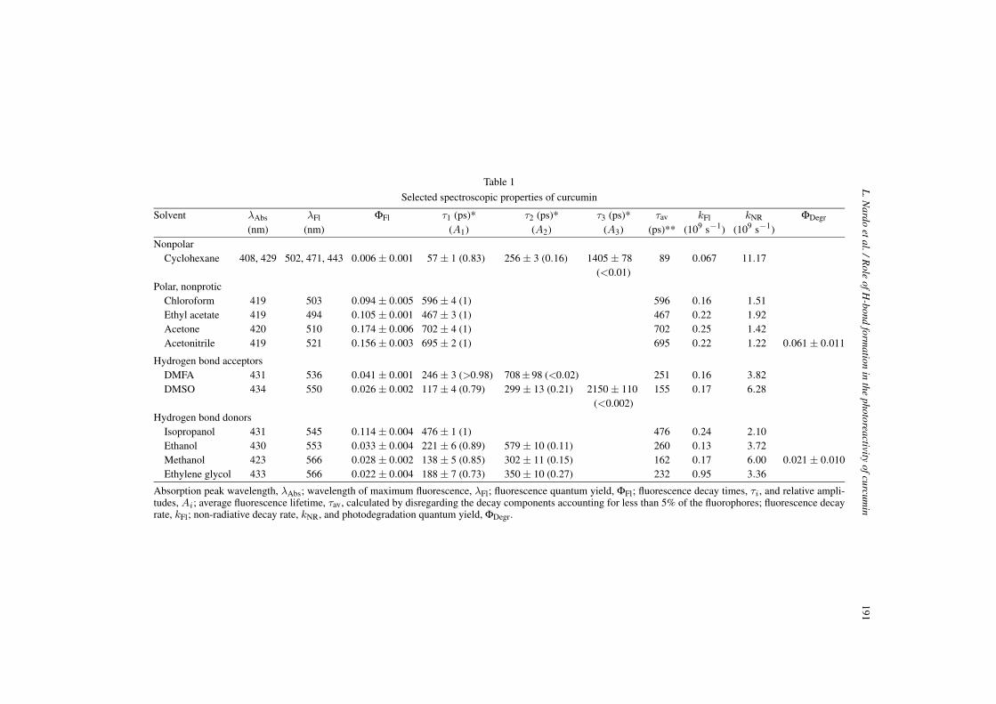
The solvents used in the present study can be divided into four main categories; non-polar (cyclo-hexane), polar non-protic (chloroform, ethyl acetate, acetone, acetonitrile), polar H-bond accepting(dimethylformamide, DMFA, and dimethylsulfoxide, DMSO), and polar H-bond donating (isopropanol,ethanol, methanol, ethylene glycol). The data are summarized in Table 1.
3.1. Absorption spectra
The absorption spectra mainly consist of broad and essentially structureless bands (see Fig. 2), evenif the spectra in protic solvents display a weak shoulder, which is also evident in ethyl acetate. Only incyclohexane the spectrum definitely exhibits two bands. The nature of the solvent slightly affects thewavelength of maximum absorption, λAbs. A small red shift is observed on going from cyclohexane tomore polar solvents (see the λAbs values in Table 1 and Fig. 2). The largest red shifts are observed forhydrogen bonding solvents. A similar trend in the absorption peak wavelength has been reported byother authors [9].
L.N
ardoetal./R
oleofH
-bondform
ationin
thephotoreactivity
ofcurcumin
191
Table 1
Selected spectroscopic properties of curcumin
Solvent λAbs λFl ΦFl τ1 (ps)* τ2 (ps)* τ3 (ps)* τav kFl kNR ΦDegr
(nm) (nm) (A1) (A2) (A3) (ps)** (109 s−1) (109 s−1)Nonpolar
Cyclohexane 408, 429 502, 471, 443 0.006 ± 0.001 57 ± 1 (0.83) 256 ± 3 (0.16) 1405 ± 78 89 0.067 11.17(<0.01)
Polar, nonproticChloroform 419 503 0.094 ± 0.005 596 ± 4 (1) 596 0.16 1.51Ethyl acetate 419 494 0.105 ± 0.001 467 ± 3 (1) 467 0.22 1.92Acetone 420 510 0.174 ± 0.006 702 ± 4 (1) 702 0.25 1.42Acetonitrile 419 521 0.156 ± 0.003 695 ± 2 (1) 695 0.22 1.22 0.061 ± 0.011
Hydrogen bond acceptorsDMFA 431 536 0.041 ± 0.001 246 ± 3 (>0.98) 708±98 (<0.02) 251 0.16 3.82DMSO 434 550 0.026 ± 0.002 117 ± 4 (0.79) 299 ± 13 (0.21) 2150 ± 110 155 0.17 6.28
(<0.002)Hydrogen bond donors
Isopropanol 431 545 0.114 ± 0.004 476 ± 1 (1) 476 0.24 2.10Ethanol 430 553 0.033 ± 0.004 221 ± 6 (0.89) 579 ± 10 (0.11) 260 0.13 3.72Methanol 423 566 0.028 ± 0.002 138 ± 5 (0.85) 302 ± 11 (0.15) 162 0.17 6.00 0.021 ± 0.010Ethylene glycol 433 566 0.022 ± 0.004 188 ± 7 (0.73) 350 ± 10 (0.27) 232 0.95 3.36
Absorption peak wavelength, λAbs; wavelength of maximum fluorescence, λFl; fluorescence quantum yield, ΦFl; fluorescence decay times, τi, and relative ampli-tudes, Ai; average fluorescence lifetime, τav, calculated by disregarding the decay components accounting for less than 5% of the fluorophores; fluorescence decayrate, kFl; non-radiative decay rate, kNR, and photodegradation quantum yield, ΦDegr.

192 L. Nardo et al. / Role of H-bond formation in the photoreactivity of curcumin
Fig. 2. Absorption spectra of curcumin in: cyclohexane (solid black line), ethyl acetate (dots), acetone (solid grey line), methanol(triangles), dimethyl-sulfoxide (diamonds). The peak absorbances were normalized to 1.
3.2. Fluorescence emission spectra
Both the fluorescence intensity and the position of the fluorescence maxima, λFl, are much more sensi-tive to the nature of the solvent as compared to absorption (see the λFl values in Table 1 and Fig. 3). Thecentral fluorescence peak is detected at approximately 470 nm in cyclohexane, while it is progressivelyred shifted in polar non-protic solvents, where the λFl values range between 494 nm in ethyl acetateand 521 nm in acetonitrile, and in H-bonding solvents. In particular, the alcohols (H-bond donors) in-duce larger red shifts (λFl being up to 566 nm in methanol and ethylene glycol) than H-bond acceptors(λFl = 536 nm in DMFA and λFl = 550 nm in DMSO). The fluorescence spectral profiles are broad andessentially structureless. Cyclohexane is the only exception, as it displays a three-band structure (Fig. 3).The fluorescence quantum yield (ΦFl) is generally low (see Table 1). The highest values are found in po-lar non-protic solvents (ranging between ΦFl = 0.094 in chloroform and ΦFl = 0.174 in acetone) andthe lowest one in cyclohexane (ΦFl = 0.006). Both H-bond accepting and H-bond donating solventsproduce intermediate ΦFl values, which are very similar to each other (ranging between ΦFl = 0.022 inethylene glycol and ΦFl = 0.041 in DMFA), except for the much higher value measured in isopropanol(ΦFl = 0.114).
3.3. Excited-state dynamics
The fluorescence decay times τi together with the relative initial amplitudes (see Ai, in brackets) arereported in Table 1.
Three decay components are identified in cyclohexane. Two fast components, with decay timesτ1 = 57 ps and τ2 = 256 ps, account for 99% of the fluorophores (83% and 16%, respectively), while

L. Nardo et al. / Role of H-bond formation in the photoreactivity of curcumin 193
Fig. 3. Fluorescence spectra of curcumin in: cyclohexane (solid black line), ethyl acetate (dots), acetone (solid grey line),dimethyl-sulfoxide (diamonds), methanol (triangles). The spectra were corrected for the samples absorbance and normalized toa unit value of the ethyl acetate solution integrated fluorescence intensity.
a much slower component with decay time τ1 = 1405 ps accounts for the remaining 1%. At differ-ence, single exponential decays are observed in polar, non-protic solvents. The decays are essentiallybiexponential in the polar, H-bond accepting solvents. In alcohols, except isopropanol, in which a sin-gle exponential decay is observed, the decays are biexponential. The average fluorescence lifetime τav
(i.e. τav =∑
i τiAi/∑
i Ai) was calculated for each solvent by disregarding the decay components withAi < 5%. The τav values are reported in Table 1. The trends of τav and of ΦFl are similar: the longest τav
values are those obtained for polar non-protic solvents (ranging between τav = 467 ps in ethyl acetateand τav = 702 ps in acetone) and the shortest one that for cyclohexane (τav = 89 ps). In both H-bond ac-cepting and H-bond donating solvents τav (between τav = 155 ps in DMSO and τav = 260 ps in ethanol)is shorter than in polar non-protic solvents, but longer than in cyclohexane. Once again isopropanol isan exception (τav = 476 ps, a value similar to those found for polar non-protic solvents).
By using the ΦFl values and the corresponding τav values displayed in Table 1, the rate constants forthe radiative (kFl) and non-radiative (kNR) decay can be calculated by means of the equations:
kFl =ΦFl
τav, (1)
kNR =1τav
− kFl. (2)
The obtained values of kFl and kNR are reported in Table 1. The values of kNR are substantially higherthan the corresponding kFl values in all solvents. The kNR values were of the order of 1–16 ns−1, while

194 L. Nardo et al. / Role of H-bond formation in the photoreactivity of curcumin
the kFl values were lower by factors between 5 (see acetonitrile) and 170 (see cyclohexane). Thus, thedeactivation of S1 occurs predominately via non-radiative pathways. The lowest kNR values are those forpolar non-protic solvents while the highest one is that for cyclohexane.
The quantum yield of photodegradation, ΦDegr, was measured in acetonitrile and methanol and itsvalues are in Table 1. We observe that ΦDegr
∼= 0.5ΦFl in acetonitrile whereas ΦDegr∼= ΦFl in methanol.
When we measured the efficiency of curcumin as a singlet oxygen photosensitizer we attained barelydetectable phosphorescence signals in both acetonitrile and methanol.
4. Discussion
In order to explain the photophysics of a symmetric β-diketone such as curcumin, one should takeinto account the three diketo conformers and six enol conformers [13]. However, in solution curcuminmainly adopts the cis enol form (Fig. 1A) [9,14,15] as confirmed by the lack of structures in both theabsorption and fluorescence spectra. The untwisting of the aliphatic chain of the cis enol conformerin the ground state is ascribed to formation of a particularly strong intramolecular H-bond (Fig. 1C)between the hydroxyl and keto groups [16]. The anti diketo isomer (Fig. 1B), whose dipole momenthas been calculated to be much smaller than that of cis enol, should not be ruled out in the case ofnon-polar solvents [14] even if the enol form dominates in both polar and non-polar solvents at roomtemperature [17]. In this frame, the two-band structure that we detected in the absorption spectrum incyclohexane, in agreement with a similar observation in toluene made by Chignell et al. [9] could beindicative of the presence of both the cis enol and the anti diketo conformers. Increasing the solventpolarity results in shifting the keto ↔ enol equilibrium towards the enol conformer, and to the loss ofspectral structure. Structureless spectra similar to ours were measured in ethanol and acetonitrile byChignell and coworkers [9].
Intermolecular H-bonding with solvent molecules perturbs the intramolecular hydrogen bond charac-terizing the cis enol conformer. In the enol tautomer, H-bond donating solvents interact with the ketomoiety, while H-bond acceptors interact with the enol proton (see Fig. 4(a)). Perturbation by polar,
(a)
(b)
Fig. 4. (a) Perturbation of intramolecular H-bonding by protic solvents; (b) perturbation of intramolecular H-bonding by polar,non-protic solvents.

L. Nardo et al. / Role of H-bond formation in the photoreactivity of curcumin 195
non-protic solvents is also likely to occur by polarity effects (Fig. 4(b)). As the weakening of the in-tramolecular H-bond makes the curcumin structure less rigid and more prone to out-of-plane vibration,we expect, and actually observe (see λAbs and λFl data in Table 1), the smallest Stokes shift in cyclo-hexane. For the same reason, the much higher Stokes shifts that we find in both non-protic and proticpolar solvents can be taken as indication that both polarity effects and intermolecular H-bond formationheavily perturb intramolecular H-bonding.
In H-bonding solvents, besides the cis conformer, substantial amounts of trans enol have been reportedfor simple β-diketones (Fig. 1A). The enhanced stabilization induced by the formation of intermolecularH-bonds with solvent molecules, which is impossible in the cis conformer for steric reasons, seems to besufficient to shift the equilibrium to the trans enol, whose presence might correlate with the appearanceof shoulders in the absorption spectra of curcumin in the protic solvents.
Instauration of the intramolecular H-bond in the cis enol conformer brings about new mechanismsthat might influence the internal conversion rate and affect kNR. Hydrogen-stretching vibrations, e.g. alengthening of the O–H bond and a shortening of the hydrogen bond in the cis enol form of S1, couldlead to radiationless decay. As we observe minimum Stokes shift in the solvent for which the H-bondis the tightest (cyclohexane, see also toluene [9]) deactivation by H-stretching vibrations is apparentlynot efficient. Charge-transfer interactions provide other radiationless decay pathways to the cis enolconformer. Three proton transfer mechanisms can occur: exchange between the vinylic CH and theenol OH sites (i.e. enol ↔ diketo tautomerism), intermolecular exchange of the labile OH proton, andits transfer from one oxygen to the other of the cis enol conformer. The last mechanism is by far thefastest one and hence the most relevant [13]. This excited-state intramolecular proton transfer (ESIPT)becomes even faster in the presence of the intramolecular H-bond in Fig. 1C [18–21], as demonstratedfor molecules having chemical structures similar to that of curcumin. Rates as high as 1011 s−1 werereported [19]. As curcumin has both electron-donating and electron-accepting properties, in protic en-vironments S1 deactivation by intermolecular electron transfer should also be considered [14]. On thecontrary, intramolecular electron transfer can be ruled out as the molecule lacks both donor and acceptorgroups [22].
Electronically excited β-diketones can also deactivate by undergoing photochemical re-ketonization[23,24]. The process accompanying the enol excitation is postulated to start with cis-trans isomerization,so that the intramolecular H-bonding stabilization of the cis enol is lost, followed by decay via eitherproton transfer to the vinylic CH (re-ketonization) or recovery of the H-bonded cis enol conformer andsubsequent ESIPT (Fig. 5) [23]. The first S1 deactivation mechanism generates an anti diketo conformerin its ground state, and it is thus favored in non-polar solvents, while the second one generates a cis enolconformer stabilized by its intramolecular H-bond, and is most likely to occur in polar solvents.
As to the decay pathways intersystem crossing and photodecomposition, our results on singlet oxygengeneration and photodegradation indicate that, at least in polar solvents (be they protic or not), none ofthem is relevant.
All ΦFl and τav data in Table 1, together with the calculated kFl and kNR, show that the non-radiativepathways are most efficient in cyclohexane, where the tightness of intramolecular H-bond ensures theoccurrence of ESIPT. This is a signal that ESIPT is the leading mechanism in curcumin decay. However,both the three-exponential fluorescence decay and the three-band fluorescence spectrum indicate theexistence of other mechanisms with comparable rates. We associate the shortest decay, and the leastStokes shifted fluorescence peak, to the cis enol curcumin molecules excited to the fluorescent statewithout breaking the H-bond. The intermediate decay, and the fluorescence band peaked at 471 nm, canbe associated to the cis enol conformers that experience enol cis-trans isomerization upon excitation and

196 L. Nardo et al. / Role of H-bond formation in the photoreactivity of curcumin
Fig. 5. Photochemically induced re-ketonization.
deactivate by re-ketonization, while the very few molecules emitting the longest-lived fluorescence arethe ground-state anti diketo conformers, that cannot undergo ESIPT. It is also reasonable to associatethese conformers to the fluorescence band peaking at 502 nm, which is the most Stokes shifted due totheir flexibility with respect to out-of-plane vibrations.
The kNR values in Table 1 are smaller in protic solvents than in the non-polar solvent. As stated above,the fluorescence decays in all the protic solvents, except isopropanol, are biexponential. The fastest decaytime values are not of the order of few tens of picoseconds, as expected for direct ESIPT driven decays,but approximately 200 ps. Possibly, the instauration of intermolecular H-bonds results in the almostcomplete intramolecular H-bond breaking, with consequent blocking of direct ESIPT. Proton transfercan no longer occur directly due to the lack of any electronic connection between the separate solvatedgroups. Desolvation is required to form the fast-decaying intramolecular H-bonded enol structure. Be-cause the ESIPT step is rapid once an intramolecular H-bond is formed, the decay time observed forthis deactivation path will depend essentially on the rate of the desolvation process. Anyway, alternativedecay mechanisms involving delivery of the excitation energy to the solvent molecules are expected tobe highly enhanced by the instauration of intermolecular H-bonds. On the contrary, even if a large num-ber of excited trans enol conformers should be found in protic solvents, re-ketonization is not probable,as the anti diketo conformer is much less polar as compared to the two enol tautomers. Biexponentialfluorescence decays can be thus explained by hypothesizing a fast, intermolecular H-bond mediated,S1 deactivation by transfer of energy to the solvent molecules and a slow, solvent-reorganization mod-erated, ESIPT. In the case of isopropanol, the first mechanism might be somehow prevented.
The ΦFl values are the highest and the τav values the longest in polar non-protic solvents. The kFl
are comparable to those obtained in the case of protic solvents, but the kNR are three to five times

L. Nardo et al. / Role of H-bond formation in the photoreactivity of curcumin 197
slower. This indicates that also polar non-protic solvents efficiently inhibit deactivation through ESIPT,but, at difference with protic solvents, their interactions with curcumin do not bring about new decaymechanisms. This hypothesis is supported by the single exponential decays we observed in all polar non-protic solvents. Interestingly, the τ1 values measured in the non-protic solvents are in the same range asthose of the longer decay times, τ2, in protic solvents, that we associated to molecules decaying throughsolvent-rearrangement moderated ESIPT.
5. Conclusion
The excited state behavior of curcumin is strongly dependent on the reaction medium. We couldsuccessfully explain the features of the spectroscopic data we measured by assuming three simple hy-pothesis, that were previously postulated by other authors:
(1) Curcumin at room temperature is virtually found in only its cis enol conformer [9,14,15,17].Lower amounts of the anti diketo and the trans enol conformers are also found in non-polar [14]and protic environment, respectively.
(2) ESIPT is the leading non-radiative decay mechanism of S1 [13,19].(3) The ESIPT rate is higher the tighter is the intramolecular H-bond between the enol and the keto
moiety [18–21].
The above results may be of help in optimizing drug delivery systems suitable for the administrationof curcumin to patients in view of its future clinical application as a photosensitizer.
References
[1] H.H. Tønnesen, Studies on curcumin and curcuminoids. XVIII. Evaluation of curcuma products by use of standardizedreference color values, Z. Lebensm. Unters. Forsch. 194 (1992), 129–130.
[2] B.B. Aggrawal, C. Sundaram, N. Malani and H. Ichikawa, Curcumin: the Indian solid gold, Adv. Exp. Med. Biol. 595(2007), 1–75.
[3] A. Duvoix, R. Blasius, S. Delhalle, M. Schnekenburger, F. Morceau, E. Henry, M. Dicato and M. Diederich, Chemopre-ventive and therapeutic effects of curcumin, Cancer Lett. 223 (2005), 181–190.
[4] M.E. Egan, M. Pearson, S. Weiner, V. Rajendran, D. Rubin, J. Glockner-Pagel, S. Canny, K. Du, G.L. Lukacs andM.J. Caplan, Curcumin, a major constituent of turmeric, corrects cystic fibrosis defects, Science 304 (2004), 600–602.
[5] Z. Sui, R. Salto, J. Li, C. Craik and P.R. Ortiz, Inhibition of the HIV-1 and HIV-2 proteases by curcumin boron complexes,Bioorg. Med. Chem. 1 (1993), 415–422.
[6] E. Bruzell, E. Morisbak and H.H. Tønnesen, Studies on curcumin and curcuminoids. XXIX. Photoinduced cytotoxicity ofcurcumin in selected aqueous preparations, Photochem. Photobiol. Sci. 4 (2005), 523–530.
[7] K.C. Das and C.K. Das, Curcumin (diferuloylmethane), a singlet oxygen (1O2) quencher, Biochem. Biophys. Res. Com.295 (2002), 62–66.
[8] A.A. Gorman, I. Hamblett, V.S. Srinivasan and P.D. Wood, Curcumin-derived transients: a pulsed laser and pulse radiolysisstudy, Photochem. Photobiol. 59 (1994), 389–398.
[9] C.F. Chignell, P. Bilski, K.J. Reszka, A.G. Motton, R.H. Sik and T.A. Dahl, Spectral and photochemical properties ofcurcumin, Photochem. Photobiol. 59 (1994), 295–302.
[10] A. Tomren, M. Màsson, T. Loftsson and H.H. Tønnesen, Studies on curcumin and curcuminoids. XXXI. Symmetric andasymmetric curcuminoids: stability, activity and complexation with cyclodextrin, Int. J. Pharm. 338 (2007), 27–34.
[11] R. Velapoldi and H.H. Tønnesen, Corrected emission spectra and quantum yields for a series of fluorescent compounds inthe visible spectral region, J. Fluoresc. 14 (2004), 465–472.
[12] D.E. Moore, Standardization of kinetic studies and photodegradation reactions, in: Photostability of Drugs and DrugFormulations, 2nd edn, H.H. Tønnesen, ed., CRC Press, Boca Raton, 2004, pp. 49–53.

198 L. Nardo et al. / Role of H-bond formation in the photoreactivity of curcumin
[13] J. Emsley, The composition, structure and hydrogen bonding of the β-diketones, in: Structure and Bonding, M.J. Clarke,J.B. Goodenough, J.A. Ibers, C.K. Jørgensen, D.M.P. Mingos, J.B. Neilands, D. Reinen, P.J. Sadler, R. Weiss andR.J.P. Williams, eds, Springer-Verlag, Berlin, 1984, pp. 148–191.
[14] K. Balasubramanian, Molecular orbital basis for yellow curry spice curcumin’s prevention of Alzheimer’s disease,J. Agric. Food Chem. 54 (2006), 3512–3520.
[15] U. Pedersen, P.B. Rasmussen and S.O. Lawesson, Synthesis of naturally occurring curcuminoids and related compounds,Liebigs Ann. Chem. 1985 (1985), 1557–1569.
[16] J.T. Mague, W.L. Alworth and F.L. Payton, Curcumin and derivatives, Acta Cryst. C 60 (2004), 608–610.[17] F. Ortica and M.A.J. Rodgers, A laser flash photolysis study of curcumin in dioxane-water mixtures, Photochem. Photo-
biol. 76 (2001), 745–751.[18] S.R. Flom and P.F. Barbara, Proton transfer and hydrogen bonding in the internal conversion of S1 anthraquinones, J. Phys.
Chem. 89 (1985), 4489–4494.[19] A.J.G. Strandjord, S.H. Courtney, D.M. Friedrich and P.F. Barbara, Excited-state dynamics of 3-hydroxyflavone, J. Phys.
Chem. 87 (1983), 1125–1133.[20] S. Mitra and S. Mukherjee, Photophysical properties of substituted intramolecularly hydrogen bonded compounds: A com-
bined experimental and theoretical study, J. Lumin. 118 (2006), 1–11.[21] P.H. Bong, Spectral and photophysical behaviours of curcumin and curcuminoids, Bull. Korean Chem. Soc. 21 (2000),
81–86.[22] Z.R. Grabowski and K. Rotkiewicz, Structural changes accompanying intramolecular electron transfer: focus on twisted
intramolecular charge-transfer states and structures, Chem. Rev. 103 (2003), 3899–4031.[23] A.C. Weedon, Photochemical reactions involving enols, in: The Chemistry of Enols, Z. Rappoport, ed., John Wiley &
Sons, New York, 1990, pp. 591–638.[24] P. Nikolov, F. Fratev, I. Petkov and P. Markov, Dimer fluorescence of some β-dicarbonyl compounds, Chem. Phys. Lett.
83 (1981), 170–173.

Submit your manuscripts athttp://www.hindawi.com
Chromatography Research International
Hindawi Publishing Corporationhttp://www.hindawi.com Volume 2013
Hindawi Publishing Corporationhttp://www.hindawi.com Volume 2013
Carbohydrate Chemistry
International Journal of
Hindawi Publishing Corporationhttp://www.hindawi.com
International Journal of
Analytical ChemistryVolume 2013
ISRN Chromatography
Hindawi Publishing Corporationhttp://www.hindawi.com Volume 2013
Hindawi Publishing Corporation http://www.hindawi.com Volume 2013Hindawi Publishing Corporation http://www.hindawi.com Volume 2013
The Scientific World Journal
Bioinorganic Chemistry and ApplicationsHindawi Publishing Corporationhttp://www.hindawi.com Volume 2013
Hindawi Publishing Corporationhttp://www.hindawi.com Volume 2013
CatalystsJournal of
ISRN Analytical Chemistry
Hindawi Publishing Corporationhttp://www.hindawi.com Volume 2013
ElectrochemistryInternational Journal of
Hindawi Publishing Corporation http://www.hindawi.com Volume 2013
Hindawi Publishing Corporationhttp://www.hindawi.com Volume 2013
Advances in
Physical Chemistry
ISRN Physical Chemistry
Hindawi Publishing Corporationhttp://www.hindawi.com Volume 2013
SpectroscopyInternational Journal of
Hindawi Publishing Corporationhttp://www.hindawi.com Volume 2013
ISRN Inorganic Chemistry
Hindawi Publishing Corporationhttp://www.hindawi.com Volume 2013
Hindawi Publishing Corporationhttp://www.hindawi.com Volume 2013
Journal of
Chemistry
Hindawi Publishing Corporationhttp://www.hindawi.com Volume 2013
Inorganic ChemistryInternational Journal of
Hindawi Publishing Corporation http://www.hindawi.com Volume 2013
International Journal ofPhotoenergy
Hindawi Publishing Corporationhttp://www.hindawi.com
Analytical Methods in Chemistry
Journal of
Volume 2013
ISRN Organic Chemistry
Hindawi Publishing Corporationhttp://www.hindawi.com Volume 2013
Hindawi Publishing Corporationhttp://www.hindawi.com Volume 2013
Journal of
Spectroscopy