Role of CFTR and TMEM16 for Regulated Cell...
68
2016 UNIVERSIDADE DE LISBOA FACULDADE DE CIÊNCIAS DEPARTAMENTO DE QUÍMICA E BIOQUÍMICA Role of CFTR and TMEM16 for Regulated Cell Death Filipa Bica Simões Mestrado em Bioquímica Especialização em Bioquímica Médica Dissertação orientada por: Prof. Dr. med. Karl Kunzelmann, Prof. Dr. Margarida Amaral
Transcript of Role of CFTR and TMEM16 for Regulated Cell...

2016
UNIVERSIDADE DE LISBOA
FACULDADE DE CIÊNCIAS
DEPARTAMENTO DE QUÍMICA E BIOQUÍMICA
Role of CFTR and TMEM16 for Regulated Cell Death
Filipa Bica Simões
Mestrado em Bioquímica
Especialização em Bioquímica Médica
Dissertação orientada por:
Prof. Dr. med. Karl Kunzelmann, Prof. Dr. Margarida Amaral


Acknowledgments/Agradecimentos
i
ACKNOWLEDGMENTS/AGRADECIMENTOS
First of all, I would like to express my appreciation to my supervisors – Prof. Karl Kunzelmann and
Prof. Margarida Amaral – for giving me the incredible opportunity of pursuing my studies in science
while gaining new insight about life abroad. For all the guidance, support, help and knowledge, I truly
thank Prof. Karl Kunzelmann. I also want to show my gratitude to Prof. Rainer Schreiber for being
available to answer my questions, give new ideas and share his experience.
A special thank goes to Ji, who followed my work very close, always showing an energetic attitude,
helping and pushing me to get the best out of my project. I will never forget all the technique and ‘small
tricks’ that she taught me! To Inês, who introduced me the life in Regensburg and always kept a positive
mind, making the craziest excuses to party all night long! Thank you for all the support, conversations
and bell ringing every morning!! To Roberta, for all the true friendship with lots of love and kicking!
Thank you for teaching me when compounds are frozen and for loving my açorda! – You will always
be my ‘you-know-what’! To Kip, for being my essential companionship in the cell culture room, for
always giving me tips about my cells and especially for never give up on trying to make me think
positive! – I will always remember that! To Gam, for all the laughter, dinners, parties and lessons in the
lab! To Joana, for all the morning coffees, opinions and Sunday afternoons doing experiments! All of
you made my days easier and brought really funny and unforgettable moments into my life! I also want
to show my appreciation to Brigitte, Tini, Susi, Patricia and Silvia for all the help and good mood in the
lab!
Obrigada a toda a minha família pela atitude sempre positiva, por todo o apoio e pela boa disposição,
mesmo nos momentos mais difíceis!
Aos meus pais, por me ensinarem a seguir os meus sonhos e a enfrentar os obstáculos na vida,
crescendo com eles. À minha mãe, por me fazer sentir que nunca estive ausente, partilhando todos os
momentos comigo e não passando uma única manhã sem me desejar um bom dia de trabalho! Ao meu
pai, por, apesar da distância, manter sempre a naturalidade, pelos conselhos e por todas as músicas
partilhadas! Ao Nuno, por todo o carinho, maluqueiras e parvoíces, conseguindo sempre pôr-me um
sorriso na cara. És a prova de que um irmão pode ser um melhor amigo!
Um agradecimento muito especial a todos os meus avós, por me acompanharem sempre e me
ensinarem a ser uma pessoa integra – o meu orgulho é eterno! Obrigada aos meus tios e primos, pelo
apoio e carinho incondicionais. Ao Miguel por ser meu amigo, companheiro e segundo irmão!
Obrigada a todos os meus amigos pela amizade verdadeira, pelas brincadeiras e por me fazerem
sentir que nunca estive longe! Em especial à Ana (Frafrá), por ter partilhado esta grande experiência
comigo, por todas as gargalhadas, viagens, cervejas e companhia: sem ti tudo teria sido muito mais
complicado!! Muito obrigada, também, por matares os bichos que apareciam no meu quarto! À Ana e
ao André, por serem amigos incansáveis, honestos, bons ouvintes e conselheiros: a festa recomeça
agora!!

Summary
ii
SUMMARY
Regulated or programmed cell death is defined as an intracellular program that plays a
complementary role to mitosis in maintaining a stable population of cells. Apoptosis is the main form
of regulated cell death, allowing the silent elimination of harmed or aged cells without triggering any
inflammatory response.
Anoctamin 6 (ANO6, TMEM16F) is a multifunctional protein from a family of ten members,
identified as an endogenous Ca2+-activated Cl- channel (CaCC). Apart from its function as a CaCC,
ANO6 is also described as a volume-regulated and outwardly rectifying Cl- channel (ORCC), a non-
selective cation channel and a Ca2+-dependent phospholipid scramblase. It has an ubiquitous expression,
being involved in many physiological processes including apoptosis. Nonetheless, ANO6 role in this
process is not entirely understood.
The Cystic Fibrosis Transmembrane conductance Regulator (CFTR) is a cyclic adenosine
monophosphate (cAMP)-gated Cl- channel expressed in the apical membrane of epithelial cells from the
intestine, pancreas, airways and sweat glands, where it is responsible to maintain ion and fluid
homeostasis. Moreover, it regulates the intracellular redox status, acidification and ceramide content in
lipid rafts, three different functions that may explain CFTR involvement in apoptosis. Mutations in the
CFTR gene are the cause for Cystic Fibrosis (CF), the most common life-threatening autosomal
recessive disease in Caucasians.
Understanding ANO6 and CFTR role in regulated cell death may help to overcome the apoptotic
dysfunction found in Cystic Fibrosis. Here it is shown that ANO6 is activated by ROS (Reactive Oxygen
Species) during apoptosis, acting as a Ca2+-activated Cl- channel, non-selective cation channel and
phospholipid scramblase in different in vitro systems. The channel has a dual contribution for this
process, transporting ions to the extracellular space and mediating phosphatidylserine (PS) exposure in
the outer membrane leaflet. Furthermore, a CFTR contribution to ROS-mediated apoptosis was found
in CFBE cells, a mechanism independent of pore opening and channel stimulation. Co-expression
studies of ANO6 and CFTR in HEK293 cells revealed a functional relationship of these proteins during
ROS-mediated apoptosis not only in terms of whole-cell current but also regarding phospholipid
scrambling.
A possible interaction between ANO6 and CFTR is also proposed to explain why cells
overexpressing both proteins have an enhanced spontaneous and ROS-induced apoptosis. Exposure of
cells to oxidative stress and consequent ROS production leads to mitochondrial permeabilization, release
of Ca2+ and pro-apoptotic proteins, responsible for caspase cleavage. These events terminate with ANO6
activation, which may support cell shrinkage and phospholipid scrambling, two apoptotic hallmarks
enhanced in the presence of CFTR.
Key-words: Regulated cell death, Apoptosis, Anoctamin 6, CFTR, Cystic Fibrosis

Resumo
iii
RESUMO
A morte celular programada é um mecanismo homeostático que desempenha um papel
complementar à mitose na manutenção de uma população estável de células nos tecidos e órgãos. Este
programa é ativado diariamente, removendo milhões de células danificadas ou envelhecidas. A apoptose
é considerada a forma predominante de morte celular programada, garantindo a eliminação silenciosa
de células, isto é, sem ativação do sistema imune.
Durante a apoptose, as células sofrem uma grande variedade de modificações morfológicas e
bioquímicas. Estas alterações são em grande parte mediadas por uma família de proteases conhecida por
caspases. As caspases são normalmente expressas como precursores inativos ou procaspases, que
adquirem atividade proteolítica na presença de um determinado estímulo. Depois de ativos, estes
enzimas clivam outras procaspases, proteínas essenciais e ADN, sendo assim responsáveis pela
amplificação da cascata apoptótica. A clivagem de todos estes substratos resulta na alteração fenotípica
das células apoptóticas, as quais passam a apresentar uma redução no seu volume (shrinkage),
condensação de cromatina e formação de protrusões na membrana plasmática (blebbing).
A composição fosfolipídica da membrana plasmática é também afetada durante a apoptose. Numa
célula viável, a bicamada lipídica é assimétrica: a fosfatidilserina (PS) e a fosfatidiletanolamina (PE)
estão presentes no folheto interno, enquanto a fosfatidilcolina (PC) e a esfingomielina (SM) localizam-
se no folheto externo. Esta assimetria é destruída pelo transporte bidirecional de fosfolípidos através da
membrana plasmática, mediado por scramblases (scrambling). Particularmente, durante a apoptose, a
PS é movida do folheto interno para o folheto externo, o que serve de sinal para a fagocitose das células
apoptóticas, um processo essencial para a manutenção da integridade do hospedeiro.
A Anoctamina 6 (ANO6) é uma proteína ubíqua e multifuncional pertencente a uma família proteica
formada por dez membros homólogos entre si (ANO1-10; TMEM16 A-K). Esta é identificada como um
canal de cloreto regulado por volume e ativado por cálcio (do inglês: Calcium activated Chloride
Channel – CaCC), canal catiónico não seletivo, componente do ORCC (do inglês: Outwardly Rectifying
Chloride Channel) e scramblase de fosfolípidos ativada por cálcio. A ANO6 participa numa enorme
variedade de processos biológicos, tais como a coagulação sanguínea, desenvolvimento do esqueleto,
regulação do volume e migração celular, entre outros. A identificação da ANO6 como uma scramblase
de fosfolípidos levantou várias questões acerca do seu envolvimento na apoptose. Diferentes estudos
indicam que esta proteína contribui para este processo como um canal iónico, participando
eventualmente no shrinkage das células e na ativação de caspases. No entanto, no que diz respeito à sua
função como scramblase, o papel da ANO6 é ainda incerto.
O CFTR (do inglês: Cystic Fibrosis Transmembrane conductance Regulator) é um canal de cloreto
regulado por cAMP (Adenosina de monofosfato cíclico), expresso na membrana apical de células
epiteliais do intestino, pâncreas, vias respiratórias e glândulas sudoríparas. Mutações no gene que
codifica para esta proteína são a causa da Fibrose Quística, a doença mortal autossómica recessiva
responsável pelo maior número de mortes na população caucasiana. Apesar da enorme diversidade de
variantes de mutações identificadas (cerca de 1.500), a deleção de um resíduo de fenilalanina (F) no
codão 508 localizado no cromossoma 7 (ΔF508) é encontrada em 90% dos pacientes com Fibrose
Quística. Esta mutação resulta num folding aberrante do CFTR e consequentemente um defeito no seu
tráfego do retículo endoplasmático para a membrana plasmática.
A Fibrose Quística é principalmente caracterizada por uma por uma perturbação da homeostase
iónica e fluídica. De entre os variados sintomas encontrados nos pacientes afetados, destaca-se a

Resumo
iv
produção excessiva de muco espesso, infeções persistentes nas vias respiratórias pela bactéria
Pseudomonas aeruginosa, infertilidade masculina e insuficiência pancreática e intestinal. Apesar dos
esforços contínuos em compreender o CFTR a um nível funcional e molecular, a Fibrose Quística
continua a ser considerada uma doença letal. A atenuação dos sintomas permitiu aumentar a esperança
média de vida para cerca de 37 anos. No entanto, a doença pulmonar crónica e a resultante perda de
função pulmonar continuam a ser os principais problemas por resolver, sendo responsáveis por 80% da
mortalidade.
Curiosamente, o CFTR foi previamente descrito como um regulador do estado redox intracelular,
da acidificação do citoplasma e organelos e dos níveis de ceramida nas jangadas lipídicas, três processos
determinantes para a apoptose e possivelmente responsáveis pela disfunção apoptótica encontrada na
Fibrose Quística. Apesar de algumas observações não serem coerentes, a maioria dos estudos defende
que mutações no CFTR aumentam a suscetibilidade das células para uma morte necrótica.
Contrariamente ao que se sucede na apoptose, durante a necrose os componentes celulares são libertados
para os tecidos circundantes e o sistema imune é ativado. Desta forma, especula-se que esta disfunção
pode contribuir para as inflamações persistentes que caracterizam a Fibrose Quística.
A interação do CFTR com outros canais e proteínas transportadoras tem sido tópico de investigação
ao longo dos anos. De entre as proteínas identificadas, destaca-se o ORCC, um canal inativo em células
viáveis e envolvido no shrinkage e scrambling de fosfolípidos durante a apoptose. Em 2010 Martins et.
al identificou a ANO6 como componente do ORCC, existindo uma possível interação entre esta proteína
e o CFTR.
Tendo em conta todas estas evidências, o objetivo deste projeto foi o estudo da relação entre a ANO6
e o CFTR durante a morte celular programada. Particularmente, o trabalho focou-se na apoptose
induzida por ROS (do inglês: Reactive Oxygen Species), uma vez que o CFTR é descrito como um
regulador do estado redox intracelular.
A primeira fase deste projeto baseou-se no estudo do impacto dos ROS na atividade da ANO6, não
só em termos de corrente elétrica, mas também no que diz respeito à sua função de scramblase. Com
este objetivo, a expressão endógena da ANO6 foi manipulada em células HEK293 e HeLa e a sua função
foi analisada por patch clamp ou citometria de fluxo, após tratamento com diferentes indutores de stress
oxidativo. Os resultados demonstraram que a ANO6 é ativada por ROS, transportando cloreto e catiões
para o espaço extracelular no início da apoptose, um evento que contribui para o shrinkage das células.
À medida que a via apoptótica avança, a ANO6 passa também a funcionar como scramblase, movendo
a PS para o folheto externo da membrana plasmática. Como o silenciamento da ANO6 não diminuiu a
morte celular, é concluído que esta proteína não é absolutamente essencial para a apoptose mediada por
ROS. No entanto, esta observação não é surpreendente, visto que a morte celular programada é um
processo essencial em qualquer sistema biológico, dependendo de uma grande variedade de moléculas
e componentes. Assim, é muito provável que uma célula que não expresse a ANO6 compense este
defeito com outras proteínas.
A segunda parte deste trabalho focou-se na importância do CFTR para apoptose mediada por ROS
e a identificação de uma possível interação funcional entre esta proteína e a ANO6. Estudos de co-
expressão em células HEK293 revelaram que a presença das duas proteínas aumenta a corrente elétrica
induzida por ROS e intensifica o scrambling de fosfolípidos durante a apoptose. Curiosamente, a co-
expressão da ANO6 e do CFTR revelou ser suficiente para induzir uma apoptose significativa em
condições controlo. Adicionalmente, a linha celular CFBE estavelmente transfetada com CFTR wt ou
ΔF508 foi escolhida como uma ferramenta para o estudo da influência da mutação mais proeminente da

Resumo
v
Fibrose Quística na apoptose. A indução de stress oxidativo nas duas linhas celulares demonstrou que a
expressão do CFTR wt intensifica a apoptose, um fenómeno independente da abertura do poro e ativação
do canal.
Em suma, este estudo permitiu demonstrar que a ANO6 e o CFTR cooperam durante a apoptose
mediada por ROS. Estas duas proteínas revelaram ter uma relação funcional no que diz respeito à
ativação de uma corrente iónica e também ao scrambling de fosfolípidos. É especulado que a expressão
do CFTR na membrana plasmática aumenta a atividade da ANO6, envolvida nestes dois fenómenos
apoptóticos. No entanto, é também provável que o contrário seja verdade, visto que a ANO6 foi já
identificada como componente das correntes induzidas por cAMP em células que expressam o CFTR.
A compreensão do papel da ANO6 e do CFTR durante a morte celular programada pode servir de motor
para obter uma nova visão acerca da disfunção apoptótica encontrada na Fibrose Quística e,
eventualmente, desenvolver novas terapias como tentativa de atenuar o processo inflamatório.
Palavras-chave: Morte programada celular, Apoptose, Anoctamina 6, CFTR, Fibrose Quística

Index
vi
INDEX
ABBREVIATIONS x
1. INTRODUCTION 1
1.1 REGULATED CELL DEATH – APOPTOSIS 1
1.2 ANOCTAMIN 6 – A UNIQUE MEMBER OF THE ANOCTAMIN FAMILY 3
1.2.1 Role of Anoctamin 6 in Apoptosis 4
1.3 CFTR 5
1.3.1 CFTR and Cystic Fibrosis – An Overview 5
1.3.2 CFTR, Cystic Fibrosis and Apoptosis 6
1.4 INTERACTION BETWEEN ANO6 AND CFTR 7
2. OBJECTIVES 8
3. MATERIALS AND METHODS 9
3.1 CELL CULTURE 9
3.1.1 Mammalian Cell Lines and Culture Conditions 9
3.1.2 Transient Transfections 9
3.1.3 Apoptosis Induction 10
3.2 PROTEIN ANALYSIS – WESTERN BLOT 10
3.3 FLUORESCENCE MICROSCOPY 11
3.3.1 ROS Detection 11
3.4 FLOW CYTOMETRY – APOPTOSIS DETECTION 12
3.4.1 Caspase-3 Activity Measurements 12
3.4.2 Annexin V and 7-AAD Labeling 13
3.5 HOLOGRAPHIC MICROSCOPY 14
3.6 CONDUCTANCE MEASUREMENTS 14
3.6.1 YFP Fluorescence Quenching Assay 14
3.6.2 Patch Clamp 15
3.7 CALCIUM SIGNALING MEASUREMENTS – FURA-2 AM 16
3.8 STATISTICAL ANALYSIS 16

Index
vii
4. RESULTS 17
4.1 ANO6 IS ACTIVATED BY ROS 17
4.1.2 ANO6 plays a role in tBHP-induced apoptosis of HEK293 cells 17
4.1.3 ANO6 contribution to STS-mediated apoptosis of HEK293 and HeLa cells 21
4.2 CFTR ENHANCES ANO6 EFFECT DURING ROS-MEDIATED APOPTOSIS 25
4.2.1 ΔF508 CFTR mutation partially rescues cells from apoptosis 27
4.2.2 CFTR contribution to apoptosis is not by GSH transport through the CFTR pore 28
5. DISCUSSION 30
5.1 ANO6 PLAYS A ROLE DURING ROS-MEDIATED APOPTOSIS 30
5.1.1 ANO6 currents are prior to phospholipid scrambling 30
5.1.2 Mechanism of ANO6 activation by ROS 30
5.2 CFTR PLAYS A ROLE DURING ROS-MEDIATED APOPTOSIS 32
5.3 ANO6 AND CFTR ARE CO-WORKERS DURING REGULATED CELL DEATH 33
6. FUTURE PERSPECTIVES 35
7. REFERENCES 36
8. APPENDICES 41
APPENDIX I – CDNA 41
APPENDIX II - INHIBITION OF TBHP-INDUCED APOPTOSIS BY IDEBENONE 41
APPENDIX III – DOWNREGULATION OF ANO6 IN HEK293 AND HELA CELLS 42
APPENDIX IV – EFFECT OF ANO6 SILENCING ON TBHP-INDUCED APOPTOSIS OF HEK293 CELLS 43
APPENDIX V - EFFECT OF CFTR IN H2O2-INDUCED CELL DEATH OF CFBE CELLS 44
APPENDIX VI – EFFECT OF STS IN WT AND SCOTT B LYMPHOCYTES 45
APPENDIX VII - CACC-AO1 DOES NOT INHIBIT TBHP-INDUCED APOPTOSIS OF HEK293 CELLS 46
APPENDIX VIII - ANO6 IS ESSENTIAL FOR CA2+-INDUCED SCRAMBLING 47
APPENDIX IX – INTRACELLULAR CA2+ CONCENTRATION DURING ROS-MEDIATED APOPTOSIS 49
APPENDIX X – PLASMA MEMBRANE TENSION IN ROS-MEDIATED APOPTOSIS 50

Index of Figures and Tables
viii
INDEX OF FIGURES AND TABLES
Figure 1.1. Representation of morphological transformations occurring during apoptosis and
engulfment of the dying cell by phagocytes. 1
Figure 1.2. Extrinsic and intrinsic apoptotic pathways. 2
Figure 1.3. Schematic representation of the function of flippases, floppases and scramblases. 3
Figure 1.4. Model of Anoctamin structure and ANO6 pore. 4
Figure 1.5. Scheme of CFTR structure. 6
Figure 3.1. Mechanism of ROS detection by H2DCFDA. 11
Figure 3.2. Mechanism of Caspase-3 activity detection by NucView™ 488 Caspase-3 substrate. 12
Figure 3.3. Schematic representation of dual staining with Annx V and 7-AAD. 13
Figure 3.4. YFP Fluorescence Quenching by Iodide (I-). 15
Figure 4.1. ROS production, cell shrinkage and caspase-3 activation by tBHP in HEK293 cells 18
Figure 4.2. tBHP activates an ANO6-dependent Cl- conductance. 19
Figure 4.3. ANO6 is activated by tBHP as a phospholipid scramblase. 20
Figure 4.4. ANO6 contribution to PS exposure induced by the ROS-producer STS. 21
Figure 4.5. STS leads to ROS increase and caspase-3 activation, mediating PS exposure enhanced by
overexpressed ANO6 in HeLa cells. 22
Figure 4.6. Downregulation of ANO6 in HeLa cells does not inhibit Annx V positivity induced by STS.
23
Figure 4.7. STS activates ANO6 currents in HeLa cells. 24
Figure 4.8. CFTR enhances ANO6 contribution to apoptosis induced by STS in HEK293 cells. 25
Figure 4.9. Enhanced apoptotic whole-cell Cl- currents in cells expressing ANO6, CFTR and ANO6 +
CFTR. 26
Figure 4.10. Effect of CFTR in tBHP-induced cell death. 27
Figure 4.11. Persistent stimulation of CFTR is not sufficient to trigger spontaneous apoptosis. 28
Figure 4.12. CFTR inhibition does not reduce tBHP-induced cell death. 29
Figure 5.1. Proposed role of ANO6 and CFTR in apoptotic cell death. 34
Figure 8.1. Inhibition of tBHP-induced apoptosis by Idebenone. 41
Figure 8.2. Western Blot analysis of ANO6 expression. 42
Figure 8.3. ANO6 silencing increases the tBHP-induced apoptosis in HEK293 cells. 43

Index of Figures and Tables
ix
Figure 8.4. CFBE cells expressing wt CFTR have a higher susceptibility to H2O2-induced cell death.
44
Figure 8.5. STS induces a differential Annx V positivity in wt and Scott lymphocytes. 45
Figure 8.6. Effect of CaCC-AO1 in tBHP-induced apoptosis of HEK293 cells. 46
Figure 8.7. Ionomycin induces an ANO6-dependent phospholipid scrambling in HEK293 cells. 47
Figure 8.8. Ionomycin induces an ANO6-dependent phospholipid scrambling in HeLa cells. 47
Figure 8.9. Ionomycin induces an ANO6-dependent phospholipid scrambling in B lymphocytes. 48
Figure 8.10. ROS effect in intracellular Ca2+ concentration. 49
Figure 8.11. Arachidonic acid enhances tBHP-induced apoptosis. 50
Figure 8.12. LPL induces an ANO6-independent PS exposure in HEK293 cells. 51
Figure 8.13. Tannic acid inhibits tBHP-induced apoptosis. 52
Table 8.1. Plasmids accession number and base pairs (bp). 41

Abbreviations
x
ABBREVIATIONS
[Ca2+]i Intracellular Ca2+ concentration
µg Microgram (10-6 g)
µL Microliter (10-6 L)
µm Micrometer (10-6 m)
µM Micromolar (10-6 M)
7-AAD 7-Aminoactinomycin D
A Ampere
ABC ATP - binding cassette
Ac Acid ceramidase
ACA N-(p-Amylcinnamoyl)anthranilic Acid
AIF Apoptosis Inducing Factor
AM Acetoxymethyl
AMP Adenosine Monophosphate
Annx V Annexin V
ANO Anoctamin
ANOVA Analysis of variance
APAF 1 Apoptotic Peptidase Activating Factor 1
ArA Arachidonic acid
Asm Acid sphingomyelinase
ATP Adenosine Triphosphate
AVD Apoptotic Volume Decrease
B Lymph B Lymphocytes
bp Base pair
C Cytosine
C Celsius
Ca2+ Calcium ion
CaCC Ca2+-activated Cl- channels
cAMP cyclic AMP
CD8 Cluster of Differentiation 8
CF Cystic Fibrosis
CFBE Cystic Fibrosis Bronchial Epithelial (cell line)
CFTR Cystic Fibrosis Transmembrane conductance Regulator
Cl- Chloride ion
CO2 Carbon Dioxide
Con Control
C-terminal Carboxyl- terminal
Cyt c Cytochrome C
D Aspartic acid
DCF 2',7'-dichlorofluorescein

Abbreviations
xi
DIC Differential Interference Contrast
DMEM Dulbecco's Modified Eagle Medium
DMSO Dimethyl sulfoxide
DNA Deoxyribonucleic acid
EGTA Ethylene Glycol Tetraacetic Acid
ER Endoplasmic Reticulum
eYFP Enhanced Yellow Fluorescent Protein
F Phenylalanine
FBS Fetal Bovine Serum
FITC Fluorescein isothiocyanate
FK Forskolin
FRET Förster Resonance Energy Transfer
FSC Forward Scatter
G Guanine
G Glutamic acid
G Gram
GSH Glutathione
GSHEE Glutathione reduced Ethyl Ester
H Hour(s)
H Histidine
H2DCFDA 2',7'-dichlorodihydrofluorescein diacetate
H2O2 Hydrogen Peroxide
HEK293 Human Embryonic Kidney 293 (cell line)
HeLa Henrietta Lacks Cervical Carcinoma (cell line)
HRP Horseradish Peroxidase
I Isoleucine
I- Iodide
IBMX 3-isobutyl-1-methylxanthine
Ide Idebenone
Iono Ionomycin
K+ Potassium ion
L Liter
L Leucine
LPA Lysophosphatidic Acid
LPL Lysophospholipids
Lyso-PS Lyso-Phosphatidylserine
m Meter
M Molar
MEM Minimum Essential Medium
Mg2+ Magnesium ion
min Minute(s)

Abbreviations
xii
mL Milliliter (10-3 L)
mm Millimeter (10-3 m)
mM Millimolar (10-3 M)
MMP Mitochondrial Membrane Permeabilization
MPTP Mitochondrial Permeability Transition Pore
MSD Membrane-spanning domain
mV Millivolt (10-3 V)
N- terminal Amino- terminal
nA Nanoampere (10-9 A)
NBD Nucleotide-binding domain
nhTMEM16 Nectria haematococca Trasmembrane Protein 16
nM Nanomolar (10-9 A)
nm Nanometer (10-9 m)
ORCC Outwardly Rectifying Cl- Channel
PBS Phosphate Buffered Saline
PC Phosphatidylcholine
PE Phosphatidylethanolamine
Pen Strep Penicillin-Streptomycin
PKA Protein Kinase A
PLA2 Phospholipase A2
PS Phosphatidylserine
PVDF Polyvinylidene fluoride
Q Glutamine
R Regulatory domain
RNA Ribonucleic acid
ROS Reactive Oxygen Species
rpm Rotations per minute
RPMI Roswell Park Memorial Institute medium
RVD Regulatory Volume Decrease
s Second(s)
scrmbld Scrambled
siRNA Small interfering RNA
SM Sphingomyelin
SSC Side Scatter
STS Staurosporine
TA Tannic Acid
tBHP Tert-butyl hydroperoxide
TMEM16 Transmembrane Protein 16
TNF Tumor Necrosis Factor
TNF-R Tumor Necrosis Receptor
V Volt

Abbreviations
xiii
V Valine
WB Western Blot
wt Wildtype
XkR8 Xk-related protein 8
YFP Yellow Fluorescent Protein


Introduction
1
1. INTRODUCTION
1.1 REGULATED CELL DEATH – APOPTOSIS
Multicellular organisms maintain a stable population of cells during their lifespan, keeping a tight
control between cell division and cell death. Every day the activation of an intracellular death
mechanism leads to the elimination of billion harmed or aged cells, which are no longer needed. This
process is known as regulated or programmed cell death and can be divided into several types, being
apoptosis the most predominant and studied form1–5.
Apoptosis was named in 1972 by Kerr and co-workers6, after a Greek word used to describe the
dropping of leaves from trees, suggesting that cell loss is beneficial for the survival of the host. The
authors intended to distinguish this process from a different type of cell death known as necrosis. Unlike
apoptosis, where cell removal happens without compromising the whole organism, necrosis is a highly
immunogenic mechanism. Furthermore, it is considered as the result of a persistent stimulus which leads
to an accidental cell death, whereas apoptosis is genetically determined1,6–8.
The apoptotic pathway involves several molecules and organelles, which are believed to be
conserved in all animals5. Inside this group, caspases play a major role. This family of proteases is
normally expressed as inactive precursors or procaspases, which acquire proteolytic activity in the
presence of a specific stimulus. Once active, these enzymes amplify the apoptotic pathway by cleaving
other procaspases and intracellular key prote ins at specific aspartic residues. Regarding apoptosis,
caspases can be divided in two classes: initiator (caspase-2, 8, 9 and 10) and executioner (caspase-3, 6
and 7). The first group is responsible to activate the executioner caspases, which will then cleave a wide
variety of substrates, leading to the final cell death1,8,9.
The phenotype of an apoptotic cell is mainly characterized by reduced volume, or shrinkage,
chromatin condensation and membrane blebbing1,7,8,10,11. After these transformations, the cytoplasm and
organelles are tightly condensed into apoptotic bodies, keeping their integrity. Once these structures are
formed, they are quickly engulfed by phagocytes (Fig. 1.1) and digested in phagolysosomes, without
production of anti-inflammatory cytokines. This allows the protection of the surrounding tissue from
potential released proteases and oxidizing molecules, avoiding inflammation1,7.
Figure 0.1. Representation of morphological transformations occurring during apoptosis and engulfment of the dying
cell by phagocytes. Initiator caspases activate executioner or effector caspases, able to cleave cellular substrates. As a
consequence, the apoptotic cell shows reduced volume, DNA damage and fragmentation into apoptotic bodies, which are
engulfed by phagocytes. Retrieved and adapted from Fink et. al8.

Introduction
2
Apoptosis can be divided in two main signaling pathways: the intrinsic or mitochondrial pathway
and the extrinsic or death receptor pathway. The first is activated when cells are exposed to inner stress
forms, such as irradiation, heat shock or reactive oxygen species (ROS)1,2. During this mechanism, the
mitochondrial permeability transition pore (MPTP) is opened, leading to the loss of the mitochondrial
transmembrane potential and a stop in the ATP (Adenosine Triphosphate) synthesis12. Pro-apoptotic
proteins are then released to the cytosol which can either function as activators of caspase-9 (e.g. cyt c
– cytochrome c) or as mediators of DNA (Deoxyribonucleic acid) fragmentation (e.g. AIF – Apoptosis
Inducing Factor)1. On the other hand, the extrinsic pathway is activated once specific ligands bind to
their complementary plasma membrane receptors, such as TNF (Tumor Necrosis Factor) to TNF-R
(TNF Receptor) or Fas ligand to CD95 (Fas ligand receptor). In this mechanism caspase-8 and 10 are
major players13. Both signaling pathways end with the activation of executioner caspases, which leads
to protein and DNA fragmentation and the consequent morphological transformations seen in apoptotic
cells1. Caspase-3 is considered a key member of this group, being activated by caspase-8, 9 and 101 (Fig.
1.2).
.
Figure 0.2. Extrinsic and intrinsic apoptotic pathways. Ligand binding to transmembrane death receptors activates the
extrinsic pathway, whereas intracellular stress triggers mitochondrial apoptosis. Both biochemical cascades begin with the
activation of initiator caspases (caspase-8 and 9, respectively), which catalyze the proteolytic maturation of executioner
caspases (caspase-3). The intrinsic pathway is characterized by mitochondrial membrane permeabilization (MMP), resulting
in the release of pro-apoptotic proteins into the cytosol, such as cyt c. The innteraction of cyt c with the adaptor protein apoptotic
peptidase activating factor 1 (APAF 1) and procaspase 9 form the apoptosome structure, responsible for the activation of
caspase-9 and consecutively caspase-3. Retrieved and adapted from Galluzzi et. al14.

Introduction
3
The activation of the apoptotic pathway is followed by modifications in the phospholipid
composition of the plasma membrane. Normally, the inner leaflet consists of phosphatidylserine (PS)
and phosphatidylethanolamine (PE), whereas phosphatidylcholine (PC) and sphingomyelin (SM) are
present in the outer leaflet. Phospholipid transport across the plasma membrane is maintained by three
different classes of proteins: flippases, floppases and scramblases. Flippases and floppases move
phospholipids in a single direction against a concentration gradient, requiring ATP to maintain
membrane asymmetry. The first class is responsible for the transport of PS and PE from the outer to the
inner leaflet, whereas floppases move PC and SM in the opposite direction. Finally, scramblases are
responsible for a bidirectional transport, acting according to the concentration gradient15,16 (Fig. 1.3).
The disruption of this asymmetry leads to the activation of immune cells17, coagulation cascade15,
exocytosis and apoptosis1,17. Particularly, during apoptosis, scramblases transport PS across the lipid
bilayer to the outer leaflet. This process is known as the “eat me signal”, since it serves as a platform for
the engulfment of the dying cells by phagocytes1,15.
Figure 0.3. Schematic representation of the function of flippases, floppases and scramblases. Flippases move PS and PE
from the outer to the inner leaflet, whereas floppases transport PC and SM in the opposite direction, using energy from ATP
hydrolysis. Scramblases are ATP-independent and move phospholipids in both directions according to a concentration gradient.
1.2 ANOCTAMIN 6 – A UNIQUE MEMBER OF THE ANOCTAMIN FAMILY
Anoctamin 6 (ANO6, TMEM16F) is a particular member of the Anoctamins, a family of ten proteins
(ANO1-10, TMEM16 A-K). ANO1, ANO2 and ANO6 have been identified as endogenous Ca2+-
activated Cl- channels (CaCC)18–20. It is a dimeric plasma membrane protein with ten transmembrane α-
helices and a pore loop21. ANO6 is homologous to ANO1, sharing a high sequence identity in the
putative transmembrane-spanning domains of the pore region22. Apart from its function as a CaCC,
ANO6 is also described as a volume-regulated23–25 and outwardly rectifying Cl- channel (ORCC)26, a
non-selective cation channel27 and a Ca2+-dependent phospholipid scramblase28,29. Because it is a
multifunctional protein and has an ubiquitous expression22, ANO6 became an interesting subject for
investigation.
Missense mutations in the TMEM16F gene and production of a defective ANO6 are the cause for
the Scott Syndrome, a rare congenital bleeding disorder. Platelets from patients with Scott Syndrome
are unable to scramble PS, a process required for the activation of coagulation factors and to keep a
regular hemostasis29–31. Studies in mice revealed that knockout of the TMEM16F gene results in an
increased bleeding time due to a suppression of platelet activation27. Moreover, ANO6 is not only
involved in blood coagulation, but also in skeletal development32, apoptosis23,26,33,34, volume

Introduction
4
regulation23,24, microparticle shedding22, innate immunity in macrophages35, breast cancer36, cell
blebbing35 and migration37.
Despite all the research, ANO6 mechanism of action is still not completely understood. Although it
is clear that high intracellular Ca2+ concentrations are required, it is unknown if Ca2+ activates ANO6
directly or through another Ca2+-dependent molecule19. Another unresolved question is the structure of
this protein and how it is able to transport lipids through an ion-conductive pore. Interestingly, Whitlock
et. al suggests that all anoctamins evolved from phospholipid scramblases and acquired ion channel
activity after structural rearrangements. It is possible that the ANO6 pore has a dual molecular
composition (lipid and protein) that allows the transport of amphipathic molecules, an idea known as
the proteolipidic pore hypothesis. In this regard, the ANO6 ion currents are probably just the result of
ions that flow across the plasma membrane together with phospholipids38. This model is supported by
crystallization of a fungal TMEM16 lipid scramblase homologue, nhTMEM16, a protein formed by ten
transmembrane domains and a pore loop. It was found that the pore has a proteolipidic nature, having a
hydrophilic region next to the lipid bilayer, surrounded by two transmembrane helices21,38 (Fig. 1.4).
Figure 0.4. Model of Anoctamin structure and ANO6 pore. A) Topology of the Anoctamin homologue in Nectria
haematococca, nhTMEM16. This structure is formed by ten transmembrane domains with a pore loop (green) and the N- and
C-terminal domains in the cytosol (blue and red, respectively). Retrieved from Brunner et al.21. B) Cartoon showing the possible
proteoplypidic furrow of ANO6 pore, viewed from the extracellular space. The particular molecular composition of the pore
allows the transport of phospholipids and ions across the plasma membrane – along with ions, phospholipid head groups interact
with the protein content of the furrow, whereas acyl chains are projected into the hydrophobic bilayer. Retrieved from Whitlock
et.al.38
1.2.1 Role of Anoctamin 6 in Apoptosis
The identification of ANO6 as a Ca2+-dependent phospholipid scramblase28,29 raised questions about
the involvement of this protein in apoptosis. Remarkably, ANO6 has a marked expression in the surface
of epithelial cells from mouse colon, which have a high apoptosis rate, compared to colonic crypt cells,
known to have a fast proliferation26.

Introduction
5
In this regard, ANO6 was found to be a component of the ORCC, an ubiquitous ion channel known
to be inactive in healthy cells and activated during apoptosis, leading to cell shrinkage and phospholipid
scrambling22,26. As a matter of fact, Martins et. al proved that stimulation of both intrinsic and extrinsic
apoptotic pathways activate ANO6-dependent currents in T Lymphocytes, human alveolar and airway
epithelial cells. These currents were sensitive to the broad anoctamin inhibitors and completely
abolished by ANO6 downregulation26. The same pattern was found in Xenopus Oocytes overexpressing
ANO6 after stimulation with Paraquat, a ROS donor22.
In parallel, Juul et. al observed a decrease in cisplatin-induced caspase-3 activity after knockdown
of ANO6 in ELA cells23. This protein is known to be involved in RVD (Regulatory Volume Decrease)23–
25 and many channels important for this process are also relevant for AVD (Apoptotic Volume
Decrease). Therefore, it was speculated that ANO6 facilitates apoptosis by mediating cell shrinkage23.
Different studies show a clear contribution of ANO6 to apoptosis as an ion channel26,34. However,
investigations in B Lymphocytes revealed that this protein is not crucial to the PS exposure detected in
apoptotic cells34. Cell death is a fundamental process in all living systems and it is very likely that other
proteins contribute for this process, compensating possible defects. It is now known that ANO3, 4, 7, 9
and 10 are also phospholipid scramblases22,39 and, aside from the anoctamin family, XkR8 (Xk-related
protein 8) is also reported to be responsible for phospholipid scrambling during apoptosis40.
1.3 CFTR
1.3.1 CFTR and Cystic Fibrosis – An Overview
Cystic Fibrosis (CF) is the most common life-threatening autosomal recessive disease in Caucasians,
affecting around 70,000 people worldwide41. It is caused by mutations in the CFTR gene (Cystic Fibrosis
Transmembrane conductance Regulator) and is mainly characterized by an imbalance of ion and fluid
homeostasis42. This gene encodes for a cyclic AMP (cAMP)-regulated Cl- channel expressed in the
apical membrane of epithelial cells from the intestine, pancreas, airways and sweat glands43.
Principal symptoms of CF include: production of thick mucus and subsequent infection by
Pseudomonas aeruginosa in the airways; pancreatic insufficiency with obstruction of pancreas ducts
and damage of the exocrine function; intestine impairment; elevated concentration of electrolytes in
sweat and male infertility41,44.
So far around 1,500 variants of mutations in the CFTR gene have been identified. Despite this
diversity, a deletion of phenylalanine (F) at codon 508 (ΔF508) on chromosome 7 is the main cause for
CF, affecting 90% of patients. This mutation results in an abnormal folding of CFTR and a trafficking
defect from the ER (Endoplasmic Reticulum) to the plasma membrane45.
The predicted CFTR sequence contains 1,480 amino acids residues43 arranged in two membrane-
spanning domains (MSD), two nucleotide-binding domains (NBD) and one regulatory domain (R)46
(Fig. 1.5). CFTR evolved from the ATP-binding cassette (ABC) transporters, sharing a high sequence
and structure similarity with these proteins42. Activation of CFTR requires phosphorylation of serine
residues in the R domain by the cAMP- dependent Protein Kinase A (PKA). ATP binding to the NBD
domains and its subsequent hydrolysis allows the pore opening and provides enough energy to the
transport of substances. Once the R domain is dephosphorylated by phosphatases CFTR returns to its
closed status. Thus, the balance between kinase and phosphatase activity together with ATP levels are
the main regulators of CFTR gating42,44,47.

Introduction
6
Figure 0.5. Scheme of CFTR structure. CFTR is inserted in the plasma membrane and contains two MSD domains (MSD1,
MSD2), two NBD domains (NBD1, NBD2) and one R domain with multiple phosphorylation sites. Retrieved and adapted
from Farinha et.al.46.
Regardless the continuous efforts in understanding CFTR at its functional and molecular level, CF
remain a lethal disease. By attenuating symptoms, life expectancy has been significantly increased to
around 37 years-old. However, chronic lung disease and resulting loss of pulmonary function is still a
major uncontrolled problem, being responsible for 80% of mortality41. Nowadays, different therapy
approaches are under development, either based on the identification of compounds that rescue the
CFTR trafficking defect (correctors) or stimulators of channel activity (potentiators)45.
1.3.2 CFTR, Cystic Fibrosis and Apoptosis
As described before, apoptosis is a non-inflammatory fundamental process essential to the clearance
of aged or harmed cells. Notably, CF disease is characterized by a defective apoptosis, a problem that
may contribute to the ongoing inflammations. The involvement of CFTR in cell death has been studied
over the years and, although some results are controversial, it seems clear that this Cl- channel is a pro-
apoptotic factor48.
CFTR has been described as a regulator of the redox status in the airways, mediating the transport
of GSH (Glutathione) to the extracellular space49. GSH is a major antioxidant, responsible for
scavenging the ROS produced as a result of cellular metabolism50. Indeed, measurements in the lung
epithelial lining fluid of CF patients revealed a lower content in GSH compared to healthy individuals51.
Therefore, mutations in CFTR impair GSH efflux and leads to an increase of the oxidative stress in the
airways52.
On the other hand, as GSH is transported, the intracellular level of this antioxidant decreases. Hence,
cells expressing wt CFTR are less capable to buffer high concentrations of ROS and have a higher
susceptibility to apoptosis. Indeed, a relationship between CFTR, GSH efflux and apoptosis was found
in mice proximal tubules where this protein may be responsible for AVD and caspase-3 activation53. In
a different study HeLa cells were transfected with wt or ΔF508 CFTR and it was found an association
among CFTR, GSH transport and Bax activation, a pro-apoptotic protein54.
Intracellular acidification is an essential phenomenon during apoptosis, required for caspase
activation and DNA cleavage. Interestingly, CFTR is known to secrete not only Cl-, but also bicarbonate,
regulating the pH from intracellular compartments. Analysis of mucus collected from lungs of CF

Introduction
7
patients revealed the presence of high molecular DNA, which contributes to the increased viscosity. One
explanation for this observation may be related to the apoptosis dysfunction found in CF epithelial
cells55. Studies in Chinese hamster lung fibroblasts and mouse mammary epithelial cells proved an
association between DNA fragmentation and CFTR. Cells expressing ΔF508 CFTR have a higher
intracellular pH, due to the lack of Cl- and bicarbonate transport. Thus, when these cells are exposed to
an apoptotic stimulus, the intracellular pH prevents nuclear condensation and DNA degradation55,56.
The apoptotic signaling cascade is complex, involving multiple molecules and signals. In this
pathway ceramide acts as a messenger mediating the activation of apoptosis57. Ceramide intracellular
level is regulated by a balance in the activity of two different enzymes: acid sphingomyelinase (Asm)
and acid ceramidase (Ac), responsible for ceramide production and degradation, respectively57,58. Asm
is triggered by Pseudomonas aeruginosa infection , leading to the increase of the ceramide content in
lipid rafts59. Ceramide-enriched lipid rafts are required for pathogen elimination and apoptosis.
However, in 2008 a study concluded that ceramide accumulation also facilitates inflammation, cell death
and infections in CF60. A high level of ceramide in the respiratory tract of uninfected CFTR-deficient
mice was found to be responsible for persistent inflammations and death of respiratory epithelial cells.
When CFTR is mutated the intracellular vesicles have a higher pH, which leads to an imbalance of Asm
and Ac activities and consequently to accumulation of ceramide in membrane lipid rafts. Therefore, by
administrating acid sphingomyelinase inhibitors it is possible to stabilize ceramide levels and decrease
inflammation61.
1.4 INTERACTION BETWEEN ANO6 AND CFTR
The concept that CFTR may interact or regulate other Cl- channels and transporters has been around
for many years. Before the identification of CFTR, ORCC was thought to be the Cl- channel defective
in Cystic Fibrosis22. Nowadays, it is known that CFTR and ORCC are independent channels that share
functional relationships62. Interestingly, Martins et. al found that ANO6 is a component of the ORCC
channel and may also be regulated by CFTR26. However, it is unknown if these ion channels interact
directly or through other scaffold proteins63. Notably, ANO6 is responsible for ORCC activation during
apoptosis, a phenomenon that becomes enhanced in the presence of CFTR. Martins et. al also reported
a decrease of the cAMP-activated whole-cell conductance in airway epithelial cells expressing CFTR
after knockdown of ANO6, which suggests a contribution of ANO6 to CFTR currents26.

Objectives
8
1. OBJECTIVES
The aim of the present work was to study the relationship between ANO6 and CFTR during ROS-
mediated apoptosis, using different in vitro systems.
The first objective was to understand the impact of ROS in ANO6 activity. To achieve this, HEK293
and HeLa cells were used either to overexpress ANO6 or downregulate the endogenous expression of
this protein. In parallel experiments, B Lymphocytes immortalized from a Scott patient and a healthy
control64 were useful to compare the presence and total absence of functional ANO6. The current
properties of ANO6 after ROS induction were studied by patch clamp. Moreover, the phospholipid
scramblase function of this protein was monitored by flow cytometry using Annexin V labeling.
Because CFTR regulates the intracellular redox status49, the next question to address would be if
CFTR-mediated GSH depletion and consequent intracellular ROS increase explain a possible
relationship between these two ion channels. In order to accomplish this, co-expression studies were
done in HEK293 cells and the same techniques were applied. Additionally, the CFBE cell line stably
transfected with wt or ΔF508 CFTR was used as a system to look for the influence in ROS-mediated
apoptosis of the most predominant CFTR mutation found in CF patients.
The understanding of the role of ANO6 and CFTR in regulated cell death can be useful to get new
insight about the apoptotic dysfunction that characterizes CF and eventually create new therapeutic
approaches.

Materials and Methods
9
2. MATERIALS AND METHODS
3.1 CELL CULTURE
Cell lines were grown inside an incubator, at 37°C in a 5% CO2-95% air humidified water-saturated
atmosphere. All cell culture was performed according to standard conditions, using sterile equipment.
Every 2 to 5 days, cells were trypsinized (70-90% confluency) from 75 or 25 cm2 flasks (Greiner bio-
one - CELLSTAR®, Frickenhausen, Germany) and, if necessary, seeded in plastic plates (M&B Stricker
Laborfachhandel GbR, Bernried, Germany) at the required density to preform experiments.
3.1.1 Mammalian Cell Lines and Culture Conditions
HEK293 (Human Embryonic Kidney 293)65 and HeLa66 (Henrietta Lacks Cervical Carcinoma) cell
lines were cultured in Dulbecco's Modified Eagle Medium (DMEM; Life Technologies - gibco®,
Karlsruhe, Germany), supplemented with 10% Fetal Bovine Serum (FBS; Life Technologies - gibco®,
Karlsruhe, Germany).
HeLa–Kyoto cells stably transfected with eYFP67 (Enhanced Yellow Fluorescent Protein) with three
point mutations (H148Q, I152L, F46L) were cultured in MEM (Minimum Essential Medium),
GlutaMAX™ (Life Technologies - gibco®, Karlsruhe, Germany), supplemented with 10% FBS, 1%
Penicillin-Streptomycin (Pen Strep; Life Technologies - gibco®, Karlsruhe, Germany) and 200 µg/mL
Hygromycin B (Promocell, Heidelberg, Germany).
B Lymphocytes, isolated from a patient with Scott Syndrome and a control subject64, were cultured
in Roswell Park Memorial Institute medium (RPMI; Life Technologies - gibco®, Karlsruhe, Germany),
supplemented with 10% FBS, 1% Pen Strep and 5 mM Hepes Buffer Solution (Life Technologies -
gibco®, Karlsruhe, Germany).
CFBE cells68 (Cystic Fibrosis Bronchial Epithelial cells) stably transfected with wt or ∆F508 CFTR
were cultured in MEM, supplemented with 10% FBS and 2,5 µg/mL Puromycin (Life Technologies -
gibco®, Karlsruhe, Germany).
To trypsinize adherent cells from flasks (HEK293, HeLa, HeLa-Kyoto, CFBE), cultured medium
was first removed by aspiration and cells were washed with Dulbecco’s Phosphate Buffered Saline
(PBS; Life Technologies - gibco®, Karlsruhe, Germany), without Ca2+ and Mg2+. Trypsin (Life
Technologies - gibco®, Karlsruhe, Germany) was added to culture flasks and incubated at 37°C, 5%
CO2 for 5 min (HEK293, HeLa, HeLa-Kyotto) or 15 min (CFBE). To stop the trypsinization process,
medium containing 10% FBS was added to cell suspension, which was then centrifuged at 20 000 rpm
for 3 min. For B Lymphocytes, suspension cells were collected and centrifuged at 20 000 rpm for 5 min.
In both cases, after the centrifugation step, supernatant was discarded and cells were resuspended in
fresh culture medium.
3.1.2 Transient Transfections
All transient transfections were performed using Lipofectamine™3000 reagent (Invitrogen,
Germany), according to manufacturer’s instructions. Lipofection is a particular method of transfection,
where the genetic material is delivered to cells through a cationic lipid that forms liposomes and interacts
with nucleic acid molecules (negatively charged). With lipofection it is possible to have a high uptake
of the genetic material, allowing an easy overexpression of a protein of interest, using DNA, or a
downregulation of a target gene, by siRNA (small interfering RNA)69,70.

Materials and Methods
10
HEK293 and HeLa cells were transfected with a pcDNA3.1 vector encoding for human ANO6.
CFTR was transfected only in HEK293 cells, using a pIRES vector with the respective sequence coupled
to a CD8 (Cluster of Differentiation 8) receptor, which was proved to be the available plasmid with the
best transfection efficiency. In every experiment, an empty pcDNA3.1 vector (mock) served as a control
and the same amount of plasmid was used in all transfections. When ANO6 and CFTR were co-
expressed in HEK293 cells, a ratio of 1:2 µg was applied. Co-transfection protocol was controlled by
co-expressing the mock plasmid either with ANO6 or CFTR (using the same ratio). For patch clamp
measurements, all transfections were done using a plasmid containing ANO6 or CFTR sequence linked
to a CD8 receptor. Experiments were performed between 48h and 72h after transfections. All the cDNA
used is described in the Appendices section (Appendix I – Table 8.1).
ANO6 endogenous expression was downregulated in HEK293 and HeLa cells, using different
siRNA’s (Invitrogen, Paisley, UK) and confirmed by Western Blot (WB). A negative control (Ambion®,
Darmstadt, Germany) with no sequence similarity for human, mouse or rat gene sequences was used to
control the effects of the siRNA delivery. Experiments and collection of protein for WB were done
between 24h and 72h after transfection.
3.1.3 Apoptosis Induction
Tert-Butyl Hydroperoxide (tBHP, Sigma, Taufkirchen, Germany) was diluted in deionized filtered
water to a final concentration of 100 mM. HEK293 cells were incubated with 100 µM tBHP in
OptiMEM (Life Technologies - gibco®, Karlsruhe, Germany), a reduced serum medium (2% serum).
CFBE cells were incubated with 100 µM tBHP diluted in MEM. Staurosporine (STS, Merck, Darmstadt,
Germany) was dissolved in DMSO (dimethyl sulfoxide) to a final concentration of 1 mM. HEK293,
HeLa and B lymphocytes were incubated with 1 µM STS diluted in OptiMEM, DMEM or RPMI,
respectively. The appropriate volume of DMSO was added to non-treated controls. Hydrogen peroxide
(H2O2, Riedel-de Haen, Seelze, Germany) was diluted in MEM to a final concentration of 900 µM and
incubated in CFBE cells. Lyso-phosphatidylserine (Lyso-PS, Avanti Lipid, Alabama, U.S.A) was
dissolved in chloroform to a final concentration of 10 mM. HEK293 cells were incubated with 10 µM
Lyso-PS for 2h diluted in DMEM. The appropriate volume of chloroform was added to non-treated
controls. All compounds were incubated for the indicated periods of time at 37°C, 5% CO2, except
Ionomycin (Iono, Biomol, Hamburg, Germany) which was added to cells after cell detachment during
experiments to a final concentration of 1, 5 or 10 µM. Inhibitors were purchased from Sigma
(Taufkirchen, Germany) and pre-incubated for at least 30 min at 37°C, 5% CO2.
3.2 PROTEIN ANALYSIS – WESTERN BLOT
ANO6 expression was downregulated in HEK293 and HeLa cells (see section 3.1.2 – 3.1.2
Transient Transfections) and confirmed by WB. Additionally, ANO6 endogenous expression was
detected in CFBE cells. In order to accomplish this, protein was collected from cells and kept in a lysis
buffer (mM: 50 Tris-HCl, 150 NaCl, 50 Tris, 100 DTT, 0.5% NP-40, 1% protease inhibitor cocktail) -
Roche, Germany. Then, protein content was separated in an 8,5% SDS-PAGE Polyacrylamide gel and
separated proteins were transferred to a PVDF (Polyvinylidene fluoride) membrane (GE Healthcare
Europe GmbH, Munich, Germany). To detect ANO6, the membrane was incubated with a primary
antibody (rabbit antihuman ANO6 – David Technology, Germany) overnight at 4°C, using a dilution
factor of 1:500 – 1:1000. Proteins were visualized using a horseradish peroxidase (HRP) - conjugated
goat antirabit secondary antibody (Dilution Factor of 1:10000) and ECL Detection Kit (GE, Healthcare,
Munich, Germany). Protein bands were detected using a FujiFilm LAS-3000 (FujiFilm, Tokyo, Japan).
Experiments were kindly performed by Podchanart Wanitchakool.

Materials and Methods
11
3.3 FLUORESCENCE MICROSCOPY
3.3.1 ROS Detection
Intracellular ROS detection is possible using fluorescence assays. H2DCFDA (2',7'-
dichlorodihydrofluorescein diacetate, Molecular Probes, Invitrogen, Germany) is a non-fluorescent
molecule able to cross plasma membranes. Once in the cytosol, this probe is deacetylated by intracellular
esterases and, as a consequence, trapped inside the cell. In the presence of ROS, the deacetylated form
is oxidized to a highly fluorescent form, DCF - 2',7'-dichlorofluorescein (Fig. 3.1). Thus, the
fluorescence intensity is an index of intracellular ROS level.
Figure 2.1. Mechanism of ROS detection by H2DCFDA. H2DCFDA is able to cross the plasma membrane. Once inside the
cell, the probe is deacetylated by esterases, ensuring probe retention in the cytosol. In the presence of ROS, the non-fluorescent
molecule is oxidized to DCF, which is highly fluorescent, allowing intracellular ROS detection.
Due to easy oxidation, H2DCFDA was dissolved shortly before experiments in DMSO to a final
concentration of 10 mM. To detect ROS, HEK293 and HeLa cells were seeded in 18 mm
collagen/fibronectin-coated glass cover-slips overnight. Cells were loaded with a solution of 10 µM of
H2DCFDA in PBS with Ca2+ and Mg2+ (Life Technologies - gibco®, Karlsruhe, Germany) for 30 min at
37°C, 5% CO2. After loading, the probe was washed 2 times with PBS and the ROS inducer was added
to cells (tBHP or STS) for the desired period of time. Fluorescence increase was detected with an
ApoTome Axiovert 200M fluorescent microscope (Zeiss, Germany), using an excitation wavelength of
493 nm and emission of 520 nm.
Alternatively, ROS was detected using a fluorogenic cytosolic sensor, MAK142 (Taufkirchen,
Germany), which becomes fluorescent in the presence of superoxide and hydroxyl radicals. In this case,
HEK293 cells were plated in 18 mm collagen/fibronectin-coated glass coverslips overnight and treated
with STS. After apoptosis induction, MAK142 was added to cells and fluorescence was monitored.
Fluorescence increase was detected with the same microscope, using an excitation wavelength of 646
nm and emission of 654 nm.

Materials and Methods
12
3.4 FLOW CYTOMETRY – APOPTOSIS DETECTION
Flow cytometry is a laser-based technology that allows single cell measurements. It is commonly
used to get information about fluorescence of a pre-labeled population of cells. Once samples are
injected into the flow cytometer, molecules are excited by a light source and fluorescence of single cells
is detected. This is a multifunctional technique, which also allows the analysis of physical properties,
such as cell size or granularity, two parameters given by forward (FSC) and side (SSC) scatter,
respectively. To study regulated cell death in the present work, a four optical filter BD Accuri™ C6
Cytometer (BD Biosciences, Heidelberg, Germany) was used.
3.4.1 Caspase-3 Activity Measurements
NucView™ 488 Caspase-3 substrate (Biotium, Fremont, USA) is a fluorogenic sensor used for the
detection of caspase-3 activity. It is formed by a DNA marker linked to a peptide sequence (DEVD).
Once this substrate crosses the cell membrane, the DEVD sequence is cleaved by caspase-3 and the
DNA dye is free to stain the nucleus (Fig. 3.2). Therefore, by measuring the fluorescence intensity of
the DNA marker it is possible to monitor caspase-3 activity.
Figure 2.2. Mechanism of Caspase-3 activity detection by NucView™ 488 Caspase-3 substrate. Caspase-3 substrate is
formed by a peptide sequence (DEVD) linked to a DNA fluorogenic sensor. Once inside the cell, the substrate is cleaved at the
DEVD sequence by caspase-3, releasing the DNA dye, which is then free to stain the nucleus.
In order to detect caspase-3 activation after ROS induction, HEK293 and HeLa cells were seeded
overnight and incubated as desired for the indicated periods of time. Following treatment, the media was
collected and cells were washed with PBS. After, adherent cells were detached with accutase (Capricorn
Scientific GmbH, Ebsdorfergrund, Germany), incubated for 3 to 5 min, at 37°C, 5% CO2. Then, the
reaction was stopped with DMEM, supplemented with 10% FBS and suspension cells were centrifuged
2 times for 10 min (1500 rpm, 4°C). Cells were resuspended in a solution of caspase-3 substrate diluted
in PBS (2.5 µL substrate in 100 µL of PBS / sample) and incubated for 30 min at room temperature,

Materials and Methods
13
protected from light. Before measurements, DNA labeling was stopped with PBS (300 µL/ sample) and
fluorescence was detected by flow cytometry, using filter 1 (excitation - 488nm; emission - 530 nm).
3.4.2 Annexin V and 7-AAD Labeling
As described in the Introduction chapter, exposure of phosphatidylserine is one of the apoptosis
hallmarks. Annexin V (Annx V) belongs to the annexin family of proteins, having the particularity of
being able to bind specifically to PS in the presence of millimolar concentrations of Ca2+ 71. Thus, by
conjugating Annx V with the fluorophore FITC (Fluorescein isothiocyanate) it became possible to
monitor PS exposure by flow cytometry.
Annx V labeling is usually coupled to 7-AAD (7-Aminoactinomycin D), a membrane impermeable
DNA marker with high affinity for G-C regions72. By combining these two dyes it is possible to
distinguish between early/late apoptosis and necrosis: PS exposure is an early event in apoptosis,
whereas changes in the plasma membrane permeability are characteristic from late apoptotic stages or
necrosis. Therefore, fluorescence intensity in an early apoptotic cell will be positive for Annx V and
negative for 7-AAD, whereas a late apoptotic or necrotic cell will be positive for both dyes.
By flow cytometry it is possible to measure Annx V and 7-AAD fluorescence intensity and conclude
about cell viability. Both parameters can be compared in a 4-quadrants plot, representing either positivity
or negativity for Annx V (x axis) and/or 7-AAD (y axis). The quadrants setting is defined with viable
cells which maintain the plasma membrane asymmetry and integrity and thus are negative for both dyes
(Fig. 3.3).
Figure 2.3. Schematic representation of dual staining with Annx V and 7-AAD. In a viable cell the plasma membrane is
intact and asymmetric, therefore Annx V and 7-AAD fluorescence intensity is negative (lower-left quadrant). In an early
apoptotic cell PS moves from the inner to the outer membrane leaflet, which allows Annx V binding (lower-right quadrant).
Once cells enter in late apoptosis/ necrosis, the plasma membrane permeability is compromised and 7-AAD can enter the cell.
Thus, cells become positive for both markers (upper-right quadrant).

Materials and Methods
14
In order to detect membrane modifications during apoptosis, cells were treated according to the
previous section and media was collected after the indicated time points. Adherent cells (HEK293, HeLa
and CFBE cells) were then washed with PBS and detached with accutase for 3-15 min at 37°C, 5% CO2.
After cell detachment, accutase reaction was stopped with DMEM/MEM, supplemented with 10% FBS.
Suspension cells (B Lymphocytes) were collected after apoptosis induction. In both cases, cells were
centrifuged 2 times for 10 min at 1 500 rpm (4°C). Following centrifugations, each sample was incubated
in a solution of 5 µL Annx V and 2,5 µL 7-AAD, diluted in 100 µL Annexin V-binding buffer
(BioLegend, Fell, Germany) for 10 min at room temperature, protected from light. After labeling, the
reaction was stopped with PBS (400 µL/sample). For each experiment, 10 000 events were collected
and fluorescence was detected by flow cytometry, using filter 1 for Annx V (excitation – 488 nm;
emission - 530 nm) and filter 3 for 7-AAD (excitation – 488 nm; emission – 670 nm). The emission
spectra of Annx V and 7-AAD overlap at some extent. Therefore, to avoid a false positive, the signal
was always compensated using a pre-determined value.
3.5 HOLOGRAPHIC MICROSCOPY
Holographic microscopy is a non-invasive label-free method used to monitor adherent cells. The
HoloMonitor™ M2 (Phase Holographic Imaging AB, Lund, Sweden) is a microscope that uses digital
holography to acquire and quantify information about cell shape, volume, area, confluence and
thickness73. For this purpose, the microscope is placed inside an incubator at 37C with a humidified
atmosphere saturated with 5% CO2, where cells can be monitored over time.
This method was useful to follow cellular morphology after apoptosis induction in HEK293 cells.
In order to accomplish this, cells were seeded one or two days before experiments in
collagen/fibronectin-coated petri-dishes and monitored for the desired period of time in the presence or
absence of ROS inducer.
3.6 CONDUCTANCE MEASUREMENTS
3.6.1 YFP Fluorescence Quenching Assay
The Yellow Fluorescent Protein (YFP) is particularly interesting due to its sensibility to surrounding
halide concentrations. This sensor has a cavity formed by a specific binding site for halide ions, such as
iodide (I-) or chloride (Cl-)74. Binding of anions to this cavity leads to a decrease in the YFP fluorescence
intensity over the time (quenching).
HeLa-Kyoto cells stably expressing eYFP were used to measure anion conductance during
apoptosis. In this cell line the YFP sequence is modified with three different point mutations (H148Q,
I152L and F46L), which enhance the halide affinity74. Briefly, cells were seeded in collagen/fibronectin-
coated transparent 96 well-plates (Thermo Fisher Scientific, Darmstadt, Germany) and incubated at
37°C, 5% CO2 with STS diluted in Ringer -40 mM Cl- solution (mM: 105 NaCl; 0,4 KH2PO4; 1,6
K2HPO4.3H2O; 5 Glucose; 1 MgCl2
.6H2O; 1,3 Ca-Gluconate.1H2O; 40 Na-Gluconate; pH 7.4). After
incubation, YFP fluorescence was measured at 37°C by a Microplate Reader (BMG LABTECH,
Offenburg, Germany; excitation – 485 nm; emission – 520 nm) each second, during 30 seconds.
Following this, a Ringer solution was injected containing NaI to a final concentration of 20 mM (mM:
105 NaCl; 0,4 KH2PO4; 1,6 K2HPO4.3H2O; 5 Glucose; 1 MgCl2
.6H2O; 1,3 Ca-Gluconate.1H2O; 40 NaI;
pH 7.4) and fluorescence was measured for 1 minute, every second.

Materials and Methods
15
Thus, by using this method it is possible to study channel activity: when ion channels are closed,
injection of I- does not interfere with YFP fluorescent signal; however, once ion channels are opened, I-
is able to cross the plasma membrane and bind to YFP, quenching its fluorescence (Fig. 3.4).
Figure 2.4. YFP Fluorescence Quenching by Iodide (I-). When ion channels are closed, I- cannot cross the plasma membrane
and YFP remains fluorescent. However, if ion channels are opened, the I- is able to permeate through the channel pore and bind
to YFP cavity, quenching the fluorescent signal.
3.6.2 Patch Clamp
Patch clamp allowed a sensitive measurement of the basal current activated by ROS. In order to
accomplish this, HEK293 and HeLa cells were grown in collagen/fibronectin - coated 18 mm glass
coverslips and transfected according to section 3.1.2 Transient Transfections, in the presence of the CD8
receptor. After transfection, HEK293 and HeLa cells were treated with tBHP or STS at 37°C, 5% CO2
for the desired period of time. Following exposure to ROS, cells were incubated 1-2 min with Dynabeads
CD8 (Invitrogen, Germany), a step that allowed the identification of the transfected cells.
Coverslips were then mounted in a chamber on the stage of an inverted microscope (IM35, Zeiss,
Germany) where whole-cell current was measured. The bath was perfused continuously at 37°C with
Ringer solution (mM: 145 NaCl; 0,4 KH2PO4; 1,6 K2HPO4.3H2O; 5 Glucose; 1 MgCl2
.6H2O; 1,3 Ca-
Gluconate.1H2O; pH 7.4). In order to prove that the detected current results from Cl- movement, Ringer
was replaced with a 5 mM Cl- solution (mM: 5 NaCl; 0,4 KH2PO4; 1,6 K2HPO4. 3H2O; 5 Glucose; 1
MgCl2.6H2O; 8 Ca-Gluconate.1H2O, 140 Na-Gluconate; pH 7.4). Regarding tBHP experiments, the
ROS inducer was diluted in the bath solution to ensure the presence of the compound during
measurements. The patch pipette was filled with a solution containing 95 mM K-Gluconate, 30 mM
KCl, 1.2 mM NaH2PO4.H2O, 4.8 mM Na2HPO4
.2H2O, 5 mM Glucose, 2.38 mM MgCl2.6H2O, 1 mM
EGTA (Ethylene Glycol Tetraacetic Acid) and 0.726 mM Ca-Gluconate.1H2O, with freshly added 3
mM ATP and pH adjusted to 7.2.

Materials and Methods
16
Membrane voltage was clamped in steps of 20 mV from -100 to +100 mV from a holding voltage
of -100 mV. Currents and voltages were recorded using a patch clamp amplifier EPC 9 and PULSE
software (HEKA, Lambrecht, Germany), as well as a Chart software (AD-Instruments, Spechbach,
Germany).
Experiments were kindly performed by Lalida Sirianant and Jiraporn Ousingsawat.
3.7 CALCIUM SIGNALING MEASUREMENTS – FURA-2 AM
HEK293 and HeLa cells were seeded in collagen/fibronectin 18 mm coated glass coverslips and
treated with tBHP and/or STS (see section 3.1.3 – Apoptosis Induction). Following ROS induction, cells
were loaded with 2 µM Fura-2 AM (Molecular Probes, Invitrogen, Germany) diluted in ringer solution,
in the presence of 0.02% Pluronic, (Molecular Probes, Invitrogen, Germany) for 1h at room temperature.
After loading, the coverslips were mounted in a cell chamber and perfused continuously with ringer
solution.
Fura-2 AM is a membrane-permeable calcium indicator formed by an ester group (AM-
acetoxymethyl). When loaded into cells, the AM group is cleaved by esterases, allowing retention of the
sensor in the cytosol.
In order to measure the intracellular concentration of Ca2+ ([Ca2+]i), Fura-2AM fluorescence was
detected using an inverted microscope Axiovert S100 (Zeiss, Germany) Flua 40x/1.30 oil objective
(Zeiss, Germany) and a high speed polychromator system (VisiChrome, Visitron Systems, Germany),
at an excitation wavelength of 340/380 nm, and emission 470-550 nm using a CCD-camera (CoolSnap
HQ, Visitron Systems, Germany).
The software package Meta-Fluor (Universal imaging, USA) allowed imaging acquisition and data
analysis. Intracellular Ca2+ was calculated according to the 340/380 nm fluorescence ratio, after
background subtraction and calibration75.
Experiments were kindly performed by Ana Fonseca.
3.8 STATISTICAL ANALYSIS
For Statistical Analysis, student’s t-test (paired and unpaired samples) and analysis of variance
(ANOVA) were used as suitable. Values were accepted as significant if p-value < 0.05.

Results
17
3. RESULTS
4.1 ANO6 IS ACTIVATED BY ROS
The first goal of this study was to determine if ANO6 is activated by ROS. To accomplish this,
overexpression studies were done in HEK293 or HeLa cells and apoptosis was induced either by tBHP
or STS. In parallel experiments ANO6 endogenous expression was downregulated by siRNA and
confirmed by Western Blot in both cell lines.
4.1.2 ANO6 plays a role in tBHP-induced apoptosis of HEK293 cells
Tert-butyl hydroperoxide or tBHP is an oxidative stress inducer commonly used in different
biological systems76, being a good tool to study the influence of ROS in ANO6 activity. In order to
identify the time frame required for ROS increase in HEK293 cells, H2DCFDA fluorescence was
monitored after incubation with 100 µM tBHP for 30 min, 1h and 2h. By this assay it was possible to
conclude that 30 min of exposure is sufficient to trigger ROS production to a detectable level, compared
to negative control. Moreover, this effect persists at least for 2h upon incubation, when a significant
difference in cell morphology can be still observed in the correspondent DIC (Differential Interference
Contrast) picture (Fig. 4.1 A).
As previously outlined, one of the apoptotic hallmarks is cell shrinkage1,11, which contrasts to
swelling observed during necrosis8. These two processes can be activated by the same physiological and
pathological stimuli, depending on the degree of exposure1. Indeed, in rat hepatocytes, tBHP is described
to induce both necrosis and apoptosis76. Because the present study is focused on regulated cell death,
the identification of the mechanism induced by 100 µM tBHP in HEK293 cells was mandatory. To
accomplish this, cell volume was monitored by holographic microscopy during 3h of exposure to the
ROS inducer. These measurements indicated that cells start to shrink substantially after 1h30min of
incubation with tBHP (Fig. 4.1 B).
A relationship between caspase activation and shrinkage is well described. In fact, it is believed that
these proteins mediate the morphological changes seen in apoptotic cells1. Interestingly, in agreement
with these statements, a 60% increase in caspase-3 activity was detected after 2h of exposure to tBHP,
a simultaneous phenomenon to volume reduction. This increase is expressed as a shift of the
fluorescence peak of caspase-3 substrate, compared to control cells (Fig. 4.1 C, D).
GSH is the reduced form of glutathione and the most prevalent thiol present in cells, responsible for
the protection of DNA, proteins and lipids. When cells are exposed to ROS, GSH is oxidized or
transported to the extracellular space, being no longer available to play its role50. Therefore,
supplementation of GSH is expected to rescue cells form ROS-induced apoptosis. This hypothesis was
tested using a membrane permeable synthetic derivative of GSH, Glutathione reduced Ethyl Ester
(GSHEE). It was observed that pre-incubation of HEK293 cells with 10 mM GSHEE for 2h resulted in
a 3-fold inhibition of tBHP-induced caspase-3 activity (Fig. 4.1 C, D).
In conclusion, tBHP increases intracellular ROS which is likely mediating cell shrinkage and
caspase-3 activation. For this reason, this compound can be used as a tool to study ANO6 involvement
in ROS-mediated apoptosis of HEK293 cells. Additionally, Annx V and 7-AAD flow cytometry
measurements revealed that tBHP-induced cell death can be strongly inhibited by Idebenone (Ide), a
synthetic analog of the coenzyme Q10/ubiquinone (Appendix II – Fig. 8.1). Ubiquinone belongs to the
mitochondrial respiratory chain, participating in the ATP synthesis by transporting electrons77. As

Results
18
previously described, during apoptosis this process is stopped due to mitochondrial permeabilization12.
Therefore, administration of Idebenone restores partially the ATP synthesis and inhibits apoptosis.
Figure 3.1. ROS production, cell shrinkage and caspase-3 activation by tBHP in HEK293 cells. A) Representative images
from three different experiments showing the over-time effect of tBHP (100 µM) in ROS production monitored by H2DCFDA
fluorescence (30 min, 1h and 2h). Bar indicates 20 µm. B) Volume (µm3) measurements of control and tBHP treated cells (100
µM). Cell shrinkage is clear after 1h30min of exposure to tBHP. C) Caspase-3 activity induced by treatment with 100 µM
tBHP for 2h (green), compared to non-treated cells (black). Inhibition of caspase-3 activation by GSH supplementation
(GSHEE 10 mM; pre-incubation for 2h). Each peak corresponds to one representative experiment from a total of 3-5. D)
Summary of caspase-3 measurements shown in C. (Values are mean ± SEM; #/# unpaired t-test to control, # unpaired t-test to
tBHP treated group, p- value < 0.05; n = number of cells, from 3 different experiments for volume measurements, n = number
of experiments for caspase-3 activity measurements).
ANO6 is identified as a Ca2+-activated Cl- channel19,20 and a non-selective cation channel18.
Therefore, to understand the ROS effect in ANO6 activity, the function of this protein was firstly studied
in terms of current. Whole-cell currents were measured by patch clamp in HEK293 cells transfected
with mock or ANO6 after incubation with 100 µM tBHP for 30 min, an exposure time sufficient to
increase the intracellular level of ROS (Fig. 4.1 A). Cells overexpressing ANO6 had a significant higher
current in the presence of tBHP, compared to mock and non-treated cells. The endogenous expression
of ANO6 in this cell line is relatively low (Appendix III – Fig. 8.2) which may explain why tBHP did
not activate a basal conductance in mock cells compared to control (Fig. 4.2 A, B, C).

Results
19
In order to know if the detected current results from Cl- movement, the Ringer bath solution was
replaced with another containing a reduced concentration of Cl- (5 mM). By applying this protocol, it
was expected an inhibition of the Cl- current, due to a shift in the concentration gradient. As predicted,
the tBHP-induced conductance of ANO6 overexpressing cells was reduced in the presence of the 5 mM
Cl-. However, because ANO6 is poor selective for Cl- ions, being described also as a non-selective cation
channel27, the current was not completely abolished (Fig. 4.2 A, B, D).
Figure 3.2. tBHP activates an ANO6-dependent Cl- conductance. A) Current overlays from tBHP treated cells (100 µM;
30 min) and inhibition by 5Cl-. Overexpression of ANO6 increases tBHP-induced current compared to mock cells. B)
Corresponding current-voltage (I/V) relationships from experiments shown in A. C) Respective current densities from
experiments shown in A. D) Summary of inhibition of tBHP- induced current by 5Cl- for mock and ANO6 transfected cells.
(Values are mean ± SEM; # unpaired t-test to control, * paired t-test to tBHP treated cells, p-value < 0.05; n = number of cells).
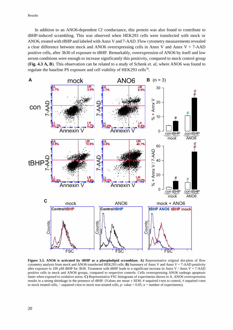
Results
20
In addition to an ANO6-dependent Cl- conductance, this protein was also found to contribute to
tBHP-induced scrambling. This was observed when HEK293 cells were transfected with mock or
ANO6, treated with tBHP and labeled with Annx V and 7-AAD. Flow cytometry measurements revealed
a clear difference between mock and ANO6 overexpressing cells in Annx V and Annx V + 7-AAD
positive cells, after 3h30 of exposure to tBHP. Remarkably, overexpression of ANO6 by itself and low
serum conditions were enough to increase significantly this positivity, compared to mock control group
(Fig. 4.3 A, B). This observation can be related to a study of Schenk et. al, where ANO6 was found to
regulate the baseline PS exposure and cell viability of HEK293 cells78.
Figure 3.3. ANO6 is activated by tBHP as a phospholipid scramblase. A) Representative original dot-plots of flow
cytometry analysis from mock and ANO6 transfected HEK293 cells. B) Summary of Annx V and Annx V + 7-AAD positivity
after exposure to 100 µM tBHP for 3h30. Treatment with tBHP leads to a significant increase in Annx V / Annx V + 7-AAD
positive cells in mock and ANO6 groups, compared to respective controls. Cells overexpressing ANO6 undergo apoptosis
faster when exposed to oxidative stress. C) Representative FSC histograms of experiments shown in A. ANO6 overexpression
results in a strong shrinkage in the presence of tBHP. (Values are mean ± SEM; # unpaired t-test to control, # unpaired t-test
to mock treated cells, # unpaired t-test to mock non-treated cells, p- value < 0.05; n = number of experiments).

Results
21
Additionally, by flow cytometry was also possible to conclude about cell size in the presence and
absence of tBHP – whereas, mock cells show just a small shift in the FSC histogram compared to control,
ANO6 overexpression resulted in a strong cell shrinkage induced by tBHP (Fig. 4.3 C).
It is interesting to note the different time points between detection of ANO6 Cl- conductance and
scrambling. While a Cl- conductance was observed only after 30 min of exposure to tBHP (Fig. 4.2),
the same conditions were not enough to induce scrambling (data not shown).
In order to know if ANO6 is essential for PS exposure triggered by ROS, the endogenous expression
of this protein was silenced in HEK293 cells by siRNA (Appendix III – Fig. 8.2) and the same protocol
was applied. However, transfection with siRNA likely resulted in off target effects, enhancing cell death
induced by tBHP compared to scrambled cells (Appendix IV – Fig. 8.3), which can be explained by
upregulation of other proteins involved in the apoptotic pathway.
4.1.3 ANO6 contribution to STS-mediated apoptosis of HEK293 and HeLa cells
Staurosporine is a cell permeable alkaloid isolated from the bacterium Streptomyces staurosporeus,
commonly used to study apoptosis79. Aside from functioning as a non-specific protein kinase inhibitor80,
this compound is also described to increase the intracellular level of ROS81. Hence, like tBHP, STS can
be used as a tool to study the possible effect of ROS in ANO6 activity. Indeed, intracellular ROS
production in HEK293 cells was detected after incubation with 1 µM STS for 24h, using the fluorogenic
sensor MAK142 (Fig. 4.4 A). In agreement with results from the previous section, overexpression of
ANO6 and exposure to STS led to an increase of the Annx V and Annx V + 7-AAD positive cells (Fig.
4.4 B, C).
Figure 3.4. ANO6 contribution to PS exposure induced by the ROS-producer STS. A) Representative images from three
different experiments showing ROS production after treatment with 1 µM STS for 24h, monitored with MAK142 fluorescence.
Bar indicates 20 µm. B) Representative original dot-plots of flow cytometry analysis from mock and ANO6 transfected
HEK293 cells in the presence and absence of 1 µM STS for 24h. C) Summary of Annx V and Annx V + 7-AAD positivity of

Results
22
experiments shown in B. Treatment with STS and ANO6 overexpression leads to a significant increase in Annx V and Annx
V + 7-AAD positive cells, compared to mock. (Values are mean ± SEM; # unpaired t-test to control, # unpaired t-test to mock
treated cells, p- value < 0.05; n = number of experiments).
After observing that ANO6 is involved in ROS-mediated apoptosis of HEK293 cells, it was
interesting to look if this is also true for another cell line. With this purpose, HeLa cells were incubated
with 1 µM STS for 5h. As expected, exposure to STS led to ROS production and caspase-3 activation
(Fig. 4.5 A, B, C). Also, overexpression of ANO6 enhanced significantly the Annx V positivity after
incubation with STS, compared to mock (Fig. 4.5 D, E). Surprisingly, downregulation of endogenous
expression by siRNA (Appendix III – Fig. 8.2) did not reduce cell death (Fig. 4.6).
Figure 3.5. STS leads to ROS increase and caspase-3 activation, mediating PS exposure enhanced by overexpressed
ANO6 in HeLa cells A) Representative images from three different experiments showing ROS production by 1 µM STS
monitored with H2DCFDA fluorescence (5h incubation). Bar indicates 20 µm. B) Representative original dot-plots of flow
cytometry analysis from mock and ANO6 transfected HeLa cells in the presence and absence of 1 µM STS for 5h. C) Summary
of Annx V positivity of experiments shown in B. ANO6 overexpression results in an increase of Annx V positive cells after
exposure to STS. (Values are mean ± SEM; # unpaired t-test to control, # unpaired t-test to mock treated cells, p-value < 0.05;
n = number of experiments).

Results
23
Figure 3.6. Downregulation of ANO6 in HeLa cells does not inhibit Annx V positivity induced by STS. A) Representative
original dot-plots of flow cytometry analysis from scrambled and siANO6 transfected HeLa cells in the presence or absence of
STS for 5h. B) Summary of Annx V positivity of experiments shown in A. STS induces Annx V positivity independently of
ANO6 expression level. (Values are mean ± SEM; # unpaired t-test to control, p-value < 0.05; n = number of experiments).
ANO6 contribution to ROS-mediated apoptosis of HeLa cells was also studied in terms of current.
First, by using the YFP-quenching assay, it was possible to detect an anion conductance activated by
STS in HeLa-YFP cells (Fig. 4.7 A, B). This conductance is also observed by patch clamp and can be
inhibited by downregulation of ANO6 and replacement with a 5 mM Cl- solution (Fig. 4.7 C, D, E).

Results
24
Figure 3.7. STS activates ANO6 currents in HeLa cells. A) YFP fluorescence measurements of HeLa-YFP cells in the
absence and presence of 20 mM I- ( ). Activation of an anion conductance after exposure to 1 µM STS for 5h, indicated by I-
efflux. B) Summary of I- uptake experiments shown in A. C) Overlay of whole-cell currents in HeLa cells, showing an
activation of a STS-induced Cl- current, reduced by ANO6 silencing. D) Current-voltage (I/V) curves of experiments shown in
B. Reduction of extracellular Cl- concentration results in a clear inhibition of baseline current detected after incubation with 1
µM STS for 3h-4h. E) Summary of delta current density at + 100 mV after current inhibition by 5Cl- solution. (Values are
mean ± SEM; # unpaired t-test to control p < 0.05, # unpaired t-test to mock treated cells, * paired t-test to tBHP treated, p-
value < 0.05; n = number of experiments).

Results
25
4.2 CFTR ENHANCES ANO6 EFFECT DURING ROS-MEDIATED APOPTOSIS
The next step of this project was focused on CFTR and the possible interaction of this protein with
ANO6. As described before, CFTR is a main regulator of the intracellular redox status, transporting
GSH to the extracellular space49. Moreover, a functional relationship between this channel and ANO6
is also described26. Thus, it is hypothesized that CFTR-mediated cytosolic ROS increase may lead to
ANO6 activation. If this is true, the presence of both proteins should enhance cell death under oxidative
stress conditions.
To address this question, HEK293 cells were co-transfected with ANO6 and CFTR and incubated
with 1 µM STS for 24h. As a control, cells were either transfected with mock, ANO6 or CFTR. Like
expected, single expression of ANO6 resulted in a significant increase of Annx V positive cells in the
presence of STS, compared to control and mock treated cells. Notably, the same was detected for CFTR
single transfection, which is in agreement with the function of this protein as a pro-apoptotic factor. Co-
expression of both proteins increased by a two-fold factor the Annx V positivity, compared to single
expression of ANO6 and CFTR. Surprisingly, the presence of both proteins was sufficient to trigger cell
death under control conditions to a significant level, evidenced by Annx V and 7-AAD positivity (Fig.
4.8).
Figure 3.8. CFTR enhances ANO6 contribution to apoptosis induced by STS in HEK293 cells. A) Representative original
dot-plots of flow cytometry analysis from cells transfected with mock, ANO6, CFTR or ANO6 and CFTR in the presence or

Results
26
absence of 1 µM STS for 24h. B) Summary of Annx V and Annx V + 7-AAD positivity of experiments shown in A. In each
group incubation with STS results in a significant increase of Annx V positive cells. Overexpression of ANO6 or CFTR
augments this effect. Co-expression of both proteins enhances ANO6 and CFTR contribution to apoptosis. (Values are mean
SEM; # unpaired t-test to control, # unpaired t-test to mock treated group, * One-way ANOVA indicates significant difference
compared to single expression, p-value < 0.05; n = number of experiments).
The finding that ANO6 and CFTR cooperate in PS exposure during STS-induced cell death led to
wonder if the same is true in terms of current activation. Therefore, single and co-expressed cells were
measured by patch clamp after incubation with 1 µM STS for 6-8h. In agreement with flow cytometry
data, overexpression of ANO6 or CFTR led to a significant increase in the basal current activated by
STS, an effect strongly enhanced by the co-expression of both proteins. In the four studied groups
(mock, ANO6, CFTR and ANO6 + CFTR), STS-induced current was inhibited by replacement with 5
mM Cl- solution in the extracellular space, showing that the whole-cell conductance is partially formed
by Cl- ions (Fig. 4.9).
Figure 3.9. Enhanced apoptotic whole-cell Cl- currents in cells expressing ANO6, CFTR and ANO6 + CFTR. A) Current
overlays from STS-treated HEK293 cells (1 µM 6h-8h) and inhibition by 5Cl-. Single expression of ANO6 or CFTR increases
STS-induced compared to mock cells. Co-expression of both proteins enhances ANO6 and CFTR effect. B) Corresponding
current-voltage relationships (I/V) from experiments shown in A. C) Respective current densities from experiments shown in
A. D) Delta of corrected current density at +100 mV after Cl- replacement. (Values are mean SEM; * paired t-test to STS
treated cells, # significant difference to indicated groups by unpaired t-test, a,b indicates significant difference calculated by
one-way ANOVA to mock and ANO6 respectively, p-value < 0.05; n = number of cells).

Results
27
4.2.1 ΔF508 CFTR mutation partially rescues cells from apoptosis
The earlier experiments preformed in HEK293 cells demonstrated that both ANO6 and CFTR
contribute to apoptosis, having a possible functional interaction when co-expressed (Fig. 4.8, 4.9).
CFBE cells stably expressing wt or F508 CFTR were chosen as an in vitro system to study CFTR
function and approach the results to a clinical significance.
The first question was related to CFTR role during cell death and the impact of the trafficking
mutation (ΔF508) in this process. With this purpose, CFBE cells (wt and ΔF508 CFTR) were exposed
to oxidative stress and their susceptibility to cell death was evaluated. Simultaneous incubation of both
cell lines with 100 µM tBHP for 24h led to a substantially difference in the Annx V and Annx V + 7-
AAD positive cells. While at the same time-point CFBE wt CFTR cells were in a late apoptotic stage –
Annx V + 7-AAD positive – cells expressing the mutant CFTR form were still in the early steps – Annx
V positive – (Fig. 4.10 A, B). It was also observed a considerable difference in the size of the two cell
lines after exposure to tBHP – CFBE wt CFTR cells showed a significant shrinkage compared to ΔF508
CFTR cells which had similar dimensions to control (Fig. 4.10 C).
Figure 3.10. Effect of CFTR in tBHP-induced cell death. A) Representative original dot-plots of flow cytometry analysis
from CFBE wt and F508 CFTR cells in the absence or presence of 100 µM tBHP for 24h. B) Summary of Annx V and Annx
V + 7-AAD positivity of experiments shown in A. C) FSC histograms of experiments shown in A. Expression of wt CFTR
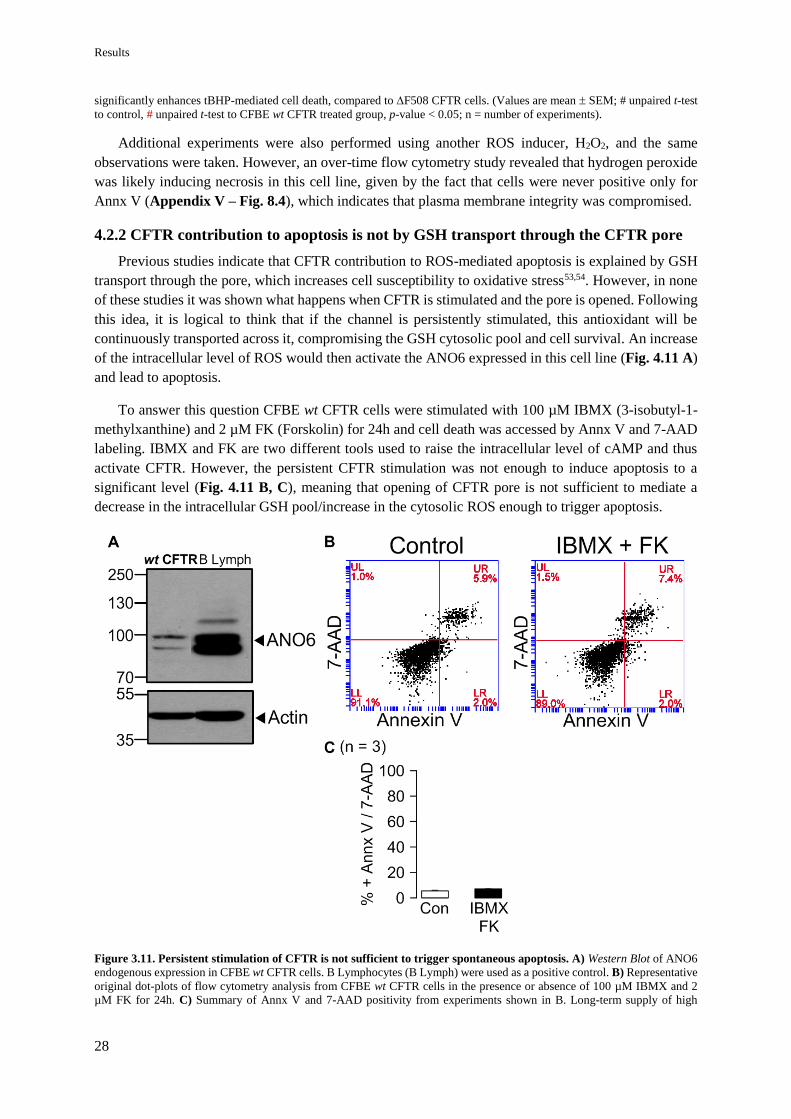
Results
28
significantly enhances tBHP-mediated cell death, compared to F508 CFTR cells. (Values are mean SEM; # unpaired t-test
to control, # unpaired t-test to CFBE wt CFTR treated group, p-value < 0.05; n = number of experiments).
Additional experiments were also performed using another ROS inducer, H2O2, and the same
observations were taken. However, an over-time flow cytometry study revealed that hydrogen peroxide
was likely inducing necrosis in this cell line, given by the fact that cells were never positive only for
Annx V (Appendix V – Fig. 8.4), which indicates that plasma membrane integrity was compromised.
4.2.2 CFTR contribution to apoptosis is not by GSH transport through the CFTR pore
Previous studies indicate that CFTR contribution to ROS-mediated apoptosis is explained by GSH
transport through the pore, which increases cell susceptibility to oxidative stress53,54. However, in none
of these studies it was shown what happens when CFTR is stimulated and the pore is opened. Following
this idea, it is logical to think that if the channel is persistently stimulated, this antioxidant will be
continuously transported across it, compromising the GSH cytosolic pool and cell survival. An increase
of the intracellular level of ROS would then activate the ANO6 expressed in this cell line (Fig. 4.11 A)
and lead to apoptosis.
To answer this question CFBE wt CFTR cells were stimulated with 100 µM IBMX (3-isobutyl-1-
methylxanthine) and 2 µM FK (Forskolin) for 24h and cell death was accessed by Annx V and 7-AAD
labeling. IBMX and FK are two different tools used to raise the intracellular level of cAMP and thus
activate CFTR. However, the persistent CFTR stimulation was not enough to induce apoptosis to a
significant level (Fig. 4.11 B, C), meaning that opening of CFTR pore is not sufficient to mediate a
decrease in the intracellular GSH pool/increase in the cytosolic ROS enough to trigger apoptosis.
Figure 3.11. Persistent stimulation of CFTR is not sufficient to trigger spontaneous apoptosis. A) Western Blot of ANO6
endogenous expression in CFBE wt CFTR cells. B Lymphocytes (B Lymph) were used as a positive control. B) Representative
original dot-plots of flow cytometry analysis from CFBE wt CFTR cells in the presence or absence of 100 µM IBMX and 2
µM FK for 24h. C) Summary of Annx V and 7-AAD positivity from experiments shown in B. Long-term supply of high
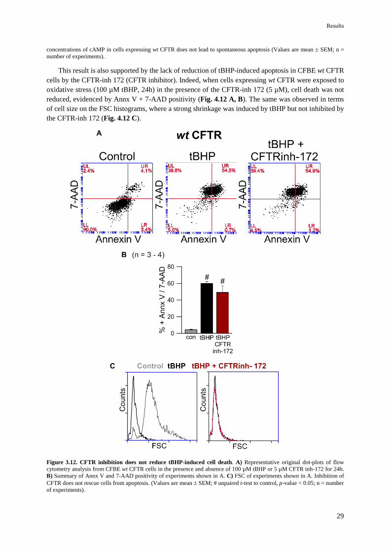
Results
29
concentrations of cAMP in cells expressing wt CFTR does not lead to spontaneous apoptosis (Values are mean SEM; n =
number of experiments).
This result is also supported by the lack of reduction of tBHP-induced apoptosis in CFBE wt CFTR
cells by the CFTR-inh 172 (CFTR inhibitor). Indeed, when cells expressing wt CFTR were exposed to
oxidative stress (100 µM tBHP, 24h) in the presence of the CFTR-inh 172 (5 µM), cell death was not
reduced, evidenced by Annx V + 7-AAD positivity (Fig. 4.12 A, B). The same was observed in terms
of cell size on the FSC histograms, where a strong shrinkage was induced by tBHP but not inhibited by
the CFTR-inh 172 (Fig. 4.12 C).
Figure 3.12. CFTR inhibition does not reduce tBHP-induced cell death. A) Representative original dot-plots of flow
cytometry analysis from CFBE wt CFTR cells in the presence and absence of 100 µM tBHP or 5 µM CFTR inh-172 for 24h.
B) Summary of Annx V and 7-AAD positivity of experiments shown in A. C) FSC of experiments shown in A. Inhibition of
CFTR does not rescue cells from apoptosis. (Values are mean SEM; # unpaired t-test to control, p-value < 0.05; n = number
of experiments).
Discussion
30
4. DISCUSSION
5.1 ANO6 PLAYS A ROLE DURING ROS-MEDIATED APOPTOSIS
This work provides evidence that ANO6 is involved in ROS-mediated apoptosis as a Ca2+- activated
Cl- channel, non-selective cation channel and phospholipid scramblase in two different in vitro systems
(HEK293 and HeLa cell lines) with two ROS inducers (tBHP and STS). It is first activated during the
early steps of apoptosis, extruding Cl- and likely cations to the extracellular space. This event may
contribute to cell shrinkage, which is known to result primarily from a loss of K+ and water82. As the
intrinsic apoptotic pathway moves forward, the phospholipid scramblase ANO6 function becomes
relevant, mediating PS exposure in the outer membrane leaflet.
Although overexpression of this protein augments apoptosis in the presence of ROS, downregulation
does not rescue cell death, indicating that ANO6 role in this process is not crucial. Additional
experiments in B lymphocytes support this conclusion, where it was observed that the absence of
functional ANO6 in Scott cells did not reduce apoptosis induced by STS compared to wt B lymphocytes.
Nonetheless, wt exhibited a higher % of cells in the early apoptotic stage - Annx V single positive
(Appendix VI – Fig. 8.5). This result may either imply that Scott cells are more sensitive to STS-
induced apoptosis, undergoing to later stages faster than wt; or that the absence of functional ANO6
leads to the activation of a different cell death pathway. Indeed, when cells die through the necrotic
pathway, the membrane integrity is compromised, allowing binding of Annx V and 7-AAD1.
The fact that ANO6 is not essential to ROS-mediated apoptosis is not surprising since regulated cell
death is a fundamental process in all living systems, depending on many molecules and cellular
components5. It is very likely that a cell with no ANO6 compensates this defect with other proteins. In
addition, Juu et. al defends that the ion channels activated during apoptosis considerably differ between
cell lines, a phenomenon that may be also true for ANO623.
5.1.1 ANO6 currents are prior to phospholipid scrambling
The different timeline between ANO6 activation as an ion channel and phospholipid scramblase
was already described in a study by Kmit et. al, where elevation of intracellular Ca2+ by ionomycin
resulted in immediate ANO6 currents but a delayed scrambling34. Here it is shown that the same is true
when ANO6 is activated by ROS. In particular, tBHP incubation for only 30 min in HEK293 cells
resulted in an ANO6-dependent Cl- conductance, whereas a differential scrambling was only detected
after 3h30min.
It is possible that PS exposure is a consequence of a persistent stimulation of the channel. Indeed,
patch clamp measurements revealed that activation of ANO6 with high concentrations of Ca2+ or for a
long period of time results in a current impossible to inhibit (data not shown). It is very likely that only
on these conditions the channel starts to scramble phospholipids and this may as well be the reason why
CaCC-AO1, a broad anoctamin inhibitor, was unable to decrease tBHP- induced scrambling in HEK293
cells (Appendix VII – Fig. 8.6).
5.1.2 Mechanism of ANO6 activation by ROS
Phospholipid scrambling can be activated through two distinct pathways: the caspase-dependent
pathway or by Ca2+ mobilizing agonists15. ANO6 contribution to both processes seems uneven – it is
very clear that this protein is essential to the Ca2+-mediated PS exposure, however when it comes to
ANO6 role in the apoptotic induced scrambling, controversial observations emerge.

Discussion
31
ANO6 is known to be activated by high intracellular Ca2+ concentrations, between 10 and 100 µM22.
In this regard, ANO6 contribution to Ca2+-mediated PS exposure was also found in this study, when
overexpression of this protein in HEK293 and HeLa cells resulted in a differential scrambling induced
by ionomycin (Appendix VIII – Fig. 8.7, 8.8). Here, downregulation of ANO6 by siRNA in HEK293
cells (Appendix III – Fig. 8.2) significantly reduced the detected scrambling (Appendix VIII – Fig.
8.7). This is supported by the correspondent experiment in B Lymphocytes (wt vs. Scott), where the
same observation was done, even though Scott cells still showed a residual scrambling that may be
explained by the expression of ANO9 known to also act as phospholipid scramblase (Appendix VIII –
Fig. 8.9).
During apoptosis, Ca2+ is released as a consequence of intracellular stress by the mitochondria and
the ER store12. Therefore, the most predictable mechanism of ANO6 activation would be through this
Ca2+ release detected in apoptotic cells. In fact, Fura-2AM measurements revealed a significant increase
in the cytosolic Ca2+, after incubation with tBHP in HEK293 cells and STS in HeLa cells (Appendix
IX – Fig. 8.10). However, the detected concentration of Ca2+ (80 nM and 300 nM, respectively) is not
sufficient to explain ANO6 activation. Interestingly, incubation with STS in HEK293 cells did not even
raise the intracellular Ca2+ to a significant level compared to control (Appendix IX – Fig. 8.10).
Another possible mechanism is related to cell shrinkage and supported by previous findings from
Martins et. al, where ANO6 was identified as component of the ORCC, a channel known to cause cell
shrinkage and phospholipid scrambling during apoptosis26. Moreover, Juul et. al found a relationship
between this protein and caspase-3 activation and thus cell shrinkage23. Hence, it is very likely that the
same relationship between ANO6, caspase-3 activation and cell shrinkage is applied to ROS-mediated
apoptosis. This is supported by the finding that HEK293 cells overexpressing ANO6 in the presence of
tBHP show a considerable difference in the cell size compared to mock cells.
Interestingly, this year Lalida et. al found an alternative mechanism for ANO6 activation,
independent of global Ca2+ rising, that relies on plasma membrane conformation. It was proved that the
cleavage of phospholipids in the plasma membrane, by phospholipase A2 (PLA2), leads to the opening
of ANO6 pore. This is explained by the release of fatty acids in the cytosol, such as arachidonic acid
(ArA), and the consequent accumulation of lysophospholipids (LPL) in the membrane. It was
established that the membrane tension, caused by the presence of LPL, and a local increase of Ca2+ in
specific domains are the two major players responsible for the activation of ANO6 as an ion channel25.
ROS are known to induce lipid peroxidation and damage of the plasma membrane83. Therefore,
another possibility to explain the findings in this work may be associated to membrane tension. This
hypothesis was also addressed to study tBHP-induced apoptosis in HEK293 cells. In this regard, ACA
(N-p-Amylcinnamoyl anthranilic Acid), a PLA2 inhibitor, was used as a tool to see if by inhibiting this
enzyme in the presence of ROS, membrane conformation would be reestablished and scrambling
reduced. However, this compound revealed to be toxic once applied for a long-term period (data not
shown). To overcome this problem, ArA was used and expected to compensate the phospholipid
oxidation during ANO6 activation. This hypothesis was supported by the fact that ArA has been shown
to inhibit PLA2-induced ANO6 currents25. Here it was found that incubation of ArA in the presence of
tBHP enhances cell death (Appendix X – Fig. 8.11). Nonetheless, this result does not necessarily
indicate that this hypothesis is not true, since ArA is also known to be an apoptotic inducer84, responsible
for the activation of the cyclooxygenase, lipoxygenase and epoxygenase pathways, three major
mechanisms involved in inflammation.

Discussion
32
Finally, because LPL was found to activate an anoctamin-dependent conductance in HEK293
cells25, it was also interesting to look for the effect in scrambling. Incubation of HEK293 with 10 µM
Lyso-PS (a kind of LPL) for 2h resulted in an increase of Annx V positive cells. However, the detected
scrambling was not inhibited by downregulation of ANO6 (Appendix III – Fig. 8.2, Appendix X –
Fig. 8.12). Thus, it is very likely that this effect is due to lysophosphatidic acid (LPA), a downstream
product of LPL known to induce apoptosis and irrelevant for ANO6 activation25,85.
As referred before, ROS-mediated cell death is not reduced by CaCC-AO1, however this is not true
when it comes to tannic acid (TA). Tannic acid is a polyphenolic biomolecule known to bind to proteins,
such as ANO6, and stabilize biological membranes. Remarkably, when applied to HEK293 cells in the
presence of tBHP, TA reduced the % of Annx V + 7-AAD positive cells, by a 3-fold factor (Appendix
X – Fig. 8.13). This strong inhibition is likely through membrane stabilization and protection of lipid
oxidation, which is in agreement with the alternative mechanism found by Lalida et. al25.
5.2 CFTR PLAYS A ROLE DURING ROS-MEDIATED APOPTOSIS
As outlined in the Introduction chapter, CFTR is described by many as a pro-apoptotic factor
involved in the regulation of the intracellular redox status, acidification and ceramide content in lipid
rafts49,52–54,56,59–61. The apoptotic process in CF has been studied over the years and, although some
conclusions are controversial, it seems that a defect on the clearance of apoptotic cells contribute to the
ongoing inflammations and tissue injury48. Also, oxidative stress and infections are two major mediators
of pulmonary damage which results in necrosis of lung epithelial cells54.
In the present project CFTR function was studied in HEK293 and CFBE cells. In the first cell line,
this protein was shown to enhance STS-mediated phospholipid scrambling and to have a Cl- conductance
during ROS-mediated apoptosis. Likewise, studies in the CFBE cell line provided evidence that CFTR
helps cells to undergo apoptosis once exposed to oxidative stress. Because the mutant CFTR form
(ΔF508) rescues partially cells from tBHP-induced apoptosis it is concluded that the presence of this
protein in the plasma membrane is essential.
However, unlike what it is reported in previous studies53, here it was found that CFTR contribution
to ROS-mediated apoptosis is independent of channel activation and pore opening. Indeed, persistent
stimulation of CFTR by supply of cAMP with IBMX and FK did not result in spontaneous apoptosis of
CFBE cells. Also, tBHP-induced cell death was not reduced by the CFTR-inh 172. A high concentration
of cytosolic ROS is known, not only to decrease GSH levels, but also to induce GSH transport to the
extracellular space50. Therefore, it is possible that GSH permeation across CFTR only happens in the
presence of intracellular oxidative stress. This might explain why it was detected a substantial difference
in tBHP-induced apoptosis between the wt and ΔF508 CFTR forms, but persistent stimulation of the
channel did not lead to cell death to a detectable level. It is also possible that the differential susceptibility
of the two CFBE variants to ROS is related to intracellular acidification. In fact, measurements done in
a study from Jungas et.al led to the conclusion that cells expressing wt CFTR have a lower intracellular
pH in the presence of H2O2 compared to ΔF508 CFTR54.
Remarkably, here CFTR was also found to contribute to cell shrinkage induced by tBHP. This result
resembles ANO6 role in tBHP-induced apoptosis of HEK293 cells and is in agreement with previous
studies where CFTR is reported to contribute to cell shrinkage53 and to act as volume-regulated Cl-
channel82.
The apoptotic dysfunction in CF is hard to establish because CF epithelial cells are characterized by
an accentuated ER stress48. Understanding this particularity of the disease may help to control the

Discussion
33
inflammatory process in CF, a symptom that remains one of the major unresolved problems. It is
believed that in the presence of a specific stimulus, cells expressing a mutant CFTR form have a higher
tendency to undergo necrosis54. One of the characteristics of the necrotic cell death is the release of
DNA debris that initiate inflammatory responses. On the contrary, if cells die through the apoptotic
pathway, fragmented DNA is tightly packed in apoptotic bodies which are then phagocytosed, protecting
the surrounding tissue from inflammations54.
Indeed, large DNA fragments are found in the lung epithelia from CF patients55, supporting this
concept. Remarkably, additional experiments in this study can be also taken as a hint that this is true.
Particularly, the over-time response of CFBE cells to H2O2 revealed that cells expressing the mutant
CFTR had a significantly higher population of cells positive for 7-AAD in the presence of the ROS
inducer, compared to wt CFTR (Appendix V – Fig. 8.4). Single binding of 7-AAD to the nucleus is not
a common event, happening once cell morphology is completely damaged and degraded. This may
indicate that stimulation of cells with ROS without CFTR in the membrane results in a burst of the dying
cell.
5.3 ANO6 AND CFTR ARE CO-WORKERS DURING REGULATED CELL DEATH
The hypothesis that ANO6 and CFTR cooperate during apoptosis emerged from previous reports
where ROS was suspected to have an influence in ANO6 activity22,26 and CFTR was described to
mediate GSH efflux49. Moreover, a functional relationship between these two channels was also
described26.
In this study it is proved that ANO6 and CFTR interact during ROS-mediated apoptosis. This is
supported by experiments in HEK293 cells where co-expression of these proteins resulted in a two-fold
increase of Annx V positive cells after exposure to STS, compared to single transfection groups.
Furthermore, it was demonstrated that ANO6 and CFTR co-expression results in a significant
spontaneous apoptosis, that is in the absence of any oxidative stress inducer.
Additionally, separate experiments of ANO6 and CFTR function during apoptosis point out for a
contribution of these two proteins to cell shrinkage, a process dependent on the activation of ion
channels. Also, overexpression of ANO6 or CFTR and exposure to STS in HEK293 cells significantly
augmented the basal current, compared to mock-transfected cells. Because this effect is stronger for
ANO6, it is speculated that CFTR expression enhances this current. Nonetheless, Martins et. al found
that cAMP- induced CFTR currents can be inhibited by downregulation of ANO6, suggesting that the
two mechanisms can occur26.
In conclusion, exposure of cells to oxidative stress and consequent ROS production leads to
mitochondrial permeabilization, release of Ca2+ and pro-apoptotic proteins, responsible for caspase
cleavage. These events terminate with ANO6 activation, which may support cell shrinkage and
phospholipid scrambling, two apoptotic hallmarks enhanced in the presence of CFTR (Fig. 5.1).

Discussion
34
Figure 4.1. Proposed role of ANO6 and CFTR in apoptotic cell death. CFTR and ANO6 are two independent channels with
a functional relationship during regulated cell death. Oxidative stress leads to mitochondrial permeabilization, release of Ca2+
and cytochrome c, which will lead to caspase-9 and 3 activation. ANO6 may support cell shrinkage and phospholipid
scrambling, two activities enhanced in the presence of CFTR.

Future Prespectives
35
5. FUTURE PERSPECTIVES
Understanding the interaction of ANO6 and CFTR during regulated cell death may help to control
CF apoptotic dysfunction and inflammations. For this reason, it is strongly encouraged that these
investigations are pursued in an attempt to gain knowledge about the molecular mechanism behind this
functional interaction.
Regarding ANO6 role in ROS-mediated apoptosis, further studies in B Lymphocytes are required.
One possibility is the analysis of the parallel response of wt and Scott cells to STS by flow cytometry
measurements of Annx V and 7-AAD labeling. This experiment may help to test the hypothesis that the
absence of functional ANO6 leads to the activation of a different cell death pathway. PLA2 inhibitors
and ArA can also be tested by path clamp experiments in order to understand if the ROS-induced basal
apoptotic current can be inhibited by a reduction of the plasma membrane tension.
Concerning CFTR involvement in apoptosis, it would be interesting to mimic inflammatory CF
conditions in the CFBE in vitro system and look if the susceptibility to cell death is still dependent on
the CFTR expression on the membrane. Similar experiments could be also done in primary cultured
cells isolated from airways of an animal model (wt or knocked-out for CFTR), where spontaneous
apoptosis could be evaluated by a simple caspase-3 assay.
Because it was concluded that CFTR involvement in tBHP-induced cell death cannot be explained
by cAMP-channel activation, thorough studies of the effect of ROS in CFTR activity could unravel why
CFBE wt CFTR cells are more susceptible to ROS-mediated apoptosis. Firstly, it would be interesting
to patch this cell line in the presence of tBHP and look for a CFTR current, which may or may not be
inhibited by the CFTR-inh 172. Additionally, measurements of the intracellular pH could prove if CFTR
contribution to intracellular acidification is a factor during tBHP-induced apoptosis. Although persistent
channel stimulation did not result in cell death, one can look for glutathione or ROS levels of CFBE
cells in the presence or absence of IBMX and FK.
The major breakthrough of this project was the evidence that ANO6 and CFTR cooperate during
regulated cell death. In this regard a co-immunoprecipitation or a FRET (Förster resonance energy
transfer) analysis can be performed to prove an interaction between these two proteins. Additionally,
because it is suggested that CFTR may enhance ANO6 function, a biotinylation of ANO6 endogenous
membrane expression in CFBE cells would be also interesting. By this experiment it would be possible
to compare if CFTR trafficking defects also affect ANO6 expression in the plasma membrane.
References
36
6. REFERENCES
1. Melorose, J., Perroy, R. & Careas, S. Apoptosis: A Review of Programmed Cell Death. Statew.
Agric. L. Use Baseline 2015 1, 495–516 (2015).
2. Ouyang, L. et al. Programmed cell death pathways in cancer: a review of apoptosis, autophagy
and programmed necrosis. Cell Prolif. 45, 487–98 (2012).
3. Williams, G. Programmed Cell Death : Apoptosis and Oncogenesis Mini review. Cell 65, 1097–
1098 (1991).
4. Ashkenazi, A. & Salvesen, G. Regulated cell death: signaling and mechanisms. Annu. Rev. Cell
Dev. Biol. 30, 337–356 (2014).
5. Ameisen, J. C. On the origin, evolution, and nature of programmed cell death: a timeline of four
billion years. Cell Death Differ. 9, 367–93 (2002).
6. KERR & J. F. R. KERR, A. H. W. A. A. R. C. Apoptosis: A basic biological phenomenon with
wide-ranging implications in human disease. J. Intern. Med. 258, 479–517 (2005).
7. Cohen, J. J. et al. APOPTOSIS AND PROGRAMMED CELL DEATH IN IMMUNITY.
8. Fink, S. L. & Cookson, B. T. MINIREVIEW Apoptosis , Pyroptosis , and Necrosis : Mechanistic
Description of Dead and Dying Eukaryotic Cells. (1907). doi:10.1128/IAI.73.4.1907
9. Li, J. & Yuan, J. Caspases in apoptosis and beyond. 6194–6206 (1993).
doi:10.1038/onc.2008.297
10. E.Gulbins, A.Jekle, K.Ferlinz, H.Grassmé, A. F. L. Physiology of apoptosis. J Physiol Ren.
Physiol 1542, 33–36 (2000).
11. Maeno, E., Ishizaki, Y., Kanaseki, T., Hazama, A. & Okada, Y. Normotonic cell shrinkage
because of disordered volume regulation is an early prerequisite to apoptosis. Proc. Natl. Acad.
Sci. U. S. A. 97, 9487–9492 (2000).
12. Kroemer, G., Galluzzi, L. & Brenner, C. Mitochondrial Membrane Permeabilization in Cell
Death. Physiol. Reveiw 99–163 (2007). doi:10.1152/physrev.00013.2006.
13. Nagata, S. Apoptosis by death factor. Cell 88, 355–365 (1997).
14. Galluzzi, L., Blomgren, K. & Kroemer, G. Mitochondrial membrane permeabilization in
neuronal injury. Nat. Rev. Neurosci. 10, 481–494 (2009).
15. Bevers, E. M. & Williamson, P. L. Phospholipid scramblase: An update. FEBS Lett. 584, 2724–
2730 (2010).
16. Devaux, P. F., Herrmann, A., Ohlwein, N. & Kozlov, M. M. How lipid flippases can modulate
membrane structure. Biochim. Biophys. Acta - Biomembr. 1778, 1591–1600 (2008).
17. Rysavy, N. M. et al. Beyond apoptosis : The mechanism and function of phosphatidylserine
asymmetry in the membrane of activating mast cells. Bioarchitecture 4, 127–137 (2014).
18. Shimizu, T. et al. TMEM16F is a component of a Ca2+-activated Cl- channel but not a volume-
sensitive outwardly rectifying Cl- channel. Am. J. Physiol. Cell Physiol. 304, C748–59 (2013).
19. Pedemonte, N. & Galietta, L. J. V. Structure and function of TMEM16 proteins (anoctamins).
Physiol. Rev. 94, 419–59 (2014).

References
37
20. Kunzelmann, K. et al. Anoctamins. Pflugers Arch. Eur. J. Physiol. 462, 195–208 (2011).
21. Brunner, J. D., Lim, N. K., Schenck, S., Duerst, A. & Dutzler, R. X-ray structure of a calcium-
activated TMEM16 lipid scramblase. Nature 516, 207–212 (2014).
22. Kunzelmann, K. et al. Molecular functions of anoctamin 6 (TMEM16F): A chloride channel,
cation channel, or phospholipid scramblase? Pflugers Arch. Eur. J. Physiol. 466, 407–414
(2014).
23. Juul, C. A. et al. Anoctamin 6 differs from VRAC and VSOAC but is involved in apoptosis and
supports volume regulation in the presence of Ca2+. Pfl??gers Arch. Eur. J. Physiol. 466, 1899–
1910 (2014).
24. Almaça, J. et al. TMEM16 proteins produce volume-regulated chloride currents that are reduced
in mice lacking TMEM16A. J. Biol. Chem. 284, 28571–28578 (2009).
25. Sirianant, L., Ousingsawat, J., Wanitchakool, P., Schreiber, R. & Kunzelmann, K. Cellular
volume regulation by anoctamin 6: Ca2+, phospholipase A2 and osmosensing. Pflugers Arch.
Eur. J. Physiol. 468, 335–349 (2016).
26. Martins, J. R. et al. Anoctamin 6 is an essential component of the outwardly rectifying chloride
channel. Proc. Natl. Acad. Sci. U. S. A. 108, 18168–72 (2011).
27. Yang, H. et al. TMEM16F forms a Ca2+-activated cation channel required for lipid scrambling
in platelets during blood coagulation. Cell 151, 111–22 (2012).
28. Yu, K. et al. Identification of a lipid scrambling domain in ANO6 / TMEM16F. 1–23 (2015).
doi:10.7554/eLife.06901
29. Suzuki, J. & Nagata, S. Phospholipid scrambling by TMEM16F. Seikagaku 83, 1050–1054
(2011).
30. Bevers, E. M. Compound heterozygosity for 2 novel TMEM16F mutations in a patient with Scott
syndrome To the editor : Chimerism levels after stem cell transplantation are primarily
determined by the ratio of donor. 117, 4399–4400 (2011).
31. Heemskerk, J. W. M., Bevers, E. M. & Lindhout, T. Platelet activation and blood coagulation.
Thromb. Haemost. 88, 186–193 (2002).
32. Ehlen, H. W. A. et al. Inactivation of anoctamin-6/Tmem16f, a regulator of phosphatidylserine
scrambling in osteoblasts, leads to decreased mineral deposition in skeletal tissues. J. Bone
Miner. Res. 28, 246–259 (2013).
33. Forschbach, V. et al. Anoctamin 6 is localized in the primary cilium of renal tubular cells and is
involved in apoptosis-dependent cyst lumen formation. Cell Death Dis. 6, e1899 (2015).
34. Kmit, A. et al. Calcium-activated and apoptotic phospholipid scrambling induced by Ano6 can
occur independently of Ano6 ion currents. Cell Death Dis. 4, e611 (2013).
35. Ousingsawat, J. et al. Anoctamin 6 mediates effects essential for innate immunity downstream
of P2X7 receptors in macrophages. Nat. Commun. 6, 6245 (2015).
36. Dutertre, M. et al. Exon-based clustering of murine breast tumor transcriptomes reveals
alternative exons whose expression is associated with metastasis. Cancer Res. 70, 896–905
(2010).
37. Jacobsen, K. S. et al. The role of TMEM16A (ANO1) and TMEM16F (ANO6) in cell migration.
Pflugers Arch. Eur. J. Physiol. 465, 1753–1762 (2013).

References
38
38. Whitlock, J. M. & Hartzell, H. C. A Pore Idea: the ion conduction pathway of TMEM16/ANO
proteins is composed partly of lipid. Pflugers Arch. Eur. J. Physiol. 468, 455–473 (2016).
39. Suzuki, J. et al. Calcium-dependent phospholipid scramblase activity of TMEM16 protein family
members. J. Biol. Chem. 288, 13305–16 (2013).
40. Suzuki, J., Imanishi, E. & Nagata, S. Exposure of phosphatidylserine by Xkrelated protein family
members during apoptosis. J. Biol. Chem. 289, 30257–30267 (2014).
41. Cutting, G. R. Cystic fibrosis genetics: from molecular understanding to clinical application. Nat.
Rev. Genet. 16, 45–56 (2015).
42. Riordan, J. R. CFTR function and prospects for therapy. Annu. Rev. Biochem. 77, 701–726
(2008).
43. Riordan, J. R. et al. Identification the Cystic Fibrosis Gene : Cloning and Characterization of
Complementary DNA. Science (80-. ). 245, 1066–1073 (1989).
44. Collins, F. S. Cystic fibrosis: molecular biology and therapeutic implications. Science (80-. ).
256, 774–779 (1992).
45. Amaral, M. D. & Kunzelmann, K. Molecular targeting of CFTR as a therapeutic approach to
cystic fibrosis. Trends Pharmacol. Sci. 28, 334–341 (2007).
46. Farinha, C. M., Matos, P. & Amaral, M. D. Control of cystic fibrosis transmembrane conductance
regulator membrane trafficking: Not just from the endoplasmic reticulum to the Golgi. FEBS J.
280, 4396–4406 (2013).
47. Sheppard, D. N. & Welsh, M. J. Structure and Function of the CFTR Chloride Channel. 23–45
(1999).
48. Soleti, R., Porro, C. & Martı, M. C. Apoptotic process in cystic fibrosis cells. 1029–1038
doi:10.1007/s10495-013-0874-y
49. Kogan, I. et al. CFTR directly mediates nucleotide-regulated glutathione flux. EMBO J. 22,
1981–1989 (2003).
50. Circu, M. L. & Aw, T. Y. Glutathione and modulation of cell apoptosis. Biochim Biophys Acta.
1, 1767–1777 (2012).
51. Tobergte, D. R. & Curtis, S. Glutathione: a radical treatment for Cystic Fibrosis lung disease? J.
Chem. Inf. Model. 53, 1689–1699 (2013).
52. Gao, L., Kim, K. J., Yankaskas, J. R. & Forman, H. J. Abnormal glutathione transport in cystic
fibrosis airway epithelia. Am. J. Physiol. - Lung Cell. Mol. Physiol. 277, L113–L118 (1999).
53. l’Hoste, S. et al. CFTR mediates apoptotic volume decrease and cell death by controlling
glutathione efflux and ROS production in cultured mice proximal tubules. Am J Physiol Ren.
Physiol 298, F435–53 (2010).
54. Jungas, T., Motta, I., Duffieux, F., Fanen, P. & Ojcius, D. M. Glutathione Levels and BAX
Activation during Apoptosis Due to Oxidative Stress in Cells Expressing Wild-type and Mutant
Cystic Fibrosis Transmembrane Conductance Regulator *. Biochemistry 277, 27912–27918
(2002).
55. Gottlieb, R. A. & Dosanjht, A. Mutant cystic fibrosis transmembrane conductance regulator
inhibits acidification and apoptosis in C127 cells : Possible relevance to cystic fibrosis.
56. Poujeol, C. et al. CFTR modulates programmed cell death by decreasing intracellular pH in

References
39
Chinese hamster lung fibroblasts. 810–824 (2001).
57. Tepper, A. D. et al. Sphingomyelin Hydrolysis to Ceramide during the Execution Phase of
Apoptosis Results from Phospholipid Scrambling and Alters Cell-surface Morphology. 155–164
(2000).
58. Smith, E. L., Schuchman, E. H. & Asm, A. The unexpected role of acid sphingomyelinase in cell
death and the pathophysiology of common diseases. 3419–3431 doi:10.1096/fj.08-108043
59. Rassmé, H. G. et al. Host defense against Pseudomonas aeruginosa requires ceramide-rich
membrane rafts. (2003). doi:10.1038/nm
60. Ulrich, M. et al. Ceramide accumulation mediates inflammation , cell death and infection
susceptibility in cystic fibrosis. 1–10 (2008). doi:10.1038/nm1748
61. Kleuser, B. & Gulbins, E. Acid Sphingomyelinase Inhibitors Normalize Pulmonary Ceramide
and Inflammation in Cystic Fibrosis. doi:10.1165/rcmb.2009-0174OC
62. Tobergte, D. R. & Curtis, S. CFTR and outward rectifying chloride channels are distinct proteins
with a regulatory relationship. J. Chem. Inf. Model. 53, 1689–1699 (2013).
63. Kunzelmann, K. & Mehta, A. CFTR: A hub for kinases and crosstalk of cAMP and Ca2+. FEBS
J. 280, 4417–4429 (2013).
64. Munnix, I. C. A. et al. Store-mediated calcium entry in the regulation of phosphatidylserine
exposure in blood cells from Scott patients. Thromb. Haemost. 89, 687–695 (2003).
65. Gerry Shaw, Silas Morse, Miguel Ararat, F. L. G. Potential transformation of human neuronal
cells by human adenoviruses and the origin of HTobergte, David R.EK293 cells. J. Chem. Inf.
Model. 53, 1689–1699 (2002).
66. Hsu, S. H. et al. Genetic characteristics of the HeLa cell. Science 191, 392–4 (1976).
67. Galietta, L. J. V, Haggie, P. M. & Verkman, A. S. Green fluorescent protein-based halide
indicators with improved chloride and iodide affinities. FEBS Lett. 499, 220–224 (2001).
68. Bebok, Z. et al. Failure of cAMP agonists to activate rescued ΔF508 CFTR in CFBE41o - airway
epithelial monolayers. J. Physiol. 569, 601–615 (2005).
69. Felgner, P. L. et al. Lipofection: a highly efficient, lipid-mediated DNA-transfection procedure.
Proc. Natl. Acad. Sci. U. S. A. 84, 7413–7 (1987).
70. Felgner, J. H. et al. Enhanced gene delivery and mechanism studies with a novel series of cationic
lipid formulations. J. Biol. Chem. 269, 2550–2561 (1994).
71. Engeland, M. Van et al. A review on an apoptosis detection system based on phosphatidyserine
exposure. Proc. Natl. Ac. Sci. USA 93, 1–9 (1996).
72. Zembruski, N. C. L., Stache, V., Haefeli, W. E. & Weiss, J. 7-Aminoactinomycin D for apoptosis
staining in flow cytometry. Anal. Biochem. 429, 79–81 (2012).
73. Mölder, A. et al. Non-invasive, label-free cell counting and quantitative analysis of adherent
cells using digital holography. J. Microsc. 232, 240–247 (2008).
74. Zhong, S., Navaratnam, D. & Santos-Sacchi, J. A genetically-encoded YFP sensor with
enhanced chloride sensitivity, photostability and reduced pH interference demonstrates
augmented transmembrane chloride movement by gerbil prestin (SLC26a5). PLoS One 9,
(2014).

References
40
75. Grynkiewicz, G., Poenie, M. & Tsien, R. Y. A new generation of Ca2+ indicators with greatly
improved fluorescence properties. J. Biol. Chem. 260, 3440–3450 (1985).
76. Martín, C. et al. tert-Butyl hydroperoxide-induced lipid signaling in hepatocytes: Involvement
of glutathione and free radicals. Biochem. Pharmacol. 62, 705–712 (2001).
77. Papucci, L. et al. Coenzyme Q10 prevents apoptosis by inhibiting mitochondrial depolarization
independently of its free radical scavenging property. J. Biol. Chem. 278, 28220–28228 (2003).
78. Schenk, L. K., Schulze, U., Henke, S., Weide, T. & Pavenstädt, H. TMEM16F Regulates
Baseline Phosphatidylserine Exposure and Cell Viability in Human Embryonic Kidney Cells.
Cell Physiol Biochem 38, 2452–2463 (2016).
79. Feng, G. & Kaplowitz, N. Mechanism of staurosporine-induced apoptosis in murine hepatocytes.
AJ-Gastrointest. Liver Physiol. 282, G825–G834 (2002).
80. Ward, N. E. & O’Brian, C. a. Kinetic analysis of protein kinase C inhibition by staurosporine:
evidence that inhibition entails inhibitor binding at a conserved region of the catalytic domain
but not competition with substrates. Mol. Pharmacol. 41, 387–92 (1992).
81. Shimizu, T., Numata, T. & Okada, Y. A role of reactive oxygen species in apoptotic activation
of volume-sensitive Cl(-) channel. Proc. Natl. Acad. Sci. U. S. A. 101, 6770–6773 (2004).
82. Kunzelmann, K. Ion channels in regulated cell death. Cell. Mol. Life Sci. 73, 1–17 (2016).
83. Circu, M. L. & Aw, T. Y. Reactive Oxygen Species, cellular redox systems and apoptosis. Free
Radic Biol Med 48, 749–762 (2010).
84. Pompeia, C., Lima, T. & Curi, R. Arachidonic acid cytotoxicity: Can arachidonic acid be a
physiological mediator of cell death? Cell Biochem. Funct. 21, 97–104 (2003).
85. Ye, X., Ishii, I., Kingsbury, M. A. & Chun, J. Lysophosphatidic acid as a novel cell
survival/apoptotic factor. Biochim. Biophys. Acta - Mol. Cell Biol. Lipids 1585, 108–113 (2002).

Appendices
41
7. APPENDICES
APPENDIX I – CDNA
Table 7.1 Plasmids accession number and base pairs (bp).
Target Accession number bp
ANO6 NM_001025356 27306
CFTR NM_000492 4440
APPENDIX II - INHIBITION OF TBHP-INDUCED APOPTOSIS BY IDEBENONE
Figure 7.1. Inhibition of tBHP-induced apoptosis by Idebenone. A) Representative original dot-plots of flow cytometry
analysis of HEK293 cells in the presence or absence of tBHP (100 µM) and/or Idebenone (20 µM) for 2h. B) Summary of
Annx V and 7-AAD positivity of experiments shown in A. Idebenone significantly reduced Annx V and 7-AAD positive cells
in the presence of tBHP, rescuing ROS-mediated cell death. (Values are mean ± SEM; # unpaired t-test to control, # unpaired
t-test to tBHP treated cells, p-value < 0.05; n = number of experiments).

Appendices
42
APPENDIX III – DOWNREGULATION OF ANO6 IN HEK293 AND HELA CELLS
Figure 7.2. Western Blot analysis of ANO6 expression in HEK293 (A) and HeLa (B) cells, after 48h and 24h of transfection
with siRNA, respectively.

Appendices
43
APPENDIX IV – EFFECT OF ANO6 SILENCING ON TBHP-INDUCED APOPTOSIS OF
HEK293 CELLS
Figure 7.3. ANO6 silencing increases the tBHP-induced apoptosis in HEK293 cells. A) Representative original dot-plots
of cells transfected with siANO6 or scrambled in the presence or absence of 100 µM tBHP for 3h30. B) Summary of Annx V
and Annx V + 7-AAD positivity of experiments shown in A. Downregulation of ANO6 likely resulted in off target effects,
enhancing cell death induced by tBHP. (Values are mean ± SEM; # unpaired t-test to control, # unpaired t-test to scrambled
treated cells, p- value < 0.05; n = number of experiments).

Appendices
44
APPENDIX V - EFFECT OF CFTR IN H2O2-INDUCED CELL DEATH OF CFBE
CELLS
Figure 7.4. CFBE cells expressing wt CFTR have a higher susceptibility to H2O2-induced cell death. A) Representative
original dot-plots from over-time response to 900 µM H2O2 (0h, 2h, 4h, 8h) of CFBE wt and ΔF508 CFTR cells. B) Summary
of 7-AAD and Annx V + 7-AAD positivity of experiments shown in A. C) FSC histograms of experiments shown in A. (Values
are mean ± SEM; # unpaired t-test to control, # unpaired t-test to H2O2 to CFBE wt CFTR treated cells, p-value < 0.05; n =
number of experiments).
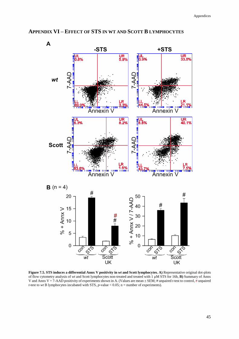
Appendices
45
APPENDIX VI – EFFECT OF STS IN WT AND SCOTT B LYMPHOCYTES
Figure 7.5. STS induces a differential Annx V positivity in wt and Scott lymphocytes. A) Representative original dot-plots
of flow cytometry analysis of wt and Scott lymphocytes non-treated and treated with 1 µM STS for 16h, B) Summary of Annx
V and Annx V + 7-AAD positivity of experiments shown in A. (Values are mean ± SEM; # unpaired t-test to control, # unpaired
t-test to wt B lymphocytes incubated with STS, p-value < 0.05; n = number of experiments).

Appendices
46
APPENDIX VII - CACC-AO1 DOES NOT INHIBIT TBHP-INDUCED APOPTOSIS OF
HEK293 CELLS
Figure 7.6. Effect of CaCC-AO1 in tBHP-induced apoptosis of HEK293 cells. A) Representative original dot-plots of flow
cytometry analysis from HEK293 cells in the presence or absence of 100 µM tBHP and 20 µM CaCC-AO1 for 2h. B) Summary
of Annx V and 7-AAD positivity of experiments shown in A. Inhibition of ANO6 by CaCC-AO1 does not rescue cells from
tBHP induced apoptosis. (Values are mean ± SEM; # unpaired t-test to control, p-value < 0.05; n = number of experiments).

Appendices
47
APPENDIX VIII - ANO6 IS ESSENTIAL FOR CA2+-INDUCED SCRAMBLING
Figure 7.7. Ionomycin induces an ANO6-dependent phospholipid scrambling in HEK293 cells. A) Representative original
dot-plots from flow cytometry analysis of HEK293 cells transfected with mock, ANO6, scrambled or siANO6 and stimulated
for 20 min with 1 µM Iono. B) Summary of Annx V positivity from experiments shown in A. (Values are mean ± SEM; #
unpaired t-test to control, # unpaired t-test to mock-transfected cells in the presence of Iono, # unpaired t-test to scrambled-
transfected cells in the presence of Iono, p-value < 0.05; n = number of experiments).
Figure 7.8. Ionomycin induces an ANO6-dependent phospholipid scrambling in HeLa cells. A) Representative original
dot-plots from flow cytometry analysis of HeLa cells transfected with mock or ANO6 and stimulated for 30 min with 10 µM
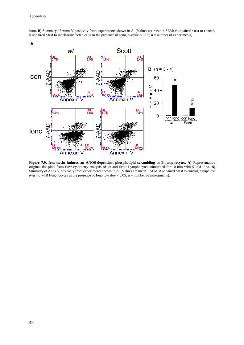
Appendices
48
Iono. B) Summary of Annx V positivity from experiments shown in A. (Values are mean ± SEM; # unpaired t-test to control,
# unpaired t-test to mock-transfected cells in the presence of Iono, p-value < 0.05; n = number of experiments).
Figure 7.9. Ionomycin induces an ANO6-dependent phospholipid scrambling in B lymphocytes. A) Representative
original dot-plots from flow cytometry analysis of wt and Scott Lymphocytes stimulated for 10 min with 5 µM Iono. B)
Summary of Annx V positivity from experiments shown in A. (Values are mean ± SEM; # unpaired t-test to control, # unpaired
t-test to wt B lymphocytes in the presence of Iono, p-value < 0.05; n = number of experiments).

Appendices
49
APPENDIX IX – INTRACELLULAR CA2+
CONCENTRATION DURING ROS-
MEDIATED APOPTOSIS
Figure 7.10. ROS effect in intracellular Ca2+ concentration. A) Fura-2 AM measurements of HEK293 cells in the presence
or absence of 100 µM tBHP for 1h30. B) Fura-2 AM measurements of HEK293 cells in the presence or absence of 1 µM STS
for 24h. C) Fura-2 AM measurements of HeLa cells in the presence or absence of 1 µM STS for 5h. Intracellular Ca2+ is
significantly increased by tBHP in HEK293 cells and STS in HeLa cells. Incubation with STS in HEK293 cells did not affect
[Ca2+]i. (Values are mean ± SEM; # unpaired t-test to control, p-value < 0.05; n = number of experiments).

Appendices
50
APPENDIX X – PLASMA MEMBRANE TENSION IN ROS-MEDIATED APOPTOSIS
Figure 7.11. Arachidonic acid enhances tBHP-induced apoptosis. A) Representative original dot-plots of HEK293 cells in
the presence or absence of 100 µM tBHP and 20 µM ArA for 2h. B) Summary of Annx V and Annx V + 7-AAD positivity of
experiments shown in A. (Values are mean ± SEM; # unpaired t-test to control, # unpaired t-test to tBHP-treated cells, p-value
< 0.05; n = number of experiments).

Appendices
51
Figure 7.12. LPL induces an ANO6-independent PS exposure in HEK293 cells. A) Representative original dot-plots of
HEK293 cells transfected with scrambled or siANO6, in the presence/absence of 10 µM Lyso-phosphatidylserine (Lyso-PS)
for 2h. B) Summary of Annx V positivity of experiments shown in A. (Values are mean ± SEM; # unpaired t-test to control,
p-value < 0.05; n = number of experiments).
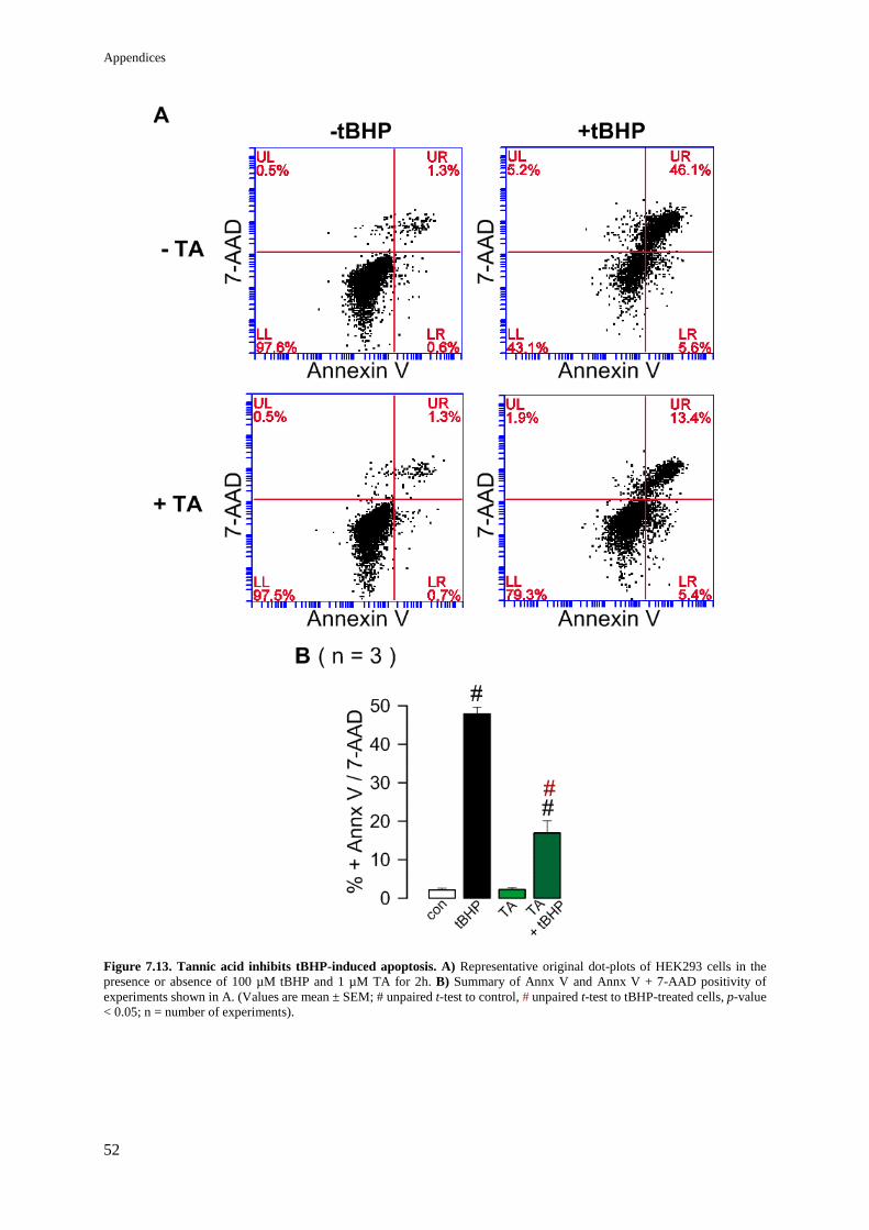
Appendices
52
Figure 7.13. Tannic acid inhibits tBHP-induced apoptosis. A) Representative original dot-plots of HEK293 cells in the
presence or absence of 100 µM tBHP and 1 µM TA for 2h. B) Summary of Annx V and Annx V + 7-AAD positivity of
experiments shown in A. (Values are mean ± SEM; # unpaired t-test to control, # unpaired t-test to tBHP-treated cells, p-value
< 0.05; n = number of experiments).