
Role for RNA:DNA hybrids in origin-independent replication priming in … · · 2015-05-01Role...
6
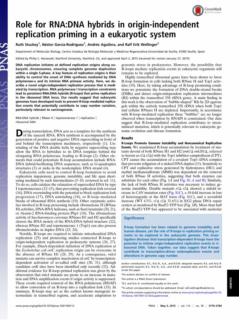
Role for RNA:DNA hybrids in origin-independent replication priming in a eukaryotic system Ruth Stuckey 1 , Néstor García-Rodríguez 1 , Andrés Aguilera, and Ralf Erik Wellinger 2 Department of Molecular Biology, Centro Andaluz de Biología Molecular y Medicina Regenerativa-Universidad de Sevilla, 41092 Seville, Spain Edited by Philip C. Hanawalt, Stanford University, Stanford, CA, and approved April 2, 2015 (received for review January 27, 2015) DNA replication initiates at defined replication origins along eu- karyotic chromosomes, ensuring complete genome duplication within a single S-phase. A key feature of replication origins is their ability to control the onset of DNA synthesis mediated by DNA polymerase-α and its intrinsic RNA primase activity. Here, we de- scribe a novel origin-independent replication process that is medi- ated by transcription. RNA polymerase I transcription constraints lead to persistent RNA:DNA hybrids (R-loops) that prime replication in the ribosomal DNA locus. Our results suggest that eukaryotic genomes have developed tools to prevent R-loop–mediated replica- tion events that potentially contribute to copy number variation, particularly relevant to carcinogenesis. RNA:DNA hybrids | RNase H | topoisomerase 1 | replication | ribosomal DNA D uring transcription, DNA acts as a template for the synthesis of the nascent RNA. RNA synthesis is accompanied by the generation of positive and negative DNA supercoiling in front of and behind the transcription machinery, respectively (1). Un- winding of the DNA double helix by negative supercoiling may allow the RNA to hybridize to its DNA template behind the elongating RNA polymerase, leading to R-loops (2). Other ele- ments that could potentiate R-loop accumulation include RNA: DNA hybrid-facilitating DNA sequences, such as G-quadruplex structures (3) or nicks in the nontemplate DNA strand (4). Eukaryotic cells need to control R-loop formation to avoid replication impairment, genome instability, and life span short- ening mediated by such intermediates (5–10; reviewed in ref. 11). To do so, cells catalyze the relaxation of supercoiled DNA by type I topoisomerases (12–15), thus preventing replication fork reversal (16), DNA overwinding with the potential to block replication fork progression (17), DNA unwinding (18), or R-loop– mediated blocks of ribosomal RNA synthesis (19). Other enzymatic activi- ties involved in R-loop processing include ribonuclease H (RNase H) activities, DNA-RNA helicases, such as Sen1/senataxin (20, 21), or Ataxin-2 RNA-binding protein Pbp1 (10). The ribonuclease activity of Saccharomyces cerevisiae RNases H1 and H2 specifically cleaves the RNA moiety of the RNA:DNA hybrid structure (22), whereas RNase H2 and topoisomerase 1 (Top1) can also process ribonucleotides in duplex DNA (23, 24). Notably, R-loops are required to initiate mitochondrial DNA replication (25) and pioneering studies connected R-loops to origin-independent replication in prokaryotic systems (26, 27). For example, DnaA-dependent initiation of DNA replication at the Escherichia coli oriC replication origin can be overcome in the absence of RNase H1 (28, 29). As a consequence, rnhA mutants can survive complete inactivation of oriC by transcription- dependent activation of so-called oriK sites (30, 31), although candidate oriK sites have been identified only recently (32). Ad- ditional evidence for R-loop–primed replication was given by the observation that rnhA mutants are prone to an increase in muta- tion and DNA amplification events if origin activity is suppressed. These events required removal of the RNA polymerase (RNAP) to allow conversion of an R-loop into a replication fork (33). In summary, R-loops may act as the earliest known mutagenic in- termediate in transcribed regions, and accelerate adaptation to genomic stress in prokaryotes. However, the possibility that R-loops mediate replication events in eukaryotic organisms still remains to be explored. Highly transcribed ribosomal genes have been shown to favor R-loop formation in cells lacking both RNase H and Top1 activ- ities (19). Here, by taking advantage of R-loop promoting condi- tions we potentiate the formation of DNA double-strand breaks (DSBs) and detect origin-independent replication intermediates (RI) within the transcribed 35S rRNA genes. A main finding in this work is the observation of “bubble-shaped” RIs by 2D agarose gels within the actively transcribed 35S rDNA when both Top1 and cellular RNases H are depleted. Importantly, in accordance with R-loop–mediated replication these “bubbles” are no longer observed when transcription by RNAPI is constrained. Our data suggest that R-loop–mediated replication contributes to stress- induced mutation, which is potentially relevant to eukaryotic ge- nome evolution and disease formation. Results R-Loops Promote Genome Instability and Noncanonical Replication Events. We maximized R-loop accumulation by treatment of mu- tants devoid of both RNase H1 and H2 (rnh1Δ rnh201Δ, referred to herein as r1Δ r2Δ) with the Top1 inhibitor camptothecin (CPT). CPT causes the accumulation of a covalent Top1–DNA complex that prevents religation of a nicked DNA duplex (15). Sensitivity to CPT and replicative stress generated by hydroxyurea (HU) and methyl methanesulfonate (MMS) was dependent on the removal of both RNase H activities, suggesting that both enzymes can substitute for each other (Fig. 1A and Fig. S1 A and B). Similarly, the lack of both RNase H activities was necessary to induce ge- nomic instability. Double mutants r1Δ r2Δ showed a sixfold in- crease in CAN mutation rates (Fig. S1C), a 10-fold increase in loss of heterozygosity at the MAT locus (Fig. S1C), and a sevenfold increase (WT 4.5%, r1Δ r2Δ 31.6%) in S/G2 phase DNA repair centers as monitored by Rad52-YFP foci (Fig. 1B). More than half of the Rad52-YFP foci appeared to be associated with nucleolar Significance R-loop formation has been related to genome instability and human disease, yet the role of R-loops in replication priming re- mains to be explored in the eukaryotic genome. This inves- tigation discloses that transcription-dependent R-loops have the potential to initiate origin-independent replication events in ri- bosomal DNA. Taken together, our data suggest that R-loops contribute to transcription-driven endoreplication events and alterations in genome copy number. Author contributions: R.S., N.G.-R., A.A., and R.E.W. designed research; R.S. and N.G.-R. performed research; R.S., N.G.-R., A.A., and R.E.W. analyzed data; and R.S. and R.E.W. wrote the paper. The authors declare no conflict of interest. This article is a PNAS Direct Submission. 1 R.S. and N.G.-R. contributed equally to this work. 2 To whom correspondence should be addressed. Email: [email protected]. This article contains supporting information online at www.pnas.org/lookup/suppl/doi:10. 1073/pnas.1501769112/-/DCSupplemental. www.pnas.org/cgi/doi/10.1073/pnas.1501769112 PNAS | May 5, 2015 | vol. 112 | no. 18 | 5779–5784 GENETICS
Transcript of Role for RNA:DNA hybrids in origin-independent replication priming in … · · 2015-05-01Role...
Role for RNA:DNA hybrids in origin-independentreplication priming in a eukaryotic systemRuth Stuckey1, Néstor García-Rodríguez1, Andrés Aguilera, and Ralf Erik Wellinger2
Department of Molecular Biology, Centro Andaluz de Biología Molecular y Medicina Regenerativa-Universidad de Sevilla, 41092 Seville, Spain
Edited by Philip C. Hanawalt, Stanford University, Stanford, CA, and approved April 2, 2015 (received for review January 27, 2015)
DNA replication initiates at defined replication origins along eu-karyotic chromosomes, ensuring complete genome duplicationwithin a single S-phase. A key feature of replication origins is theirability to control the onset of DNA synthesis mediated by DNApolymerase-α and its intrinsic RNA primase activity. Here, we de-scribe a novel origin-independent replication process that is medi-ated by transcription. RNA polymerase I transcription constraintslead to persistent RNA:DNA hybrids (R-loops) that prime replicationin the ribosomal DNA locus. Our results suggest that eukaryoticgenomes have developed tools to prevent R-loop–mediated replica-tion events that potentially contribute to copy number variation,particularly relevant to carcinogenesis.
RNA:DNA hybrids | RNase H | topoisomerase 1 | replication |ribosomal DNA
During transcription, DNA acts as a template for the synthesisof the nascent RNA. RNA synthesis is accompanied by the
generation of positive and negative DNA supercoiling in front ofand behind the transcription machinery, respectively (1). Un-winding of the DNA double helix by negative supercoiling mayallow the RNA to hybridize to its DNA template behind theelongating RNA polymerase, leading to R-loops (2). Other ele-ments that could potentiate R-loop accumulation include RNA:DNA hybrid-facilitating DNA sequences, such as G-quadruplexstructures (3) or nicks in the nontemplate DNA strand (4).Eukaryotic cells need to control R-loop formation to avoid
replication impairment, genome instability, and life span short-ening mediated by such intermediates (5–10; reviewed in ref. 11).To do so, cells catalyze the relaxation of supercoiled DNA by typeI topoisomerases (12–15), thus preventing replication fork reversal(16), DNA overwinding with the potential to block replication forkprogression (17), DNA unwinding (18), or R-loop–mediatedblocks of ribosomal RNA synthesis (19). Other enzymatic activi-ties involved in R-loop processing include ribonuclease H (RNaseH) activities, DNA-RNA helicases, such as Sen1/senataxin (20, 21),or Ataxin-2 RNA-binding protein Pbp1 (10). The ribonucleaseactivity of Saccharomyces cerevisiaeRNases H1 and H2 specificallycleaves the RNA moiety of the RNA:DNA hybrid structure (22),whereas RNase H2 and topoisomerase 1 (Top1) can also processribonucleotides in duplex DNA (23, 24).Notably, R-loops are required to initiate mitochondrial DNA
replication (25) and pioneering studies connected R-loops toorigin-independent replication in prokaryotic systems (26, 27).For example, DnaA-dependent initiation of DNA replication atthe Escherichia coli oriC replication origin can be overcome inthe absence of RNase H1 (28, 29). As a consequence, rnhAmutants can survive complete inactivation of oriC by transcription-dependent activation of so-called oriK sites (30, 31), althoughcandidate oriK sites have been identified only recently (32). Ad-ditional evidence for R-loop–primed replication was given by theobservation that rnhA mutants are prone to an increase in muta-tion and DNA amplification events if origin activity is suppressed.These events required removal of the RNA polymerase (RNAP)to allow conversion of an R-loop into a replication fork (33). Insummary, R-loops may act as the earliest known mutagenic in-termediate in transcribed regions, and accelerate adaptation to
genomic stress in prokaryotes. However, the possibility thatR-loops mediate replication events in eukaryotic organisms stillremains to be explored.Highly transcribed ribosomal genes have been shown to favor
R-loop formation in cells lacking both RNase H and Top1 activ-ities (19). Here, by taking advantage of R-loop promoting condi-tions we potentiate the formation of DNA double-strand breaks(DSBs) and detect origin-independent replication intermediates(RI) within the transcribed 35S rRNA genes. A main finding inthis work is the observation of “bubble-shaped” RIs by 2D agarosegels within the actively transcribed 35S rDNA when both Top1and cellular RNases H are depleted. Importantly, in accordancewith R-loop–mediated replication these “bubbles” are no longerobserved when transcription by RNAPI is constrained. Our datasuggest that R-loop–mediated replication contributes to stress-induced mutation, which is potentially relevant to eukaryotic ge-nome evolution and disease formation.
ResultsR-Loops Promote Genome Instability and Noncanonical ReplicationEvents. We maximized R-loop accumulation by treatment of mu-tants devoid of both RNase H1 and H2 (rnh1Δ rnh201Δ, referredto herein as r1Δ r2Δ) with the Top1 inhibitor camptothecin (CPT).CPT causes the accumulation of a covalent Top1–DNA complexthat prevents religation of a nicked DNA duplex (15). Sensitivity toCPT and replicative stress generated by hydroxyurea (HU) andmethyl methanesulfonate (MMS) was dependent on the removalof both RNase H activities, suggesting that both enzymes cansubstitute for each other (Fig. 1A and Fig. S1 A and B). Similarly,the lack of both RNase H activities was necessary to induce ge-nomic instability. Double mutants r1Δ r2Δ showed a sixfold in-crease in CAN mutation rates (Fig. S1C), a 10-fold increase in lossof heterozygosity at the MAT locus (Fig. S1C), and a sevenfoldincrease (WT 4.5%, r1Δ r2Δ 31.6%) in S/G2 phase DNA repaircenters as monitored by Rad52-YFP foci (Fig. 1B). More than halfof the Rad52-YFP foci appeared to be associated with nucleolar
Significance
R-loop formation has been related to genome instability andhuman disease, yet the role of R-loops in replication priming re-mains to be explored in the eukaryotic genome. This inves-tigation discloses that transcription-dependent R-loops have thepotential to initiate origin-independent replication events in ri-bosomal DNA. Taken together, our data suggest that R-loopscontribute to transcription-driven endoreplication events andalterations in genome copy number.
Author contributions: R.S., N.G.-R., A.A., and R.E.W. designed research; R.S. and N.G.-R.performed research; R.S., N.G.-R., A.A., and R.E.W. analyzed data; and R.S. and R.E.W.wrote the paper.
The authors declare no conflict of interest.
This article is a PNAS Direct Submission.1R.S. and N.G.-R. contributed equally to this work.2To whom correspondence should be addressed. Email: [email protected].
This article contains supporting information online at www.pnas.org/lookup/suppl/doi:10.1073/pnas.1501769112/-/DCSupplemental.
www.pnas.org/cgi/doi/10.1073/pnas.1501769112 PNAS | May 5, 2015 | vol. 112 | no. 18 | 5779–5784
GEN
ETICS

DNA in r1Δ r2Δ cells (61% in the absence and 65% in thepresence of CPT), indicating an increased susceptibility of ribo-somal DNA (rDNA) to DNA damage caused by impaired pro-cessing of RNA:DNA hybrids.The rDNA, which is hosted in the nucleolus, is localized on
chromosome XII and consists of ∼150 rDNA copies per haploidthat are susceptible to stress-induced changes in repeat numberand the formation of extrachromosomal rDNA circles (ERCs)caused by the excision of rDNA repeats (34, 35). Consistent withan increase in replicative stress upon CPT treatment, the numberof Rad52 foci-containing cells doubled 30–90 min after releasefrom α-factor (Fig. S2A) and CPT-treated r1Δ r2Δ mutant cellsaccumulated in late S or G2 phase (Fig. 2A). To further investigatethe impact of CPT on rDNA maintenance, we monitored the fateof replication forks in S-phase cells. Following G1-synchronizationby α-factor and release into CPT-containing medium, RIs wereisolated and analyzed by 2D agarose gel electrophoresis (for in-terpretation of the results, see Fig. 2B). Although the S-phasespecific pattern of replication (Y-arc) and recombination (X-spike)intermediates in WT and r1Δ r2Δ strains were similar, a cleardifference was observed at late S/G2 phase. Whereas replication inthe WT had finished, Y-arc and X-spike RIs remained in the r1Δr2Δ mutants, and replication fork pausing sites (RFPs) (Fig. 2C,open arrowheads) appeared in the nontranscribed spacer region(Fig. 2C, probe A). These pausing sites were CPT-dependent be-cause we could not detect them in RIs isolated from untreated r1Δr2Δ mutants (Fig. S3A). The majority of these RFP sites over-lapped with those previously described in cells lacking the Rrm3DNA helicase (36) (Fig. 3). The observed pausing sites may beenriched at hot spots of Top1–DNA interaction sites and corre-spond to CPT-trapped Top1–DNA complexes, yet they correlatedwith potential sites of R-loop formation (37, 38), such as the 3′ endof 35S genes (d in Fig. 3) or 5S genes (c in Fig. 3), but also includedsites of protein barriers such as the ribosomal autonomously rep-licating sequence (ARS) or 35S promoter (a/b and e in Fig. 3).Next, we monitored replication intermediates present within the
35S gene (Fig. 2C, probe B). Although the 35S gene lacks commoncharacteristics of replication origins that allow the binding of theprereplication complex (39), intermediates migrating as expectedfor bubble-shaped molecules were detected (indicated by a black
arrowhead at the 105-min timepoint in Fig. 2C; see ref. 40 for amore detailed explanation of RI characteristics), indicative ofreplication initiation events within the 35S gene. We analyzed thebubble-shaped RIs by several means to determine the structure ofthese molecules (Fig. S3B). Previously it had been shown that invitro RNase H treatment of mtDNA replication intermediatesremoves RNA:DNA hybrids and leads to the appearance of sim-ple-Y structures in 2D agarose gels (41, 42). In accordance with areplicon-like structure, the bubble-shaped molecules were resistantto in vitro RNase H treatment and heat-induced branch migration.Furthermore, as expected for replicating molecules having an ex-tendable 3′ end, in vitro DNA synthesis by Klenow/gp32 treatmentcould destroy the bubble-shaped molecules by strand displacement(43). These analyses rule out the presence of long stretches ofR-loops, which could have a bubble-like appearance, and suggest arapid conversion of R-loops into noncanonical replicons.
A CPT 10 μg/mlYPAD
WT
B
Cel
ls w
ith R
ad52
-YFP
foci
(%) WT
Control CPT 0
100
Ph
Rad52-YFP
DAPI
merge
Nop1-mRFP
r1∆
r2∆ r1∆ r2∆
r1∆ r2∆
80
60
40
20
Nop1-overlapping
Fig. 1. Lack of RNase H activities predominantly affects nucleolar DNAstability. (A) Tenfold serial dilutions of cells grown for 3 d on YPAD (control)or YPAD-containing 10 μg/mL CPT. (B) Rad52-YFP foci represent DNA repaircenters associated to nuclear versus nucleolar DNA (rDNA; hatched). Nop1-mRFP was used as nucleolar marker. (Scale bars, 2.5 μm.) Percentage of cellscontaining Rad52-YFP foci counted in exponentially growing cells growing inthe absence (control) or presence of CPT (10 μg/mL, 3-h treatment; Right). Datarepresent mean± SD from three independent experiments. Note that the ratioof Rad52-YFP/Nop1-RFP colocalization increased from 29% (control) and 34%(+CPT) in WT cells to 61% (control) and 65% (+CPT) in r1Δ r2Δ mutants.
A
release in CPT (min)
B5S ARS
RFB
B
Probe A
35S
Probe B
B B35S
αF 30 45 1057515
r1∆ r2∆
C
WT
WT
r1∆ r2∆
Xs RFB
1x
2x
Bubbles
Ys
r1∆ r2∆
αF 30 45 75 10515
WT
release in CPT (min)
Bn 2n
Fig. 2. Two-dimensional gel analysis detects exceptional replication events inthe rDNA of CPT-treated r1Δ r2Δ mutants. (A) FACS analysis of S-phase pro-gression upon α-factor release of cells (WT, r1Δ r2Δ) in the presence of 10 μg/mLCPT. G1 (n) and G2/M phase (2n) are indicated. (B) Schematic representation ofmajor 2D gel signals including Y-, X-, and bubble-shaped molecules aredisplayed. The accumulation of replication intermediates at the RFB site aswell as nonreplicating DNA (1×) are indicated. (C) Two-dimensional gelanalysis of the BglII (B) digested rDNA locus. Probe A (Upper) detects theintergenic spacer region (IGS) containing the ribosomal origin of replica-tion (ARS); the 5S gene, the RFB, and the 5′ end of the 35S gene. Repli-cation pausing sites are indicated (open arrowheads). Probe B hybridizesto the RNAPI transcribed 35S gene. Replication intermediates indicative ofbubble-shaped molecules are indicated (black arrowhead). The images arereduced by 5×. Quantification is shown in Fig. S2B.
5780 | www.pnas.org/cgi/doi/10.1073/pnas.1501769112 Stuckey et al.

Depletion of Top1 in the Absence of RNase H Leads to UnscheduledReplication Within the 35S rDNA. Next we generated an indoleacetic acid (IAA)-inducible Top1-degron mutant (TOP1AID*)and assayed the impact of Top1 degradation on RNA:DNA hy-brid formation. Top1 levels rapidly decreased upon IAA addition(Fig. 4A, Left and Fig. S4A), and consistent with previous ob-servations (19), growth of r1Δ r2Δ mutants was inhibited in theabsence of Top1 (Fig. S4A). Although 30% of r1Δ r2Δ mutantcells cross-reacted with the R-loop specific S9.6 antibody, 61% ofcells were S9.6+ upon CPT addition, and similar values (39% vs.76%) were obtained for the IAA-stimulated Top1 degradation inr1Δ r2Δ mutants, respectively (Fig. 4A, Right and Fig. S4B). Thisresult supports the idea that the concomitant loss of RNase Hand Top1 activities has an additive effect on RNA:DNA hybridformation (13, 33).Top1 depletion in r1Δ r2Δ mutants was accompanied by an
increase in Rad52 foci, indicative of an increase in DSB accu-mulation in the absence of both Top1 and RNase H activities(Fig. S4C) and a cell cycle arrest in S/G2 (Fig. S4D). Some of theCPT-mediated pausing sites characterized within the intergenicspace region in r1Δ r2Δ mutants (Fig. 3) were barely detectablein the triple mutant (Fig. 4B, probe A). However, a strong pausingsite at the 3′ end of the 35S gene (Fig. 4B, open arrowhead) mayindicate a failure of the replication machinery to bypass torsionalstress generated ahead of the transcribing RNAPI. Interestingly,the appearance of this strong RFP correlates with a decrease ofRF accumulation at the Fob1-dependent replication fork barrier(RFB) (Fig. 4B, asterisk). A cease in ribosomal ARS firing, in-creased torsional stress, or weakened Fob1 binding in the absenceof Top1 (44) could contribute to the loss of the Fob1-dependentRFB signal. Nevertheless, the presence of replicon-like structureswithin the 35S gene (Fig. 4B; probe B, black arrowhead) uponTop1 depletion in cells lacking RNase H strengthens the ideathat R-loops could mediate origin-independent replication initi-ation events.
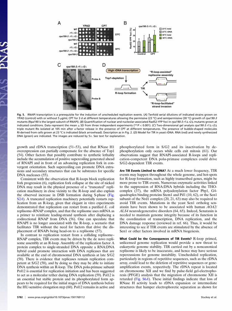
Unscheduled Replication Events Are Transcription-Dependent. Ourmodel predicts that RNAPI transcription would be a prerequisitefor R-loop–initiated replication. To slow down rDNA transcrip-tion rates, we made use of the rpa190-3 mutant of the largestRNAPI subunit Rpa190 and the rrn3-8 mutant of Rnr3, whichrecruits RNAPI to the promoter of 35S rRNA genes (45). Strik-ingly, both rpa190-3 and rrn3-8 mutants alleviated the CPT-sen-sitivity of r1Δ r2Δ mutants at semipermissive temperature (Fig. 5Aand Fig. S5A), reduced the formation of nucleolus-associatedRad52-foci formation and suppressed S/G2-phase cell cycle arrest(Fig. 5B and Fig. S5B). These observations suggest that the ma-jority of the CPT-induced Top1-dependent DNA lesions arelinked to rDNA transcription, as well as a potential link betweenrDNA damage, checkpoint activation, and growth rate. Next, weexamined the fate of replication in rpa190-3 r1Δ r2Δ mutants by2D gel electrophoresis. Cells were grown at 26 °C, before α-factor
synchronization and released in CPT-containing media at per-missive (23 °C) or semipermissive (30 °C) temperature. The ab-sence of bubble-shaped replication intermediates in cells releasedfrom α-factor synchronization at semipermissive temperature (Fig.5C and Fig. S5C) confirms a mechanism where transcription-mediated R-loops initiate replication at late S/G2 phase within the35S rDNA.
DiscussionDecades ago it became evident that R-loops take part in repli-cation initiation of prokaryotic cells (46, 47). Here we presentevidence that this is also the case for eukaryotic cells based on theobservation that persistent R-loops can mediate unscheduled,origin-independent replication initiation in yeast chromosomalDNA. These replication events were observed in the highly tran-scribed 35S rRNA gene, and occurred spatially and temporallyoutside of the regular replication schedule. Unscheduled replica-tion was not linked to a defined replication origin, and it wasobserved in late S/G2 phase of the cell cycle where replicationtermination and completion is expected to take place.
Which Factors and Mechanism Would Participate in Transcription-Initiated Replication Events? Various, nonexclusive mechanismscould cooperate to trigger such transcription-initiated replication(TIR) (Fig. 5D). At present we do not know whether the pres-ence of single-stranded DNA within an R-loop may permitstrand invasion-dependent replication events, favored by therepetitive nature of the rDNA array. DSBs seem to be particu-larly frequent in the rDNA locus. Thus, R-loops and DSBs couldstimulate recombination-driven replication events as observed inCandiada albicans mtDNA (42) or break-induced replication(48, 49) and involve the transient formation of simple Y-likereplication intermediates.The synthetic lethality observed in the absence of Top1 and
RNase H activities (19, 50, and present work) complement pre-vious notions that R-loops have an evolutionary conserved impacton transcription. E. coli Top1 mutants suffer from impaired
A
B
Top1 AID*
Tir1
0 30IAA (min)
0- -+ + 30IAA (min)CPT
WTr1∆ r2∆ TOP1AID*
r1∆ r2∆
20
40
60
80
% c
ells
with
hyb
rids
0
release in IAA (min)αF 15 30 45 75 105
Probe B
Probe A
Fig. 4. Top1AID*-depletion stimulates R-loop formation and unscheduledreplication events in r1Δ r2Δ mutants. (A, Left) Western blot analysis of IAA(1 mM) induced Top1AID* protein degradation. Tir1 serves as loading control.Note that both proteins contain multiple Myc epitopes for detection. (Right)Quantification of RNA:DNA hybrids by S9.6 antibody detection in WT, r1Δr2Δ, and r1Δ r2Δ TOP1AID* mutants in the absence or presence of CPT orIAA, respectively (Right). (B) Two-dimensional gel analysis of RIs isolatedfrom the r1Δ r2Δ TOP1AID* mutant cells grown in the presence of 1 mM IAAfollowing α-factor release. The disappearance of the RFB signal is indicated(white asterisk). The images are reduced by 5.5×. Description as in Fig. 2.
r1∆ r2∆
Prob
e A
fob1∆ rrm3∆
c
ba
d
c
ba
d
e
RFB
5S ARS
RFB
B B35S
a/b
RFB
c
d
e
Fig. 3. Characterization of unscheduled replication intermediates in therDNA. (Left) Comparison of r1Δ r2Δ and rrm3Δ-dependent RFPs (indicated bylowercase letters a to d) detected by probe A. The open arrowmarks a r1Δ r2Δspecific pausing site. (Right) A low-resolution mapping of the pausing sitesbased on their Y-arc location is shown. The images are reduced by 3.5×.
Stuckey et al. PNAS | May 5, 2015 | vol. 112 | no. 18 | 5781
GEN
ETICS
growth and rDNA transcription (51–53), and that RNase H1overexpression can partially compensate for the absence of Top1(54). Other factors that possibly contribute to synthetic lethalityinclude the accumulation of positive supercoiling generated aheadof RNAPI and in front of an advancing replication fork in con-vergent orientation. Such supercoiling can promote DNA extru-sions and secondary structures that can be substrates for specificDNA nucleases (55).Consistent with the observation that R-loops block replication
fork progression (6), replication fork collapse at the site of nickedDNA may result in the physical presence of a “truncated” repli-cation machinery in close vicinity to the R-loop and also explainthe observed increase in DSB formation during S-phase (Fig.S2A). A truncated replication machinery potentially restarts rep-lication from an R-loop, given that elegant in vitro experimentsdemonstrated that replication can restart from a purified E. colireplisome–RNAP complex, and that the replisome uses mRNA asa primer to reinitiate leading-strand synthesis after displacing acodirectional RNAP from DNA (56). One can speculate thatRNAPI is no longer associated with the R-loop, a scenario thatfacilitates TIR without the need for factors that drive the dis-placement of RNAPs being head-on to a replisome (57).In contrast to replication restart from a colliding replisome–
RNAP complex, TIR events may be driven by the de novo repli-some assembly at an R-loop. Assembly of the replication factor Aprotein complex to single-stranded DNA opposite a RNA:DNAhybrid could promote interaction with DNA replicases that areavailable at the end of chromosomal DNA synthesis at late S/G2(58). There is evidence that replicases remain replication com-petent at S/G2 (58), and by doing so they may be able to initiateDNA synthesis within an R-loop. The DNA polα-primase subunitPol12 is essential for replication initiation and has been suggestedto act as a molecular tether during DNA replication (59). Pol12 isan essential but stable protein and its phosphorylated form ap-pears to be required for the initial stages of DNA synthesis beforethe HU-sensitive elongation step (60). Pol12 remains in active and
phosphorylated form in S/G2 and its inactivation by de-phosphorylation only occurs while cells exit mitosis (61). Ourobservations suggest that RNAPI-associated R-loops and repli-cation-competent DNA polα-primase complexes could driveS/G2-dependent TIR events.
Are TIR Events Limited to rDNA? At a much lower frequency, TIRevents may happen throughout the whole genome, and hot-spotsfor R-loop formation, such as highly transcribed genes, might bemore prone to TIR events. Numerous enzymatic activities linkedto the suppression of RNA:DNA hybrids including the THO-complex (37), the mRNA polyadenylation factor Pbp1, G4-quadruplex binding proteins Stem1 and Pif1 (10, 62), or the Sen1subunit of the Nrd1 complex (20, 21, 63) may also be required toavoid TIR events. Mutations in the yeast Sen1 ortholog sen-ataxin have been shown to be associated with human AOA2/ALS4 neurodegenerative disorders (64, 65). Indeed, Senataxin isneeded to maintain genome integrity because of its function inthe coordination of transcription, DNA replication, and theDNA damage response (reviewed in ref. 66). Thus, it would beinteresting to see if TIR events are stimulated by the absence ofSen1 or other factors involved in mRNA biogenesis.
What Could be the Consequences of TIR Events? R-loop primed,unlicensed genome replication would provide a new threat toeukaryotic genome stability. TIR carried out by a noncanonicalreplisome is likely to be inaccurate, and hence may have seriousrepercussions for genome instability. Unscheduled replication,particularly in regions of repetitive sequences, such as the rDNAarray, could lead to the deletion of repetitive sequences or gene-amplification events, respectively. The rDNA repeat is locatedon chromosome XII and we find by pulse-field gel-electropho-resis (PFGE) analysis that the migration of chromosome XII isretarded (Fig. S6A). These initial findings indicate that loss ofRNase H activity leads to rDNA expansion or intermediatestructures that hamper electrophoretic separation as shown for
A B23°C total23°C rDNA30°C total30°C rDNA
rpa190-3 r1∆ r2∆40
30
20
10
0
Rad
52-Y
PF fo
ci (%
)
Control CPT
23ºC
30ºC
WT
rpa190-3 r1∆ r2∆rpa190-3
YPAD CPT
r1∆ r2∆
WT
rpa190-3 r1∆ r2∆rpa190-3
r1∆ r2∆
**
C rpa190-3 r1∆ r2∆
Probe B
Probe A
23ºC 30ºC
R-loop formation
High rDNA transcription
Top1-depletion
TIR
“R-loop-assisted” replisome assembly
“R-loop-facilitated”strand invasion
D
Fig. 5. RNAPI transcription is a prerequisite for the induction of unscheduled replication events. (A) Tenfold serial dilutions of indicated strains grown onYPAD (control) with or without 5 μg/mL CPT for 3 d at different temperatures allowing the permissive (23 °C) and semipermissive (30 °C) growth of rpa190-3mutants (Rpa190 is the largest subunit of RNAPI). (B) Quantification of nuclear and nucleolar-associated Rad52-YFP foci in rpa190-3 r1Δ r2Δmutants grown atindicated conditions. Data represent the mean ± SD from three independent experiments (**P < 0.001). (C) Two-dimensional gel analysis rpa190-3 r1Δ r2Δtriple mutant RIs isolated at 105 min after α-factor release in the presence of CPT at different temperatures. The presence of bubble-shaped moleculesRI-derived from cells grown at 23 °C is indicated (black arrowhead). Description as in Fig. 2. (D) Model for TIR in yeast rDNA. RNA (red) and newly synthesizedDNA (green) are indicated. The images are reduced by 5×. See text for explanation.
5782 | www.pnas.org/cgi/doi/10.1073/pnas.1501769112 Stuckey et al.

replicating chromosomes (67). The observation that ERC forma-tion is enhanced in r1Δ r2Δ mutants (Fig. S6B) confirms that theloss of RNase H activities causes genetic instability (Fig. S1). Itwould be particularly interesting to determine to which extentimpaired RNA:DNA hybrid processing contributes to rereplica-tion, gene amplification, and alterations in chromosome copynumber in human cells. These events would have disastrous con-sequences for eukaryotic genome function and are particularlyrelevant to carcinogenesis (68).Selective gene amplification is frequently observed in dif-
ferentiating eukaryotic cells and results in transcript numberand gene product increases in a dosage-dependent manner (69).The mechanisms by which gene amplification is achieved includeDSB-dependent sister chromatid fusion and repeated breakage-fusion-bridge cycles evident in the dihydrofolate reductase locusof CHO cells (70), endoreplication of diptera chorion genes bymultiple activation of replication origins within the same S-phase(71), RNA-template derived nanochromosome amplification inStylonychia lemnae (72), or rolling circle amplification of extra-chromosomal rDNA circles in Xenopus oocytes (73). Although inour study we could not distinguish if ERCs are more prone to TIRevents, our results provide the possibility that ERC replication canbe driven by impaired RNA transcript-processing suggesting thatR-loops could have a physiological role in the control of geneamplification linked to nuclear differentiation events.
Experimental ProceduresYeast Strains and Growth Conditions. Yeast strains used in this study are listedin Table S1. Gene deletions were constructed by PCR-based methods (74). Ifnot generated by a direct knock-out in the YKL background, mutant strainswere backcrossed at least twice to the YKL83 strain background. Yeaststrains were grown in YPAD, or synthetic complete (SC) minimal mediumsupplemented with 2% (wt/vol) glucose and amino acids. IAA (Sigma) wasadded at 500 μM for solid and 1 mM for liquid YPAD media.
Viability Assays. To test for sensitivity to genotoxic agents, 10-fold serialdilutions of cells were grown for 3 d on YPAD plates or YPAD-containing HU(50 mM; USBiological), MMS (10 mM; Fluka), or CPT (5 μg/mL; Santa CruzBiotechnology), unless otherwise specified.
Quantification and Colocalization of Rad52-YFP Foci. For colocalization ofRad52-YFP foci with the nucleolar marker Nop1, cells were cotransformedwith the RAD52-YFP expressing plasmid (F. Prado, Sevilla, Spain) and a plasmidexpressing NOP1-mRFP (75). Transformants were grown in exponential phase inSC with plasmid selection, in the presence or absence of 10 μg/mL CPT for 3 h,and fixed using 2.5% (vol/vol) formaldehyde. YFP fluorescence (480-nm excita-tion/527-nm emission) and RFP fluorescence (584-nm excitation/607-nm emission)were detected by wide-field fluorescence microscopy (DM-6000B, Leica) at 100×magnification. Images were taken using LAS AF software (Leica). For eachsample, 600 cells were counted from three independent experiments. For time-course analysis of Rad52-YFP foci, cells expressing the RAD52-YFP plasmid weresynchronized with α-factor and released in the presence or absence of 10 μg/mLCPT. Samples were retrieved at the specified time points following release andfixed with 2.5% (vol/vol) formaldehyde. Approximately 600 cells derived fromthree independent colonies were analyzed for each condition.
Cell Cycle Progression Analysis and DNA Isolation. For G1 synchronization,MATa cells were grown to an OD600 of 0.4 in YPAD medium before α-factor
(1 μg/mL; Biomedal) synchronization for 180 up to 240 min. Cells were re-leased from α-factor treatment by washing three times in prewarmed, freshYPAD medium containing 0.1 mg/mL Pronase E (Sigma). For FACS analysis,1-mL samples were taken at indicated times and fixed in 70% ethanol.Samples were resuspended in PBS containing 5 μg/mL propidium iodide, fol-lowing RNase A overnight treatment, and were analyzed by flow cytometryusing a FACSCalibur (Becton Dickinson) and CellQuest software. For DNAanalysis, cells were released in fresh media containing 10 μg/mL CPT. 100 mLsamples were retrieved at the specified time points following release andarrested by adding sodium azide (to a final concentration of 0.1%). DNA wasextracted according to ref. 6.
Two Dimensional Agarose Gel Electrophoresis. In a total volume of 100 μL,about 5 μg of genomic DNA was digested with 40 units BglII for 6 h, iso-propanol precipitated, and resuspended in 20 μL loading buffer. First, di-mension electrophoresis was performed in 0.4% TBE-buffered agarose gels at40 V for 20 h. A gel slice containing DNA fragments between 3 and 12 kb wascut out for second dimension resolution in 1.1% TBE-buffered agarose at 140 Vfor 16 h. Denatured DNA was transferred to a Hybond XL membrane(Amersham) by standard procedures. Replication intermediates were detectedby hybridization with specific 32P-labeled DNA probes, matching to nucleo-tides 452691–453344 (probe A) and 453834–454699 (probe B) on chromosomeXII. Signals were quantified using a PhosphorImager with ImageGaugesoftware (Fuji). The relative intensity of replication intermediates wasnormalized to the signal intensity obtained in the 1×-spot (nonsaturatingexposure).
TOP1AID* Degron Construct. The Top1 protein was tagged at the C terminuswith the IAA17 auxin-binding domain (amplified by PCR from the plasmidpKan–AID* (76) in a r1Δ r2Δ strain, which expresses TIR1 under control of theconstitutive ADH promoter. Note that both proteins contain multiple Mycepitopes that can be detected by a c-Myc primary antibody. The functionalityof the Top1AID* degron was confirmed by Western blot analysis against theMyc tag to confirm degradation of the Top1 protein in response to 1 mM IAA(60). Protein extracts for immunoblotting were separated by SDS/PAGE us-ing 8% (wt/vol) polyacrylamide (37.5:1), and protein bound membraneswere incubated overnight with a mouse anti–c-Myc primary antibody(Abcam) at 1:1,000 and 1:20,000 in PBS-5% (wt/vol) milk, respectively. ForFACS and 2D-gel analysis of the r1Δ r2Δ TOP1AID* strain, 1 mM IAA wasadded to YPAD medium for the last 30 min of α-factor incubation, and cellswere released in the presence of IAA.
Statistics. Values are expressed as the mean ± SD. Significance values (P) wereobtained following Student’s t test for pairwise comparisons of data andconsidered statistically significant for P values < 0.05.
ACKNOWLEDGMENTS. We thank K. Labib, H. Ulrich, F. Prado, J. Baßler, andJ. de la Cruz for kindly providing strains and plasmids; H. Gaillard, A. El Hage,and D. Tollervey for suggestions and critical reading of the document; andE. Fernandéz-García and H. Heluani for technical assistance. This work wassupported in part by Spanish Ministry of Science and Innovation GrantsBIO2006-08051 and BFU2010-21339 (to R.E.W.); Junta de Andalucía GrantsBIO-026, P08-CTS-04297, and P11-CTS-7962 (to R.E.W.); a European UnionFEDER grant (to R.E.W.); a Junta para la Ampliación de Estudios predoctoraltraining grant from the Spanish National Research Council (CSIC) (to R.S.); apredoctoral training grant from the University of Seville/El Monte Founda-tion (to N.G.-R.); the Spanish Ministry of Science and Innovation GrantBFU2010-16372 and Consolider Ingenio 2010 CSD2007-0015 (to A.A.); Juntade Andalucía Grants BIO-102 and P09/CVI4567 (to A.A.); and the EuropeanUnion (FEDER) (A.A.).
1. Liu LF, Wang JC (1987) Supercoiling of the DNA template during transcription. Proc
Natl Acad Sci USA 84(20):7024–7027.2. Thomas M, White RL, Davis RW (1976) Hybridization of RNA to double-stranded DNA:
Formation of R-loops. Proc Natl Acad Sci USA 73(7):2294–2298.3. Duquette ML, Handa P, Vincent JA, Taylor AF, Maizels N (2004) Intracellular tran-
scription of G-rich DNAs induces formation of G-loops, novel structures containing G4
DNA. Genes Dev 18(13):1618–1629.4. Roy D, Zhang Z, Lu Z, Hsieh CL, Lieber MR (2010) Competition between the RNA
transcript and the nontemplate DNA strand during R-loop formation in vitro: A nick
can serve as a strong R-loop initiation site. Mol Cell Biol 30(1):146–159.5. Huertas P, Aguilera A (2003) Cotranscriptionally formed DNA:RNA hybrids mediate
transcription elongation impairment and transcription-associated recombination.Mol
Cell 12(3):711–721.
6. Wellinger RE, Prado F, Aguilera A (2006) Replication fork progression is impaired bytranscription in hyperrecombinant yeast cells lacking a functional THO complex. MolCell Biol 26(8):3327–3334.
7. Gan W, et al. (2011) R-loop-mediated genomic instability is caused by impairment ofreplication fork progression. Genes Dev 25(19):2041–2056.
8. Helmrich A, Ballarino M, Tora L (2011) Collisions between replication and transcrip-tion complexes cause common fragile site instability at the longest human genes.MolCell 44(6):966–977.
9. Castellano-Pozo M, García-Muse T, Aguilera A (2012) R-loops cause replication im-pairment and genome instability during meiosis. EMBO Rep 13(10):923–929.
10. Salvi JS, et al. (2014) Roles for Pbp1 and caloric restriction in genome and lifespanmaintenance via suppression of RNA-DNA hybrids. Dev Cell 30(2):177–191.
11. Aguilera A, García-Muse T (2012) R loops: From transcription byproducts to threats togenome stability. Mol Cell 46(2):115–124.
Stuckey et al. PNAS | May 5, 2015 | vol. 112 | no. 18 | 5783
GEN
ETICS

12. Wang JC (1971) Interaction between DNA and an Escherichia coli protein omega.J Mol Biol 55(3):523–533.
13. Wang JC (2002) Cellular roles of DNA topoisomerases: A molecular perspective. NatRev Mol Cell Biol 3(6):430–440.
14. Leppard JB, Champoux JJ (2005) Human DNA topoisomerase I: Relaxation, roles, anddamage control. Chromosoma 114(2):75–85.
15. Pommier Y (2009) DNA topoisomerase I inhibitors: Chemistry, biology, and interfacialinhibition. Chem Rev 109(7):2894–2902.
16. Sogo JM, Lopes M, Foiani M (2002) Fork reversal and ssDNA accumulation at stalledreplication forks owing to checkpoint defects. Science 297(5581):599–602.
17. Tuduri S, et al. (2009) Topoisomerase I suppresses genomic instability by preventinginterference between replication and transcription. Nat Cell Biol 11(11):1315–1324.
18. French SL, et al. (2011) Distinguishing the roles of topoisomerases I and II in relief oftranscription-induced torsional stress in yeast rRNA genes. Mol Cell Biol 31(3):482–494.
19. El Hage A, French SL, Beyer AL, Tollervey D (2010) Loss of topoisomerase I leads toR-loop-mediated transcriptional blocks during ribosomal RNA synthesis. Genes Dev24(14):1546–1558.
20. Skourti-Stathaki K, Proudfoot NJ, Gromak N (2011) Human senataxin resolves RNA/DNA hybrids formed at transcriptional pause sites to promote Xrn2-dependent ter-mination. Mol Cell 42(6):794–805.
21. Mischo HE, et al. (2011) Yeast Sen1 helicase protects the genome from transcription-associated instability. Mol Cell 41(1):21–32.
22. Cerritelli SM, Crouch RJ (2009) Ribonuclease H: The enzymes in eukaryotes. FEBS J276(6):1494–1505.
23. Lazzaro F, et al. (2012) RNase H and postreplication repair protect cells from ribo-nucleotides incorporated in DNA. Mol Cell 45(1):99–110.
24. Kim N, et al. (2011) Mutagenic processing of ribonucleotides in DNA by yeast top-oisomerase I. Science 332(6037):1561–1564.
25. Xu B, Clayton DA (1996) RNA-DNA hybrid formation at the human mitochondrialheavy-strand origin ceases at replication start sites: An implication for RNA-DNAhybrids serving as primers. EMBO J 15(12):3135–3143.
26. Kogoma T (1978) A novel Escherichia coli mutant capable of DNA replication in theabsence of protein synthesis. J Mol Biol 121(1):55–69.
27. Kogoma T, Maldonado RR (1997) DNA polymerase I in constitutive stable DNA rep-lication in Escherichia coli. J Bacteriol 179(7):2109–2115.
28. Horiuchi T, Maki H, Sekiguchi M (1984) RNase H-defective mutants of Escherichia coli:A possible discriminatory role of RNase H in initiation of DNA replication. Mol GenGenet 195(1-2):17–22.
29. Kogoma T (1984) Absence of RNase H allows replication of pBR322 in Escherichia colimutants lacking DNA polymerase I. Proc Natl Acad Sci USA 81(24):7845–7849.
30. Kogoma T, von Meyenburg K (1983) The origin of replication, oriC, and the dnaAprotein are dispensable in stable DNA replication (sdrA) mutants of Escherichia coliK-12. EMBO J 2(3):463–468.
31. de Massy B, Fayet O, Kogoma T (1984) Multiple origin usage for DNA replication insdrA(rnh) mutants of Escherichia coli K-12. Initiation in the absence of oriC. J Mol Biol178(2):227–236.
32. Maduike NZ, Tehranchi AK, Wang JD, Kreuzer KN (2014) Replication of the Escher-ichia coli chromosome in RNase HI-deficient cells: Multiple initiation regions and forkdynamics. Mol Microbiol 91(1):39–56.
33. Wimberly H, et al. (2013) R-loops and nicks initiate DNA breakage and genome in-stability in non-growing Escherichia coli. Nat Commun 4:2115.
34. Sinclair DA, Guarente L (1997) Extrachromosomal rDNA circles—A cause of aging inyeast. Cell 91(7):1033–1042.
35. Takeuchi Y, Horiuchi T, Kobayashi T (2003) Transcription-dependent recombinationand the role of fork collision in yeast rDNA. Genes Dev 17(12):1497–1506.
36. Ivessa AS, Zhou JQ, Zakian VA (2000) The Saccharomyces Pif1p DNA helicase and thehighly related Rrm3p have opposite effects on replication fork progression in ribo-somal DNA. Cell 100(4):479–489.
37. Chan YA, et al. (2014) Genome-wide profiling of yeast DNA:RNA hybrid prone siteswith DRIP-chip. PLoS Genet 10(4):e1004288.
38. El Hage A, Webb S, Kerr A, Tollervey D (2014) Genome-wide distribution of RNA-DNAhybrids identifies RNase H targets in tRNA genes, retrotransposons and mitochondria.PLoS Genet 10(10):e1004716.
39. Raghuraman MK, et al. (2001) Replication dynamics of the yeast genome. Science294(5540):115–121.
40. Brewer BJ, FangmanWL (1987) The localization of replication origins on ARS plasmidsin S. cerevisiae. Cell 51(3):463–471.
41. Holt IJ, Lorimer HE, Jacobs HT (2000) Coupled leading- and lagging-strand synthesis ofmammalian mitochondrial DNA. Cell 100(5):515–524.
42. Gerhold JM, Aun A, Sedman T, Jõers P, Sedman J (2010) Strand invasion structures inthe inverted repeat of Candida albicans mitochondrial DNA reveal a role for ho-mologous recombination in replication. Mol Cell 39(6):851–861.
43. Wellinger RE, Schär P, Sogo JM (2003) Rad52-independent accumulation of jointcircular minichromosomes during S phase in Saccharomyces cerevisiae. Mol Cell Biol23(18):6363–6372.
44. Krawczyk C, Dion V, Schär P, Fritsch O (2014) Reversible Top1 cleavage complexes arestabilized strand-specifically at the ribosomal replication fork barrier and contributeto ribosomal DNA stability. Nucleic Acids Res 42(8):4985–4995.
45. Wittekind M, et al. (1988) Isolation and characterization of temperature-sensitivemutations in RPA190, the gene encoding the largest subunit of RNA polymerase Ifrom Saccharomyces cerevisiae. Mol Cell Biol 8(10):3997–4008.
46. Marians KJ (1992) Prokaryotic DNA replication. Annu Rev Biochem 61:673–719.47. Gowrishankar J (2015) End of the beginning: Elongation and termination features of
alternative modes of chromosomal replication initiation in bacteria. PLoS Genet 11(1):e1004909.
48. Saini N, et al. (2013) Migrating bubble during break-induced replication drives con-servative DNA synthesis. Nature 502(7471):389–392.
49. Kraus E, Leung WY, Haber JE (2001) Break-induced replication: A review and an ex-ample in budding yeast. Proc Natl Acad Sci USA 98(15):8255–8262.
50. Chon H, et al. (2013) RNase H2 roles in genome integrity revealed by unlinking itsactivities. Nucleic Acids Res 41(5):3130–3143.
51. Hraiky C, Raymond MA, Drolet M (2000) RNase H overproduction corrects a defect atthe level of transcription elongation during rRNA synthesis in the absence of DNAtopoisomerase I in Escherichia coli. J Biol Chem 275(15):11257–11263.
52. Drolet M (2006) Growth inhibition mediated by excess negative supercoiling: Theinterplay between transcription elongation, R-loop formation and DNA topology.Mol Microbiol 59(3):723–730.
53. Baaklini I, et al. (2008) Hypernegative supercoiling inhibits growth by causing RNAdegradation. J Bacteriol 190(22):7346–7356.
54. Drolet M, et al. (1995) Overexpression of RNase H partially complements the growthdefect of an Escherichia coli delta topA mutant: R-loop formation is a major problemin the absence of DNA topoisomerase I. Proc Natl Acad Sci USA 92(8):3526–3530.
55. Lobachev KS, Gordenin DA, Resnick MA (2002) The Mre11 complex is required forrepair of hairpin-capped double-strand breaks and prevention of chromosome re-arrangements. Cell 108(2):183–193.
56. Pomerantz RT, O’Donnell M (2008) The replisome uses mRNA as a primer after col-liding with RNA polymerase. Nature 456(7223):762–766.
57. Pomerantz RT, O’Donnell M (2010) Direct restart of a replication fork stalled by ahead-on RNA polymerase. Science 327(5965):590–592.
58. Longhese MP, Plevani P, Lucchini G (1994) Replication factor A is required in vivo forDNA replication, repair, and recombination. Mol Cell Biol 14(12):7884–7890.
59. Collins KL, Russo AA, Tseng BY, Kelly TJ (1993) The role of the 70 kDa subunit ofhuman DNA polymerase alpha in DNA replication. EMBO J 12(12):4555–4566.
60. Foiani M, Marini F, Gamba D, Lucchini G, Plevani P (1994) The B subunit of the DNApolymerase alpha-primase complex in Saccharomyces cerevisiae executes an essentialfunction at the initial stage of DNA replication. Mol Cell Biol 14(2):923–933.
61. Foiani M, Liberi G, Lucchini G, Plevani P (1995) Cell cycle-dependent phosphorylationand dephosphorylation of the yeast DNA polymerase alpha-primase B subunit. MolCell Biol 15(2):883–891.
62. Paeschke K, et al. (2013) Pif1 family helicases suppress genome instability at G-quadruplex motifs. Nature 497(7450):458–462.
63. Tudek A, et al. (2014) Molecular basis for coordinating transcription termination withnoncoding RNA degradation. Mol Cell 55(3):467–481.
64. Moreira MC, et al. (2004) Senataxin, the ortholog of a yeast RNA helicase, is mutant inataxia-ocular apraxia 2. Nat Genet 36(3):225–227.
65. Chen YZ, et al. (2006) Senataxin, the yeast Sen1p orthologue: characterization of aunique protein in which recessive mutations cause ataxia and dominant mutationscause motor neuron disease. Neurobiol Dis 23(1):97–108.
66. Skourti-Stathaki K, Proudfoot NJ (2014) A double-edged sword: R loops as threats togenome integrity and powerful regulators of gene expression. Genes Dev 28(13):1384–1396.
67. Cha RS, Kleckner N (2002) ATR homolog Mec1 promotes fork progression, thusaverting breaks in replication slow zones. Science 297(5581):602–606.
68. Schwab M (1998) Amplification of oncogenes in human cancer cells. Bioessays 20(6):473–479.
69. Tanaka H, Yao MC (2009) Palindromic gene amplification—An evolutionarily con-served role for DNA inverted repeats in the genome. Nat Rev Cancer 9(3):216–224.
70. Trask BJ, Hamlin JL (1989) Early dihydrofolate reductase gene amplification events inCHO cells usually occur on the same chromosome arm as the original locus. Genes Dev3(12A):1913–1925.
71. Heck MM, Spradling AC (1990) Multiple replication origins are used during Drosophilachorion gene amplification. J Cell Biol 110(4):903–914.
72. Heyse G, Jönsson F, Chang WJ, Lipps HJ (2010) RNA-dependent control of gene am-plification. Proc Natl Acad Sci USA 107(51):22134–22139.
73. Hourcade D, Dressler D, Wolfson J (1973) The amplification of ribosomal RNA genesinvolves a rolling circle intermediate. Proc Natl Acad Sci USA 70(10):2926–2930.
74. Longtine MS, et al. (1998) Additional modules for versatile and economical PCR-basedgene deletion and modification in Saccharomyces cerevisiae. Yeast 14(10):953–961.
75. Ulbrich C, et al. (2009) Mechanochemical removal of ribosome biogenesis factors fromnascent 60S ribosomal subunits. Cell 138(5):911–922.
76. Morawska M, Ulrich HD (2013) An expanded tool kit for the auxin-inducible degronsystem in budding yeast. Yeast 30(9):341–351.
5784 | www.pnas.org/cgi/doi/10.1073/pnas.1501769112 Stuckey et al.