Risk factors for abutment and implant fracture after loading
6
1 Abstract Purpose: Implant component fractures are one of the most serious compli- cations in implant treatment. With a better understanding of the risk factors for fracture in the preoperative, surgery, superstructure, and post-loading phases of implant treatment, low-risk treatment could reduce implant com- ponent fractures, leading to a better prognosis. The aim of this study was to clarify the risk factors for abutment and implant fractures that occur after loading, and to perform a retrospective, approximately 10-year follow-up study to explore the risk factors in each treatment phase. Methods: Subjects were fitted with an implant prosthesis between Janu- ary 2008 and December 2009. In total, 1,126 Ankylos implants in 430 patients were included for analysis. Binary logistic regression analysis was performed to extract factors related to non-fracture and fracture of the abutment or implant as a dependent variable. Results: Gender (OR = 3.466, 95% CI 1.296-9.268, P = 0.013), gonial angle (OR = 3.420, 95% CI 1.308-8.945, P = 0.012), and splinting status of the superstructure (OR = 4.456, 95% CI 1.861-10.669, P = 0.001) were identified as significant risk factors. Conclusion: The risk of fracture is increased in males, especially those with a mandibular angle of less than 120° on panoramic radiographs, and those with a non-splinted superstructure. Keywords; gonial angle, implant fracture, retrospective cohort study, risk factors Introduction Implant treatment has a stable long-term prognosis with a high chance of achieving osseointegration. However, mechanical and biological complica- tions related to implants have been reported [1-3]. Fracture of the veneering material; loosening or fracture of the abutment screw; and fracture of the framework, abutment, or implant are the worst-case scenarios and have been reported following long-term prosthesis use [4,5]. The fracture rate of implant components, including the implant, abutments, and abutment screws, is 5.6% [6]. Bruxism and the crown-implant ratio (C/I) are possible causes of internal stress that lead to mechanical complications [7]. It has been reported that bruxism is a factor for implant fracture due to the strong force applied [8] and that the strain on the implant body’s buccolingual side is directly proportional to an increase in the C/I [9]. Prosthetic problems caused by mechanical complications occur after superstructure installation. Therefore, it is necessary to minimize the occurrence of complications, considering the resulting discomfort to the patient, time needed for repair and remanufacturing, the cost and damage, as well as the distress caused by removal of the fractured implant. It is thus desirable to predict the case selection, response to risk factors, and prog- nosis by evaluating risk factors from information obtained prior to surgery. It is difficult to evaluate risks such as the presence or absence of brux- ism by only examining the dentition and skeleton through preoperative oral and X-ray examinations. Therefore, these risks should be evaluated using an established or simple inspection method. Implant treatment is divided into preoperative examination, surgery, superstructure, and post-loading phases. If the risk factors for mechanical complications per phase can be determined, it will be possible to reduce implant treatment failure. Previous studies [1-10] have examined the risk factors for fracture of the implant component; the incidence of implant and abutment fractures is reportedly low. Possible causes are multifactorial, such as dental implant manufacturing and design failure, bruxism or large occlusal force, super- structure design, implant positioning, implant diameter, metal fatigue, and bone resorption around the implant. However, conflicting reports [11-13] deny the role of the above factors, and no clear cause is stated. Few stud- ies have described the long-term observation of multiple factors from the preoperative to post-loading periods and compared and evaluated cases with and without fractures. By investigating the risk factors for fracture during each phase, it is expected that low-risk implant treatment can reduce the fracture of implant components, leading to a better prognosis. There- fore, the aim of this study was to clarify the risk factors for abutment and implant fracture that occur after loading, and to perform a retrospective, approximately 10-year follow-up study to evaluate these factors in each phase of implant treatment (i.e. preoperative examination, surgery, and superstructure and post-loading phases). Materials and Methods Study design This was a retrospective cohort study. Subjects Subjects underwent insertion of an implant prosthesis between January 2008 and December 2009 at the Oral Implant Clinic, Nihon University Hospital at Matsudo. There were 1,148 implants in 434 patients (129 men, 305 women) with follow-up data until December 2019. Only patients with Ankylos implants (Dentsply Implants Manufacturing GmbH, Hanau, Ger- many) were included; those with non-Ankylos implants were excluded. In line with the manufacturer’s recommended protocol, all Ankylos implantations were performed in two stages. First, the implant body (com- mercially pure titanium [cpTi] grade 2) was inserted so that its platform was located at or below the margin of the bone, and the mucosa was sutured. Following a 3-month healing period, a permucosal extension was fashioned during second-stage surgery after taking an impression and prosthesis insertion. All abutments were made of ready-made, formable titanium alloys (Ti-6Al-4V) that correspond to mucosal penetration with different neck lengths. All superstructures were fixed with temporary cement. The occlusal contact applied to the superstructure was adjusted so that the implant and natural tooth parts were in the same occlusal contact state. Maintenance was performed at 1, 3, and 6 months after superstruc- ture installation, and at 6-month intervals thereafter. The subjects were divided into two groups. Patients with or without J-STAGE Advance Publication: December 14, 2020 Journal of Oral Science Original article Risk factors for abutment and implant fracture after loading Hiroshi Murakami 1) , Kentaro Igarashi 2) , Megumi Fuse 3) , Tsuyoshi Kitagawa 1) , Mitsuhiko Igarashi 4) , Satoshi Uchibori 1) , Chiaki Komine 5) , Hiroya Gotouda 6) , Hiroyuki Okada 4) , and Yasuhiko Kawai 2) 1) Department of Fixed Prosthodontics and Oral Implantology, Nihon University School of Dentistry at Matsudo, Matsudo, Japan 2) Department of Removable Prosthodontics, Nihon University School of Dentistry at Matsudo, Matsudo, Japan 3) Liberal Arts (Chemistry), Nihon University School of Dentistry at Matsudo, Matsudo, Japan 4) Department of Histology, Nihon University School of Dentistry at Matsudo, Matsudo, Japan 5) Department of Oral Health Science, Division of Laboratory Medicine for Dentistry, Nihon University School of Dentistry at Matsudo, Matsudo, Japan 6) Department of Community Oral Health, Nihon University School of Dentistry at Matsudo, Matsudo, Japan (Received August 29, 2020; Accepted November 11, 2020) Correspondence to Dr. Hiroshi Murakami, Department of Fixed Prosthodontics and Oral Implantology, Nihon University School of Dentistry at Matsudo, 2-870-1 Sakaecho-nishi, Matsudo, Chiba 271-8587, Japan Fax: +81-47-360-9411 E-mail: [email protected] doi.org/10.2334/josnusd.20-0443 DN/JST.JSTAGE/josnusd/20-0443
Transcript of Risk factors for abutment and implant fracture after loading
1
Abstract
Purpose: Implant component fractures are one of the most serious compli-cations in implant treatment. With a better understanding of the risk factors for fracture in the preoperative, surgery, superstructure, and post-loading phases of implant treatment, low-risk treatment could reduce implant com-ponent fractures, leading to a better prognosis. The aim of this study was to clarify the risk factors for abutment and implant fractures that occur after loading, and to perform a retrospective, approximately 10-year follow-up study to explore the risk factors in each treatment phase. Methods: Subjects were fitted with an implant prosthesis between Janu-ary 2008 and December 2009. In total, 1,126 Ankylos implants in 430 patients were included for analysis. Binary logistic regression analysis was performed to extract factors related to non-fracture and fracture of the abutment or implant as a dependent variable. Results: Gender (OR = 3.466, 95% CI 1.296-9.268, P = 0.013), gonial angle (OR = 3.420, 95% CI 1.308-8.945, P = 0.012), and splinting status of the superstructure (OR = 4.456, 95% CI 1.861-10.669, P = 0.001) were identified as significant risk factors. Conclusion: The risk of fracture is increased in males, especially those with a mandibular angle of less than 120° on panoramic radiographs, and those with a non-splinted superstructure.
Keywords; gonial angle, implant fracture, retrospective cohort study, risk factors
Introduction
Implant treatment has a stable long-term prognosis with a high chance of achieving osseointegration. However, mechanical and biological complica-tions related to implants have been reported [1-3]. Fracture of the veneering material; loosening or fracture of the abutment screw; and fracture of the framework, abutment, or implant are the worst-case scenarios and have been reported following long-term prosthesis use [4,5]. The fracture rate of implant components, including the implant, abutments, and abutment screws, is 5.6% [6]. Bruxism and the crown-implant ratio (C/I) are possible causes of internal stress that lead to mechanical complications [7]. It has been reported that bruxism is a factor for implant fracture due to the strong force applied [8] and that the strain on the implant body’s buccolingual side is directly proportional to an increase in the C/I [9].
Prosthetic problems caused by mechanical complications occur after superstructure installation. Therefore, it is necessary to minimize the occurrence of complications, considering the resulting discomfort to the patient, time needed for repair and remanufacturing, the cost and damage, as well as the distress caused by removal of the fractured implant. It is thus
desirable to predict the case selection, response to risk factors, and prog-nosis by evaluating risk factors from information obtained prior to surgery.
It is difficult to evaluate risks such as the presence or absence of brux-ism by only examining the dentition and skeleton through preoperative oral and X-ray examinations. Therefore, these risks should be evaluated using an established or simple inspection method. Implant treatment is divided into preoperative examination, surgery, superstructure, and post-loading phases. If the risk factors for mechanical complications per phase can be determined, it will be possible to reduce implant treatment failure.
Previous studies [1-10] have examined the risk factors for fracture of the implant component; the incidence of implant and abutment fractures is reportedly low. Possible causes are multifactorial, such as dental implant manufacturing and design failure, bruxism or large occlusal force, super-structure design, implant positioning, implant diameter, metal fatigue, and bone resorption around the implant. However, conflicting reports [11-13] deny the role of the above factors, and no clear cause is stated. Few stud-ies have described the long-term observation of multiple factors from the preoperative to post-loading periods and compared and evaluated cases with and without fractures. By investigating the risk factors for fracture during each phase, it is expected that low-risk implant treatment can reduce the fracture of implant components, leading to a better prognosis. There-fore, the aim of this study was to clarify the risk factors for abutment and implant fracture that occur after loading, and to perform a retrospective, approximately 10-year follow-up study to evaluate these factors in each phase of implant treatment (i.e. preoperative examination, surgery, and superstructure and post-loading phases).
Materials and Methods
Study designThis was a retrospective cohort study.
SubjectsSubjects underwent insertion of an implant prosthesis between January 2008 and December 2009 at the Oral Implant Clinic, Nihon University Hospital at Matsudo. There were 1,148 implants in 434 patients (129 men, 305 women) with follow-up data until December 2019. Only patients with Ankylos implants (Dentsply Implants Manufacturing GmbH, Hanau, Ger-many) were included; those with non-Ankylos implants were excluded.
In line with the manufacturer’s recommended protocol, all Ankylos implantations were performed in two stages. First, the implant body (com-mercially pure titanium [cpTi] grade 2) was inserted so that its platform was located at or below the margin of the bone, and the mucosa was sutured. Following a 3-month healing period, a permucosal extension was fashioned during second-stage surgery after taking an impression and prosthesis insertion. All abutments were made of ready-made, formable titanium alloys (Ti-6Al-4V) that correspond to mucosal penetration with different neck lengths. All superstructures were fixed with temporary cement. The occlusal contact applied to the superstructure was adjusted so that the implant and natural tooth parts were in the same occlusal contact state. Maintenance was performed at 1, 3, and 6 months after superstruc-ture installation, and at 6-month intervals thereafter.
The subjects were divided into two groups. Patients with or without
J-STAGE Advance Publication: December 14, 2020 Journal of Oral Science
Original article
Risk factors for abutment and implant fracture after loadingHiroshi Murakami1), Kentaro Igarashi2), Megumi Fuse3), Tsuyoshi Kitagawa1), Mitsuhiko Igarashi4), Satoshi Uchibori1), Chiaki Komine5), Hiroya Gotouda6), Hiroyuki Okada4), and Yasuhiko Kawai2)
1) Department of Fixed Prosthodontics and Oral Implantology, Nihon University School of Dentistry at Matsudo, Matsudo, Japan2) Department of Removable Prosthodontics, Nihon University School of Dentistry at Matsudo, Matsudo, Japan3) Liberal Arts (Chemistry), Nihon University School of Dentistry at Matsudo, Matsudo, Japan4) Department of Histology, Nihon University School of Dentistry at Matsudo, Matsudo, Japan5) Department of Oral Health Science, Division of Laboratory Medicine for Dentistry, Nihon University School of Dentistry at Matsudo, Matsudo, Japan6) Department of Community Oral Health, Nihon University School of Dentistry at Matsudo, Matsudo, Japan
(Received August 29, 2020; Accepted November 11, 2020)
Correspondence to Dr. Hiroshi Murakami, Department of Fixed Prosthodontics and Oral Implantology, Nihon University School of Dentistry at Matsudo, 2-870-1 Sakaecho-nishi, Matsudo, Chiba 271-8587, JapanFax: +81-47-360-9411 E-mail: [email protected]
doi.org/10.2334/josnusd.20-0443DN/JST.JSTAGE/josnusd/20-0443
2
fracture of the implant or abutment during the observation period were categorized into the fracture group and non-fracture groups, respectively.
Investigation parametersPreoperative itemsBasic attributes: Age at the time of implant placement and gender were obtained from medical records.
Medical history: The absence or onset of hypertension, heart disease, cerebrovascular disease, diabetes, osteoporosis, hepatic or renal dysfunc-tion, autoimmune disease, psychiatric disorders, and smoking, which are risk factors for implant treatment [14], were obtained from medical records.
Gonial angle: Gonial angle was obtained from a panoramic radiograph. Abe et al. [15] defined the gonial angle as the angle between the ramus and mandibular planes, excluding the mandibular condyle’s posterior margin. This angle was expressed as the mean of both sides. It was classified as either greater than 120 degrees or less than 120 degrees, based on a report indicating that the fracture of vital teeth tends to increase when the mandibular angle is less than 120 degrees [15]. Synapse Vincent (Fujifilm Medical Corp., Tokyo, Japan) was used to obtain the measurements.
Miyachi’s Index: The relationship between the number of residual teeth and that of occlusal supports preoperatively was acquired from the medi-cal records and panoramic radiographs. It was evaluated in four groups: occlusal miss, occlusal defect, occlusal collapse, and occlusal loss, as per Miyachi’s triangular classification [16].
Eichner Index: The occlusal support area of the premolar and molar regions was graded as A, B, or C, as per Eichner’s classification [17] based on data from the medical records and panoramic radiographs.
Operative itemsThe presence or absence of hard tissue management: When normal implant placement was impossible due to insufficient bone mass, hard tissue man-agement (maxillary sinus floor elevation, guided bone regeneration (GBR), bone graft, or split crest) was performed. Its presence or absence was con-firmed using the medical records.
Implant body: A relationship between the Ankylos implant diameter and abutment fracture has been reported [18]. Implant diameter and length measurements were obtained from medical records.
Placement position: Because large forces are involved with the last molar [19], whether the implant placement position was at the end of the dental arch or not was evaluated using panoramic radiographs.
Superstructure itemsMorphological evaluation: The possibility of mechanical complications depending on the difference in C/I and the superstructure’s splinting or non-splinting [9,20] was evaluated, and the following items were acquired: the distance between the platform and crown margin, superstructure length, and distance between the platform and cusp were measured from the medi-
cal records and X-ray photographs. C/I was then calculated.Splinting or non-splinting: The splinting status of the superstructure
was acquired from the medical records and X-ray photographs.Superstructure material: Differences in the transmission speed and
absorption of impact force due to differences in superstructure materials [21,22] have been reported. The use of porcelain, hybrid composite resin, and gold platinum alloy in superstructure materials was acquired from the medical records.Post-loading itemsThe presence or absence of fractures of veneering materials or loosening of abutment screws or superstructure falling (mechanical trouble): a previ-ous report [23] suggested that abutment screw fractures preceded implant fractures. The presence or absence of these items was obtained from the medical records.
Presence or absence of fracture of abutment or implant: Presence or absence of this fracture was acquired from the medical records and X-ray photographs.
Marginal bone loss: Marginal bone loss was measured as the distance between the platform and bottom of the marginal bone loss from the X-ray photographs. A report [23] described the relationship between implant fracture and marginal bone loss. The presence or absence of this informa-tion was acquired from the medical records and X-ray photographs.
Statistical analysisRegarding each evaluation item, depending on the fracture and non-fracture groups in relation to abutment and implant, Mann-Whitney U-test was performed to examine continuous variables between groups, and chi-square test was conducted to examine categorical variables. Binary logistic regression analysis in forced entry was performed to extract factors related to non-fracture or fracture as dependent variables. The analysis was performed at the implant level, where component fracture occurs, rather than the patient level. Furthermore, the risk ratio was calculated. SPSS version 23.0 for Windows (IBM Corp., Armonk, NY, USA) was used for all statistical analyses. Statistical significance was set at 5%.
Ethical considerationsThis study was conducted with the approval of the Ethics Committee of Nihon University School of Dentistry at Matsudo (approval number: EC18-18-15-038-2) and in accordance with the Declaration of Helsinki. All participants provided written informed consent.
Results
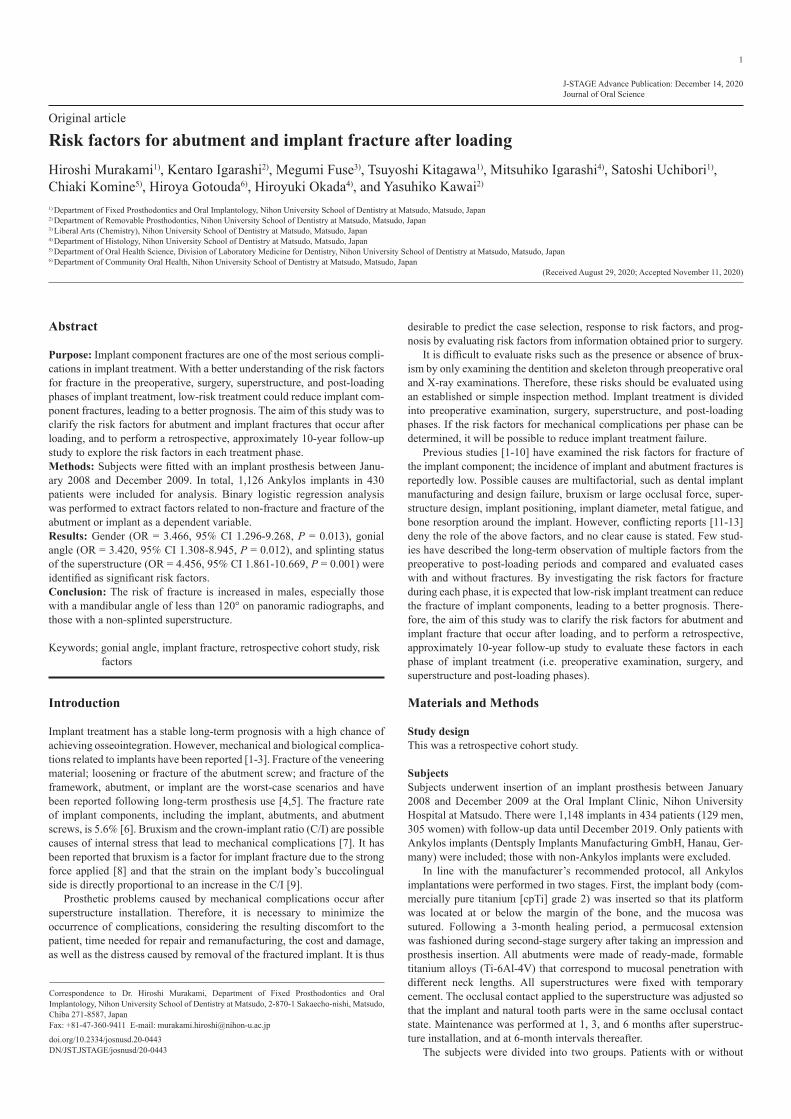
Subjects The participant flow chart is shown in Fig. 1. During the study period, 1,126 Ankylos implants from 430 patients (128 men, 302 women; mean age 55.3 ± 10.6 years) were inserted. There were 1,100 implants in 409
Fig. 1 Participant flow in this study
3
patients that did not develop abutment or implant fracture; while 26 com-ponents (18 abutments, 8 implants) in 21 patients (abutment: 16 patients, implant: 5 patients) experienced abutment or implant fracture.
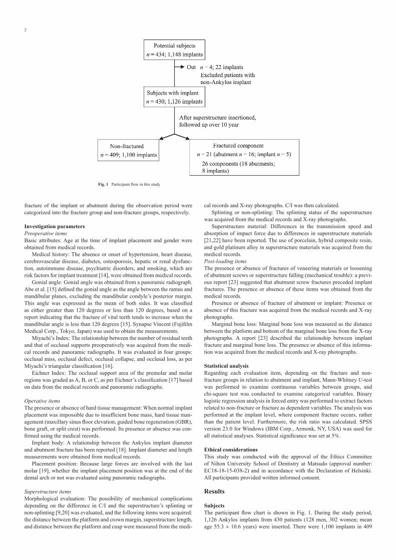
Comparison of the non-fracture and fracture groups at the implant level As shown in Table 1, the average gonial angle was 122.9 ± 7.6 degrees in the non-fracture group and 116.4 ± 6.0 degrees in the fracture group, which was significantly different (P < 0.001). The marginal bone loss was 0.7 ± 1.6 mm in the non-fracture group and 1.0 ± 1.6 mm in the fracture group, with the bone loss in the fracture group being significantly higher than that observed in the non-fracture group (P = 0.046). There were also significant differences in the gender distribution between the fracture and non-fracture groups (P < 0.001), in the proportion of individuals with a gonial angle
more or less than 120 degrees (P = 0.001), the presence of a history of hypertension (P = 0.008), renal dysfunction (P = 0.002), smoking status (P = 0.005), use of hard tissue management (P = 0.040), splinting status (P = 0.002), and the material used in the superstructure (P < 0.001).
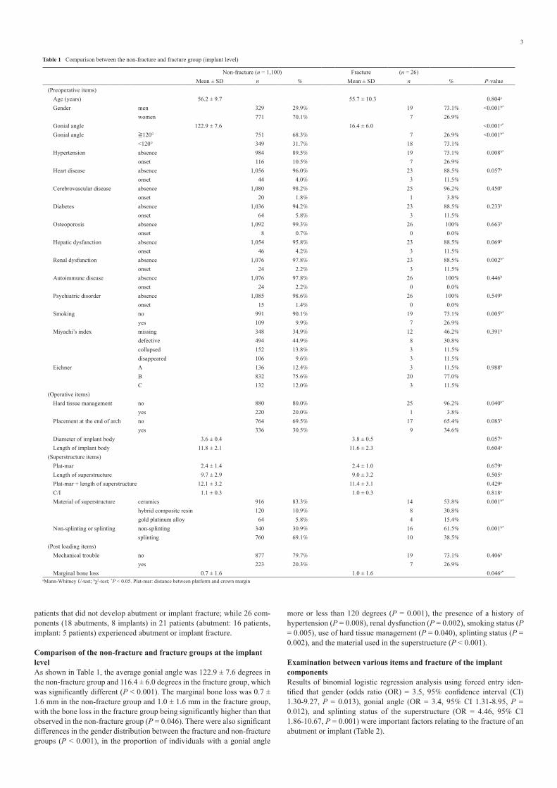
Examination between various items and fracture of the implant components Results of binomial logistic regression analysis using forced entry iden-tified that gender (odds ratio (OR) = 3.5, 95% confidence interval (CI) 1.30-9.27, P = 0.013), gonial angle (OR = 3.4, 95% CI 1.31-8.95, P = 0.012), and splinting status of the superstructure (OR = 4.46, 95% CI 1.86-10.67, P = 0.001) were important factors relating to the fracture of an abutment or implant (Table 2).
Table 1 Comparison between the non-fracture and fracture group (implant level)
Non-fracture (n = 1,100) Fracture (n = 26)Mean ± SD n % Mean ± SD n % P-value
(Preoperative items)Age (years) 56.2 ± 9.7 55.7 ± 10.3 0.804a
Gender men 329 29.9% 19 73.1% <0.001b*
women 771 70.1% 7 26.9%Gonial angle 122.9 ± 7.6 16.4 ± 6.0 <0.001a*
Gonial angle ≧120° 751 68.3% 7 26.9% <0.001b*
<120° 349 31.7% 18 73.1%Hypertension absence 984 89.5% 19 73.1% 0.008b*
onset 116 10.5% 7 26.9%Heart disease absence 1,056 96.0% 23 88.5% 0.057b
onset 44 4.0% 3 11.5%Cerebrovascular disease absence 1,080 98.2% 25 96.2% 0.450b
onset 20 1.8% 1 3.8%Diabetes absence 1,036 94.2% 23 88.5% 0.233b
onset 64 5.8% 3 11.5%Osteoporosis absence 1,092 99.3% 26 100% 0.663b
onset 8 0.7% 0 0.0%Hepatic dysfunction absence 1,054 95.8% 23 88.5% 0.069b
onset 46 4.2% 3 11.5%Renal dysfunction absence 1,076 97.8% 23 88.5% 0.002b*
onset 24 2.2% 3 11.5%Autoimmune disease absence 1,076 97.8% 26 100% 0.446b
onset 24 2.2% 0 0.0%Psychiatric disorder absence 1,085 98.6% 26 100% 0.549b
onset 15 1.4% 0 0.0%Smoking no 991 90.1% 19 73.1% 0.005b*
yes 109 9.9% 7 26.9%Miyachi’s index missing 348 34.9% 12 46.2% 0.391b
defective 494 44.9% 8 30.8%collapsed 152 13.8% 3 11.5%disappeared 106 9.6% 3 11.5%
Eichner A 136 12.4% 3 11.5% 0.988b
B 832 75.6% 20 77.0%C 132 12.0% 3 11.5%
(Operative items)Hard tissue management no 880 80.0% 25 96.2% 0.040b*
yes 220 20.0% 1 3.8%Placement at the end of arch no 764 69.5% 17 65.4% 0.083b
yes 336 30.5% 9 34.6%Diameter of implant body 3.6 ± 0.4 3.8 ± 0.5 0.057a
Length of implant body 11.8 ± 2.1 11.6 ± 2.3 0.604a
(Superstructure items)Plat-mar 2.4 ± 1.4 2.4 ± 1.0 0.679a
Length of superstructure 9.7 ± 2.9 9.0 ± 3.2 0.505a
Plat-mar + length of superstructure 12.1 ± 3.2 11.4 ± 3.1 0.429a
C/I 1.1 ± 0.3 1.0 ± 0.3 0.818a
Material of superstructure ceramics 916 83.3% 14 53.8% 0.001b*
hybrid composite resin 120 10.9% 8 30.8%gold platinum alloy 64 5.8% 4 15.4%
Non-splinting or splinting non-splinting 340 30.9% 16 61.5% 0.001b*
splinting 760 69.1% 10 38.5%(Post loading items)
Mechanical trouble no 877 79.7% 19 73.1% 0.406b
yes 223 20.3% 7 26.9%Marginal bone loss 0.7 ± 1.6 1.0 ± 1.6 0.046a*
aMann-Whitney U-test; bχ2-test; *P < 0.05. Plat-mar: distance between platform and crown margin
4
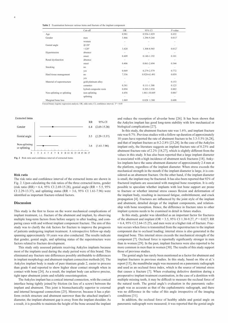
Risk ratio The risk ratio and confidence interval of the extracted items are shown in Fig. 2. Upon calculating the risk ratios of the three extracted items, gender (risk ratio (RR) = 6.4, 95% CI 2.69-15.28), gonial angle (RR = 5.5, 95% CI 2.29-13.37), and splinting status (RR = 3.6, 95% CI 1.61-7.96) were identified as important fracture-related factors.
Discussion
This study is the first to focus on the worst mechanical complications of implant treatment, i.e. fracture of the abutment and implant, by observing multiple long-term factors from before surgery to after loading, and com-paring cases with and without implant component fracture. The aim of this study was to clarify the risk factors for fracture to improve the prognosis of patients undergoing implant treatment. A retrospective follow-up study spanning approximately 10 years was also conducted. The results indicate that gender, gonial angle, and splinting status of the superstructure were factors related to fracture development.
This study only assessed patients receiving Ankylos implants because most of the implants used during the study period were of this brand. This eliminated any fracture rate differences possibly attributable to differences in implant morphology and abutment-implant connection methods [6]. The Ankylos implant body is made of pure titanium grade 2, easier to process than grade 4 and reported to show higher shear contact strength when in contact with bone [24]. As a result, the implant body can achieve precise, tight taper abutment joints and reliable osseointegration.
The Ankylos implant has a conical internal connection, with the conical interface being tightly joined by friction (in lieu of a screw) between the implant and abutment. This joint is biomechanically superior to external and internal hexagonal connections [25]. Moreover, because it has a plat-form switch structure and abutment narrower than the attached implant’s diameter, the implant-abutment gap is away from the implant shoulder. As a result, it is possible to maintain the height of the bone around the implant
and reduce the resorption of alveolar bone [26]. It has been shown that the Ankylos implant has good long-term stability with few mechanical or biological complications [27].
In this study, the abutment fracture rate was 1.6%, and implant fracture rate was 0.7%. Previous studies with a follow-up duration of approximately 10 years have reported the rate of abutment fracture to be 3.3-5.5% [6,28], and that of implant fracture as 0.2-2.8% [23,28]. In the case of the Ankylos implant only, the literature suggests an implant fracture rate of 0.23% and abutment fracture rate of 2.2% [18,27], which is slightly different from the values in this study. It has also been reported that a large implant diameter is associated with a high incidence of abutment neck fractures [18]. Anky-los implants have the same abutment diameter of approximately 2.4 mm at the platform, regardless of the implant diameter. When stress exceeds the mechanical strength in the mouth if the implant diameter is large, it is con-sidered as an abutment fracture. On the other hand, if the implant diameter is small, the implant may be fractured. It has also been reported that 92% of fractured implants are associated with marginal bone resorption. It is only possible to speculate whether implants with lost bone support are prone to fracture or whether internal stress causes flexion and deformation of the implant body, resulting in increased fatigue, embrittlement, and crack propagation [4]. Fractures are influenced by the joint style of the implant and abutment, detailed design of the implant components, and relation-ship with bone resorption. Hence, the difference in fracture rates to other implant systems needs to be examined in detail in future studies.
In this study, gender was identified as an important factor for fractures of the abutment and implant (OR = 3.5, 95% CI 1.30-9.27, P = 0.027; RR = 6.4, 95% CI 2.64-15.25), and men were at a higher risk of fracture. Frac-ture occurs when force is transmitted from the superstructure to the implant component due to occlusal loading; internal stress is also generated in the marginal bone. This internal stress exceeds the mechanical strength of the component [7]. Occlusal force is reportedly significantly stronger in men than in women [29]. In the past, implant fractures were also reported to be more common in men than in women [30]. The results of this study support those of previous studies.
The gonial angle has rarely been mentioned as a factor for abutment and implant fractures in previous studies. In this study, based on Abe et al.’s report [15], the mandibular angle was measured on a panoramic radiograph and used as an occlusal force index, which is the source of internal stress that causes a fracture [7]. When evaluating defective dentition during a preoperative implant treatment examination, in the case of a dentition with multiple missing teeth, it may be difficult to measure the occlusal force of the natural tooth. The gonial angle’s evaluation in the panoramic radio-graph was as accurate as that of the cephalometric radiograph, and there was no difference in the value of this angle irrespective of the imaging conditions [31].
In addition, the occlusal force of healthy adults and gonial angle on panoramic radiograph were measured; it was reported that the gonial angle
Table 2 Examination between various items and fracture of the implant component
Cut-off OR 95% CI P-valueAge 0.981 0.936-1.029 0.432Gender men 3.466 1.296-9.268 0.013*
women – – –Gonial angle ≧120° – – –
<120° 3.420 1.308-8.945 0.012*
Hypertension absence – – –onset 0.409 0.140-1.192 0.101
Renal dysfunction absence – – –onset 0.406 0.061-2.694 0.544
Smoking no – – –yes 0.849 0.279-2.579 0.772
Hard tissue management no 7.556 0.928-61.491 0.059yes – – –
Material of superstructure gold platinum alloy – – 0.153ceramics 0.381 0.111-1.308 0.125hybrid composite resin 0.894 0.203-3.930 0.882
Non-splinting or splinting non-splinting 4.456 1.861-10.669 0.001*
splinting – – –Marginal bone loss 1.069 0.828-1.380 0.608
Forced binary logistic regression analysis. OR, odds ratio; CI, confidence interval; *P < 0.05
Fig. 2 Risk ratio and confidence interval of extracted items
5
and maximum occlusal force were negatively correlated [29]. Bruxism is a factor that induces internal stress. Bruxers reportedly have a stronger maxi-mum occlusal force than non-bruxers and occlusal force and tooth wear are correlated [32]. However, there is no difference in maximum occlusal force between bruxers and non-bruxers [33], and the relationship between bruxism and occlusal forces is not clear. Because it is not easy to determine clinically whether a subject is a bruxer, it was exempted from inclusion as an evaluation item in this study. From these facts, it can be concluded that the gonial angle measured on a panoramic radiograph, widely used in dentistry, can be more objectively and easily applied in a clinical setting. Therefore, the gonial angle was deemed to be suitable as an occlusal force index and was thus chosen as an evaluation item.
The gonial angle was also established as an important factor (OR = 3.4, 95% CI 1.31-8.95, P = 0.019; RR = 5.5, 95% CI 2.29-13.37). The fracture group’s mean gonial angle was 116.4 degrees, smaller than that of the non-fracture group’s (122.9 degrees). It is speculated that a relatively large occlusal force was applied to the fracture group. The cut-off value was set to 120 degrees, referring to the report that fractures of vital teeth tended to increase when the gonial angle on the panoramic radiographs was less than 120 degrees [15]. Fracture risk for gonial angles less than 120 degrees was also higher than that of angles exceeding 120 degrees. The potential occlusal force was presumably exerted more via restoration of the posterior part’s occlusal support by the bone-anchored bridge than by tissue-borne dentures. Therefore, although the gonial angle is a simple index, it may be used as a preoperative risk factor of fracture. However, the occlusal force is affected not only by the gonial angle but also by the shape of the entire mandible [34] and thickness of the masseter muscles [35]. It follows that further studies need to evaluate the relationship between the manifestation of occlusal force affected by the gonial angle and abutment and implant fractures.
Splinting status of the superstructure was also extracted as an important factor for fractures of the abutment or implant (OR = 4.46, 95% CI 1.86-10.67, P = 0.001; RR = 3.6, 95% CI 1.61-7.96). Non-splinted patients were at higher risk of fractures than splinted patients. There are almost no pub-lished reports on the relationship between abutment or implant fracture and the superstructure’s splinting status. Based on past studies, splinted resto-rations were associated with decreased loosening of screws and ceramic veneer fractures [36]. These biological and mechanical complications were not related to the splinting status of the superstructure [12]. The Ankylos implant supposedly results in several fractures of the abutment neck [18], and its thin morphological characteristics make it well-adapted to sur-rounding tissues yet mechanically fragile. Therefore, there is a possibility that one can easily avoid the risk of fracture by splinting the abutment with a superstructure.
Ceramic veneer fractures and the loosening of screws also may lead to mechanical complications such as fractures [4,23]. However, these com-plications were not related to abutment or implant fractures in this study. Because the Ankylos implant is joined by friction between the abutment and implant, the abutment often remains fixed even with a loose screw. It is difficult to investigate the relationship between the looseness of the screw and fracture of the abutment or implant. Furthermore, neither slight veneer fractures or all minor fractures may be recorded in the patient’s medical record. Therefore, it is a challenge to accurately investigate the relationship between veneer fractures and implant or abutment fractures.
A higher C/I is also said to cause internal stress and mechanical compli-cations. Still, no relationship between fracture of the abutment or implant and C/I, including the superstructure length, was found (P = 0.818) in this study. There is also a report that mechanical complications are more likely to occur when the C/I increases or the superstructure becomes longer [9], but this was opposed by another report stating that a relationship between fracture and C/I cannot be confirmed [13]. C/I increases when a short implant is placed in the absorbed ridge and a long superstructure is attached, but the success rate is similar to that of a long implant [37]. These reports may have promoted the recent increase in the use of short implants.
Three findings were noted on univariate analysis. First, it showed that hypertension (P = 0.017), renal dysfunction (P = 0.022), and smoking (P = 0.005) were associated with fracture of the abutment or implant. Although these diseases are sometimes considered risk factors during implant sur-gery, there have been no reports showing their direct relationship with fracture. Similarly, no association with fracture was found in our analysis
after adjusting for related factors.Second, a relationship between the absence of hard tissue manage-
ment and fracture of the abutment or implant was also found on univariate analysis (P = 0.040). Implants at the bone grafting sites reportedly have a low risk of fracture [38]. Because several factors are interrelated, it is considered that no association was found on multivariate analysis after adjusting for confounders.
Last, marginal bone loss resorption was associated with fracture of the abutment or implant on univariate analysis (P = 0.046). Similar to this study, fractured implants reportedly have marginal bone loss [4]. How-ever, it is not clear whether the cause of the fracture is bone resorption or overload.
This study had some limitations. Given that this was a retrospective study, it was difficult to accurately investigate all risk factors involved in the fracture of abutments or implants. In addition to the survey conducted, other evaluation items are thought to be related to such fractures. In partic-ular, internal stress enhancement factors [39], such as incorrect mandibular position, occlusal plane disorder, and occlusal interference caused by poor occlusal morphology, may be important. However, it is very difficult to judge whether the mandibular position and occlusion are correct or not and whether there is an occlusal abnormality. In the statistical analysis, both the odds and risk ratios were calculated, and the relationship between the extracted factor and fracture was examined. Because the odds ratio is an approximate value, and the risk ratio was not adjusted to account for other related factors, there is a possibility that the results’ accuracy was affected. In addition, while the bias due to difference in manufacturer was elimi-nated because only Ankylos implants were investigated, the characteristics of the Ankylos implants may have influenced the results. Collecting data to identify the cause of a fracture is tedious due to the rarity of a fracture. Fur-thermore, many factors can cause a fracture, and the interaction between these factors makes it more challenging to pinpoint the causative factor. Long-term studies involving more cases are required to corroborate these results in the future. In spite of the above limitations, this study was able to investigate the factors related to fractures, the most serious complication associated with implant treatment, with an approximately 10-year follow-up period. The risk factors were evaluated in the preoperative examination and superstructure phases. This information is considered meaningful for patients who will have implant treatment, as it will help avoid complica-tions and improve the prognosis.
In conclusion, this study investigated the risk factors related to abut-ment and implant fractures in Ankylos implants during preoperative examination, surgery, superstructure, and post-loading phases. Gender, gonial angle, and splinting status of the superstructure were identified as factors related to their occurrence. This suggests that fracture risk is increased in males, especially those with a mandibular angle of less than 120° on panoramic radiographs and non-splinted superstructures.
Acknowledgments We would like to thank our colleagues for their great support in this study.
Conflict of interestThe authors declare no conflict of interest in relation to this study.
References
1. Jung RE, Pjetursson BE, Glauser R, Zembic A, Zwahlen M, Lang NP (2008) A systematic review of the 5-year survival and complication rates of implant-supported single crowns. Clin Oral Implants Res 19, 119-130.
2. Albrektsson T, Donos N, Working Group 1 (2012) Implant survival and complications. The Third EAO consensus conference 2012. Clin Oral Implants Res 23, 63-65.
3. Jung RE, Zembic A, Pjetursson BE, Zwahlen M, Thoma DS (2012) Systematic review of the survival rate and the incidence of biological, technical, and aesthetic complications of single crowns on implants reported in longitudinal studies with a mean follow-up of 5 years. Clin Oral Implants Res 23, 2-21.
4. Taylor TD (1998) Prosthodontic problems and limitations associated with osseointegration. J Prosthet Dent 79, 74-78.
5. Papaspyridakos P, Chen CJ, Chuang SK, Weber HP, Gallucci GO (2012) A systematic review of biologic and technical complications with fixed implant rehabilitations for eden-tulous patients. Int J Oral Maxillofac Implants 27, 102-110.
6. Yi Y, Koak JY, Kim SK, Lee SJ, Heo SJ (2018) Comparison of implant component frac-tures in external and internal type: a 12-year retrospective study. J Adv Prosthodont 10, 155-162.
7. Huang HM, Tsai CM, Chang CC, Lin CT, Lee SY (2005) Evaluation of loading conditions on fatigue-failed implants by fracture surface analysis. Int J Oral Maxillofac Implants 20, 854-859.
6
8. Zhou Y, Gao J, Luo L, Wang Y (2016) Does bruxism contribute to dental implant failure? A systematic review and meta-analysis. Clin Implant Dent Relat Res 18, 410-420.
9. Nissan J, Gross O, Ghelfan O, Priel I, Gross M, Chaushu G (2011) The effect of splinting implant-supported restorations on stress distribution of different crown-implant ratios and crown height spaces. J Oral Maxillofac Surg 69, 2990-2994.
10. Gealh WC, V Mazzo V, Barbi F, Camarini ET (2011) Osseointegrated implant fracture: causes and treatment. J Oral Implantol 37, 499-503.
11. Tawil G, Aboujaoude N, Younan R (2006) Influence of prosthetic parameters on the sur-vival and complication rates of short implants. Int J Oral Maxillofac Implants 21, 275-282.
12. Vigolo P, Mutinelli S, Zaccaria M, Stellini E (2015) Clinical evaluation of marginal bone level change around multiple adjacent implants restored with splinted and nonsplinted restorations: a 10-year randomized controlled trial. Int J Oral Maxillofac Implants 30, 411-418.
13. Meijer HJA, Boven C, Delli K, Raghoebar GM (2018) Is there an effect of crown-to-implant ratio on implant treatment outcomes? A systematic review. Clin Oral Implants Res 29 Suppl 18, 243-252.
14. Mombelli A, Cionca N (2006) Systemic diseases affecting osseointegration therapy. Clin Oral Implants Res 17, 97-103.
15. Abe S, Takayanagi A, Nakazawa A, Kobayashi A (2010) Risk assessment of tooth fractures from the modified gonial angles. Int J Clin Dent 3, 103-110.
16. Sato N, Ono T, Kon H, Sakurai N, Kohno S, Yoshihara A et al. (2016) Ten-year longitudinal study on the state of dentition and subjective masticatory ability in community-dwelling elderly people. J Prosthodont Res 60, 177-184.
17. Ikebe K, Matsuda K, Murai S, Maeda S, Nokubi T (2010) Validation of the eichner index in relation to occlusal force and masticatory performance. Int J Prosthodont 23, 521-524.
18. Shim HW, Yang BE (2015) Long-term cumulative survival and mechanical complications of single-tooth Ankylos implants: focus on the abutment neck fractures. J Adv Prosthodont 7, 423-430.
19. Kumagai H, Suzuki T, Hamada T, Sondang P, Fujitani M, Nikawa H (1999) Occlusal force distribution on the dental arch during various levels of clenching. J Oral Rehabil 26, 932-935.
20. Ravidà A, Saleh MHA, Muriel MC, Maska B, Wang HL (2018) Biological and techni-cal complications of splinted or nonsplinted dental implants: a decision tree for selection. Implant Dent 27, 89-94.
21. Gracis SE, Nicholls JI, Chalupnik JD, Yuodelis RA (1991) Shock-absorbing behavior of five restorative materials used on implants. Int J Prosthodont 4, 282-291.
22. Soumeire J, Dejou J (1999) Shock absorbability of various restorative materials used on implants. J Oral Rehabil 26, 394-401.
23. Balshi TJ (1996) An analysis and management of fractured implants: a clinical report. Int J Oral Maxillofac Implants 11, 660-666.
24. Kwon TK, Choi JY, Park JI, Yeo IL (2019) A clue to the existence of bonding between bone
and implant surface: an in vivo study. Materials 12, 1187.25. Merz BR, Hunenbart S, Belser UC (2000) Mechanics of the implant-abutment connection:
an 8-degree taper compared to a butt joint connection. Int J Oral Maxillofac Implants 15, 519-526.
26. Cappiello M, Luongo R, Di Iorio D, Bugea C, Cocchetto R, Celletti R (2008) Evaluation of peri-implant bone loss around platform-switched implants. Int J Periodontics Restorative Dent 28, 347-355.
27. Krebs M, Schmenger K, Neumann K, Weigl P, Moser W, Nentwig GH (2015) Long-term evaluation of ANKYLOS® dental implants, part i: 20-year life table analysis of a longitudi-nal study of more than 12,500 implants. Clin Implant Dent Relat Res 17, e275-e286.
28. Shemtov-Yona K, Rittel D (2015) An overview of the mechanical integrity of dental implants. Biomed Res Int 2015, 547384.
29. Miwa S, Wada M, Murakami S, Suganami T, Ikebe K, Maeda Y (2019) Gonial angle mea-sured by orthopantomography as a predictor of maximum occlusal force. J Prosthodont 28, e426-e430.
30. Gargallo Albiol J, Satorres-Nieto M, Puyuelo Capablo JL, Sánchez Garcés MA, Pi Urgell J, Gay Escoda C (2008) Endosseous dental implant fractures: an analysis of 21 cases. Med Oral Patol Oral Cir Bucal 13, E124-E128.
31. Mattila K, Altonen M, Haavikko K (1977) Determination of the gonial angle from the orthopantomogram. Angle Orthod 47, 107-110.
32. Dıraçoğlu D, Alptekin K, Cifter ED, Güçlü B, Karan A, Aksoy C (2011) Relationship between maximal bite force and tooth wear in bruxist and non-bruxist individuals. Arch Oral Biol 56, 1569-1575.
33. Cosme DC, Baldisserotto SM, Canabarro Sde A, Shinkai RS (2005) Bruxism and voluntary maximal bite force in young dentate adults. Int J Prosthodont 18, 328-332.
34. Sella-Tunis T, Pokhojaev A, Sarig R, O’Higgins P, May H (2018) Human mandibular shape is associated with masticatory muscle force. Sci Rep 8, 6042.
35. Bakke M, Tuxen A, Vilmann P, Jensen BR, Vilmann A, Toft M (1992) Ultrasound image of human masseter muscle related to bite force, electromyography, facial morphology, and occlusal factors. Scand J Dental Res 100, 164-171.
36. de Souza Batista VE, Verri FR, Lemos CAA, Cruz RS, Oliveira HFF, Gomes JML et al. (2019) Should the restoration of adjacent implants be splinted or nonsplinted? A systematic review and meta-analysis. J Prosthet Dent 121, 41-51.
37. Morand M, Irinakis T (2007) The challenge of implant therapy in the posterior maxilla: providing a rationale for the use of short implants. J Oral Implantol 33, 257-266.
38. Lee DW, Kim NH, Lee Y, Oh YA, Lee JH, You HK (2019) Implant fracture failure rate and potential associated risk indicators: an up to 12-year retrospective study of implants in 5,124 patients. Clin Oral Implants Res 30, 206-217.
39. Johansson A, Omar R, Carlsson GE (2011) Bruxism and prosthetic treatment: a critical review. J Prosthodont Res 55, 127-136.