Influence of implant-abutment connection structure on peri ... · The implant-abutment connection...
8
The Journal of Advanced Prosthodontics 147 Influence of implant-abutment connection structure on peri-implant bone level in a second molar: A 1-year randomized controlled trial Jin-Cheol Kim 1 , Jungwon Lee 2 , Sungtae Kim 3 , Ki-Tae Koo 3 , Hae-Young Kim 4 , In-Sung Luke Yeo 1 * 1 Department of Prosthodontics, School of Dentistry and Dental Research Institute, Seoul National University, Seoul, Republic of Korea 2 Department of Periodontology, Seoul National University Dental Hospital, Seoul, Republic of Korea 3 Department of Periodontology, Seoul National University School of Dentistry, Seoul, Republic of Korea 4 Department of Health Policy and Management, College of Health Science, Korea University, Seoul, Republic of Korea PURPOSE. This study aimed to evaluate the effect of two different implant-abutment connection structures with identical implant design on peri-implant bone level. MATERIALS AND METHODS. This clinical study was a patient-blind randomized controlled trial following the CONSORT 2010 checklists. This trial was conducted in 24 patients recruited between March 2013 and July 2015. Implants with internal friction connection were compared to those with external hex connection. One implant for each patient was installed, replacing the second molar. Implant-supported crowns were delivered at four months after implant insertion. Standardized periapical radiographs were taken at prosthesis delivery (baseline), and one year after delivery. On the radiographs, distance from implant shoulder to first bone-to-implant contact (DIB) and peri-implant area were measured, which were the primary and secondary outcome, respectively. RESULTS. Eleven external and eleven internal implants were analyzed. Mean changes of DIB from baseline to 1-year postloading were 0.59 (0.95) mm for the external and 0.01 (0.68) mm for the internal connection. Although no significant differences were found between the two groups, medium effect size was found in DIB between the connections (Cohen’s d = 0.67). CONCLUSION. Considering the effect size in DIB, this study suggested the possibility of the internal friction connection structure for more effective preservation of marginal bone. [ J Adv Prosthodont 2019;11:147-54] KEYWORDS: Randomized controlled trial; Dental implant-abutment design; Alveolar bone loss https://doi.org/10.4047/jap.2019.11.3.147 https://jap.or.kr J Adv Prosthodont 2019;11:147-54 INTRODUCTION The stability of hard and soft tissues around dental implants is one of the most decisive factors for long-term implant prognosis. 1,2 Marginal bone loss is a major factor in implant success or failure. 3 Peri-implant infection plays a role in the marginal bone resorption around a dental implant. 4 A previous study has suggested several factors that cause the marginal bone loss, including surgical trauma, reforma- tion of biologic width, implant-abutment connection struc- ture, history of periodontitis, and occlusal overloading. 5,6 Some studies have assessed the effects of implant-abutment connection structure on the marginal bone level change. 7-10 The implant-abutment connection structure is an important etiologic factor for peri-implant bone remodeling and crestal bone loss, as the highest number of inflammatory cells is infiltrated and the bacteria causing periodontitis are colo- nized at the microgap of implant-abutment connection. 11,12 Biomechanical bone responses depending on implant- abutment connection structure are also considered to affect marginal bone level at peri-implant sites. Compared to the external hex connection, the internal friction connection structure has been shown to be mechanically more stable and advantageous in force distribution. 13,14 Some studies Corresponding author: In-Sung Luke Yeo Department of Prosthodontics, School of Dentistry and Dental Research Institute, Seoul National University, 101 Daehak-ro, Jongno-gu, Seoul 03080, Republic of Korea Tel. +82220722661: e-mail, [email protected] Received July 12, 2018 / Last Revision April 30, 2019 / Accepted June 11, 2019 © 2019 The Korean Academy of Prosthodontics This is an Open Access article distributed under the terms of the Creative Commons Attribution Non-Commercial License (http://creativecommons. org/licenses/by-nc/4.0) which permits unrestricted non-commercial use, distribution, and reproduction in any medium, provided the original work is properly cited. The authors report grants and non-financial support from Shinhung Corporation, Seoul, Korea (Grant No.: NCR13002). This work was also supported by the Mid-Career Researcher Program through a grant from the National Research Foundation of Korea, funded by the Korean government (MSIP) (2016R1A2B4014330).
Transcript of Influence of implant-abutment connection structure on peri ... · The implant-abutment connection...
The Journal of Advanced Prosthodontics 147
Influence of implant-abutment connection structure on peri-implant bone level in a second molar: A 1-year randomized controlled trial
Jin-Cheol Kim1, Jungwon Lee2, Sungtae Kim3, Ki-Tae Koo3, Hae-Young Kim4, In-Sung Luke Yeo1*1Department of Prosthodontics, School of Dentistry and Dental Research Institute, Seoul National University, Seoul, Republic of Korea2Department of Periodontology, Seoul National University Dental Hospital, Seoul, Republic of Korea3Department of Periodontology, Seoul National University School of Dentistry, Seoul, Republic of Korea4Department of Health Policy and Management, College of Health Science, Korea University, Seoul, Republic of Korea
PURPOSE. This study aimed to evaluate the effect of two different implant-abutment connection structures with identical implant design on peri-implant bone level. MATERIALS AND METHODS. This clinical study was a patient-blind randomized controlled trial following the CONSORT 2010 checklists. This trial was conducted in 24 patients recruited between March 2013 and July 2015. Implants with internal friction connection were compared to those with external hex connection. One implant for each patient was installed, replacing the second molar. Implant-supported crowns were delivered at four months after implant insertion. Standardized periapical radiographs were taken at prosthesis delivery (baseline), and one year after delivery. On the radiographs, distance from implant shoulder to first bone-to-implant contact (DIB) and peri-implant area were measured, which were the primary and secondary outcome, respectively. RESULTS. Eleven external and eleven internal implants were analyzed. Mean changes of DIB from baseline to 1-year postloading were 0.59 (0.95) mm for the external and 0.01 (0.68) mm for the internal connection. Although no significant differences were found between the two groups, medium effect size was found in DIB between the connections (Cohen’s d = 0.67). CONCLUSION. Considering the effect size in DIB, this study suggested the possibility of the internal friction connection structure for more effective preservation of marginal bone. [ J Adv Prosthodont 2019;11:147-54]
KEYWORDS: Randomized controlled trial; Dental implant-abutment design; Alveolar bone loss
https://doi.org/10.4047/jap.2019.11.3.147https://jap.or.kr J Adv Prosthodont 2019;11:147-54
INTRODUCTION
The stability of hard and soft tissues around dental implants is one of the most decisive factors for long-term implant
prognosis.1,2 Marginal bone loss is a major factor in implant success or failure.3 Peri-implant infection plays a role in the marginal bone resorption around a dental implant.4
A previous study has suggested several factors that cause the marginal bone loss, including surgical trauma, reforma-tion of biologic width, implant-abutment connection struc-ture, history of periodontitis, and occlusal overloading.5,6 Some studies have assessed the effects of implant-abutment connection structure on the marginal bone level change.7-10 The implant-abutment connection structure is an important etiologic factor for peri-implant bone remodeling and crestal bone loss, as the highest number of inflammatory cells is infiltrated and the bacteria causing periodontitis are colo-nized at the microgap of implant-abutment connection.11,12
Biomechanical bone responses depending on implant-abutment connection structure are also considered to affect marginal bone level at peri-implant sites. Compared to the external hex connection, the internal friction connection structure has been shown to be mechanically more stable and advantageous in force distribution.13,14 Some studies
Corresponding author: In-Sung Luke YeoDepartment of Prosthodontics, School of Dentistry and Dental Research Institute, Seoul National University, 101 Daehak-ro, Jongno-gu, Seoul 03080, Republic of KoreaTel. +82220722661: e-mail, [email protected] July 12, 2018 / Last Revision April 30, 2019 / Accepted June 11, 2019
© 2019 The Korean Academy of ProsthodonticsThis is an Open Access article distributed under the terms of the Creative Commons Attribution Non-Commercial License (http://creativecommons.org/licenses/by-nc/4.0) which permits unrestricted non-commercial use, distribution, and reproduction in any medium, provided the original work is properly cited.
The authors report grants and non-financial support from Shinhung Corporation, Seoul, Korea (Grant No.: NCR13002). This work was also supported by the Mid-Career Researcher Program through a grant from the National Research Foundation of Korea, funded by the Korean government (MSIP) (2016R1A2B4014330).
148
have reported high stress and marginal bone loss around the external hex connection structure compared with the inter-nal friction.15-17 In contrast, other studies have reported that marginal bone loss between two different connection struc-tures is not statistically significant.18,19 Inconsistent results might stem from uncontrolled confounding factors of implant design, such as microthread, platform switching, and surface texture.
An occlusal overload could affect peri-implant marginal bone loss.20 Because of the biomechanics of the lever sys-tem of the mandible and jaw elevator muscles, the occlusal force is greater on the posterior than on the anterior region; hence, relative distribution of occlusal bite force in the pos-terior region is higher than that in the anterior region.21 Therefore, it is necessary to limit implant sites to exclude the difference of occlusal force.
A direct comparison between two different implant-abutment connection structures (external hex and internal friction connections) with identical implant design in the posterior region has been rare, especially in the same poste-rior region.22,23 The current study aims to evaluate the effect of implant-abutment connection structure with identical
implant design in the second molar region on the peri-implant bone level.
MATERIALS AND METHODS
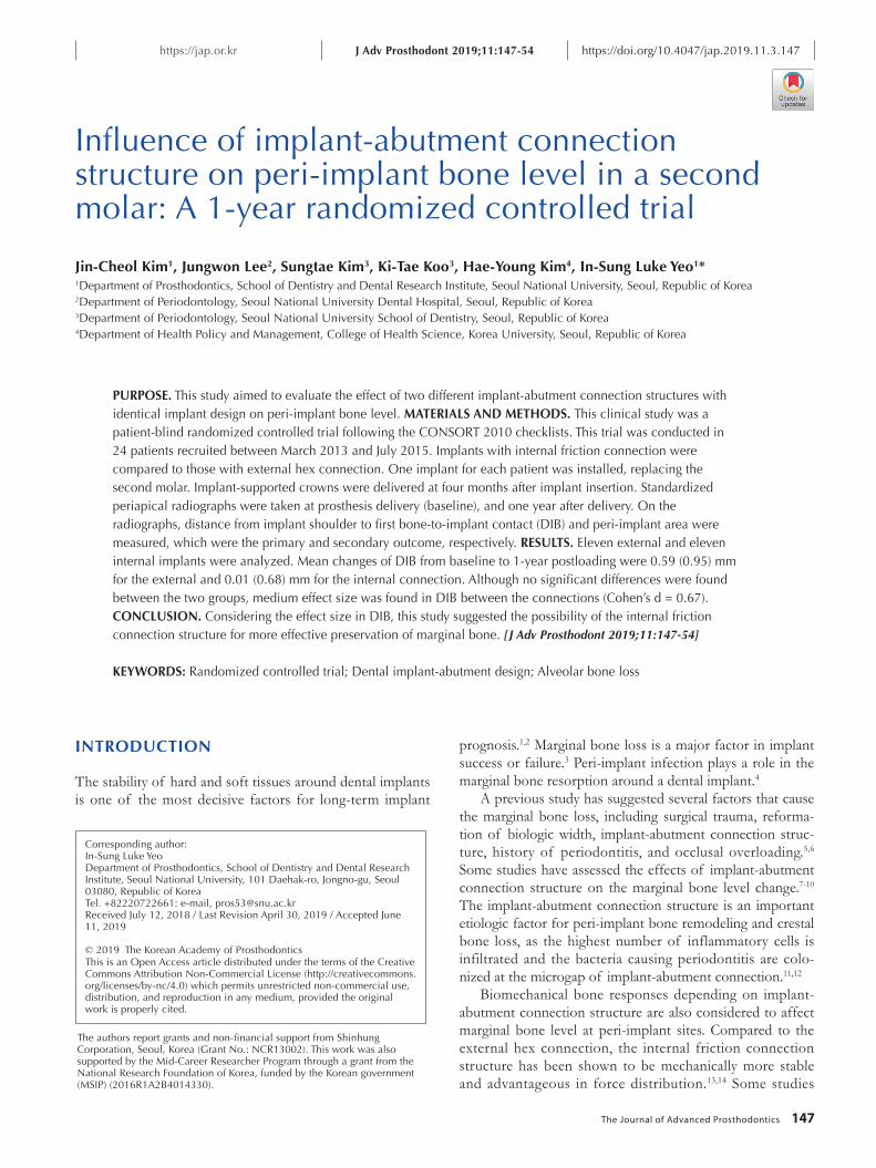
This single-blind (patient-blind), randomized, parallel, con-trolled clinical trial was performed according to the princi-ples of the Declaration of Helsinki and was approved by the Institutional Review Board (IRB #CMP13001), Seoul National University Dental Hospital, Seoul, Korea. The CONSORT 2010 checklists for clinical trials were followed.24 Two different connection structures of the implants were compared: external hex connection structure (the control group) and internal friction connection structure (the test group). The study was performed between March 2013 and July 2015 at at Seoul National University Dental Hospital, Korea. The flowchart of this study is presented in Fig. 1.
Patients who met all of the following criteria were eligi-ble for inclusion.
1) Patients aged 20 - 66 years who could undergo surgi-cal treatment
2) Patients who needed to restore a single second molar
Fig. 1. Flow chart of the study. Twelve subjects were allocated to the control and test groups each. Two patients did not receive allocated interventions. One participant in the control group changed decision and one in the test group did not return to the treatment program. In total, 11 subjects in the control group and 11 in the test group received treatment and were analyzed.
J Adv Prosthodont 2019;11:147-54
The Journal of Advanced Prosthodontics 149
due to the tooth loss3) Patients with sufficient healing time of at least 3
months following tooth extraction4) Patients who agreed to take the test and sign the
informed consent form5) Patients with at least 9 mm width and 9 mm height of
alveolar bone in cone beam computed tomography analysis.
The exclusion criteria were as follows:1) Untreated periodontal disease2) Acute abscess with pain3) Heavy smokers (>10 cigarettes per day)4) Parafunctional habit (bruxism, clenching)5) General contraindications to surgery6) Participation in other clinical trials that may interfere
with the present protocolRandomization sequence was created using the random-
ization program on http://www.randomization.com by KTK (Ki-Tae Koo) who didn’t perform the treatment. In total, 24 subjects were randomized into six blocks. The allo-cation was concealed by sealing it in an opaque envelope, and the envelope was opened immediately after the final drilling procedure performed at implant surgery.
Patients were recruited and treated by two different peri-odontists (JL and SK) and one prosthodontist (ISLY). The treatment was performed using a standardized protocol, and
the surgery was conducted by two periodontists on 24 patients (12 in each group). No information was provided to the patients, of which connection structure was received. Detailed explanations were given to all recruited patients and a written informed consent form was obtained before enrolling in the clinical trial.
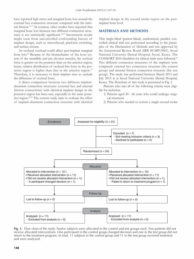
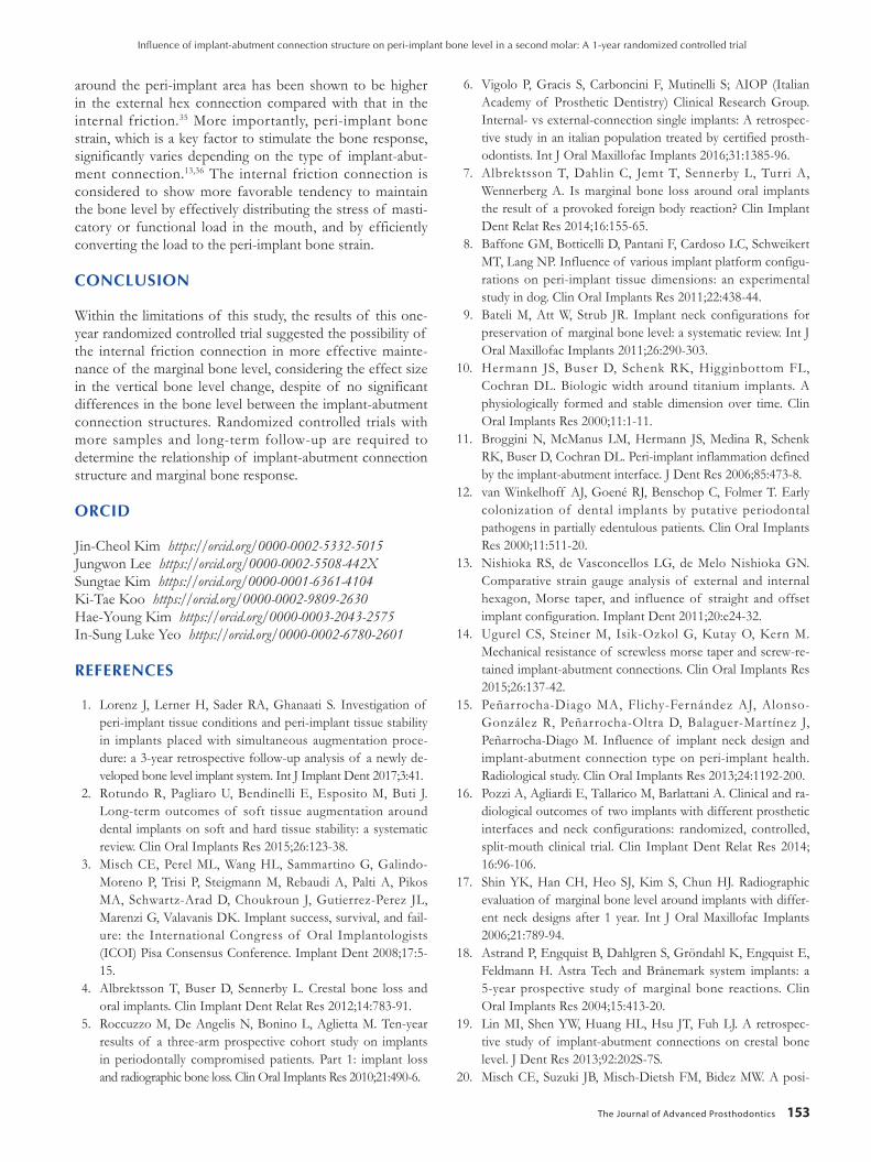
After local anesthesia using 2% lidocaine solution with epinephrine 1:100,000 (Huons, Seongnam, Gyeonggi, Korea), a flap was reflected and dental implants (diameter 5.0 mm; length 8.5, 10, or 11.5 mm; Shinhung, Seoul, Korea) were placed at the buccal bone crest level at maxillary or mandibular second molar region according to the manufac-turer’s recommendation using a non-submerged protocol, and a healing abutment was immediately connected. At implant placement, patients were allocated to either control (external hex connection type; Sola, Shinhung, Seoul, Korea) or test group (internal friction connection type; Luna, Shinhung, Seoul, Korea). The same implant drilling proce-dure was applied to each group because the implant designs were identical except the implant-abutment connection structures (Fig. 2). No bone augmentation procedure was conducted around the implant placement site. Instructions not to brush the surgical area and to rinse with 0.1% chlorhexidine (Bukwang Pharmaceutical Co., Seoul, Korea) until suture removal were given to the patients, and the suture was removed 7 - 10 days after surgery. Antibiotics
Fig. 2. Two implants with different implant-abutment connection structures were used in this study. The control group was an external connection structure (A), and the test group was an internal connection structure (B). Two implants have an identical design such as thread geometry, implant body profile, and surface topography (middle) with the exception of implant-abutment connection structure. The implant thread pitch is 0.8 mm, the thread depth is 0.3 - 0.45 mm, and the inclination angle of thread flank is 35°. Implant surface was blasted by resorbable blast media, and its arithmetic mean height (Ra) was 1.50 - 2.00 μm.
A B
Influence of implant-abutment connection structure on peri-implant bone level in a second molar: A 1-year randomized controlled trial
150
(Augmentin 625 mg) and analgesics (acetaminophen 650 mg) were also prescribed every 8 hour for 5 days. The pros-thetic procedure was started 3 months following implant surgery. Definitive impressions were taken at the implant-level using polyvinyl siloxane (Examix, GC Korea, Seoul, Korea) and transfer impression copings (Shinhung). Customized gold or titanium abutments were made on the master casts in the dental laboratory and metal ceramic gold crowns with the abutment screw access holes were also fab-ricated. The customized abutments were seated on the implants in the patients’ mouths and the abutment screws were tightened to 35 Ncm torque. The definitive screw- and cement-retained crowns with the screw access holes were cemented to the abutments by resin-modified glass ionomer (Fujicem, GC Corp., Tokyo, Japan) and the holes were sealed. These implant-supported restorations were delivered to the patients at 4 months after surgery, having occlusal contacts at the centric positions and no contacts at the eccentric.
A standardized periapical radiograph (Kodak Ektaspeed Plus film, 1512 × 1134 pixels, 40 × 30 mm, 256 grayscale, Eastman Kodak Co., Rochester, NY, USA) was taken using the paralleling technique (60 kV, 10 mA, 0.250 s) with RINN XCP positioners (Dentsply Sirona, York, PA, USA) at prosthesis delivery (baseline, 4 months after implant placement) and 1 year postloading. This radiographic series for each recruited patient was standardized with the individ-ualized polyvinyl siloxane positioning template for radiogra-
phy (Blu-Mousse, Parkell, Edgewood, NY, USA), which had been described in a previous study.22 Radiographic images were stored at 300 dpi in tiff format (Infinitt PACS, Infinitt, Seoul, Korea).
An examiner (JCK) conducted repeated measurements of the radiographic parameters using an image analysis pro-gram (ImageJ 1.60, NIH, Bethesda, MD, USA). Radiographs were assessed on a 24-inch liquid crystal display monitor (Samsung, Seoul, Korea) under standardized conditions (ISO 12646:2015). Parameters in the radiograph were cali-brated with the known width and length of the implants. After training 10 samples, high intraexaminer reliability was achieved. The intraclass correlation coefficients for the radiographic parameters were 0.918 and 0.924, respectively.
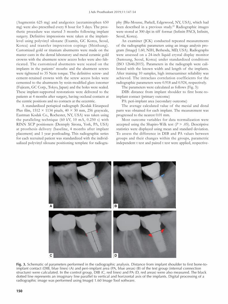
The parameters were calculated as follows (Fig. 3):DIB: distance from implant shoulder to first bone-to-
implant contact (primary outcome)PA: peri-implant area (secondary outcome)The average calculated value of the mesial and distal
parts was obtained for each implant. The measurement was progressed to the nearest 0.01 mm.
Most outcome variables for data normalization were accepted using the Shapiro-Wilk test (P > .05). Descriptive statistics were displayed using mean and standard deviation. To assess the difference in DIB and PA values between groups and their changes within the groups, parametric independent t test and paired t test were applied, respective-
Fig. 3. Schematic of parameters performed in the radiographic analysis. Distance from implant shoulder to first bone-to-implant contact (DIB, blue lines) (A) and peri-implant area (PA, blue areas) (B) of the test group (internal connection structure) were calculated. In the control group, DIB (C, red lines) and PA (D, red areas) were also measured. The black dotted line represents an imaginary line parallel to vertical and horizontal axis of the implants. Digital processing of a radiographic image was performed using ImageJ 1.60 Image Tool software.
A B
C D
J Adv Prosthodont 2019;11:147-54
The Journal of Advanced Prosthodontics 151
ly. An effect size of Cohen’s d was calculated to assess the actual difference in changes of DIB and PA as following formula:
The interpretation of Cohen’s d is as follows: 0.2 as small, 0.5 as medium, and 0.8 as large effect size.25 The sta-tistical software IBM SPSS Statistics Version 22 (IBM Corp., Armonk, NY, USA) was used for the analysis. Statistical sig-nificance was set at P < .05.
RESULTS
A total of 24 subjects were recruited and 22 received treat-ment. One patient in the control group did not receive the allocated intervention due to the participant’s decision to change, and one patient in the test group did not return to the treatment program. The antagonistic maxillary second molars were all natural teeth. Eleven implants in 11 patients were assigned to control group (the external connection) and test group (the internal connection). Patient characteris-tics related to sex, age, smoking, systemic disease, history of periodontitis, reason for extraction, presence of the adja-cent 3rd molar, implant length (data not shown), gingival thickness, and bone quality are presented in Table 1. No sta-tistically significant differences were found between the two groups.
Insertion torque was between 25 and 40 Ncm in all implants. No remarkable complications were reported through-out the study. Implants success rate was 100%, according to the criteria proposed by the International Congress of Oral Implantologists Consensus Conference.3 Prosthetic screw loos-ening was observed once in one subject and two times in one subject (two patients were in the control group). All loose screws were replaced according to the manufacturer’s recommendation.
The means of DIB and PA at implant prosthesis deliv-ery (baseline) and at 1 year postloading are shown in Table 2. There were no statistically significant differences between the control and test groups at baseline. The mean changes of DIB from baseline to 1 year were 0.59 mm (95% confi-dence interval, -0.05 - 1.23 mm) and 0.04 mm (-0.45 - 0.47 mm) for external and internal connection structure, respec-tively. No significance was found in the DIB change between the two groups (P = 0.116). However, the effect size of average change between two groups was observed to be medium (Cohen’s d = 0.67). Average changes in PA between baseline and 1 year postloading were 0.10 mm (-0.21 - 0.41 mm) and 0.09 mm (-0.25 - 0.43) in the control and test groups, respectively, and there was no significant difference between the two groups (P = 0.923). The effect size of average change between two group in PA was small (Cohen’s d = 0.02).
Table 1. Description of patient distribution recruited in this study
Treatment group
Control(n = 11)
Test(n = 11)
Total(N = 22)
Significance
Sex
Male 9 4 130.080
Female 2 7 9
Age
Under 45 years 5 4 91.000
≥ 45 years old 6 7 13
Smoking
Non-smoker 7 10 17
0.214 Former smoker 1 1 2
Mild smoker 3 0 3
Systemic disease
Hypertension 2 2 4
1.000 Diabetes mellitus 1 0 1
None 8 9 17
History of periodontitis
Yes 5 2 70.361
No 6 9 15
Reason for extraction
Dental caries 1 2 3
0.678 Endodontic failure* 3 2 5
Periodontitis† 4 6 9
Root fracture/crack 3 1 4
The 3rd molar
Absence 8 11 190.534
Presence 3 0 3
Gingival width
< 3 mm 4 5 91.000
≥ 3 mm 7 6 13
Bone quality‡
1 0 0 0
0.230 2 6 5 11
3 3 6 9
4 2 0 2
* Endodontic failure is defined here as recurrence of signs or symptoms like periapical lesion and pain after endodontic therapy is finished.† Periodontitis is defined here as bone resorption over 70% of the length of the root with the 3rd degree of tooth mobility.‡ Bone quality was assessed at implant surgery according to the classification suggested by Lekholm and Zarb (1985).
Influence of implant-abutment connection structure on peri-implant bone level in a second molar: A 1-year randomized controlled trial
152
DISCUSSION
The present clinical study aimed to evaluate the crestal bone response to implant-abutment connection structures. To date, no published randomized controlled trial has evaluated the effect of implant-abutment connection on single implant-supported crowns replacing only the missing sec-ond molar. Previous studies that have estimated marginal bone level change have focused on diverse factors, especial-ly with respect to implant location, implant-abutment junc-tion, surgical approach (submerged or non-submerged), implant surface, presence of adjacent tooth, and history of periodontitis.5,15,16 However, to preclude aforementioned factors as a variable, the present study had to adhere to strict inclusion criteria (with the only difference being the implant-abutment connection) to have same implant thread design and texture, second molar position, non-submerged protocol, implant diameter (5 mm), and length falling within the ranges of 8.5 - 11.5 mm. Fortunately, in our recruited subjects, there were no differences between two groups for extraction option (P = 0.678) and history of periodontitis (P = 0.361). Therefore, the authors of this study considered the effect of localized and/or generalized periodontitis was minimized enough to compare test and control group.
In the present study, the DIB of the external hex con-nection tended to increase at 1 year postloading despite of no significance between the baseline and the 1 year post-loading due to the small sample size. In this study, we calcu-lated the effect size of Cohen’s d. It was interpreted as small, medium, and large corresponding to values of 0.2, 0.5, and 0.8, respectively.25 Although the differences could not be confirmed in PA and DIB in terms of P-value, the effect size in DIB was observed to be medium. In this point of view, internal connection type might be favorable to peri-implant bone response compared to external hex type. However, we could not observe any differences between
control and test group in PA. Initial marginal bone loss is considered to progress linearly to the apical direction and then expand to peri-implant area. As a result, one-dimen-sional parameter, DIB seems to show a medium effect size (Cohen’s d = 0.67), whereas PA, two-dimensional parame-ter, exhibits small effect size (Cohen’s d = 0.02). However, this result should be accepted carefully and further clinical long-term studies with larger sample size are needed to elu-cidate the effect of implant-abutment connection type.
Although there are some limits to making direct compar-isons, similar studies have shown that internal connection structures exhibited lower values of marginal bone loss with no statistical difference between the connection types.19,26,27 Some other studies have shown that internal connection structures exhibited lower levels of marginal bone loss, showing a statistical difference compared with external con-nection structures although no significant difference was found in implant survival.28-32 Those authors concluded that the platform switching concept was largely responsible for marginal bone loss. This concept is based on the research that a bacterial contamination of the implant-abutment interface appears to provoke the inflammatory response. Preventing microbial leakage at the implant-abutment junc-tion has been reported to be a major challenge to minimize inflammatory reactions and to maintain the bone crest level at the junction.33 However, there has been a study demon-strating that no difference in bacterial infiltration (Escherichia coli and Streptococcus sanguis) was found between the implant-abutment connection structures.34
The different biomechanics of implant-abutment con-nection structures can explain the different tendency in crestal bone responses to implant-supported restorations. The connection type exerts a significant influence on the stress distribution in bone because of the different load transfer mechanisms and differences in the spread of the contact area between the abutment and implant.30,31 Stress
Table 2. Comparative mean (95% confidence interval) and changes in distance from implant shoulder to first bone-to-implant contact (DIB) and peri-implant area (PA) according to different implant connections
External(n = 11)
Internal(n = 11)
P valuea Effect sizec
DIB at baseline -0.06 (-0.62 - 0.50) 0.21 (-0.45 - 0.87) .813
DIB at 1 year loading 0.53 (-0.23 - 1.29) 0.26 (-0.21 - 0.74) .105
△DIB (baseline–1 year postloading) 0.59 (-0.05 - 1.23) 0.04 (-0.45 - 0.47) .116 0.67
P valueb .067 .837
PA at baseline 0.34 (-0.12 - 0.80) 0.31 (-0.13 - 0.75) .917
PA at 1 year loading 0.44 (-0.22 - 1.10) 0.40 (-0.02 - 0.82) .198
△PA (baseline–1 year postloading) 0.10 (-0.21 - 0.41) 0.09 (-0.25 - 0.43) .923 0.02
P valueb .495 .566
a P value by independent samples t test.b P value by related samples paired t test.c Cohen’s d was used as the effect size.
J Adv Prosthodont 2019;11:147-54
The Journal of Advanced Prosthodontics 153
around the peri-implant area has been shown to be higher in the external hex connection compared with that in the internal friction.35 More importantly, peri-implant bone strain, which is a key factor to stimulate the bone response, significantly varies depending on the type of implant-abut-ment connection.13,36 The internal friction connection is considered to show more favorable tendency to maintain the bone level by effectively distributing the stress of masti-catory or functional load in the mouth, and by efficiently converting the load to the peri-implant bone strain.
CONCLUSION
Within the limitations of this study, the results of this one-year randomized controlled trial suggested the possibility of the internal friction connection in more effective mainte-nance of the marginal bone level, considering the effect size in the vertical bone level change, despite of no significant differences in the bone level between the implant-abutment connection structures. Randomized controlled trials with more samples and long-term follow-up are required to determine the relationship of implant-abutment connection structure and marginal bone response.
ORCID
Jin-Cheol Kim https://orcid.org/0000-0002-5332-5015Jungwon Lee https://orcid.org/0000-0002-5508-442XSungtae Kim https://orcid.org/0000-0001-6361-4104Ki-Tae Koo https://orcid.org/0000-0002-9809-2630Hae-Young Kim https://orcid.org/0000-0003-2043-2575In-Sung Luke Yeo https://orcid.org/0000-0002-6780-2601
REFERENCES
1. Lorenz J, Lerner H, Sader RA, Ghanaati S. Investigation of peri-implant tissue conditions and peri-implant tissue stability in implants placed with simultaneous augmentation proce-dure: a 3-year retrospective follow-up analysis of a newly de-veloped bone level implant system. Int J Implant Dent 2017;3:41.
2. Rotundo R, Pagliaro U, Bendinelli E, Esposito M, Buti J. Long-term outcomes of soft tissue augmentation around dental implants on soft and hard tissue stability: a systematic review. Clin Oral Implants Res 2015;26:123-38.
3. Misch CE, Perel ML, Wang HL, Sammartino G, Galindo-Moreno P, Trisi P, Steigmann M, Rebaudi A, Palti A, Pikos MA, Schwartz-Arad D, Choukroun J, Gutierrez-Perez JL, Marenzi G, Valavanis DK. Implant success, survival, and fail-ure: the International Congress of Oral Implantologists (ICOI) Pisa Consensus Conference. Implant Dent 2008;17:5-15.
4. Albrektsson T, Buser D, Sennerby L. Crestal bone loss and oral implants. Clin Implant Dent Relat Res 2012;14:783-91.
5. Roccuzzo M, De Angelis N, Bonino L, Aglietta M. Ten-year results of a three-arm prospective cohort study on implants in periodontally compromised patients. Part 1: implant loss and radiographic bone loss. Clin Oral Implants Res 2010;21:490-6.
6. Vigolo P, Gracis S, Carboncini F, Mutinelli S; AIOP (Italian Academy of Prosthetic Dentistry) Clinical Research Group. Internal- vs external-connection single implants: A retrospec-tive study in an italian population treated by certified prosth-odontists. Int J Oral Maxillofac Implants 2016;31:1385-96.
7. Albrektsson T, Dahlin C, Jemt T, Sennerby L, Turri A, Wennerberg A. Is marginal bone loss around oral implants the result of a provoked foreign body reaction? Clin Implant Dent Relat Res 2014;16:155-65.
8. Baffone GM, Botticelli D, Pantani F, Cardoso LC, Schweikert MT, Lang NP. Influence of various implant platform configu-rations on peri-implant tissue dimensions: an experimental study in dog. Clin Oral Implants Res 2011;22:438-44.
9. Bateli M, Att W, Strub JR. Implant neck configurations for preservation of marginal bone level: a systematic review. Int J Oral Maxillofac Implants 2011;26:290-303.
10. Hermann JS, Buser D, Schenk RK, Higginbottom FL, Cochran DL. Biologic width around titanium implants. A physiologically formed and stable dimension over time. Clin Oral Implants Res 2000;11:1-11.
11. Broggini N, McManus LM, Hermann JS, Medina R, Schenk RK, Buser D, Cochran DL. Peri-implant inflammation defined by the implant-abutment interface. J Dent Res 2006;85:473-8.
12. van Winkelhoff AJ, Goené RJ, Benschop C, Folmer T. Early colonization of dental implants by putative periodontal pathogens in partially edentulous patients. Clin Oral Implants Res 2000;11:511-20.
13. Nishioka RS, de Vasconcellos LG, de Melo Nishioka GN. Comparative strain gauge analysis of external and internal hexagon, Morse taper, and influence of straight and offset implant configuration. Implant Dent 2011;20:e24-32.
14. Ugurel CS, Steiner M, Isik-Ozkol G, Kutay O, Kern M. Mechanical resistance of screwless morse taper and screw-re-tained implant-abutment connections. Clin Oral Implants Res 2015;26:137-42.
15. Peñarrocha-Diago MA, Flichy-Fernández AJ, Alonso-González R, Peñarrocha-Oltra D, Balaguer-Martínez J, Peñarrocha-Diago M. Influence of implant neck design and implant-abutment connection type on peri-implant health. Radiological study. Clin Oral Implants Res 2013;24:1192-200.
16. Pozzi A, Agliardi E, Tallarico M, Barlattani A. Clinical and ra-diological outcomes of two implants with different prosthetic interfaces and neck configurations: randomized, controlled, split-mouth clinical trial. Clin Implant Dent Relat Res 2014; 16:96-106.
17. Shin YK, Han CH, Heo SJ, Kim S, Chun HJ. Radiographic evaluation of marginal bone level around implants with differ-ent neck designs after 1 year. Int J Oral Maxillofac Implants 2006;21:789-94.
18. Astrand P, Engquist B, Dahlgren S, Gröndahl K, Engquist E, Feldmann H. Astra Tech and Brånemark system implants: a 5-year prospective study of marginal bone reactions. Clin Oral Implants Res 2004;15:413-20.
19. Lin MI, Shen YW, Huang HL, Hsu JT, Fuh LJ. A retrospec-tive study of implant-abutment connections on crestal bone level. J Dent Res 2013;92:202S-7S.
20. Misch CE, Suzuki JB, Misch-Dietsh FM, Bidez MW. A posi-
Influence of implant-abutment connection structure on peri-implant bone level in a second molar: A 1-year randomized controlled trial
154
tive correlation between occlusal trauma and peri-implant bone loss: literature support. Implant Dent 2005;14:108-16.
21. Shinogaya T, Bakke M, Thomsen CE, Vilmann A, Matsumoto M. Bite force and occlusal load in healthy young subjects-a meth-odological study. Eur J Prosthodont Restor Dent 2000;8:11-5.
22. Koo KT, Lee EJ, Kim JY, Seol YJ, Han JS, Kim TI, Lee YM, Ku Y, Wikesjö UM, Rhyu IC. The effect of internal versus external abutment connection modes on crestal bone changes around dental implants: a radiographic analysis. J Periodontol 2012;83:1104-9.
23. Canullo L, Rosa JC, Pinto VS, Francischone CE, Götz W. Inward-inclined implant platform for the amplified platform-switching concept: 18-month follow-up report of a prospec-tive randomized matched-pair controlled trial. Int J Oral Maxillofac Implants 2012;27:927-34.
24. Schulz KF, Altman DG, Moher D; CONSORT Group. CONSORT 2010 statement: updated guidelines for reporting parallel group randomised trials. BMJ 2010;340:c332.
25. Cohen J. The t test for means. In: Cohen J, ed. Statistical power analysis for the behavioral sciences. 2nd ed. New York; Lawrence Erlbaum Associates; 1988. p. 20-7.
26. Crespi R, Capparè P, Gherlone E. Radiographic evaluation of marginal bone levels around platform-switched and non-plat-form-switched implants used in an immediate loading proto-col. Int J Oral Maxillofac Implants 2009;24:920-6.
27. Esposito M, Maghaireh H, Pistilli R, Grusovin MG, Lee ST, Trullenque-Eriksson A, Gualini F. Dental implants with inter-nal versus external connections: 5-year post-loading results from a pragmatic multicenter randomised controlled trial. Eur J Oral Implantol 2016;9:129-41.
28. De Angelis N, Nevins ML, Camelo MC, Ono Y, Campailla M, Benedicenti S. Platform switching versus conventional tech-nique: a randomized controlled clinical trial. Int J Periodontics Restorative Dent 2014;34:s75-9.
29. Pozzi A, Tallarico M, Moy PK. Three-year post-loading re-sults of a randomised, controlled, split-mouth trial comparing implants with different prosthetic interfaces and design in partially posterior edentulous mandibles. Eur J Oral Implantol 2014;7:47-61.
30. Caricasulo R, Malchiodi L, Ghensi P, Fantozzi G, Cucchi A. The influence of implant-abutment connection to peri-im-plant bone loss: A systematic review and meta-analysis. Clin Implant Dent Relat Res 2018;20:653-64.
31. Lemos CAA, Verri FR, Bonfante EA, Santiago Júnior JF, Pellizzer EP. Comparison of external and internal implant-abutment connections for implant supported prostheses. A systematic review and meta-analysis. J Dent 2018;70:14-22.
32. Palacios-Garzón N, Mauri-Obradors E, Roselló-LLabrés X, Estrugo-Devesa A, Jané-Salas E, López-López J. Comparison of marginal bone loss between implants with internal and ex-ternal connections: A systematic review. Int J Oral Maxillofac Implants 2018;33:580-9.
33. Hermann JS, Schoolfield JD, Schenk RK, Buser D, Cochran DL. Influence of the size of the microgap on crestal bone changes around titanium implants. A histometric evaluation of unloaded non-submerged implants in the canine mandible. J Periodontol 2001;72:1372-83.
34. Guerra E, Pereira C, Faria R, Jorge AO, Bottino MA, de Melo RM. The impact of conical and nonconical abutments on bacterial infiltration at the implant-abutment interface. Int J Periodontics Restorative Dent 2016;36:825-31.
35. Khraisat A, Stegaroiu R, Nomura S, Miyakawa O. Fatigue re-sistance of two implant/abutment joint designs. J Prosthet Dent 2002;88:604-10.
36. Yang TC, Maeda Y. The biomechanical effect of platform switching on external- and internal-connection implants. Int J Oral Maxillofac Implants 2013;28:143-7.
J Adv Prosthodont 2019;11:147-54