Review Surface engineerings of polyacrylonitrile-based ...membrane.ustc.edu.cn/class/ref/20 Surface...
16
Journal of Membrane Science 304 (2007) 8–23 Review Surface engineerings of polyacrylonitrile-based asymmetric membranes towards biomedical applications: An overview Zhen-Gang Wang, Ling-Shu Wan, Zhi-Kang Xu ∗ Institute of Polymer Science, Key Laboratory of Macromolecular Synthesis and Functionalization (Ministry of Education), and State Key Laboratory of Chemical Engineering, Zhejiang University, Hangzhou 310027, China Received 28 March 2007; received in revised form 11 May 2007; accepted 12 May 2007 Available online 17 May 2007 Abstract Polyacrylonitrile (PAN) membranes have attracted much attention due to a variety of excellent characteristics, which include thermal stability and tolerance to most solvents, atmosphere, bacteria and photo irradiation. Therefore, their exploitation also covers many fields, like pervaporation, water treatment, enzyme immobilization, and hemodialysis. However, the relative poor hydrophilicity and biocompatibility get the membranes susceptible to protein adsorption and cell adhesion that may cause biofouling and immunoreactions, especially when the membranes are used in blood-related systems. This review presents various surface engineerings for PAN-based membranes, which mainly refers to grafting polymerization (also known as “grafting-from” method), partially hydrolysis, macromolecule immobilization (also known as “grafting-to” method), and enzyme immobilization. To render the membrane surface with biocompatibility for hemodialysis, the concept of mimicking the outer surface of cells is critical. For example, biomacromolecules such as chitosan, heparin or insulin can be tethered on the PAN-based membranes to improve their surface hemocompatibility remarkably. In addition, one of the potential applications of PAN-based membranes is as enzyme immobilization supports. Over 15 relative literatures are listed and briefly discussed with different enzymes, modified supports, and coupling methods. However, the application trial for these enzyme-immobilized membranes is still elementary. © 2007 Published by Elsevier B.V. Keywords: Polyacrylonitrile membrane; Surface engineering; Grafting polymerization; Biomedical application; Enzyme immobilization Contents 1. Introduction ............................................................................................................... 9 2. Grafting on the PAN-based membrane surface ............................................................................... 10 2.1. Surface modifications by plasma treatment and plasma-initiated graft polymerization ..................................... 10 2.2. Surface modification by photo-induced grafting ....................................................................... 11 2.3. Surface modification by metal ions-initiated grafting ................................................................... 11 3. Hydrolysis of the PAN-based membrane surface ............................................................................. 12 4. (Bio-)macromolecule immobilization on the PAN-based membrane surface .................................................... 15 4.1. Homo-surface immobilization ....................................................................................... 15 4.2. Co-surface immobilization .......................................................................................... 16 4.2.1. Tethering PEG onto PANCMA membrane [48–51] ............................................................ 16 4.2.2. Anchoring of phospholipid moieties onto the PANCHEMA membrane [54–56] .................................. 18 5. PAN-based membrane surface for enzyme immobilization .................................................................... 19 Concluding remarks and perspectives ....................................................................................... 20 Acknowledgement ....................................................................................................... 21 References .............................................................................................................. 21 ∗ Corresponding author. Fax: +86 571 8795 1773. E-mail addresses: [email protected], [email protected] (Z.-K. Xu). 0376-7388/$ – see front matter © 2007 Published by Elsevier B.V. doi:10.1016/j.memsci.2007.05.012
Transcript of Review Surface engineerings of polyacrylonitrile-based ...membrane.ustc.edu.cn/class/ref/20 Surface...
A
awsb(ich1t©
K
C
0d
Journal of Membrane Science 304 (2007) 8–23
Review
Surface engineerings of polyacrylonitrile-based asymmetricmembranes towards biomedical applications: An overview
Zhen-Gang Wang, Ling-Shu Wan, Zhi-Kang Xu ∗Institute of Polymer Science, Key Laboratory of Macromolecular Synthesis and Functionalization (Ministry of Education),
and State Key Laboratory of Chemical Engineering, Zhejiang University, Hangzhou 310027, China
Received 28 March 2007; received in revised form 11 May 2007; accepted 12 May 2007Available online 17 May 2007
bstract
Polyacrylonitrile (PAN) membranes have attracted much attention due to a variety of excellent characteristics, which include thermal stabilitynd tolerance to most solvents, atmosphere, bacteria and photo irradiation. Therefore, their exploitation also covers many fields, like pervaporation,ater treatment, enzyme immobilization, and hemodialysis. However, the relative poor hydrophilicity and biocompatibility get the membranes
usceptible to protein adsorption and cell adhesion that may cause biofouling and immunoreactions, especially when the membranes are used inlood-related systems. This review presents various surface engineerings for PAN-based membranes, which mainly refers to grafting polymerizationalso known as “grafting-from” method), partially hydrolysis, macromolecule immobilization (also known as “grafting-to” method), and enzymemmobilization. To render the membrane surface with biocompatibility for hemodialysis, the concept of mimicking the outer surface of cells isritical. For example, biomacromolecules such as chitosan, heparin or insulin can be tethered on the PAN-based membranes to improve their surfaceemocompatibility remarkably. In addition, one of the potential applications of PAN-based membranes is as enzyme immobilization supports. Over5 relative literatures are listed and briefly discussed with different enzymes, modified supports, and coupling methods. However, the application
rial for these enzyme-immobilized membranes is still elementary.2007 Published by Elsevier B.V.
eywords: Polyacrylonitrile membrane; Surface engineering; Grafting polymerization; Biomedical application; Enzyme immobilization
ontents
1. Introduction. . . . . . . . . . . . . . . . . . . . . . . . . . . . . . . . . . . . . . . . . . . . . . . . . . . . . . . . . . . . . . . . . . . . . . . . . . . . . . . . . . . . . . . . . . . . . . . . . . . . . . . . . . . . . . . 92. Grafting on the PAN-based membrane surface . . . . . . . . . . . . . . . . . . . . . . . . . . . . . . . . . . . . . . . . . . . . . . . . . . . . . . . . . . . . . . . . . . . . . . . . . . . . . . . 10
2.1. Surface modifications by plasma treatment and plasma-initiated graft polymerization . . . . . . . . . . . . . . . . . . . . . . . . . . . . . . . . . . . . . 102.2. Surface modification by photo-induced grafting . . . . . . . . . . . . . . . . . . . . . . . . . . . . . . . . . . . . . . . . . . . . . . . . . . . . . . . . . . . . . . . . . . . . . . . 112.3. Surface modification by metal ions-initiated grafting . . . . . . . . . . . . . . . . . . . . . . . . . . . . . . . . . . . . . . . . . . . . . . . . . . . . . . . . . . . . . . . . . . . 11
3. Hydrolysis of the PAN-based membrane surface . . . . . . . . . . . . . . . . . . . . . . . . . . . . . . . . . . . . . . . . . . . . . . . . . . . . . . . . . . . . . . . . . . . . . . . . . . . . . 124. (Bio-)macromolecule immobilization on the PAN-based membrane surface . . . . . . . . . . . . . . . . . . . . . . . . . . . . . . . . . . . . . . . . . . . . . . . . . . . . 15
4.1. Homo-surface immobilization . . . . . . . . . . . . . . . . . . . . . . . . . . . . . . . . . . . . . . . . . . . . . . . . . . . . . . . . . . . . . . . . . . . . . . . . . . . . . . . . . . . . . . . 154.2. Co-surface immobilization . . . . . . . . . . . . . . . . . . . . . . . . . . . . . . . . . . . . . . . . . . . . . . . . . . . . . . . . . . . . . . . . . . . . . . . . . . . . . . . . . . . . . . . . . . 16
4.2.1. Tethering PEG onto PANCMA membrane [48–51] . . . . . . . . . . . . . . . . . . . . . . . . . . . . . . . . . . . . . . . . . . . . . . . . . . . . . . . . . . . . 164.2.2. Anchoring of phospholipid moieties onto the PANCHEMA membrane [54–56] . . . . . . . . . . . . . . . . . . . . . . . . . . . . . . . . . . 18
5. PAN-based membrane surface for enzyme immobilization . . . . . . . . . . . . . . . . . . . . . . . . . . . . . . . . . . . . . . . . . . . . . . . . . . . . . . . . . . . . . . . . . . . . 19
Concluding remarks and perspectives. . . . . . . . . . . . . . . . . . . . . . . . . . . . . . . . . . . . . . . . . . . . . . . . . . . . . . . . . . . . . . . . . . . . . . . . . . . . . . . . . . . . . . . 20Acknowledgement . . . . . . . . . . . . . . . . . . . . . . . . . . . . . . . . . . . . . . . . . . . . . . . . . . . . . . . . . . . . . . . . . . . . . . . . . . . . . . . . . . . . . . . . . . . . . . . . . . . . . . . 21References . . . . . . . . . . . . . . . . . . . . . . . . . . . . . . . . . . . . . . . . . . . . . . . . . . . . . . . . . . . . . . . . . . . . . . . . . . . . . . . . . . . . . . . . . . . . . . . . . . . . . . . . . . . . . . 21∗ Corresponding author. Fax: +86 571 8795 1773.E-mail addresses: [email protected], [email protected] (Z.-K. Xu).
376-7388/$ – see front matter © 2007 Published by Elsevier B.V.oi:10.1016/j.memsci.2007.05.012
emb
1
tattfipomabbumhsibImdb[iilaIaspaciefsgahstahc
hmcowtstt
terahytspotwo
hnfoilcawotfisfbmliititasa6rAaabibbe
rr
Z.-G. Wang et al. / Journal of M
. Introduction
As a simple, energy-saving and powerful tool for separa-ion and purification processes, membrane technologies haverrested growing interests of industrial managers since the lasthree decades. It is well known that various membranes arehe centers of these technologies. Polyacrylonitrile-based ultra-ltration (UF) membranes constitute a family of such porousroducts. They obviously turn up to be more advantageous overther conventional membranes in various aspects, such as ther-al stability, resistance to most organic solvents, commercial
vailability, etc. The family has attracted much attention in theiomedical fields like protein filtration and biocatalysts immo-ilization. Especially, PAN hollow fiber membranes are alreadysed in dialyzers that enable low (e.g. urea, creatinine, etc.) toiddle molecule protein (e.g. �2-microglobulin) removals and
igh-flux dialysis therapy [1]. Nevertheless, every coin has twoides, and it is also the same with PAN membranes herein. Its well known that in the development of ultrafiltration, mem-rane fouling has largely and often limited the use in practice.n the progress of filtration, solute molecules deposit on/in theembrane surfaces/pores. The permeate flux is then reduced
ramatically, the process has to be stopped, and the fouled mem-rane must be regenerated or replaced. As pointed out by Nilsson2], the fouling of UF membranes mainly involves three steps,ncluding solute transfer onto the membrane surface, transfernto the membrane after it either adsorbs or passes through fol-owing a set of sorption–desorption events, and surface bindingccompanied by structural rearrangement in the adsorbed state.n the case of protein as the solute, factors affecting proteindsorption include the properties of protein molecules such asize, shape, stability, charge and charge distribution; the solutionroperties such as protein concentration, pH, and ionic strength;nd the physical nature of membrane such as its hydrophobicity,harge, and charge density. The interplay between the factorss complex; however, electrostatic, hydrophobic, and entropicffects are often regarded as the main driving forces for sur-ace adsorption. PAN membranes have a relatively hydrophobicurface; therefore, the driving force for protein adsorption isenerally attributed to hydrophobicity. This poor hydrophilicitylso causes the requirement of anticoagulant injection duringemodialysis. A commonly applied way to adjust the chemicaltructure for improving the membrane performance is to renderhe surface hydrophilic, which can obviously decrease the inter-ctions between the proteins and the surfaces. In addition, theydrophilicity can also protect the membrane surfaces againstell adsorption, which will benefit the dialysis application.
When the membranes are used in the biological environment,ydrophilic/hydrophobic balance is usually emphasized. Thateans, when applied in vivo, the membrane surfaces must be
ompatible with the surroundings. Or else, especially in the fieldf dialysis, serious biological consequences such as thrombosisill happen, which results in the adverse effect of membrane
ransplantation. Therefore, in this aspect, it is difficult to achieveuch request only by creating a hydrophilic surface. Thereafter,he concept of biocompatibility is introduced herein. It referso any harmful effects induced by the contact of the blood with
eftt
rane Science 304 (2007) 8–23 9
he dialysis membrane, as well as anaphylactoid reactions. Anxtended definition of biocompatibility also includes factorselated to the patient’s clinical conditions and factors associ-ted with the specific supportive technique: i.e. hemodialysis,emofiltration, etc. [3]. Hemocompatibility is an aspect of dial-sis biocompatibility, which has been particularly studied inhe context of extracorporeal circulation for cardiopulmonaryurgery. How to make the membrane surfaces biologically com-atible to the blood component is critical to the developmentf ultrafiltration membranes used in the hemodialysis. Fromhis point of view, making a biocompatible membrane surfaceill not only aim to reduce the fouling, but also to prevent theccurrence of pathological changes.
As to the selection of hemodialyser membrane, the historyas to be simply introduced. Since the 1970s, a cuprammo-ium regenerated cellulose (Cuprophan, CU) membrane, maderom cotton fibers dissolved in an ammonia solution of cupricxide, was developed as filtration membrane in hemodialysis. Its relatively porous to fluid and microsolutes but did not allowarge molecules (albumin, vitamin B12) to pass freely. It washaracteristic of high transports rates, low protein adsorptionnd high wet strength. However, two shortcomings stood in theay of its commercial application. Firstly, the abundant glycanligomer in the cellulose could activate the complement sys-em [4,5] and secondly, CU produced a decrease in the immuneunctions in the blood, leaving the patient more susceptible tonfection. Alternatively, membranes from synthetic polymershowed their advantages in these aspects. AN-69 membranerom poly(acrylonitrile-co-methallyl sulfonate) was producedy Rhone Poulenc in France for hemodialysis. It was more per-eable, and coincided with first use of volume controllers to
imit plasma water loss during dialysis. Moreover, character-zed by complement activation, biocompatibility of AN-69 wasmproved; in other words, AN-69 were much less activatinghan cellulose [6]. The negatively charged sulfonate absorbed anntermediate in the complement chain, thus stopping the reac-ion. However, AN-69 could still adsorb the proteins in the bloodnd promote blood coagulation despite the presence of repul-ion between its negatively charged surface and proteins (at pHbove the isoelectric point). Therefore, hemodialysis using AN-9 also required anticoagulant, as other kinds of membranes,egardless of the hydrophilicity and hydrophobicity. Moreover,N-69 could only be sterilized by radiation, during which small
mount of acrylonitrile monomer were found. It was well knowncrylonitrile harmed the cells. Today the most widely used mem-rane is polysulfone membrane for hemodialysis, because afterncorporation of a trapped hydrophilic polymer (PVP), the mem-rane surface was made slightly wettable and showed highiofunctional characteristics and moreover biocompatibility andconomical efficiency [7].
Nevertheless, PAN-based membrane still showed some supe-iority over polysulfone membrane. The former surface iselatively active and much easier to be modified, and the achieved
ffect can be maintained much more longer than the latter sur-ace, because the incorporated PVP is more facile to leap out ofhe membrane. Therefore, considering the past and the perspec-ives of PAN membranes, an enormous number of researches
1 emb
hbii[pbgcaocttcieffi
2
““tTimttGbcc
2p
opbascrcitoa[sbo
irbgtt
PthinMousswfathaabseapi
boptipTvpabmla(fcgstsp
0 Z.-G. Wang et al. / Journal of M
ave been devoted to the surface engineerings of PAN mem-ranes. Until now, how to improve the surface biocompatibilitynvolves decreasing the roughness, increasing the hydrophilic-ty, charging, and fabricating microphase separation morphology8]. The current focus with regard to rendering biocompatibilityartially includes the direct reaction occurring on the PAN mem-rane surfaces followed by immobilizing biomacromolecules orrafting other hydrophilic polymers. Because of the absence ofhemical reactive functional groups on the membrane surface,surface activation process is needed to create reactive sites
n it for further grafting or immobilization. Practically, onean generate reactive groups through UV irradiation, plasmareatment, metal ions activation and hydrolysis, etc. In addition,he active groups for immobilization can also be generated byopolymerization of acrylonitrile with other vinyl monomers,n which the amount of introduced groups seems to be moreasily controlled. Therefore, considering that the membrane sur-aces are mainly modified by grafting or immobilization, theollowing sections will be developed according to the two mod-fications.
. Grafting on the PAN-based membrane surface
The grafting methods can be generally divided intografting-to” and “grafting-from” processes [9]. In the case ofgrafting-to” method, polymer chains carrying reactive groups athe ends or sides are covalently coupled to the membrane surface.he “grafting-from” method utilizes the active species exist-
ng on the membrane surfaces to initiate the polymerization ofonomer from the surface towards the outside bulk phase. This
echnique has a key advantage that the surface properties can beailored rather flexibly by choosing different grafting monomers.raft chains with a high density and exact localization can alsoe introduced easily and controllably. Compared with the physi-ally coated polymer chains, the covalent attachment of polymerhains maintains a long-term stability.
.1. Surface modifications by plasma treatment andlasma-initiated graft polymerization
Plasma, which is sometimes referred to as the fourth statef matter, generally consists of negatively charged electrons,ositively charged ions, and neutral atoms or molecules oroth [10]. It contains equal numbers of ions and electrons insufficient density so that its Debye shielding length is much
maller than the dimension of medium. Plasma can be typi-ally obtained when gases are excited into energetic states byadio frequency, microwave, or electrons from a hot filament dis-harge. It is a highly unusual and reactive chemical environmentn which many surface reactions can take place. Modifica-ion can be rapidly and cleanly achieved due to the formationf various active species on the surface. In practice, plasma-ssisted modification has been intensively studied since 1960s
11–14]. Especially in the biomedical field, plasma-assistedurface modification is gaining popularity and with regard toiomaterial engineering. Chu et al. [11] specifically pointedut the advantages offered by plasma-based techniques, whichiots
rane Science 304 (2007) 8–23
nclude the improvement of biocompatibility. Siow et al. [13]eviewed the plasma-assisted generation of reactive surfaces foriomacromolecule immobilization and cell colonization withood stability. Therefore, the plasma process and its effect onhe polymer surface will not be described in detail and only thereatment of PAN membranes is discussed here.
Ulbricht et al. were the first to apply this technique to modifyAN ultrafiltration membranes. As the first example [15], theyreated the membrane surface with low temperature helium orelium/water plasma followed by exposure to air. The resultsndicated that hydrophilicity for the membrane surface was sig-ificantly increased with only minor changes in permeability.eanwhile, surface analysis showed high concentrations of per-
xide species were created, which suggests the surface chemistrynderwent fast and intensive changes after excitation and expo-ure. Usually, plasma treatment is performed on the membraneurface to generate active sites for subsequent grafting. This ishy He and water were used here, because they are known to
avor the formation of radical sites (i.e. peroxides) and function-lizations on the membrane surface. Under thermal condition,he graft polymerization of hydrophilic monomers such as 2-ydroxy-ethylmethacrylate (HEMA) and acrylic or methacryliccid was initiated via the decomposition of peroxides [16]. Asresult, both for the plasma-treated and HEMA-grafted mem-rane surfaces, protein fouling was strongly reduced due to lesstatic protein adsorption, and higher permeation fluxes werenabled with the same protein retention. Similarly, Bryjak etl. [17] studied the effect of air plasma treatment on the micro-orous PAN membranes. They found the surface polarity wasncreased at low energy treatment.
To prepare hydrophilic PAN-based nanofiltration mem-ranes, Zhao et al. [18,19] studied the grafting modificationsf PAN ultrafiltration membranes induced by low temperaturelasma. Herein, it is necessary to give a brief introduction of low-emperature plasma, which can be divided into (1) hot plasma,.e. gas temperature normally of the order of 104 K, and (2) coldlasma, i.e. gas temperature normally of the order of 102 K.he success of cold plasma in surface treatment relies on itsery high electronic temperature and relatively low gas tem-erature. The former affords a sputtering effect on the surfacend the possibility of chemical modification, when the latter,eing as low as room temperature, in most cases, enable theatrix to experience such plasma surface modification without
oss of their mechanical properties. A hydrophilic monomer,crylic acid, was introduced on the PAN membrane by argonAr) plasma treating and subsequent grafting reaction. It wasound Ar plasma radiation did not break the C N bonds butaused the scission of C H bonds, which would favor the nextrafting. The surface pores were both affected by grafting andurface etching; the former resulted in smaller pore size due tohe coverage of poly(acrylic acid) but the latter increased it. Con-idering the balance of surface hydrophilization and membraneermeability, a short graft reaction time was recommended. Sim-
larly, N-vinylpyrrolidone in aqueous solution was also graftedn the PAN membrane under Ar plasma. At the low concen-ration range of monomer, the water permeation flux was notignificantly affected.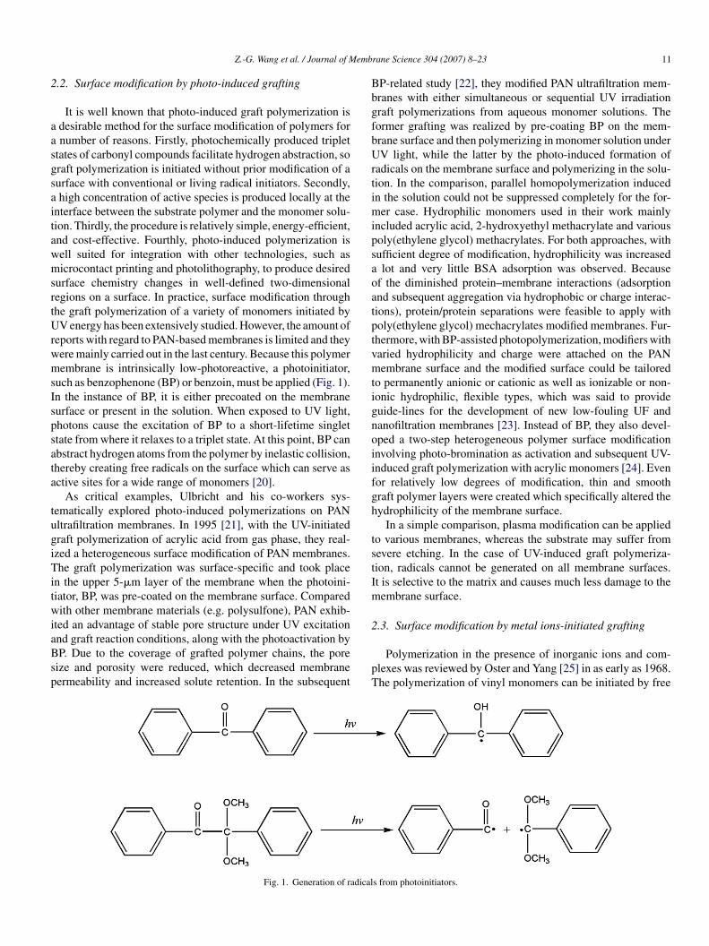
emb
2
aasgsaitawmsrtUrwmsIspsata
tugiTitwiaBsp
BbgfbUrtimipsaoatptvmtignoiifgh
tstIm
2
Z.-G. Wang et al. / Journal of M
.2. Surface modification by photo-induced grafting
It is well known that photo-induced graft polymerization isdesirable method for the surface modification of polymers fornumber of reasons. Firstly, photochemically produced triplet
tates of carbonyl compounds facilitate hydrogen abstraction, soraft polymerization is initiated without prior modification of aurface with conventional or living radical initiators. Secondly,high concentration of active species is produced locally at the
nterface between the substrate polymer and the monomer solu-ion. Thirdly, the procedure is relatively simple, energy-efficient,nd cost-effective. Fourthly, photo-induced polymerization isell suited for integration with other technologies, such asicrocontact printing and photolithography, to produce desired
urface chemistry changes in well-defined two-dimensionalegions on a surface. In practice, surface modification throughhe graft polymerization of a variety of monomers initiated byV energy has been extensively studied. However, the amount of
eports with regard to PAN-based membranes is limited and theyere mainly carried out in the last century. Because this polymerembrane is intrinsically low-photoreactive, a photoinitiator,
uch as benzophenone (BP) or benzoin, must be applied (Fig. 1).n the instance of BP, it is either precoated on the membraneurface or present in the solution. When exposed to UV light,hotons cause the excitation of BP to a short-lifetime singlettate from where it relaxes to a triplet state. At this point, BP canbstract hydrogen atoms from the polymer by inelastic collision,hereby creating free radicals on the surface which can serve asctive sites for a wide range of monomers [20].
As critical examples, Ulbricht and his co-workers sys-ematically explored photo-induced polymerizations on PANltrafiltration membranes. In 1995 [21], with the UV-initiatedraft polymerization of acrylic acid from gas phase, they real-zed a heterogeneous surface modification of PAN membranes.he graft polymerization was surface-specific and took place
n the upper 5-�m layer of the membrane when the photoini-iator, BP, was pre-coated on the membrane surface. Comparedith other membrane materials (e.g. polysulfone), PAN exhib-
ted an advantage of stable pore structure under UV excitation
nd graft reaction conditions, along with the photoactivation byP. Due to the coverage of grafted polymer chains, the poreize and porosity were reduced, which decreased membraneermeability and increased solute retention. In the subsequent
pT
Fig. 1. Generation of radical
rane Science 304 (2007) 8–23 11
P-related study [22], they modified PAN ultrafiltration mem-ranes with either simultaneous or sequential UV irradiationraft polymerizations from aqueous monomer solutions. Theormer grafting was realized by pre-coating BP on the mem-rane surface and then polymerizing in monomer solution underV light, while the latter by the photo-induced formation of
adicals on the membrane surface and polymerizing in the solu-ion. In the comparison, parallel homopolymerization inducedn the solution could not be suppressed completely for the for-
er case. Hydrophilic monomers used in their work mainlyncluded acrylic acid, 2-hydroxyethyl methacrylate and variousoly(ethylene glycol) methacrylates. For both approaches, withufficient degree of modification, hydrophilicity was increasedlot and very little BSA adsorption was observed. Because
f the diminished protein–membrane interactions (adsorptionnd subsequent aggregation via hydrophobic or charge interac-ions), protein/protein separations were feasible to apply witholy(ethylene glycol) mechacrylates modified membranes. Fur-hermore, with BP-assisted photopolymerization, modifiers witharied hydrophilicity and charge were attached on the PANembrane surface and the modified surface could be tailored
o permanently anionic or cationic as well as ionizable or non-onic hydrophilic, flexible types, which was said to provideuide-lines for the development of new low-fouling UF andanofiltration membranes [23]. Instead of BP, they also devel-ped a two-step heterogeneous polymer surface modificationnvolving photo-bromination as activation and subsequent UV-nduced graft polymerization with acrylic monomers [24]. Evenor relatively low degrees of modification, thin and smoothraft polymer layers were created which specifically altered theydrophilicity of the membrane surface.
In a simple comparison, plasma modification can be appliedo various membranes, whereas the substrate may suffer fromevere etching. In the case of UV-induced graft polymeriza-ion, radicals cannot be generated on all membrane surfaces.t is selective to the matrix and causes much less damage to theembrane surface.
.3. Surface modification by metal ions-initiated grafting
Polymerization in the presence of inorganic ions and com-lexes was reviewed by Oster and Yang [25] in as early as 1968.he polymerization of vinyl monomers can be initiated by free
s from photoinitiators.
1 emb
rapp[sri
ga(mlgtsTfwStbtrmttbMFihwhdgh
tma
pimTeosmcTe(maistpnafTpi
3
qc
2 Z.-G. Wang et al. / Journal of M
adicals or metal ions in their unstable valence states, whichre produced by excited electron transfer. This process can takelace spontaneously in “redox” initiating systems or can berovoked by externally supplied energy. Based on the theories26,27], metal ions abstract hydrogen atoms from the monomer,olvent and polymer substrates and then produce abundant freeadicals, which initiate graft polymerization and homopolymer-zation at the same time.
Very few works have been done about the ions initiatedrafting on PAN-based ultrafiltration membranes but by Yuannd her co-workers [23,24]. Using ceric ammonium nitrateCe4+) as initiator, they grafted acrylamide (AAm) onto theembrane surface of poly(acrylonitrile-co-methyl methacry-
ate) (PANCMMA). A basic consideration is that the functionalroups on the main chains of the copolymer could be oxidizedo generate radicals by Ce4+ [28]. After grafting, the membraneurface got rougher and the pore structure had little change.herefore, the wettability was improved. In a succeeding work,
errous ammonium sulfate (Fe2+)/H2O2 as an initiator systemas used to graft AAm on the same copolymer membrane [29].imilar goal was achieved that the wettability was improved
hrough the attachment of polyacrylamide chains on the mem-rane surface with the pores slightly blocked. Compared withhe Ce4+ initiated grafting process, the initiator of Fe2+/H2O2esulted in much higher grafting degree, because the formeright use the reactive groups in the early stage but the lat-
er system could generate hydroxyl radicals in the mediumhat transferred into PAN-based macromolecules on the mem-rane surface so that the reaction could last for a longer time.oreover, they brought forward a mechanism to interpret how
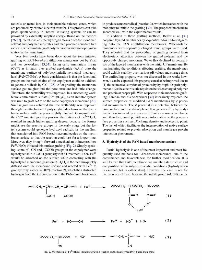
e2+/H2O2 initiated this surface grafting (Fig. 2). Simply speak-ng, some of –CN and –COOR groups in the copolymer wereydrolyzed into –COOH groups by NaOH treatment. Then, Fe2+
ould be adsorbed on the surface while contacting with the
ydrolyzed membrane (reaction 1). H2O2 in the medium quicklyiffused onto the membrane surface and reacted with Fe2+ toive hydroxyl radicals (OH•) (reaction 2), which then abstractedydrogen from the tertiary carbon in the PAN-based backboneswcit
Fig. 2. Mechanism of Fe2+/H2O2 initiating grafting re
rane Science 304 (2007) 8–23
o produce a macroradical (reaction 3), which interacted with theonomer to initiate the grafting [30]. The proposed mechanism
ccorded well with the experimental results.In addition to these grafting methods, Belfer et al. [31]
repared layered membranes by sequential redox-initiated graft-ng onto the PAN ultrafiltration membranes. Water-soluble
onomers with oppositely charged ionic groups were used.hey reported that the proceeding of grafting derived fromlectrostatic attraction between the grafted polymer and theppositely charged monomer. Water flux declined in compari-on of the layered membranes with the initial UF membrane. Byanipulating the conditions affecting grafting, the membranes
ould exhibit stability over various pH values and storage time.he antifouling property was not discussed in the work; how-ver, it can be expected this property can also be improved due to1) the reduced adsorption of proteins by hydrophilic graft poly-er and (2) the electrostatic repulsion between charged polymer
nd protein at proper pH. With respect to ionic monomers graft-ng, Tanioka and his co-workers [32] intensively explored theurface properties of modified PAN membranes by ζ poten-ial measurement. The ζ potential is a potential between theore surface and the shear plane. It is generated by hydrody-amic flow induced by a pressure difference across a membranend, therefore, could provide much information on the pore sur-ace properties such as pK, charge density and isoelectric point.he last of which facilitates the interpretation of native surfaceroperties related to protein adsorption and membrane-proteinnteraction phenomena.
. Hydrolysis of the PAN-based membrane surface
Partial hydrolysis is one of the most important and most fre-uently used methods for PAN-based membranes, due to theonvenience and favorableness for further modification. It is
ell known that PAN membrane can maintain its structure andomposition when subject to acidic conditions (hydrolyzations existent, but is rather slow). However, the case is not forhe presence of base, because the nitrile group (–C N) can be
action on the hydrolyzed PAN-based membrane.
Z.-G. Wang et al. / Journal of Membrane Science 304 (2007) 8–23 13
Fig. 3. Reaction mechanism of PAN with (a) NaOH and (b) primary amines.

1 emb
ebtpmPta[wtcs
F
1
2
3
soth
wgrGiblainoCbfiBgletamipwha
ig
thpAtmtfitoa1atdIpsepfsit
wfpdmtalebtbmtw(odAdiflemc
4 Z.-G. Wang et al. / Journal of M
asily hydrolyzed by NaOH or amine, and converted into car-oxyl, acylamide or amide groups. Hydrolyzation can be usedo render the PAN membrane surface hydrophilic and chargedroperty, which helps to improve the protein-resistant perfor-ance. From the inherence (surface property, structure, etc.) of
AN membrane, hydrolysis has impact on the surface throughwo aspects, i.e. hydrophilicity and pore size. A possible mech-nism for NaOH-induced hydrolysis is indicated in Fig. 3(a)33]. PAN with a certain content of –COOH is easily swollenhen expose to aqueous medium. The swollen macromolecules
hen become more mobile to move towards the pores. This pro-ess decreases the pore size and makes the membrane surfacemoother.
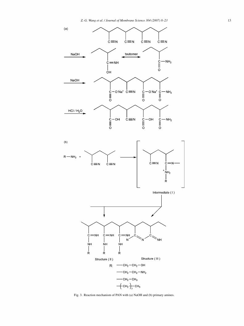
The reaction of PAN chain with primary amine is shown inig. 3(b) [34]. A possible mechanism for this reaction is:
. An intermediate (I) could be obtained in the initiation step bythe nucleophilic attack of the amine groups in primary amineon the nitrile groups in PAN.
. The intermediate with its separation of charges could rear-range into imine (structure II).
. A further reaction might occur simultaneously among theintermediates, and the nitrile groups could form a carbon-nitrogen conjugated structure (structure III).
When the amine used is ethanolamine or ethylenediamine,tructure II could enhance the hydrophilicity of PAN becausef the increase of hydrophilic –OH or –NH2 groups withinhe polymer chains. On the contrary, structure III increases theydrophobicity for the linear sequences of fused rings.
Carboxyl groups derived from the partial hydrolysis of PANith NaOH are facile to be ionized by surrounding water, so theireneration does not only improve the hydrophilicity, but alsoesults in negative surface charge for the PAN-based membranes.odjevargova and Dimov [35] investigated the permeabil-
ty (vitamin B12) and protein adsorption (albumin solution)ehavior of PAN-based membranes modified by hydroxy-amine (NH2OH), diethylaminoethyl methacrylate (DEAEM)nd NaOH (DEAEM + NaOH). The grafting of DEAEM wasnitiated by an oxidation reduction system of H2O2–Fe(II). Theitrile groups were converted by NH2OH into primary amine,ximes (simply speaking, –NH2 and –OH, respectively, added toand N atoms of –C N to form primary amine and oxime) and
y DEAEM into tertiary amino group. They got that the modi-ed membranes gave better permeability coefficient of vitamin12 than the original membrane with the exception of DEAEM-rafted membranes, while the increase in hydrophilicity did notead to the expected increase in permeability coefficient, anffect attributed to the decrease in the pore volume in the selec-ive layer; partially hydrolyzed membrane showed the smallestdsorption of proteins. The amount of protein adsorbed on theembrane surface was determined by the membrane charge
nstead of the membrane hydrophilicity. Above the isoelectric
oint (IEP), albumin with negative charges repulsively interactedith the negatively charged membrane surface. Therefore, theydrolyzed membrane surface showed small amount of proteindsorption but the DEAEM-grafted membrane surface resultedwioa
rane Science 304 (2007) 8–23
n higher adsorption, because the latter consumed some carboxylroups on the membrane surface.
Yang and Tong [36] studied the ultrafiltration of pro-eins through the ionized PAN hollow fiber membrane withydrolyzation and investigated its capability of separating tworoteins having two similar molecular weight but different IEP.fter treating in definite conditions including hydrolyzing time,
emperature and NaOH concentration, charge density on theembrane surface could reach highest, while too high concen-
ration of NaOH could cause severe degradation of the hollowber. Due to the pH-sensitive poly(acrylic acid) (PAA) layer,
he hydraulic permeability and the ultrafiltration performancef proteins were greatly affected by pH. For both buffer solutionnd protein solution, the permeability at pH above 5 was about/3 of that at pH below 5. It was possibly due to the swollennd thicker PAA layer at higher pH. The retention of both pro-eins (myoglobin and cytochrome c) increased with the ionicensity of PAN hollow fiber and reached the minimum at theEP for the hydrolyzed membranes. It is known that, when theH is equal to the IEP, the protein is neutral and its size is themallest. Most of the pores on the ionic PAA layer were largenough for single particles of myoglobin and cytochrome c toass through, despite the electrostatic interaction. When the sur-ace charge density was higher, the electrostatic interaction wastronger and the retention was hence increased. Their resultsndicated that using the hollow fiber at varied pH, the selectivityowards a mixture of proteins could therefore be controlled.
Bryjak et al. [37] also modified a porous PAN membraneith NaOH. They found that the average pore diameter changed
rom 2.6 to 0.6 nm and the modified membranes were lessrone for protein deposition, in which fouling caused a poreiameter reduction of 80% for the untreated and 20% for theodified membranes. As is known, transformation of nitrile
o carboxylic group on the membrane surface may result indecrease of pore lumen up to filling them with a PAA-
ike gel. However, when the pores in the membrane are smallnough and the membrane material is charged, such a mem-rane may work in the nanofiltration mode. Results indicatedhat a membrane immersed in the modification bath was capa-le of rejecting about 50% of calcium while the unmodifiedembrane rejected no salt. Lohokare et al. [38] compared
he effects of different treatment modes on the hydrolyzationith organic (ethanolamine/triethylamine) and inorganic bases
NaOH/KOH). The modes included dead-end and cross-flowne. It was found the extent of change in permeation was highlyependent on the treatment mode and treatment temperature.
maximum increase of 152% in water flux was achieved byead-end mode within 20 h, while cross-flow offered maximumncrease of 230% in just 2.5 h duration at 45 ◦C, because cross-ow mode did not cause a noticeable pore swelling. As a typicalxample in application, Oh et al. [33,39] functionalized PAN UFembranes by NaOH aiming at the preparation of nanofiltration
omposite membranes, in which polyacrylamide active layers
ere interconnected with support layers via the formation ofonic bonds. Kim et al. [40] also studied the effect of hydrolysisf annealed membrane using different concentrations of NaOHnd CH3ONa.
emb
4P
mdTmcdw2iorbogup
rssoTa
4
acbttYpp
NdNilitfipthtuwao
ignaihatpdi[rcbsuwersledthbaatreih
puPpthlplCeotftbht
Z.-G. Wang et al. / Journal of M
. (Bio-)macromolecule immobilization on theAN-based membrane surface
Surface immobilization, also referred to as “graft-to” methodentioned previously, involves the covalent attachment of
esignated macromolecular chains on the membrane surface.his usually requires the presence of reactive groups on theembrane, which can be generated through hydrolysis or
opolymerization. The former method has been described inetail, and the latter one can be realized using vinyl monomersith reactive groups, such as acrylic acid, maleic anhydride and-hydroxyethyl methacrylate. The obvious advantage of surfacemmobilization over surface polymerization is that more kindsf macromolecules are available for selection, including natu-al polymers and synthesized polymers that are hardly obtainedy radicals-initiated polymerization (e.g. PEG). The principle isnly based on that (1) the immobilized polymers possess activeroups and (2) the selected polymers are hemocompatible whensed in hemodialysis, such as chitosan, heparin, some kinds ofroteins, PEG, etc.
Hemodialysis is an important clinical procedure for theemoval of toxic biological metabolites in patients with end-tage renal disease. The key element of a hemodialyzer is theemipermeable membrane, which allows for selective transportf low molecular weight biological metabolites from blood.herefore, the discussion below is mainly focused on function-lizing PAN-based membranes for the purpose of hemodialysis.
.1. Homo-surface immobilization
It was mentioned that PAN membranes are susceptible tolkaline condition and the nitrile groups can be hydrolyzed intoarboxyl groups. Therefore, macromolecule immobilization cane carried out via the reaction of reactive carboxyl groups onhe membrane and suitable polymer chains. With respect tohis immobilization process on the hydrolyzed-PAN membrane,ang et al. [39,40] investigated the effect of chitosan/heparinolyelectrolyte complex (PEC) and some proteins on the surfaceroperties.
Chitosan, the principal derivative of chitin, is obtained by-deacetylation to a varying extent that is characterized by theegree of deacetylation, and is consequently a copolymer of-acetyl-d-glucosamine and d-glucosamine. Chitosan has been
dentified as biocompatible, biodegradable, non-toxic, physio-ogically inert, antibacterial properties, etc. Moreover, chitosans one of the most plentiful, renewable organic resources andhus can be commercially obtained at a relatively low costrom the shells of shellfish, wastes of the seafood processingndustry [41]. Despite so many advantages, chitosan enhanceslasma protein adsorption, platelet adhesion and activation, andhrombus development; in other words, chitosan is an effectiveemostatic agent [42,43], possibly due to electrostatic attrac-ion. Therefore, chitosan cannot be applied for contacting blood
nless it is modified. Contrary to chitosan, heparin is the mostidely used blood anticoagulant. Heparin, a mixture of vari-bly sulfated polysaccharide chains composed of repeating unitsf d-glucosamine and either l-iduronic or d-glucuronic acids,
Ioht
rane Science 304 (2007) 8–23 15
s commonly used in hemodialysis. The presence of sulfateroups in the carbohydrate moiety gives heparin its highlyegative charge. Heparin binds to antithrombin III, acceler-ting the enzyme-neutralizing effect of this serine proteasenhibator, and prevents thrombin formation. In the presence ofeparin, the interaction of antithrombin with thrombin is virtu-lly instantaneous. The heparin–antithrombin complex inhibitshe conversion of other coagulation proteins to active serineroteases (Factors XII, XI, IX and X). Considering these fun-amentals, the chitosan/heparin complex (PEC) was covalentlymmobilized onto the surface of hydrolyzed PAN membrane44,45]. Briefly, the immobilization process involved that theeaction of chitosan with 1-ethyl-3-(3-di-methylaminopropyl)arbodiimide (EDC)-activated carboxyl groups on the mem-rane and the attachment of heparin onto immobilized chitosanpacer by glutaraldehyde. A series of characterizations and eval-ations were performed, revealing that the surface hydrophilicityas increased, thrombus formation was reduced, the prolif-
ration of bacterial was suppressed and the permeability wasetained. Therefore, the conjugation of chitosan with heparinimultaneously endowed the membrane surface with anticoagu-ation activity and antibacterial activity, the latter of which wasmphasized because anticoagulant must be injected continuousuring hemodialysis. And the conjugate covalent immobiliza-ion possessed additional advantage of reducing release ofeparin from the surface, comparing with physical or ioniconding. The undesirable release into the blood can eventu-lly leads to the disadvantages such as systemic heparinizationnd increases the risk of abnormal hemorrhage or the symp-om of thrombocytopenia [46]. The less loss of heparin alsoeduced the injection of heparin in clinical applications, andven resulted in the heparin-free therapy. This work was mean-ngful in potentializing the application of PAN membrane inemodialysis.
Yang and co-workers [47] also covalently immobilizedlasma proteins onto hydrolyzed PAN membrane to eval-ate the hemocompatibility and anaphylatoxin formation.latelet-adhesion-inhibiting human serum albumin (HSA) andlatelet-adhesion-promoting collagen (COL) were used asypical model proteins. Both immobilizations increased theydrophilicity of the membrane surface, but just a little. Immobi-ized HSA reduced the platelet adhesion, fibrinogen adsorption,rolonged the blood coagulation times, as well as reduced theeukopenia and anaphylatoxin formation. On the other hand,OL immobilizing on PAN membrane exhibited the oppositeffect when contacting with blood, though induced less increasef C3, C4 antigens of serum. During the process of hemodialysis,he plasma proteins will always more or less adsorb onto the sur-ace of hemodialyzer, even after intensive washing. Therefore,he results helped to explain why reused hemodialyzers wereeneficial to hemodialysis therapy. That was because the reusedemodialyzers usually suppressed the leukopenia, anaphyla-oxin formation and avoided inducing the first use syndrome.
t was also obtained that HSA immobilization on the surfacef PAN membrane was useful and practicable in improving theemocompatibility and diminishing the anaphylatoxin forma-ion during hemodialysis treatment.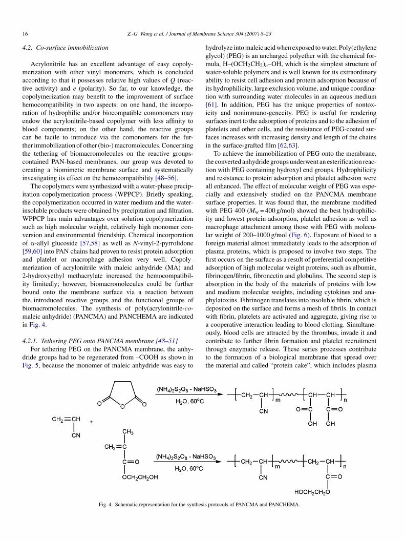
1 emb
4
matchrebcttcci
itiWsvo[am2ibtbmi
4
dF
hgmwait[ispfi
ttaacswimlfpfiafiaapdwao
6 Z.-G. Wang et al. / Journal of M
.2. Co-surface immobilization
Acrylonitrile has an excellent advantage of easy copoly-erization with other vinyl monomers, which is concluded
ccording to that it possesses relative high values of Q (reac-ive activity) and e (polarity). So far, to our knowledge, theopolymerization may benefit to the improvement of surfaceemocompatibility in two aspects: on one hand, the incorpo-ation of hydrophilic and/or biocompatible comonomers mayndow the acrylonitrile-based copolymer with less affinity tolood components; on the other hand, the reactive groupsan be facile to introduce via the comonomers for the fur-her immobilization of other (bio-) macromolecules. Concerninghe tethering of biomacromolecules on the reactive groups-ontained PAN-based membranes, our group was devoted toreating a biomimetic membrane surface and systematicallynvestigating its effect on the hemocompatibility [48–56].
The copolymers were synthesized with a water-phase precip-tation copolymerization process (WPPCP). Briefly speaking,he copolymerization occurred in water medium and the water-nsoluble products were obtained by precipitation and filtration.
PPCP has main advantages over solution copolymerizationuch as high molecular weight, relatively high monomer con-ersion and environmental friendship. Chemical incorporationf �-allyl glucoside [57,58] as well as N-vinyl-2-pyrrolidone59,60] into PAN chains had proven to resist protein adsorptionnd platelet or macrophage adhesion very well. Copoly-erization of acrylonitrile with maleic anhydride (MA) and
-hydroxyethyl methacrylate increased the hemocompatibil-ty limitedly; however, biomacromolecules could be furtheround onto the membrane surface via a reaction betweenhe introduced reactive groups and the functional groups ofiomacromolecules. The synthesis of poly(acrylonitrile-co-aleic anhydride) (PANCMA) and PANCHEMA are indicated
n Fig. 4.
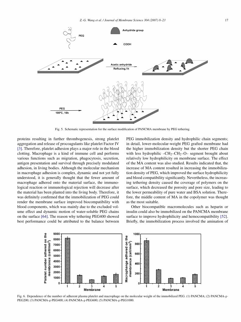
.2.1. Tethering PEG onto PANCMA membrane [48–51]For tethering PEG on the PANCMA membrane, the anhy-
ride groups had to be regenerated from –COOH as shown inig. 5, because the monomer of maleic anhydride was easy to
cttt
Fig. 4. Schematic representation for the synthesis
rane Science 304 (2007) 8–23
ydrolyze into maleic acid when exposed to water. Poly(ethylenelycol) (PEG) is an uncharged polyether with the chemical for-ula, H–(OCH2CH2)n–OH, which is the simplest structure ofater-soluble polymers and is well known for its extraordinary
bility to resist cell adhesion and protein adsorption because ofts hydrophilicity, large exclusion volume, and unique coordina-ion with surrounding water molecules in an aqueous medium61]. In addition, PEG has the unique properties of nontox-city and nonimmuno-genecity. PEG is useful for renderingurfaces inert to the adsorption of proteins and to the adhesion oflatelets and other cells, and the resistance of PEG-coated sur-aces increases with increasing density and length of the chainsn the surface-grafted film [62,63].
To achieve the immobilization of PEG onto the membrane,he converted anhydride groups underwent an esterification reac-ion with PEG containing hydroxyl end groups. Hydrophilicitynd resistance to protein adsorption and platelet adhesion werell enhanced. The effect of molecular weight of PEG was espe-ially and extensively studied on the PANCMA membraneurface properties. It was found that, the membrane modifiedith PEG 400 (Mw = 400 g/mol) showed the best hydrophilic-
ty and lowest protein adsorption, platelet adhesion as well asacrophage attachment among those with PEG with molecu-
ar weight of 200–1000 g/mol (Fig. 6). Exposure of blood to aoreign material almost immediately leads to the adsorption oflasma proteins, which is proposed to involve two steps. Therst occurs on the surface as a result of preferential competitivedsorption of high molecular weight proteins, such as albumin,brinogen/fibrin, fibronectin and globulins. The second step isbsorption in the body of the materials of proteins with lownd medium molecular weights, including cytokines and ana-hylatoxins. Fibrinogen translates into insoluble fibrin, which iseposited on the surface and forms a mesh of fibrils. In contactith fibrin, platelets are activated and aggregate, giving rise tocooperative interaction leading to blood clotting. Simultane-
usly, blood cells are attracted by the thrombus, invade it and
ontribute to further fibrin formation and platelet recruitmenthrough enzymatic release. These series processes contributeo the formation of a biological membrane that spread overhe material and called “protein cake”, which includes plasmaprotocols of PANCMA and PANCHEMA.
Z.-G. Wang et al. / Journal of Membrane Science 304 (2007) 8–23 17
odific
pa[cvaaiumltwrbuob
Pitwroitaistfa
FP
Fig. 5. Schematic representation for the surface m
roteins resulting in further thrombogenesis, strong plateletggregation and release of procoagulants like platelet Factor IV3]. Therefore, platelet adhesion plays a major role in the bloodlotting. Macrophage is a kind of immune cell and performsarious functions such as migration, phagocytosis, secretion,ntigen presentation and survival through precisely modulateddhesion, in living bodies. Although the molecular mechanismn macrophage adhesion is complex, dynamic and not yet fullynderstood, it is generally thought that the fewer amount ofacrophage adhered onto the material surface, the immuno-
ogical reaction or immunological rejection will decrease afterhe material has been planted into the living body. Therefore, itas definitely confirmed that the immobilization of PEG could
ender the membrane surface improved biocompatibility with
lood components, which was mainly due to the excluded vol-me effect and dynamic motion of water-soluble PEG chainsn the surface [64]. The reason why tethering PEG400 showedest performance could be attributed to the balance betweenisB
ig. 6. Dependence of the number of adherent plasma platelet and macrophage on thEG200; (3) PANCMA-g-PEG400; (4) PANCMA-g-PEG600; (5) PANCMA-g-PEG
ation of PANCMA membrane by PEG tethering.
EG immobilization density and hydrophilic chain segments;n detail, lower-molecular-weight PEG grafted membrane hadhe higher immobilization density but the shorter PEG chainith less hydrophilic –CH2–CH2–O– segment brought about
elatively low hydrophilicity on membrane surface. The effectf the MA content was also studied. Results indicated that, thencrease of MA content resulted in increasing the immobiliza-ion density of PEG, which improved the surface hydrophilicitynd blood compatibility significantly. Nevertheless, the increas-ng tethering density caused the coverage of polymers on theurface, which decreased the porosity and pore size, leading tohe lower permeability of pure water and BSA solution. There-ore, the middle content of MA in the copolymer was thoughts the most suitable.
Other biocompatible macromolecules such as heparin ornsulin could also be immobilized on the PANCMA membraneurface to improve hydrophilicity and hemocompatibility [52].riefly, the immobilization process involved the amination of
e molecular weight of the immobilized PEG. (1) PANCMA; (2) PANCMA-g-1000.

18 Z.-G. Wang et al. / Journal of Membrane Science 304 (2007) 8–23
rin/or
–tattbeapoaiP[p
4P
bppssochswwmat[mb
btbpigcram(tmntgrwbtmtWbtl
gbaas
Fig. 7. Schematic representation for the immobilization of (a) hepa
COOH contained membrane surface and then the immobiliza-ion of heparin/ or insulin previously activated with EDC on themino-bound membrane surface (Fig. 7(a)). Heparin is referredo as an excellent anticoagulant and insulin can also reducehe aggregation of platelets; therefore, immobilizing the twoiomacromolecules was expected to improve the surface prop-rties. Results indicated the bound heparin/or insulin formedthin layer covering on the porous surface, the hydrophilicity
lus hemocompatibility were markedly improved (the numberf adherent platelet and macrophage decreased significantly)nd tethering heparin showed more efficient improvement thannsulin. Chitosan or gelatin was also immobilized on theANCMA membrane surface using EDC coupling (Fig. 7(b)53]) and it was also observed that the number of adherentlatelet and macrophage decreased.
.2.2. Anchoring of phospholipid moieties onto theANCHEMA membrane [54–56]
The concept of anchoring is inspired by the mimicry ofiomembranes mainly constructed of neutral phospholipids andhosphorylcholines. Phosphorylcholine is the headgroup ofhosphatidylcholine, the major component of the extracellularide of cell membranes. The fact that proteins in the blood-tream do not foul onto the surface of cells shows that theuter surface of the cell walls, comprising PC groups, is bio-ompatible [65,66]. Accordingly, phosphorylcholines materialsave the potential to reduce protein adsorption significantly,ince their hydrated surfaces are able to interact with proteinsithout inducing three-dimensional conformational changes,hich only result in the reversible adsorption of protein. Surfaceodifications with phospholipids analogous polymers (PAPs)
nd phospholipid moieties were extensively studied to improve
he biocompatibility for various biomaterials in recent years67–70]. It is recognized that just a small amount of phospholipidoieties can improve the surface biocompatibility of materials,ecause the zwitterionic nature of the phospholipid moiety, com-
tmbm
insulin and (b) chitosan/ or gelatin onto the PANCMA membrane.
ined with its ability to bind water tightly around itself, giveshese materials remarkable biocompatibility. Therefore, it woulde rather meaningful to perform the investigation of anchoringhospholipid moieties onto the PANCHEMA membrane. Thentroduction process mainly involved the reaction of hydroxylroups on the HEMA-contained membrane surface with 2-hloro-2-oxo-1,3,2-dioxaphospholane (COP) followed by theing-opening reaction of COP with trimethylamine (Fig. 8). Asresult, for the phospholipid moieties modifying PANCHEMAembranes, the hydrophilicity and the permeation of protein
bovine serum albumin) solution were greatly enhanced, the pro-ein adsorption and the fouling were obviously weakened, and
uch less platelet and macrophage adhesion occurred. Untilow, how the phospholipids interact with proteins to achievehe biocompatibility is not entirely understood. However, it isenerally recognized that the chemical and electronic configu-ation of the zwitterionic moieties structures and the associationith a large amount of water allow reversible interaction withiomacromolecules that approach the surface. By controllinghe adsorption of biomacromolecules and the formation of any
ediating layers, cellular adhesion can also be controlled. Byhese basic principles, biofouling is governed on any scale.
ith regard to the PANCHEMA membrane surface anchoredy phospholipid moieties, the hemocompatibility could be easilyailored by adjusting the content of HEMA and thus phospho-ipid moieties (Fig. 9).
So far, for the purpose of surface immobilization, the reactiveroups can be generated on the PAN-based membranes eithery copolymerizing or hydrolysis. The two methods have eachdvantages and disadvantages. PAN and its various membranesre relatively more commercially available; however, the expo-ure to the base, especially NaOH, may more or less damage
he membrane structure and thus the mechanical strength, andore at serious condition such as long treatment time and highase concentration. On the other hand, the copolymerizationethod can be used to control the reactive groups introduced

Z.-G. Wang et al. / Journal of Membrane Science 304 (2007) 8–23 19
e PA
mtihthtwa
ith
Fb(c
hbamhitoa
Fig. 8. Schematic representation for the surface modification of th
ore easily, maintain the mechanical strength and even bet-er, because the comonomer segments may reduce the strongnteraction between PAN chains and make them more flexible;owever, the copolymer was usually synthesized in the lab andhe monomers are usually not easy to obtain. Moreover, whateverydrolysis or copolymerization for grafting-to method is advan-ageous over the mentioned graft-from method, over preventingaste of materials due to homopolymerization and crosslinking
nd more easily controlling graft density.
Functionalization of PAN-based membranes for the antifoul-ng and blood-contacting purpose has been generally reviewedhrough the above paragraphs. A variety of modificationsave been utilized to endow the membrane surface with the
ig. 9. Adhesion of platelet on PAN, PANCHEAM and PCANCP dense mem-rane surface. The HEMA mole fraction in the copolymer membrane is (a) 0%;b) 6.4%; (c) 9.3%; (d) 17.8%. The mole fraction of phospholipid moiety on theopolymer membrane surface is (e) 6.09%; (f) 9.19%; (g) 17.1%.
tmwPem
5i
afimbdtcpahots
NHEMA membrane by the formation of phospholipids moieties.
ydrophilicity and the inertness to protein adsorption followedy antifouling performance, some of which, however, are notvailable to the hemodialysis field. Collagen and chitosan asodifiers are the typical examples. To meet the request of the
emodialysis process, the mimicry of biological inert interfaces concerned. In modifying PAN-based membranes, mimickinghe surfaces of blood cells only ever involved the introductionf phospholipid moieties. Nevertheless, the attempt providedn alternative avenue to develop the PAN-based membranesowards the application to hemodialyzer, so the method of the
imicry in preparing such membrane is rather promising. Any-ay, we can expect with confidence that the modifications ofAN-based membranes with so many excellent intrinsic prop-rties have great potential to deepen and expand its influence inore biomedical fields.
. PAN-based membrane surface for enzymemmobilization
Enzymes, proteins with high efficiency in catalysis and unpar-lleled selectivity, have been extensively realized in variouselds, such as fine-chemical synthesis, pharmaceuticals, com-odity catalysts in food processing and detergent applications,
iosensors, bioremediation, polymerase chain reaction, proteinigestion in proteomic analysis, and biofuel cells. And due toheir biological character, enzymes are well known as “greenatalysts”. However, some inevitable disadvantages of enzymesractically restrict their further development in large-scale oper-tions, such as the short lifetime from the intrinsic instability and
igh cost from disable recycling. In practice, almost all industrialperations prefer the enzymes in immobilizing form, becausehey offer easy recycling, feasible continuous operations, andimple product purification. Among the large amount of sup-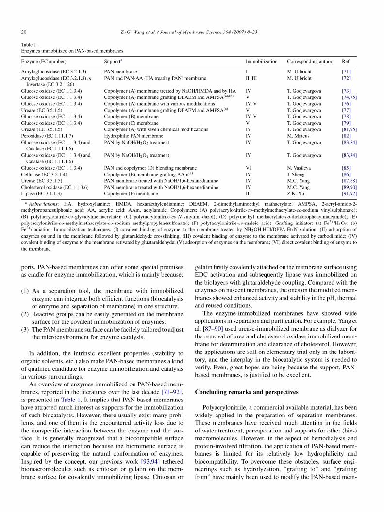
20 Z.-G. Wang et al. / Journal of Membrane Science 304 (2007) 8–23
Table 1Enzymes immobilized on PAN-based membranes
Enzyme (EC number) Supporta Immobilization Corresponding author Ref
Amyloglucosidase (EC 3.2.1.3) PAN membrane I M. Ulbricht [71]Amyloglucosidase (EC 3.2.1.3) or
Invertase (EC 3.2.1.26)PAN and PAN-AA (HA treating PAN) membrane II, III M. Ulbricht [72]
Glucose oxidase (EC 1.1.3.4) Copolymer (A) membrane treated by NaOH/HMDA and by HA IV T. Godjevargova [73]Glucose oxidase (EC 1.1.3.4) Copolymer (A) membrane grafting DEAEM and AMPSA(a),(b) V T. Godjevargova [74,75]Glucose oxidase (EC 1.1.3.4) Copolymer (A) membrane with various modifications IV, V T. Godjevargova [76]Urease (EC 3.5.1.5) Copolymer (A) membrane grafting DEAEM and AMPSA(a) V T. Godjevargova [77]Glucose oxidase (EC 1.1.3.4) Copolymer (B) membrane IV, V T. Godjevargova [78]Glucose oxidase (EC 1.1.3.4) Copolymer (C) membrane V T. Godjevargova [79]Urease (EC 3.5.1.5) Copolymer (A) with seven chemical modifications IV T. Godjevargova [81,95]Peroxidase (EC 1.11.1.7) Hydrophilic PAN membrane IV M. Mateus [82]Glucose oxidase (EC 1.1.3.4) and
Catalase (EC 1.11.1.6)PAN by NaOH/H2O2 treatment IV T. Godjevargova [83,84]
Glucose oxidase (EC 1.1.3.4) andCatalase (EC 1.11.1.6)
PAN by NaOH/H2O2 treatment IV T. Godjevargova [83,84]
Glucose oxidase (EC 1.1.3.4) PAN and copolymer (D) blending membrane VI N. Vasileva [85]Cellulase (EC 3.2.1.4) Copolymer (E) membrane grafting AAm(a) IV J. Sheng [86]Urease (EC 3.5.1.5) PAN membrane treated with NaOH/1,6-hexanediamine IV M.C. Yang [87,88]Cholesterol oxidase (EC 1.1.3.6) PAN membrane treated with NaOH/1,6-hexanediamine IV M.C. Yang [89,90]Lipase (EC 3.1.1.3) Copolymer (F) membrane III Z.K. Xu [91,92]
a Abbreviations: HA, hydroxylamine; HMDA, hexamethylendiamine; DEAEM, 2-dimethylaminoethyl mathacrylate; AMPSA, 2-acryl-amido-2-methylpropanesulphonic acid; AA, acrylic acid; AAm, acrylamide. Copolymers: (A) poly(acrylonitrile-co-methylmethacrylate-co-sodium vinylsulphonate);(B) poly(acrylonitrile-co-glycidylmethacrylate); (C) poly(acrylonitrile-co-N-vinylimi-dazol); (D) poly(methyl methacrylate-co-dichlorophenylmaleimide); (E)poly(acrylonitrile-co-methylmethacrylate-co-sodium methylpropylenesulfonate); (F) poly(acrylonitrile-co-maleic acid). Grafting initiator: (a) Fe2+/H2O2; (b)Fe2+/radiation. Immobilization techniques: (I) covalent binding of enzyme to the membrane treated by NH2OH·HCl/DPPA-Et3N solution; (II) adsorption ofe III) cc adsort
pa
(
(
(
ooi
biholtfccIbb
gEteba
aatbttvb
C
wTomp
nzymes on and in the membrane followed by glutaraldehyde crosslinking; (ovalent binding of enzyme to the membrane activated by gluataraldehyde; (V)he membrane.
orts, PAN-based membranes can offer some special promisess cradle for enzyme immobilization, which is mainly because:
1) As a separation tool, the membrane with immobilizedenzyme can integrate both efficient functions (biocatalysisof enzyme and separation of membrane) in one structure.
2) Reactive groups can be easily generated on the membranesurface for the covalent immobilization of enzymes.
3) The PAN membrane surface can be facilely tailored to adjustthe microenvironment for enzyme catalysis.
In addition, the intrinsic excellent properties (stability torganic solvents, etc.) also make PAN-based membranes a kindf qualified candidate for enzyme immobilization and catalysisn various surroundings.
An overview of enzymes immobilized on PAN-based mem-ranes, reported in the literatures over the last decade [71–92],s presented in Table 1. It implies that PAN-based membranesave attracted much interest as supports for the immobilizationf such biocatalysts. However, there usually exist many prob-ems, and one of them is the encountered activity loss due tohe nonspecific interaction between the enzyme and the sur-ace. It is generally recognized that a biocompatible surfacean reduce the interaction because the biomimetic surface is
apable of preserving the natural conformation of enzymes.nspired by the concept, our previous work [93,94] tetherediomacromolecules such as chitosan or gelatin on the mem-rane surface for covalently immobilizing lipase. Chitosan orbbnf
ovalent binding of enzyme to the membrane activated by carbodiimide; (IV)ption of enzymes on the membrane; (VI) direct covalent binding of enzyme to
elatin firstly covalently attached on the membrane surface usingDC activation and subsequently lipase was immobilized on
he biolayers with glutaraldehyde coupling. Compared with thenzymes on nascent membranes, the ones on the modified mem-ranes showed enhanced activity and stability in the pH, thermalnd reused conditions.
The enzyme-immobilized membranes have showed widepplications in separation and purification. For example, Yang etl. [87–90] used urease-immobilized membrane as dialyzer forhe removal of urea and cholesterol oxidase immobilized mem-rane for determination and clearance of cholesterol. However,he applications are still on elementary trial only in the labora-ory, and the interplay in the biocatalytic system is needed toerify. Even, great hopes are being because the support, PAN-ased membranes, is justified to be excellent.
oncluding remarks and perspectives
Polyacrylonitrile, a commercial available material, has beenidely applied in the preparation of separation membranes.hese membranes have received much attention in the fieldsf water treatment, pervaporation and supports for other (bio-)acromolecules. However, in the aspect of hemodialysis and
rotein-involved filtration, the application of PAN-based mem-
ranes is limited for its relatively low hydrophilicity andiocompatibility. To overcome these obstacles, surface engi-eerings such as hydrolyzation, “grafting to” and “graftingrom” have mainly been used to modify the PAN-based mem-
emb
bicbimhbpofimctmbee
fiHmTspwtit
A
d5
R
[
[
[
[
[
[
[
[
[
[
[
[
[
[
[
[
[
[
[
[
Z.-G. Wang et al. / Journal of M
ranes. The membranes after the above treatment showed muchmproved resistance to the adsorption of protein or bloodomponent (platelet, macrophage). Especially, the concept ofiomimicry, i.e. mimicking the outer surface of the cells, wasntroduced. In this case, the hemocompatibility of the PAN
embrane surface was improved a lot. PAN-based membranesave more superiority over other materials when subject toiomimicry modifications. One of the advantages is that itsolymer derivatives possess reactive groups favoring anchoringther biocompatible molecules. In addition to the dialysis andltration, one of the most important applications of the PANembrane is for attaching enzyme. Such enzyme-membrane
an simultaneously realize the functions of catalysis and separa-ion and turn out to be a bioactive membrane. The modification
ethods previously described to improve surface biocompati-ility can also enhance the activity and stability of immobilizednzyme, i.e. biomimicry is beneficial to the surface attachingnzymes.
Because of the unique characteristics of PAN, the sur-ace engineerings of the membranes seem rather potential inmproving its present use and widening its further applications.owever, until now, there still exist two problems. Firstly, theodification methods are relatively expensive and complex.herefore, to make the modified membranes more available,implifying the modification procedure is necessary. For exam-le, by copolymerizing with other biocompatible monomersith, the PAN-based membranes showed rather strong resis-
ance to protein and platelet adhesion. Secondly, the mechanismnvolving the interaction between the modified surface and pro-eins (enzymes, etc.) is still obscure and needed to verify.
cknowledgement
Financial support from the National Natural Science Foun-ation of China for Distinguished Young Scholars (Grant no.0625309) is gratefully acknowledged.
eferences
[1] K.K. Jindal, J. McDougall, B. Woods, L. Nowakowski, M.B. Goldstein,A study of the basic principles determining the performance of severalhigh-flux dialyzers, Am. J. Kidney Dis. 14 (1989) 507–511.
[2] J.L. Nilsson, Protein fouling of UF membranes: causes and consequences,J. Membr. Sci. 52 (1990) 121–142.
[3] J. Chanard, S. Lavaud, C. Randoux, P. Rieu, New insights in dialysismembrane biocompatibility: relevance of adsorption properties and heparinbinding, Nephrol. Dial. Transpl. 18 (2003) 252–257.
[4] J.K. Czop, K.F. Austen, Properties of glycans that activate the humanalternative complement pathway and interact with the human monocytebeta-glucan receptor, J. Immunol. 135 (1985) 3388–3393.
[5] Y. Yokota, T. Arai, T. Kawasaki, Oligomeric structures required for com-plement activation of serum mannan-binding proteins, J. Biochem. (Japan)117 (1995) 414–419.
[6] E. Klein, The modern history of hemodialysis membranes and controllers,Nephrobgy 4 (1998) 255–265.
[7] M. Hayama, K. Yamamoto, F. Kohori, K. Sakai, How polysulfone dialysismembranes containing polyvinylpyrrolidone achieve excellent biocompat-ibility, J. Membr. Sci. 234 (2004) 41–49.
[8] S.C. Lin, Protein’s natural conformation and biomaterials’ biocompatibil-ity, Chin. Polym. Bull. 1 (1998) 1–10.
[
rane Science 304 (2007) 8–23 21
[9] E. Ruckenstein, Z.F. Li, Surface modification and functionalization throughthe self-assembled monolayer and graft polymerization, Adv. Colloid Inter-face Sci. 113 (2005) 43–63.
10] R.Z. Li, L. Ye, Y.W. Mai, Application of plasma technologies infibre-reinforced polymer composites: a review of recent developments,Composites Part A-Appl. Sci. 28 (1997) 73–86.
11] P.K. Chu, J.Y. Chen, L.P. Wang, N. Huang, Plasma-surface modification ofbiomaterials, Mater. Sci. Eng. R 36 (2002) 143–206.
12] R. Forch, Z.H. Zhang, W. Knoll, Soft plasma treated surfaces: tailoringof structure and properties for biomaterial applications, Plasma Process.Polym. 2 (2005) 351–372.
13] K.S. Siow, L. Britcher, S. Kumar, H.J. Griesser, Plasma methods for thegeneration of chemically reactive surfaces for biomolecule immobilizationand cell colonization—a review, Plasma Process. Polym. 3 (2006) 392–418.
14] K. Kato, E. Uchida, E.T. Kang, Y. Uyama, Y. Ikada, Polymer surface withgraft chains, Prog. Polym. Sci. 28 (2003) 209–259.
15] M. Ulbricht, B. G., Surface modification of ultrafiltration membranes bylow temperature plasma I. Treatment of polyacrylonitrile, J. Appl. Polym.Sci. 56 (1995) 325–343.
16] M. Ulbricht, G. Belfort, Surface modification of ultrafiltration membranesby low temperature plasma II. Graft polymerization onto polyacrylonitrileand polysulfone, J. Membr. Sci. 111 (1996) 193–215.
17] M. Bryjak, I. Gancarz, A. Krajciewicz, J. Pigowski, Air plasma treatment ofpolyacrylonitrile porous membrane, Angew. Makromol. Chem. 234 (1996)21–29.
18] Z.P. Zhao, J.D. Li, D. Wang, C.X. Chen, Nanofiltration membrane pre-pared from polyacrylonitrile ultrafiltration membrane by low-temperatureplasma: 4. Grafting of N-vinylpyrrolidone in aqueous solution, Desalina-tion 184 (2005) 37–44.
19] Z.P. Zhao, J.D. Li, D.X. Zhang, C.X. Chen, Nanofiltration membrane pre-pared from polyacrylonitrile ultrafiltration membrane by low-temperatureplasma I. Graft of acrylic acid in gas, J. Membr. Sci. 232 (2004) 1–8.
20] U. Edlund, M. Kallrot, A.C. Albertsson, Single-step covalent functional-ization of polylactide surfaces, J. Am. Chem. Soc. 127 (2005) 8865–8871.
21] M. Ulbricht, A. Oechel, C. Lehmann, G. Tomaschewski, H.G. Hicke,Gas-phase photoinduced graft polymerization of acrylic acid onto poly-acrylonitrile ultrafiltration membranes, J. Appl. Polym. Sci. 55 (1995)1707–1723.
22] M. Ulbricht, H. Matuschewski, A. Oechel, H.G. Hicke, Photo-induced graftpolymerization surface modifications for the preparation of hydrophilicand low-protein-adsorbing ultrafiltration membranes, J. Membr. Sci. 115(1996) 31–47.
23] M. Ulbricht, K. Richau, H. Kamusewitz, Photomodification of ultrafil-tration membranes—Part 11—chemically and morphologically definedultrafiltration membrane surfaces prepared by heterogeneous photo-initiated graft polymerization, Colloid Surf. A 138 (1998) 353–366.
24] M. Ulbricht, A. Oechel, Photo-bromination and photo-induced graftpolymerization as a two-step approach for surface modification of polyacry-lonitrile ultrafiltration membranes, Eur. Polym. J. 32 (1996) 1045–1054.
25] G. Oster, N.L. Yang, Photopolymerization of vinyl monomers, Chem. Rev.68 (1968) 125–151.
26] F.S. Dainton, W.D. Sisley, Polymerization of methacylamide in aqueoussolution. Part 2. The ferric-ion-photosensitized reaction, Trans. FaradaySoc. 59 (1963) 1377–1384.
27] S.S. Tripathy, S.B. Misra, N.P. Padhi, B.C. Singh, A study on graft copoly-merization of methyl methacrylate onto jute fiber, J. Appl. Polym. Sci. 30(1985) 1399–1406.
28] X.Y. Yuan, J. Sheng, F. He, X.L. Lu, N.X. Shen, Surface modification ofacrylonitrile copolymer membranes by grafting acrylamide. 1. Initiationby ceric ions, J. Appl. Polym. Sci. 66 (1997) 1521–1529.
29] X.Y. Yuan, J. Sheng, F. He, Y. Tang, N.X. Shen, Surface modification ofacrylonitrile copolymer membranes by grafting acrylamide. II. Initiation
2+
by Fe /H2O2, J. Appl. Polym. Sci. 69 (1998) 1907–1915.30] X.Y. Yuan, J. Sheng, N.X. Shen, Surface modification of acrylonitrilecopolymer membranes by grafting acrylamide. III. Kinetics and reac-tion mechanism initiating by Fe2+/H2O2, J. Appl. Polym. Sci. 69 (1998)1917–1921.

2 emb
[
[
[
[
[
[
[
[
[
[
[
[
[
[
[
[
[
[
[
[
[
[
[
[
[
[
[
[
[
[
[
[
[
[
[
[
[
[
[
[
[
[
2 Z.-G. Wang et al. / Journal of M
31] S. Belfer, A. Bottino, G. Capannelli, Preparation and characterization oflayered membranes constructed by sequential redox-initiated grafting ontopolyacrylonitrile ultrafiltration membranes, J. Appl. Polym. Sci. 98 (2005)509–520.
32] T. Jimbo, M. Higa, N. Minoura, A. Tanioka, Surface characterization ofpoly(acrylonitrile) membranes graft-polymerized with ionic monomersas revealed by zeta potential measurement, Macromolecules 31 (1998)1277–1284.
33] N.W. Oh, J. Jegal, K.H. Lee, Preparation and characterization of nanofil-tration composite membranes using polyacrylonitrile (PAN). I. Preparationand modification of PAN supports, J. Appl. Polym. Sci. 80 (2001)1854–1862.
34] Y.W. Chiang, C.M. Hu, Studies of reactions with polymers. VI. The modi-fication of pan with primary amines, J. Polym. Sci. Part A: Polym. Chem.28 (1990) 1623–1636.
35] T. Godjevargova, A. Dimov, Permeability protein adsorption of modifiedcharged acrylonitrile copolymer membranes, J. Membr. Sci. 67 (1992)283–287.
36] M.C. Yang, J.H. Tong, Loose ultrafiltration of proteins using hydrolyzedpolyacrylonitrile hollow fiber, J. Membr. Sci. 132 (1997) 63–71.
37] M. Bryjak, H. Hodge, B. Dach, Modification of porous polyacrylonitrilemembrane, Angew. Makromol. Chem. 260 (1998) 25–29.
38] H.R. Lohokare, S.C. Kumbharkar, Y.S. Bhole, U.K. Kharul, Surface modi-fication of polyacrylonitrile based ultrafiltration membrane, J. Appl. Polym.Sci. 101 (2006) 4378–4385.
39] N.W. Oh, J. Jegal, K.H. Lee, Preparation and characterization of nanofiltra-tion composite membranes using polyacrylonitrile (PAN). II. Preparationand characterization of polyamide composite membranes, J. Appl. Polym.Sci. 80 (2001) 2729–2736.
40] I.C. Kim, H.G. Yun, K.H. Lee, Preparation of asymmetric polyacryloni-trile membrane with small pore size by phase inversion and post-treatmentprocess, J. Membr. Sci. 199 (2002) 75–84.
41] B. Krajewska, Application of chitin- and chitosan-based materials forenzyme immobilizations: a review, Enzyme Microb. Technol. 35 (2004)126–139.
42] M.M. Amiji, Platelet adhesion and activation on an amphoteric chitosanderivative bearing sulfonate groups, Colloid. Surf. B 10 (1998) 263–271.
43] T.C. Chou, E. Fu, C.J. Wu, J.H. Yeh, Chitosan enhances platelet adhesionand aggregation, Biochem. Biophys. Res. Commun. 302 (2003) 480–483.
44] W.C. Lin, T.Y. Liu, M.C. Yang, Hemocompatibility of polyacrylonitriledialysis membrane immobilized with chitosan and heparin conjugate, Bio-materials 25 (2004) 1947–1957.
45] M.C. Yang, W.C. Lin, Surface modification and blood compatibility ofpolyacrylonitrile membrane with immobilized chitosan-heparin conjugate,J. Polym. Res. 9 (2002) 201–206.
46] G. Nowak, Anticoagulation with �-hirudin in regular hemodialysis withheparin-induced thrombocytopenia (HIT II), Wien Klin Wochenschr 109(1997) 343–345.
47] T.Y. Liu, W.C. Lin, L.Y. Huang, S.Y. Chen, M.C. Yang, Hemocompatibil-ity and anaphylatoxin formation of protein-immobilizing polyacrylonitrilehemodialysis membrane, Biomaterials 26 (2005) 1437–1444.
48] F.Q. Nie, Z.K. Xu, X.J. Huang, P. Ye, J. Wu, Acrylonitrile-based copolymermembranes containing reactive groups: surface modification by the immo-bilization of poly(ethylene glycol) for improving antifouling property andbiocompatibility, Langmuir 19 (2003) 9889–9895.
49] F.Q. Nie, Z.K. Xu, Q. Yang, J. Wu, L.S. Wan, Surface modification ofpoly(acrylonitrile-co-maleic acid) membranes by the immobilization ofpoly(ethylene glycol), J. Membr. Sci. 235 (2004) 147–155.
50] F.Q. Nie, Z.K. Xu, P. Ye, J. Wu, P. Seta, Acrylonitrile-based copoly-mer membranes containing reactive groups: effects of surface-immobilizedpoly(ethylene glycol)s on anti-fouling properties and blood compatibility,Polymer 45 (2004) 399–407.
51] Z.K. Xu, F.Q. Nie, C. Qu, L.S. Wan, J. Wu, K. Yao, Tethering poly(ethylene
glycol)s to improve the surface biocompatibility of poly(acrylonitrile-co-malele acid) asymmetric membranes, Biomaterials 26 (2005) 589–598.52] A.F. Che, F.Q. Nie, X.D. Huang, Z.K. Xu, K. Yao, Acrylonitrile-basedcopolymer membranes containing reactive groups: surface modifica-
[
rane Science 304 (2007) 8–23
tion by the immobilization of biomacromolecules, Polymer 46 (2005)11060–11065.
53] Z.W. Dai, F.Q. Nie, Z.K. Xu, Acrylonitrile-based copolymer membranescontaining reactive groups: fabrication dual-layer biomimetic membranesby the immobilization of biomacromolecules, J. Membr. Sci. 264 (2005)20–26.
54] X.J. Huang, X.D. Huang, A.F. Che, Z.K. Xu, K. Yao, Suppression ofcell adhesion on polyacrylonitrile-based membranes by the anchoring ofphospholipid moieties, Chin. J. Polym. Sci. 24 (2006) 103–106.
55] X.J. Huang, Z.K. Xu, X.D. Huang, Z.G. Wang, K. Yao, Biomimetic surfacemodification on polyacrylonitrile-based asymmetric membranes via directformation of phospholipid moieties, Polymer 47 (2006) 3141–3149.
56] X.J. Huang, Z.K. Xu, L.S. Wan, Z.G. Wang, J.L. Wang, Surface mod-ification of polyacrylonitrile-based membranes by chemical reactions togenerate phospholipid moieties, Langmuir 21 (2005) 2941–2947.
57] Z.K. Xu, R.Q. Kou, Z.M. Liu, F.Q. Nie, Incorporating alpha-allyl gluco-side into polyacrylonitrile by water-phase precipitation copolymerizationto reduce protein adsorption and cell adhesion, Macromolecules 36 (2003)2441–2447.
58] Z.K. Xu, Q. Yang, R.Q. Kou, J. Wu, J.Q. Wang, First results of hemocom-patible membranes fabricated from acrylonitrile copolymers containingsugar moieties, J. Membr. Sci. 243 (2004) 195–202.
59] L.S. Wan, Z.K. Xu, X.J. Huang, Asymmetric membranes fabricated frompoly(acrylonitrile-co-N-vinyl-2-pyrrolidone)s with excellent biocompati-bility, J. Appl. Polym. Sci. 102 (2006) 4577–4583.
60] L.S. Wan, Z.K. Xu, X.J. Huang, Z.G. Wang, H.L. Wang, Copolymerizationof acrylonitrile with N-vinyl-2-pyrrolidone to improve the hemocompati-bility of polyacrylonitrile, Polymer 46 (2005) 7715–7723.
61] S. Jo, K. Park, Surface modification using silanated poly(ethylene glycol)s,Biomaterials 21 (2000) 605–616.
62] S.I. Jeon, G.H. Lee, J.D. Andrade, P.J. De Gennes, Protein–surface interac-tions in the presence of polyethylene oxide: I. Simplified theory, J. ColloidInterface Sci. 142 (1991) 149–158.
63] T. McPherson, A. Kidane, I. Szleifer, K. Park, Prevention of protein adsorp-tion by tethered poly(ethylene oxide) layers: experiments and single-chainmean-field analysis, Langmuir 14 (1998) 176–186.
64] Y.G. Ko, Y.H. Kim, K.D. Park, H.J. Lee, W.K. Lee, H.D. Park, S.H. Kim,G.S. Lee, D.J. Ahn, Immobilization of poly(ethylene glycol) or its sul-fonate onto polymer surfaces by ozone oxidation, Biomaterials 22 (2001)2115–2123.
65] J.A. Hayward, D. Chapman, Biomembrane surfaces as models for poly-mer design: the potential for hemocompatibility, Biomaterials 5 (1984)135–142.
66] E.F. Murphy, J.R. Lu, A.L. Lewis, J. Brewer, J. Russell, P. Stratford, Char-acterization of protein adsorption at the phosphorylcholine incorporatedpolymer-water interface, Macromolecules 33 (2000) 4545–4554.
67] A.L. Lewis, P.D. Hughes, L.C. Kirkwood, S.W. Leppard, R.P. Red-man, L.A. Tolhurst, P.W. Stratford, Synthesis and characterisation ofphosphorylcholine-based polymers useful for coating blood filtrationdevices, Biomaterials 21 (2000) 1847–1859.
68] T. Uchiyama, J. Watanabe, K. Ishihara, Biocompatible polymer alloy mem-brane for implantable artificial pancreas, J. Membr. Sci. 208 (2002) 39–48.
69] Z.K. Xu, Q.W. Dai, J. Wu, X.J. Huang, Q. Yang, Covalent attachmentof phospholipid analogous polymers to modify a polymeric membranesurface: a novel approach, Langmuir 20 (2004) 1481–1488.
70] S.H. Ye, J. Watanabe, Y. Iwasaki, K. Ishihara, Novel cellulose acetate mem-brane blended with phospholipid polymer for hemocompatible filtrationsystem, J. Membr. Sci. 210 (2002) 411–421.
71] H.G. Hicke, P. Bohme, M. Becker, H. Schulze, M. Ulbricht, Immobilizationof enzymes onto modified polyacrylonitrile membranes: application of theacyl azide method, J. Appl. Polym. Sci. 80 (1996) 1147–1161.
72] M. Ulbricht, A. Papra, Polyacrylonitrile enzyme ultrafiltration membranes
prepared by adsorption, cross-linking, and covalent binding, EnzymeMicrob. Technol. 20 (1997) 61–68.73] T. Godjevargova, A. Dimov, N. Vasileva, Immobilization of glucose oxidaseonto membranes of modified acrylonitrile copolymer, J. Appl. Polym. Sci.355–359 (1994) 355–359.
emb
[
[
[
[
[
[
[
[
[
[
[
[
[
[
[
[
[
[
[
[
Z.-G. Wang et al. / Journal of M
74] T. Godjevargova, A. Dimov, Grafting of acrylonitrile copolymer mem-branes with hydrophilic monomers for immobilization of glucose oxidase,J. Appl. Polym. Sci. 57 (1995) 487–491.
75] T. Godjevargova, Radiation grafting of AMPSA and DMAEM onto mem-branes of acrylonitrile copolymer and polyamide for immobilization ofglucose oxidase, J. Appl. Polym. Sci. 61 (1996) 343–349.
76] T. Godjevargova, A. Dimov, N. Vassileva, Effects of chemical modificationsand immobilization of glucose oxidase onto acrylonitrile copolymer mem-branes on membrane potential and membrane charge density, J. Membr.Sci. 116 (1996) 273–278.
77] T. Godjevargova, A. Dimov, Immobilization of urease onto membranesof modified acrylonitrile copolymer, J. Membr. Sci. 135 (1997) 93–98.
78] T. Godjevargova, V. Konsulov, A. Dimov, Preparation of an ultrafiltrationmembrane from the copolymer of acrylonitrile-glycidylmethacrylate uti-lized for immobilization of glucose oxidase, J. Membr. Sci. 152 (1999)235–240.
79] T. Godjevargova, V. Konsulov, A. Dimov, N. Vasileva, Behavior of glucoseoxidase immobilized on ultrafiltration membranes obtained by copoly-merizing acrylonitrile and N-vinylimidazol, J. Membr. Sci. 172 (2000)279–285.
80] T. Godjevargova, N. Vasileva, Z. Letskovska, Study of the thermal stabilityof glucose oxidase in the presence of water-soluble polymers, J. Appl.Polym. Sci. 90 (2003) 1393–1397.
81] T. Godjevargova, K. Gabrovska, Influence of matrix on external mass trans-fer resistance in immobilized urease membranes, Enzyme Microb. Technol.38 (2006) 338–342.
82] P.R.S. Leiriao, L.J.P. Fonseca, M.A. Taipa, J.M.S. Cabral, M. Mateus,Horseradish peroxidase immobilized through its carboxylic groups ontoa polyacrylonitrile membrane—comparison of enzyme performances
with inorganic beaded supports, Appl. Biochem. Biotechnol. 110 (2003)1–10.83] R. Dayal, T. Godjevargova, Polyacrylonitrile enzyme ultrafiltration andpolyamide enzyme microfiltration membranes prepared by diffusion andconvection, Macromol. Biosci. 5 (2005) 222–228.
[
rane Science 304 (2007) 8–23 23
84] T. Godjevargova, R. Dayal, I. Marinov, Simultaneous covalent immobiliza-tion of glucose oxidase and catalase onto chemically modified acrylonitrilecopolymer membranes, J. Appl. Polym. Sci. 91 (2004) 4057–4063.
85] N. Vasileva, T. Godjevargova, V. Konsulov, A. Simeonova, S. Turmanova,Behavior of immobilized glucose oxidase on membranes from polyacry-lonitrile and copolymer of methylmethacrylate-dichlorophenylmaleimide,J. Appl. Polym. Sci. 101 (2006) 4334–4340.
86] X.Y. Yuan, N.X. Shen, J. Sheng, X. Wei, Immobilization of cellulase usingacrylamide grafted acrylonitrile copolymer membranes, J. Membr. Sci. 155(1999) 101–106.
87] C.C. Lin, M.C. Yang, Urea permeation and hydrolysis through hollowfiber dialyzer immobilized with urease: storage and operation properties,Biomaterials 24 (2003) 1989–1994.
88] M.C. Yang, C.C. Lin, Urea permeation and hydrolysis through hollow fiberdialyzer immobilized with urease, Biomaterials 22 (2001) 891–896.
89] C.C. Lin, M.C. Yang, Cholesterol oxidation using hollow fiber dialyzerimmobilized with cholesterol oxidase: preparation and properties, Biotech-nol. Progr. 19 (2003) 361–364.
90] C.C. Lin, M.C. Yang, Cholesterol oxidation using hollow fiber dialyzerimmobilized with cholesterol oxidase: effect of storage and reuse, Bioma-terials 24 (2003) 549–557.
91] P. Ye, Z.K. Xu, Z.G. Wang, J. Wu, H.T. Deng, P. Seta, Comparison ofhydrolytic activities in aqueous and organic media for lipases immobilizedon poly(acrylonitrile-co-maleic acid) ultrafiltration hollow fiber membrane,J. Mol. Catal. B-Enzym. 32 (2005) 115–121.
92] P. Ye, Z.K. Xu, J. Wu, H.T. Deng, P. Seta, Covalent immobilization of lipaseon poly(acrylonitrile-co-maleic acid) ultrafiltration hollow fiber membrane,Chem. Res. Chin. Univ. 21 (2005) 723–727.
93] P. Ye, Z.K. Xu, A.F. Che, J. Wu, P. Seta, Chitosan-tetheredpoly(acrylonitrile-co-maleic acid) hollow fiber membrane for lipase immo-
bilization, Biomaterials 26 (2005) 6394–6403.94] P. Ye, Z.K. Xu, J. Wu, C. Innocent, P. Seta, Entrusting poly(acrylonitrile-co-maleic acid) ultrafiltration hollow fiber membranes with biomimeticsurfaces for lipase immobilization, J. Mol. Catal. B-Enzym. 40 (2006)30–37.