RET signaling and ureteric bud development
12
INTRODUCTION Development of the metanephric kidney has long been known to depend on reciprocal inductive interactions between the ureteric bud (UB) and the metanephric mesenchyme (Saxen, 1987). While the mesenchyme induces the growth and branching of the UB, which gives rise to the renal collecting system (Erickson, 1968; Grobstein, 1953, 1955), the UB branches induce the surrounding mesenchymal cells to condense into epithelial vesicles, which differentiate into the various segments of the nephron (Grobstein, 1953, 1955; Saxen, 1970). While nephrogenesis has been intensively studied for many years, until recently less attention has been paid to the developmental control of UB growth and branching. However, this process is equally critical for normal renal development: as the kidney develops, each new branch of the UB induces new nephrons, so that the histoarchitecture of the mature kidney is determined largely by the pattern of growth and branching of the UB (Ekblom, 1992). Gene expression studies (reviewed by Bard et al., 1994) have suggested a number of growth factors and receptors that may play a role in UB development, and the importance of several such gene products has been demonstrated through gene knock-out studies or through inhibition of gene expression in organ culture systems (reviewed by Lechner and Dressler, 1997). The first gene with a demonstrated role in UB development was the proto-oncogene RET, which encodes a receptor tyrosine kinase (Takahashi and Cooper, 1987). RET is initially expressed throughout the Wolffian duct, and when the ureteric bud evaginates from the Wolffian duct and branches within the metanephric mesenchyme, RET expression continues throughout the entire UB epithelium. By the time the UB has branched several times, expression of RET is restricted to the distal tips of the UB in the peripheral nephrogenic zone (Pachnis et al., 1993; Tsuzuki et al., 1995). In RET -/- mouse embryos, the UB either fails to form, resulting in renal agenesis, or its growth and branching are severely retarded leading to hypodysplasia (Schuchardt et al., 1994, 1996). This 1375 Development 126, 1375-1386 (1999) Printed in Great Britain © The Company of Biologists Limited 1999 DEV3940 During kidney development, factors from the metanephric mesenchyme induce the growth and repeated branching of the ureteric bud, which gives rise to the collecting duct system and also induces nephrogenesis. One signaling pathway known to be required for this process includes the receptor tyrosine kinase RET and co-receptor GFRα-1, which are expressed in the ureteric bud, and the secreted ligand GDNF produced in the mesenchyme. To examine the role of RET signaling in ureteric bud morphogenesis, we produced transgenic mice in which the pattern of RET expression was altered, or in which a ligand-independent form of RET kinase was expressed. The Hoxb7 promoter was used to express RET throughout the ureteric bud branches, in contrast to its normal expression only at the bud tips. This caused a variable inhibition of ureteric bud growth and branching reminiscent of, but less severe than, the RET knockout phenotype. Manipulation of the level of GDNF, in vitro or in vivo, suggested that this defect was due to insufficient rather than excessive RET signaling. We propose that RET receptors expressed ectopically on ureteric bud trunk cells sequester GDNF, reducing its availability to the normal target cells at the bud tips. When crossed to RET knockout mice, the Hoxb7/RET transgene, which encoded the RET9 isoform, supported normal kidney development in some RET -/- animals, indicating that the other major isoform, RET51, is not required in this organ. Expression of a Hoxb7/RET-PTC2 transgene, encoding a ligand-independent form of RET kinase, caused the development of abnormal nodules, outside the kidney or at its periphery, containing branched epithelial tubules apparently formed by deregulated growth of the ureteric bud. This suggests that RET signaling is not only necessary but is sufficient to induce ureteric bud growth, and that the orderly, centripetal growth of the bud tips is controlled by the spatially and temporally regulated expression of GDNF and RET. Key words: RET signaling, Ureteric bud, GDNF, Kidney development, Mouse SUMMARY Dominant effects of RET receptor misexpression and ligand-independent RET signaling on ureteric bud development Shankar Srinivas 1 , Zaiqi Wu 1 , Chiann-Mun Chen 1 , Vivette D’Agati 2 and Frank Costantini 1, * 1 Department of Genetics and Development and 2 Department of Pathology, College of Physicians and Surgeons, Columbia University, 701 W 168 th Street, New York, NY 10032, USA *Author for correspondence (e-mail: [email protected]) Accepted 11 January; published on WWW 3 March 1999
Transcript of RET signaling and ureteric bud development

INTRODUCTION
Development of the metanephric kidney has long been knownto depend on reciprocal inductive interactions between theureteric bud (UB) and the metanephric mesenchyme (Saxen,1987). While the mesenchyme induces the growth andbranching of the UB, which gives rise to the renal collectingsystem (Erickson, 1968; Grobstein, 1953, 1955), the UBbranches induce the surrounding mesenchymal cells tocondense into epithelial vesicles, which differentiate into thevarious segments of the nephron (Grobstein, 1953, 1955;Saxen, 1970). While nephrogenesis has been intensivelystudied for many years, until recently less attention has beenpaid to the developmental control of UB growth and branching.However, this process is equally critical for normal renaldevelopment: as the kidney develops, each new branch of theUB induces new nephrons, so that the histoarchitecture of themature kidney is determined largely by the pattern of growthand branching of the UB (Ekblom, 1992).
Gene expression studies (reviewed by Bard et al., 1994) havesuggested a number of growth factors and receptors that mayplay a role in UB development, and the importance of severalsuch gene products has been demonstrated through geneknock-out studies or through inhibition of gene expression inorgan culture systems (reviewed by Lechner and Dressler,1997). The first gene with a demonstrated role in UBdevelopment was the proto-oncogene RET, which encodes areceptor tyrosine kinase (Takahashi and Cooper, 1987). RETisinitially expressed throughout the Wolffian duct, and when theureteric bud evaginates from the Wolffian duct and brancheswithin the metanephric mesenchyme, RET expressioncontinues throughout the entire UB epithelium. By the time theUB has branched several times, expression of RETis restrictedto the distal tips of the UB in the peripheral nephrogenic zone(Pachnis et al., 1993; Tsuzuki et al., 1995). In RET−/− mouseembryos, the UB either fails to form, resulting in renalagenesis, or its growth and branching are severely retardedleading to hypodysplasia (Schuchardt et al., 1994, 1996). This
1375Development 126, 1375-1386 (1999)Printed in Great Britain © The Company of Biologists Limited 1999DEV3940
During kidney development, factors from the metanephricmesenchyme induce the growth and repeated branching ofthe ureteric bud, which gives rise to the collecting ductsystem and also induces nephrogenesis. One signalingpathway known to be required for this process includes thereceptor tyrosine kinase RET and co-receptor GFRα-1,which are expressed in the ureteric bud, and the secretedligand GDNF produced in the mesenchyme. To examine therole of RET signaling in ureteric bud morphogenesis, weproduced transgenic mice in which the pattern of RETexpression was altered, or in which a ligand-independentform of RET kinase was expressed. The Hoxb7 promoterwas used to express RET throughout the ureteric budbranches, in contrast to its normal expression only at thebud tips. This caused a variable inhibition of ureteric budgrowth and branching reminiscent of, but less severe than,the RET knockout phenotype. Manipulation of the level ofGDNF, in vitro or in vivo, suggested that this defect wasdue to insufficient rather than excessive RET signaling. Wepropose that RET receptors expressed ectopically on
ureteric bud trunk cells sequester GDNF, reducing itsavailability to the normal target cells at the bud tips. Whencrossed to RETknockout mice, the Hoxb7/RET transgene,which encoded the RET9 isoform, supported normalkidney development in some RET−/− animals, indicatingthat the other major isoform, RET51, is not required in thisorgan. Expression of a Hoxb7/RET-PTC2 transgene,encoding a ligand-independent form of RET kinase, causedthe development of abnormal nodules, outside the kidneyor at its periphery, containing branched epithelial tubulesapparently formed by deregulated growth of the uretericbud. This suggests that RET signaling is not only necessarybut is sufficient to induce ureteric bud growth, and that theorderly, centripetal growth of the bud tips is controlled bythe spatially and temporally regulated expression of GDNFand RET.
Key words: RET signaling, Ureteric bud, GDNF, Kidneydevelopment, Mouse
SUMMARY
Dominant effects of RET receptor misexpression and ligand-independent RET
signaling on ureteric bud development
Shankar Srinivas 1, Zaiqi Wu 1, Chiann-Mun Chen 1, Vivette D’Agati 2 and Frank Costantini 1,*1Department of Genetics and Development and 2Department of Pathology, College of Physicians and Surgeons, ColumbiaUniversity, 701 W 168th Street, New York, NY 10032, USA*Author for correspondence (e-mail: [email protected])
Accepted 11 January; published on WWW 3 March 1999

1376
led to the proposal that RET serves as the receptor for a ligand,produced by the metanephric mesenchyme, which stimulatesthe outgrowth of the UB from the Wolffian duct as well as itssubsequent growth and repeated branching (Schuchardt et al.,1996).
This model was substantiated by the discovery of a RETligand, glial cell line-derived neurotrophic factor (GDNF)(Durbec et al., 1996; Lin et al., 1993; Robertson and Mason,1997; Trupp et al., 1996; Vega et al., 1996; Worby et al., 1996),which is expressed in the metanephric mesenchyme (Hellmichet al., 1996; Miyamoto et al., 1997; Suvanto et al., 1996).GDNF binds to RET in conjunction with GFRα-1, aglycosylphosphatidylinositol- (GPI) linked cell surface co-receptor (Jing et al., 1996; Treanor et al., 1996). In thedeveloping kidney, GFRα-1 mRNA is expressed at the highestlevels in the peripheral portion of the UB, although in a broaderdomain than RET (Baloh et al., 1997; Sainio et al., 1997), andat lower levels in condensed mesenchyme and early epithelialtubules (Sainio et al., 1997; Suvanto et al., 1997). SolubleGFRα-1, produced by cleaving the GPI linkage, can alsomediate the binding of GDNF to RET (Jing et al., 1996;Treanor et al., 1996). Targeted mutagenesis of either GDNF(Moore et al., 1996; Pichel et al., 1996; Sanchez et al., 1996)or GFRα-1 (Enomoto et al., 1998) results in bilateral renalagenesis in homozygotes. These observations have establishedthat GDNF is an inductive factor produced by the metanephricmesenchyme, which signals through GFRα-1 and RET, thusstimulating the evagination of the UB from the Wolffian duct,and its subsequent growth and branching within themetanephros.
While these studies have demonstrated that GDNF/RETsignaling is requiredfor normal UB development, the specificconsequences of this signal for the UB remain to be elucidated.Does the GDNF/RET signal promote survival, proliferation, ordifferentiation of the cells at the tips of the developing UB?Does it stimulate branching, or influence the specific pattern ofgrowth and branching? While several recent studies haveattempted to address these questions through the addition ofGDNF to kidney rudiments growing in organ culture (Pepicelliet al., 1997; Sainio et al., 1997; Vega et al., 1996), they remainto be fully resolved. In this paper, we address the relatedquestion of the significance of the restricted sites of expressionof RET and GDNF. After the first few branches of the UB,expression of RET is restricted to the distal tips of the UBbranches, the sites at which new UB branches are formed.GDNF is expressed in the undifferentiated metanephricmesenchyme, which is also localized in the peripheralnephrogenic zone. It has been observed that the early UB willgrow toward localized sources of GDNF (Durbec et al., 1996;Pepicelli et al., 1997; Sainio et al., 1997), and that addition ofsoluble GDNF to organ cultures results in increased numbersof UB tips (Pepicelli et al., 1997; Vega et al., 1996). Together,these observations suggested that the restricted expression ofRET and GDNF might be important to regulate the extent, andpossibly the pattern, of UB growth and branching.
To examine this question, we have produced transgenic micein which a wild-type RET cDNA is expressed under the controlof the Hoxb7promoter. Unlike RET, Hoxb7 is expressed in aconstitutive pattern throughout the developing UB, and itsexpression persists in the collecting ducts, pelvis and ureter inthe adult kidney. Hoxb7promoter sequences have been shown
to direct expression of a lacZ reporter gene in the same patternas the endogenous Hoxb7gene (Kress et al., 1990; Vogels etal., 1993). Our hypothesis was that constitutive expression ofwild-type RET would cause excessive or ectopic branching ofthe ureteric bud, and thus disrupt or transform the normalhistoarchitecture of the kidney. However, we were concernedthat RET receptors ectopically expressed in the UB trunkregions might be ineffective if GDNF were restricted to theperiphery of the kidney. Therefore, we also producedtransgenic mice in which a ligand-independent, constitutivelyactive form of RET was expressed under the Hoxb7promoter.
MATERIALS AND METHODS
Mouse strainsAll constructs were injected into (B6×CBA)F2 zygotes, and transgenicmice were bred to the same F1 hybrid strain, or to RET+/− mice (on amixed 129/SvEv and MF1 background). GDNF+/− mice (Sanchez etal., 1996) were obtained from Dr M. Barbacid (Bristol-MyersSquibb).
Transgene constructsThe Hoxb7promoter fragment (sequences from −1316 to +81 of theHoxb7gene), obtained from Dr Jacqueline Deschamps (Kress et al.,1990), was excised from the pGEM-Blue vector with KpnI and AvaI,the ends blunted, and recloned into the EcoRV site of pBluescript KS+
in both orientations. A cDNA encoding the RET9 isoform, cloned intothe EcoRI and SacI sites of pBluescript KS+, was provided by DrVassilis Pachnis. The PTC2 RET cDNA (Bongarzone et al., 1993),cloned into the XbaI site of the pMAM-neo vector, was obtained fromDr Marco Pierotti. The human β-globin sequence was a 1.7 kbBamHI/PstI fragment that included the last 20 base pairs of exon2, allof intron 2 and exon 3, and the polyadenylation signal. This fragmentwas cloned into the BamHI and PstI sites of pBluescript KS+.
To make the Hoxb7/RET9construct, the human β-globin sequencewas excised with BamHI and ClaI, and the BamHI site blunted withKlenow DNA polymerase. This fragment was ligated downstream ofthe RET cDNA by digesting the RET clone with EcoRV and ClaI.The ligated RET and globin sequences were subsequently excisedwith SacII and ClaI, and the SacII end was blunted. This fragmentwas ligated downstream of the Hoxb7promoter by cutting the vectorwith HindIII and ClaI (the HindIII site was blunted prior to ligation).The entire transgene was excised for microinjection using XbaI andClaI. The RET cDNA portion of the final RET9 construct wassequenced using multiple overlapping primers generated on the basisof the published sequence.
The PTC2 cDNA was excised from pMAM-neo by partial digestionwith EcoRI, digested with DraI to delete the 3′ poly(A) tail, andcloned into pBluescript KS+ cut with EcoRI and SmaI. To produce thetransgene, a NotI/HindIII Hoxb7 fragment and a HindIII/SpeI PTC2fragment were ligated into a human β-globin-containing pBluescriptKS+ vector cut with SpeI and NotI. The entire transgene was thenexcised with KpnI and NotI.
Production of transgenic miceTransgene DNA for microinjection was prepared as described byHogan et al. (1994). Briefly, the transgene fragment was gel purifiedand then subject to CsCl gradient centrifugation. Peak fractions weredialyzed against a vast excess of injection buffer (10 mM Tris-HCl,0.1 mM EDTA) at 4°C for >48 hours. The transgene solution was thencentrifuged through ‘Ultrafree-MC’ centrifugal filtration units(Millipore) to remove particulate impurities. The DNA was diluted to3 ng/µl and used for pronuclear injection into mouse eggs. Potentialtransgenic mice were screened by the PCR and confirmed by Southernblotting.
S. Srinivas and others

1377RET signaling and ureteric bud development
Polymerase chain reactionThe primers used to screen the RET9mice spanned the RET/β-globinjunction. The primer in RET (Ab: 5′TTCGGACTCACTGCTGTA-TGAC3′) and the primer in globin (K: 5′ACGATCCTGAGACTT-CCACACT3′) amplified a band of 370 bp. The primers used to screenthe PTC2 mice spanned the junction of the Hoxb7promoter and PTC2cDNA. The Hoxb7 primer (R2: 5′GGGGGTCCTTTTGGTGTA-AATC3′) and the PTC2 primer (Ad: 5′CCGCATCTTCCTCCGTG-TAGAC3′) amplified a 550 bp band. The primers used to screen forthe RET knockout allele (Schuchardt et al., 1994) were P1:5′TGGGAGAAGGCGAGTTTGGAAA3′ and P2: 5′TTCAGGAAC-ACTGGCTACCATG3′ which amplified a wild-type band of 220 bp,and P3: 5′AGAGGCTATTCGGCTATGACTG3′ and P4: 5′CCTG-ATCGACAAGACCGGCTTC3′ which amplified a mutant band of420 bp.
The primers used to screen for the GDNF knockout allele wereBH128: 5′GCTGGCGAAAGGGGGATGTG3′, which anneals to the5′ end of the wild-type allele, BH122: 5′GGAGGGAGCGG-TTCTTACAG3′ which anneals to the 3′ end of the wild-type allele,and BH129: 5′CTTGCCTGGTGCGGTTCTCT3′which anneals tothe 3′ end of the targeted allele. They amplified a 260 bp wild-typeband and a 170 bp targeted band.
Southern and northern blotsSouthern and northern blot hybridization was performed usingstandard methods. Total RNA was isolated from adult brains andkidneys by homogenizing the organs in GNTC buffer (4 M guanidinethiocyanate, 25 mM sodium citrate, 0.5% Sarcosyl, 0.1 M β-mercaptoethanol), lowering the pH with 2 M sodium acetate pH 4.0,extracting with phenol and precipitating with isopropanol. Poly(A)+
RNA was selected using the PolyAttract mRNA isolation system(Promega). Probes for Southern and northern blots were randomprimed using the ‘Rediprime DNA labeling system’ (Amersham LifeSciences). Probes were labeled using [α32P]dCTP (NEN Dupont).Hybridized blots were exposed to a Molecular Dynamics ‘StoragePhosphor Screen’ and developed using a Molecular DynamicsPhosphorImager.
A 1.1 kb HindIII/XhoI fragment of the mouse RET cDNA was usedas a template in probe preparation for both Southern and northern blothybridization on nucleic acids from Hoxb7/RET9transgenic mice.This fragment contains a portion of the extracellular domain, thetransmembrane domain and a portion of the intracellular domain.Genomic DNA was digested with ScaI, to cut once within thetransgene, and Southern blots detected a transgene band at 6.5 kb inaddition to endogenous bands. The entire PTC2 cDNA was used as atemplate for the probe used on Southern blots of DNA fromHoxb7/PTC2mice. Genomic DNA was digested with XbaI, and theprobe detected a 5.0 kb target band. We determined that none of theadult Hoxb7/PTC2 transgenic mice expressed the transgene byperforming northern blot hybridization on poly(A)+ RNA from thebrain and kidneys. A 1.1 kb EcoRI/XbaI fragment from the 3′ end ofthe PTC2 cDNA (corresponding to the intracellular domain of RET)was used as template for the probe. As a positive control, we usedpoly(A)+ RNA from the SK-N-SH human neuroblastoma cell line(ATCC).
Analysis of transgenic kidneysThe cross-sectional area of newborn kidneys was estimated by placingthem on a 0.25 mm grid and measuring their length (rostrocaudalaspect) and width (mediolateral aspect) under a dissectionmicroscope. For organ culture, kidneys dissected from E11.5 embryoswere cultured on ‘Transwell Clear’ filter units (Costar) in DMEM/F12medium containing 5% fetal bovine serum (Hyclone), 0.1 unit/mlpenicillin/streptomycin (Gibco BRL) and 2 mM glutamine (GibcoBRL). Kidneys were cultured at 37°C in the presence of 5% CO2.Lyophilized recombinant rat GDNF (R&D Technologies) was re-suspended according to the manufacturers recommendations, and
added directly to the culture medium to a final concentration of 100ng/ml. Culture medium was replaced every 2 days. To visualize theureteric bud branches, the cultured kidneys were fixed in fresh 2%paraformaldehyde in PBS at 4°C, permeabilized in 0.1% saponin inPBS at room temperature and stained in 50 µg/ml FITC-conjugatedDolichos bifloruslectin (DB) at 37°C. They were then washed, post-fixed in 2% paraformaldehyde, mounted on glass slides andphotographed. To quantitate the extent of ureteric bud branching, thenumbers of bud tips in each DB-stained kidney was counted, withoutprior knowledge of genotype or culture conditions. The numbers ofbud tips in wild-type or transgenic kidneys cultured with and withoutGDNF were compared using Student’s t-test for paired samples.
For histological analysis, kidneys were fixed in 10% formalin or4% paraformaldehyde, washed in PBS, dehydrated and embedded inparaffin. Six micron sections were cut and stained with hematoxylinand eosin. Dolichos biflorusand Tetragonolobus lotuslectin stainingof kidney sections was performed as described by D’Agati and Trudel(1992).
In situ hybridizationIn situ hybridization was performed as described by Wilkinson (1992)with modifications. Briefly, samples were fixed in fresh ice cold 4%paraformaldehyde in PBS, washed in PBS, then saline, and thendehydrated through an ethanol series. They were then embedded inparaffin and sectioned at 8 µm. Hybridization was performed usingdigoxigenin-labeled riboprobes, which were detected using analkaline-phosphatase-conjugated monoclonal antibody againstdigoxigenin (Boehringer Mannheim). Alkaline phosphatase activitywas detected with BCIP and NBT (Boehringer Mannheim).
The entire RET cDNA was used as a template for riboprobesynthesis. For sense probe, the vector was linearized with EcoRV, andT7 polymerase was used to transcribe the RNA. For antisense probe,the vector was linearized with SacII, and transcribed using T3polymerase. A GDNF cDNA, obtained from Dr Andreas Zimmer, waslinearized with XbaI and transcribed using T7 polymerase (senseprobe), or linearized with HindIII and transcribed with Sp6polymerase (antisense probe). The template for the GFRα-1 probewas a partial cDNA cloned by RT-PCR from newborn mouse brainRNA, using the primers Gtop: 5′AACTGCCTGAGTCTGCAAG-ACTCCTGCAAG3′ and Gbot: 5′GGAGCAGCCATTGATTTTG-TGGTTATGTGG3′. The amplified cDNA fragment, whichcorresponds to 645 base pairs of the 3′ end of the coding region, wascloned into the pCRII vector in both orientations. To make the senseand antisense probes, the two vectors were linearized using HindIII,and transcribed with T7 polymerase.
GDNF expression was examined in E12.5 and E13.5 (as well aslater) embryos by another method (data not shown). Hoxb7/RET9transgenic mice were crossed with a mouse strain carrying a lacZknock-in at the GDNFlocus, in which lacZ expression serves as areporter for the transcriptional activity of GDNF (Sanchez et al.,1996). Embryos heterozygous for the GDNF knock-in allele, andeither transgenic or non-transgenic for Hoxb7/RET9, were stained forβ-galactosidase (Hogan et al., 1994).
RESULTS
A Hoxb7/RET transgene causes dominant renalhypodysplasia of varying severityTo ask whether the expression of RET throughout the uretericbud, instead of only at its tips, would alter the pattern of UBgrowth and branching, we expressed a wild-type RETcDNAunder the control of the Hoxb7 promoter (Fig. 1A). Fourtransgenic mouse lines were generated, all of which expressedthe transgenic RET mRNA in the adult kidney as well as in the

1378
brain (Fig. 1B). The three lines with the highest expression inthe kidney (Tg5, Tg6 and Tg8) were used for further studies.The sustained expression of RET in the kidneys of adulttransgenic mice contrasts with the transient fetal expression ofendogenous RET in the kidneys of non-transgenic mice(Pachnis et al., 1993), and confirms that the Hoxb7promoteris constitutively active in the mature kidney (Kress et al., 1990;Vogels et al., 1993).
While some adultHoxb7/RET transgenic mice appearednormal and healthy, many others became sick or died, and theirkidneys were found to be small and cystic (data not shown).Subsequently, to avoid selecting for less severely affectedindividuals, the kidneys from a random sample of newborntransgenic progeny and their non-transgenic littermates wereanalyzed histologically. The transgenic kidneys ranged fromnormal, to moderately small and cystic, to very small and highlydysplastic (Fig. 2). The most severely affected kidneys werecomparable in size and dysmorphology to the kidneys observedin some newborn mice homozygous for the RETknockout (e.g.Fig. 7E). However, in contrast to RETknockout mice, most of
which display unilateral or bilateral renal agenesis, all of thetransgenic mice had two kidneys and ureters. Similar resultswere obtained with all three transgenic lines.
In general, the size of the newborn transgenic kidneysshowed an inverse correlation with the degree of dysplasiadetermined histologically. Therefore, the cross-sectional areaof the newborn kidneys was employed as a quantitativemeasure of the degree of dominant abnormality in Hoxb7/RETtransgenic mice. As shown in Fig. 3, most kidneys of newbornwild-type mice had a cross-sectional area of 6-10 mm2. Incontrast, the transgenic kidneys displayed a broad, bimodalsize distribution, with about half in the near-normal size range(5-9 mm2) and the other half reduced several-fold in size (2-3mm2). The variable severity of renal hypodysplasia did notcorrelate with level of expression of the transgene (data notshown), and is most likely a consequence of the mixed geneticbackground of the mice.
Developmental basis of dominant renal defects inHoxb7/RET transgenic miceTo examine the developmental basis of the renal abnormalitiesin Hoxb7/RETtransgenic mice, fetal kidneys were dissected atvarious stages and subjected to histological analysis. Thisrevealed that the first signs of developmental retardation couldbe observed at about E12.5 (data not shown). In situ
S. Srinivas and others
Hoxb7 RET9 globin
Hoxb7 RET-PTC2 globin
A.pA
pA
Fig. 1.Hoxb7transgene constructs, and expression of RET mRNA inbrain and kidney of adult Hoxb7/RETtransgenic mice.(A) Schematic diagram of transgene constructs. The Hoxb7promoterand 5′ flanking sequences were joined to cDNA sequences encodingeither the wild-type murine RET9 isoform or the human RET-PTC2oncogene product. The arrow indicates the start of transcription.Both constructs contained 3′ sequences from the human β-globingene, including an intron (open bar), untranslated exon sequences(black bars) a polyadenylation signal and 3′ flanking DNA (greybars). RET-PTC2 consists of the tyrosine kinase domain of RETfused to a portion of the RIα regulatory subunit of protein kinase A,which allows ligand-independent dimerization and constitutive RETkinase activity (Bongarzone et al., 1993). (B) Northern blot analysisof RNAs from brain (B) and kidney (K) of adult wild-type (WT)mice and individuals from four Hoxb7/RETtransgenic lines. Thearrows on the left indicate the positions of endogenous RET mRNAsin the brain: 4.5 kb (the major species), 6.0 kb and 7.0 kb. Theendogenous RET gene is not expressed in the adult kidney, aspreviously reported (Pachnis et al., 1993). The arrow at rightindicates the position of the major 4.1 kb mRNA encoded by thetransgene, which is expressed in the kidney as well as the brain.
Fig. 2.Dominant renal hypodysplasia in Hoxb7/RETtransgenic mice. (A) Excretory systems dissected from one wild-type (WT) and threetransgenic newborn mice. (B) Sagittal section of a kidney of a wild-type newborn mouse. (C-E) sagittal sections of kidneys of three transgenicnewborn mice, showing a range of severity of hypodysplasia. H&E stain. Scale bars, 0.5 mm.
A
1379RET signaling and ureteric bud development
hybridization studies of the fetal transgenic kidneys showed theexpected ectopic expression of RET, as well as abnormalitiesin the expression of several other genes. RET mRNA wasfound throughout the ureter and medullary trunks of theureteric bud (Fig. 4B), in contrast to its normal pattern at theUB tips (Fig. 4A). GFRα-1 mRNA, which is normallyexpressed in the tip and distal trunk regions of the UB, as wellas in nephrogenic vesicles (Fig. 4C), showed greatly reducedexpression (Fig. 4D). Similarly, GDNF mRNA, which isnormally expressed in the undifferentiated mesenchymesurrounding the UB tips (Fig. 4E), was much reduced (Fig. 4F).
Next, we dissected metanephroi from transgenic E11.5embryos and their non-transgenic littermates, and monitoredtheir development in organ culture. In normal mouse embryos,the ureteric bud evaginates from the Wolffian duct at E10.5,and by E11.5 it has grown into the metanephric mesenchymeand branched once. All of the transgenic metanephroicontained normal T-shaped ureteric buds at E11.5, and theyremained indistinguishable from wild type through the first dayof culture (Fig. 5A). Subsequently, the transgenic kidneysbegan to show delayed development, and by the fifth day ofculture many of them were grossly retarded, with relatively fewureteric bud branches and nephron elements (Fig. 5B). Toquantitate this effect, we counted the number of UB tips inpreparations stained with the UB-specific lectin Dolichosbiflorus (DB) (Fig. 5C and data not shown). While wild-typekidneys developed an average of 41 (25 UB tips; n=16) by the4th-5th day, the transgenic kidneys had, on average, fewer thanhalf as many UB tips, 15 (14 (n=18, P=0.0003)
In summary, rather than causing increased or ectopic growthand branching of the UB, the Hoxb7/RETtransgene caused a
retardation of kidney development beginning at about E12.5,and reduced expression of two other genes involved in signaltransduction through RET.
The role of GDNF levels in the dominant kidneydefect in Hoxb7/RET9 transgenic miceOne potential explanation for the inhibitory effects of thetransgene on kidney development was that ectopic RETsignaling in the UB trunk cells interfered with their normalgrowth or differentiation. Alternatively, since the Hoxb7promoter is also active in the UB tip cells, which expressendogenous RET, overexpression of RET in these cells could
0
0.1
0.2
0.3
0.4
0.5
0.6
0.7
0.8
0 1 2 3 4 5 6 7 8 9 10 11 12
cross sectional area (mm2)
freq
uen
cy
wild type
Transgenic
RET -/-
RET -/-, Transgenic
Fig. 3. Kidney sizes in wild-type, Hoxb7/RETtransgenic, RET−/− andtransgenic RET−/− newborn mice. Kidneys were dissected fromnewborn mice and the cross-sectional area of each kidney (in thesagittal plane) was measured. The frequency of kidneys in each sizecategory is plotted for each of four genotypic classes: non-transgenic onwild-type background (n=92), transgenic on wild-type background(n=54), non-transgenic RET−/− (n=38) and transgenic RET−/− (n=92) (nindicates the number of individual kidneys measured). Data for threedifferent transgenic lines, Tg5, Tg6 and Tg8 are pooled, as there wereno obvious differences between the lines. RET +/+ and +/− animalswere both included in the wild type category as they wereindistinguishable in phenotype. A size ‘0’ indicates absence of a kidney.
Fig. 4. Expression of RET (A,B), GFRα-1 (C,D) and GDNF (E,F)mRNAs in wild-type (A,C,E) or Hoxb7/RETtransgenic (B,D,F)kidneys at E15.5, analyzed by in situ hybridization. In wild-type,RET mRNA is expressed only at the tips of the UB (A), while in thetransgenic (B) it is expressed throughout the ureter and branches ofthe UB (arrows). GFRα-1 mRNA, which is normally expressed inthe peripheral regions of the UB and in nephrogenic vesicles (C), andGDNF mRNA, which is normally expressed in the mesenchymesurrounding the UB tips (E) are both greatly reduced in thetransgenic kidneys (D,F) except in a few areas (arrows). Scale bar,0.25 mm in all panels.

1380
result in an abnormally high level of RET signaling, whichmight also be deleterious. To test whether either of theseexplanations might be valid, we examined the effects of varyingthe level of GDNF to which the kidneys were exposed, eitherin vitro or in vivo. The rationale was that if RET signaling(either excessive or ectopic) were responsible for the observeddevelopmental defects, then an increase in the level of GDNFshould worsen the defects, while a moderate reduction in thelevel of GDNF should improve kidney development.
One metanephros from each transgenic or wild-type E11.5embryo was cultured in medium containing 100 ng/ml ofrecombinant GDNF, and compared to the contralateralmetanephros cultured in normal medium. After 4 or 5 days, thekidneys were stained with the DB lectin to examine thebranching patterns (Fig. 5C,D). Consistent with previousreports (Pepicelli et al., 1997; Vega et al., 1996), wild-typekidneys cultured with added GDNF (n=16) showed a modestincrease (average 23%) in the number of UB tips compared tothose cultured in normal medium. The transgenic kidneys(n=18) showed an even greater improvement in growth andbranching of the UB when cultured with 100 ng/ml GDNF,with an average 88% increase in the number of UB tipscompared to the contralateral controls (P=0.05). Thus, additionof GDNF to the transgenic kidneys partiallyrescued the defect in UB branching, rather thanworsening it.
In a complementary experiment, theHoxb7/RETtransgenic mice were crossed withGDNF heterozygous (+/−) knockout mice toreduce by half the level of GDNF to which thetransgenic UB is exposed in vivo. The kidneys ofnewborn mice of various genotypes (transgenicor non-transgenic, GDNF+/+ or +/−) weredissected and measured. As observed in previoussamples of newborn mice, most non-transgenic,wild-type (GDNF+/+) kidneys had a crosssectional area in the range 7-10 mm2 (Fig. 6) witha mean of 8.2 mm2. Transgenic mice on a wild-type background showed a heterogeneousreduction in kidney size, with a mean of 5.45mm2, and 28% of kidneys 3 mm2 or smaller. Non-transgenic GDNF+/− mice showed a mildreduction in the kidney size distribution (mean6.3 mm2), consistent with previous reports(Moore et al., 1996; Pichel et al., 1996; Sanchezet al., 1996). However, transgenic GDNF+/−
kidneys were more severely affected thantransgenic GDNF+/+ kidneys, with a mean area of2.8 mm2, and 69% with an area of 3 mm2 or less.Therefore, a 2-fold reduction in the gene dosageof GDNF throughout prenatal developmentexacerbated the developmental defects inHoxb7/RETtransgenic kidneys.
In conclusion, the results of both experimentsargue against the hypothesis that the inhibition ofkidney development in Hoxb7/RET transgenicmice is due to excessive or ectopic RET signaling.On the contrary, the ability of added GDNF torescue partially the defects in vitro suggests thatexpression of the transgene can somehow interferewith normal GDNF/RET signaling.
The Hoxb7/RET transgene can rescue kidneydevelopment in RET knockout miceSince the Hoxb7 promoter is active throughout the UB(including the normal site of RET expression), and since onlya fraction of Hoxb7/RET transgenic animals show severedominant defects, we expected that the transgene might be ableto substitute for endogenous RETduring kidney development,in at least some homozygous RETknockout mice. When theHoxb7/RETtransgene was bred into the RET−/− background, itwas indeed able to support apparently normal kidneydevelopment in some RET−/− individuals, although in othercases the rescue was only partial (Fig. 7). This variability inthe degree of rescue may be due to a combination of theincompletely penetrant, deleterious effect of the transgene onkidney development, and its ability to substitute forendogenous RET. Nevertheless, given that kidneys >1 mm2
were never observed in RET−/− mice, it is highly significant that>15% of the kidneys in Hoxb7/RETtransgenic, RET−/− micewere within the normal size range and histologically normal,and 70% were ≥2 mm2 (Fig. 3).
The endogenous RETgene encodes two major proteinisoforms, RET9 and RET51, which are produced bydifferential splicing and polyadenylation, and differ only in
S. Srinivas and others
Fig. 5. In vitro culture of wild-type and Hoxb7/RETtransgenic kidneys and theeffects of added GDNF. (A,B) Kidneys were dissected at E11.5, cultured in normalmedium and photographed after 1 (A) and 5 (B) days of culture. After 1 day,transgenic kidneys were indistinguishable from wild type, but after 5 days many ofthe transgenic kidneys showed moderate to severe developmental retardation.(C,D) Improved development of transgenic kidneys with added GDNF. One kidneyfrom each E11.5 embryo was cultured in medium containing 100 ng/ml GDNF (D),while the contralateral kidney from each embryo was cultured in normal medium(C). After 5 days, the kidneys were stained with Dolichos biflorusto reveal thebranching pattern of the UB. Representative examples of wild-type and transgenickidneys are shown. Scale bars, 0.5 mm (all images at same magnification).

1381RET signaling and ureteric bud development
their C-terminal tail (Tahira et al., 1990). While the twoisoforms are co-expressed in most if not all tissues where RETis expressed (Pachnis et al., 1993), it is not known whether theyserve different functions. As the RET cDNA used to constructthe Hoxb7/RETtransgene encoded only the RET9 isoform, ourresults indicate that the RET51 isoform is not required fornormal kidney development.
A Hoxb7/RET -PTC2 transgene causes extra-renalgrowth of the ureteric budIn addition to the restricted expression of RET in thedeveloping kidney, the specific expression patterns of the RETligand GDNF and the co-receptor GFRα-1 may also beimportant for the control of ureteric bud development. Becausereceptor tyrosine kinases are active only when induced todimerize by ligand binding, RET can only transduce a signalin cells exposed to a soluble ligand, such as GDNF, and amembrane bound (or potentially, soluble) co-receptor such as
GFRα-1. Since GDNF is only synthesized in the peripheralnephrogenic zone, and GFRα-1 is expressed primarily in theperipheral regions of the ureteric bud, RET receptorsectopically expressed in the medullary (trunk) portions of theUB might not be induced to signal. Therefore, the ectopicexpression of wild-type RET receptors by the Hoxb7/RETtransgene may not fully test its ability to alter the pattern ofUB growth and branching.
To address this question, we attempted to produce transgenicmice expressing a ligand-independent, constitutively activeform of RET, also under the Hoxb7 promoter (Fig. 1A). RET-PTC2 is a rearranged form of human RET found in certainpapillary thyroid carcinomas, in which the RET intracellulardomain is fused to a heterologous protein as a result of achromosomal rearrangement. The RET-PTC2 fusion proteinundergoes spontaneous dimerization, resulting in constitutiveand ligand-independent tyrosine kinase activity (Bongarzone etal., 1993; Durick et al., 1995). All of the five founder transgenicmice produced with the Hoxb7/RET-PTC2construct had verylow transgene copy numbers, and when mated to normal micethey transmitted the transgene to only a small fraction ofprogeny, if any, suggesting that they were highly mosaic forthe transgene (data not shown). In addition, we were unable todetect any expression of the transgene by northern blot analysisof adult kidney and brain RNA (data not shown). We thereforesuspected that mice with high transgene copy numbers in everycell, expressing this transgene at high levels, had not survivedto birth or to weaning age.
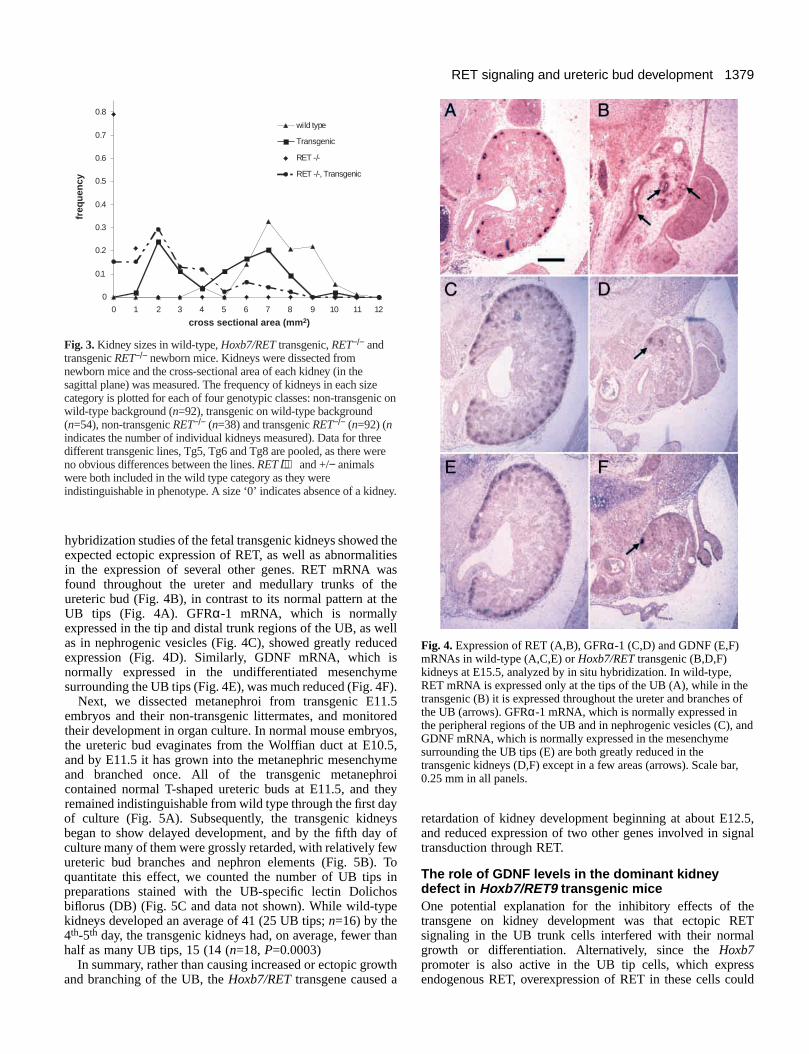
We therefore microinjected additional eggs with theHoxb7/RET-PTC2 construct, and analyzed the resulting micebefore birth. Four of the seven transgenic mice analyzed atE16-E19 displayed renal abnormalities, which were quitedistinct from the abnormalities in Hoxb7/RETtransgenic mice.The Hoxb7/RET-PTC2transgenic kidneys were close tonormal in size, and none of them had the cystic and dysplasticappearance of Hoxb7/RETkidneys. Instead, they containedabnormal nodules, either outside the kidney capsule in one case(Fig. 8A,B) or near the periphery of an otherwise normalkidney in other cases (e.g., Fig. 8C). All of the nodulesconsisted of branched tubular epithelial structures whosemorphology was reminiscent of ureteric bud branches,surrounded by undifferentiated mesenchyme or stroma, withno visible glomeruli or comma- or S-shaped bodies (Fig. 8D-F). Staining with DB lectin confirmed the UB origin of thebranched tubular structures (Fig. 9A-C). The nodules failed tostain with the lectin Tetragonolobus lotus, a specific marker forproximal tubules, confirming the morphological evidence thatthe nodules do not contain differentiated nephron elements(Fig. 9D-F).
Thus, expression of a constitutively active form of the RETtyrosine kinase under the Hoxb7promoter does not inhibitkidney development in the same way as a similar transgeneexpressing wild-type RET, but it results in the unrestrictedgrowth of the ureteric bud at the periphery of the kidney, oroutside the normal confines of the kidney.
DISCUSSION
It is known that the secreted factor GDNF and its receptor,composed of the RET receptor tyrosine kinase and a co-receptor,
A
0
0.1
0.2
0.3
0.4
0.5
0 1 2 3 4 5 6 7 8 9 10 11 12
cross sectional area (mm2)
freq
uen
cy
wild type
Transgenic
GDNF+/-, non-transgenic
GDNF+/-, Transgenic
2.78
6.34
8.23
5.45
0
2
4
6
8
10
wild type Transgenic GDNF +/-,non-
transgenic
GDNF+/-,Transgenic
Genotype
Ave
rage
Are
a
B
Fig. 6.Effect of heterozygosity for a GDNFknockout ondevelopment of Hoxb7/RETtransgenic kidneys. Hemizygoustransgenic mice were crossed with heterozygous GDNF+/− mice togenerate four possible genotypes, and the kidney sizes (sagittal crosssectional area) of all the progeny were measured at birth. The kidneysize distributions of the four genotypes are shown in A, and theaverage size and standard deviation are shown in B. Wild type, n=76;Transgenic, n=50; GDNF+/−, non-transgenic, n=50; GDNF+/−,Transgenic, n=66. For ‘Transgenic’ vs. ‘GDNF+/−, Transgenic’ the Pvalue was <10−6.

1382
GFRα-1, are essential for normal growth and branching of theureteric bud during kidney development. However, exactly howthis signal transduction pathway regulates the development ofthe UB is not well understood. RET and GFRα-1 are normallyco-expressed only in the UB tips at the periphery of the kidney,while GDNF is expressed in the surrounding metanephricmesenchyme. In this study, we sought to test the developmentalsignificance of these restricted expression patterns, bymisexpressing two different forms of the RET tyrosine kinasethroughout the UB, under the control of the Hoxb7 promoter.One transgene encoded a wild-type RET protein, and the seconda ligand-independent, constitutively active form (RET-PTC2).Because the UB tips, where RET is normally expressed, are thesites of active growth and branching, we expected that thesetransgenes might alter the pattern of branching of the UB, andindeed both of them did, but in different ways. The Hoxb7/RETtransgene, rather than causing extra or ectopic branches,inhibited kidney development in a manner reminiscent of(although less severe than) a RETknockout. Studies in whichwe manipulated the level of GDNF suggested that this defectwas caused by a decrease, rather than an increase, in RETsignaling. We suggest that ectopically expressed wild-type RETreceptors act as a sink, decreasing the available concentration ofGDNF around the UB tips. The expression of aligand-independent form of RET kinase under thesame promoter did not inhibit kidney developmentin the same manner, but instead allowed the UB togrow in abnormal patterns, and in one case tooutgrow the normal confines of the kidney. Thisimplies that the growth of the UB is normallyrestricted by the limited and spatially restrictedsupply of RET ligand.
A dominant inhibitory effect of ectopicRET expression on kidney developmentMany of the Hoxb7/RETtransgenic mice on a wild-type background had small, cystic kidneys, with adysplastic histoarchitecture similar to that seen inthe rudimentary kidneys that develop in somehomozygous RETknockout mice. Histological andorgan culture studies with transgenic fetal kidneysshowed that these defects resulted from a deficiencyin UB growth and branching beginning at aboutE12.5, after the first few branches of the UB formednormally. Although there was no specific defect innephrogenesis, there was a progressive deteriorationin kidney development, as indicated by the reducedexpression of GDNF mRNA in the metanephricmesenchyme and GFRα-1mRNA in the UB andnephrogenic vesicles. As the transgene is expressedspecifically in the UB, the effect on GDNFexpression is likely to be an indirect one, in whichdefective growth of the UB tips leads to reducedproliferation of metanephric stem cells (whichexpress GDNF), and in turn to further deteriorationof UB development (as evinced by the weakexpression of GFRα-1). This conclusion wassupported by the observation that GDNF expressionwas normal at E12.5-E13.5, before the stagewhen kidney development becomes severelycompromised (data not shown). A greatly reduced
level of GDNFmRNA in the metanephric mesenchyme has alsobeen seen in mice defective for Emx2, a homeobox geneexpressed primarily in the UB, whose disruption inhibits UBgrowth and branching (Miyamoto et al., 1997).
Why did the expression of RET under the Hoxb7 promoterinterfere with normal UB growth and branching? Thehypothetical mechanisms can be divided into two classes: (1)those that involve signaling by the exogenous RET molecules,either excessive signaling in the UB tip cells due to increasedRET expression, or ectopic signaling in regions of the UB thatdo not normally express RET; and (2) those that involveinhibition of endogenous RET function. Mechanisms that involveincreased or ectopic RET signaling appear to be ruled out by twotypes of evidence. First, two different experiments, in which wemanipulated the level of GDNF available to the transgenickidneys, indicated that the developmental defects were caused byinsufficient rather than excessive RET signaling. Addition ofextra GDNF to transgenic kidney primordia cultured in vitroimproved the growth and branching of the UB, while halving theGDNF gene dosage in vivo, by breeding the transgene into aGDNF heterozygous knockout background, exacerbated thedeleterious effect of the transgene on kidney development.Second, the Hoxb7/RET-PTC2 transgene, which encodes a
S. Srinivas and others
Fig. 7.Ability of the Hoxb7/RETtransgene to rescue renal development in RET−/−
knockout mice. (A) Excretory systems dissected from one wild-type (RET+/+)newborn mouse (left) and three RET−/− newborn mice, two of which inherited theHoxb7/RETtransgene. (B-E) Sagittal sections of wild-type newborn kidney (B),two examples of RET−/− kidneys rescued by the transgene (C,D) and one kidneyfrom a RET−/− non-transgenic newborn mouse (E). C shows an example of acomplete rescue, and D shows a partial rescue. H&E stain. Scale bars, 0.5 mm.
1383RET signaling and ureteric bud development
ligand-independent form of the RET tyrosine kinase that signalsconstitutively, did not inhibit kidney development in a similarmanner, although it caused a different type of abnormality.
There are several potential mechanisms by which the transgenemight have interfered with endogenous RET function. First, itmight have carried a mutation so that it encoded a hypomorphicreceptor, which could inhibit normal RET signaling through adominant negative mechanism. This appeared unlikely, since thetransgene was able to support normal kidney development inmice homozygous for a knockout of the endogenous RETgene.Nevertheless, to rule out this possibility, we sequenced the entirecoding sequence of the transgene, and found that it matched thepublished RET cDNA sequence (Iwamoto et al., 1993) at all butone base pair, which appears to be either apolymorphism or an error in the publishedsequence1. Second, co-expression of the RET9 andRET51 isoforms might be important for kidneydevelopment. Overexpression of RET9 (encodedby the transgene) would alter the ratio of the twoisoforms, which might somehow interfere withnormal signal transduction. This possibility hasbeen ruled out by producing Hoxb7/RET51transgenic mice and breeding this transgenetogether with the Hoxb7/RET9 transgene. Thedoubly transgenic mice showed no improvement inkidney development compared to the Hoxb7/RET9transgenic mice (unpublished data).
We propose an alternative model to explain thedominant defect, in which the ectopically expressedRET receptors on UB trunk cells can bind andsequester GDNF, thus reducing the amountavailable to the UB tip cells, for whose growth thelevel of GDNF is critical (Fig. 9). It is possible thatthe UB trunk cells, which are phenotypicallydistinct from tip cells (Bard et al., 1994; Sariola andSainio, 1997; Vainio and Muller, 1997), may beunable to respond to RET signaling with the samebiological response as UB tip cells and so cannotovercome the defect. Several observations areconsistent with this model. First, GDNF isdiffusible, as localized sources of the factor can actat a distance to stimulate UB outgrowth from theWolffian duct (Durbec et al., 1996; Sainio et al.,1997). It is therefore plausible that GDNF producedby the peripheral mesenchyme cells may bind toreceptors in more proximal regions of the UB.Second, GDNF is produced in limiting quantities,as GDNF knockout heterozygotes display reducedkidney size (Fig. 6) and occasional renal agenesis(Moore et al., 1996; Pichel et al., 1996; Sanchez etal., 1996). Therefore, a reduction in the level ofavailable GDNF could account for the observedgrowth inhibition, as well as the ability of addedGDNF to partially rescue this defect. Third, GFRα-1 is expressed not only in the UB tip cells but also
in the immediately proximal UB trunk cells (Baloh et al., 1997;Pachnis et al., 1993; Sainio et al., 1997), which may become asink for GDNF when they co-express RET. Alternatively, solubleGFRα-1 whose GPI anchor has been cleaved might also mediatethe binding of GDNF to RET receptors in more medullaryregions of the UB in the transgenic mice. Fourth, at early stages(E10.5-E12.0) when RET is normally expressed throughout theUB (Pachnis et al., 1993; Pepicelli et al., 1997), the transgene hasno deleterious effect. Only at the stage when RET expressionnormally becomes restricted to the UB tips (E12.5-E13.5) doesthe transgene begin to inhibit kidney development. A similarmechanism has been invoked to explain the dominant effects ofseveral naturally occurring mutations that cause the abnormal
Fig. 8.Uncontrolled growth of the ureteric bud in Hoxb7/RET-PTC2transgenic miceat E16.5 and E18.5. (A) Kidneys from a wild-type mouse (left) and a transgenicmouse (right) analyzed at E18.5, the latter showing multiple extra-renal nodules.(B) Section of kidney and attached nodules shown in A, H&E stain. (C) Section ofkidney from an E16.5 Hoxb7/RET-PTC2transgenic mouse, showing an abnormalnodule at the periphery of the kidney. (D,E) Higher magnification views of two areas(yellow boxes) in B, showing branched epithelial tubules (arrows) surrounded byundifferentiated mesenchyme (asterisks). (F) Higher magnification view of thenodule in C, containing ureteric bud-like tubules and undifferentiated mesenchyme.Scale bars, 0.1 mm.
1Nucleotide 521 of RET was a T, whereas in the published sequence itis a C. Several RET cDNA clones were sequenced in this region, andall had a T at position 521. This would encode a Phe at amino acid 174,instead of the Ser in the published mouse RET sequence. However,human and chicken RET both have a Phe at this position (Iwamoto etal., 1993; Schuchardt et al., 1995; Takahashi et al., 1989; Takahashi etal. 1988).

1384
expression of c-kit, a tyrosine kinase receptor for the Steel factor,in somites or other ectopic sites. In these mutants, which includeWsash, Wbandedand Patch, ectopically expressed c-kit is believedto sequester Steel, which normally promotes the survival orproliferation of migrating melanoblasts, resulting in pigmentationdefects (Duttlinger et al., 1995, 1993; Kluppel et al., 1997;Wehrle-Haller et al., 1996).
One question we initially posed was why the normalexpression of RET is so tightly restricted to a small populationof cells at the tips of the UB. We initially hypothesized that thispattern of gene regulation might be part of a mechanism to limitthe sites of UB branching. However, in the cultured transgenickidneys, either in normal medium or with added GDNF, weobserved no signs of abnormal branching or unusual growth ofUB trunks. Furthermore, expression of a constitutively activeform of RET throughout the UB did not visibly perturb theoverall histoarchitecture of the kidney, which might have beenexpected if it induced ectopic branching. Therefore, while ourdata do not rule out the initial hypothesis, they suggest another
explanation. Because the RET ligand GDNF is produced inlimiting quantity and is diffusible, the expression of RET in aless tightly regulated pattern (e.g., throughout the UB) wouldresult in binding and sequestering of the ligand by UB trunkcells at the expense of the UB tip cells.
The Hoxb7/RET transgene can support kidneydevelopment in RET knockout miceWhen Hoxb7/RETtransgenic mice were bred with RETknockoutmice, the transgene was able to rescue kidney development insome of the newborn RET−/− mice (Figs 3 and 7). Fifteen percentof the kidneys in these mice were normal in size and histology,and an additional 55% were partially rescued (i.e., smaller thannormal but larger than RET−/− kidneys, displaying mild tomoderate dysplasia). The incomplete rescue in some animals maybe due to two competing effects of the transgene: the ability ofexogenous RET to substitute for endogenous RET in the UB tips,and the incompletely penetrant, inhibitory effect of theectopically expressed RET. The Hoxb7/RETtransgene did not
S. Srinivas and others
Fig. 9.Lectin staining of abnormal nodules in Hoxb7/RET-PTC2transgenic kidneys. (A) Dolichos bifloruslectin (DB) specificallystains the collecting ducts in a normal kidney. (B,C) Dolichosbiflorus lectin staining of epithelia in nodules from an E18.5 kidney(shown in Fig. 8A and B) confirms their origin from the ureteric bud.(D) Tetragonolobus lotuslectin specifically stains the maturingproximal tubules in the inner cortex of an E16.5 kidney.(E,F) Epithelia in abnormal nodules are negative withTetragonolobus lotusstain. Hematoxylin counterstain. Scale bars,0.05 mm.
Wild Type TransgenicHoxb7/RET
GDNF
Activated RET/GFRα-1
Inactive RETUB tip cells
UB trunk cellsFig. 10.A model for the growth-inhibitory effect of ectopic RETexpression in the developing kidneys of Hoxb7/RETtransgenic mice.In the wild type, the RET receptor tyrosine kinase (Y symbols) isexpressed only at the distal tips of the UB branches (yellow regions),while GDNF (red triangles) is expressed by the surroundingmesenchyme cells at the periphery of the metanephros. GFRα-1, aco-receptor required for binding of GDNF to RET (not indicated indiagram), is co-expressed with RET in the UB tip cells, but isexpressed in more proximal regions of the UB as well, and may alsobe present in a soluble form. In the presence of normal levels ofGDNF, many of the RET receptors bind GDNF and are activated(black Ysymbols). In the transgenic kidney, RET is no longerrestricted to the UB tips, and some of the GDNF molecules bind toRET receptors in more proximal regions of the UB (UB trunks,shown in blue). This reduces the amount of GDNF available to bindto RET receptors on the UB tip cells, with the result that many moreRET receptors on the UB tips fail to be activated (grey Y symbols),and growth stimulation of the UB is sub-optimal. Evidence insupport of this model is described in the text.

1385RET signaling and ureteric bud development
rescue the other defects in the RET−/− mice (e.g., intestinalaganglionosis), presumably because it is not expressed in manyof the normal sites of RETexpression in the developing nervoussystem (Kress et al., 1990; Vogels et al., 1993).
Two important conclusions can be drawn from this result.First, it formally demonstrates that the renal agenesis phenotypein RETknockout mice is due to the RETmutation, and not to aninadvertently introduced mutation in another gene linked to RET.Second, it demonstrates that the RET9 isoform is sufficient tosupport normal kidney development. RET9 and RET51 differonly at the C terminus, where the last 9 amino acids of RET9are replaced by a different 51 amino acid tail in RET51. Onedifference between the isoforms is that only RET51 containsbinding sites for Grb2, an adaptor protein that links RET to Rasand the MAP kinase pathway (Borrello et al., 1994; Liu et al.,1996; Lorenzo et al., 1997). Apparently, the ability to bind Grb2is not essential for the role of RET signaling in kidneydevelopment. This may be because both RET isoforms havebinding sites for Shc, another adaptor protein through whichRET signaling can activate Ras (Arighi et al., 1997; Asai et al.,1996; Lorenzo et al., 1997; Ohiwa et al., 1997). Our observationis consistent with previous findings that RET9 and RET51 areboth biologically active in a number of systems that depend onRas activation, including fibroblast transformation and inductionof neuronal differentiation in PC12 cells (Asai et al., 1995;Iwashita et al., 1996; Rossel et al., 1997; Santoro et al., 1995).
A ligand-independent form of RET uncouplesureteric bud growth from the growth of thenephrogenic zoneExpression of RET-PTC2 under the Hoxb7promoter resulted inabnormal growth of the UB in localized nodules at the peripheryof the kidney, and in one case, in multiple nodules outside thekidney. Surprisingly, with the exception of these nodules, thekidneys of newborn transgenic mice were histologically normal,and their size was unaffected. This suggests that throughout theearly stages of kidney development, the expression of aconstitutively active RET kinase had little or no effect, perhapsbecause there is already sufficient GDNF to maintain RETsignaling in the UB tips. As noted above, this result also suggeststhat ectopic RET signaling in the UB trunk cells isinconsequential. The ability of the UB to eventually outgrow therest of the kidney, forming nodules that protrude through thenephrogenic zone, suggests that at later stages of kidneydevelopment, the centripetal growth of the UB tips is normallyrestricted by the availability of GDNF. GDNF is expressed by athin layer of mesenchymal cells near the periphery of the kidney,and any UB tips that grow through this layer would normallyoutgrow their source of ligand. In transgenic mice expressing aligand-independent RET kinase, this level of regulation of UBgrowth is apparently circumvented. This indicates that RETsignaling is not only necessaryfor UB development (as shownby the RETand GDNFknockouts), but that RET signalingactively stimulates the growth of the UB. In previous in vitrostudies, addition of GDNF to the culture medium was observedto stimulate UB cell proliferation within intact metanephroi(Pepicelli et al., 1997), but not in isolated ureteric buds (Sainioet al., 1997). While our observations are consistent with amitogenic role for GDNF/RET signaling, other factors producedby the kidney or surrounding tissues may also be required forthe stimulation of UB growth.
It is interesting that the tubular UB elements in the noduleswere surrounded only by undifferentiated mesenchymal orstromal cells, lacking mature glomeruli or immaturenephrogenic vesicles. One likely explanation is that the UB hasoutgrown the nephrogenic stem cells at the periphery of thekidney, or (in the animal with extra-renal nodules) has growninto regions of mesenchyme lacking any nephrogenic potential.Alternatively, the abnormally growing UB may have lost theability to induce nephrogenesis.
The authors would like to thank Doris Herzlinger and CathyMendelsohn for critical reviews of the manuscript, Qais Al-Awqati,Vassilis Pachnis and Jonathan Barasch for helpful discussions, and TonyWard and Xiaolin Liang for excellent technical assistance. We alsothank Vassilis Pachnis, Mariano Barbacid, Bristol-Myers Squibb,Jacqueline Deschamps, Marco Pierotti and Andreas Zimmer forreagents. This work was supported by a grant from the NIDDK to F. C.
REFERENCES
Arighi, E., Alberti, L., Torriti, F., Ghizzoni, S., Rizzetti, M. G., Pelicci, G.,Pasini, B., Bongarzone, I., Piutti, C., Pierotti, M. A. et al.(1997).Identification of Shc docking site on Ret tyrosine kinase. Oncogene14, 773-782.
Asai, N., Iwashita, T., Matsuyama, M. and Takahashi, M. (1995).Mechanism of activation of the ret proto-oncogene by multiple endocrineneoplasia 2A mutations. Mol. Cell. Biol.15, 1613-1619.
Asai, N., Murakami, H., Iwashita, T. and Takahashi, M.(1996). A mutationat tyrosine 1062 in MEN2A-Ret and MEN2B-Ret impairs their transformingactivity and association with shc adaptor proteins. J. Biol. Chem.271,17644-17649.
Baloh, R. H., Tansey, M. G., Golden, J. P., Creedon, D. J., Heuckeroth, R.O., Keck, C. L., Zimonjic, D. B., Popescu, N. C., Johnson, E. M., Jr. andMilbrandt, J. (1997). TrnR2, a novel receptor that mediates neurturin andGDNF signaling through Ret. Neuron18, 793-802.
Bard, J. B., McConnell, J. E. and Davies, J. A.(1994). Towards a geneticbasis for kidney development. Mech. Dev.48, 3-11.
Bongarzone, I., Monzini, N., Borrello, M. G., Carcano, C., Ferraresi, G.,Arighi, E., Mondellini, P., Della, P. G. and Pierotti, M. A. (1993). Molecularcharacterization of a thyroid tumor-specific transforming sequence formed bythe fusion of ret tyrosine kinase and the regulatory subunit RI alpha of cyclicAMP-dependent protein kinase A. Mol. Cell. Biol.13, 358-366.
Borrello, M. G., Pelicci, G., Arighi, E., De Filippis, L., Greco, A.,Bongarzone, I., Rizzetti, M., Pelicci, P. G. and Pierotti, M. A.(1994).The oncogenic versions of the Ret and Trk tyrosine kinases bind Shc andGrb2 adaptor proteins. Oncogene9, 1661-1668.
D’Agati, V. and Trudel, M. (1992). Lectin characterization of cystogenesisin the SBM transgenic model of polycystic kidney disease. J. Am. Soc.Nephrol.3, 975-983.
Durbec, P., Marcos-Gutierrez, C. V., Kilkenny, C., Grigoriou, M.,Wartiowaara, K., Suvanto, P., Smith, D., Ponder, B., Costantini, F.,Saarma, M. et al. (1996). GDNF signalling through the Ret receptortyrosine kinase. Nature381, 789-793.
Durick, K., Yao, V. J., Borrello, M. G., Bongarzone, I., Pierotti, M. A. andTaylor, S. S.(1995). Tyrosines outside the kinase core and dimerization arerequired for the mitogenic activity of RET/ptc2. J. Biol. Chem. 270, 24642-24645.
Duttlinger, R., Manova, K., Berrozpe, G., Chu, T. Y., DeLeon, V.,Timokhina, I., Chaganti, R. S., Zelenetz, A. D., Bachvarova, R. F. andBesmer, P.(1995). The Wsh and Ph mutations affect the c-kit expressionprofile: c-kit misexpression in embryogenesis impairs melanogenesis inWsh and Ph mutant mice. Proc. Natl. Acad. Sci. USA92, 3754-3758.
Duttlinger, R., Manova, K., Chu, T. Y., Gyssler, C., Zelenetz, A. D.,Bachvarova, R. F. and Besmer, P.(1993). W-sash affects positive andnegative elements controlling c-kit expression: ectopic c-kit expression at sitesof kit-ligand expression affects melanogenesis. Development118, 705-717.
Ekblom, P. (1992). Renal Development. In The Kidney: Physiology andPathophysiology, (ed. Seldin, D. W. and Giebisch, G.), pp. 475-501. NewYork: Raven Press.
Enomoto, H., Araki, T., Jackman, A., Heuckeroth, R. O., Snider, W. D.,

1386
Johnson, E. M. J. and Milbrandt, J. (1998). GFRα1-deficient mice havedeficits in the enteric nervous system and kidneys. Neuron21, 317-324.
Erickson, R. A. (1968). Inductive interactions in the development of themouse metanephros. J. Exp. Zool.169, 33-42.
Grobstein, C.(1953). Inductive epithelio-mesenchymal interaction in culturedorgan rudiments of the mouse. Science118, 52-55.
Grobstein, C. (1955). Inductive interaction in the development of the mousemetanephros. J. Exp. Zool.130, 319-340.
Hellmich, H. L., Kos, L., Cho, E. S., Mahon, K. A. and Zimmer, A.(1996).Embryonic expression of glial cell-line derived neurotrophic factor (GDNF)suggests multiple developmental roles in neural differentiation andepithelial-mesenchymal interactions. Mech. Dev.54, 95-105.
Hogan, B., Bedington, R., Costantini, F. and Lacy, E.(1994). Manipulatingthe Mouse Embryo: A Laboratory Manual.Cold Spring Harbor: Cold SpringHarbor Laboratory.
Iwamoto, T., Taniguchi, M., Asai, N., Ohkusu, K., Nakashima, I. andTakahashi, M. (1993). cDNA cloning of mouse ret proto-oncogene and itssequence similarity to the cadherin superfamily. Oncogene8, 1087-1091.
Iwashita, T., Asai, N., Murakami, H., Matsuyama, M. and Takahashi, M.(1996). Identification of tyrosine residues that are essential for transformingactivity of the ret proto-oncogene with MEN2A or MEN2B mutation.Oncogene12, 481-487.
Jing, S., Wen, D., Yu, Y., Holst, P. L., Luo, Y., Fang, M., Tamir, R., Antonio,L., Hu, Z., Cupples, R. et al.(1996). GDNF-induced activation of the retprotein tyrosine kinase is mediated by GDNFR-alpha, a novel receptor forGDNF. Cell 85, 1113-1124.
Kluppel, M., Nagle, D. L., Bucan, M. and Bernstein, A.(1997). Long-rangegenomic rearrangements upstream of Kit dysregulate the developmentalpattern of Kit expression in W57 and Wbanded mice and interfere withdistinct steps in melanocyte development. Development124, 65-77.
Kress, C., Vogels, R., De, G. W., Bonnerot, C., Meijlink, F., Nicolas, J. F.and Deschamps, J.(1990). Hox-2.3 upstream sequences mediate lacZexpression in intermediate mesoderm derivatives of transgenic mice.Development109, 775-786.
Lechner, M. S. and Dressler, G. R.(1997). The molecular basis of embryonickidney development. Mech. Dev.62, 105-120.
Lin, L. F., Doherty, D. H., Lile, J. D., Bektesh, S. and Collins, F.(1993).GDNF: a glial cell line-derived neurotrophic factor for midbraindopaminergic neurons. Science260, 1130-1132.
Liu, X., Vega, Q. C., Decker, R. A., Pandey, A., Worby, C. A. and Dixon,J. E. (1996). Oncogenic RET receptors display differentautophosphorylation sites and substrate binding specificities. J. Biol. Chem.271, 5309-5312.
Lorenzo, M. J., Gish, G. D., Houghton, C., Stonehouse, T. J., Pawson, T.,Ponder, B. A. and Smith, D. P.(1997). RET alternate splicing influencesthe interaction of activated RET with the SH2 and PTB domains of Shc, andthe SH2 domain of Grb2. Oncogene14, 763-771.
Miyamoto, N., Yoshida, M., Kuratani, S., Matsuo, I. and Aizawa, S.(1997).Defects of urogenital development in mice lacking Emx2. Development124,1653-1664.
Moore, M. W., Klein, R. D., Farinas, I., Sauer, H., Armanini, M., Phillips,H., Reichardt, L. F., Ryan, A. M., Carver-Moore, K. and Rosenthal, A.(1996). Renal and neuronal abnormalities in mice lacking GDNF. Nature382, 76-79.
Ohiwa, M., Murakami, H., Iwashita, T., Asai, N., Iwata, Y., Imai, T.,Funahashi, H., Takagi, H. and Takahashi, M.(1997). Characterization ofRet-Shc-Grb2 complex induced by GDNF, MEN 2A, and MEN 2Bmutations. Biochem. Biophys. Res. Commun.237, 747-751.
Pachnis, V., Mankoo, B. S. and Costantini, F.(1993). Expression of the c-retproto-oncogene during mouse embryogenesis. Development119, 1005-1017.
Pepicelli, C. V., Kispert, A., Rowitch, D. H. and McMahon, A. P.(1997).GDNF induces branching and increased cell proliferation in the ureter ofthe mouse. Dev. Biol.192, 193-198.
Pichel, J. G., Shen, L., Sheng, H. Z., Granholm, A. C., Drago, J., Grinberg,A., Lee, E. J., Huang, S. P., Saarma, M., Hoffer, B. J. et al.(1996).Defects in enteric innervation and kidney development in mice lackingGDNF. Nature382, 73-76.
Robertson, K. and Mason, I.(1997). The GDNF-RET signalling partnership.Trends Genet.13, 1-3.
Rossel, M., Pasini, A., Chappuis, S., Geneste, O., Fournier, L., Schuffenecker,I., Takahashi, M., van Grunsven, L. A., Urdiales, J. L., Rudkin, B. B. etal. (1997). Distinct biological properties of two RET isoforms activated byMEN 2A and MEN 2B mutations. Oncogene14, 265-275.
Sainio, K., Suvanto, P., Davies, J., Wartiovaara, J., Wartiovaara, K.,
Saarma, M., Arumae, U., Meng, X., Lindahl, M., Pachnis, V. et al.(1997). Glial-cell-line-derived neurotrophic factor is required for budinitiation from ureteric epithelium. Development124, 4077-4087.
Sanchez, M. P., Silos-Santiago, I., Frisen, J., He, B., Lira, S. A. andBarbacid, M. (1996). Renal agenesis and the absence of enteric neurons inmice lacking GDNF. Nature382, 70-73.
Santoro, M., Carlomagno, F., Romano, A., Bottaro, D. P., Dathan, N. A.,Grieco, M., Fusco, A., Vecchio, G., Matoskova, B., Kraus, M. H. et al.(1995). Activation of RET as a dominant transforming gene by germlinemutations of MEN2A and MEN2B. Science267, 381-383.
Sariola, H. and Sainio, K.(1997). The tip-top branching ureter. Curr. Opin.Cell. Biol. 9, 877-884.
Saxen, L. (1970). Failure to demonstrate tubule induction in a heterologousmesenchyme. Dev. Biol.23, 511-523.
Saxen, L. (1987). Organogenesis of the Kidney. Cambridge: CambridgeUniversity Press.
Schuchardt, A., D’Agati, V., Larsson-Blomberg, L., Costantini, F. andPachnis, V. (1994). Defects in the kidney and enteric nervous system ofmice lacking the tyrosine kinase receptor Ret. Nature367, 380-383.
Schuchardt, A., D’Agati, V., Pachnis, V. and Costantini, F.(1996). Renalagenesis and hypodysplasia in ret-k- mutant mice result from defects inureteric bud development. Development122, 1919-1929.
Schuchardt, A., Srinivas, S., Pachnis, V. and Costantini, F.(1995). Isolationand characterization of a chicken homolog of the c-ret proto-oncogene.Oncogene10, 641-649.
Suvanto, P., Hiltunen, J. O., Arumae, U., Moshnyakov, M., Sariola, H.,Sainio, K. and Saarma, M.(1996). Localization of glial cell line-derivedneurotrophic factor (GDNF) mRNA in embryonic rat by in situhybridization. Eur. J. Neurosci.8, 816-822.
Suvanto, P., Wartiovaara, K., Lindahl, M., Arumae, U., Moshnyakov, M.,Horelli-Kuitunen, N., Airaksinen, M. S., Palotie, A., Sariola, H. andSaarma, M. (1997). Cloning, mRNA distribution and chromosomallocalisation of the gene for glial cell line-derived neurotrophic factor receptorbeta, a homologue to GDNFR-alpha. Hum. Mol. Genet.6, 1267-1273.
Tahira, T., Ishizaka, Y., Itoh, F., Sugimura, T. and Nagao, M.(1990).Characterization of ret proto-oncogene mRNAs encoding two isoforms of theprotein product in a human neuroblastoma cell line. Oncogene5, 97-102.
Takahashi, M., Buma, Y. and Hiai, H.(1989). Isolation of ret proto-oncogenecDNA with an amino-terminal signal sequence. Oncogene4, 805-806.
Takahashi, M., Buma, Y., Iwamoto, T., Inaguma, Y., Ikeda, H. and Hiai,H. (1988). Cloning and expression of the ret proto-oncogene encoding atyrosine kinase with two potential transmembrane domains. Oncogene3,571-578.
Takahashi, M. and Cooper, G. M.(1987). ret transforming gene encodes afusion protein homologous to tyrosine kinases. Mol Cell Biol 7, 1378-1385.
Treanor, J. J., Goodman, L., de Sauvage, F., Stone, D. M., Poulsen, K. T.,Beck, C. D., Gray, C., Armanini, M. P., Pollock, R. A., Hefti, F. et al.(1996).Characterization of a multicomponent receptor for GDNF. Nature382, 80-83.
Trupp, M., Arenas, E., Fainzilber, M., Nilsson, A. S., Sieber, B. A.,Grigoriou, M., Kilkenny, C., Salazar-Grueso, E., Pachnis, V., Arumae,U. et al. (1996). Functional receptor for GDNF encoded by the c-ret proto-oncogene. Nature381, 785-788.
Tsuzuki, T., Takahashi, M., Asai, N., Iwashita, T., Matsuyama, M. andAsai, J. (1995). Spatial and temporal expression of the ret proto-oncogeneproduct in embryonic, infant and adult rat tissues. Oncogene10, 191-198.
Vainio, S. and Muller, U. (1997). Inductive tissue interactions, cell signaling,and the control of kidney organogenesis. Cell90, 975-978.
Vega, Q. C., Worby, C. A., Lechner, M. S., Dixon, J. E. and Dressler, G.R. (1996). Glial cell line-derived neurotrophic factor activates the receptortyrosine kinase RET and promotes kidney morphogenesis. Proc. Natl. Acad.Sci. USA93, 10657-10661.
Vogels, R., Charite, J., de Graff, W. and Deschamps, J.(1993). Proximalcis-acting elements cooperate to set Hoxb-7 (Hox-2.3) expressionboundaries in transgenic mice. Development118, 71-82.
Wehrle-Haller, B., Morrison-Graham, K. and Weston, J. A.(1996). Ectopicc-kit expression affects the fate of melanocyte precursors in Patch mutantembryos. Dev. Biol.177, 463-474.
Wilkinson, D. G. (1992). Whole mount in situ hybridization of vertebrateembryos. In In Situ Hybridization: A Practical Approach(ed. Wilkinson, D.G.), pp. 75-83. Oxford: IRL Press.
Worby, C. A., Vega, Q. C., Zhao, Y., Chao, H. H. J., Seasholtz, A. F. andDixon, J. E. (1996). Glial cell line-derived neurotrophic factor signalsthrough the RET receptor and activates mitogen-activated protein kinase. J.Biol. Chem.271, 23619-23622.
S. Srinivas and others