Resolution of Abfraction-Associated Gingival Fenestration ...
5
Case Report Resolution of Abfraction-Associated Gingival Fenestration Utilizing Connective Tissue Grafting Braydon Haskell, J. Kobi Stern, Jordan Ghiassi, Andrew Kurialacherry , Sadja Gaud-Quintana, and Mark E. Peacock The Dental College of Georgia at Augusta University, Augusta, GA, USA Correspondence should be addressed to Andrew Kurialacherry; [email protected] Received 7 February 2019; Revised 13 May 2019; Accepted 23 May 2019; Published 12 June 2019 Academic Editor: Mine Dündar Copyright © 2019 Braydon Haskell et al. This is an open access article distributed under the Creative Commons Attribution License, which permits unrestricted use, distribution, and reproduction in any medium, provided the original work is properly cited. Introduction. Gingival fenestration (GF) is a distinct clinical entity of uncertain etiology that is seldom documented in the literature. It has been associated mainly with submucosal mechanical irritants such as calculus that subsequently create an opening in the oral soft tissue, usually at facial anterior sites. Surgical correction may be indicated to address functional and/or esthetic concerns. Case Presentation. The patient, a 74-year-old male, presented to the clinic with a chief complaint of “something is poking through my gum.” Clinical exam revealed a gingival fenestration on the facial of tooth #11, associated with what appeared to be a pronounced noncarious cervical lesion (NCCL). Surgical treatment consisted of a connective tissue graft and odontoplasty of the sharp protruding edge of the root surface. Healing was uneventful with excellent closure of the fenestration and no evidence of recurrence after 18 months of follow-up. Conclusion. GF is a perforation of the mucosa typically associated with underlying sharp mechanical etiology. This report describes a fenestration that developed from a probable abfractive lesion, which later was successfully closed and exhibits long-term stability. 1. Introduction Gingival fenestration is an opening through oral keratinized mucosa that is reported infrequently, possibly due to lack of symptoms and/or patient unawareness [1]. Unlike fenestra- tions, the gingival margin for a dehiscence is noncontinuous and apically positioned. Although previously mentioned in the literature, Lane in 1977 was the first to describe it as a dis- tinct pathologic entity [2]. The etiological basis for GF has not been completely elucidated but is probably multifactorial. Local factors that have been reported to be associated with GF include plaque/calculus [1–4], cervical enamel pro- jections (CEP) [5], tooth malpositioning [1, 2], trauma [3], chewing habits/foreign body (gutka) [6], occlusal factors [7], and implant fixture threads [8]. Anatomic factors associ- ated with noncarious cervical lesions (abfraction, abrasion) could possibly lead to sharp irregular cervical “notching,” [9] resulting in a soft tissue fenestration [10]. Almost all reported cases of GF occurred on the facial surfaces of incisor teeth. Soft tissue fenestrations linked with NCCLs are almost never documented. NCCLs may have very rough and ser- rated line angles that could lead to GF. A strong relationship between NCCLs and occlusal overload (bruxism, grinding) has been reported [11], and patients with group function can have NCCLs occurring six times more frequently than those with cuspid-protected occlusion [12]. The theoretical concept of abfraction, described by Lee and Eakle [13], is controversial, but this phenomenon is still supported by a multitude of practitioners. Most gingival fenestrations described have occurred on the facial surfaces of thin unattached gingiva and are associ- ated with heavy calculus deposits. This case report docu- ments a GF linked to a subgingival NCCL, along with its surgical treatment and follow-up. 2. Case Report A 74-year-old Caucasian male was referred to the Peri- odontics Clinic of the Dental College of Georgia, Augusta Hindawi Case Reports in Dentistry Volume 2019, Article ID 6810670, 4 pages https://doi.org/10.1155/2019/6810670
Transcript of Resolution of Abfraction-Associated Gingival Fenestration ...
Case ReportResolution of Abfraction-Associated Gingival FenestrationUtilizing Connective Tissue Grafting
Braydon Haskell, J. Kobi Stern, Jordan Ghiassi, Andrew Kurialacherry ,Sadja Gaud-Quintana, and Mark E. Peacock
The Dental College of Georgia at Augusta University, Augusta, GA, USA
Correspondence should be addressed to Andrew Kurialacherry; [email protected]
Received 7 February 2019; Revised 13 May 2019; Accepted 23 May 2019; Published 12 June 2019
Academic Editor: Mine Dündar
Copyright © 2019 Braydon Haskell et al. This is an open access article distributed under the Creative Commons AttributionLicense, which permits unrestricted use, distribution, and reproduction in any medium, provided the original work isproperly cited.
Introduction. Gingival fenestration (GF) is a distinct clinical entity of uncertain etiology that is seldom documented in the literature.It has been associated mainly with submucosal mechanical irritants such as calculus that subsequently create an opening in the oralsoft tissue, usually at facial anterior sites. Surgical correction may be indicated to address functional and/or esthetic concerns. CasePresentation. The patient, a 74-year-old male, presented to the clinic with a chief complaint of “something is poking through mygum.” Clinical exam revealed a gingival fenestration on the facial of tooth #11, associated with what appeared to be apronounced noncarious cervical lesion (NCCL). Surgical treatment consisted of a connective tissue graft and odontoplasty of thesharp protruding edge of the root surface. Healing was uneventful with excellent closure of the fenestration and no evidence ofrecurrence after 18 months of follow-up. Conclusion. GF is a perforation of the mucosa typically associated with underlyingsharp mechanical etiology. This report describes a fenestration that developed from a probable abfractive lesion, which later wassuccessfully closed and exhibits long-term stability.
1. Introduction
Gingival fenestration is an opening through oral keratinizedmucosa that is reported infrequently, possibly due to lack ofsymptoms and/or patient unawareness [1]. Unlike fenestra-tions, the gingival margin for a dehiscence is noncontinuousand apically positioned. Although previously mentioned inthe literature, Lane in 1977 was the first to describe it as a dis-tinct pathologic entity [2]. The etiological basis for GF hasnot been completely elucidated but is probably multifactorial.
Local factors that have been reported to be associatedwith GF include plaque/calculus [1–4], cervical enamel pro-jections (CEP) [5], tooth malpositioning [1, 2], trauma [3],chewing habits/foreign body (gutka) [6], occlusal factors[7], and implant fixture threads [8]. Anatomic factors associ-ated with noncarious cervical lesions (abfraction, abrasion)could possibly lead to sharp irregular cervical “notching,”[9] resulting in a soft tissue fenestration [10]. Almost allreported cases of GF occurred on the facial surfaces ofincisor teeth.
Soft tissue fenestrations linked with NCCLs are almostnever documented. NCCLs may have very rough and ser-rated line angles that could lead to GF. A strong relationshipbetween NCCLs and occlusal overload (bruxism, grinding)has been reported [11], and patients with group functioncan have NCCLs occurring six times more frequently thanthose with cuspid-protected occlusion [12]. The theoreticalconcept of abfraction, described by Lee and Eakle [13], iscontroversial, but this phenomenon is still supported by amultitude of practitioners.
Most gingival fenestrations described have occurred onthe facial surfaces of thin unattached gingiva and are associ-ated with heavy calculus deposits. This case report docu-ments a GF linked to a subgingival NCCL, along with itssurgical treatment and follow-up.
2. Case Report
A 74-year-old Caucasian male was referred to the Peri-odontics Clinic of the Dental College of Georgia, Augusta
HindawiCase Reports in DentistryVolume 2019, Article ID 6810670, 4 pageshttps://doi.org/10.1155/2019/6810670
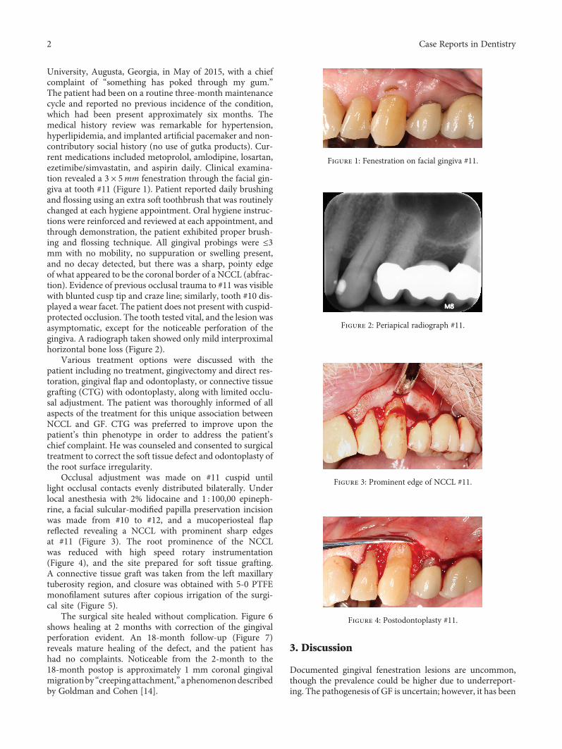
University, Augusta, Georgia, in May of 2015, with a chiefcomplaint of “something has poked through my gum.”The patient had been on a routine three-month maintenancecycle and reported no previous incidence of the condition,which had been present approximately six months. Themedical history review was remarkable for hypertension,hyperlipidemia, and implanted artificial pacemaker and non-contributory social history (no use of gutka products). Cur-rent medications included metoprolol, amlodipine, losartan,ezetimibe/simvastatin, and aspirin daily. Clinical examina-tion revealed a 3 × 5mm fenestration through the facial gin-giva at tooth #11 (Figure 1). Patient reported daily brushingand flossing using an extra soft toothbrush that was routinelychanged at each hygiene appointment. Oral hygiene instruc-tions were reinforced and reviewed at each appointment, andthrough demonstration, the patient exhibited proper brush-ing and flossing technique. All gingival probings were ≤3mm with no mobility, no suppuration or swelling present,and no decay detected, but there was a sharp, pointy edgeof what appeared to be the coronal border of a NCCL (abfrac-tion). Evidence of previous occlusal trauma to #11 was visiblewith blunted cusp tip and craze line; similarly, tooth #10 dis-played a wear facet. The patient does not present with cuspid-protected occlusion. The tooth tested vital, and the lesion wasasymptomatic, except for the noticeable perforation of thegingiva. A radiograph taken showed only mild interproximalhorizontal bone loss (Figure 2).
Various treatment options were discussed with thepatient including no treatment, gingivectomy and direct res-toration, gingival flap and odontoplasty, or connective tissuegrafting (CTG) with odontoplasty, along with limited occlu-sal adjustment. The patient was thoroughly informed of allaspects of the treatment for this unique association betweenNCCL and GF. CTG was preferred to improve upon thepatient’s thin phenotype in order to address the patient’schief complaint. He was counseled and consented to surgicaltreatment to correct the soft tissue defect and odontoplasty ofthe root surface irregularity.
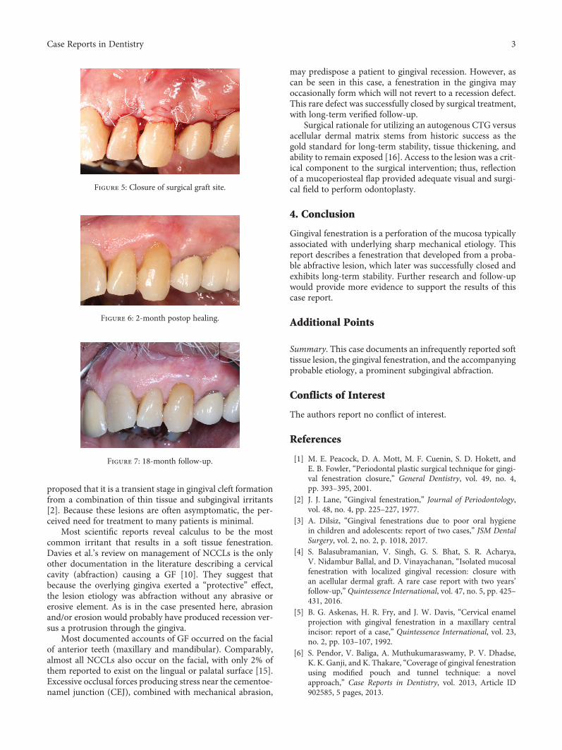
Occlusal adjustment was made on #11 cuspid untillight occlusal contacts evenly distributed bilaterally. Underlocal anesthesia with 2% lidocaine and 1 : 100,00 epineph-rine, a facial sulcular-modified papilla preservation incisionwas made from #10 to #12, and a mucoperiosteal flapreflected revealing a NCCL with prominent sharp edgesat #11 (Figure 3). The root prominence of the NCCLwas reduced with high speed rotary instrumentation(Figure 4), and the site prepared for soft tissue grafting.A connective tissue graft was taken from the left maxillarytuberosity region, and closure was obtained with 5-0 PTFEmonofilament sutures after copious irrigation of the surgi-cal site (Figure 5).
The surgical site healed without complication. Figure 6shows healing at 2 months with correction of the gingivalperforation evident. An 18-month follow-up (Figure 7)reveals mature healing of the defect, and the patient hashad no complaints. Noticeable from the 2-month to the18-month postop is approximately 1 mm coronal gingivalmigrationby “creeping attachment,”aphenomenondescribedby Goldman and Cohen [14].
3. Discussion
Documented gingival fenestration lesions are uncommon,though the prevalence could be higher due to underreport-ing. The pathogenesis of GF is uncertain; however, it has been
Figure 1: Fenestration on facial gingiva #11.
Figure 2: Periapical radiograph #11.
Figure 3: Prominent edge of NCCL #11.
Figure 4: Postodontoplasty #11.
2 Case Reports in Dentistry
proposed that it is a transient stage in gingival cleft formationfrom a combination of thin tissue and subgingival irritants[2]. Because these lesions are often asymptomatic, the per-ceived need for treatment to many patients is minimal.
Most scientific reports reveal calculus to be the mostcommon irritant that results in a soft tissue fenestration.Davies et al.’s review on management of NCCLs is the onlyother documentation in the literature describing a cervicalcavity (abfraction) causing a GF [10]. They suggest thatbecause the overlying gingiva exerted a “protective” effect,the lesion etiology was abfraction without any abrasive orerosive element. As is in the case presented here, abrasionand/or erosion would probably have produced recession ver-sus a protrusion through the gingiva.
Most documented accounts of GF occurred on the facialof anterior teeth (maxillary and mandibular). Comparably,almost all NCCLs also occur on the facial, with only 2% ofthem reported to exist on the lingual or palatal surface [15].Excessive occlusal forces producing stress near the cementoe-namel junction (CEJ), combined with mechanical abrasion,
may predispose a patient to gingival recession. However, ascan be seen in this case, a fenestration in the gingiva mayoccasionally form which will not revert to a recession defect.This rare defect was successfully closed by surgical treatment,with long-term verified follow-up.
Surgical rationale for utilizing an autogenous CTG versusacellular dermal matrix stems from historic success as thegold standard for long-term stability, tissue thickening, andability to remain exposed [16]. Access to the lesion was a crit-ical component to the surgical intervention; thus, reflectionof a mucoperiosteal flap provided adequate visual and surgi-cal field to perform odontoplasty.
4. Conclusion
Gingival fenestration is a perforation of the mucosa typicallyassociated with underlying sharp mechanical etiology. Thisreport describes a fenestration that developed from a proba-ble abfractive lesion, which later was successfully closed andexhibits long-term stability. Further research and follow-upwould provide more evidence to support the results of thiscase report.
Additional Points
Summary. This case documents an infrequently reported softtissue lesion, the gingival fenestration, and the accompanyingprobable etiology, a prominent subgingival abfraction.
Conflicts of Interest
The authors report no conflict of interest.
References
[1] M. E. Peacock, D. A. Mott, M. F. Cuenin, S. D. Hokett, andE. B. Fowler, “Periodontal plastic surgical technique for gingi-val fenestration closure,” General Dentistry, vol. 49, no. 4,pp. 393–395, 2001.
[2] J. J. Lane, “Gingival fenestration,” Journal of Periodontology,vol. 48, no. 4, pp. 225–227, 1977.
[3] A. Dilsiz, “Gingival fenestrations due to poor oral hygienein children and adolescents: report of two cases,” JSM DentalSurgery, vol. 2, no. 2, p. 1018, 2017.
[4] S. Balasubramanian, V. Singh, G. S. Bhat, S. R. Acharya,V. Nidambur Ballal, and D. Vinayachanan, “Isolated mucosalfenestration with localized gingival recession: closure withan acellular dermal graft. A rare case report with two years’follow-up,” Quintessence International, vol. 47, no. 5, pp. 425–431, 2016.
[5] B. G. Askenas, H. R. Fry, and J. W. Davis, “Cervical enamelprojection with gingival fenestration in a maxillary centralincisor: report of a case,” Quintessence International, vol. 23,no. 2, pp. 103–107, 1992.
[6] S. Pendor, V. Baliga, A. Muthukumaraswamy, P. V. Dhadse,K. K. Ganji, and K. Thakare, “Coverage of gingival fenestrationusing modified pouch and tunnel technique: a novelapproach,” Case Reports in Dentistry, vol. 2013, Article ID902585, 5 pages, 2013.
Figure 5: Closure of surgical graft site.
Figure 6: 2-month postop healing.
Figure 7: 18-month follow-up.
3Case Reports in Dentistry
[7] A. M. Geiger, “Malocclusion as an etiologic factor in peri-odontal disease: a retrospective essay,” American Journal ofOrthodontics and Dentofacial Orthopedics, vol. 120, no. 2,pp. 112–115, 2001.
[8] L. G. Breault, R. C. Brentson, E. B. Fowler, and F. C. Bisch,“Repair of a gingival fenestration using an acellular dermalmatrix allograft,” US Army Medical Department Journal,vol. 1, pp. 81–85, 2016.
[9] W. D. Sneed, “Noncarious cervical lesions: why on the facial?A theory,” Journal of Esthetic and Restorative Dentistry,vol. 23, no. 4, pp. 197–200, 2011.
[10] S. J. Davies, R. J. M. Gray, and A. J. E. Qualtrough, “Manage-ment of tooth surface loss,” British Dental Journal, vol. 192,no. 1, pp. 11–23, 2002.
[11] D. A. Brandini, C. L. Trevisan, S. R. Panzarini, and D. Pedrini,“Clinical evaluation of the association between noncariouscervical lesions and occlusal forces,” The Journal of ProstheticDentistry, vol. 108, no. 5, pp. 298–303, 2012.
[12] L. R. Marion, S. C. Bayne, D. A. Shugars et al., “Effects of occlu-sion type and wear on cervical lesion frequency,” Journal ofDental Research, vol. 76, p. 2364, 1997.
[13] W. C. Lee andW. S. Eakle, “Possible role of tensile stress in theetiology of cervical erosive lesions of teeth,” The Journal ofProsthetic Dentistry, vol. 52, no. 3, pp. 374–380, 1984.
[14] H. M. Goldman and D. W. Cohen, Periodontal Therapy, CVMosby Company, 1973.
[15] F. Khan, W. G. Young, S. Shahabi, and T. J. Daley, “Dental cer-vical lesions associated with occlusal erosion and attrition,”Australian Dental Journal, vol. 44, no. 3, pp. 176–186, 1999.
[16] C. G. Hutton, G. K. Johnson, C. A. Barwacz, V. Allareddy,and G. Avila-Ortiz, “Comparison of two different surgicalapproaches to increase peri-implant mucosal thickness: arandomized controlled clinical trial,” Journal of Periodontol-ogy, vol. 89, no. 7, pp. 807–814, 2018.
4 Case Reports in Dentistry

DentistryInternational Journal of
Hindawiwww.hindawi.com Volume 2018
Environmental and Public Health
Journal of
Hindawiwww.hindawi.com Volume 2018
Hindawi Publishing Corporation http://www.hindawi.com Volume 2013Hindawiwww.hindawi.com
The Scientific World Journal
Volume 2018Hindawiwww.hindawi.com Volume 2018
Public Health Advances in
Hindawiwww.hindawi.com Volume 2018
Case Reports in Medicine
Hindawiwww.hindawi.com Volume 2018
International Journal of
Biomaterials
Scienti�caHindawiwww.hindawi.com Volume 2018
PainResearch and TreatmentHindawiwww.hindawi.com Volume 2018
Preventive MedicineAdvances in
Hindawiwww.hindawi.com Volume 2018
Hindawiwww.hindawi.com Volume 2018
Case Reports in Dentistry
Hindawiwww.hindawi.com Volume 2018
Surgery Research and Practice
Hindawiwww.hindawi.com Volume 2018
BioMed Research International Medicine
Advances in
Hindawiwww.hindawi.com Volume 2018
Hindawiwww.hindawi.com Volume 2018
Anesthesiology Research and Practice
Hindawiwww.hindawi.com Volume 2018
Radiology Research and Practice
Hindawiwww.hindawi.com Volume 2018
Computational and Mathematical Methods in Medicine
EndocrinologyInternational Journal of
Hindawiwww.hindawi.com Volume 2018
Hindawiwww.hindawi.com Volume 2018
OrthopedicsAdvances in
Drug DeliveryJournal of
Hindawiwww.hindawi.com Volume 2018
Submit your manuscripts atwww.hindawi.com


![Case Report Coverage of Gingival Fenestration Using ...downloads.hindawi.com/journals/crid/2013/902585.pdf · morphologic changes and membrane damage [ ]. In the present case the](https://static.fdocuments.net/doc/165x107/5f055a877e708231d4128be9/case-report-coverage-of-gingival-fenestration-using-morphologic-changes-and.jpg)