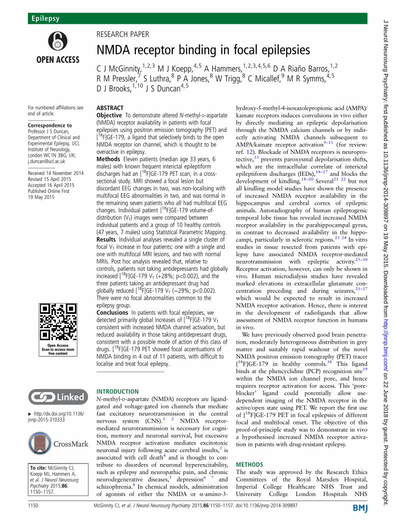
RESEARCH PAPER NMDA receptor binding in focal...
8
RESEARCH PAPER NMDA receptor binding in focal epilepsies C J McGinnity, 1,2,3 M J Koepp, 4,5 A Hammers, 1,2,3,4,5,6 D A Riaño Barros, 1,2 R M Pressler, 7 S Luthra, 8 P A Jones, 8 W Trigg, 8 C Micallef, 9 M R Symms, 4,5 D J Brooks, 1,10 J S Duncan 4,5 For numbered affiliations see end of article. Correspondence to Professor J S Duncan, Department of Clinical and Experimental Epilepsy, UCL Institute of Neurology, London WC1N 3BG, UK; [email protected] Received 14 November 2014 Revised 15 April 2015 Accepted 16 April 2015 Published Online First 19 May 2015 ▸ http://dx.doi.org/10.1136/ jnnp-2015-310333 To cite: McGinnity CJ, Koepp MJ, Hammers A, et al. J Neurol Neurosurg Psychiatry 2015;86: 1150–1157. ABSTRACT Objective To demonstrate altered N-methyl-D-aspartate (NMDA) receptor availability in patients with focal epilepsies using positron emission tomography (PET) and [ 18 F]GE-179, a ligand that selectively binds to the open NMDA receptor ion channel, which is thought to be overactive in epilepsy. Methods Eleven patients (median age 33 years, 6 males) with known frequent interictal epileptiform discharges had an [ 18 F]GE-179 PET scan, in a cross- sectional study. MRI showed a focal lesion but discordant EEG changes in two, was non-localising with multifocal EEG abnormalities in two, and was normal in the remaining seven patients who all had multifocal EEG changes. Individual patient [ 18 F]GE-179 volume-of- distribution (V T ) images were compared between individual patients and a group of 10 healthy controls (47 years, 7 males) using Statistical Parametric Mapping. Results Individual analyses revealed a single cluster of focal V T increase in four patients; one with a single and one with multifocal MRI lesions, and two with normal MRIs. Post hoc analysis revealed that, relative to controls, patients not taking antidepressants had globally increased [ 18 F]GE-179 V T (+28%; p<0.002), and the three patients taking an antidepressant drug had globally reduced [ 18 F]GE-179 V T (-29%; p<0.002). There were no focal abnormalities common to the epilepsy group. Conclusions In patients with focal epilepsies, we detected primarily global increases of [ 18 F]GE-179 V T consistent with increased NMDA channel activation, but reduced availability in those taking antidepressant drugs, consistent with a possible mode of action of this class of drugs. [ 18 F]GE-179 PET showed focal accentuations of NMDA binding in 4 out of 11 patients, with difficult to localise and treat focal epilepsy. INTRODUCTION N-methyl-D-aspartate (NMDA) receptors are ligand- gated and voltage-gated ion channels that mediate fast excitatory neurotransmission in the central nervous system (CNS). 1 2 NMDA receptor- mediated neurotransmission is necessary for cogni- tion, memory and neuronal survival, but excessive NMDA receptor activation mediates excitotoxic neuronal injury following acute cerebral insults, 3 is associated with cell death 4 and is thought to con- tribute to disorders of neuronal hyperexcitability, such as epilepsy and neuropathic pain, and chronic neurodegenerative diseases, 5 depression 6 7 and schizophrenia. 8 In chemical models, administration of agonists of either the NMDA or α-amino-3- hydroxy-5-methyl-4-isoxazolepropionic acid (AMPA)/ kainate receptors induces convulsions in vivo either by directly mediating an epileptic depolarisation through the NMDA calcium channels or by indir- ectly activating NMDA channels subsequent to AMPA/kainate receptor activation 9–11 (for review: ref. 12). Blockade of NMDA receptors is neuropro- tective, 13 prevents paroxysmal depolarisation shifts, which are the intracellular correlate of interictal epileptiform discharges (IEDs), 14–17 and blocks the development of kindling. 18–20 Several 21 22 but not all kindling model studies have shown the presence of increased NMDA receptor availability in the hippocampus and cerebral cortex of epileptic animals. Autoradiography of human epileptogenic temporal lobe tissue has revealed increased NMDA receptor availability in the parahippocampal gyrus, in contrast to decreased availability in the hippo- campi, particularly in sclerotic regions. 23 24 In vitro studies in tissue resected from patients with epi- lepsy have associated NMDA receptor-mediated neurotransmission with epileptic activity. 25–30 Receptor activation, however, can only be shown in vivo. Human microdialysis studies have revealed marked elevations in extracellular glutamate con- centration preceding and during seizures, 31–37 which would be expected to result in increased NMDA receptor activation. Hence, there is interest in the development of radioligands that allow assessment of NMDA receptor function in humans in vivo. We have previouslyobserved good brain penetra- tion, moderately heterogeneous distribution in grey matter and suitably rapid washout of the novel NMDA positron emission tomography (PET) tracer [ 18 F]GE-179 in healthy controls. 38 This ligand binds at the phencyclidine (PCP) recognition site 39 within the NMDA ion channel pore, and hence requires receptor activation for access. This ‘pore- blocker’ ligand could potentially allow use- dependent imaging of the NMDA receptor in the active/open state using PET. We report the first use of [ 18 F]GE-179 PET in focal epilepsies of different focal and multifocal onset. The objective of this proof-of-principle study was to demonstrate in vivo a hypothesised increased NMDA receptor activa- tion in patients with drug-resistant epilepsy. METHODS The study was approved by the Research Ethics Committees of the Royal Marsden Hospital, Imperial College Healthcare NHS Trust and University College London Hospitals NHS Open Access Scan to access more free content 1150 McGinnity CJ, et al. J Neurol Neurosurg Psychiatry 2015;86:1150–1157. doi:10.1136/jnnp-2014-309897 Epilepsy on 22 June 2018 by guest. Protected by copyright. http://jnnp.bmj.com/ J Neurol Neurosurg Psychiatry: first published as 10.1136/jnnp-2014-309897 on 19 May 2015. Downloaded from
Transcript of RESEARCH PAPER NMDA receptor binding in focal...
RESEARCH PAPER
NMDA receptor binding in focal epilepsiesC J McGinnity,1,2,3 M J Koepp,4,5 A Hammers,1,2,3,4,5,6 D A Riaño Barros,1,2
R M Pressler,7 S Luthra,8 P A Jones,8 W Trigg,8 C Micallef,9 M R Symms,4,5
D J Brooks,1,10 J S Duncan4,5
For numbered affiliations seeend of article.
Correspondence toProfessor J S Duncan,Department of Clinical andExperimental Epilepsy, UCLInstitute of Neurology,London WC1N 3BG, UK;[email protected]
Received 14 November 2014Revised 15 April 2015Accepted 16 April 2015Published Online First19 May 2015
▸ http://dx.doi.org/10.1136/jnnp-2015-310333
To cite: McGinnity CJ,Koepp MJ, Hammers A,et al. J Neurol NeurosurgPsychiatry 2015;86:1150–1157.
ABSTRACTObjective To demonstrate altered N-methyl-D-aspartate(NMDA) receptor availability in patients with focalepilepsies using positron emission tomography (PET) and[18F]GE-179, a ligand that selectively binds to the openNMDA receptor ion channel, which is thought to beoveractive in epilepsy.Methods Eleven patients (median age 33 years, 6males) with known frequent interictal epileptiformdischarges had an [18F]GE-179 PET scan, in a cross-sectional study. MRI showed a focal lesion butdiscordant EEG changes in two, was non-localising withmultifocal EEG abnormalities in two, and was normal inthe remaining seven patients who all had multifocal EEGchanges. Individual patient [18F]GE-179 volume-of-distribution (VT) images were compared betweenindividual patients and a group of 10 healthy controls(47 years, 7 males) using Statistical Parametric Mapping.Results Individual analyses revealed a single cluster offocal VT increase in four patients; one with a single andone with multifocal MRI lesions, and two with normalMRIs. Post hoc analysis revealed that, relative tocontrols, patients not taking antidepressants had globallyincreased [18F]GE-179 VT (+28%; p<0.002), and thethree patients taking an antidepressant drug hadglobally reduced [18F]GE-179 VT (−29%; p<0.002).There were no focal abnormalities common to theepilepsy group.Conclusions In patients with focal epilepsies, wedetected primarily global increases of [18F]GE-179 VTconsistent with increased NMDA channel activation, butreduced availability in those taking antidepressant drugs,consistent with a possible mode of action of this class ofdrugs. [18F]GE-179 PET showed focal accentuations ofNMDA binding in 4 out of 11 patients, with difficult tolocalise and treat focal epilepsy.
INTRODUCTIONN-methyl-D-aspartate (NMDA) receptors are ligand-gated and voltage-gated ion channels that mediatefast excitatory neurotransmission in the centralnervous system (CNS).1 2 NMDA receptor-mediated neurotransmission is necessary for cogni-tion, memory and neuronal survival, but excessiveNMDA receptor activation mediates excitotoxicneuronal injury following acute cerebral insults,3 isassociated with cell death4 and is thought to con-tribute to disorders of neuronal hyperexcitability,such as epilepsy and neuropathic pain, and chronicneurodegenerative diseases,5 depression6 7 andschizophrenia.8 In chemical models, administrationof agonists of either the NMDA or α-amino-3-
hydroxy-5-methyl-4-isoxazolepropionic acid (AMPA)/kainate receptors induces convulsions in vivo eitherby directly mediating an epileptic depolarisationthrough the NMDA calcium channels or by indir-ectly activating NMDA channels subsequent toAMPA/kainate receptor activation9–11 (for review:ref. 12). Blockade of NMDA receptors is neuropro-tective,13 prevents paroxysmal depolarisation shifts,which are the intracellular correlate of interictalepileptiform discharges (IEDs),14–17 and blocks thedevelopment of kindling.18–20 Several21 22 but notall kindling model studies have shown the presenceof increased NMDA receptor availability in thehippocampus and cerebral cortex of epilepticanimals. Autoradiography of human epileptogenictemporal lobe tissue has revealed increased NMDAreceptor availability in the parahippocampal gyrus,in contrast to decreased availability in the hippo-campi, particularly in sclerotic regions.23 24 In vitrostudies in tissue resected from patients with epi-lepsy have associated NMDA receptor-mediatedneurotransmission with epileptic activity.25–30
Receptor activation, however, can only be shown invivo. Human microdialysis studies have revealedmarked elevations in extracellular glutamate con-centration preceding and during seizures,31–37
which would be expected to result in increasedNMDA receptor activation. Hence, there is interestin the development of radioligands that allowassessment of NMDA receptor function in humansin vivo.We have previously observed good brain penetra-
tion, moderately heterogeneous distribution in greymatter and suitably rapid washout of the novelNMDA positron emission tomography (PET) tracer[18F]GE-179 in healthy controls.38 This ligandbinds at the phencyclidine (PCP) recognition site39
within the NMDA ion channel pore, and hencerequires receptor activation for access. This ‘pore-blocker’ ligand could potentially allow use-dependent imaging of the NMDA receptor in theactive/open state using PET. We report the first useof [18F]GE-179 PET in focal epilepsies of differentfocal and multifocal onset. The objective of thisproof-of-principle study was to demonstrate in vivoa hypothesised increased NMDA receptor activa-tion in patients with drug-resistant epilepsy.
METHODSThe study was approved by the Research EthicsCommittees of the Royal Marsden Hospital,Imperial College Healthcare NHS Trust andUniversity College London Hospitals NHS
Open AccessScan to access more
free content
1150 McGinnity CJ, et al. J Neurol Neurosurg Psychiatry 2015;86:1150–1157. doi:10.1136/jnnp-2014-309897
Epilepsy on 22 June 2018 by guest. P
rotected by copyright.http://jnnp.bm
j.com/
J Neurol N
eurosurg Psychiatry: first published as 10.1136/jnnp-2014-309897 on 19 M
ay 2015. Dow
nloaded from
Foundation Trust. Permission to administer [18F]GE-179 wasobtained from the Administration of Radioactive SubstancesAdvisory Committee (ARSAC), UK. All participants providedwritten, informed consent.
Epilepsy and control populationsThis was a proof-of-principle, cross-sectional pilot study withtargets of 12 participants per group. Eleven patients with refrac-tory focal epilepsies (median age 33 years; range 20–50 years; 6males) were recruited from the outpatient clinics at the NationalHospital for Neurology and Neurosurgery. Demographics andclinical details are listed in table 1. Their diagnoses were basedon history, seizure semiology, prolonged and repeated interictaland ictal EEG recordings (where available), and MRI data.Patients were chosen who had frequent interictal spikes on pre-vious EEG recordings, which we hypothesised would maximiseour chances to detect increased binding to open NMDA recep-tors. None of the patients were taking an antiepileptic drug(AED) known to act at the NMDA receptor. Exclusion criteriaincluded inability to provide informed consent, claustrophobia,standard MR contraindications, a positive urinary pregnancytest on the day of the PET scan and history of drug abuse.Patient 4, whose seizures consisted of a sustained fluctuation ofperception of brightness with pupillary hippus, has been pre-sented in a detailed case report.40
The control group, 9 of whom have been described previ-ously,38 comprised 10 healthy volunteers without history ofneurological or psychiatric illness (median age 46 years; range25–62 years; 7 males). Additional exclusion criteria were asdescribed for the patients above. A further three seeminglyhealthy individuals were subsequently excluded; one due toexcessive movement throughout the PET scan acquisition, onewhose MRI revealed evidence of a cerebral infarct; and onewho was discovered to have a history of benzodiazepine abuse.The original control group data were used, rather than repeatimaging; the patient data were acquired at approximately thesame time as those of the control group (i.e. within 12 months)using the same imaging protocol.
Median age and body mass index (BMI) were comparedbetween patients and control groups by Mann-Whitney U statis-tic in SPSS. Gender balance was compared between groups withthe (Pearson) χ2 test.
MRI data acquisitionThree-dimensional volumetric T1-weighted coronal MRIsequences were acquired at Epilepsy Society (Chalfont St. Peter,UK), as previously described.38 MRIs were reviewed by anexperienced neuroradiologist (CM). MRIs were not availablefor one control participant, in whom 3.0 T MRI was contra-indicated.
EEGAll patients had an EEG during the PET scan using a Trackit 18/8 (Lifelines Limited, Hants, UK) ambulatory EEG recorder andan ECI E1 Cap (Electro-Cap International, Eaton, Ohio, USA)with 19 electrodes placed according to the International 10–20system. An additional reference electrode (Fpz) was sited justanterior to Fz. The O1 and O2 electrodes were removed fromthe cap prior to scanning for several patients in order to minim-ise discomfort. The participants were closely observed forevidence of seizures throughout the scan. EEGs were reviewedby an experienced clinical neurophysiologist (RMP). Thenumber of IEDs during the first 30 min of scan acquisition was
quantified and correlated with [18F]GE-179 global volume-of-distribution (VT) using Spearman’s r correlation coefficient.
PET image acquisition and data analysisPET image acquisition has been described previously.38 Briefly,images were acquired using a Siemens/CTI ECAT EXACT3D962 HR+PET camera (Siemens, Erlangen, Germany) atHammersmith Imanet Limited. Each participant had a 90 mindynamic emission scan with a smooth bolus intravenous injec-tion of median 187 MBq (range 173–192 MBq) [18F]GE-179administered 30 seconds after starting image acquisition. For cal-culation of continuous decay-corrected and metabolite-correctedparent plasma input functions, discrete arterial blood sampleswere taken throughout the scan, with continuous arterial bloodsampling for the first 15 minutes.
The area under the metabolite model curves (AUCmetabs) wasused as a measure of the rate of metabolism for each individual.The AUCmetabs over t=0–30 minutes and t=0–90.5 minutes wascompared between groups by multivariate general linear model(GLM), with gender as a fixed factor, and age and BMI as cov-ariates. The residual sum of squares (RSSmetabs) for the metabol-ite model curve was compared between groups by univariateGLM. The threshold for statistical significance was p=0.05.
The VT of [18F]GE-179 was computed at the voxel level foreach participant by exponential spectral analysis, as describedpreviously.38
Each participant’s VT image was spatially normalised using ascanner-specific template. To enable group-wise comparisons,images acquired from patients with an epileptogenic zone in theright hemisphere (3 patients) were left-right flipped prior tonormalisation; three control participants’ images were also left-right flipped prior to group-wise comparison (controls 1, 4 and6, selected at random). For individual (1 patient vs 10 controls)comparisons, the native (unflipped) patient and control imageswere used.
The primary outcome measure, global VT, was computed asan overall mean over the entire matrix, thresholded at 1/8 ofthat value to create a brain mask, and averaged again within thismask. The global VT values were compared between groupsusing a univariate GLM, with gender as a fixed factor, and ageand BMI as covariates.
In order to identify changes in the regional distribution ofactivated NMDA receptors, group-wise SPM8 analyses based onthe smoothed (12 mm Full Width at Half Maximum (FWHM)isotropic Gaussian kernel), transformed parametric VT images,were performed, comparing patients with focal epilepsy againstthe controls. The images were compared on a voxel-by-voxelbasis using a two-sample t test, assuming unequal variances,with global VT taken into account via an analysis of covariance(ANCOVA) by group. The images were grey and white matter(explicit) masked, with relative threshold masking at 0.4. Wereport differences in [18F]GE-179 VT at p<0.001 (uncorrected),clusters at p<0.05 (uncorrected) and an extent threshold of 15voxels.
In order to identify participant-specific changes in theregional distribution of activated NMDA receptors, individualSPM8 analyses based on the smoothed transformed parametricVT images were also performed for each of the patients with fre-quent IEDs against the 10 controls. Equal variances wereassumed and global VT taken into account via an ANCOVA bygroup. The unflipped VT image for each control participant wascompared with those of the nine other controls in an identicalfashion.
McGinnity CJ, et al. J Neurol Neurosurg Psychiatry 2015;86:1150–1157. doi:10.1136/jnnp-2014-309897 1151
Epilepsy on 22 June 2018 by guest. P
rotected by copyright.http://jnnp.bm
j.com/
J Neurol N
eurosurg Psychiatry: first published as 10.1136/jnnp-2014-309897 on 19 M
ay 2015. Dow
nloaded from
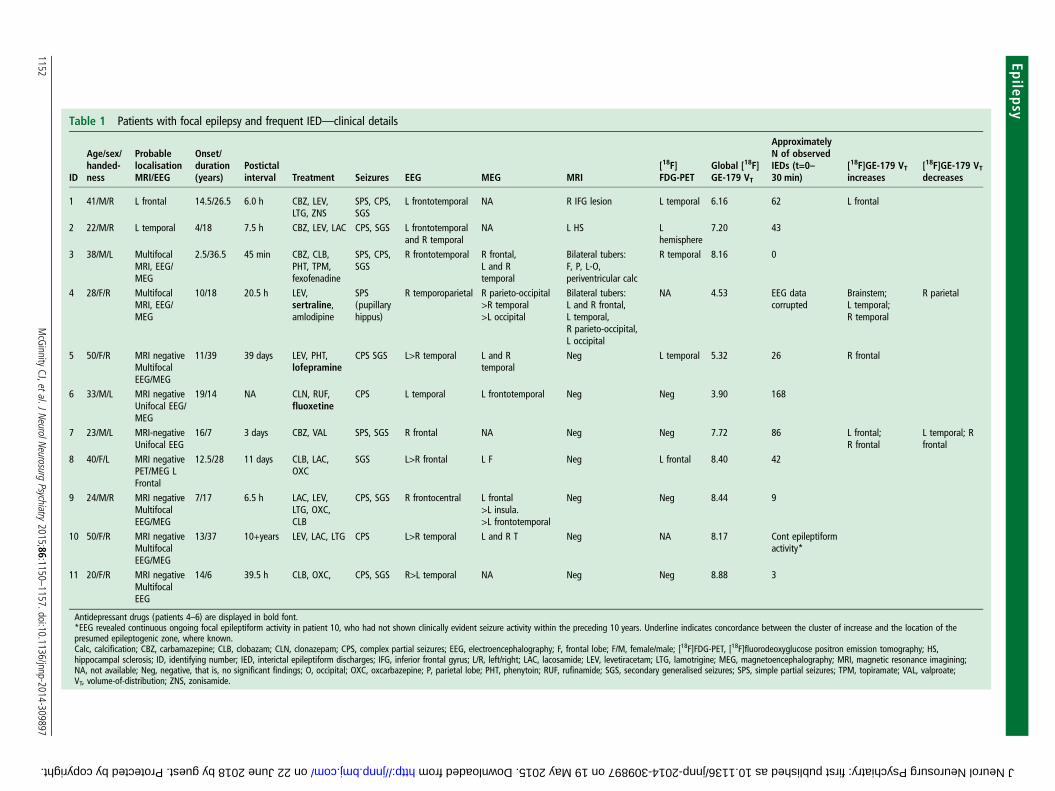
Table 1 Patients with focal epilepsy and frequent IED—clinical details
ID
Age/sex/handed-ness
ProbablelocalisationMRI/EEG
Onset/duration(years)
Postictalinterval Treatment Seizures EEG MEG MRI
[18F]FDG-PET
Global [18F]GE-179 VT
ApproximatelyN of observedIEDs (t=0–30 min)
[18F]GE-179 VTincreases
[18F]GE-179 VTdecreases
1 41/M/R L frontal 14.5/26.5 6.0 h CBZ, LEV,LTG, ZNS
SPS, CPS,SGS
L frontotemporal NA R IFG lesion L temporal 6.16 62 L frontal
2 22/M/R L temporal 4/18 7.5 h CBZ, LEV, LAC CPS, SGS L frontotemporaland R temporal
NA L HS Lhemisphere
7.20 43
3 38/M/L MultifocalMRI, EEG/MEG
2.5/36.5 45 min CBZ, CLB,PHT, TPM,fexofenadine
SPS, CPS,SGS
R frontotemporal R frontal,L and Rtemporal
Bilateral tubers:F, P, L-O,periventricular calc
R temporal 8.16 0
4 28/F/R MultifocalMRI, EEG/MEG
10/18 20.5 h LEV,sertraline,amlodipine
SPS(pupillaryhippus)
R temporoparietal R parieto-occipital>R temporal>L occipital
Bilateral tubers:L and R frontal,L temporal,R parieto-occipital,L occipital
NA 4.53 EEG datacorrupted
Brainstem;L temporal;R temporal
R parietal
5 50/F/R MRI negativeMultifocalEEG/MEG
11/39 39 days LEV, PHT,lofepramine
CPS SGS L>R temporal L and Rtemporal
Neg L temporal 5.32 26 R frontal
6 33/M/L MRI negativeUnifocal EEG/MEG
19/14 NA CLN, RUF,fluoxetine
CPS L temporal L frontotemporal Neg Neg 3.90 168
7 23/M/L MRI-negativeUnifocal EEG
16/7 3 days CBZ, VAL SPS, SGS R frontal NA Neg Neg 7.72 86 L frontal;R frontal
L temporal; Rfrontal
8 40/F/L MRI negativePET/MEG LFrontal
12.5/28 11 days CLB, LAC,OXC
SGS L>R frontal L F Neg L frontal 8.40 42
9 24/M/R MRI negativeMultifocalEEG/MEG
7/17 6.5 h LAC, LEV,LTG, OXC,CLB
CPS, SGS R frontocentral L frontal>L insula.>L frontotemporal
Neg Neg 8.44 9
10 50/F/R MRI negativeMultifocalEEG/MEG
13/37 10+years LEV, LAC, LTG CPS L>R temporal L and R T Neg NA 8.17 Cont epileptiformactivity*
11 20/F/R MRI negativeMultifocalEEG
14/6 39.5 h CLB, OXC, CPS, SGS R>L temporal NA Neg Neg 8.88 3
Antidepressant drugs (patients 4–6) are displayed in bold font.*EEG revealed continuous ongoing focal epileptiform activity in patient 10, who had not shown clinically evident seizure activity within the preceding 10 years. Underline indicates concordance between the cluster of increase and the location of thepresumed epileptogenic zone, where known.Calc, calcification; CBZ, carbamazepine; CLB, clobazam; CLN, clonazepam; CPS, complex partial seizures; EEG, electroencephalography; F, frontal lobe; F/M, female/male; [18F]FDG-PET, [18F]fluorodeoxyglucose positron emission tomography; HS,hippocampal sclerosis; ID, identifying number; IED, interictal epileptiform discharges; IFG, inferior frontal gyrus; L/R, left/right; LAC, lacosamide; LEV, levetiracetam; LTG, lamotrigine; MEG, magnetoencephalography; MRI, magnetic resonance imagining;NA, not available; Neg, negative, that is, no significant findings; O, occipital; OXC, oxcarbazepine; P, parietal lobe; PHT, phenytoin; RUF, rufinamide; SGS, secondary generalised seizures; SPS, simple partial seizures; TPM, topiramate; VAL, valproate;VT, volume-of-distribution; ZNS, zonisamide.
1152McGinnity
CJ,etal.JNeurolN
eurosurgPsychiatry
2015;86:1150–1157.doi:10.1136/jnnp-2014-309897
Epilepsy on 22 June 2018 by guest. Protected by copyright. http://jnnp.bmj.com/ J Neurol Neurosurg Psychiatry: first published as 10.1136/jnnp-2014-309897 on 19 May 2015. Downloaded from
RESULTSThere was no difference between patient and control group interms of age (p=1.00), BMI (p=1.00) or gender mix (p=0.47).
There were no significant differences in the AUCmetabs or theRSSmetabs between the groups (p=0.19 and p=0.47, respect-ively). Age, BMI and gender also did not significantly influencethe AUCmetabs (all p>0.09).
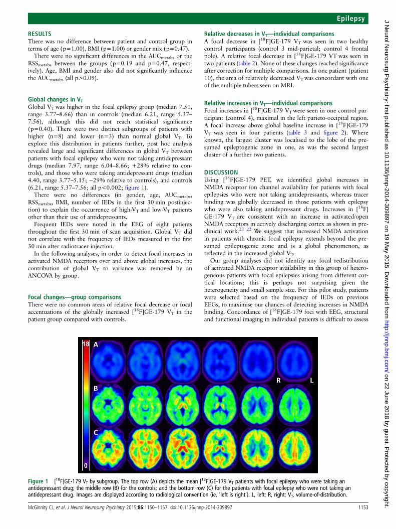
Global changes in VTGlobal VTwas higher in the focal epilepsy group (median 7.51,range 3.77–8.66) than in controls (median 6.21, range 5.37–7.56), although this did not reach statistical significance(p=0.40). There were two distinct subgroups of patients withhigher (n=8) and lower (n=3) than normal global VT. Toexplore this distribution in patients further, post hoc analysisrevealed large and significant differences in global VT betweenpatients with focal epilepsy who were not taking antidepressantdrugs (median 7.97, range 6.04–8.66; +28% relative to con-trols), and those who were taking antidepressant drugs (median4.40, range 3.77–5.15; −29% relative to controls), and controls(6.21, range 5.37–7.56; all p<0.002; figure 1).
There were no differences (in gender, age, AUCmetabs,RSSmetabs, BMI, number of IEDs in the first 30 min postinjec-tion) to explain the occurrence of high-VT and low-VT patientsother than their use of antidepressants.
Frequent IEDs were noted in the EEG of eight patientsthroughout the first 30 min of scan acquisition. Global VT didnot correlate with the frequency of IEDs measured in the first30 min after radiotracer injection.
In the following analyses, in order to detect focal increases inactivated NMDA receptors over and above global increases, thecontribution of global VT to variance was removed by anANCOVA by group.
Focal changes—group comparisonsThere were no common areas of relative focal decrease or focalaccentuations of the globally increased [18F]GE-179 VT in thepatient group compared with controls.
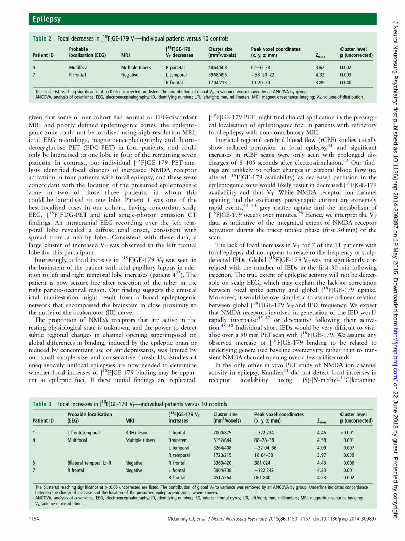
Relative decreases in VT—individual comparisonsA focal decrease in [18F]GE-179 VT was seen in two healthycontrol participants (control 3 mid-parietal; control 4 frontalpole). A relative focal decrease in [18F]GE-179 VT was seen intwo patients (table 2). None of these changes reached significanceafter correction for multiple comparisons. In one patient (patient10), the area of relatively decreased VTwas concordant with oneof the multiple tubers seen on MRI.
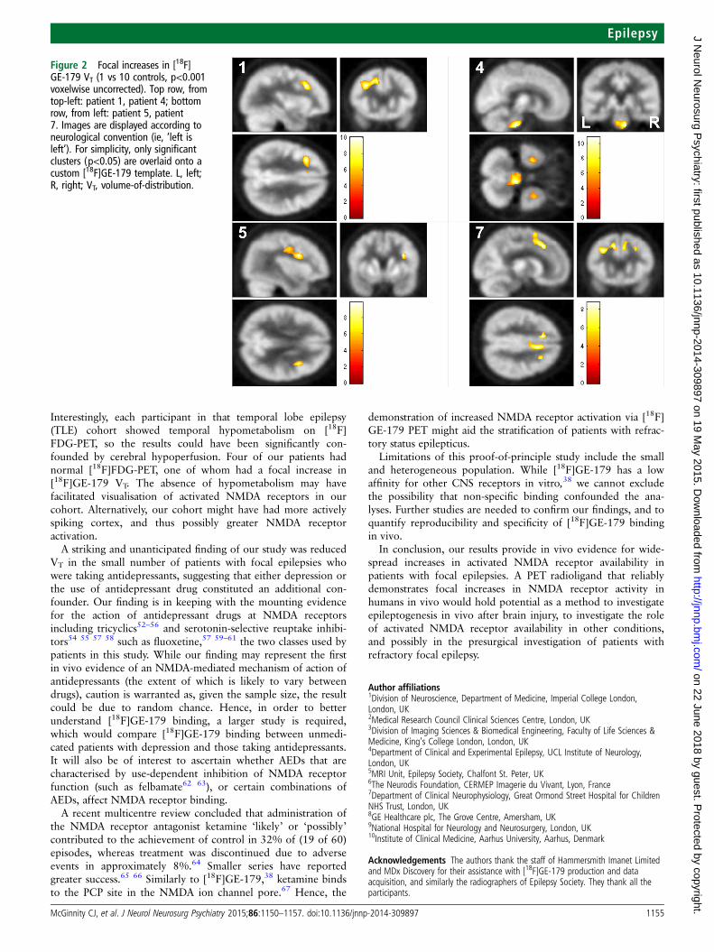
Relative increases in VT—individual comparisonsFocal increases in [18F]GE-179 VTwere seen in one control par-ticipant (control 4), maximal in the left parieto-occipital region.A focal increase above global baseline increase in [18F]GE-179VT was seen in four patients (table 3 and figure 2). Whereknown, the largest cluster was localised to the lobe of the pre-sumed epileptogenic zone in one, as was the second largestcluster of a further two patients.
DISCUSSIONUsing [18F]GE-179 PET, we identified global increases inNMDA receptor ion channel availability for patients with focalepilepsies who were not taking antidepressants, whereas tracerbinding was globally decreased in those patients with epilepsywho were also taking antidepressant drugs. Increases in [18F]GE-179 VT are consistent with an increase in activated/openNMDA receptors in actively discharging cortex as shown in pre-clinical work.21 22 We suggest that increased NMDA activationin patients with chronic focal epilepsy extends beyond the pre-sumed epileptogenic zone and is a global phenomenon, asreflected in the increased global VT.
Our group analyses did not identify any focal redistributionof activated NMDA receptor availability in this group of hetero-geneous patients with focal epilepsies arising from different cor-tical locations; this is perhaps not surprising given theheterogeneity and small sample size. For this pilot study, patientswere selected based on the frequency of IEDs on previousEEGs, to maximise our chances of detecting increases in NMDAbinding. Concordance of [18F]GE-179 foci with EEG, structuraland functional imaging in individual patients is difficult to assess
Figure 1 [18F]GE-179 VT by subgroup. The top row (A) depicts the mean [18F]GE-179 VT patients with focal epilepsy who were taking anantidepressant drug; the middle row (B) for the controls; and the bottom row (C) for the patients with focal epilepsy who were not taking anantidepressant drug. Images are displayed according to radiological convention (ie, ‘left is right’). L, left; R, right; VT, volume-of-distribution.
McGinnity CJ, et al. J Neurol Neurosurg Psychiatry 2015;86:1150–1157. doi:10.1136/jnnp-2014-309897 1153
Epilepsy on 22 June 2018 by guest. P
rotected by copyright.http://jnnp.bm
j.com/
J Neurol N
eurosurg Psychiatry: first published as 10.1136/jnnp-2014-309897 on 19 M
ay 2015. Dow
nloaded from
given that some of our cohort had normal or EEG-discordantMRI and poorly defined epileptogenic zones: the epilepto-genic zone could not be localised using high-resolution MRI,ictal EEG recordings, magnetoencephalography and fluoro-deoxyglucose PET (FDG-PET) in four patients, and couldonly be lateralised to one lobe in four of the remaining sevenpatients. In contrast, our individual [18F]GE-179 PET ana-lysis identified focal clusters of increased NMDA receptoractivation in four patients with focal epilepsy, and these wereconcordant with the location of the presumed epileptogeniczone in two of those three patients, in whom thiscould be lateralised to one lobe. Patient 1 was one of thebest-localised cases in our cohort, having concordant scalpEEG, [18F]FDG-PET and ictal single-photon emission CTfindings. An intracranial EEG recording over the left tem-poral lobe revealed a diffuse ictal onset, consistent withspread from a nearby lobe. Consistent with these data, alarge cluster of increased VT was observed in the left frontallobe for this participant.
Interestingly, a focal increase in [18F]GE-179 VT was seen inthe brainstem of the patient with ictal pupillary hippus in add-ition to left and right temporal lobe increases (patient 423). Thepatient is now seizure-free after resection of the tuber in theright parieto-occipital region. Our finding suggests the unusualictal manifestation might result from a broad epileptogenicnetwork that encompassed the brainstem in close proximity tothe nuclei of the oculomotor (III) nerve.
The proportion of NMDA receptors that are active in theresting physiological state is unknown, and the power to detectsubtle regional changes in channel opening superimposed onglobal differences in binding, induced by the epileptic brain orreduced by concomitant use of antidepressants, was limited byour small sample size and conservative thresholds. Studies ofunequivocally unifocal epilepsies are now needed to determinewhether focal increases of [18F]GE-179 binding may be appar-ent at epileptic foci. If these initial findings are replicated,
[18F]GE-179 PET might find clinical application in the presurgi-cal localisation of epileptogenic foci in patients with refractoryfocal epilepsy with non-contributory MRI.
Interictal regional cerebral blood flow (rCBF) studies usuallyshow reduced perfusion in focal epilepsy,41 and significantincreases in rCBF scans were only seen with prolonged dis-charges of 8–105 seconds after electrostimulation.42 Our find-ings are unlikely to reflect changes in cerebral blood flow (ie,altered [18F]GE-179 availability) as decreased perfusion in theepileptogenic zone would likely result in decreased [18F]GE-179availability and thus VT. While NMDA receptor ion channelopening and the excitatory postsynaptic current are extremelyrapid events,43 44 grey matter uptake and the metabolism of[18F]GE-179 occurs over minutes.38 Hence, we interpret the VT
data as indicative of the integrated extent of NMDA receptoractivation during the tracer uptake phase (first 30 min) of thescan.
The lack of focal increases in VT for 7 of the 11 patients withfocal epilepsy did not appear to relate to the frequency of scalp-detected IEDs. Global [18F]GE-179 VTwas not significantly cor-related with the number of IEDs in the first 30 min followinginjection. The true extent of epileptic activity will not be detect-able on scalp EEG, which may explain the lack of correlationbetween focal spike activity and global [18F]GE-179 uptake.Moreover, it would be oversimplistic to assume a linear relationbetween global [18F]GE-179 VT and IED frequency. We expectthat NMDA receptors involved in generation of the IED wouldrapidly internalise45–47 or desensitise following their activa-tion.48–50 Individual short IEDs would be very difficult to visu-alise over a 90 min PET scan with [18F]GE-179. We assume anyobserved increase of [18F]GE-179 binding to be related tounderlying generalised baseline overactivity, rather than to tran-sient NMDA channel opening over a few milliseconds.
In the only other in vivo PET study of NMDA ion channelactivity in epilepsy, Kumlien51 did not detect focal increases inreceptor availability using (S)-[N-methyl-11C]ketamine.
Table 2 Focal decreases in [18F]GE-179 VT—individual patients versus 10 controls
Patient IDProbablelocalisation (EEG) MRI
[18F]GE-179VT decreases
Cluster size(mm3/voxels)
Peak voxel coordinates(x, y, z; mm) Zmax
Cluster levelp (uncorrected)
4 Multifocal Multiple tubers R parietal 4864/608 62–32 38 3.62 0.0027 R frontal Negative L temporal 3968/496 −58–20–22 4.32 0.003
R frontal 1704/213 10 20–20 3.89 0.040
The cluster(s) reaching significance at p<0.05 uncorrected are listed. The contribution of global VT to variance was removed by an ANCOVA by group.ANCOVA, analysis of covariance; EEG, electroencephalography; ID, identifying number; L/R, left/right; mm, millimeters; MRI, magnetic resonance imaging; VT, volume-of-distribution.
Table 3 Focal increases in [18F]GE-179 VT—individual patients versus 10 controls
Patient IDProbable localisation(EEG) MRI
[18F]GE-179 VTincreases
Cluster size(mm3/voxels)
Peak voxel coordinates(x, y, z; mm) Zmax
Cluster levelp (uncorrected)
1 L frontotemporal R IFG lesion L frontal 7000/875 −322 234 4.46 <0.001
4 Multifocal Multiple tubers Brainstem 5152/644 08–28–38 4.58 0.001L temporal 3264/408 −32 04–36 4.09 0.007R temporal 1720/215 18 04–30 3.97 0.039
5 Bilateral temporal L>R Negative R frontal 3360/420 381 024 4.43 0.0067 R frontal Negative L frontal 5904/738 −122 242 4.23 0.001
R frontal 4512/564 061 840 4.23 0.002
The cluster(s) reaching significance at p<0.05 uncorrected are listed. The contribution of global VT to variance was removed by an ANCOVA by group. Underline indicates concordancebetween the cluster of increase and the location of the presumed epileptogenic zone, where known.ANCOVA, analysis of covariance; EEG, electroencephalography; ID, identifying number; IFG, inferior frontal gyrus; L/R, left/right; mm, millimeters; MRI, magnetic resonance imaging;VT, volume-of-distribution.
1154 McGinnity CJ, et al. J Neurol Neurosurg Psychiatry 2015;86:1150–1157. doi:10.1136/jnnp-2014-309897
Epilepsy on 22 June 2018 by guest. P
rotected by copyright.http://jnnp.bm
j.com/
J Neurol N
eurosurg Psychiatry: first published as 10.1136/jnnp-2014-309897 on 19 M
ay 2015. Dow
nloaded from
Interestingly, each participant in that temporal lobe epilepsy(TLE) cohort showed temporal hypometabolism on [18F]FDG-PET, so the results could have been significantly con-founded by cerebral hypoperfusion. Four of our patients hadnormal [18F]FDG-PET, one of whom had a focal increase in[18F]GE-179 VT. The absence of hypometabolism may havefacilitated visualisation of activated NMDA receptors in ourcohort. Alternatively, our cohort might have had more activelyspiking cortex, and thus possibly greater NMDA receptoractivation.
A striking and unanticipated finding of our study was reducedVT in the small number of patients with focal epilepsies whowere taking antidepressants, suggesting that either depression orthe use of antidepressant drug constituted an additional con-founder. Our finding is in keeping with the mounting evidencefor the action of antidepressant drugs at NMDA receptorsincluding tricyclics52–56 and serotonin-selective reuptake inhibi-tors54 55 57 58 such as fluoxetine,57 59–61 the two classes used bypatients in this study. While our finding may represent the firstin vivo evidence of an NMDA-mediated mechanism of action ofantidepressants (the extent of which is likely to vary betweendrugs), caution is warranted as, given the sample size, the resultcould be due to random chance. Hence, in order to betterunderstand [18F]GE-179 binding, a larger study is required,which would compare [18F]GE-179 binding between unmedi-cated patients with depression and those taking antidepressants.It will also be of interest to ascertain whether AEDs that arecharacterised by use-dependent inhibition of NMDA receptorfunction (such as felbamate62 63), or certain combinations ofAEDs, affect NMDA receptor binding.
A recent multicentre review concluded that administration ofthe NMDA receptor antagonist ketamine ‘likely’ or ‘possibly’contributed to the achievement of control in 32% of (19 of 60)episodes, whereas treatment was discontinued due to adverseevents in approximately 8%.64 Smaller series have reportedgreater success.65 66 Similarly to [18F]GE-179,38 ketamine bindsto the PCP site in the NMDA ion channel pore.67 Hence, the
demonstration of increased NMDA receptor activation via [18F]GE-179 PET might aid the stratification of patients with refrac-tory status epilepticus.
Limitations of this proof-of-principle study include the smalland heterogeneous population. While [18F]GE-179 has a lowaffinity for other CNS receptors in vitro,38 we cannot excludethe possibility that non-specific binding confounded the ana-lyses. Further studies are needed to confirm our findings, and toquantify reproducibility and specificity of [18F]GE-179 bindingin vivo.
In conclusion, our results provide in vivo evidence for wide-spread increases in activated NMDA receptor availability inpatients with focal epilepsies. A PET radioligand that reliablydemonstrates focal increases in NMDA receptor activity inhumans in vivo would hold potential as a method to investigateepileptogenesis in vivo after brain injury, to investigate the roleof activated NMDA receptor availability in other conditions,and possibly in the presurgical investigation of patients withrefractory focal epilepsy.
Author affiliations1Division of Neuroscience, Department of Medicine, Imperial College London,London, UK2Medical Research Council Clinical Sciences Centre, London, UK3Division of Imaging Sciences & Biomedical Engineering, Faculty of Life Sciences &Medicine, King’s College London, London, UK4Department of Clinical and Experimental Epilepsy, UCL Institute of Neurology,London, UK5MRI Unit, Epilepsy Society, Chalfont St. Peter, UK6The Neurodis Foundation, CERMEP Imagerie du Vivant, Lyon, France7Department of Clinical Neurophysiology, Great Ormond Street Hospital for ChildrenNHS Trust, London, UK8GE Healthcare plc, The Grove Centre, Amersham, UK9National Hospital for Neurology and Neurosurgery, London, UK10Institute of Clinical Medicine, Aarhus University, Aarhus, Denmark
Acknowledgements The authors thank the staff of Hammersmith Imanet Limitedand MDx Discovery for their assistance with [18F]GE-179 production and dataacquisition, and similarly the radiographers of Epilepsy Society. They thank all theparticipants.
Figure 2 Focal increases in [18F]GE-179 VT (1 vs 10 controls, p<0.001voxelwise uncorrected). Top row, fromtop-left: patient 1, patient 4; bottomrow, from left: patient 5, patient7. Images are displayed according toneurological convention (ie, ‘left isleft’). For simplicity, only significantclusters (p<0.05) are overlaid onto acustom [18F]GE-179 template. L, left;R, right; VT, volume-of-distribution.
McGinnity CJ, et al. J Neurol Neurosurg Psychiatry 2015;86:1150–1157. doi:10.1136/jnnp-2014-309897 1155
Epilepsy on 22 June 2018 by guest. P
rotected by copyright.http://jnnp.bm
j.com/
J Neurol N
eurosurg Psychiatry: first published as 10.1136/jnnp-2014-309897 on 19 M
ay 2015. Dow
nloaded from
Contributors CJM contributed to the design of the study, was primarilyresponsible for data acquisition and analysis, and prepared the manuscript drafts.MJK contributed to conceptualisation and design of the study, data review andmanuscript revision. AH contributed to data analysis and review, and manuscriptrevision. DARB contributed to PET data acquisition and manuscript revision. RMPreported the EEG data. SL contributed to conceptualisation and design of the study,and facilitated production of the radioligand at Hammersmith Imanet Ltd. PAJ andWT contributed to conceptualisation and design of the study, data review andmanuscript revision. They both also facilitated production of the radioligand atHammersmith Imanet Ltd. CM reported on the MRIs. MRS assisted with MRIacquisition, and co-registration of MR and PET data sets. DJB contributed toconceptualisation and design of the study, submissions to regulatory bodies andmanuscript review. JSD contributed to conceptualisation and design of the study,data review and manuscript revision. JSD was primarily responsible for identificationof the patients with focal epilepsy.
Funding This study was supported by the Medical Research Council (MRC) ClinicalSciences Centre and GE Healthcare plc. Open access fee and colour figures fees arepaid by GE Healthcare plc.
Competing interests CJM, MJK and JSD are grateful for support from the NIHRBiomedical Research Centre funding scheme to the UCLH/UCL BRC. Authorsaffiliated with the Division of Neuroscience, Department of Medicine, ImperialCollege London, London, UK, are grateful for support by the NIHR BiomedicalResearch Centre. SL, PAJ, WT and DJB are or were employees of GE Healthcare plcat the time of the study, which they report as a conflict of interest. CJM, JSD andMJK have received fees from GE Healthcare plc. CJM was supported by a MRCDoctoral Training Account (3+1) studentship that was awarded by Imperial CollegeLondon. JSD and MJK were supported by Epilepsy Society, UCL, UCL Hospitals andUCLH/UCL Biomedical Research Centre. JSD has received fees for organisingsymposia and lecturing for UCB Pharma, Eisai, GSK and GE Healthcare. AH wassupported by an MRC Clinician Scientist Fellowship (G108/585) and by the NeurodisFoundation.
Ethics approval Royal Marsden Hospital National Health Service Research EthicsCommittee.
Provenance and peer review Not commissioned; externally peer reviewed.
Data sharing statement The authors have submitted all major results from thestudy for publication. Unpublished data consist of individual imaging data sets andare only available to the investigators at present. Any request for data can beaddressed to the corresponding author; any data sharing will be subject torestrictions based on anonymisation rules.
Open Access This is an Open Access article distributed in accordance with theCreative Commons Attribution Non Commercial (CC BY-NC 4.0) license, whichpermits others to distribute, remix, adapt, build upon this work non-commercially,and license their derivative works on different terms, provided the original work isproperly cited and the use is non-commercial. See: http://creativecommons.org/licenses/by-nc/4.0/
REFERENCES1 Monaghan DT, Bridges RJ, Cotman CW. The excitatory amino acid receptors: their
classes, pharmacology, and distinct properties in the function of the central nervoussystem. Annu Rev Pharmacol Toxicol 1989;29:365–402.
2 Wollmuth LP, Sobolevsky AI. Structure and gating of the glutamate receptor ionchannel. Trends Neurosci 2004;27:321–8.
3 Leker RR, Shohami E, Leker RR, et al. Cerebral ischemia and trauma-differentetiologies yet similar mechanisms: neuroprotective opportunities. Brain Res Rev2002;39:55–73.
4 Sattler R, Xiang ZG, Lu WY, et al. Specific coupling of NMDA receptor activation tonitric oxide neurotoxicity by PSD-95 protein. Science 1999;284:1845–8.
5 Waxman EA, Lynch DR, Waxman EA, et al. N-methyl-D-aspartate receptor subtypes:multiple roles in excitotoxicity and neurological disease. Neuroscientist2005;11:37–49.
6 Sanacora G, Treccani G, Popoli M. Towards a glutamate hypothesis of depression:an emerging frontier of neuropsychopharmacology for mood disorders.Neuropharmacology 2012;62:63–77.
7 Musazzi L, Treccani G, Mallei A, et al. The action of antidepressants on theglutamate system: regulation of glutamate release and glutamate receptors. BiolPsychiatry 2013;73:1180–8.
8 Lau CG, Zukin RS, Lau CG, et al. NMDA receptor trafficking in synaptic plasticityand neuropsychiatric disorders. Nat Rev Neurosci 2007;8:413–26.
9 Turski L, Niemann W, Stephens DN. Differential effects of antiepileptic drugs andbeta-carbolines on seizures induced by excitatory amino acids. Neuroscience1990;39:799–807.
10 Chiamulera C, Costa S, Reggiani A. Effect of NMDA- and strychnine-insensitiveglycine site antagonists on NMDA-mediated convulsions and learning.Psychopharmacology (Berl) 1990;102:551–2.
11 Scerrati M, Onofrj M, Pacifici L, et al. Electrocerebral and behavioural analysis ofsystemic kainic acid-induced epilepsy in the rat. Drugs Exp Clin Res1986;12:671–80.
12 De Deyn PP, D’Hooge R, Marescau B, et al. Chemical models of epilepsy with somereference to their applicability in the development of anticonvulsants. Epilepsy Res1992;12:87–110.
13 Brandt C, Potschka H, Loscher W, et al. N-methyl-D-aspartate receptor blockadeafter status epilepticus protects against limbic brain damage but not againstepilepsy in the kainate model of temporal lobe epilepsy. Neuroscience2003;118:727–40.
14 Anderson WW, Lewis DV, Swartzwelder HS, et al. Magnesium-free medium activatesseizure-like events in the rat hippocampal slice. Brain Res 1986;398:215–19.
15 Mody I, Lambert JD, Heinemann U. Low extracellular magnesium inducesepileptiform activity and spreading depression in rat hippocampal slices.J Neurophysiol 1987;57:869–88.
16 Baldino F Jr, Wolfson B, Heinemann U, et al. An N-methyl-D-aspartate (NMDA)receptor antagonist reduces bicuculline-induced depolarization shifts in neocorticalexplant cultures. Neurosci Lett 1986;70:101–5.
17 Uchida K. Excitatory amino acid receptors appear to mediate paroxysmaldepolarizing shifts in rat neocortical neurons in vitro. Brain Res 1992;577:151–4.
18 Croucher MJ, Bradford HF, Sunter DC, et al. Inhibition of the development ofelectrical kindling of the prepyriform cortex by daily focal injections of excitatoryamino acid antagonists. Eur J Pharmacol 1988;152:29–38.
19 Croucher MJ, Cotterell KL, Bradford HF. Competitive NMDA receptor antagonists raiseelectrically kindled generalized seizure thresholds. Neurochem Res 1992;17:409–13.
20 Vezzani A, Wu HQ, Moneta E, et al. Role of the N-methyl-D-aspartate-typereceptors in the development and maintenance of hippocampal kindling in rats.Neurosci Lett 1988;87:63–8.
21 Savage DD, Werling LL, Nadler JV, et al. Selective increase in L-[3H]glutamatebinding to a quisqualate-sensitive site on hippocampal synaptic membranes afterangular bundle kindling. Eur J Pharmacol 1982;85:255–6.
22 Yeh GC, Bonhaus DW, Nadler JV, et al. N-methyl-D-aspartate receptor plasticity inkindling: quantitative and qualitative alterations in the N-methyl-D-aspartatereceptor-channel complex. Proc Natl Acad Sci USA 1989;86:8157–60.
23 Geddes JW, Cahan LD, Cooper SM, et al. Altered distribution of excitatory aminoacid receptors in temporal lobe epilepsy. Exp Neurol 1990;108:214–20.
24 Hosford DA, Crain BJ, Cao Z, et al. Increased AMPA-sensitive quisqualate receptorbinding and reduced NMDA receptor binding in epileptic human hippocampus.J Neurosci 1991;11:428–34.
25 Louvel J, Pumain R, Avoli M, et al. GABAA inhibition controls the calcium flowsduring NMDA-dependent epileptiform activity in human epileptogenic neocortex.Epilepsy Res Suppl 1996;12:293–300.
26 Cohen I, Navarro V, Clemenceau S, et al. On the origin of interictal activity inhuman temporal lobe epilepsy in vitro. Science 2002;298:1418–21.
27 Huberfeld G, Menendez de la Prida L, Pallud J, et al. Glutamatergic pre-ictaldischarges emerge at the transition to seizure in human epilepsy. Nat Neurosci2011;14:627–34.
28 Franck JE, Pokorny J, Kunkel DD, et al. Physiologic and morphologic characteristics ofgranule cell circuitry in human epileptic hippocampus. Epilepsia 1995;36:543–58.
29 Isokawa M, Fried I. Extracellular slow negative transient in the dentate gyrus ofhuman epileptic hippocampus in vitro. Neuroscience 1996;72:31–7.
30 Kim YI, Peacock WJ, Dudek FE. Properties and synaptic mechanisms ofbicuculline-induced epileptiform bursts in neocortical slices from children withintractable epilepsy. J Neurophysiol 1993;70:1759–66.
31 Wilson CL, Maidment NT, Shomer MH, et al. Comparison of seizure related aminoacid release in human epileptic hippocampus versus a chronic, kainate rat model ofhippocampal epilepsy. Epilepsy Res 1996;26:245–54.
32 Carlson H, Ronne-Engstrom E, Ungerstedt U, et al. Seizure related elevations ofextracellular amino acids in human focal epilepsy. Neurosci Lett 1992;140:30–2.
33 During MJ, Spencer DD. Extracellular hippocampal glutamate and spontaneousseizure in the conscious human brain. Lancet 1993;341:1607–10.
34 Thomas PM, Phillips JP, O’Connor WT. Hippocampal microdialysis during spontaneousintraoperative epileptiform activity. Acta Neurochir (Wien) 2004;146:143–51.
35 Hamberger A, Nystrom B, Larsson S, et al. Amino acids in the neuronalmicroenvironment of focal human epileptic lesions. Epilepsy Res 1991;9:32–43.
36 Ronne-Engstrom E, Hillered L, Flink R, et al. Intracerebral microdialysis ofextracellular amino acids in the human epileptic focus. J Cereb Blood Flow Metab1992;12:873–6.
37 Thomas PM, Phillips JP, Delanty N, et al. Elevated extracellular levels of glutamate,aspartate and gamma-aminobutyric acid within the intraoperative, spontaneouslyepileptiform human hippocampus. Epilepsy Res 2003;54:73–9.
38 McGinnity CJ, Hammers A, Riano Barros DA, et al. Initial evaluation of18F-GE-179, a putative PET tracer for activated N-methyl D-aspartate receptors.J Nucl Med 2014;55:423–30.
39 Waterhouse RN, Waterhouse RN. Imaging the PCP site of the NMDA ion channel.Nucl Med Biol 2003;30:869–78.
40 Centeno M, Feldmann M, Harrison NA, et al. Epilepsy causing pupillary hippus:an unusual semiology. Epilepsia 2011;52:e93–6.
1156 McGinnity CJ, et al. J Neurol Neurosurg Psychiatry 2015;86:1150–1157. doi:10.1136/jnnp-2014-309897
Epilepsy on 22 June 2018 by guest. P
rotected by copyright.http://jnnp.bm
j.com/
J Neurol N
eurosurg Psychiatry: first published as 10.1136/jnnp-2014-309897 on 19 M
ay 2015. Dow
nloaded from
41 Devous MD, Thisted RA, Morgan GF, et al. SPECT brain imaging in epilepsy:a meta-analysis. J Nucl Med 1998;39:285–93.
42 Kahane P, Merlet I, Gregoire MC, et al. An H(2) (15)O-PET study of cerebral bloodflow changes during focal epileptic discharges induced by intracerebral electricalstimulation. Brain 1999;122(Pt 10):1851–65.
43 Lester RA, Jahr CE. NMDA channel behavior depends on agonist affinity. J Neurosci1992;12:635–43.
44 Korinek M, Sedlacek M, Cais O, et al. Temperature dependence of N-methyl-D-aspartate receptor channels and N-methyl-D-aspartate receptor excitatorypostsynaptic currents. Neuroscience 2010;165:736–48.
45 Nong Y, Huang YQ, Ju W, et al. Glycine binding primes NMDA receptorinternalization. Nature 2003;422:302–7.
46 Roche KW, Standley S, McCallum J, et al. Molecular determinants of NMDAreceptor internalization. Nat Neurosci 2001;4:794–802.
47 Han L, Campanucci VA, Cooke J, et al. Identification of a single amino acid inGluN1 that is critical for glycine-primed internalization of NMDA receptors. MolBrain 2013;6:36.
48 Vyklický L, Benveniste M, Mayer ML. Modulation of N-methyl-D-aspartic acidreceptor desensitization by glycine in mouse cultured hippocampal neurones.J Physiol 1990;428:313–31.
49 Lin F, Stevens CF. Both open and closed NMDA receptor channels desensitize.J Neurosci 1994;14:2153–60.
50 Nahum-Levy R, Lipinski D, Shavit S, et al. Desensitization of NMDA receptorchannels is modulated by glutamate agonists. Biophys J 2001;80:2152–66.
51 Kumlien E. NMDA-receptor activity visualized with (S)-[N-methyl-11C]ketamine andpositron emission tomography in patients with medial temporal lobe epilepsy.Epilepsia 1999;40:30–7.
52 Boyer PA, Skolnick P, Fossom LH. Chronic administration of imipramine andcitalopram alters the expression of NMDA receptor subunit mRNAs in mouse brain:a quantitative in situ hybridization study. J Mol Neurosci 1998;10:219–33.
53 Tokarski K, Bobula B, Wabno J, et al. Repeated administration of imipramineattenuates glutamatergic transmission in rat frontal cortex. Neuroscience2008;153:789–95.
54 Paul IA, Nowak G, Layer RT, et al. Adaptation of the N-methyl-D-aspartate receptorcomplex following chronic antidepressant treatments. J Pharmacol Exp Ther1994;269:95–102.
55 Skolnick P, Layer RT, Popik P, et al. Adaptation of N-methyl-D-aspartate (NMDA)receptors following antidepressant treatment: implications for the pharmacotherapyof depression. Pharmacopsychiatry 1996;29:23–6.
56 Nowak G, Trullas R, Layer RT, et al. Adaptive changes in the N-methyl-D-aspartate receptor complex after chronic treatment with imipramine and1-aminocyclopropanecarboxylic acid. J Pharmacol Exp Ther 1993;265:1380–6.
57 Nowak G, Legutko B, Skolnick P, et al. Adaptation of cortical NMDA receptors bychronic treatment with specific serotonin reuptake inhibitors. Eur J Pharmacol1998;342:367–70.
58 Ryan B, Musazzi L, Mallei A, et al. Remodelling by early-life stress of NMDAreceptor-dependent synaptic plasticity in a gene-environment rat model ofdepression. Int J Neuropsychopharmacol 2009;12:553–9.
59 Pittaluga A, Raiteri L, Longordo F, et al. Antidepressant treatments and function ofglutamate ionotropic receptors mediating amine release in hippocampus.Neuropharmacology 2007;53:27–36.
60 Musazzi L, Milanese M, Farisello P, et al. Acute stress increasesdepolarization-evoked glutamate release in the rat prefrontal/frontal cortex: thedampening action of antidepressants. PLoS ONE 2010;5:e8566.
61 Bonanno G, Giambelli R, Raiteri L, et al. Chronic antidepressants reducedepolarization-evoked glutamate release and protein interactions favoring formationof SNARE complex in hippocampus. J Neurosci 2005;25:3270–9.
62 Kuo CC, Lin BJ, Chang HR, et al. Use-dependent inhibition of theN-methyl-D-aspartate currents by felbamate: a gating modifier with selectivebinding to the desensitized channels. Mol Pharmacol 2004;65:370–80.
63 Chang HR, Kuo CC. Characterization of the gating conformational changes in thefelbamate binding site in NMDA channels. Biophys J 2007;93:456–66.
64 Gaspard N, Foreman B, Judd LM, et al. Intravenous ketamine for the treatment ofrefractory status epilepticus: a retrospective multicenter study. Epilepsia2013;54:1498–503.
65 Rosati A, L’Erario M, Ilvento L, et al. Efficacy and safety of ketamine in refractorystatus epilepticus in children. Neurology 2012;79:2355–8.
66 Synowiec AS, Singh DS, Yenugadhati V, et al. Ketamine use in the treatment ofrefractory status epilepticus. Epilepsy Res 2013;105:183–8.
67 MacDonald JF, Bartlett MC, Mody I, et al. Actions of ketamine, phencyclidine andMK-801 on NMDA receptor currents in cultured mouse hippocampal neurones.J Physiol 1991;432:483–508.
McGinnity CJ, et al. J Neurol Neurosurg Psychiatry 2015;86:1150–1157. doi:10.1136/jnnp-2014-309897 1157
Epilepsy on 22 June 2018 by guest. P
rotected by copyright.http://jnnp.bm
j.com/
J Neurol N
eurosurg Psychiatry: first published as 10.1136/jnnp-2014-309897 on 19 M
ay 2015. Dow
nloaded from