
Mallon, Andrew P. (2004) An investigation of NMDA receptor...
246
Glasgow Theses Service http://theses.gla.ac.uk/ [email protected] Mallon, Andrew P. (2004) An investigation of NMDA receptor subunit pharmacology. PhD thesis. http://theses.gla.ac.uk/3127/ Copyright and moral rights for this thesis are retained by the Author A copy can be downloaded for personal non-commercial research or study, without prior permission or charge This thesis cannot be reproduced or quoted extensively from without first obtaining permission in writing from the Author The content must not be changed in any way or sold commercially in any format or medium without the formal permission of the Author When referring to this work, full bibliographic details including the author, title, awarding institution and date of the thesis must be given
Transcript of Mallon, Andrew P. (2004) An investigation of NMDA receptor...

Glasgow Theses Service http://theses.gla.ac.uk/
Mallon, Andrew P. (2004) An investigation of NMDA receptor subunit pharmacology. PhD thesis. http://theses.gla.ac.uk/3127/ Copyright and moral rights for this thesis are retained by the Author A copy can be downloaded for personal non-commercial research or study, without prior permission or charge This thesis cannot be reproduced or quoted extensively from without first obtaining permission in writing from the Author The content must not be changed in any way or sold commercially in any format or medium without the formal permission of the Author When referring to this work, full bibliographic details including the author, title, awarding institution and date of the thesis must be given

An investigation of NMDA
receptor subunit pharmacology
Andrew Peter Mallon
Institute of Biomedical and Life Sciences,
Division of Neuroscience and Biomedical Systems,
University of Glasgow.
April 2004
Thesis submitted in part fulfilment of the requirement for admission to the
degree of Doctor of Philosophy of the University of Glasgow.

Abstract
N-Methyl-D-aspartate (NMDA) receptors are critically involved in
synaptic transmission, neural development and various forms of neuronal
plasticity including long-term potentiation (LTP) and long-term depression
(LTD). They are also involved in the production of neuronal damage
following excessive activation by glutamate released as a result of hypoxia
or ischaemia. Each heteromeric receptor includes one or two NRl
subunits, at least two of the four NR2A-D subunits and less usually the
NR3AJB subunits. This study demonstrates that the putative NR2B
subunit-containing NMDA receptor antagonist Ro 25-6981 potentiates the
effects ofNMDA on rat hippocampal slices. The NR2A subunit antagonist
PEAQX blocks the effects of NMDA alone and the potentiated response
following Ro 25-6981 application. Furthermore, Ro 25-6981 was not
neuroprotective as reported previously but unexpectedly precipitated
excitotoxicity. The potentiating effect of Ro 25-6981 required around 20
minutes to become apparent, took a further 30 minutes to reach its
maximum effect and was irreversible. It was not prevented by
staurosporine (a broad-spectrum protein kinase inhibitor), okadaic acid (a
potent inhibitor of the serine/threonine protein phosphatases types 1 and
2A) or anisomycin (a protein synthesis inhibitor). However, the
potentiation was prevented by cyclosporin A (an inhibitor of
Ca2+/calmodulin-dependent phosphatase 2B [calcineurin]). The results
" :.::2~·,.
indicate that in an intact neuronal network, NR2B subunits tonically gate
NR2A subunit-containing receptor function by a negative coupling
mechanism involving ca1cineurin activation.
NMDA receptor-dependent LTP induced by high frequency stimulation
was prevented by PEAQX, an NR2A antagonist. Ro 25-6981 was unable
to prevent L TP induction but was associated with a marginal reduction in
the magnitude of LTP induced.
There is evidence for the binding of homoquinolinic acid to an NMDA-
insensitive novel binding site in the brain. This study investigated the
pharmacology of homoquinolinate on the evoked field excitatory synaptic
potential (fEPSP) recorded from the CAl area of rat hippocampal slices.
Two NMDA receptor agonists, quinolinic acid 150/lM and homoquinolinic
acid 2.5/lM, caused an approximately 50% inhibition of fEPSP slope.
Paired-pulse studies suggested there might be a presynaptic component to
this action that is independent of presynaptic adenosine Al receptor
activation. The broad-spectrum EAA antagonist kynurenic acid and the
NMDA receptor blockers 2-amino-5-phosphonopentanoic acid and
dizocilpine could prevent the inhibition of fEPSP slope. None of these
antagonists revealed any other NMDA-insensitive activity of
homoquinolinic acid. The use of 2-carboxy-3-carboxymethylquinoline
(CCMQ) to displace the reported NMDA-insensitive binding had no effect
on either baseline fEPSP slope or the depression caused by homoquinolinic 3

acid. It was also apparent that responses to homoquinolinic acid were
blocked completely by the NR2A subunit-selective antagonist PEAQX, but
not by the NR2B subunit-selective blocker Ro 25-6981. It was concluded
that the novel binding site for homoquinolinic acid does not affect synaptic
potentials in the hippocampus and that homoquinolinic acid appears to be a
selective agonist at NMDA receptors that include the NR2A subunit.
Although the NR2B agonist site may be maximally activated under normal
conditions and therefore it is not possible to observe any additional effects
upon fEPSP slope.
This study next investigated the negative coupling between NR2B and
NR2A subunit-containing receptors, combining the NR2A1B subunit
selective agonist HQA with the NR2B and NR2A selective antagonists
Ro 25-6981 and PEAQX. The negative coupling observed previously with
applications of NMDA was also seen using HQA and QA. The
potentiation of responses to HQA by Ro 25-6981 application was also
associated with an enhancement of paired-pulse interactions. The
subsequent application of PEAQX was able to block both the depression of
fEPSP slope and the associated enhancement of paired-pulse interactions.
The presence of a presynaptic element during applications of HQA alone
and potentiated responses alike and the blockade of these effects by
PEAQX suggests the NR2A subunit-containing NMDA receptor is
responsible for the presynaptic effects acting either directly at presynaptic
4
sites or indirectly at postsynaptic sites leading to the raising of a retrograde
signal. The NR2B subunit in both its activated and antagonised state was
associated with enhancements in paired-pulse interactions which suggest
that it is not able to modulate directly the presynaptic element. However,
whilst paired-pulse interactions are generally accepted to he presynaptic
phenomena, it does not follow that postsynaptic effects cannot influence
the appearance of changes in these interactions in field recordings. The
absence of any observable difference between HQA, QA and NMDA
results suggests that the NR2D subunit is not obviously involved in these
processes.
5

Dedication
To my grandparents,
Peter and Agnes
Anthony and Agnes
6
Acknowledgements
I would like to extend my gratitude to Professor Trevor Stone and to
Professor Hugh Nimmo for the opportunity to pursue research in their
laboratories and for their help and expertise in realising my aims.
Special thanks go to the efforts made by the Bower Fire committee in their
handling of this eponymous incident and the efforts made to prevent
excessive ill effects to the course of my research.
I would also like to thank the many members of staff who provided me
with help and advice.
I gratefully acknowledge Dr. Y. Auberson, Novartis Pharmaceuticals for
the gift of PEAQX and Dr. Georg Jaeschke, F. Hoffman-LaRoche
Pharmaceuticals for the gift of Ro 25-6981.
I would especially like to acknowledge the congenial company and
assistance provided by the many members of the various laboratories I have
researched in and frequented.
My fondest regards for my friends especially Ali, Annamieke, Chand,
Chris, John, Kara, Leanne, Melissa, Sevil, Torfi and my girlfriend Julia.
In particular, I am indebted to my family, both immediate and extended, for
their dependable love and support, especially Ben.
7
Contents
Abstract ........................................................................................................ 2
Dedica tio n .•.•.•.•.•.•........................................................................................ 6
Acknowledgements ...................................................................................... 7
Contents ........................................................................................................ 8
List of figures ............................................................................................. 12
List of tables ............................................................................................... 16
Abbreviations ............................................................................................ 17
Declaration ................................................................................................. 20
Publications ................................................................................................ 21
CHAPTER 1. INTRODUCTION .................................................... 23
NMDA receptor: an historical perspective ............................................. 23
NMDA receptor physiology ..................................................................... 24
NMDA receptor molecular biology ......................................................... 26
NMDA receptor distribution ................................................................... 28
NMD A receptor development .................................................................. 30
NMDA receptor pharmacology ............................................................... 34
AMP A and kainate receptors .................................................................. 37
Inhibitory GABA-ergic transmission ...................................................... 38
Anatomy of the hippocampus .................................................................. 42 8

Neuronal circuits in the hippocampus .................................................... 44
The hippocampal slice preparation ................................................... c ••••• 47
Extracellular responses ............................................................................. 51
Paired-pulse interactions .......................................................................... 52
Paired-pulse facilitation .......................................................................... 52
Paired-pulse depression ........................................................................... 53
Aims ............................................................................................................ 56
CHAPTER 2. MATERIAL AND METHODS .................................. 57
Preparation of hippocampal slices .......................................................... 57
Composition of ACSF ............................................................................... 58
Bath superfusion and application of drugs ............................................ 58
Stimulation ................................................................................................. 59
Reco rding ................................................................................................... 61
Data analysis .............................................................................................. 62
Statistical analysis ..................................................................................... 62
Chemical agents and drugs ...................................................................... 63
CHAPTER 3. THE NEGATIVE COUPLING OF NR2B TO NR2A
SUBUNIT-CONTAINING RECEPTORS ........................................ 65
Introduction ............................................................................................... 65
Results ........................................................................................................ 72
NMDA ..................................................................................................... 72
Ro 25-6981 .............................................................................................. 77
9

NMDA receptor antagonism ................................................................... 83
PEAQX .................................................................................................... 95
Protein phosphorylation ........................................................................... 97
Protein synthesis .................................................................................... 104
Discussion ................................................................................................. 106
CHAPTER 4. THE NMDA SUBUNIT SPECIFICITY OF L TP ..... 112
Introduction ............................................................................................. 112
Results ...................................................................................................... 115
Discussion ................................................................................................. 122
CHAPTER 5. THE NR2A & NR2B SUBUNIT-SELECTIVITY OF
HOMOQUINOLINIC ACiD ........................................................... 124
Introduction ............................................................................................. 124
Results ...................................................................................................... 126
HQA and QA ......................................................................................... 126
DPCPX .................................................................................................. 137
Inhibition by kynurenic acid, 2-AP5 and dizoci1pine ........................... 141
Ro 25-6981 ............................................................................................ 146
Excitotoxicity ......................................................................................... 150
NR2A antagonists .................................................................................. 153
CCMQ ................................................................................................ 153
Zinc ..................................................................................................... 158
PEAQX ............................................................................................... 160
PEAQX's inhibition of the Ro 25-69811HQA potentiation .................. 165
Protein synthesis .................................................................................... 168
AMP A and kainate receptors ................................................................. 170
10

Discussion ................................................................................................. 173
CHAPTER 6. GENERAL DISCUSSION ..................................... 177
Negative coupling ofNR2B to NR2A subunit-containing receptors .... 177
Multimeric NMDA receptor composition ............................................. 178
Presynaptic NMDA receptors ................................................................ 179
Excitotoxicity ......................................................................................... 184
Phosphatase hypothesis ......................................................................... 187
Synaptic plasticity .................................................................................. 189
APPENDIX 1 ................................................................................ 195
Methods .................................................................................................... 195
Results ...................................................................................................... 197
APPENDIX 2 ............................................................................... 200
REFERENCES ............................................................................ 201
11
List of figures
Figure 1. The NMDA receptor.. ................................................................. 36
Figure 2. Diagram showing the anatomy of the rat hippocampal formation
and the placement of the stimulating and recording electrodes ........... 46
Figure 3. Example responses recorded from the CAl region of the rat
hippocampal slice ................................................................................. 60
Figure 4. Concentration curve of the inhibitory effect of NMDA upon
fEPSP slope .......................................................................................... 73
Figure 5. Effect ofNMDA 10/-lM on fEPSP slope .................................... 74
Figure 6. Effect ofNMDA 10/-lM on paired-pulse interactions ................ 75
Figure 7. Example records of paired-pulse interactions during baseline
recording and under the influence of 1 O/-lM NMDA ........................... 76
Figure 8. Repeated 10-minute applications of 10J..lM NMDA and the effect
of3J..lM Ro 25-6981 .............................................................................. 80
Figure 9. Representative fEPSPs showing excitotoxic damage ................ 81
Figure 10. Effect ofRo 25-6981 and NMDA 4/-lM on fEPSP slope ......... 82
Figure 11. Effect of Ro 25-6981 on the continuous application of NMDA
4J..lM and its reversal by 2-AP5 ............................................................ 86
Figure 12. Effect of NMDA 4/-lM and Ro 25-6981 on paired-pulse
interactions ........................................................................................... 87
Figure 13. Effect ofRo 25-6981 on the continuous application ofNMDA
7.5J..lM and its reversal by 2-AP5 ......................................................... 88
Figure 14. The concentration-response curve of NMDA alone and after
Ro 25-6981 ........................................................................................... 89
Figure 15. Effect of 4J..lM NMDA and Ro 25-6981 in the presence ofMK
801 on fEPSP slope .............................................................................. 91
Figure 16. Effect of 2-AP5 continuously bath-applied during the
application of 4J..lM NMDA and Ro 25-6981 on fEPSP slope ............. 94
12

Figure 17. Effect ofNMDA, Ro 25-6981 and PEAQX on fEPSP slope .. 96
Figure 18. Action of staurosporine upon the effect of the co-application of
4~M NMDA and Ro 25-6981 on fEPSP slope .................................. 100
Figure 19. Action of okadaic acid upon the effect of the co-application of
4~M NMDA and Ro 25-6981 on fEPSP slope .................................. 101
Figure 20. Prevention by cyc1osporin A of the effect of the co-application
of 4~M NMDA and Ro 25-6981 on fEPSP slope .............................. 102
Figure 21. Column bar graph summary of phosphorylation modulators. 103
Figure 22. Effect of anisomycin on the Ro 25-6981/NMDA effect upon
fEPSP slope ........................................................................................ 105
Figure 23. Effect of high frequency stimulation on fEPSP slope ............ 116
Figure 24. Representative fEPSPs demonstrating LTP ........................... 117
Figure 25. Blockade ofNMDA-dependent LTP induction by 2-AP5 ..... 118
Figure 26. Blockade ofNMDA-dependent LTP induction by PEAQX .. 119
Figure 27. Effect ofRo 25-6981 on NMDA-dependent LTP induction. 120
Figure 28. Role of NR2A and NR2B subunits in long-term potentiation .
............................................................................................................ 121
Figure 29. The concentration-response curve ofHQA ............................ 127
Figure 30. Effect ofHQA 2.5~M on fEPSP slope .................................. 128
Figure 31. Representative fEPSPs showing effect of 2.5 ~M HQA ......... 129
Figure 32. Effect of2.5~M HQA on paired-pulse interactions ............... 130
Figure 33. Example records of fEPSPs during paired-pulse interactions .
............................................................................................................ 131
Figure 34. The concentration-response curve ofQA ............................... 132
Figure 35. Effect of 150~ QA on fEPSP slope .................................... 133
Figure 36. Representative fEPSPs showing the effect ofQA 150~M ..... 134
Figure 37. Effect ofQA on paired-pulse interactions .............................. 135
Figure 38. Example records of fEPSPs during paired-pulse interactions .
............................................................................................................ 136
13

Figure 39. Effect of DPCPX upon HQA-evoked depression of fEPSP
slope ................................................................................................... 139
Figure 40. Effect ofHQA and DPCPX upon paired-pulse interactions .. 140
Figure 41. Effect of kynurenic acid on QA and HQA-induced depressions
offEPSP slope .................................................................................... 143
Figure 42. Effect of 2-AP5 on QA and HQA on fEPSP slope ................ 144
Figure 43. Effect ofMK 801 on QA and HQA on fEPSP slope ............. 145
Figure 44. Effect ofRo 25-6981 with QA and HQA on fEPSP slope .... 147
Figure 45. Effect ofRo 25-6981 and QA on paired-pulse interactions ... 148
Figure 46. Effect ofRo 25-6981 and HQA on paired-pulse interactions. 149
Figure 47. Effect ofHQA 4~M and Ro 25-6981 on fEPSP slope .......... 151
Figure 48. Representative fEPSP recordings showing excitotoxicity ..... 152
Figure 49. Effect ofHQA 2.5~M and CCMQ 0.5mM on fEPSP slope .. 155
Figure 50. Effect ofCCMQ and Ro 25-6981 on fEPSP slope ................ 156
Figure 51. Effect ofHQA 2~M, Ro 25-6981 and CCMQ 0.5mM on fEPSP
slope ................................................................................................... 157
Figure 52. Effect ofHQA 2.5~M and Zn2+ 100nM on fEPSP slope ....... 159
Figure 53. Effect ofHQA and PEAQX on fEPSP slope ......................... 162
Figure 54. The concentration-response curve of PEAQX on HQA-induced
depression of fEPSP slope ................................................................. 163
Figure 55. Effect ofHQA and PEAQX on paired-pulse interactions ..... 164
Figure 56. Effect ofHQA, Ro 25-6981 and PEAQX on fEPSP slope .... 166
Figure 57. Effect of HQA, Ro 25-6981 and PEAQX on paired-pulse
interactions ......................................................................................... 167
Figure 58. Effect ofHQA, Ro 25-6981 and anisomycin upon fEPSP slope .
............................................................................................................ 169
Figure 59. Effect of Ro 25-6981 and AMP A on fEPSP slope ................ 171
Figure 60. Effect ofRo 25-6981 and KA on fEPSP slope ...................... 172
Figure 61. Phosphatase hypothesis .......................................................... 194
14

Figure 62. Intracellular recordings of NMDA responses and the effect of
Ro 25-6981 ......................................................................................... 198
Figure 63. Intracellular recordings showing the effect ofNMDA alone and
after the application of Ro 25-6981 ................................................... 199
15

List of tables
Table 1. Summary of the distribution and developmental course ofNMDA
receptor subunits .................................................................................. 31
Table 2. Summary of the functional heterogeneity of NMDA receptors
depending on the presence of specific NR2 subunits .......................... 32
Table 3. Summary of the general distribution of excitatory amino acid
(EEA) receptors in the mammalian eNS ............................................. 33
Table 4. List of pharmacological agents, suppliers and solvents used to
produce stock solution ......................................................................... 64
16

Abbreviations
ACSF
AMPA
ANOVA
2-AP5
BCM
CaMKII
CCMQ
CNS
CREB
CyA
DMSO
DPCPX
EC50
ffiPSP
GAB A
modification threshold
artificial cerebrospinal fluid
a-amino-3-hydroxy-5-methyl-4-
isoxazolepropionic acid
analysis of variance
D-2-amino-5-phosphono-pentanoic acid
Bienenstock -Cooper-Munro
Ca2+/calmodulin-dependent protein kinase II
2-carboxy-3 -carboxymethylquinoline
central nervous system
cAMP response element binding protein
cyclosporin A
dimethyl sulfoxide
l,3-dipropyl-8-cyclopentyl-xanthine
50% excitatory concentration
field excitatory postsynaptic potential
y-aminobutyric acid
17

GlnBP
HEK
HFS
HQA
ICso
IPSP
KA
KYA
LFS
LIVBP
LTD
LTP
mRNA
nNOS
NO
n.s.
NMDA
glutamine binding protein
human embryonic kidney
high frequency stimulation
homoquinolinic acid
50% inhibitory concentration
inhibitory postsynaptic potential
kainate
kynurenic acid
low frequency stimulation
leucine/isoleucine/valine binding protein
long-term depression
long-term potentiation
messenger ribonucleic acid
neuronal NO synthase
nitric oxide
not significant
N-methyl-D-aspartic acid
18

NRxy
OA
PEAQX
PKA
PKC
PP
PPD
PPF
PSD
QA
Ro 25-6981
SAP
s.e.m.
STA
NMDA receptor, x=subunit family,
y=splice variant or family member
okadaic acid
(R)-[(S)-l-( 4-bromo-phenyl)-ethylamino ]-(2,3-
dioxo-l,2,3,4-tetrahydroquinoxalin-5-yl)
methyl]-phosphonic acid (NVP-AAM077)
protein kinase A
protein kinase C
protein phosphatase
paired-pulse depression
paired-pulse facilitation
postsynaptic density
quinolinic acid
(R * ,S*)-a-( 4-hydroxyphenyl)-~-methyl-4-
(phenylmethyl)-l-piperidinepropanolol.
synapse-associated protein
standard error of the mean
staurosporine
19

Declaration
I declare that I myself carried out all the work in this thesis except where
referenced and that it has not been submitted for any previous higher
degree.
20
Publications
Papers
Mallon A.P., Auberson, Y.P., Stone T.W. (2005) Selective subunit
antagonists suggest an inhibitory relationship between NR2B and NR2A
subunit-containing N-methyl-D-aspartate receptors in hippocampal slices.
Experimental Brain Research, 162, 374-383.
Mallon A.P., MacGregor D.G., O'Kane M., Ross F., Nimmo H.G. & Stone
T.W. (2004) The purification of an unknown protein from a crude source
which induces long-term Depression (LTD) in hippocampal CAl neurones.
(Publication Pending)
Mallon A.P. & Stone T.W. (2004) Actions and NR2A subunit selectivity of
homoquinolinic acid in the rat hippocampus. (Publication Pending)
Mallon A.P. & Stone T.W. (2004) NR2A and NR2B subunit-containing
NMDA receptor pharmacology: excitotoxicity, hypoxia and epilepsy. (In
preparation)
Mallon A.P., Morris B. & Stone T.W. (2004) Changes in phosphorylation
levels of NMDA subunits following the disinhibition of the NR2B
subunits. (In preparation)
21

Abstracts
A.P. Mallon, A.K. Shahraki, T.W. Stone. (2003) Effects ofhomoquinolinic
acid in the rat hippocampal slice. Program No. 153.19. 2003 Abstract
Viewer/Itinerary Planner. Washington, DC: Society for Neuroscience,
2003. Online.
A.P. Mallon, Stone T.W. The negative coupling of NR2B to NR2A
subunits. (2004) Program No. 957.11. 2004 Abstract Viewer/Itinerary
Planner. Washington, DC: Society for Neuroscience, 2004. Online.
A.P. Mallon, Stone T.W. NMDA subunit pharmacology: excitotoxicity,
hypoxia and epilepsy. (2004) Program No. 957.11. 2004 Abstract
Viewer/Itinerary Planner. Washington, DC: Society for Neuroscience,
2005. (Publication Pending)
22

Chapter 1. Introduction
NMDA receptor: an historical perspective
The excitatory amino acid transmitter field has its historical origins in the
finding of the convulsive effects of L-glutamate and L-aspartate upon
mammalian brain tissue (Hayashi, 1952; 1954) and their depolarising and
excitatory effects on central neurones (Curtis, 1959; 1960). However the
concept that such an amino acid, found in relatively high concentrations in
the central nervous system (CNS), may be responsible for the majority of
neurotransmission only became accepted with the accumulation of later
evidence from electrophysiological, pharmacological and molecular
biological studies. L-glutamate is now considered the principal excitatory
neurotransmitter in the mammalian CNS acting upon a variety of receptor
types. The existence of glutamate receptor subtypes was suggested by the
observations that glutamate agonist analogues showed different potencies
on different subsets of neurones (Curtis & Johnston, 1974; McLennan,
1983). Furthermore, the development of several antagonists in the early
1970' s that had differential effects upon agonist responses resulted in an
initial classification ofNMDA, quisqualate and kainate receptors (Evans et
ai., 1978; Davies & Watkins, 1979; McLennan & Lodge, 1979; Ault et aI.,
1980). Since quisqualate was later shown to activate metabotropic
glutamate receptors (Sladeczek et ai., 1985; Nicoletti et ai., 1986) and a
23

new agonist, a-amino-3-hydroxy-5-methyl-4-isoxazolepropionic acid
(AMPA) (Krogsgaard Larsen et a/., 1980), was demonstrated to be
selective for the quisqualate-activated non-NMDA receptors the ionotropic
receptor family was reclassified as NMDA, AMP A and kainate
(Collingridge & Lester, 1989). This has since been confirmed with the
cloning and functional expression of various NMDA, AMP A and kainate
receptor subunits (Hollmann & Heinemann, 1994).
NMDA receptor physiology
The NMDA receptor is a heteromeric ligand-gated ion channel that
interacts with multiple intracellular proteins by way of different subunits
(McBain & Mayer, 1994; Husi et ai., 2000). NMDA receptors are
involved in a variety of neural processes, including long-term potentiation
(LTP) (Collingridge & Singer, 1990; Bliss & Collingridge, 1993), long
term depression (LTD) (Dudek & Bear, 1992; Mulkey & Malenka, 1992),
learning, memory (Olney, 1990; Nakanishi et ai., 1992), brain
development, excitotoxicity (Choi & Rothman, 1990), neuropathologies
such as epilepsy, psychosis and neurodegenerative diseases (Choi, 1988;
Greenamyre & Young, 1989; Meldrum & Garthwaite, 1990; Doble, 1995).
The NMDA receptor is characterised by a number of unique and important
properties that distinguishes it from other ligand gated ion-channels.
Firstly, extracellular Mg2+ at resting membrane potentials blocks the
24

NMDA receptor channel and it is only opened with simultaneous
depolarisation and agonist binding (Mayer et aI., 1984). Secondly, NMDA
receptor channels are highly permeable to both monovalent cations and
Ca2+ ions. NMDA receptor activation results in an influx of Ca2+ as well as
the influx of Na+ and efflux of K+ that is characteristic of all excitatory
amino acid (EAA) ionotropic receptors (MacDermott et at., 1986; Mayer &
Westbrook, 1987; Schneggenburger et aI., 1993). This characteristic Ca2+
entry is the key trigger for many important physiological phenomenon
including LTP (Lynch et at., 1983; Bliss & Collingridge, 1993) and LTD
(Dudek & Bear, 1992; Selig et at., 1995) where the relative magnitude of
the rise in intracellular Ca2+ concentration and its temporal and spatial
character determines which plasticity is induced (Lisman, 1989; Artola &
Singer, 1993; Neveu & Zucker, 1996). Furthermore, prolonged activation
of NMDA receptors results in excessive Ca2+ influx producing neuronal
cell death in hypoxia, ischaemia and neurodegenerative disorders (Choi,
1988). Thirdly, the coincident binding of glutamate and the co-agonist
glycine is necessary for receptor activation (e.g. two molecules of glycine
and two molecules of glutamate in a tetrameric, heterodimeric,
NR11NR11NR21NR2 receptor) (Johnson & Ascher, 1987; Kleckner &
Dingledine, 1988). Glycine is present in cerebrospinal fluid at a
concentration of about 10~M (Curtis & Johnston, 1974) and could be
expected to attain a similar concentration in the extracellular fluid. At
these concentrations the NMDA receptor could be fully saturated with
25

glycine, however it is conceivable that glycine is also released from glia or
from the presynaptic terminal as a co-transmitter (Davanger et al., 1994).
Furthermore, the widespread expression of mRNA for glycine transporter-l
throughout the brain, with a distribution colocalized to that of the NRI
subunit suggests that the concentration of glycine is closely regulated
(Smith et al., 1992). It has been reported by some groups that the
application of glycine potentiates NMDA receptor-mediated synaptic
transmission (Danysz et al., 1989; Wilcox et al., 1996; Lim et al., 2004)
but not by others (Fletcher & Lodge, 1988; Obrenovitch et al., 1997).
NMDA receptor molecular biology
Molecular studies have reported that native rat NMDA receptors may be
either tetrameric or pentameric membrane proteins comprising the NMDA
receptor 1 (NR1) subunit family, of which there are eight splice variants
(NRla-h based upon the presence or absence of 3 exons) encoded by a
single gene, and the NR2 subunit family, of which there are four members
(NR2A-D) (Laube et al., 1998), each encoded by four distinct genes
(Seeburg et al., 1994; Dingledine et al., 1999). A third family ofNMDA
receptor subunits, NR3, has also been described (Ciabarra et al., 1995;
Sucher et al., 1995); of which there are two types NR3A and NR3B that
less commonly contribute to NMDA receptors (Nishi et al., 2001;
Chatterton et al., 2002; Eriksson et al., 2002). The NR3A subunit has been
26

shown to function during early development and to modulate negatively
ion channel activity (Das et ai., 1998). It has been reported that mutations
in the NR2 subunit alters the NMDA receptor's response to glutamate
whilst mutations in the NR1 subunit do not (Laube et ai., 1997; Anson et
ai., 1998; Anson et ai., 2000). This is consistent with the pronounced
glutamatergic heterogeneity of the native NMDA receptor being dependant
upon the NR2A-D subunit complement and the slight heterogeneity seen
with NR1 splice variants (Buller & Monaghan, 1997). In particular, the
presence of different NR2A-D subunits within a heteromeric receptor
confers different degrees of affinity for glutamate and dictates the
functional properties of the receptor (table2). Thus, the NR2 subunit
contains the glutamate binding site (Laube et aI., 1997) whilst the co
agonist glycine's binding site is located on the NR1 and the NR3 subunit
(Kuryatov et aI., 1994). Native heteromeric receptor complexes are
thought to be combinations of one or two NR1 and/or NR3 subunits
forming the glycine-binding site and two or three NR2 subunits providing
the glutamate-binding site. The exact subunit composition influencing the
NMDA receptors' pharmacology and function (Kohr & Seeburg, 1996;
Krupp et ai., 1996; Monaghan & Larsen, 1997; Vicini et ai., 1998; Pizzi et
ai., 1999). The subunit composition of the NMDA receptor is dynamic,
changing during synaptic development (Kew et ai., 1998a; Tovar &
Westbrook, 1999), synaptic plasticity (Kiyama et ai., 1998; Manabe et ai.,
2000) and other physiological and pathophysiological processes. The
27

activity-dependent trafficking of NMDA receptors is achieved both by
receptors recycling through exocytosis-endocytosis from intracellular sites
and lateral diffusion of receptors between synaptic and extrasynaptic sites
(Tovar & Westbrook, 1999; Grosshans et al., 2002; Groc et al., 2004;
Lavezzari et al., 2004).
NMDA receptor distribution
Glutamate receptors are predominantly expressed in the CNS, but have also
been reported in pancreatic islet cells, osteoclasts, osteoblasts, nerve
terminals in the skin, mast cells, taste buds, cardiac ganglia and even in
plant cells. The first evidence for differential NMDA receptor distribution
came from electrophysiological studies into the endogenous agonist
quinolinic acid (QA), which demonstrated variable potency in different
brain regions (Stone & Burton, 1988). In the brain NMDA receptors are
now known to be heterogeneously distributed according to their subunit
composition, in particular to that of the NR2 family (Monaghan & Buller,
1994) (see table 1). The NR1 subunit is expressed throughout the brain
with little differential distribution of splice variants (Nakanishi et al., 1992;
Laurie & Seeburg, 1994) The NR2A-D subunits are expressed in a very
distinct pattern (Watanabe et al., 1993; Buller et a!., 1994; Laurie &
Seeburg, 1994). In the adult brain the NR1 and NR2A subunits are
ubiquitous, the NR2B subunit is expressed mainly in the forebrain, NR2C
28
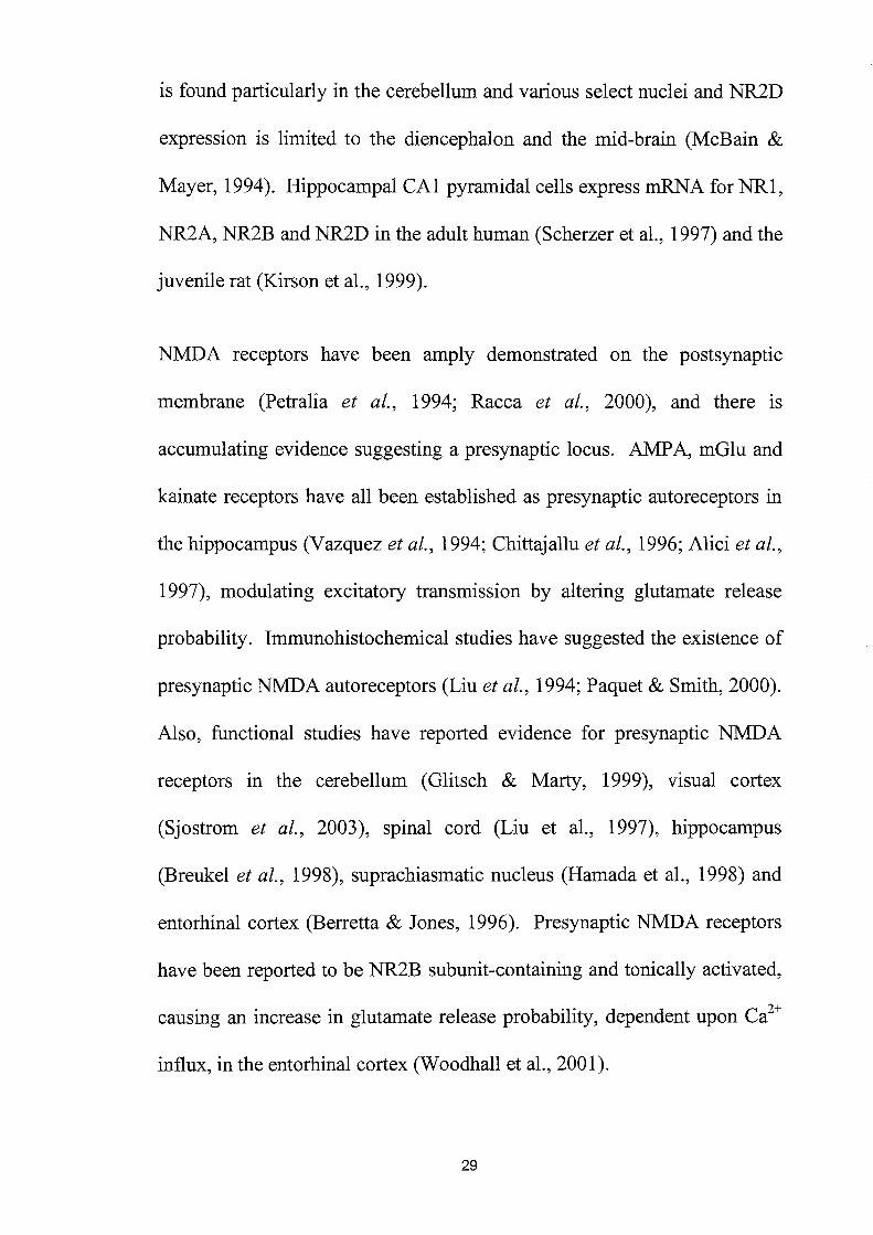
is found particularly in the cerebellum and various select nuclei and NR2D
expression is limited to the diencephalon and the mid-brain (McBain &
Mayer, 1994). Hippocampal CAl pyramidal cells express mRNA for NRl,
NR2A, NR2B and NR2D in the adult human (Scherzer et aI., 1997) and the
juvenile rat (Kirson et aI., 1999).
NMDA receptors have been amply demonstrated on the postsynaptic
membrane (Petralia et aI., 1994; Racca et aI., 2000), and there is
accumulating evidence suggesting a presynaptic locus. AMP A, mGlu and
kainate receptors have all been established as presynaptic autoreceptors in
the hippocampus (Vazquez et aI., 1994; Chittajallu et al., 1996; Alici et al.,
1997), modulating excitatory transmission by altering glutamate release
probability. Immunohistochemical studies have suggested the existence of
presynaptic NMDA autoreceptors (Liu et al., 1994; Paquet & Smith, 2000).
Also, functional studies have reported evidence for presynaptic NMDA
receptors in the cerebellum (Glitsch & Marty, 1999), visual cortex
(Sjostrom et al., 2003), spinal cord (Liu et aI., 1997), hippocampus
(Breukel et aI., 1998), suprachiasmatic nucleus (Hamada et aI., 1998) and
entorhina1 cortex (Berretta & Jones, 1996). Presynaptic NMDA receptors
have been reported to be NR2B subunit-containing and tonically activated,
causing an increase in glutamate release probability, dependent upon Ca2+
influx, in the entorhina1 cortex (Woodhall et aI., 2001).
29

NMDA receptor development
Developmentally only NR2B and NR2D subunits are present and
widespread in the prenatal brain but shortly after birth, NR2A and NR2C
subunits quickly predominate while the NR2B and NR2D subunits decline
to adult levels and a limited distribution. NR2B subunits are expressed
from late embryonic stages up to adulthood, while expression of NR2A
subunits only develops during early postnatal stages and rises to mature
levels by post-natal day 21 (Williams et al., 1993; Mori & Mishina, 1995;
Wenzel et al., 1995; Portera Cailliau et al., 1996). This transformation is
particularly significant for the age-dependence of synaptic plasticity (Kemp
et aI., 2000) and glutamate toxicity (Liu et aI., 1996). NR1 subunits are
present and widespread both pre- and post-natally (Lynch & Guttmann,
2001)(see tables 1,2 and 3).
30

Subunit Localisation in adult Developmental animals course
NRI Ubiquitous with splice variant Prenatal and postnatal distribution limited to specific cells
NR2A Widespread throughout the Develops post-natally forebrain. Less prominent in the basal ganglia
NR2B Largely limited to forebrain Widespread with later decline to adult levels and distribution
NR2C Largely limited to cerebellum Appears postnatally
NR2D Isolated cells of thalamus, sub Widespread prenatally thalamus and nNOS containing cells of cortex
Table 1. Summary of the distribution and developmental course of NMDA
receptor subunits.
31

NMDA receptor Subunit heterogeneity property
Glutamate and glycine 2D > 28> 2C affinity
Channel open 2A (2-5 fold higher) > 28
probability
Peak current density 2A (4 times larger) > 28
Channel conductance 2A, 28 » 2C, 2D
Receptor kinetics 2A > 28 = 2C > 2D
Current decay time 2A > 28 > 2C > 2D
Ca~'" influx 28 >2A
Sensitivity to Mg~· 2A, 28 » 2C, 2D
block
Deactivation time 2D » 28, 2C > 2A
Desensitisation 2A » 2D » 28, 2C sensitivity
Speed of recovery from 2A > 2A128 > 28 desensitisation
Proton sensitivity 2A > 28 > 20 > 2C
Table 2. Summary of the functional heterogeneity of NMDA receptors
depending on the presence of specific NR2 subunits.
32

Widely distributed in mammalian CNS (especially NMDA
enriched in hippocampus and cerebral cortex).
Widespread in CNS; similar distribution to NMDA AMPA
receptors.
Concentrated in a few specific areas of CNS, Kainate
corresponding to NMDA and AMP A receptor distribution.
A class of receptors positively linked to inositol Metabotropic
triphosphate or negatively to cyclic AMP formation.
Table 3. Summary of the general distribution of excitatory amino acid (EEA)
receptors in the mammalian eNS.
33

NMDA receptor pharmacology
The complicated physiology of the NMDA receptor complex provides
agonists and antagonists with several distinct binding sites: glutamate,
strychnine-insensitive glycine, ion-channel, polyamine and other
modulatory sites (Figure 1).
Glutamate, NMDA, aspartate, quinolinic acid, homoquinolinic acid and
other agonists activate the glutamate site; the prototypic antagonist of this
site is 2-AP5. The glycine site is activated by glycine (Johnson & Ascher,
1987) and several antagonists exist including ACBC, CGP 78608 and 7-
chlorokynurenic acid (Fletcher & Lodge, 1988; Kemp et al., 1988). The
NMDA receptor ion channel is blocked by Mg2+ ions at resting membrane
potentials. Other modulators have also been discovered that block the ion
channel in a non-competitive manner; notably, dizocilpine (MK 801), PCP
and ketamine. Zinc, like other group IIB metal ions produces inhibition of
the NMDA receptor and is selective at nanomolar concentrations to NR2A
subunit-containing NMDA receptors (Paoletti et al., 1997). Polyamines
such as spermine and spermidine can inhibit or potentiate the NMDA
receptor, at high and low concentrations respectively. Polyamines promote
channel opening at low micromolar concentrations by glycine-dependent
and glycine-independent mechanisms (Rock & Macdonald, 1992;
Williams, 1997). At higher concentrations, they block the channel in a
34

voltage-dependent manner. Antagonists at this site, inhibiting the NMDA
receptor, include ifenprodil and haloperidol (Reynolds & Miller, 1989;
Ilyin et ai., 1996; Lynch & Gallagher, 1996). Modulation also occurs at the
Ro 25-6981 site, the redox site and with ethanol and the volatile
anaesthetics.
35

Modulatory f lIVBP·like
Domain \
NR1
Agonist f binding
GlnBP·iike . Glycine-Domain l
Cytoplasmic
Figure 1. The NMDA receptor.
Polvamine Mg2+
NR2
"--. { Zn2 + (MR2A) Ifenprodil (NR2B)
-- Glutamate
Extracellll/af
A model showing the agonist binding sites for glutamate and glycine and the
competitive antagonists that can block these sites. Additionally, other
modulators can block the ion-channel and modulate the receptor at discrete
sites (Kemp & McKernan, 2002).
36

AMPA and kainate receptors
The a-amino-3-hydroxy-5-methyl-4-isoxazole propionic acid (AMP A) and
kainate (KA) receptors are the non-NMDA ionotropic receptor subtypes
that activate Na+ and K+ conductances whilst the NMDA receptors activate
Na+, K+ and Ca+ conductances (Monaghan et at., 1989; Barnard & Henley,
1990). AMP A receptors are hetero-oligomeric proteins consisting of the
subunits GluR1 to GluR4, each receptor comprising four subunits
(Hollmann & Heinemann, 1994; Rosenmund et at., 1998). The AMPA and
kainate receptors have at least three separate binding sites at which agonists
and antagonists can act: glutamate binding, desensitisation and intra-ion
channel binding sites. Kainate receptors consist of GluR5-7 and KA1-2
subunits, and have approximately 40% sequence homology to AMP A
receptor GluR1-4 subunits. High affinity binding of kainate is observed to
the KA1 and KA2 subunits compared to low affinity binding to GluR5-7.
The same agonists and antagonists can modulate both AMP A and kainate
receptors with little differentiation. However, certain desensitisation
inhibitors, which act allosterically to modulate positively sites on the ion
channel, can distinguish between AMP A and kainate receptors (Table 3).
37

Inhibitory GABA-ergic transmission
As well as excitatory transmission mediated by glutamate there is also the
presence of GABA-ergic interneurones that mediate feedforward and
feedback inhibition of excitatory transmission. Activation of these
inhibitory interneurones by glutamate results in a dual component
inhibitory postsynaptic potential (IPSP)(Alger & Nicoll, 1982; Benardo,
1994). This consists of a GABAA-mediated fast component which results
in the activation of GABAA ionotropic receptors releasing hyperpolarising
cr ions into the neurone. The other component is a slow GABAB-
mediated component which results in the activation of the GABAB
metabotropic receptor (Alger, 1984) leading to the hyperpolarising influx
of K+ ions and the negative modulation of the influx of Ca2+ ions. IPSPs
can inhibit excitatory synaptic transmission by moving the membrane
potential to values that are more negative and increasing the amount of
depolarisation required by EPSPs to reach the threshold for action potential
generation. GABAB mediated slow IPSPs are possibly better poised to
counter longer-lasting EPSPNMDA which because of their long slow kinetics
have been reported to escape fast GABAA inhibition (Benardo, 1995)
although one report suggested that when stimulation evokes only a small
EPSP (2-5m V) the GABAB component is negligible (Otmakhova &
Lisman,2004). In addition to postsynaptic GABA receptors there is also a
presynaptic GABAB autoreceptor in the Schaffer collateral feedforward
38

interneurones (but not in the perforant pathway). When the interneurone is
repeatedly activated, GABA is released in sufficient quantity to feedback
on these GABAB receptors, which inhibits the release of further GABA,
and synaptic inhibition is depressed (Davies et al., 1990).
39

Excitotoxicity
The term excitotoxicity was first coined by John Olney who observed that a
number of excitatory amino acids (EAAs) killed neurones in the
hypothalamus with a potency that correlated with their efficacy in
depolarising cells. Excitotoxicity is a critical mechanism contributing to
neurodegeneration during ischaemia and hypoxia. Both the loss of calcium
homeostasis and the activation of EAA receptors are postulated to be
responsible for excitotoxic neuronal damage (Olney, 1969; 1986; Seisjo,
1988; Choi, 1991; Sattler et al., 1999). With the activation of NMDA,
receptors both of these hypotheses are fulfilled. The over-activation of
NMDA receptors leads to an influx of Ca2+ resulting in necrosis or
apoptosis by several proposed mechanisms. The subsequent activation of
Ca2+ -sensitive enzymes (nNOS, endonucleases and proteases), the
production of highly reactive free radical species, mitochondrial
impairment and the activation of immediate-early response sense genes all
contributing to neuronal death. Blockade of this influx, by NMDA
antagonists and calcium antagonists, is neuroprotective in animal models of
stroke and seizure (Lee et aI., 1999). The clinical use of non-selective
NMDA receptors as neuroprotective drugs has been hindered by their
narrow therapeutic window, which produces adverse drug reactions
including sedation, muscular weakness, ataxia, confusion and tinnitus.
Subunit selective antagonists such as ifenprodil have been associated with a
40

decrease in side effects allowing an effective therapeutic dose to be titrated
(Gotti et aI., 1988). Nevertheless, therapeutic NMDA receptor antagonists
have yet to be developed with clinical success (De Keyser et at., 1999).
Several studies have specifically implicated both the NR2A- and NR2B
containing NMDA receptors in excitotoxicity. Excitotoxicity in cultured
embryonic forebrain neurones coincides with a dramatic increase in NR2B
mRNA levels during the first 10 days, thereafter remaining stable (Cheng
et aI., 1999). However, decreased expression of NR2A and NR2B mRNA
has also been reported in CAl and other hippocampal areas, in response to
a severe ischaemic insult, altering the function of the NMDA receptors
(Zhang et aI., 1997). In addition to changes in subunit expression,
ischaemia has also been shown to increase the tyrosine phosphorylation of
both the NR2A and NR2B subunits thereby altering their function (Kim et
at., 1998; Cheung et at., 2000).
41

Anatomy of the hippocampus
The electrophysiological experiments described in this study were carried
out in the hippocampal slice preparation of the 7-8 week old Wistar rat.
The hippocampus along with the amygdala, adjacent regions of the limbic
cortex and the septal area constitute the main structures of the limbic
system. These structures form major interconnections with portions of the
thalamus and the cerebral cortex (cingulated gyrus).
For three reasons the hippocampus is one of the most studied areas of
central nervous system. Firstly, it has an easily identifiable structure at the
gross and histological levels. Secondly, it has been recognized that the
hippocampus plays a fundamental role in learning and memory (patients
who have had bilateral hippocampalectomy have suffered from a
permanent loss of storing new information into their long-term memory).
Thirdly, the hippocampus is especially susceptible to seizure disorders and
is particularly vulnerable to the effects of ischaemia and hypoxia (Johnston
& Amaral, 1998).
In the brains of mammals, the hippocampus is a bilateral structure that
appears as a ridge extending into the lateral ventricle. The outer surface of
the hippocampus is composed of myelinated fibres, appears white and is
called the alveus. The hippocampal formation can be divided into
Ammon's hom, the subiculum, the dentate gyrus and the entorhinal cortex.
42
Based on the Golgi method of staining, the anatomist Lorente de N6 (1934)
(see Johnstone & Amaral, 1998) divided Ammon's hom into four cornu
Ammonis subfields: CAl to CA4. The designation CA4 is no longer used
because it referred to the region occupied by the polymorphic layer of the
dentate gyrus. CAl is equivalent to the regio superior and CA2 and CA3
fields are equivalent to the regio inferior. A narrow transitional zone, CA2,
separates CAl and CA3 (Figure 2).
The dentate gyrus contains round, tightly packed neurons called granule
cells and consists of three layers: the granule layer, which is the principal
layer, the molecular layer above the granule cell layer and a polymorphic
layer below the granule cell layer (Isaccson, 1987).
In all CA fields below the alveus is the stratum oriens, which contains the
basal dendrites of the pyramidal cells. The cell bodies are clearly visible
under a microscope as a dark band. Below this is the stratum radiatum
consisting of the apical dendrites of the pyramidal cells and the Schaffer
collaterals, which are collateral branches from axons of pyramidal cells in
the CA3 region (Cajal, 1968).
43

Neuronal circuits in the hippocampus
The excitatory neuronal circuitry of the hippocampal formation is
illustrated in figure 2. The functional organisation of the hippocampus has
been described in terms of a 'trisynaptic' circuit. Information that flows
from the neocortex into and out of the hippocampus travels in a
unidirectional manner through this trisynaptic pathway. The entorhinal
cortex is considered the starting point of the circuit. Neurones located in
layer II of the entorhinal cortex give rise to the perforant pathway that
projects through the subiculum and terminates both in the dentate gyrus and
in the CA3 region. Neurones forming the medial entorhinal cortex produce
axons that terminate in the middle layer of the molecular layer of the
dentate gyrus; the outer third of the molecular layer receive axons from the
lateral entorhinal cortex. These two components of the perforant pathway
also terminate in the stratum lacunosum-molculare of the CA2 and CA3
regions. N eurones located in layer III of the entorhinal cortex proj ect to the
CA 1 and the subiculum.
The granule cells also synapse onto neurones of the polymorphic layer,
which supplies associational connections to other levels of the dentate
gyrus. The CA3 pyramidal cells project heavily within the CA3 and as the
Schaffer collateral projection to the CAl stratum radiatum and stratum
oriens. CAl neurones project to both the subiculum and the deep layers of
44

the entorhinal cortex, which in turn produce projections back into many of
the cortical areas that originally terminated in the entorhinal cortex.
Therefore information sent from cortical neurones to the entorhinal cortex
can then negotiate the complete hippocampal circuit and be returned back
to its cortical origin (Witter, 1989).
As well as the main excitatory circuitry, there are also inhibitory pathways
throughout the hippocampus. Three types of interneurones control this
circuit in the pyramidal cell layers: axo-axonic cells, basket cells and
bistratified cells. The majority of interneurones in the hippocampus use the
inhibitory transmitter y-aminobutyric acid (GABA). The axo-axonic cells
synapse upon the initial segment of pyramidal neurones and have a strong
influence over the generation of action potentials. Basket cells synapse
onto the somata and bistratified cell synapse at apical and basal dendrites of
the pyramidal neurones. The dendrites of these three classes of
interneurone proj ect into the stratum radiatum and stratum oriens where
they can encounter excitatory inputs from the Schaffer collaterals,
commissural-association fibres and feedback synapses from local
pyramidal neurones (lsaccson, 1987; Johnston, 1998).
45

A
B 1
~,:::;.::.t ......
.. , ,
r c ;: ,
2
.. ~ c
Figure 2. Diagram showing the anatomy of the rat hippocampal formation and
the placement of the stimulating and recording electrodes.
A, location of the left hippocampus in rat brain. B, structure of a hippocampal
slice showing the principal excitatory neurons. The stimulating electrode (1)
was positioned in the stratum radiatum at the CA1/CA2 junction. Orthodromic
population spikes and excitatory postsynaptic potentials were recorded from the
stratum pyramidale and stratum radiatum respectively using electrodes (2) and
(3) placed in the CA 1 region. C, hippocampal circuitry. DG, dentate gyrus;
ENT, entorhinal cortex; mf, mossy fibre; pp, perforant pathway; rc, recurrent
collateral; sc, Schaffer collateral; SUB, subiculum. (Revest & Longstaff, 1998).
46

The hippocampal slice preparation
Ever since cortical slices were demonstrated to have comparable electrical
activity with the intact animal (Yamamoto & McIlwain, 1966) the slice
preparation has been used extensively in the study of synaptic transmission
and electrophysiological experiments in the mammalian CNS. The studies
leading to our present understanding of how NMDA, AMP A, and
metabotropic glutamate receptors contribute to synaptic transmission; and
investigations of LTD and L TP have come almost exclusively from brain
slice studies. Hippocampal slices have also shown good conditions to
allow intracellular recording (Yamamoto, 1972; Kerkut, 1981). The
hippocampus is an essentially laminar structure, each transverse slice
having the same 'trisynaptic' circuitry. The neuronal function and anatomy
of the slice preparation, in the plane in which it is cut, is calculated to be
similar to the intact brain producing representative electrophysiological and
pharmacological responses. The slices should be well enough prepared to
allow recording at physiological temperatures (36-37°C for rodent slices)
however, this is not usually achievable and a sub-physiological temperature
of 28-30°C is normally used.
47

Advantages of the In vitro slice preparation over III VIVO techniques
include:
1. Technical simplicity: it is relatively easy to record via extracellular,
intracellular and whole-cell patch-clamp compared with in vivo
experiments.
2. Control over the conditions of the preparation. If one can reduce the
number of contributory variables, greater significance can be drawn
upon results from a constant environment (e.g. temperature,
movement, 02/C02 saturation, absence of the influence of an
anaesthetic maintenance dose).
3. Improved visualization of tissue. The tissue is easily identified
under low magnification facilitating the positioning of stimulating
and recording electrodes.
4. Control of the extracellular environment and the field potential to
study neurophysiology and pharmacology. This is helpful when
applying drugs and doses that are impractical in whole animal
studies.
5. Humane treatment of animals, because slice preparation does not
require restraint or the maintenance of physiological functions it is a
relatively more humane method.
48

6. The ability to use technically demanding methods that are difficult or
impossible in intact preparations. For example, the ability to isolate
specific pathways and record from specific postsynaptic cells
without contamination of other synaptic inputs from other brain
regions which have been dissected (Grover & Teyler, 1990), or the
ability to use optical techniques.
These advantages of the slice preparation have led to increasing numbers of
neuroscientists to use this technique. However, there are also some
disadvantages with brain slice preparations:
1. The dissection and slicing process severs the hippocampal slices'
normal sensory input and motor output. Long feedback loops,
some excitatory pathways and some inhibitory pathways are also
lost. One obvious symptom of this is the markedly lower levels of
spontaneous activity found in the slice preparation compared to the
intact animal. This brings into question the similarity and relevance
of the in vitro slice preparation compared to the in VIVO
environment. Furthermore, it has been demonstrated that
phenomena such as LTD induction are more readily induced in
vitro than in vivo (Bear & Abraham, 1996)
49

2. The crude method of slicing causes damage along the faces of the
slice, this results in cell damage and the release of substances that
may affect the slice.
3. The tissue is subject to an anoxic period during preparation (Lipton
& Whittingham, 1979).
4. The ionic environment does not mImIC exactly the normal
extracellular conditions in vivo. This is of particular relevance in
the induction and maintenance of LTD and L TP that are dependent
upon relative ci+ and Mg2+ ion concentrations. The continuous
perfusion of artificial cerebrospinal fluid (ACSF) can also wash out
any modulatory compounds endogenous to the slice (such as
growth factors released following stimulation).
5. The addition of drugs to the artificial cerebrospinal fluid (ACSF)
results in the exposure of the whole slice to pharmacological
modulation. This confounds the significance of results seen with
the subset of cells that are being recorded.
6. Finally, the diversity of methods used in each brain slice laboratory
makes the comparison of findings difficult (Lipton et aI., 1995).
The most obvious differences include: (A) the maintenance of brain
slices at the interface of the ACSF and oxygenated atmosphere
50

versus those maintained entirely submerged in ACSF, (B) cutting
brain slices on a manual tissue chopper versus a vibratome or a
vibraslice, (C) differing ACSF recipes and (D) temperature
differences.
Extracellular responses
Single electrical stimulations of the Schaffer collaterals result in a
distinctive sequence of excitation followed by inhibition in the target CAl
pyramidal neurones. Field extracellular recordings represent the summed
responses from a population of neurones close to the recording electrode.
The field excitatory postsynaptic potential (fEPSP) recorded in the stratum
radiatum consists of an initial fibre volley of action potentials in the
presynaptic fibres producing a negative-going transient; this is followed by
a slower negative-going potential with a time-period similar to that of the
underlying synaptic currents. The current flowing into these dendrites
during the fEPSP will exit the neurons near the cell body layer in the
stratum pyramidale where a recording electrode will record a concurrent
positive going potential. If the strength of the synaptic input is adequate to
evoke action potentials in the neurons, then a negative-going potential
(population spike) can also be recorded in the stratum pyramidale resulting
from the inward current during the postsynaptic action potentials.
51

Paired-pulse interactions
The magnitude of synaptic transmission in the mammalian brain can be
modulated in different ways. Two examples of such modulation are
paired-pulse facilitation (PPF) and paired-pulse depression (PPD).
Paired-pulse facilitation
Paired-pulse facilitation (PPF) is a form of short-term plasticity in which
the synaptic response to the test pulse (second stimulus) given after the
conditioning pulse (first stimulus) is enhanced in comparison to the
conditioning pulse when interstimulus intervals are relatively long
(~20ms). The most widely accepted hypothesis to explain the mechanism
of PPF is based on the accumulation of presynaptic ci+ after the first
stimulation. The first pulse induces a Ca2+ influx that lingers in the
presynaptic terminal for several hundred milliseconds. This residual Ca2+
combined with fresh Ca2+ entering during the second pulse leads to the
increased transmitter release (Katz & Miledi, 1968; Thomson, 2000;
Zucker & Regehr, 2002). The Ca2+ concentration in the presynaptic nerve
terminal affects directly the release probability of neurotransmitter.
Furthermore, reports at the neuromuscular junction have suggested that the
second of the two action potentials induces release with higher probability
than the first one because of a small but persistent increase of the
intracellular Ca2+ concentration in the axon terminal. This residual Ca2+
52

can enhance the release probability by increasing the fusion of synaptic
vesicles with the presynaptic membrane and enhancing the number of
vesicles released by the action potential (Katz & Miledi, 1968; Mennerick
& Zorumski, 1995; Debanne et at., 1996). Neuromodulators and
physiological processes that alter transmitter release probability also affect
the paired-pulse ratio. Increasing the external Mg2+/Ca2+ ratio (Davies &
Collingridge, 1993; Lambert & Wilson, 1994; Wilcox & Dichter, 1994)
and applying adenosine (Lupica et at., 1992; Higgins & Stone, 1995)
decreases the probability of release of neurotransmitter during the first
stimulus and increases the probability of release by the second stimulus
producing enhanced PPF. In the hippocampus, PPF of excitatory synaptic
potentials in the CAl and CA3 is observed when large numbers of axon are
concurrently stimulated (Creager et at., 1980; Manabe et at., 1993)
whereas, during conditions in which the release probability is enhanced by
decreasing the Mg2+/Ca2+ ratio then PPF is reduced (Nathan et at., 1990;
Kahle & Cotman, 1993).
Paired-pulse depression
When two evoked potentials are elicited when interstimulus intervals are
small «1 Oms), the size of the second response is smaller than the first, a
phenomenon called paired-pulse depression (PPD). The mechanism
underlying PPD of synaptic transmission is unclear. One hypothesis is the
activation of GABAB autoreceptors (Deisz & Prince, 1989), but studies 53

have shown that GABAB receptor antagonists did not attenuate PPD of
unitary inhibitory post synaptic potentials (IPSPs) in pairs of cultured
hippocampal neurons (Wilcox & Dichter, 1994). An alternative presynaptic
mechanism for PPD is a transient decrease in the quantal content caused by
depletion of the readily releasable vesicle pool by the first stimulus
(Mennerick & Zorumski, 1995; Stevens & Wang, 1995; Debanne et al.,
1996). Postsynaptic mechanisms such as the desensitisation of GABAA
receptors (Alger, 1991) and a reduced driving force due to intracellular
accumulation of chlorine ions and/or extracellular accumulation of
potassium ions may also explain PPD (McCarren & Alger, 1985). Since
paired-pulse depression is observed most clearly and reliably when the
interstimulus interval is around 15-100 ms, which corresponds to the time
course of the GABAA mediated fast inhibitory potential recorded
intracellularly in pyramidal cells (Davies et al., 1990), it is thought to be
largely caused by the activation of GABAA receptors (Wilcox & Dichter,
1994). It is difficult to test this proposition using bath application of
GABAA receptor antagonists such as bicuculline which increase the
excitability of the pyramidal cells by blocking the effects of spontaneously
released GABA.
54
Whilst changes in paired-pulse interactions are widely considered to be due
to presynaptic alterations in release probability; when studied on fEPSPs
and especially where there are depolarisation-induced changes in the size
of the postsynaptic potential in response to the test agents many factors
affect them which can confound the interpretation. These include changes
in presynaptic release probability (Andreasen & Hablitz, 1994), activity of
presynaptic autoreceptors on inhibitory or excitatory terminals (Davies et
at., 1990; Stanford et at., 1995), modulation of postsynaptic conductances
(e.g. voltage-dependent NMDA receptor) and the presence of GABA-ergic
inhibition following the fEPSP and the depression cause by NMDA agonist
action (Duguid & Smart, 2004). In particular, it has been reported that
there is a non-linear inverse relationship between feedforward inhibition
and the NMDA dependent PPF component of the fEPSP
(Papatheodoropoulos & Kostopoulos, 1998) although, this NMDA
component of PPF is manifested by an increase in the duration of the
fEPSP and not the amplitude of the slope.
In hippocampal slices paired-pulse inhibition has been reported at
interstimulus intervals less than 40 ms and in the present experiments at
less than 20 ms while PPF is observed at longer time intervals (20 and 50
IDS) (Lynch et at., 1983; Higgins & Stone, 1995; Nikbakht & Stone, 2000).
55

Aims
1. To investigate NMDA receptor subunit pharmacology in the intact,
adult hippocampal slice preparation using the newly developed
NR2A and NR2B subunit-selective NMDA receptor antagonists
Ro 25-6981 and PEAQX.
2. To address the contribution of NR2A and NR2B subunit-containing
NMDA receptors in the induction of NMDA receptor dependent
synaptic plasticity in the Schaffer collateral-CAl pyramidal synapse.
3. A report had suggested that the NR2AJ2B subunit selective NMDA
receptor agonist homoquinolinic acid was binding to a novel
NMDA-insensitive site in the brain. An investigation of NMDA
insensitive effects on the fEPSP was undertaken to reveal any
residual effects.
Furthermore, the selectivity of homoquinolinic acid allowed the
investigation of the NR2A and NR2B components of the previous
study (1) and to find evidence for the involvement of the NR2D
subunit. In addition, this study investigated changes of paired-pulse
interactions using these compounds.
56

Chapter 2. Material and methods
Preparation of hippocampal slices
Male Wistar rats (130-180g, 7-8 weeks) were anaesthetised by an
intraperitoneal injection of urethane solution (1.5 g/Kg); urethane has been
shown not to interfere with synaptic plasticity, a problem encountered
when using phenobarbitone (unpublished observations). The animals were
then killed by cervical dislocation, decapitated using a guillotine, and the
brain gently and rapidly removed to ice-cold and oxygenated artificial
cerebrospinal fluid (ACSF). The intact brain was kept cold and moist by
copious amounts of ACSF whilst it was transferred to a petri dish lined
with filter paper to prevent sliding. The cerebellum was removed and the
two cerebral hemispheres were separated with a scalpel blade. Each
hippocampus was dissected free from the surrounding tissue using small
spatulas and cut transversely using a McIlwain tissue chopper into slices
450~m thick, perpendicular to their longitudinal axis. The slices were
gently separated from each other using blunt glass microelectrodes to
ensure that they receive adequate amounts of oxygen and nutrients and to
allow individual slices to be transferred easily. The slices were incubated
at room temperature (21-230C) on a fresh filter paper lined petri dish
containing a small amount of freshly gassed ACSF to cover the slices. The
petri dish was kept in an incubation chamber saturated in an atmosphere of
57

95% O2 and 5% CO2 for at least 1 hour prior to individual slices being
transferred to the recording chamber.
Composition of ACSF
The composition of the ACSF was (in mM): KH2P04 2.2, KC12, NaHC03
25, NaCll15, CaCh 2.5, MgS04 1.2, and glucose 10. It was gassed with a
mixture of95% O2 and 5% CO2 yielding a pH ,..,7.4.
Bath superfusion and application of drugs
Following incubation, individual slices were transferred to a I ml
submerged recording chamber using a wide tipped plastic Pasteur pipette.
A seeker wire, the end of which was modified in such a way as to form a ~
shape was used to gently hold the slice in place. The slice was left to
stabilise whilst being continuously superfused with ACSF by a gravity-fed
silicone tube at a rate of 4 ml/min. The ACSF was kept saturated with 95%
O2 and 5% CO2 and heated using a thermostatically controlled water bath
to 28-30°C. Drugs were added to the ACSF and fed via a three-way tap
through the silicon tubing to the recording chamber. Due to the length of
tubing, it took 1 minute for fluid to reach the recording chamber. To
prevent the build-up of organic matter the silicon tubing and recording
chamber were flushed with distilled water before and after use.
Additionally, the system was more aggressively cleaned using diluted
58

bleach once a month and the tubing replaced every six months and as
required.
Stimulation
Stimuli were AC amplified square wave constant-current pluses of 300~s
duration. Paired stimuli, where used, were delivered through the same
electrode. The slices were stimulated using a concentric bipolar electrode
(Clark Electromedical Instruments Ltd, Harvard Apparatus) positioned in
the stratum radiatum near the commissural border of CAlICA2 for
orthodromic activation of pyramidal cells. The slice was briefly and
occasionally stimulated at I Hertz (Hz) until a population of neurones was
identified; once this was achieved, a stimulus frequency of 0.05Hz was
used. The population of neurones was then allowed to stabilise.
Population spikes were recorded at 70% of maximum amplitude whilst
ffiPSP slopes were recorded at 50% of their maximum amplitudes. Only
field responses that produced clear ffiPSP and population spikes with
maximum amplitudes greater than 2m V were used in experiments.
Furthermore, responses that showed greater than 5% drift over the 20-
minute baseline-recording period were excluded (Figure 3).
59

A
~ Population spike
Presynaptic
lmV L Sms
B fiT volley Example points of maximum slope measurement
t Stimulation artefacts
~fEPSP
ImV L Sms
Figure 3. Example responses recorded from the CA 1 region of the rat
hippocampal slice.
Trace (A) shows a population spike recorded from the stratum pyramidale and
trace (8) shows a fEPSP, recorded from the stratum radiatum.
60

Recording
Extracellular population spike potentials and field excitatory postsynaptic
potentials (fEPSPs) were recorded extracellularly from the stratum
pyramidale and the stratum radiatum, respectively, using borosilicate glass
microelectrodes, which were produced on a Kopf vertical puller. The
electrode tips were broken back to produce an opening 2-4~m in diameter
under low magnification using a glass probe, resistances approximately 2-
5MQ. The electrodes were filled with sodium chloride 0.9% solution using
a fine 36-gauge needle.
Evoked responses were amplified through a Neurolog system, displayed on
a digital oscilloscope and recorded onto a personal computer via a
Cambridge Electronic Device (CED) micro 1401 interface and Signal
analysis software (Cambridge Electronic Design, version 1). Responses
were filtered between DC and 5kHz; line frequency interference was
removed by digitally filtering (Humbug, Digitimer Ltd).
61

Data analysis
Responses were quantified as the amplitude of the population spike in m V
(measured as the difference between peak negativity and the averaged
values of the two peaks of the positive-going synaptic potential) and the
fEPSP was measured as the maximum slope of the initial negative going
gradient with two static cursors using Signal software; manually selecting
and recording the slope in response to changes in the size of the fEPSP
response and electrical interference (figure 3). Individual responses were
measured, normalised and compared to the 20-minute initial baseline
recordings. Every time point was pooled and graphed using GraphPad
Prism software (GraphPad Prism Software, San Diego, CA, version 3)
showing mean ± standard error of the mean (s.e.m.). Rarely, single
responses had to be excluded due to anomalous electrical interference
disrupting the recording of evoked responses.
When paired-pulse interactions were investigated, inhibition or facilitation
was expressed as the ratio of the second response of a pair compared with
the first response.
Statistical analysis
The statistical significance of a difference was assessed on raw data by a
paired Student's t-test or, if the difference was between three or more
62

means, significance was calculated with a one-way or repeated measures
analysis of variance (ANOV A) followed by a Student-Newman-Keuls
post-test using GraphPad Prism software; p values less than 0.05 were
considered statistically significant. IC50 and EC50 values were calculated
using GraphPad Prism software by generating non-linear regression,
sigmoidal concentration-response curves. In the figures statistically
differences between data points are sometimes indicated by asterisks:
* p<0.05; ** p<0.01; *** p<O.OOl.
Chemical agents and drugs
Constituents of ACSF were of analar grade and were dissolved in distilled
water to prepare fresh every day. N-Methyl-D-aspartic acid (NMDA) ,
D-(-)-2-amino-5-phosphonopentanoic acid (2-AP5), dizocilpine (MK 801),
zinc acetate (Zn2+), (RS)-AMPA hydrobromide (AMP A) and kainate (KA)
were dissolved in distilled water to obtain stock solutions. Quinolinic acid
(QA) and homoquinolinic acid (HQA) were dissolved in 1 molar
equivalencies of NaOH to give stock solutions. Dimethyl sulphoxide
(DMSO) was used to dissolve kynurenic acid (KYA) , 2-carboxy-3-
carboxymethylquinoline (CCMQ), okadaic acid (OA), anisomycin, (R:(*),
S:(*))-alpha-(4-hydroxyphenyl)-beta-methyl-4-(phenylmethy1)-1-
piperidinepropanol (Ro 25-6981), staurosporine (STA) and 1,3-dipropyl-8-
cyclopentyl-xanthine (DPCPX) to produce stock solutions. The final
63
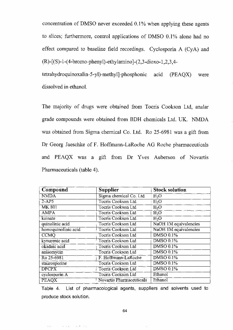
concentration of DMSO never exceeded 0.1 % when applying these agents
to slices; furthermore, control applications of DMSO 0.1% alone had no
effect compared to baseline field recordings. Cyclosporin A (CyA) and
(R)-[(S)-I-( 4-bromo-phenyl)-ethylamino ]-(2,3-dioxo-l ,2,3,4-
tetrahydroquinoxalin-5-yl)-methyl]-phosphonic acid (PEAQX) were
dissolved in ethanol.
The majority of drugs were obtained from Tocris Cookson Ltd, analar
grade compounds were obtained from BDH chemicals Ltd. UK. NMDA
was obtained from Sigma chemical Co. Ltd. Ro 25-6981 was a gift from
Dr Georg J aeschke of F. Hoffmann-LaRoche AG Roche pharmaceuticals
and PEAQX was a gift from Dr Yves Auberson of Novartis
Pharmaceuticals (table 4).
Compound Supplier Stock solution NMDA Sigma chemical Co. Ltd H2O 2-AP5 Tocris Cookson Ltd H2O MK801 Tocris Cookson Ltd H2O AMPA Tocris Cookson Ltd H2O kainate Tocris Cookson Ltd H2O quinolinic acid Tocris Cookson Ltd NaOH 1M equivalencies homoquinolinic acid Tocris Cookson Ltd NaOH 1M equivalencies CCMQ Tocris Cookson Ltd DMSOO.1% kynurenic acid Tocris Cookson Ltd DMSOO.1% okadaic acid Tocris Cookson Ltd DMSOO.1%
. . Tocris Cookson Ltd DMSOO.1% amsomycm
Ro 25-6981 F. Hoffinann-LaRoche DMSO O.l% staurosporine Tocris Cookson Ltd DMSOO.1% DPCPX Tocris Cookson Ltd DMSOO.1% cyclosporin A Tocris Cookson Ltd Ethanol PEAQX Novartis Pharmaceuticals Ethanol
Table 4. List of pharmacological agents, suppliers and solvents used to
produce stock solution.
64

Chapter 3. The negative coupling of NR2B to
NR2A subunit-containing receptors.
Introduction
There has been a considerable amount of research over the last 10 years
that has identified the NR2B subunit as being especially important in
determining the function of the NMDA receptor. For example, in forming
the glutamate binding site (Laube et aI., 1997), controlling the Mg2+ block
(Williams et al., 1998) and in a variety of phenomena including learning
and memory formation, synaptic plasticity (Hrabetova et aI., 2000), pain
(Chizh et aI., 2001), schizophrenia (Grimwood et aI., 1999; Gao et al.,
2000), Parkinson's disease (Nash et al., 1999; Oh et al., 1999; Dunah et al.,
2000; Steece Collier et al., 2000), Huntington's disease (Chen et al., 1999;
Zeron et al., 2001) and excitotoxicity (Zhang et al., 1997; Cheng et al.,
1999). The NR2B subunit consists of 1456 amino acids with a molecular
weight of approximately 170-180 kDa. All of the NR2 subunits have the
same basic structure as the NR1 subunit (Hollmann & Heinemann, 1994)
but varying in that they have an especially large intracellular C-terminal
domain. This C-terminal domain, it has been suggested, plays a role as the
target for modulatory or accessory proteins in promoting receptor
assembly, sorting or targeting and playing a part in different channel
conformations to modulate receptor function (Kohr et aI., 2003; Loftis &
65

Janowsky, 2003). The NR2B subunit is modulated by its co-assembly with
other subunits, alterations in the interaction with associated proteins such as
PSD-95 and SAP102, proteases, growth factors, hormones and post-
translational modifications such as phosphorylation. The phosphorylation
of tyrosine residues can contribute to enhanced receptor responses and
signal transduction into the neurone (Gurd, 1985; Gurd & Bissoon, 1997;
Hisatsune et al., 1999; Manabe et al., 2000; Nakazawa et al., 2001).
Receptor function is also modulated by the phosphorylation of serine
residues by protein kinase A (PKA) , protein kinase C (PKC), and
calmodulin-dependent protein kinase II (CaMKII) (Smart, 1997).
Ro 25-6981 is an ifenprodil derivative that has high affinity and selectivity
for the NR2B subunit-containing NMDA receptor but without the affinity
for aI-adrenergic and serotonergic receptors found with ifenprodil itself
(Chenard et al., 1991; McCool & Lovinger, 1995). It is a high affinity,
selective (5000-fold greater for the NR2B than the NR2A subunit),
activity-dependent, voltage-independent and non-competitive NR2B
subunit-containing NMDA receptor antagonist (Fischer et al., 1997; Mutel
et al., 1998; Lynch et al., 2001).
F. Hoffman-La Roche Pharmaceuticals first described Ro 25-6981 in 1997.
They used cloned NMDA receptors expressing NR1cINR2A, NRlclNR2B
and NR1flNR2B subunits to confirm that Ro 25-6981 was selective for the
NR2B subunit-containing receptors. 66
They also studied the

electrophysiology of Ro 25-6981 in young cultured rat cortical neurones,
which express NR2B as the dominant NMDA receptor subunit. It was
observed that the effect of Ro 25-6981 was dependent upon the relative
level of NMDA receptor activation. Ro 25-6981 potently blocked the
current evoked by NMDA lOOIlM but it potentiated the slight responses to
NMDA 11lM. Protons (H+) exert a tonic inhibition of the NMDA receptor
with an ICso at physiological pH 7.4 by binding at a proton sensor site. The
NR2A and NR2B subunit-containing NMDA receptors are the most
sensitive to this effect. The proton sensor's binding site overlaps
structurally and mechanistically with the binding site of Ro 25-6981,
ifenprodil and the endogenous modulators spermine and spermidine (Mott
et al., 1998). The binding stabilises a protonated, agonist-bound state of
the receptor that has a low open probability. This shifts the pKa of the
proton sensor to values that are more alkaline and increases tonic
inhibition. However, the potentiation of responses to NMDA by these
compounds is reported to be due to an allosteric modulation with the
glutamate and glycine binding sites. The binding of these compounds
causes a decrease in NMDA receptor affinity for glycine and an increase in
affinity for glutamate and NMDA leading to potentiation. However, while
ifenprodil and Ro 25-6981 both bind to the N-terminal third of the NR2B
receptor, Ro 25-6981 has a distinct binding site that has greater three
dimensional requirements. Furthermore, the binding of ifenprodil is
67

decreased and its IC50 is increased by rising concentrations of spermidine
whilst under the same conditions the binding of Ro 25-6981 is unaffected
and its IC50 decreases (Kew et al., 1996; Kew et al., 1998b; Grimwood et
al., 2000; Lynch et al., 2001; Perin-Dureau et al., 2002). In addition, Ro
25-6981 had no direct effect on the ion channel as seen with ifenprodii.
This clearly demonstrates distinct properties of action by Ro 25-6981,
which have not yet been fully investigated. This activity-dependent
property is of great significance in therapeutic development as it predicts
that NMDA receptors that are pathologically and continuously activated
will be blocked whilst leaving inactive receptors relatively unaffected. In
neuroprotective experiments, Ro 25-6981 was found to be very effective in
protecting cultured cortical neurones from excitotoxic and ischaemic
insults (Fischer et al., 1997).
A further study followed, using autoradiography to investigate the binding
of Ro 25-6981 to adult rat brain membranes and tissue sections. It was
observed that Ro 25-6981 binding was similar to NR2B subunit
distribution and dissimilar to NR2A subunit distribution (Mutel et aI.,
1998).
More recently, a study of Ro 25-6981 was carried out to characterise its
NR2B selective pharmacology using human embryonic kidney (HEK) 293
cells transfected with heterodimers consisting of NRI a and either wildtype,
chimeric or mutant NR2B subunits (Lynch et al., 2001) which co-assemble 68

into tetrameric or pentameric receptors. It was observed that specific
regions of the N-terminal leucine/isoleucine/valine-binding protein
(LIVBP)-like domain of the NR2B subunit protein modulated the
interaction between Ro 25-6981 and the NMDA receptor. Ro 25-6981 may
be binding directly to the NR2B subunit or indirectly to the NR1 subunit at
a site that is modulated by the NR2B subunit in a way that is analogous to
glycine binding to the NRl subunit where the NR2 subunit influences
glycine affinity (Gallagher et al., 1996; Masuko et al., 1999; Perin-Dureau
et a!., 2002). Whilst Ro 25-6981 is an ifenprodil analogue it has a binding
site and pharmacology that is distinct from the polyamine site (Lynch et al.,
2001).
Although there are several NR2B antagonists available, Ro 25-6981 is the
first compound to be developed that is a highly selective and full antagonist
at NR2B subunit-containing NMDA receptors. Ifenprodil is a partial
antagonist only inhibiting NR11NR2B receptors by 80% on average and
affecting NR11NR2A!NR2B receptors with little (Luo et al., 1997) or
intermediate effect (Tovar & Westbrook, 1999). Also, ifenprodi1 is not
very selective for the NR2B subunit (400-fold, recombinant NR11NR2B
compared to NR1INR2A) and also blocks aI-adrenergic, 5HT lA, 5HT 2,
5HT3 and sigma receptors and N, P and Q type calcium channels (Chenard
et al., 1991; McCool & Lovinger, 1995). Interestingly, Pfizer has recently
developed an NR2B subunit-selective antagonist CP-10 1, 606, which has a
69

very low affinity for the NR11NR2A1NR2B synaptic receptors but a high
affinity for the purely extrasynaptic NR11NR2B population (Chenard et al.,
1995; Tovar & Westbrook, 1999). It would be of particular interest to
repeat this study using CP-101, 606 to investigate further the contribution
of synaptic versus extrasynaptic receptor populations (Hardingham et al.,
2002). There have been several recent reports demonstrating that the
different subunit-containing NMDA receptors and the locus of activation
has a great effect on determining the direction of synaptic plasticity (Liu et
al., 2004; Massey et al., 2004; Rusakov et al., 2004; Wong et al., 2004;
Mallon et al., 2005) and the phosphorylation of CREB (cAMP response
element binding protein) leading to BDNF expression and pro-survival
pathways (Hardingham et al., 2002; Riccio & Ginty, 2002).
Novartis Pharmaceuticals have recently developed an NR2A subunit
containing NMDA receptor antagonist (R)-[(S)-l-( 4-bromo-phenyl)
ethyl amino ]-(2,3-dioxo-1 ,2,3 ,4-tetrahydroquinoxalin-5-yl)-methyl]
phosphonic acid (PEAQX, also known as NVP-AAM077, appendix 2).
PEAQX is a water-soluble 5-methyl-quinoxalinedione with a high affinity
for the glutamate-binding site of the NMDA receptor ([3H]-CGP39653
binding assay: ICso = llnM). At lOflM PEAQX showed no additional
activity in a series of 68 radio ligand binding assays for CNS receptors.
PEAQX has 126-fold selectivity for the NMDA NR1A1NR2A over the
NR1A1NR2B receptors expressed in Xenopus oocytes (IC50 = 14nM and
70

1800nM respectively). Experiments on embryonic cortical cells and on
neocortical slices have confirmed these results with native receptors.
Cultured embryonic cortical cells express NR2A subunits at a very low
level, resulting in a low potency of PEAQX (ICso = 1 ~M). In adult slices
where NR2A subunits are predominantly expressed the effect of PEAQX is
stronger (IC50 = 68nM) than in cultured embryonic cortical cells that
express NR2A at very low levels (undetectable) (ICso = 1 ~M).
Furthermore, in vivo PEAQX was found to be a potent anticonvulsant
following intravenous dosing with few of the side effects associated with
non-selective or NR2B selective antagonists (Auberson et al., 2002).
Although these compounds have been examined on recombinant receptors
and immature cultures, which are systems that over-express the NR2B
subunit-containing NMDA receptors, this study has used these compounds
to examine the role of NR2 subunits in an intact neuronal network in which
adult heteromeric NMDA receptors are involved in synaptic transmission
and plasticity. Of great importance in this study was the use of adult
animals that are 7-8 weeks old because they are past the 3rd postnatal-week
threshold for the maturation ofNMDA receptor subunit expression (where
NR2A subunits are the last to reach their mature levels) and the 4th
postnatal-week threshold during which the basal levels of receptor tyrosine
phosphorylation are attained (Cudmore & Gurd, 1991; Tovar & Westbrook,
1999).
71
Results
NMDA
We started this study usmg lO-minute applications of NMDA, which
reversibly depressed the slope of the fEPSP. The depression of fEPSP
slope being a reflection of changes in membrane conductance and
depolarisation of the neurones. This leads to the depolarisation block of the
CAl neurones recorded from and the subsequent blockade of excitatory
transmission. The concentration-response relationship was very steep, with
l/-lM having had no effect, 4/-lM producing a response that was discernible
but not statistically significant, while 7.5/-lM produced a depression to
69.36±16.33%, lO/-lM produced a depression to l5.l8±4.43% (Figure 5)
and 20/-lM producing a depression to -1.05±1.61 % of fEPSP slope (Figure
4). Furthermore, investigation of changes to the paired-pulse paradigm
during the depression elicited by NMDA lO/-lM found a significant
decrease of paired-pulse inhibition, and increase of paired-pulse facilitation
at all interstimulus intervals (lOms p<O.OOl, 20ms, p<O.Ol and 50ms
p<O.Ol, n=5) (Figure 6 and 7).
72

Q) C- 100 I o en a.. en a.. w 'I-
Q) c: .--Q) en C'CS .c
'I-0 ~ c
80
60
40
20
0 . ,
-6 -5 Log[NMDA]M
Figure 4. Concentration curve of the inhibitory effect of NMDA upon fEPSP
slope.
Graph showing the non-cumulative concentration-response curve of depression
of fEPSP slope in response to 10-minute applications of NMDA concentrations
of 1~M, 4~M, 7.5~M, 10~M and 20~M, n=4, 13, 4, 8 and 4 respectively. A
concentration of 8.6~M is predicted to cause a 50% depression of fEPSP slope.
Each point shows the mean±s.e.m.
73

II NMDA 10flM
Figure 5. Effect of NMDA 10flM on fEPSP slope.
Plot showing the effect of a 10-minute application of 10IJM NMDA, which elicits
a depression in the slope to 15.18±4.43% of fEPSP (p<O.001). Points (c), (1)
and (2) indicate the timing of the paired-pulse tests summarised in Figure 6.
Each point shows the mean±s.e.m. for n=8 experiments.
74

..... o 0-2 ~ (l) • cu tJ) '- c -0 Q)c.. o tJ) 2 C Q) • Q) ''-..,
~~ :C'i:1 Q).s •
.!!1-o ::l C c..o 101 -0 Q) •
~ tJ) .-cu c..
**
***
10 20 30 40 inter-stimulus interval (ms)
Figure 6. Effect of NMDA 10!-lM on paired-pulse interactions.
**
50
1
2 c
Graph showing the paired-pulse interactions of fEPSP slope at interstimulus
intervals of 10ms, 20ms and 50ms during the baseline control responses (c),
10!-lM NMDA (1) and the washout period (2) (10ms p<O.001, 20ms p<O.01,
50ms p<O.01, n=5, 1 compared to 2 and c)(see plot in figure 5 for
corresponding times points and example records in figure 7). Each point shows
the mean±s.e.m. for n=5 experiments.
75

Control (c) 10IJM NMDA (1)
a) d)
b) e)
c) f)
lmV L 5ms
Figure 7. Example records of paired-pulse interactions during baseline
recording and under the influence of 10IJM NMDA.
Showing paired-pulse interactions recorded during baseline recording: (a) 10ms
(b) 20ms and (c) 50ms and during the depression caused by NMDA 10~M: (d)
10ms (e) 20ms and (f) 50ms (points relate to same pOints in figures 5 and 6).
76

Ro 25-6981
The large depressions of fEPSP slope elicited by applications of NMDA
were caused by the activation of native heteromeric NMDA receptors
containing a combination ofNR1, NR2A, NR2B and NR2D subunits. This
study investigated the contribution of NR2B subunit-containing NMDA
receptors using the NR2B antagonist Ro 25-6981. A concentration of 3 ~
Ro 25-6981 was chosen as it has previously been shown to block
selectively the NR2B component of the NMDA excitatory postsynaptic
current (NMDAEPSc) in 3-4 week old rats and not the NR2A component
(Liu et al., 2004). Repeated applications of NMDA 10J.lM caused two
similar reversible depressions of fEPSP slope to 16.06±9.16% then to
12.85±7.85% (p<0.001, n=4). The co-application of3J.lM Ro 25-6981 had
no immediate effect upon this depression, which again reversibly depressed
the fEPSP slope to 8.96±2.77% (p<0.001, n=4). However, the next 10-
minute application of NMDA started 5 minutes after the end of the 20-
minute application of Ro 25-6981 3 J.lM totally abolished the fEPSP, which
then slowly and partially recovered to 37.56±7.26% of the baseline
responses and remained depressed for at least the 100 minutes recorded
(p<0.001, n=4) (figure 8 and 9). Attempts to recover the response by
increasing afferent stimulation or by HFS stimulation, which normally
induces LTP (responses not shown), were unsuccessful. This observation
is consistent with a terminal excitotoxic insult to the neurones recorded
77
from and inconsistent with the induction of LTD as witnessed by the failure
to reverse it with an L TP inducing protocol or by increasing the stimulation
of the presynaptic afferents (Schurr et aI., 1995).
Following the intensity of the response to lOI-LM NMDA, a concentration of
NMDA 4I-LM was used, a concentration that produced a smaller control
depression of baseline fEPSP slope (figure 10). This would be less likely
to result in an excitotoxic insult after the application of Ro 25-6981.
Applications of NMDA 4I-LM caused two small yet discernible depressions
of fEPSP slope that were not statistically significant (i and ii). Again the
co-application of Ro 25-6981 3I-LM with a third application of NMDA (iii)
had no immediate effect but a fourth application, during the washout phase
of Ro 25-6981 (iv), showed a significant enhancement of the depression of
fEPSP slope. The fifth application of NMDA (v) caused an even greater
depression of fEPSP slope that was matched in size by the sixth and
seventh (vi and vii) applications with no washout of the effect being
observed up to 100 minutes after Ro 25-6981 had been removed from the
perfusate (significant depressions of fEPSP slope to (iv) 40.94±4.56%, (v)
19.78±6.7%, (vi) 19.28±10.74% and (vii) 13.23±4.18% (all p<O.OOl
compared to the second NMDA 4I-LM induced depression, n=4)). Lastly,
the application of the NR2A subunit-selective antagonist PEAQX O.lI-LM
during the eighth and final application of NMDA 4I-LM (viii) completely
78
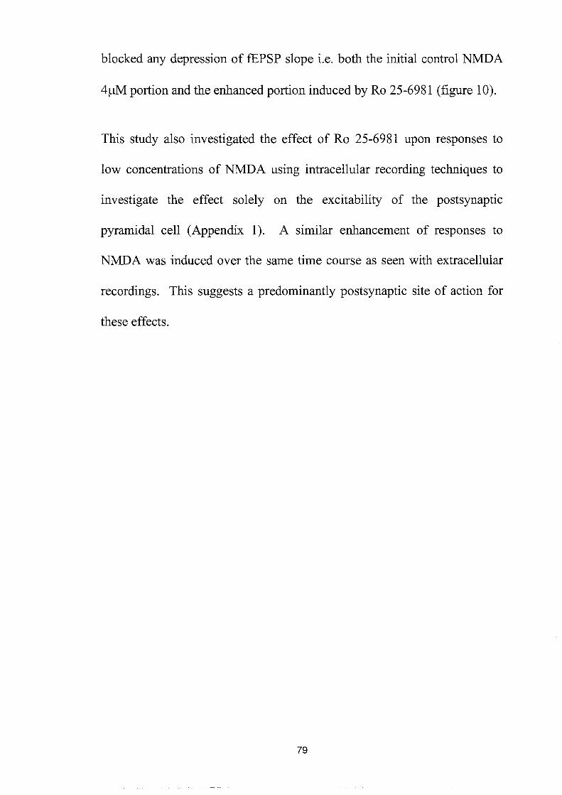
blocked any depression of fEPSP slope i.e. both the initial control NMDA
4~M portion and the enhanced portion induced by Ro 25-6981 (figure 10).
This study also investigated the effect of Ro 25-6981 upon responses to
low concentrations of NMDA using intracellular recording techniques to
investigate the effect solely on the excitability of the postsynaptic
pyramidal cell (Appendix 1). A similar enhancement of responses to
NMDA was induced over the same time course as seen with extracellular
recordings. This suggests a predominantly postsynaptic site of action for
these effects.
79

Cl) c. ~ 120 en
c.. 100 en c.. !i!:! 80 Cl) s:::: .- 60
40
20
o i
o
II NMDA 10~M
Ro 25-6981 3~M
I I
2
i i i i
50 100 150 200 time (mins)
Figure 8. Repeated 10-minute applications of 10j..lM NMDA and the effect of
3j..1M Ro 25-6981.
Plot showing the co-application of 3j..1M Ro 25-6981 had no immediate effect
upon the response to 10j..lM NMDA, whereas the next application of NMDA was
potentiated to the extent that the fEPSP was initially abolished and then shows
only partial recovery (p<O.001, n=4). Each point shows the mean±s.e.m. for
n=4 experiments.
80

1
2
lmV
5 ms
Figure 9. Representative fEPSPs showing excitotoxic damage.
Recordings of fEPSPs showing the irrecoverable depression of baseline
responses caused by the excitotoxic insult of NMDA 10l-lM and Ro 25-6981
31-lM (2) compared with baseline responses (1). Numbered traces correspond
to the time course in figure 8.
81

II NMDA4~M Ro 25-6981 3~M
PEAQX O.1~M
(1) 12 c. . .. . . .. 1 11 111 IV V VI Vll Vll1
0 -tn 10 c.. en c.. 8 w .... (1) 6 c: .--(1)
4 tn ca .c .... 2 0 ~ 0
, , , , , i
o 50 100 150 200 250 time (mins)
Figure 10. Effect of Ro 25-6981 and NMDA 4~M on fEPSP slope.
Plot showing the effect of repeated 10-minute applications of 4j..1M NMDA, which
produced a small depression of the fEPSP slope. The application of 3j..1M Ro
25-6981 had no immediate effect upon this, but subsequent applications of 4j..1M
NMDA show the development of potentiation in the fEPSP slope (p<O.001,
n=4). The co-application of PEAQX at O.1j..1M completely blocked the final
potentiated response to NMDA. Each point shows the mean±s.e.m. for n=4
experiments
82

NMDA receptor antagonism
A surprising feature of these experiments was that the effect of Ro 25-6981
developed slowly, required at least 20 minutes to elicit a clearly significant
decrease in response size and the effect gradually continued to develop
during the washout phase. In addition this effect, once fully developed,
was long lasting, with no attenuation of the depressed response size being
observed for up to 100 minutes post-washout. The continuous bath
application of NMDA 4f.!M was employed in order to observe better how
the effect of Ro 25-6981 developed. As shown in figure 11, NMDA 4f.!M
initially produced a small, stable depression of baseline fEPSP slope, the
co-perfusion of Ro 25-6981 3f.!M produced a slight recovery of the
responses but near the end of the 20-minute perfusion the depression
gradually develops. As Ro 25-6981 started to washout the depression had
reached 70.42±12.18% of baseline fEPSP slope. In spite of the absence of
perfusing Ro 25-6981 the depression continued to gradually develop. The
depression stabilised 30 minutes after the washout of Ro 25-6981 at
33.84±0.32% (p<0.001; n=5) of baseline fEPSP slope. The application of
the competitive, non-selective NMDA receptor antagonist 2-AP5 50llM
reversed the depression to near-baseline responses. However, after the
washout of 2-AP5 the depression reasserted itself within approximately 18
minutes (p<O.OOl; n=5). At the end of the experiment and with the
washout of NMDA, the fEPSP slope returned to a level not significantly
83

different from the initial baseline responses (Figure 11). An examination
of paired-pulse interactions during this experiment found that paired-pulse
ratios were enhanced during the Ro 2S-6981-mediated enhancement of
NMDA 4f.lM (p<O.OOI, n=S at all interstimulus intervals) but were returned
to baseline responses during the reversal of this depression caused by 2-
APS (figure 12).
Repeating the same protocol using a slightly higher level of NMDA at a
concentration of 7 .S f.lM (figure 13) produced a modest and stable
depression of fEPSP slope to 64.6±10.4% (not significant, n=4). The co
application of Ro 2S-698l 3 f.lM caused an enhancement of this depression,
which started during Ro 2S-6981 co-perfusion and continued during
washout until it reached a stable plateau of -0.16±0.24% (p<O.OI; n=4) of
baseline fEPSP slope within approximately 18 minutes of the depression
beginning. The application of the competitive, non-selective NMDA
receptor antagonist 2-APS SOf.lM significantly reversed the depression of
fEPSP slope back to the level previously seen with NMDA alone (p<O.OI;
n=4). Again, during the washout of 2-APS the depression reasserted itself
within approximately 18 minutes to O.80±O.29% (p<O.OOI; n=4). At the
end of the experiment and with the washout of the NMDA, the fEPSP
returned to a level not significantly different from the baseline responses
although it was apparent that the level and duration ofNMDA agonism had
had a deleterious effect upon the response size. The rate of onset of effect
84

was quicker with NMDA 7.5!J.M (18 minutes) than with NMDA 4!J.M (30
minutes) perhaps reflecting the increased rate of binding with increased
concentration of NMDA as previously reported with glutamate (Mutel et
al., 1998) and with ifenprodil (Grimwood et al., 2000). Figure 14 shows a
concentration-response curve of the effect of NMDA concentrations and
the degree to which they are potentiated to under the influence of Ro 25-
6981 3 !J.M. This shows a shift to the left of the NMDA concentration
response curve after the application of Ro 25-6981. The concentration
required to elicit a 50% depression of fEPSP slope changed from 1.7!-lM
with NMDA alone to 8.6!-lM under the influence ofRo 25-6981.
85

Cl)
CO
U; 100 a.. tJ)
a.. 80 w ..... ~ 60 .-Cl)
en ns .c ..... o
40
20
II NMDA 4~M Ro 25-6981 3J.lM
2-AP550J.lM
c 2
I111111111111111111111111111111111111111111111111111111111111111111111111111111
O+---~--~--~--~--~--~---o 25 50 75 100 125 150 time (mins)
Figure 11. Effect of Ro 25-6981 on the continuous application of NMDA 4IJM
and its reversal by 2-AP5.
4IJM NMDA caused a slight depression in the fEPSP slope (c). The co
application of 3IJM Ro 25-6981 increased this response; the increase occurred
slowly during Ro 25-6981 washout into a stable plateau, (1) fEPSP slope of
33.84±O.32% (p<O.001; n=5) compared with the baseline. The competitive
NMDA receptor antagonist 2-AP5 (50IJM) reversed the depression (2) (p<O.001;
n=5). During the washout of 2-AP5, the potentiated response to NMDA re
asserted itself (p<O.001; n=5), but recovered to baseline values upon removing
the NMDA. Each point shows the mean±s.e.m. for n=5 experiments.
86

0..-.. +:;Q) ca t/) s- s::::
""-"'0 Q)o. (.) t/) s:::: Q) Q) ss-Q) ......
==~ .- .-""0 ..... Q) 0 t/) ...... -""0 :::s s:::: 0.0
I (.)
""OQ) ~ t/) .- ..... [0
3.0 ***
2.5 ***
2.0 ***
1.5
1.0
0.5~....-----r--...---......-----..-10 20 30 40 50 inter-stimulus interval (ms)
1
2 c
Figure 12. Effect of NMDA 4J..lM and Ro 25-6981 on paired-pulse interactions.
This graph summarises paired-pulse interactions at interstimulus intervals of
10 ms, 20 ms and 50 ms, corresponding to points (c), (1) and (2) in figure 11.
The results demonstrate a significant enhancement of paired-pulse interactions
during the trough of the depression caused by Ro 25-6981 and NMDA (1)
(10 ms p<0.001, 20 ms p<0.001, 50 ms p<0.001, n=5) compared with baseline
responses (c). These changes were reversed by 2-AP5 (2). Each point shows
the mean±s.e.m. for n=5 experiments
87

II NMDA 7.5J.1M
R 0 25 -6 9 81 3 J.1 M
2-APV 50 J.1M
CJ)
C-O I111111111111111111111111111111111111111111111111111111111111111111111111111111111 -en c.. 100 en c.. w 80 \I-
CJ)
c 60 .--CJ)
en 40 co .Q
\I- 20 0
~ 0
0 i i i , , , , 0 25 50 75 100 125 150
time (mins) Figure 13. Effect of Ro 25-6981 on the continuous application of NMDA 7.51JM
and its reversal by 2-AP5.
7.51JM NMDA caused a modest (64.6±10.4%; n.s. n=4) depression of the
fEPSP slope. The co-application of 3IJM Ro 25-6981 increased this response;
the increase occurred slowly during Ro 25-6981 washout into a stable plateau
fEPSP slope of -O.16±O.24% (p<O.01; n=4) compared with the baseline. The
competitive NMDA receptor antagonist 2-AP5 (50IJM) reversed the depression
(p<O.01; n=4). During the washout of 2-AP5, the potentiated response to
NMDA re-asserted itself (p<O.001; n=4), but recovered to baseline values upon
removing the NMDA. Each point shows the mean±s.e.m. for n=4 experiments.
88

tJ'J
a.. 80 en a.. ~ 60 Cl) c: .--~ 40 co .c '0 20 ~ Q
o
Ro 25-6981 +NMDA
-7
NMDA
-6 Log[NMDA]M
-5
Figure 14. The concentration-response curve of NMDA alone and after
Ro 25-6981.
Graph summarising the concentration-response curves for responses to NMDA
in the presence and absence of 3IJM Ro 25-6981, and showing the marked
leftward shift produced by the combination. NMDA alone (1, 4, 7.5, 10 and
20/-lM) and Ro 25-6981 potentiated responses to NMDA (0.1, 1, 4, 7.5 and
10/-lM). The concentration eliciting a 50% depression of fEPSP slope changes
from 8.61JM for NMDA alone to 1.71JM under the influence of Ro 25-6981. Each
point shows the mean±s.e.m.
89

2-APS is a competitive antagonist at the NMDA receptor's glutamate
binding site, in order to test that this was not a compounding factor in the
blockade of the effect of Ro 2S-698l the non-competitive, open channel
blocker dizocilpine (MK 801) was used to antagonise the NMDA receptor
during the co-application of Ro 2S-698l and NMDA. The application of
NMDA 4~M caused a small, stable depression of fEPSP slope that was
reversed by the application of MK 801 10~M to baseline response size.
The perfusion of Ro 2S-6981 3 ~M had no effect upon the fEPSP slope and
during the washout of MK 801 the response size remained at baseline
levels (figure IS). However, the non-competitive antagonism provided by
MK 801 was not washed out of the slice over the time-period recorded
from and this is a likely reason for the lack of any depression observed.
90

0)
0.120 0 -en c.. U) c.. w 80 "t-
O)
c: 60 .--0)
en ns 40 .c
"t-o
20 ~ 0
0 0
II NMDA 4flM
III MK801 10flM
Ro 25 6981 3J.lM
111111111111111111111111111111111111
111111111111111111111111111111111111111111111111//////////////////////1
20 40 60 80 100 120 time (mins)
Figure 15. Effect of 4~M NMDA and Ro 25-6981 in the presence of MK 801 on
fEPSP slope.
Plot showing the effect of 1 O~M MK 801 applied throughout the combined
application of 4~M NMDA and 3~M Ro 25-6981. An initial, small depression
(approximately 5%) produced by NMDA was blocked by 10~M MK 801. This
prevents the development of a potentiated response to NMDA plus Ro 25-6981
even after the washout of MK 801. Each point is the mean±s.e.m. for n=3.
91

Having previously demonstrated that the Ro 25-6981-mediated potentiation
of responses to NMDA was transiently reversible by applying 2-AP5 50~M
this study investigated whether, by applying this competitive antagonist
before, during and after Ro 25-6981, the induction of the depression of
fEPSP slope could be prevented. As shown in figure 16 the application of
NMDA 4~M produced a slight, stable depression of the fEPSP slope that
was reversed to above the baseline levels by the subsequent application of
2-AP5. The co-application ofRo 25-6981 3~M had no further effect upon
the fEPSP response, 2-AP5 preventing the depression normally elicited.
However, as 2-AP5 was washed out the depression of fEPSP slope
developed rapidly to 14.06±5.09% of baseline fEPSP slope (p=0.0178,
n=4) within approximately 20 minutes of it starting to reduce. The gradient
of the development of this depression was similar to that seen with 2-AP5
washout in figure 11 and 13; i.e. a depression that is linear and rate
dependent upon the washout of 2-AP5. The gradient was dissimilar to the
Ro 25-69811NMDA 4~ gradient of figure 11, which is rate-dependent on
the effect initiated by Ro 25-6981, which is a slower developing effect
(approximately 30 minutes). Therefore, the effect of Ro 25-6981 would
seem to have already been realized during 2-AP5 co-perfusion suggesting
that it does not require NMDA receptor activation. The difference in these
gradients may be explained by the binding of Ro 25-6981 having already
taken place during 2-AP5 perfusion; 2-AP5 was able to suppress the NR2A
92

activation-dependent expression of its effect but not the binding of Ro 25-
6981 to its unique site on the NR2B subunit and the mechanism it puts into
effect to produce the enhanced depression of fEPSP slope.
93

II NMDA4f!M
2-APS so J.lM
Ro 2S-6981 3J.lM
Q) 12 C-o - 10 (/)
a. tIJ a. w .... Q) r::: ---Q) (/) ca .c .... 0 ~ 0
i i ,
0 20 40 60 80 100 120 140 time (mins)
Figure 16. Effect of 2-AP5 continuously bath-applied during the application of
4jJM NMDA and Ro 25-6981 on fEPSP slope.
A plot showing the effect of 50jJM 2-AP5 applied throughout the combined
application of 4jJM NMDA and 3jJM Ro 25-6981. An initial, small depression
(approximately 5%) produced by NMDA was blocked by 50jJM 2-AP5. Despite
this, the application of 2-AP5 did not prevent the development of a potentiated
response to NMDA plus Ro 25-6981, which appeared after washout of the 2-
AP5 and persisted until the NMDA was removed. Each point is the
mean±s.e.m. for n=4 experiments.
94

PEAQX
The bath application of NMDA in the adult rat hippocampus will activate
NMDA receptors containing NR2A, NR2B and NR2D subunits. In view of
the potentiating activity produced when the NR2B element to the action of
NMDA was blocked by Ro 25-6981, the putative NR2A subunit antagonist
PEAQX was used to investigate the residual response to NMDA. NMDA
4f.lM was continuously bath-applied producing a small, stable depression of
fEPSP slope (figure 17). The co-perfusion of 3f.lM Ro 25-6981
transformed this into a large depression to 31.90±1.0 1 % of the baseline
fEPSP slope (p<0.05, n=3) that developed over approximately 30 minutes.
It was then reversed by the application of PEAQX 0.1 f.lM back to baseline
fEPSP responses. The rate of washout of PEAQX was initially rapid but
after 8 minutes, it slowed to such an extent that it still did not completely
washout for at least 2 hours. These experiments were therefore concluded
before the depression might be observed to fully re-establish.
95
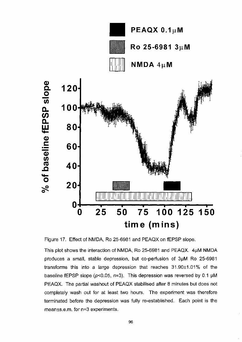
~ 120 o -U)
a. t/J a. w '+-Q) s:::: ---Q) U) ca .c '+-o ~ o
80
60
40
20
PEAQX O.1~M
Ro 25-6981 3~M
II NMDA 4JlM
O+---~--~---P--~--~--~~ o 25 50 75 100 125 150
time (mins)
Figure 17. Effect of NMDA, Ro 25-6981 and PEAQX on fEPSP slope.
This plot shows the interaction of NMDA, Ro 25-6981 and PEAQX. 4iJM NMDA
produces a small, stable depression, but co-perfusion of 3iJM Ro 25-6981
transforms this into a large depression that reaches 31.90±1.01 % of the
baseline fEPSP slope (p<0.05, n=3). This depression was reversed by 0.1 iJM
PEAQX. The partial washout of PEAQX stabilised after 8 minutes but does not
completely wash out for at least two hours. The experiment was therefore
terminated before the depression was fully re-established. Each point is the
mean±s.e.m. for n=3 experiments.
96

Protein phosphorylation
The phosphorylation state of the NMDA receptor and many other cellular
proteins determines their activity and function (Smart, 1997). This study
next investigated the involvement of protein kinases and protein
phosphatases on the Ro 25-6981 effect.
Staurosporine (STA) is a broad-spectrum protein kinase inhibitor, enzymes
inhibited include: PKC (ICso=3nM), PKA (ICso=7nM), tyrosine protein
kinase (ICso=6nM) and CaMKII (ICso=20nM) (Tamaoki et al., 1986;
Ruegg & Burgess, 1989; Yanagihara et al., 1991). As shown in figure 18
the initial 10-minute application ofNMDA 4~M caused a small depression
of the fEPSP slope, staurosporine lOOnM was continuously bath-applied
and had no effect upon baseline fEPSP slope or the depression produced by
a further 10-minute application ofNMDA 4~M. The application ofRo 25-
6981 3~M and the subsequent application ofNMDA produced a significant
depression of fEPSP slope to 17.66±lO.83% (p<O.Ol, n=3) compared to
baseline responses. After washout, a final application of NMDA 4~M
produced another significant depression of fEPSP slope to 1.77±O.87%
(p<O.OOl, n=3) compared to baseline responses (Figure 18). Therefore,
staurosporine was unable to prevent the potentiation of responses to
NMDA 4~M by Ro 25-6981.
97

The first phosphatase inhibitor investigated was okadaic acid (OA), a
potent inhibitor of the serine/threonine protein phosphatases types 1 and 2A
(PP1 and PP2A) (Bia10jan & Takai, 1988). The continuous application of
NMDA 4~M elicited a small, stable depression of fEPSP slope. The
addition of okadaic acid before, during and after the co-application of Ro
25-6981 3 ~M was unable to prevent the development of a significant
depression of the fEPSP slope to 1.56±0.3l % (p<O.OOl, n=4).
Interestingly, upon washout the fEPSP slope recovered to an average of
156.5±14.62% (p<0.05, n=4) of baseline responses, a significant LTP of
responses that was stable (figure 19). This LTP-inducing effect was not
investigated further at this time.
Cyc1osporin A (CyA) , a widely used serine/threonine Ca2+/calmodulin-
dependent phosphatase 2B (calcineurin) inhibitor, was used next. The
continuous bath-application ofNMDA 4~M again produced a slight, stable
depression of fEPSP slope. The application of 1 ~M cyc1osporin A had no
significant effect on the baseline fEPSP slope but it was able to prevent the
anticipated enhancement of the NMDA 4~M-mediated depression of
fEPSP slope previously seen after the application of Ro 25-6981 3 ~M
(figure 20). Interestingly the baseline fEPSP returned to values that were
significantly potentiated (132±0.81 %, p<0.05, n=4) compared to baseline
responses. The application of cyc1osporin A prevented the depression of
fEPSP slope seen with both the control group and the experiments using 98

okadaic acid and staurosporine, which were unable to inhibit the
development of the depression. Interestingly, the magnitude of the
depression of fEPSP slope was significantly greater than the control group
for both okadaic acid and staurosporine (figure 21).
99

Q) c. 0 120 -tn c..
100 tJ) c.. w 80 "-Q) r::
60 .--Q) tn C'G 40 ..c "-0 20 ~ Q
0 , 0
[[Ill
,
n NMDA 4J.1M
D STA 100nM
Ro 25-6981 3J.lM
-[[Ill [[Ill
, , , , , 50 100 150
time (mins)
,
Figure 18. Action of staurosporine upon the effect of the co-application of 4~M
NMDA and Ro 25-6981 on fEPSP slope.
Plot showing repeated applications of NMDA 4~M produced a small depression
of fEPSP slope that was not prevented by the co-application of 100nM
staurosporine. The co-application of staurosporine was also not able to prevent
the enhancement of NMDA responses by Ro 25-6981 3~M which produced a
significant depression of the fEPSP slope to 17.66±10.83% (p<O.01). Each
point shows the mean±s.e.m. for n=3 experiments.
100

",
Cl) C-o -U)
a.. en a.. w ..... Cl) c: .--Cl) U) ca
..c ..... 0
'#.
175
150
125
100
75
50
25
0 , 0
OA 50nM
Ro 25-6981 3J.lM
II NMDA 4~M
I11111111111111111111111111111111111111111111111111111111111111111111
i , , , , ,
20 40 60 80 100 120 time (mins)
Figure 19. Action of okadaic acid upon the effect of the co-application of 4IJM
NMDA and Ro 25-6981 on fEPSP slope.
Plot showing that the continuous application of NMDA 4IJM produced a small
depression of fEPSP slope. The co-application of staurosporine before, during
and after Ro 25-6981 was not able to prevent the enhancement of NMDA
responses by Ro 25-6981 3IJM which produced a significant depression of the
fEPSP slope to 1.56±O.31 % (p<O.001, n=4). Each point shows the
mean±s.e.m. for n=4 experiments.
101
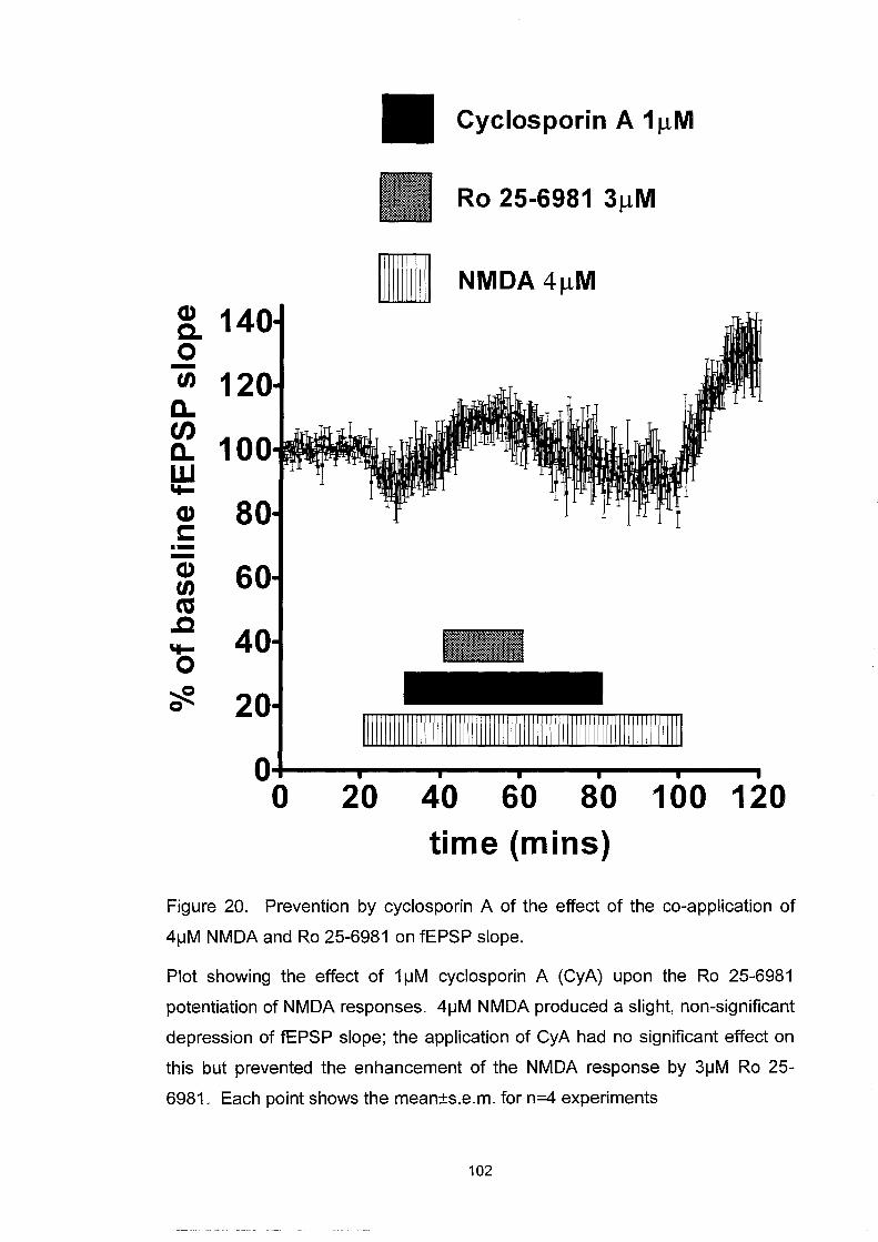
Cyclosporin A 1 f.lM
II NMDA4/lM Q) c. 0 -tn 12 c.. t/J
10 c.. w ~
Q) 8 c: ---Q) 6 tn ca .c ~
0 ~ 0
I1111111111111111111111111111111111111111111111111111111111111111111111111
o 20 40 60 80 100 120 time (mins)
Figure 20. Prevention by cyclosporin A of the effect of the co-application of
4\-1M NMDA and Ro 25-6981 on fEPSP slope.
Plot showing the effect of 1 \-1M cyclosporin A (CyA) upon the Ro 25-6981
potentiation of NMDA responses. 4\-1M NMDA produced a slight, non-significant
depression of fEPSP slope; the application of CyA had no significant effect on
this but prevented the enhancement of the NMDA response by 3\-1M Ro 25-
6981. Each point shows the mean±s.e.m. for n=4 experiments
102

*** (1) ,10 c. 0 -en a.. 8 en a.. w ..... 6 (1) c: .--(1) en 4 cu .c ..... 0
2 ';je
** ** control CyA OA STA
Figure 21. Column bar graph summary of phosphorylation modulators.
This histogram summarises the depression caused by the co-application of 4!JM
NMDA and 3!JM Ro 2S-6981 under control conditions (figure 11, n=4) and with
the co-perfusion of 1!JM cyclosporin A (CyA) (figure 20, n=4), SOnM okadaic
acid (OA) (figure 19, n=4) and 100nM staurosporine (STA) (figure 18, n=3).
Cyclosporin A prevented the depression of the fEPSP slope induced by NMDA
in the control group (p<0.001). However, okadaic acid and staurosporine
increased the magnitude of the depression (both p<0.01).
103

Protein synthesis
In view of the latency of onset, the long-lasting effect of Ro 25-6981, and
the involvement of protein phosphorylation the question arose of whether
any permanent change was required and induced of new protein synthesis.
To investigate this possibility anisomycin, a widely used reversible
translation inhibitor was used (GroHman, 1967). The application of
NMDA 4~M produced a small, stable depression of fEPSP slope. The
continuous application of anisomycin 30~M had no significant effect upon
the fEPSP slope nor was it able to interfere with the subsequent NMDA
and Ro 25-6981-mediated depression of fEPSP slope to 10.65±O.3628%
(p=O.0128, n=4)(figure 22).
104

(1) 15 c. 0 - 12 en c.. en 10 c.. w .... (1) 7 c: .--(1)
5 en ns .c .... 2 0 ~ 0
, o
,
Anisomycin 30J.lM
Ro 25-6981 3J.lM
II NMDA4JlM
, , , , 25 50 75 100 125
time (mins) Figure 22. Effect of anisomycin on the Ro 25-6981/NMDA effect upon fEPSP
slope.
Plot showing that 30jJM anisomycin was unable to prevent the development of
the Ro 25-6981-mediated enhancement of NMDA responses (p=O.0128, n=4).
Each point shows the mean±s.e.m. for n=4 experiments.
105

Discussion
Applications of NMDA produced a concentration dependent depression of
evoked fEPSP slope that was associated with an enhancement of paired
pulse ratios at all interstimulus intervals. The fEPSP under normal 0.05Hz
stimulation is predominantly a reflection of the fast kinetics of the AMP A
receptors which underlie this recording, with fast rise times (10-90% rise
time of ~400!ls) and fast decay time constant (~1.3ms). The NMDA
component, in contrast, has slow kinetics with a slow rise time (~9ms) and
a slow mean decay time constant (~52ms) (Gibb et al., 1994). Schaffer
collateral inputs onto CAl neurones elicit fEPSPs where the NMDA
receptors contribute only very a small component (~3%) (Arrigoni &
Greene, 2004), principally as a result of the magnesium block of the
channel at resting membrane potentials. At these negative membrane
potentials, the magnesium ion blocks the ion channel in a voltage
dependent non-competitive manner. The depolarisation induced by a
single fEPSP is not sufficient to overcome the threshold leading to NMDA
receptor activation because the membrane potential does not reach a
sufficient level to expel the magnesium ion. However, repetitive activation
of the synapse such as that seen in HFS and LFS stimulation protocols to
induce LTP and LTD produces a non-linear summating effect which
overcomes this threshold and leads to the activation of the NMDA
receptors. It is apparent from current-voltage plots for NMDA-induced
106

conductances that even at -90m V, although the NMDA current is very
small it is never entirely prevented by magnesium block (Mayer et a/.,
1984; Nowak et at., 1984). The bath application of NMDA receptor
agonists will lead to the activation of synaptic and extrasynaptic NMDA
receptors which will result in large conductance through their SOpS
channels over longer time periods than that seen with AMP A receptors,
producing a regenerative feedback that leads to the unblocking of NMDA
receptors and their activation. This conductance then leads to the
depolarisation of the cells recorded from and the subsequent depolarisation
block of excitatory synaptic transmission, manifested as a loss of the fEPSP
slope. The washout of NMDA agonist allows the neurones to rebuild the
ionic gradients that underlie the membrane potential and the fEPSP
response recovers. The application of NMDA agonists which leads to this
depolarisation will also lead to the activation of GABAA and GABAB
receptors responsible for fast and slow components of feed-forward and
feedback inhibitory transmission. Although this study did not address the
contribution of these effects on the changes in the depression of fEPSP
slope or on paired-pulse interactions previous investigations found that
GABA, bath-applied to the hippocampal slice, elicited no inhibition of the
fEPSP slope or altered the paired-pulse interactions in concentrations up to
lmM (unpublished observations). This suggested that the contribution of
additional GABA release in this preparation is limited or already saturated
by concentrations of GABA released in response to afferent stimulation.
107

Ro 25-6981 has been described as a selective antagonist at the NR2B
subunit of the NMDA receptor. However, the previous work was carried
out deliberately on immature tissue or cloned receptors overexpressing the
NR2B subunit and not in adult tissues expressing mature NMDA receptors.
When tested on the intrinsic neuronal circuitry of the adult hippocampal
slice Ro 25-6981 unexpectedly potentiated NMDA receptor activation at all
concentrations of NMDA tested and showed no inhibition of NMDA
responses. The increased depression of fEPSP slope was generated through
NR2A subunit-containing receptors, since the potentiated response could
be blocked selectively by PEAQX. Furthermore, this enhancement
appeared to have a presynaptic component, since the paired-pulse ratios
were significantly enhanced during potentiated responses to NMDA at
concentrations that previously showed no such change from baseline
responses. It is widely recognised that a loss of paired-pulse inhibition and
an increase of paired-pulse facilitation represents a presynaptic action of
the test agents. Although it does not follow that postsynaptic effects cannot
influence the appearance of PPD and PPF in field recordings (Seabrook et
a!., 1999). Additionally, evidence from intracellular recordings of the
enhancement of NMDA responses by Ro 25-6981 suggests that this
phenomenon has a predominantly postsynaptic locus. The presynaptic
element of responses to NMDA suggested in this study may be mediated
via NR2A or NR2A1NR2B subunit-containing receptors. This is consistent
with evidence for the existence of presynaptic NMDA receptors, which
108

may be responsible for changes in presynaptic release probability and the
changes in paired-pulse interactions described here.
The most parsimonious explanation of the present results would be that the
NR2B subunits exert a tonic inhibitory restraint on the activation of NR2A
subunit-containing NMDA receptors. Blockade of the NR2B subunits then
results in disinhibition of NR2A subunit-containing receptors, which
become the primary effectors of the increased response to NMDA. This
would explain both the potentiating effect of Ro 25-6981 and the blockade
of the potentiated responses by PEAQX. It is not possible to conclude
from the present data whether this inhibitory subunit interaction occurs
between subunits within single heterotrimeric NR11NR2A1NR2B receptors
or between discrete heterodimeric receptors containing exclusively
NR11NR2A and NR1INR2B subunits. Additionally, the interaction may be
between receptor populations located at different sites interacting by way
of intracellular or retrograde messengers.
These results are partially consistent with reports that ifenprodil and Ro 25-
6981 caused small inward currents produced by NMDA 1/J-M to be
potentiated, while larger currents produced by NMDA 100/J-M were
markedly inhibited (Kew et at., 1996; Fischer et at., 1997). However, no
inhibition against any concentrations of NMDA in the intact neuronal
network of the adult hippocampal slice was observed. This is most simply
109

explained by the very different expressIOn of subunits and their
phosphorylation within cloned receptor constructs, immature tissues and
with the mature tissue used in this study but merits further investigation.
It also appears from the data in this study that the interaction between
NR2B and NR2A subunits was not prevented by inhibitors of protein
kinases or the serine/threonine protein phosphatases types 1 and 2A (PP 1
and PP2A) (Bialojan & Takai, 1988). A finding consistent with our later
observation that Ro 25-6981 was unable to block LTP induction, which
requires the activation of protein kinases (Bliss & Collingridge, 1993).
Although the interaction between NR2B and NR2A subunit-containing
receptors was not prevented by the broad-spectrum protein kinase inhibitor
staurosporine, it is nevertheless likely that constitutively active kinases are
essential to phosphorylate the unknown moiety to maintain the inhibitory
restraint described. The subunit interaction does appear to involve
Ca2+/calmodulin-dependent phosphatase 2B (calcineurin), since it was
prevented by cyclosporin A. Ca1cineurin has previously been shown to
enhance both the presynaptic release of glutamate and postsynaptic
responsiveness to it (Yakel, 1997) partly by altering channel gating
properties and promoting desensitisation in cultured hippocampal neurons
(Lieberman & Mody, 1994; Nichols et ai., 1994; Sihra et ai., 1995; Tong et
ai., 1995; Victor et ai., 1995). Furthermore, the calcineurin inhibitors
cyclosporin A and FK506 are able to prevent the induction of LTD
110

(Mulkey et at., 1994; Hodgkiss & Kelly, 1995). Interestingly, it has been
reported that calmodulin-dependent inhibition of NMDA receptors will
reduce the amplitude and time course of excitatory synaptic currents in 12-
day-old rat hippocampal NMDA receptors (Rycroft & Gibb, 2002). NR2
subunits, unlike NRI subunits, are known to be highly phosphorylated
under basal conditions (Hall & Soderling, 1997), therefore the
phosphorylation step described could be acting directly upon the NR2A
and/or the NR2B subunit to produce the enhancement of responses to
NMDA. It is however possible that the phosphorylation site is elsewhere
on the NMDA receptor complex, which has been reported from proteomic
analysis to associate with at least 77 proteins (Husi et al., 2000) e.g. NRl,
PSD-95, SAP 102, or at a non-NMDA site.
The present work also reveals that the potentiating effect of Ro 25-6981 is
not dependent upon new protein synthesis, although it is expected that
relevant proteins are constitutively present. The reason for the slow onset
and apparently irreversible nature of the enhancement effect remains
unclear. It may be due for example to the Ro 25-6981 binding irreversibly,
to a persistent modification of the NR2B, NR2A or NRI subunit that is
then independent of Ro 25-6981 binding, to an irreversible uncoupling of
the NR2B subunit from NR2A or to a change in NMDA receptor functional
expression. Radiolabelled forms of Ro 25-6981 are unavailable at this time
to investigate the possibility of it binding irreversibly to the NR2B subunit.
111

Chapter 4. The NMDA subunit specificity of L TP
Introduction
It is widely accepted that a persistent change in synaptic strength is the
cellular basis of learning and memory (Alkon & Nelson, 1990; Kandel,
1997). Both long-term potentiation (LTP) and long-term depression (LTD)
have now been shown to fulfil three out of the four criteria that are required
to verify that they are both 'necessary and sufficient for the information
storage underlying the type of memory mediated by the brain area in which
that plasticity is observed'. Alterations in synaptic strength are detectable
after learning, interfering with the induction and expression mechanisms
results in concurrent interference in learning, and altering the pattern of
synaptic weights after learning affects the ability of animals to remember a
previously learned experience. The, unfulfilled, fourth criterion is
mimicry, where an implanted pattern of synaptic weights generates in the
animal the behaviour of a specific recalled memory (Moser et aI., 1998).
LTP, first reported in the dentate gyrus (Lomo, 1966; Bliss & Lomo, 1973),
has since been demonstrated in all three excitatory pathways in the
hippocampus (Schwartzkroin & Wester, 1975; Alger & Teyler, 1976) and
elsewhere in the eNS (Kuba & Kumamoto, 1990). LTP is characterised by
input specificity, co-operativity and associativity, leading to the proposal
that LTP is a Hebbian process (Hebb, 1946), where the co-incident activity 112

of pre- and postsynaptic neurones is required to increase persistently
synaptic strength. LTP was first shown to be involved in the cellular basis
of learning and memory with experiments to block L TP in vivo coincided
with deficits in hippocampal-dependent place navigation (Morris et aI.,
1986). LTP induction in the CAl area is NMDA receptor dependent: it is
triggered by NMDA receptor activation and blocked by NMDA receptor
antagonism (Collingridge et ai., 1983). AMPA receptor-mediated
depolarisation of the postsynaptic cell during L TP induction, by high
frequency stimulation (HFS), releases the Mg2+ block of NMDA receptors
allowing Ca2+ to flow into the cell (Lynch et ai., 1983; Mayer et ai., 1984;
Nowak et ai., 1984; Malinow & Miller, 1986; Wigstrom et ai., 1986). A
large rise in Ca2+ in the postsynaptic spine sets off a cascade of events that
results in LTP (Bliss & Collingridge, 1993). The induction of LTP is
widely considered to be postsynaptic and the expression both pre- and
postsynaptic (Kullmann & Nicoll, 1992; Manabe et ai., 1993). The
maintenance of LTP is characterised by an early phase which is dependent
on post-translational modifications and lasts for a few hours, followed by
later phases which are dependent on de novo gene expression and protein
synthesis that lasts for many hours (Krug et ai., 1984; Otani et ai., 1989;
Nguyen et ai., 1994). LTP can persist for varying amounts of time
dependent upon the induction protocol and the experimental model. It has
recently been demonstrated that stable L TP can last for up to a year
(Abraham et aI., 2002).
113

There is much evidence that supports a role for the NR2B subunit in
synaptic plasticity and specifically in LTP. Increases in the expression of
the NR2B and not of the NR2A, NR2C or NR2D subunit after L TP
induction and the prevention of L TP induction in an NR2B knock-down
model support a role for the NR2B subunit in the early phase of LTP
(Thomas et al., 1994; Thomas et al., 1996). Furthermore, enhanced
tyrosine phosphorylation of the NR2B subunit has been demonstrated after
the induction of LTP (Rosenblum et aI., 1996; Manabe et al., 2000;
Nakazawa et aI., 2001). LTP and LTD are bi-directional mechanisms of
synaptic plasticity, induced by different patterns of afferent stimulation
(Dudek & Bear, 1993). A recent investigation was made into whether
these different patterns reflect the activation of specific subunits to produce
either LTD or LTP. It was found that antagonists with a higher affinity for
the NR2AIB subunits over the NR2C/D subunits produced greater
inhibition of LTP than LTD induction (Hrabetova et aI., 2000). There are
also reports that the NR2B subunit is associated with age-related changes
(Magnusson, 2000), the decrease in NR2B expression with increasing age
leading to the blockage of L TP induction and impairments in spatial
learning performance (Clayton et aI., 2002). Potentiating the NMDA
receptors using spermine, an endogenous polyamine, could reverse this.
Taking advantage of the new NR2B and NR2A subunit-selective
antagonists, Ro 25-6981 and PEAQX, this investigation sought to
determine the subunit specificity ofNMDA-dependent LTP induction.
114

Results
L TP was induced by high frequency stimulation (3 identical trains of 1
second stimulation bursts at 100Hz 10 minutes apart; HFS). This produced
an enhancement of fEPSP slope to 197.9±16.4% (p<0.001, n=6) that
persisted steadily for 140 minutes from the start of the induction protocol
(figure 23 and 24).
To demonstrate that the LTP induced by HFS was NMDA receptor
dependent 50)lM 2-AP5 was applied during the induction protocol and
prevented LTP induction (figure 25). PEAQX O.l)lM was then used, which
had previously been seen in chapter 3 to block NMDA responses in an
analogous manner to 2-AP5. The perfusion of this NR2A antagonist also
prevented the induction of LTP (figure 26). Finally, the NR2B antagonist
Ro 25-6981 was used but it was unable to prevent the induction of LTP to
170.4±15.7% (p<O.Ol, n=6) of baseline fEPSP slope, which was less than
control HFS induced LTP but not significantly so (figure 27).
This result clearly demonstrates that the NR2A subunit, and not the NR2B
subunit, is critically required for the induction of NMDA-dependent LTP
(figure 28).
115

Cl) c. 0 -fA
D.. tJ) D.. w ..... Cl) c: .--Cl) fA ca .c ..... 0
cf!.
250
200
150 C
0 0
100Hz stirn u lation of 1 second duration.
1 1 1 20 40 60
time (mins)
Figure 23. Effect of high frequency stimulation on fEPSP slope.
1
~O
Plot showing L TP induced by three trains of stimuli using 100 pulses at 100 Hz,
ten minutes apart (HFS) at 20, 30 and 40 minute time points, producing a
persistent enhancement of the fEPSP slope to 197.9±16.4% of the control size
(p<0.001, n=6). Each point shows the mean±s.e.m. for n=6 experiments.
116
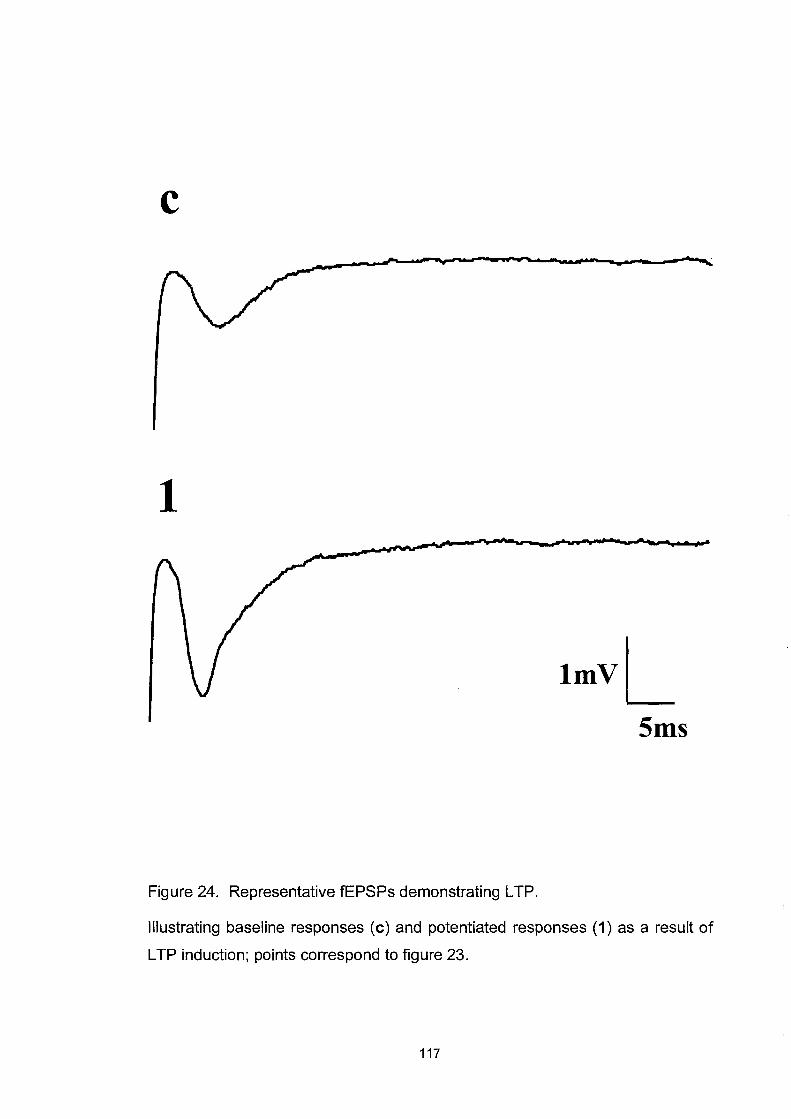
c
1
ImvL 5ms
Figure 24. Representative fEPSPs demonstrating L TP.
Illustrating baseline responses (c) and potentiated responses (1) as a result of
L TP induction; pOints correspond to figure 23.
117

(1) c. o -tn a.. en a.. w ..... (1) c: .--(1) tn ca .c ..... o
.H 100Hz stim u lation of 1 second duration.
2-AP5 50J.lM
100~
75
50
?fl. 25
O+-------~---------~------~-------o 20 40 60 time (mins)
Figure 25. Blockade of NMDA-dependent L TP induction by 2-AP5.
Plot showing the perfusion of 2-AP5 50flM during the HFS induction protocol for
L TP which prevented the induction of L TP, n=3. Each point shows the
mean±s.e.m. for n=3 experiments.
118

Q) Co -(/) a. en a. w "I-Q) c: .-Q) (/) ns .c "I-o ~ o
J~ 100Hz stirn u lation of 1 second_ duration.
PEAQX O.1~M
Ol+-----~------~----~----o 20 40 60
time (mins) Figure 26. Blockade of NMDA-dependent L TP induction by PEAOX.
Plot showing the effect of PEAOX 0.1 J.lM upon the induction of L TP. The
perfusion of PEAOX prevented the induction of L TP. Each point shows the
mean±s.e.m. for n=4 experiments.
119

CI) cO -'" a.
en a. w "I-
CI) c: .--CI)
'" co .c "Io ?ft.
200
150
50
.4~ 100Hz stirn u lation of 1 seconds duration.
Ro 25-6981 3f.lM
ttt O+----~---~--~---~---~---~~ o 20 40 60 80 100 120
time (mins)
Figure 27. Effect of Ro 25-6981 on NMDA-dependent L TP induction.
Plot showing that the application of 3f-lM Ro 25-6981 during the HFS-L TP
induction protocol was unable to prevent the induction of a significant L TP
(170.4±15.7% (p<O.01, n=6) of baseline fEPSP slope. Each point shows the
mean±s.e.m. for n=6 experiments.
120

CI> n.s. e.
*** 0 -'" ** a..
en a.. w 1 'I-
CI> c: .--CI> 1
'" CO .c 'I-0
?fe.
LTP 2-APV PEAQX Ro 25-6981
Figure 28. Role of NR2A and NR2B subunits in long-term potentiation.
Histogram showing L TP induction by high frequency stimulation (HFS, three
stimulation trains of 100 pulses at 100 Hz, ten minutes apart). Bath application
of either SO\-lM 2-APS or 0.1 \-1M PEAQX prevented the induction of LTP,
whereas 3IJM Ro 2S-6981 did not, reducing LTP to 170.4±1S.7% (p<O.01, n=6)
of baseline fEPSP slope. This was not significantly less than L TP in the
absence of drugs (p=0.2S26, n=6), but was significantly greater than the original
fEPSP baseline slope (**p<0.01, n=6).
121
Discussion
The present study has shown that NMDA receptor-dependent LTP
induction is critically dependent upon the NR2A subunit-containing
receptor activation and not the NR2B subunit in the Schaffer collateral
CAl synapse. This result is consistent with a recent report that LTP is
associated with the rapid surface expression of postsynaptic NR2A subunit
containing NMDA receptors (Grosshans et aI., 2002). Additionally, whilst
Ro 25-6981 does not block the induction of LTP, the LTP induced was
marginally reduced consistent with a recent report (Kohr et aI., 2003).
Several reports have now been published investigating the subunit
selectivity ofLTP and LTD (Sjostrom et ai., 2003; Liu et ai., 2004; Massey
et ai., 2004; Mallon et aI., 2005). Liu and colleagues reported that at
hippocampal CAl synapses of 3-4 week old rats LTP was blocked by
PEAQX. Furthermore, using an LTP protocol pairing Schaffer collateral
stimulation with postsynaptic depolarisation under whole-cell recording
conditions LTP could be blocked by PEAQX and LTD was induced. The
induction of LTD has been reported to be blocked by NR2B antagonists,
including Ro 25-6981 and ifenprodil (Sjostrom et aI., 2003; Liu et ai.,
2004; Massey et aI., 2004). However the investigation of LTD in this
study was prevented because of the difficulty in induction of LTD in adult
animals, in agreement with reports by other laboratories (Kemp & Bashir,
122

2001). Massey and colleagues reported that the induction of L TP and
depotentiation in the perirhinal cortex of 7 -12 week old rat was blocked by
PEAQX. They also found that LTD was difficult to induce but that
blockade of glutamate uptake, which led to the activation of postsynaptic,
extrasynaptic NR1/NR2B receptors, overcame this obstacle. This allowed
the induction of LTD, which could also be prevented by Ro 25-6981, the
NR2B antagonist. However, the distribution of NMDA receptor subunits
varies widely developmentally and between brain regions and synaptic
pathways (Otmakhova et a!., 2002; Arrigoni & Greene, 2004). Therefore,
different local conditions can regulate the sensitivity and characteristics of
synaptic plasticity to different NR2 subunits. It has also been reported that
LTP is sensitive to NR2B antagonists in the young Schaffer collateral-CAl
synapse (R. Malenka, personal communication), visual cortex (Yoshimura
et al., 2003), amygdala (T. Sigurdsson, personal communication) and the
hippocampal commissural-CA3 synapse (Ito et al., 2000). The NR2B
subunit, unlike the NR2A subunit-containing receptors or AMPA receptors,
seem to be of particular importance in regulating learning and memory and
have recently been reported to be differentially distributed in the left-right
asymmetry of the brain, which has important implications for higher order
functions (Kawakami et al., 2003; Shinohara et al., 2003).
123

Chapter 5. The NR2A & NR2B subunit-selectivity
of homoquinolinic acid
Introduction
The tryptophan metabolite, quinolinic acid (QA), has been recognized for
many years to be a weak, endogenous NMDA receptor agonist (Stone &
Perkins, 1981; Stone, 1984; 1993). Shortly after its discovery, a study of
structure-activity relationships among a series of related compounds
revealed that a close analogue, homoquinolinic acid (HQA), had a potent
excitatory activity. Indeed, HQA appeared to be between 10 and 100-fold
more potent at activating glutamate receptors (Stone, 1984).
Autoradiographical and electrophysiological studies have recently been
undertaken on native and recombinant receptors to evaluate the subunit
specificity of HQA. It has been reported from electrophysiological studies
that HQA is a potent NMDA receptor agonist that has a higher affinity for
Xenopus oocytes expressing NR2A and NR2B subunit-containing
receptors, compared to NR2C and NR2D subunit-containing receptors
(Buller & Monaghan, 1997). A recent study also found that HQA had an
approximately 2-fold higher affinity for NR1a/NR2B and
NR1 a/NR2A/NR2B receptors compared to NR1 a/NR2A receptors (EC50
13.8, 9.04 and 25.2JlM, respectively). Yet showed greater intrinsic activity
for NR1 a/NR2A and NR1 a/NR2A1NR2B compared to NR1 a/NR2B
124
transfected receptors (compared to glutamate responses (100%), 148±15,
125±5.6 and 93.3±4%, respectively) (Grimwood et al., 2002). In
autoradiographical studies, it has been observed that 3[H]-homoquinolinic
acid selectively radiolabels a subpopulation of NMDA receptors in rat
brain that is consistent with the distribution of the NR2B subunit and
clearly dissimilar to NR2C and NR2D subunit distribution (Buller et al.,
1994; De Carvalho et al., 1996; Brown et al., 1998), although there are
contradictory reports where HQA was not as highly specific for NR2B
containing receptors (Grimwood et al., 2002). In radioligand binding
studies 3[H]-HQA also bound to a novel binding site from which it was not
displaced by NMDA or glutamate but was displaced by 2-carboxy-3-
carboxymethylquinoline (CCMQ), a HQA derivative (Brown et ai., 1998;
Grimwood et al., 2002). The identity and function of this proposed site are
unknown.
This investigation was designed to establish whether there was any
evidence for this unidentified site in the generation of evoked synaptic
potentials in the hippocampal slice. Additionally, by using an
NR2A1NR2B subunit-containing NMDA receptor agonist (HQA) to
investigate subunit-pharmacology in combination with NR2A and NR2B
subunit-selective antagonists (PEAQX and Ro 25-6981) this study can
consider the role of NR2D subunit-containing receptors on the depression
of fEPSP slope and paired-pulse interactions.
125

Results
HQAand QA
In order to determine the most suitable concentrations of agonists to use
concentration-response curves were constructed from which it was clear
that HQA and QA gave useful depressions of approximately 50% of the
baseline fEPSP slope at 2.5~M and l50~M respectively (figures 29-31, 34-
36). These concentrations were selected for further experiments since both
increases and decreases of the depression of fEPSP slope could easily be
observed. The depression elicited by the application of HQA 2.5~M and
QA l50~M both coincided with a significant decrease of paired-pulse
inhibition and increase of paired-pulse facilitation at all interstimulus
intervals (HQA: 10ms p<O.Ol, 20ms p<O.OOl, 50ms p<O.OOl, n=lO; QA:
10ms p<0.05; 20ms p<O.OOl; 50ms p<O.OOl; n=7) (figures 32,33,37,38).
126

12
-6.1 -5.9 -5.7 -5.5 -5.3 Log[homoquinolinic acid]M
Figure 29. The concentration-response curve of HQA.
A plot showing concentration-response curve for homoquinolinic acid-produced
depression of the fEPSP slope at 1, 2, 2.5, 3 and 4IJM (n = 3, 5, 8, 5 and 4,
respectively). The concentration inducing a 50% reduction is 2.4 IJM. Each
point shows the mean±s.e.m.
127
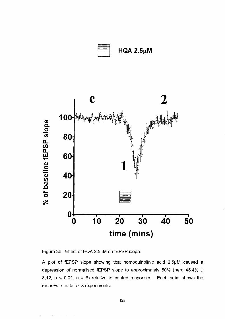
III HQA 2.5JlM
c 2 100 ~IW;!WI~ ~r (1)
a. 0 U) 80 a..
en a.. w 60 '+-(1) 1 c: .-- 40 (1) U) co .c '+- 20 III 0 ~ 0
0 0 10 20 30 40 50
time (mins)
Figure 30. Effect of HQA 2.5f-LM on fEPSP slope.
A plot of fEPSP slope showing that homoquinolinic acid 2.51JM caused a
depression of normalised fEPSP slope to approximately 50% (here 45.4% ±
8.12, p < 0.01, n = 8) relative to control responses. Each point shows the
mean±s.e.m. for n=8 experiments.
128

c
1
tmV
tOms
Figure 31. Representative fEPSPs showing effect of 2.51JM HQA.
fEPSPs corresponding to baseline (c) responses and during the depression
elicited by HQA 2.51JM (1) in the time course of figure 30.
129

..... o 0_
+:,(1) ca tn I.. c:
2.5
-0 B c.. 2.0 c: tn (I) f 1.. ... (I) tn
== ... ~ 'i: 1.5 (I),S
.!!l."'O :::l c: c..o -c ~ 1.0 f tn .-ca c..
*** ***
** 1
2 c
0.5'~--r-----~--------------~----------10 20 30 40 50
inter-stimulus interval (ms)
Figure 32. Effect of 2.51JM HQA on paired-pulse interactions.
Shows the paired-pulse interactions of fEPSP slope at interstimulus intervals of
10ms, 20ms and 50ms during the depression elicited by HQA 2.5~M. The
results are presented as the interactions during baseline control responses (c),
the effect of homoquinolinic acid 2.5~M (1) and the washout period (2) (1 Oms
p<0.01, 20ms p<0.001, 50ms p<0.001, compared to baseline control
responses, n=10), points correspond to the time course of figure 30. The data
show a loss of paired-pulse inhibition at 10ms, and increased paired-pulse
facilitation at 20 and 50ms caused by homoquinolinic acid. *P < 0.05, **P <
0.01, ***P < 0.001. Each point shows the mean±s.e.m. for n=10 experiments
130

control HQA 2.51JM
a) d)
b) e)
c) 1)
lmVL
lOms
Figure 33. Example records of fEPSPs during paired-pulse interactions.
During baseline recording: (a) 10ms (b) 20ms and (c) 50ms and during the
depression elicited by HQA 2.5~M: (d) 10ms (e) 20ms and (f) 50ms.
131

Q) 120 c. ~ 100 c.. en c.. w I+-
Q) c: Q) tn co .c I+-o 'te
o -5
, , , , -4
Log[quinolinic acid]M
Figure 34. The concentration-response curve of QA.
, , , , -3
A plot showing the concentration-response curve of the fEPSP slope to 5-
minute applications of QA: 10, 50, 100, 150, 300 and 1 OOO~M (n=4, 3, 6, 8, 3,
and 4 respectively). The concentration inducing a 50% reduction is 157.2IlM.
Each point shows the mean±s.e.m.
132

QA 150f.!M
eI) c 2 c. 0 -U)
c.. (1J c.. w .... eI) c .-- 1 eI) U) co
..Q .... 0 ~ 0
20
0 0 10 20 30 40
time (mins)
Figure 35. Effect of 150l-lM QA on fEPSP slope.
A plot of fEPSP slope showing that quinolinic acid 150l-lM caused a depression
of normalised fEPSP slope to 55.7%±8.12% (p=O.0017; n=8) relative to control
responses. Each point shows the mean±s.e.m. for n=8 experiments.
133

c
1
lIDVL
IOms
Figure 36. Representative fEPSPs showing the effect of QA 150\JM.
fEPSPs corresponding to baseline (c) responses and during the depression
elicited by QA 150\JM (1) in the time course of figure 35.
134

'I-o 0_ .- 0) 10 f/) 2.5 '" s:: -0 0) 0. o f/)
~ ~ 2.0 "' ..... ~~ :o~ 0) .s 1.5
.!!l."C ::::s s:: 0.0
I 0 "CO) 0) 1.0 '" f/) .-rcs 0.
*** * *** 1
2 c
0.5~--~-----~----------------~-----~---10 20 30 40 50
inter-stimulus interval (ms)
Figure 37. Effect of QA on paired-pulse interactions.
Shows paired-pulse interactions of the fEPSP slope under resting conditions,
(c), in the presence of quinolinic acid 150 !-1M (1), and during washout (2).
Quinolinic acid caused a significant decrease of paired-pulse inhibition at 10ms,
and a facilitation of the paired-pulse ratio at greater intervals. Points
correspond to in the time course of figure 35. *P < 0.05, **P < 0.01, ***P <
0.001 compared to baseline control responses. Each point shows the
mean±s.e.m. for n=7 experiments.
135

control
a) d)
b) e)
c) f)
quinolinic acid 150IJM
tmV I -
tOms
Figure 38. Example records of fEPSPs during paired-pulse interactions.
During baseline recording: (a) 10ms (b) 20ms and (c) 50ms and during the
depression elicited by QA 150jJM: (d) 10ms (e) 20ms and (f) 50ms.
136

DPCPX
Adenosine is a neuromodulator with an important role in synaptic
transmission and neuronal excitability, inhibiting transmission via the
activation of presynaptic Al receptors by decreasing release probability
which enhances the paired-pulse interaction (Dunwidde, 1985; Greene,
1991). 1,3-dipropyl-8-cyclopentyl-xanthine (DPCPX) is a potent and
selective adenosine Al receptor antagonist (Linden, 1991). Since the
depolarisation of excitable tissues elicits the release of adenosine, it was
important to confirm that the depression of fEPSP slope and the associated
changes in paired-pulse interactions were indeed directly attributable to the
activation of NMDA receptors and not to the release of endogenous
adenosine.
An initial application of HQA 2.5/-lM caused a depression of baseline
fEPSP slope to 13.13±3.88% (p<O.OOI, n=5). In the presence of DPCPX
20nM, a concentration that selectively blocks adenosine Al receptor, the
second application of HQA 2.5/-lM caused a similar depression to
15.20±7.04% (p<O.OOI, n=5) compared to baseline responses (figure 39).
During the initial HQA application the paired-pulse interactions were
significantly enhanced at all interstimulus intervals (p<O.O 1, n=5 at all
intervals) compared to baseline control responses. The co-application of
DPCPX during the second application of HQA produced an exactly similar
137

effect (p<0.01, n=5 at all intervals) compared to baseline control responses
(figure 40). This suggests that the presynaptic element of the depression of
fEPSP slope is not dependent upon the activation of presynaptic adenosine
Al receptors.
138

II HQA 2.5JlM
DPCPX 20nM
c 1 2 Q) 12 c. 0 10 -tn a.. en 8 a.. w 'to- 6 Q) c: --- 4 Q) tn ca .c 2 'to-0 ~ 0
o 20 40 60 80 time (mins)
Figure 39. Effect of DPCPX upon HQA-evoked depression of fEPSP slope.
A plot of fEPSP slope showing the depression of baseline (c) fEPSP slope
caused by HQA 2.5/lM alone (1), and in the presence of DPCPX 20nM (2).
Each point shows the mean±s.e.m. for n=5 experiments.
139

** ** **
** ** **
10 20 30 40 50 inter-stimulus interval (ms)
Figure 40. Effect of HQA and DPCPX upon paired-pulse interactions.
1 2
c
Showing the paired-pulse interactions of fEPSP slope at interstimulus inteNals
of 10ms, 20ms and 50ms; the results are presented as HQA 2.5~M (1) (p<0.01,
n=5 at all inteNals) and HQA 2.5~M in the presence of DPCPX 20nM (2)
(p<0.01, n=5 at all inteNals) both produced significant enhancements of paired
pulse interactions compared with baseline control responses (c). Each point
shows the mean±s.e.m. for n=5 experiments.
140

Inhibition by kynurenic acid, 2-AP5 and dizocilpine
The broad-spectrum EAA antagonist, kynurenic acid (KYA) 100/-lM, was
used at a concentration that selectively blocks NMDA and not AMP A or
kainate receptors (Perkins & Stone, 1982; Stone & Burton, 1988). This
blocked the depressant effect of both QA and HQA on the evoked tEPSP
with no residual response that might indicate a novel binding site (figure
41). The selective and competitive NMDA receptor antagonist 2-APS
SO/-lM was used, which also blocked the effect of both QA lSO/-lM and
HQA 2.S /-lM but again left no residual effect upon the evoked tEPSP
(p<0.001 and p<O.OOl; respectively, n=4) (figure 42).
It was thought that the concentration of HQA required to activate the novel
binding site might be higher than that required for NMDA receptor
activation, but that raising the agonist concentration would simply out
compete the blockade of 2-APS. Therefore, the selective and non
competitive NMDA receptor antagonist dizocilpine (MK 801) 10/-lM was
used, which produces a voltage and use-dependent non-competitive
blockade of the NMDA receptor ion-channel. This blockade allowed the
increase by 10-fold the concentration of both QA and HQA to 1.SmM and
2S /-lM respectively so that any novel binding site should be more strongly
activated. The blockade of the NMDA receptor was confirmed using
NMDA itself at 10/-lM. However, in the presence of MK 801 these high
141

concentrations of agonists still did not induce any observable changes in
the fEPSP. Unfortunately, it was not possible to perform control responses
to these higher concentrations of QA and HQA because, without the
presence of the antagonist, they produced a complete and largely
irreversible loss of the fEPSP, through excitotoxic neuronal damage (figure
43).
142

lillliliiiilil!1 KYA 100~M
QA 150~M
• HQA2.5~M
(U c.. 0 .. - ~~ tn . . ....
" , . c.. , . . .. . . en . c.. ~
· W · · · ..... (U · · c: · . . . - · - · . (U · tn CO . .c · ': ..... · · 0 · .
: . ~ ••
'Ii Q
~i
1""······· .. ··· .... ······ .. ······················ .... 1 l:::::::::::::::::::::::::::::::::::::::::::::::::::::::::::::;
~ ~ o 50 100 150 200
time (mins)
Figure 41. Effect of kynurenic acid on QA and HQA-induced depressions of
fEPSP slope.
Representative plot showing the blockade by kynurenic acid 1 OO~M of the
depression of fEPSP slope caused by QA 150~M and HQA 2.5~M.
143

QA 15011M
II HQA 2.5!!M
Cl) c. 12 0 -tn c.. 10 t/) c..
8 w 't-Cl) !: 6 .--Cl) tn 4 ca .c 't-0 2 ~ 0
iiiiiliiiiiiiiiiiiliiii\!iiiiiilliiiiiiiiiiiiliiiiii,jliliiiiiliiiiiiiiilillliiiiiiiiiiiiiiii!
o 20 40 60 80 100 time (mins)
Figure 42. Effect of 2-AP5 on QA and HQA on fEPSP slope.
A plot showing the depression of fEPSP slope during the application of
quinolinic acid 150J-LM and homoquinolinic acid 2.5J-LM and their subsequent
inhibition by 2-AP5 50J-LM (P < 0.001 and P < 0.001; respectively). Each point
shows the mean±s.e.m. for n=4 experiments.
144
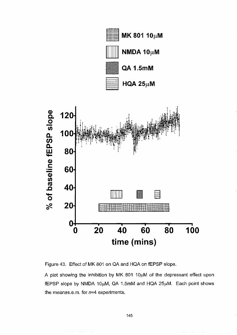
CI) 12 c.
0 -tn a.. 10 en a.. w 8 ~
CI) c: 6 ---CI) tn co 4 ..c ~
0
?ft. 2
o
II MK 80110!lM
II NMDA 10!lM
QA 1.SmM
II HQA 25!lM
• I ~ 1111111111111I11111111111111111111111111111111111111111111111111111
20 40 60 80 100 time (mins)
Figure 43. Effect of MK 801 on QA and HQA on fEPSP slope.
A plot showing the inhibition by MK 801 10llM of the depressant effect upon
fEPSP slope by NMDA 10IlM, QA 1.5mM and HQA 251lM. Each point shows
the mean±s.e.m. for n=4 experiments.
145

Ro 25-6981
As illustrated in figure 44, HQA 2~M (i) and QA 100~M (1) are
concentrations that produced small yet discernible depressions of baseline
fEPSP slope (c). With the co-perfusion of Ro 25-6981 3~M, the same
concentrations of QA (2) and HQA (ii) produced significant depressions of
the fEPSP slope to 24.19±15.39% and -1.88±4.93% respectively compared
to baseline responses (p<O.OOl and p<O.OOl respectively n=5). Ro 25-6981
was then allowed to wash out and QA (3) and HQA (iii) were re-applied
where a significant depression to 8.98±5.21 % and -5.59±4.04% of control
respectively was still observed (p<O.OOl and p<O.OOl respectively n=5)
(figure 44). It was noticed that the effect of Ro 25-6981 seemed to develop
slowly and did not diminish during the washout phase of the experiment. It
was also observed that paired-pulse interactions were significantly
enhanced during the enhanced depressions of QA and HQA at all
interstimulus intervals after the application of Ro 25-6981 compared to
during the initial applications (QA: 10ms p<O.Ol, 20ms p<O.05, 50ms
p<O.Ol; HQA: 10ms p<O.OOl, 20ms p<O.OOl, 50ms p<O.05, n=6; figure 45
and 46).
146

QA 100flM
III
(1)
c. 0
tn 120 a.. en
100 a.. w .....
80 (1)
c: .- 60 (1)
tn ns 40 .c
..... 0 20 ~ Q
0
0 50 100 150 200 tim e (m in 5)
Figure 44. Effect of Ro 25-6981 with QA and HQA on fEPSP slope.
A plot showing the effect of QA 100IJM (1) and homoquinolinic acid 2IJM (i)
compared to the baseline fEPSP response (c). During the co-perfusion of Ro
25-6981 3~M, the same concentrations of QA (2) and homoquinolinic acid (ii)
produced a significant depression to 24.19±15.39% and -1.88±4.93% of the
fEPSP slope respectively compared to baseline responses (p<0.001 and
p<0.001 respectively). Ro 25-6981 was then washed out for 20 minutes until
QA (3) was re-applied and 45 minutes until homoquinolinic acid (iii) was re
applied where a significant depression to 8.98±5.21 % and -5.59±4.04%
respectively was observed compared to the baseline fEPSP responses
(p<0.001 and p<0.001 respectively). Each point shows the mean±s.e.m.
147

quinolinic acid 'I- ** 0 3.0 0_ +=i Cl)
2 cu t/) ":"t: Cl) 0 2.5 00-t: t/) Cl) Cl) s.. s.. Cl)+"
==~ 2.0
:C'i: Cl) 0 t/)+" 1.5 1 -\J :::l t: 0.0
I 0 C "'CCl) 1.0 ~ t/) .-cu 0.
0.5 10 20 30 40 50
inter-stimulus interval (ms)
Figure 45. Effect of Ro 25-6981 and QA on paired-pulse interactions.
A plot showing the paired-pulse interactions at interstimulus intervals of 10ms,
20ms and 50ms during baseline recordings (c), the initial application of QA
100~M (1) and the second application of QA 100~M during the co-perfusion
with Ro 25-6981 3~M (2) (10ms p<0.01, 20ms p<0.05, 50ms p<0.01, n=6).
Points correspond to figure 44. *P < 0.05, **P < 0.01, ***P < 0.001. Each point
shows the mean±s.e.m. for n=6 experiments.
148
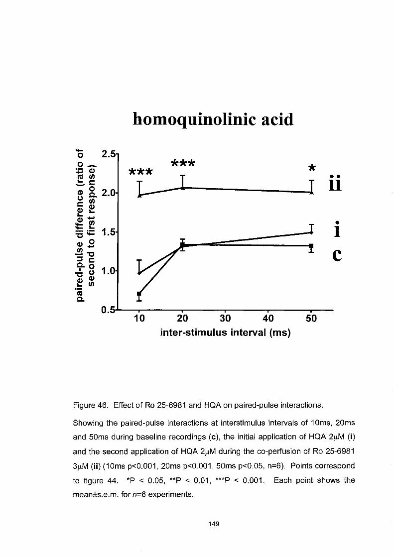
homoquinolinic acid
'to- 2.S 0 0_ *** * :,jjCl) *** ns tn I •• a... s:::::: L I II -0 2.0 Cl)o. (.) tn s:::::: CI) CI) a... a... ~u; • 'to- a... 1.S I .- .-"0 'to-
CI) 0 tn"""
C -"0 ::::s s:::::: 0.0 1.0 I (.)
"0 CI)
f tn .-ns 0.
O.S 10 20 30 40 SO
inter-stimulus interval (ms)
Figure 46. Effect of Ro 25-6981 and HQA on paired-pulse interactions.
Showing the paired-pulse interactions at interstimulus intervals of 10ms, 20ms
and 50ms during baseline recordings (c), the initial application of HQA 2~M (i)
and the second application of HQA 2~M during the co-perfusion of Ro 25-6981
3~M (ii) (10ms p<0.001, 20ms p<0.001, 50ms p<0.05, n=6). Points correspond
to figure 44. *P < 0.05, **P < 0.01, ***P < 0.001. Each point shows the
mean±s.e.m. for n=6 experiments.
149
Excitotoxicity
An application of HQA that significantly depressed the fEPSP slope was
selected to investigate the effect of Ro 25-6981 on higher concentrations of
NMDA receptor agonists. In previous studies of young cultured rat cortical
neurones Ro 25-6981 had potentiated small depolarising concentrations of
NMDA but blocked larger depolarising concentrations (Fischer et al.,
1997). A 5-minute application of HQA 4/-lM caused a maximal depression
offEPSP slope to 6.63±3.69% (p<0.001, n=4) that reverted to near baseline
responses upon washout. Ro 25-6981 3/-lM was co-perfused with a second
application of 4/-lM HQA which produced a depression of fEPSP slope to
-9.27±1.13% (p<0.001, n=4). However, this depression did not fully
recover to baseline responses. The fEPSP slope was irreversibly reduced to
an average 34.70±0.38% compared to baseline responses (p<0.001, n=4).
Maximum evokable amplitudes of fEPSP slope (100%), which were taken
as an additional measure of excitotoxic damage, were significantly reduced
(p=O.OOll, n=4) compared to the baseline maximums recorded and they
did not recover during the 70-minute washout phase of the experiment.
Attempts to induce L TP at the end of the experiments also failed, again
suggesting that irreversible excitotoxic damage had occurred (figure 47 and
48). This specifically ruled out the induction of NMDA-receptor
dependent LTD, which was reversible by high frequency stimulation (data
not shown).
150

II HQA 4/-lM
Ro 25-6981 3f.lM
Cl) ~ c. 12 0 -tn a.. 10 en a.. 8 w ~
Cl)
6 1 c: .--Cl) tn 4 ns ..c ~
2 0 ~ 0
i i i i i
o 25 50 75 100 125 time (mins)
Figure 47. Effect of HQA 4J.!M and Ro 25-6981 on fEPSP slope.
A 5-minute application of homoquinolinic acid 4J.!M elicited a reversible
depression of baseline fEPSP slope to 6.63±3.69% (p<O.001). Ro 25-6981
3J.!M was co-perfused and a second 5-minute application of homoquinolinic acid
4J.!M produced a depression of fEPSP slope to -9.27±1.13% (p<O.001).
However, the fEPSP slope did not fully recover but was significantly and
irreversibly reduced to an average 34.70±O.38% compared to the initial baseline
(p<O.001). Each point shows the mean±s.e.m. for n=4 experiments.
151

c
1
tmV
tOms
Figure 48. Representative fEPSP recordings showing excitotoxicity.
During baseline recording (c) and during the irrecoverable depression (1)
corresponding to figure 47.
152
NR2A antagonists
HQA is reported to be a selective agonist for NR2A and NR2B subunit
containing NMDA receptors. Using Ro 25-6981, an NR2B antagonist, to
produce the potentiation of the depression of fEPSP slope by low
concentrations of HQA suggests that the remaining HQA-dependent effect
could be modulated by the remaining NR2A component of HQA activity.
In order to test this theory several candidate NR2A selective antagonists
including zinc, CCMQ and PEAQX were investigated (Paoletti et aI., 1997;
Brown et ai., 1998; Auberson et ai., 2002; Grimwood et aI., 2002).
CCMQ
Previous autoradiographical studies found that 2-carboxy-3-
carboxymethylquinoline (CCMQ), a derivative of HQA, displaces bound
eH]-HQA from its NMDA-insensitivenovel binding site at a concentration
of 1mM, leaving behind a binding pattern which closely resembles the
distribution of NR2B subunits in the rat brain (Brown et ai., 1998;
Grimwood et ai., 2002). These results and the structural homology with
HQA suggest that CCMQ may be able to displace HQA from the NR2A
subunit-containing NMDA receptors thereby leaving only the NR2B
population, acting as an NR2A-selective ligand.
153

The effect of CCMQ 0.5mM upon the HQA-induced depression of fEPSP
slope was investigated first. As shown in figure 49 two initial applications
of HQA 2.5 f.lM produced two similar depressions of fEPSP slope. The
third application of HQA was co-applied with CCMQ but this had no effect
on an identical depression of fEPSP slope. This suggests that CCMQ was
not acting as an antagonist of the NR2A site activated by HQA. This study
next investigated if CCMQ was an NR2A agonist and used Ro 25-6981 to
block the NR2B subunit in order to help reveal any NR2A effect. As
shown in figure 50 the perfusion of Ro 25-6981 alone had no effect upon
baseline fEPSP slope but CCMQ elicited a small potentiation of the fEPSP
slope to 108.65±1.71 % (p=0.0058, n=3). Finally, the effect of CCMQ
upon the potentiation of HQA 2f.lM caused by Ro 25-6981 3 f.lM was
investigated in order to address the subunit specificity of this potentiation.
As shown in figure 51 the application of HQA 2f.lM produced little
response but the application of Ro 25-6981 potentiated this to a significant
depression to 14.72±10.49% (p<O.OOI, n=3) of baseline fEPSP slope,
which was not inhibited by CCMQ. The co-perfusion of CCMQ resulted
again in the same small but significant increase in fEPSP slope to
124.25±2.75% (p<O.05, n=3). A third application ofHQA 20 minutes after
the end of the perfusion of Ro 25-6981 also produced a large depression to
7.18±3.24% (p<O.OOI, n=3) offEPSP slope.
154

cv c. o -U)
c.. UJ c.. W \I-
cv c: .-cv U) ns .c \I-o ?fl.
12
10
8
6
4
2
o 20
III HQA 2.51lM
III CCMQ O.5mM
40 60 80 time (mins)
Figure 49. Effect of HQA 2.5~M and CCMQ O.5mM on fEPSP slope.
100
A plot showing the effect of repeated 5-minute perfusions of homoquinolinic
acid 2.5~M, that caused three consecutive depressions of the fEPSP slope.
The co-perfusion of CCMQ O.5mM with the third perfusion of homoquinolinic
acid had no significant effect upon the peak magnitude or duration of the
response. Each point shows the mean±s.e.m. for n=3 experiments.
155

CD C. o -tn a.. en a.. w "I-
CD c: ---CD tn ca .c "I-o ~ Q
Ro 25-6981 3J.!M
II CCMQ O.SmM
80
60
40
lll~!lliiiii!liliiiilillllrrjililiii!iiiiiiiliiiiliii~illillilililililililililiiililililiiiilliliiiilt 20 • O+---~--~--...~--...--~--~--~ o 10 20 30 40 50 60
time (mins)
Figure 50. Effect of CCMQ and Ro 25-6981 on fEPSP slope.
A plot showing the effect of 3jJM Ro 25-6981 and the subsequent co-perfusion
of O.5mM CCMQ which produced a small potentiation of the fEPSP slope to
108.65±1.71 % (p=O.0058). Each point shows the mean±s.e.m. for n=3
experiments.
156

Q) Q. 0 -tn
D.. tJ) D.. W 'I-
Q) c: ---Q) tn ca .c 'I-0 ~ 0
120
100
80
60
40
20
0 0
II HQA 211M
D Ro 25-6981 311 M
III CCMQ 0.5 mM
20 40 60 80 time (mins)
100
Figure 51. Effect of HQA 2f.lM, Ro 25-6981 and CCMQ 0.5mM on fEPSP slope.
Plot showing that the first application of HQA 2f.lM produced little response but
the co-application of Ro 25-6981 potentiated a second HQA-application
producing a significant decrease to 14.72±10.49% (p<0.001) of baseline fEPSP
slope. The co-perfusion of CCMQ again resulted in the same small but
significant increase in fEPSP slope to 124.25±2.75% (p<0.05). A third
application of HQA 20 minutes after the end of the perfusion of Ro 25-6981 also
produced a large depression to 7.18±3.24% (p<0.001) of fEPSP slope. Each
point shows the mean±s.e.m. for n=3 experiments.
157

Zinc
Zinc ions (Zn2+) have been reported to produce a selective and non
competitive inhibition of the NR2A subunit-containing recombinant
NMDA receptors, producing a voltage-independent inhibition at nanomolar
concentrations (Paoletti et aI., 1997). As shown in figure 52 an initial
application of HQA 2.5 JlM caused a significant depression of fEPSP slope
to O.7±O.57% (p<O.OOl, n=4) , a second application of HQA with Zn2+
100nM also produced an identical depression of fEPSP slope to 2.6±2.2%
(p<O.Ol, n=4). Thus, 100nM Zn2+ was unable to prevent the depression of
fEPSP slope.
158
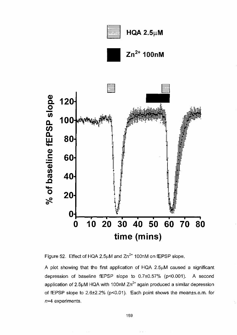
Cl) c. 0 -U)
a.. en a.. w "I-
Cl) t: .--Cl) U) ns .c "I-0
cf:.
12
10
8
6
4
2
II HQA2.5~M Zn2+ 100nM
o 10 20 30 40 50 60 70 80 time (mins)
Figure 52. Effect of HQA 2.5flM and Zn2+ 100nM on fEPSP slope.
A plot showing that the first application of HQA 2.5flM caused a significant
depression of baseline fEPSP slope to O.7±O.57% (p<O.001). A second
application of 2.51JM HQA with 100nM Zn2+ again produced a similar depression
of fEPSP slope to 2.6±2.2% (p<O.01). Each point shows the mean±s.e.m. for
n=4 experiments.
159
PEAQX
Having been unsuccessful with CCMQ and zinc in attempts to antagonise
the NR2A subunit, PEAQX, a novel NR2A antagonist was obtained as a
gift from Dr Yves Auberson of Novartis Pharmaceuticals. This compound
has l26-fold selectivity for the NMDA NRlAINR2A over the
NRlAINR2B receptors expressed in Xenopus oocytes (ICso = l4nM and
l800nM respectively)(Auberson et al., 2002). This investigation was
started by constructing a concentration-response curve for the inhibitory
effect of PEAQX upon the depression of fEPSP slope elicited by HQA
3 ~M. This was achieved by applying 3 ~M HQA, which produced a large
depression of the fEPSP slope, and then PEAQX was co-applied at varying
concentrations. Subsequent co-applications of the same concentration of
HQA were made and the degree of antagonism produced was observed
(figures 53 and 54). From the concentration-response curve it was
determined that using a concentration of 0.1 ~M provided the most selective
and complete antagonism of the NR2A subunit-containing NMDA
response. This concentration-response curve is very similar to that found
for the effect of PEAQX on NMDA-induced currents in Xenopus oocytes
expressing human NRlAl2A subunits (Appendix 2).
This study also investigated paired-pulse interactions during the interaction
of HQA and PEAQX. It was found that during the initial depression of
160

fEPSP slope caused by HQA 3 JlM the paired-pulse ratios were increased at
all interstimulus intervals (lOms p<O.OOI, 20ms p<O.OI, 50ms p<O.OOI;
n=6) but that during the subsequent co-application of PEAQX 0.1 JlM and
HQA 3 JlM they remained similar to baseline responses (figure 55).
161
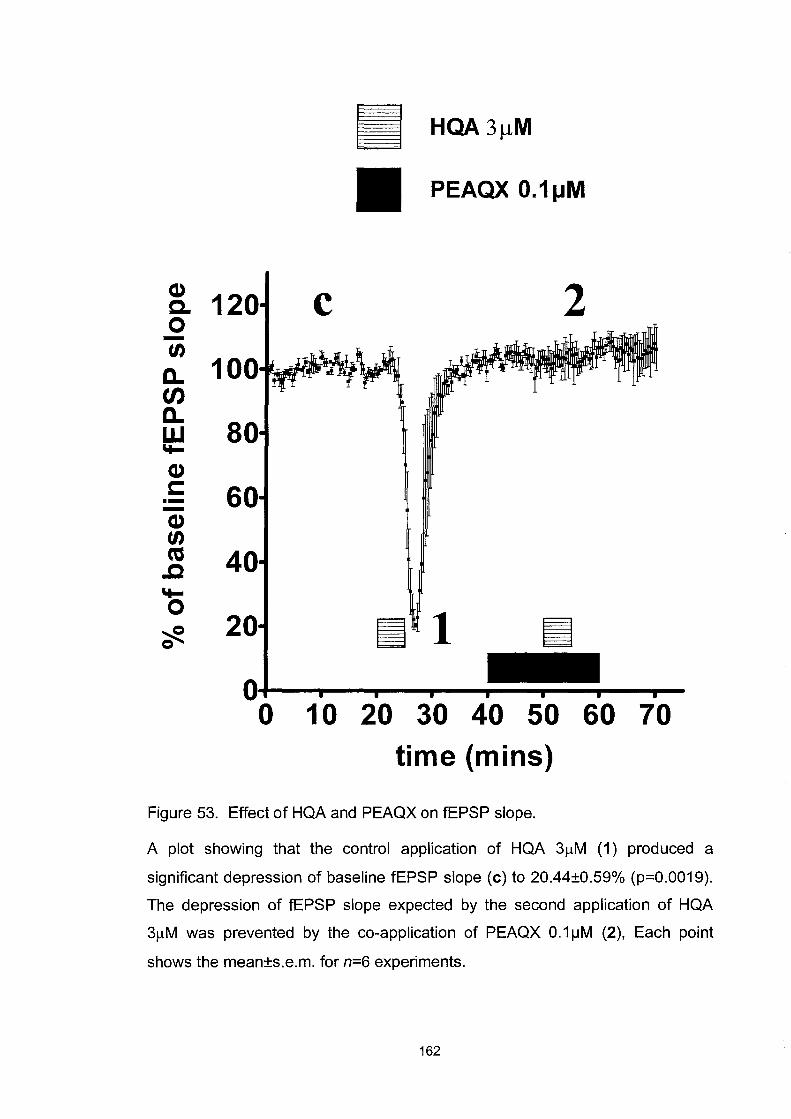
Cl) c. o -U)
Den Dw "t-Cl) c: .--Cl) U) ctS ..c "t-O ~ o
PEAQX O.1tJM
c 2
60
40
20 ~ 1 O+-~~~--~--~--~~--~-o 1 0 20 30 40 50 60 70
time (mins)
Figure 53. Effect of HQA and PEAQX on fEPSP slope.
A plot showing that the control application of HQA 31lM (1) produced a
significant depression of baseline fEPSP slope (c) to 20.44±O.59% (p=O.0019).
The depression of fEPSP slope expected by the second application of HQA
31lM was prevented by the co-application of PEAQX O.1j..1M (2), Each point
shows the mean±s.e.m. for n=6 experiments.
162

Q) c. 0 tn
D.. en D.. w 't-
Q) c: .--Q) tn CO .c 't-0 ~ Q 20
0 -9 -8 -7
Log [PEAQX]M
Figure 54. The concentration-response curve of PEAQX on HQA-induced
depression of fEPSP slope.
A plot showing the concentration-response curve of the inhibitory action of
PEAQX at concentrations of 1 nM, 5nM, 10nM, 50nM, 100nM and 500nM (n=3
at all intervals, except n=6 for 100nM PEAQX) upon HQA 3f.lM, which normally
caused a significant depression of fEPSP slope. A concentration of 9.23nM is
predicted to produce a 50% inhibition of the depression elicited by HQA 31JM.
Each point shows the mean±s.e.m.
163

o~ ** +=iQ) co tJ) ~ t: ---0
Q) 0. *** (J tJ) t: Q) Q) ~ ~
~(j) .... ~ .- .- *** 1 "'0 .... Q) 0 tJ) ..... -"'0 :::l t: 0.0 • (J "'OQ) ~ tJ) 1.0 -- .... [0
10 20 30 40 50 inter-stimulus interval (ms)
Figure 55. Effect of HQA and PEAQX on paired-pulse interactions.
c 2
A plot showing the paired-pulse interactions during baseline responses (c),
which were significantly enhanced at interstimulus intervals of 10ms, 20ms and
50ms during the first application of HQA 3~M (1) and returned to control levels
during the blockade of the second application of HQA 3~M by PEAQX 0.11JM
(2). Points correspond to the time-course in figure 53. Each point shows the
mean±s.e.m. for n=6 experiments. *P < 0.05, **P < 0.01, ***P < 0.001.
164

PEAQX's inhibition of the Ro 2S-6981/HQA potentiation.
However, of greater interest was the finding that the potentiated depression
of fEPSP slope obtained by HQA 2~M with Ro 25-6981 3 ~M was also
completely blocked by PEAQX 0.1~. As shown in figure 56, a
concentration of 2~M HQA (1) initially produced a small and insignificant
depression of the baseline fEPSP slope (c) but following the perfusion of
Ro 25-6981 3 ~M, a second application of HQA 2~M (2) produced a
significant depression of fEPSP slope to 37.33±4.43% (p=O.0261, n-4).
The co-perfusion of PEAQX O.l~M prevented the depression of a third
application of HQA 2~M (3).
It was also observed that paired-pulse interactions during the first
application of HQA 2~M (1) were not significantly different from baseline
responses (c). However, after the application of Ro 25-6981 3~M, the
second application of HQA 2~M (2) caused a significant enhancement of
paired-pulse ratios (p<O.OOI, n=4 at all interstimulus intervals). During the
inhibition by PEAQX O.I~M, the third application of HQA 2~M (3) saw
paired-pulse interactions remain similar to baseline responses (figure 57).
165

III HQA 2~M
PEAQX 100nM
Ro 25-6981 3f.!M
CI) c 1 2 3 c. 12 0 -tn
a.. en a.. w \t-
CI) c: 6 ---CI) tn co 4 ..c
\t-o 2 ~ 0
0 0 20 40 60 80 100
time (mins)
Figure 56. Effect of HQA, Ro 25-6981 and PEAQX on fEPSP slope.
A plot showing the effect of a low concentration of homoquinolinic acid 2JlM (1)
that initially produced a small, insignificant depression of the baseline fEPSP
slope (c). Following the perfusion of Ro 25-6981 3JlM, a second application of
homoquinolinic acid 2JlM (2) produced a significant depression of fEPSP slope
to 37.33±4.43% (p=O.0261). The co-perfusion of PEAQX O.1JlM, an NR2A
selective antagonist, inhibited the depression of the third application of
homoquinolinic acid 2JlM (3). Each point shows the mean±s.e.m. for n=4
experiments.
166

~ G) 2.5 *** co U) s... C ""-'0
(1) 0. *** g ~ 2.0 (1) s... s........, ~U) ..... s... :ct;:1 (1) £ .5 .!!l-c ::l c 0.0 -b ~ 1.0 ~ U) .- ..... [0
***
0.5 ................. ---.---...--.....--....--10 20 30 40 50 inter-stimUlus interval (ms)
2
c
1 3
Figure 57. Effect of HQA, Ro 25-6981 and PEAQX on paired-pulse
interactions.
This plot shows the paired-pulse interactions recorded during the co-perfusions
of HQA 2/lM, Ro 25-6981 3/lM and PEAQX 0.1IJM. Points correspond to the
time course of figure 56. Paired-pulse interactions during the first application of
homoquinolinic acid 2/lM(1) were not significantly different from baseline
responses (c). However, after the application of Ro 25-6981 3/lM the second
application of homoquinolinic acid 2/lM (2) caused an enhancement of the
paired-pulse interactions at 10ms (p<0.001), 20ms (p<0.001) and 50ms
(p<0.001) compared to baseline interactions. During the NR2A inhibition of
PEAQX the third application of homoquinolinic acid 2/lM (3) saw the paired
pulse interactions return to baseline responses (n=4). Each point shows the
mean±s.e.m. for n=4 experiments. *P < 0.05, **P < 0.01, ***P < 0.001. Points
correspond to the time course in figure 56.
167

Protein synthesis
In view of the latency of onset, the long-lasting effect of Ro 25-6981 and
the involvement of a protein phosphatase signal transduction step the
question arose of whether any new protein synthesis was required and
induced. To investigate this possibility anisomycin 30f.lM was used, which
has previously been shown to block the maintenance of L TP and LTD at
this concentration and over this timescale (Grollman, 1967; Otani et at.,
1989; Manahan Vaughan et at., 2000; Karachot et at., 2001). An initial
application of HQA 2f.lM caused a small depression to 82.68±3.l6% of
baseline fEPSP slope. Anisomycin 30f.lM and the subsequent co-perfusion
of Ro 25-6981 3 f.lM had no significant effect on the baseline fEPSP slope.
A second application of HQA 2f.lM produced a large and significant
depression of fEPSP slope to 0.91±3.0% (p=0.0152, n=4) compared to the
initial application of HQA 2f.lM. A similar profound depression of fEPSP
slope to 6.05±4.86% (p=0.0128, n-4) , again in comparison to the initial
application of HQA 2f.lM, was obtained with a subsequent application of
HQA 2f.lM (figure 58). This suggests that new protein synthesis is not
required for the potentiation by Ro 25-6981.
168

CD C-O -fJ)
a.. CJ) a.. w 'to-
CD s::: .--CD fJ) ca .c 'to-0 ~ 0
140
120
100
80
60
40
20
0 0
[:::::::::1 Anisomycin 30~M
Ro 25-6981 3~M
• HQA2~M
~ ~ 111::::::::::::1:1:11:1::1::1:::::1::111::1:::::111::1::1::1::::11:1::::11
20 40 60 80 100 120 140 time (mins)
Figure 58. Effect of HQA, Ro 25-6981 and anisomycin upon fEPSP slope.
Plot showing that an initial application of homoquinolinic acid 2~M caused a
small depression of baseline fEPSP slope, the perfusion of anisomycin 30~M
and subsequent co-perfusion of Ro 25-6981 3~M had no significant effect on
the baseline fEPSP slope. The second application of homoquinolinic acid 2~M
produced a depression to 0.91±3.04% (p=0.0152,n=4) compared to the trough
of the initial application of homoquinolinic acid 2~M. After washing out for 20
minutes a further application of homoquinolinic acid 2~M caused a similar
depression of fEPSP slope to 6.05±4.86% (p=0.0128, n=4) again in comparison
to the trough depression of the initial application of homoquinolinic acid 2~M.
Each point shows the mean±s.e.m. for n=4 experiments.
169

AMPA and kainate receptors
The earlier paired-pulse data suggested that there might be a presynaptic
component during the NMDA, HQA and QA evoked depressions of fEPSP
slope. AMP A and kainate receptors are the main glutamatergic receptors
responsible for basal synaptic transmission and the generation of currents
underlying the fEPSP. AMP A and kainate were used at 100nM and 25nM
respectively, concentrations that produced small depressions of the fEPSP
slope. In both cases, the application of Ro 25-6981 3 ~M had no effect on
the responses, confirming that this effect is specific to NMDA receptor
activation (figures 59 and 60). This is consistent with the absence of effect
of Ro 25-6981 alone upon baseline responses.
170

Ro 25-6981 3f.lM
AMPA 100nM
12 Q) c. 0 10 ~ -tn a.. en
8 a.. w "I-Q)
6 s:: .--Q) tn
4 ca ..c "I-0 ~ 2 0
o 20 40 60 80 100 time (mins)
Figure 59. Effect of Ro 25-6981 and AMPA on fEPSP slope.
Plot showing the effect of 100nM AMPA applied before and after the perfusion
of 3IJM Ro 25-6981, the response to AMPA was unchanged. Each point shows
the mean±s.e.m. for n=3 experiments.
171

(1) Q. 0 -(/)
a.. tJ) a.. w 'I-
(1) c: .--(1) (/) ca .c 'I-0 ~ 0
12
10
8
6
4
2
o
Ro 25-6981 311M
D kainate 25nM
D D
20 40 60 80 time (mins)
Figure 60. Effect of Ro 25-6981 and KA on fEPSP slope
100
Plot showing the effect of 25nM kainate applied before and after the perfusion
of 3IJM Ro 25-6981, the response to kainate was unchanged. Each point
shows the mean±s.e.m. for n=3 experiments.
172

Discussion
In order to expose a response to the activation by homoquinolinic acid of
the uncharacterised binding site reported by others the non-selective EAA
antagonist kynurenic acid, the competitive NMDA receptor antagonist 2-
AP5 and the non-competitive antagonist dizocilpine (MK-801) were used.
2-AP5, as with some competitive antagonists (straight chain, piperidine and
piperazine structures) displays the subunit potency NR2A > NR2B >
NR2D > NR2C (Laurie & Seeburg, 1994). However, neither kynurenic
acid, 2-AP5, dizocilpine nor PEAQX revealed a novel action of
homoquinolinic acid on the fEPSP slope. Therefore, the novel binding site
for homoquinolinic acid does not affect acutely recorded synaptic
potentials in the hippocampus and homoquinolinic acid appears to be a
selective agonist at NMDA receptors that include the NR2A subunit.
Particularly when studied on fEPSPs, the paired-pulse paradigm represents
a valuable means of assessing the actions of compounds on presynaptic
terminals. The paired-pulse inhibition obtained at interpulse intervals of
10ms results from the depletion of transmitter from presynaptic stores
(Burke & Hablitz, 1994; Wilcox & Dichter, 1994; Hashimoto & Kano,
1998), and is reduced by agents or procedures which decrease transmitter
release. Paired-pulse facilitation, observed at longer interpulse intervals, is
due to the residual intraterminal Ca2+ which increases transmitter release
173

(Hess et ai., 1987; Wu & Saggau, 1994; Debanne et aI., 1996;
Kleschevnikov et ai., 1997). The modification of paired-pulse interactions
by homoquinolinic acid and quinolinic acid suggests that at least part of its
ability to depress the fEPSP slope is presynaptic in origin. Indeed, there is
ample evidence corroborating the existence of presynaptic NMDA
receptors in several regions of the brain (Berretta & Jones, 1996; Hamada
et ai., 1998; Glitsch & Marty, 1999; Woodhall et ai., 2001), including the
hippocampus (Breukel et ai., 1998). Alternatively, a retrograde signalling
molecule released postsynaptically in response to depo1arisation by
homoquinolinic acid may mediate this effect indirectly or other
postsynaptic effects may confound the changes in PPD and PPF (Foster &
Richardson, 1997). The possibility that adenosine released in response to
neuronal depo1arisation could act as a retrograde inhibitor was investigated
using the selective Al receptor antagonist DPCPX. However, DPCPX had
no effect upon the magnitude of the depression caused by homoquinolinic
acid or paired-pulse interactions suggesting that the presynaptic activity of
homoquinolinic acid is not dependent upon the activation of presynaptic
adenosine Al receptors.
As previously seen with NMDA, Ro 25-6981 potentiated the responses to
HQA and QA on fEPSP slope and altered paired-pulse interactions. One
possibility for the potentiating effect of Ro 25-6981 is that it causes an
increase in the release probability of glutamate from the presynaptic
174

boutons. This is unlikely as there is no effect of Ro 25-6981 on baseline
ffiPSP or on comparable responses to AMP A and kainate. Another
possibility is that Ro 25-6981 increases the local release of an endogenous
NMDA agonist such as glycine, homocysteate, cysteine sulphinate or
aspartate (Patneau & Mayer, 1990; Davanger et al., 1994). Whilst glycine
has a high affinity and is thought to saturate NRIINR2B receptors, it has a
lower affinity for NRIINR2A receptors, which under resting concentrations
of glycine could be unsaturated and may be potentiated with rising glycine
concentrations.
CCMQ was not able to block the depression of ffiPSP slope at a similar
concentration at which it displaced the binding of HQA in binding studies.
Furthermore, the Ro 25-6981-enhanced response to HQA was similarly not
blocked. This suggests that CCMQ is not acting as an antagonist in its
ability to displace HQA from NR2A subunits in the previous studies.
Considering the slight, yet significant, increases in ffiPSP slope elicited by
CCMQ under the influence of Ro 25-6981 it is possible that CCMQ is a
weak NR2A agonist. This will best be confirmed using recombinant
NRIINR2A receptors expressed in Xenopus oocytes. Once this has been
achieved, a study of structure-activity relationships could lead to more
potent and selective NR2A agonists.
The absence of effect of zinc as an NR2A antagonist may be due to the
experimental conditions where the level of NR2A inhibition is already 175
saturated by the concentrations of zinc contaminating the ACSF. Also
there may be physiological mechanisms that regulate zinc inhibition still
active in the intact slice preparation, such as tyrosine kinase src
phosphorylation and chelation of zinc by thiol-reducing agents (Paoletti et
al., 1997; Arden et al., 1998; Zheng et al., 1998). In addition, differences
between native heteromeric receptors and the recombinant receptors may
weaken the effectiveness of zinc. Zinc is concentrated by certain neurones
into synaptic vesicles and can be released in a Ca2+ -dependent manner at
some synapses including the mossy fibre-CA3 pyramidal synapse
(Dingledine et al., 1999).
The effect of the NR2A antagonist PEAQX demonstrates that the NR2A
subunit-containing NMDA receptors gate the currents involved in the
depression of fEPSP slope and the associated paired pulse interactions.
The enhancements of paired-pulse interactions are associated both with
HQA alone and under the influence of Ro 25-6981, where the NR2B
subunit is in both an activated and inhibited state. This suggests that the
NR2B subunit cannot independently alter the presynaptic element
suggested by paired-pulse results although it may be able to do so
indirectly by its modulation of NR2A subunits, which alone seems to
modulate directly the enhancement of paired-pulse interactions and the
depression of the fEPSP slope.
176

Chapter 6. General Discussion
Negative coupling of NR2B to NR2A subunit-containing
receptors
This study has suggested that acutely activated, tonically activated or
constitutively active NR2B subunits maintain an inhibitory restraint upon
the activation of the NR2A subunit-containing NMDA receptors by way of
a protein phosphatase 2B (ca1cineurin) transduction step. The NR2A
subunit-containing receptor seems to be the principal receptor involved in
postsynaptic depolarisation and presynaptic alteration of release
probability. The NR2B subunit has no observable function other than to
gate the activity of the NR2A subunit-containing NMDA receptors. A
recent report investigating the properties of NRllNR2BINR2D receptors
expressed in newborn rat hippocampal granule cells has also observed
evidence of negative coupling (Pina Crespo & Gibb, 2002). Additionally,
NR3A subunits have been shown to negatively modulate NMDA receptors
(Das et al., 1998). Although the effect ofRo 25-6981 is dependent on the
presence of an NMDA receptor agonist to observe its effect, it is a
persistent change in the weight of the synapse over a timescale reported to
be associated with a potentiation of NMDA (Watt et al., 2004).
Furthermore, it may involve co-incident pre- and postsynaptic activity
suggestive of a Hebbian process. The NMDA receptor has a central role in
177

the induction of LTP and LTD, the negative coupling demonstrated in this
study has significant implications for determining both the threshold for
L TP and LTD induction and perhaps the magnitude of plasticity induced.
The cellular mechanism by which this enhancement is produced is not
entirely understood. Such an enhancement has very exciting implications
for our understanding ofNMDA receptor physiology and pharmacology.
Multimeric NMDA receptor composition
Nicotinic acetylcholine receptors are pentamers, voltage-gated potassium
channels are tetramers and gap junction proteins mediating coupling at
electrical synapses are hexameric (or hexamonic). Glutamate receptors
were often supposed to be pentamers because they, like the acetylcholine
receptor are ligand gated. Equal evidence exists for both a tetrameric or a
pentameric structure from receptor subunit studies (Behe et ai., 1995;
Ferrer Montiel & Montal, 1996; Premkumar & Auerbach, 1997; Laube et
ai., 1998; Mano & Teichberg, 1998; Rosenmund et ai., 1998). However,
these studies are designed to identify the number of functional binding sites
in a receptor but are based upon the assumption that each binding site in a
receptor operates independently of other subunits. This study has
suggested that there is a negative coupling between the NR2A and the
NR2B receptor subunits and it has been reported by others of a negative
coupling in receptors containing NR3A and NR2A subunits (Ciabarra et
ai., 1995; Das et ai., 1998) and NR2B and NR2D subunits (Pina Crespo & 178

Gibb, 2002). This observation suggests that the analysis of dose-response
curves (Laube et al., 1998) and patterns of main and sub-conductance states
(Behe et al., 1995; Premkumar & Auerbach, 1997) may be more
complicated and the heteromeric structure of the native NMDA receptor
may have been miscalculated.
Presynaptic NMDA receptors
Demonstrating the role of presynaptic receptors definitively ideally
requires the fulfilment of five criteria (McGehee & Role, 1996;
MacDermott et al., 1999):
1. Evidence for the presence of presynaptic receptors.
2. Activation of these receptors exogenously produces the
presynaptic effect that alters release probability (that mimics
physiological control).
3. Presence of an endogenous ligand at the synapse that can be
released under physiological conditions for activation of the
presynaptic receptor.
4. Antagonists that block the presynaptic ionotropic receptors block
synaptic plasticity.
179

5. Activation of the presynaptic receptor leads to the observed
change in release through a characterised mechanism.
In answer to these criteria:
1. AMP A, kainate and metabotropic glutamate receptors have all been
established as presynaptic autoreceptors in the hippocampus
(Vazquez et al., 1994; Chittajallu et al., 1996; A1ici et al., 1997),
modulating excitatory transmission by altering glutamate release
probability. NMDA receptors have been amply demonstrated on the
postsynaptic membrane (Petralia et al., 1994; Racca et al., 2000) and
there is accumulating evidence demonstrating a presynaptic locus.
Presynaptic NMDA autoreceptors have been reported in the
cerebellum (Glitsch & Marty, 1999), spinal cord (Liu et aI., 1997),
hippocampus (Breukel et al., 1998), suprachiasmatic nucleus
(Hamada et aI., 1998) and entorhinal cortex (Berretta & Jones,
1996). Furthermore, presynaptic NMDA receptors have been
reported to be NR2B subunit-containing and tonically activated,
causing an increase in glutamate release probability, dependent upon
Ca2+ influx, in the entorhinal cortex (Woodhall et aI., 2001).
Additionally, immunohistochemical studies have found evidence for
presynaptic NMDA receptors (Liu et ai., 1994; Paquet & Smith,
2000).
180

2. This report has suggested, through the changes observed in paired
pulse interactions, that NMDA receptor activation by HQA, QA and
NMDA generated an effect with a presynaptic element. However,
whilst PPF and PPD are generally accepted to be presynaptic
phenomenon, it does not follow that postsynaptic effects cannot
influence their appearance in field recordings (Foster & Richardson,
1997).
3. The endogenous ligand at these synapses is glutamate, which could
act under physiological conditions to activate a presynaptic receptor.
Furthermore, this study has demonstrated this effect by using another
endogenous ligand quinolinic acid.
4. This investigation has demonstrated that both 2-AP5 and PEAQX
can block NMDA-receptor dependent LTP. Furthermore, it has been
reported recently that Ro 25-6981 is able to inhibit LTD (Sjostrom et
aI., 2003; Liu et at., 2004; Massey et at., 2004)
5. The inhibition of this effect by cyclosporin A, which modulates
protein phosphatase 2B (ca1cineurin) activity, demonstrates a distinct
and well-characterised signal transduction pathway.
The lack of sufficiently compelling evidence on point 2 in this study means
that there is not full qualification for a presynaptic NMDA receptor.
181

Therefore, the possible involvement of a postsynaptic receptor with a
retrograde signalling mechanism must be considered. In NMDA receptor
dependent synaptic plasticity, postsynaptically induced processes such as
LTD and LTP are still able to trigger presynaptic changes through
retrograde messengers such as nitric oxide activating PKG (Reyes Harde et
al., 1999) and endocannibinoids acting upon CBl receptors (Cheval eyre &
Castillo, 2003; Sjostrom et at., 2003) or release from other sources such as
glia (Parpura et al., 1994).
Whilst changes in paired-pulse interactions are widely regarded to be due
to presynaptic alterations in release probability; when studied on ffiPSPs
and especially where there are changes in the size of the postsynaptic
potential in response to the test agents many factors affect them which can
affect their interpretation. Including, changes in presynaptic release
probability (Andreasen & Hablitz, 1994), activity of presynaptic
autoreceptors on inhibitory or excitatory terminals (Davies et al., 1990;
Stanford et al., 1995), modulation of postsynaptic conductances and the
presence of GABA-ergic inhibition following the ffiPSP and the depression
cause by NMDA agonist action (Duguid & Smart, 2004).
Also, considering the intracellular data showing a purely postsynaptic
manifestation of these effects a further study of the contribution of different
NMDA subunits is required on clear presynaptic phenomenon such as
spontaneous miniature EPSPs or by concurrently measuring by patch-182

clamp whole cell recording corresponding presynaptic and postsynaptic
neurones.
It is clear that NR2A and NR2B subunit-containing NMDA receptors are
found postsynaptically, as NR1INR2A, NRIINR2B or NR1A1NR2A1NR2B
heteromers, at the synaptic site whilst NR11NR2B receptors are found
exclusively at the extrasynaptic site (Li et at., 1998; Stocca & Vicini, 1998;
Tovar & Westbrook, 1999). A tonically activated state of the agonist
preferring NR2B subunit-containing receptor could be achieved by being
located near to the release site of glycine and glutamate on the presynaptic
bouton. However, the NR2B subunit in both it's activated and inhibited
state is associated with enhancements in paired-pulse inhibition whereas,
only the blockade of the NR2A subunit prevents the enhancement of
paired-pulse interactions. Alternatively, the NR2B subunit may be in a
constitutively active state that is independent of agonist binding as has been
observed with other receptors (Scheer & Cotecchia, 1997; Behan &
Chalmers, 2001; Richard et at., 2001). Interestingly NR2B subunit
containing receptors have a higher glutamate and glycine affinity, are less
sensitive to tonic proton inhibition, and are insensitive to desensitisation,
which suggests that they would be more sensitive detectors of glutamate
than NR2A subunit-containing receptors, especially at extrasynaptic sites.
Applications of HQA, QA and NMDA by themselves and under the
influence of NR2B antagonism are associated with depressions of fEPSP
183

slope and increases in paired-pulse ratios. These are both blocked by
PEAQX, the NR2A antagonist, suggesting that the presynaptic element
may be NR2A subunit-mediated. It is difficult from the current study to
make any definitive resolution of the receptor populations found pre- and
postsynaptically or the subunit makeup of such receptors.
It may be possible to investigate these further using single channel analysis
of outside-out patches from these tissues. Using selective antagonists, it
can be determined whether the negative coupling of NR2B upon NR2A is
between subunits in a single heterotrimeric NR1INR2A1NR2B receptor
(rather than between discrete heterodimeric NR11NR2A and NR1A1NR2B
receptors). This may also address the location of these subunits at pre- or
postsynaptic sites. There is evidence for the co-localisation of NR2A and
NR2B subunits in the hippocampus, and specifically within the CA 1, CA2
and CA3 regions (Fritschy et aI., 1998).
Excitotoxicity
This report has demonstrated that, far from being neuroprotective, Ro 25-
6981 actually precipitates an excitotoxic insult from otherwise benign,
depolarising concentrations of NMDA and HQA. This observation is
contradictory to prevIOUS reports of NR2B antagonists being
neuroprotective in excitotoxic studies (Gotti et al., 1988; Fischer et al.,
1997; Lynch et al., 2001; Gill et al., 2002) but some reports have
184

corroborated this study (Zou & Crews, 2003). The age of tissue used in
investigations could account for the reason for the difference in these
results. In previous studies immature tissues were used whereas this study
has used adult tissues in a well-proven experimental model of
excitotoxicity (Schurr et aI., 1995). As mentioned in the introduction, the
expression levels and distribution of NMDA subunits change dramatically
during development, especially in respect of the NR2A and the NR2B
subunits. This developmental change is responsible for the alteration in
susceptibility of tissues to excitotoxicity and also to the induction of
synaptic plasticity (Dudek & Bear, 1993; Liu et al., 1996). The use of
immature tissues to investigate excitotoxicity is dictated to some extent by
methodological constraints. The rationale for using these tissues in the
investigation of stroke, which is an almost exclusive threat to the adult
population, is difficult to justify, especially here, considering the disparity
between these results. Additionally, the study of tissue from different areas
of the brain and even different regions of the hippocampus will dictate the
influence of NMDA receptors and the expression of different types and
quantity of NR2 subunits. A recent study of cultured hippocampal
neurones reported that the selective activation of synaptic NR2A subunit
containing NMDA receptors is associated with the potent activation of
cAMP response element binding protein (CREB) and subsequent brain
derived neurotrophic factor (BDNF) gene expression. This leads to an anti
apoptotic cell-signalling pathway. However, the activation of NR2B
185

sensitive extrasynaptic NMDA receptors by bath application of NMDA
agonists or exposure to hypoxic/ischaemic conditions, which cause the
reversal of glutamate transporters (Rossi et al., 2000), is associated with the
Ca2+-dependent transient activation of CREB. This leads to a CREB shut
off signal of the synaptic pro-survival pathway and leads to cell death
(Hardingham et al., 2002). It can be concluded that exceSSIve
overactivation of the NR2A-subunit associated channel could be directly
responsible for the large Ca2+ influx that triggers excitotoxicity. The NR2B
subunit acting to gate the activity of the NR2A subunit-containing receptor
and when it is inhibited this restraint is lost.
An obvious question anses as to whether there a correlation between
decreased levels of NR2B subunit expression, mRNA levels and genetic
expression in adult patients susceptible to stroke or in geriatric patients who
have an age-related susceptibility to stroke and in other relevant disease
states. Indeed, the NR2B subunit is already implicated in an impressive
number of age related diseases and developmentally, the mRNA and the
protein expression of the NR2B subunit decreases significantly between the
ages of 3 and 30 months in mice (Magnusson, 2000; Magnusson et al.,
2002) which is similar to the decrease in the human hippocampus (Law et
aI., 2003). It is possible to further develop this study by investigating the
effect of selective subunit NMDA antagonists in hypoxia, ischaemia and
seizure disorder, which are known to have NMDA receptor components.
186

The potential for the development of drugs to modulate this NR2B-NR2A
interaction is very exciting.
Phosphatase hypothesis
This report has suggested that NR2B subunits maintain an inhibitory
control over NR2A subunit-containing receptors. It could achieve this by
being acutely activated, tonically activated or remaining constitutively
active physiologically, maintaining the phosphorylation state of an
unidentified protein. The superfusion of Ro 25-6981 blocks the NR2B
subunit leading to the ca1cineurin-mediated dephosphorylation of an
unknown protein residue resulting in the potentiation of NR2A subunit-
containing NMDA receptor responses. NR2 subunits are known to be
highly phosphorylated under basal conditions whilst NR1 subunits are very
weakly phosphorylated (Lau & Huganir, 1995; Hall & Soderling, 1997;
Smart, 1997). Therefore, the dephosphorylation step described here may
be acting directly upon either the NR2A or the NR2B subunit to produce
the enhancement of responses to NMDA and HQA. However, it may also
be acting on the NR1 subunit or associated proteins such as PSD-95 and
SAP 102 or an unrelated and possibly novel site. Interestingly, there is
strong evidence for a tyrosine residue that is phosphorylated and leads to
potentiation of the NMDA receptor and is found on the NR2A or NR2B
subunit-containing receptor, as seen with insulin potentiation and LTP
(Chen & Leonard, 1996; Kohr & Seeburg, 1996; Rosenblum et al., 1996; 187

Smart, 1997; Zheng et at., 1998; Christie et at., 1999). The absence of
effect of okadaic acid, a potent inhibitor of the serine/threonine protein
phosphatases types 1 and 2A, rules out these serine residues being
involved. The next step is to identify if there are changes in the levels of
phosphorylation of these subunits. This can be achieved by a combination
of immunoprecipitation using NR1, NR2A and NR2B subunit specific
antibodies to these proteins, then immunoblotting with anti
phosphotyrosine and anti-phosphoserine specific antibodies and estimating
changes in levels of phosphorylation compared to controls with a
chemiluminescent secondary antibody. There are many studies that
correlate increases in phosphorylation with increases in receptor activity
and that dephosphorylation is associates with reductions in receptor activity
(Smart, 1997). However, it was found in this study that the activity of
ca1cineurin, which de-phosphorylates is associated with the potentiation of
responses to NMDA. This is blocked by Cyclosporin A, a phosphatase
inhibitor. Many of the previous studies were carried out using immature
tissue as with previous excitotoxicity reports as mentioned previously. In
addition to the developmental switch of subunit expression (Tovar &
Westbrook, 1999) the basal levels of phosphorylation of the adult NMDA
receptor do not reach maturity until 3-4 weeks post-natally (Cudmore &
Gurd, 1991).
188

Synaptic plasticity
The entry of calcium into the neurone contributes ~ 16% of the NMDA
receptor current but the subsequent activation of Ca2+ -dependent enzymes
and signal transduction pathways induce potent effects (Schneggenburger
et a!., 1993). Both LTP and LTD can be induced by NMDA receptor
activation, resulting in an influx of Ca2+ that triggers plasticity. The
relative level, kinetics and location of the Ca2+ influx determining whether
L TP or LTD is induced; a large, fast, synaptic influx inducing L TP and a
relatively modest, prolonged, extrasynaptic influx inducing LTD (Lisman,
1989; Artola & Singer, 1993; Neveu & Zucker, 1996; Yang et a!., 1999;
Cho et al., 2001; Liu et al., 2004; Massey et a!., 2004). The receptor
kinetics of the NR2B subunit-containing receptor is relatively slower than
the NR2A subunit-containing receptor resulting in calcium influxes that are
consistent with LTD and LTP induction respectively (figure 61). This
suggests that there is a role of NR2B in LTD that requires an NMDA
mediated influx of Ca2+ consistent with the NR2B subunit-containing
NMDA receptor kinetics. Indeed, it has recently been reported that
neocortical LTD is inhibited by NR2B antagonists affecting presynaptic
receptors (Sjostrom et aI., 2003), that LTD in the perirhinal cortex is
inhibited by the NR2B antagonist Ro 25-6981 but not by the NR2A
antagonist PEAQX (Massey, 2003; Massey et al., 2004) and that LTD in
the hippocampus is NR2B dependent (Liu et al., 2004). The NR2A
189

subunit-containing receptor has kinetics consistent with those required for a
large and rapid rise in intracellular calcium concentrations leading to L TP
induction. During LFS protocols that induce LTD the NRIINR2B
receptors make a larger contribution to the total charge transfer and Ca2+
influx whereas during HFS protocols that induce LTP the NRIINR2A
mediates the greater total charge transfer and Ca2+ influx (Erreger et aI.,
2005). This study has demonstrated that the NR2A subunit is critically
required in the induction of L TP. Furthermore, it is known that LTP
requires protein kinase activation (Malenka et aI., 1989) while LTD
requires protein phosphatase activation (Mulkey et aI., 1993; Mulkey et ai.,
1994). This study has also shown that the protein phosphatase 2B
(calcineurin) inhibitor cyc1osporin A is able to prevent the effect of Ro 25-
6981. This is consistent with reports of calcineurin being critical in the
induction of LTD (Mulkey et aI., 1994). This study cannot rule out the
involvement of the NR2B subunit in Schaffer collateral-CAl LTP,
especially considering the interaction of NR2B with the critical NR2A
subunit-containing NMDA receptors described, the marginal reduction in
L TP induced and the many reports implicating the NR2B subunit in L TP.
This report has suggested that NR2B subunit-containing NMDA receptors,
which have been reported to be tonically or constitutively active (Sah et ai.,
1989; Berretta & Jones, 1996; Breukel et ai., 1998; Woodhall et ai., 2001)
and are critically involved in the induction of LTD (Sjostrom et aI., 2003),
190

negatively gate under physiological conditions the NR2A subunit
containing NMDA receptor. It is widely reported and our studies have also
found that successful NMDA dependent LTD induction is very difficult to
achieve whilst L TP induction is relatively easy (Kemp & Bashir, 200 I;
Massey et al., 2004). It could be that such tissues, rather than being in a
resting state, are in a maximal LTD state and are therefore only able to be
potentiated towards an L TP state from which level they can then be easily
depotentiated. This could be explained by differential expression of NR2
subunits in NMDA receptors. This study has not demonstrated that there is
a way to increase the levels of the NR2B-mediated restraint and this may
be because the level of restraint is saturated. A caveat to this link is the
exclusivity to the NMDA receptor and the requirement of NMDA agonist
to observe this phenomenon. The induction of LTP and LTD critically
require the NR2A and NR2B subunits respectively but they are further
associated with the trafficking of AMP A receptors to and from the
postsynaptic membrane and an associated change in the synaptic weight of
neuronal transmission independent of NMDA receptor agonists (Malinow
& Malenka, 2002; Watt et a!., 2004). This is missing from the interaction
this study has shown but if bridged might lead to the induction of L TP
proper.
191

Donald Hebb's neurophysiological hypothesis states that' When an axon of
cell A is near enough to excite a cell B and repeatedly or persistently takes
part in firing it, some growth process or metabolic change takes place in
one or both cells such that A's efficiency, as one of the cells firing B, is
increased.' (Hebb, 1946).
A modification of this hypothesis has led to the Bienenstock-Cooper-
Munro (BCM) theory of the sliding threshold of synaptic plasticity
(Bienenstock et aI., 1982). It hypothesizes that active synapses grow
stronger or weaker when postsynaptic activity exceeds or falls below a
'modification threshold', 8m • Presynaptic activity triggers synaptic
depression or potentiation depending on the concurrent level of
postsynaptic activity. The history of integrated postsynaptic activity
determines the threshold level at which plasticity is induced; the value of
8m varies as a function of the history of integrated postsynaptic activity. As
average activity falls or rises, so does the value of 8m •
This hypothesis led to the observations in the visual cortex that experience
drives NMDA receptor subunit switching from LTD-induction-preferring
NRllNR2B heterodimeric to LTP induction-preferring NRlINR2BINR2A
heterotrimeric receptors (Kirkwood et aI., 1996; Philpott et al., 2001).
NR2A and NR2B subunit-containing receptors provide the biological basis
for the BCM sliding threshold of synaptic plasticity. This model is further
strengthened by the recent reports of NR2A and NR2B antagonist sensitive 192

induction of LTP and LTD respectively (Liu et at., 2004; Massey et at.,
2004; Mallon et at., 2005). The negative coupling of NR2B to NR2A
subunit-containing receptors may represent a further mechanism for fine
tuning the strength of NMDA mediated synaptic transmission and for
maintaining a threshold for induction of synaptic plasticity that only
requires changes in the NR2B subunit. Perhaps by lateral diffusion
between synaptic and extrasynaptic sites, in agreement with recent studies
where the NR2A subunit does not appear to be very mobile (Groc et at.,
2004; Triller & Choquet, 2005). Furthermore, as well as the induction
threshold for LTP or LTD induction there is also the magnitude of change
in strength elicited (Castellani et at., 2001). It was found in this study that
whilst LTD could not be induced, the level of LTP that was reliably
induced was of a particularly high magnitude. A level at which it was then
possible to depotentiate from (data not shown). It may be that the
experience-deprived environment the rats are familiar with, means that the
biological basis for the sliding threshold is set such that they are in a
maximal state of LTD and that only LTP can be induced. It has been
reported that rats from enriched environments are more likely to exhibit
LTD (T.V.P. Bliss, personal communication). The investigation of this
could help to discover more about the biological basis for the magnitude of
synaptic plasticity induced and the effects of environment (Castellani et at.,
2001).
193

e NR2A
.: 1 (PEAQX)
· · · · · o
. o . · · · · ·
CaM
. 00 Low[Ca2+]. ° I
\.... 1 (Ro 25-6981)
NR2B
Adenylate cyclase
+ (staurosporine) LTP
+ PKA
f CaMKII-®
cAMP
(okadaic acid)
Phosphatase 1
-0/
(cyclosporin A)
Calcineurin
:f( e
CaMKII
~ LTD
Figure 61. Phosphatase hypothesis.
This scheme summarises the intracellular transduction systems that account for
the differing pharmacology and physiology of the NR2A and NR2B subunit
containing receptors, the associated phosphoprotein steps and their influence
over L TP and L TO. In brackets are the relevant antagonists (Mulkey et al.,
1994; Revest & Longstaff, 1998).
194

Appendix 1
Methods
Experiments were carried out by Professor T.W. Stone to further
investigate the actions of Ro 25-6981 using intracellular recording
techniques.
Intracellular recordings were made using sharp glass microelectrodes (o.d.
1.5mm) (Clark Electromedical, Harvard Instruments). These were pulled
on a Narashige vertical puller in a manually controlled two-stage process to
produce pipettes with a shank length of approximately 20mm. The
electrodes were filled with 1M potassium acetate, yielding a resistance of
80-l00MO when measured in the extracellular space of the brain slices via
the Neurolog amplifier. Potentials were amplified by a Neurolog 102 DC
amplifier or by an Axoclamp-2A operated in bridge balance mode, and
filtered between DC and 20KHz. The electrode was advanced in 2.51lm
steps using a remotely controlled World Precision Instruments motorised
stepper. When a neurone was approached, indicated by a rise in electrical
noise, a brief increase of electrode capacitance ('buzz') was induced in
order to facilitate entry. Cells were allowed to recover from penetration for
at least 10 minutes before examining them for electrical properties and
stability. Neurones were then used for experimentation if they displayed a
195

stable resting potential (±10% over 20 minutes) greater than 60m V and a
spike of at least 70m V in response to an intracellular depolarising current
pulse. Current pulses (OJ-InA) were used to monitor membrane input
resistance throughout the experiments. Records were calibrated using the
amplifier's own calibration pulse (1 Om V), and were recorded directly onto
a high frequency thermal chart recorder (Grass instruments Dash IV),
although the filter settings were placed at DC to 10Hz to reduce interfering
transients and action potentials.
196

Results
NMDA
Four neurones were tested with a combination ofNMDA and Ro 25-6981.
In each case, NMDA was superfused at a concentration of 2.5uM for 10
minutes, with 20 minutes between each application. At least two control
applications were made. At this concentration, the cells showed little or no
response, the two applications of NMDA immediately prior to Ro 25-6981
yielding barely discernible depolarisations of 0.19±0.048, 0.26±0.059mV
(n=4). Ten minutes after the second of these controls, superfusion of
Ro 25-6981 (3uM) was commenced for 20 minutes. The next application
of NMDA (parallel with the second 10 minutes of Ro 25-6981) produced a
similarly weak effect (0.26±0.066m V). However, the next application,
which was begun 20 minutes after ending the superfusion of Ro 25-6981,
produced a substantial depolarisation (l0.67±1.25mV; p<O.OOOOl, n=4)
(figures 62 and 63). This suggests that the negative coupling of NR2B to
NR2A subunit-containing NMDA receptors is occurring at postsynaptic
sites.
197

a d
b e
c f
Figure 62. Intracellular recordings of NMDA responses and the effect of Ro 25-
6981
Intracellular recordings from a pyramidal neurone of resting potential -74 mV,
with input resistance monitored by hyperpolarising pulses of 0.3nA amplitude,
300 ms duration every 20 s. Recordings are from one of six neurons tested
with 2.5~M NMDA and 3~M Ro 25-6981. In panels a-c, NMDA was superfused
alone at 2.5~M for ten minutes, with twenty minutes between each application.
Ten minutes after record c, Ro 25-6981 (3~M) was applied for twenty minutes.
The next application of NMDA (d) (parallel with the second ten minutes of Ro
25-6981) still produced no clear response. The next application (e, twenty
minutes after ending Ro 25-6981) indicates an increased frequency of
excitatory synaptic potentials elicited by NMDA, while in panel (f), NMDA
produced a substantial depolarisation with superimposed action potentials
(truncated).
198

Figure 63. Intracellular recordings showing the effect of NMDA alone and after
the application of Ro 25-6981.
Intracellular recordings from a pyramidal neurone of resting potential -68 mV,
with input resistance monitored by hyperpolarising pulses of 0.3nA amplitude,
300 ms duration every 20 s. Recordings are from a second of the six neurons
tested with 2.5~M NMDA and 3~M Ro 25-6981. NMDA was superfused alone
at 2.5~M for ten minutes, with twenty minutes between three repeated
applications (a). Ten minutes after record the third application had ended, Ro
25-6981 (3~M) was applied for twenty minutes. The next application of NMDA
(parallel with the second ten minutes of Ro 25-6981) still produced no clear
response. The next application (twenty minutes after ending Ro 25-6981)
indicated an increased frequency of excitatory synaptic potentials elicited by
NMDA, while in panel b, a further application of NMDA (fifty minutes after
ending Ro 25-6981) produced a substantial depolarisation with superimposed
action potentials (truncated).
199

Appendix 2
PEAQX is an NMDA receptor antagonists with a preference for the 1A12A, rather than 1A12B receptor composition.
Y. P. Auberson*, S. Bischoff, K. Lingenhoehl, H. Neijt, M. F. Pozza, M. Schmutz
PEAQX is a water-soluble quinoxalinedione with a high affinity for the glutamatebinding site of NMDA receptors (eH]CGP39653 binding assay: IC50 = I1nM). At a concentration of 10 /-lM, PEAQX showed no additional activity in a battery of 68 radio ligand binding assays for CNS receptors.
In vitro, PEAQX possesses a 126-fold selectivity for the NMDA 1A12A over 1A12B receptor composition expressed in Xenopus oocytes (Figure 1, IC50 = 14nM and 1800nM respectively). Experiments on embryonic cortical cells and on neocortical slices confirm these results on native receptors: Cultured embryonic cortical cells express NR2A subunits at a very low level, resulting in a low potency of PEAQX (IC5o = 1 /-lM). NR2A subunits are expressed to a much larger extent in adult slices, and the effect ofPEAQX is therefore stronger in this model (IC5o = 68nM).
Figure 1: Effect ofPEAQX on NMDA-induced currents in Xenopus oocytes expressing human NMDAR 1A12A or 1A12B. Structure of(lRS,l 'S)-PEAQX.
100 0
80 0 .....
····.r '("'1.
".
o hNMDA 1A12A, IGso = 14 nM
• hNMDA 1A12B, IGso = 1800 nM
.. 60 \J. _0 - 40 -
20 .~\ ...
0 ...................................... ~~:::::9' ...... -.. O"..u~':.-.:.:~::.::;';..
In vivo, PEAQX is a potent anticonvulsant in both DBAl2 mice (tonic convulsions: ED50 = 1.2 mg/kg, 1h i.p.) and in the mouse maximal electroshock test (ED50 = 5 mg/kg, 1h i.p.), with a long duration of action (~24h). Anticonvulsant activity after intravenous administration indicates a rapid brain penetration. The side effect profile of PEAQX is clearly different from non-selective or NMDA 1A12B selective NMDA antagonists, as it causes no motor stimulation at anticonvulsant doses, does not disrupt the prepulse inhibition in the rat startle model, and does not substitute for PCP in rats trained in a two lever operant task.
* K-136.6.82, Novartis PharmaAG, 4002 Basel, Switzerland. Tel. +41616968404, Fax +41616968676, [email protected]
200
References
Abraham, W.C., Logan, B., Greenwood, 1.M. & Dragunow, M. (2002)
Induction and experience-dependent consolidation of stable long
term potentiation lasting months in the hippocampus. J Neurosci, 22,
9626-9634.
Alger, B.E. (1984) Characteristics of a slow hyperpolarizing synaptic
potential in rat hippocampal pyramidal cells in vitro. J Neurophys,
52, 892-910.
Alger, B.E. (1991) Gating of GABAergic inhibition in hippocampal
pyramidal cells. Ann N Y Acad Sci, 627, 249-263.
Alger, B.E. & Nicoll, R.A. (1982) Pharmacological evidence for two kinds
of GABA receptors on rat hippocampal pyramidal cells studied in
vitro. J Physioi, 328, 125-141.
Alger, B.E. & Teyler, TJ. (1976) Long-term and short-term plasticity in
the CAl, CA3, and dentate regions of the rat hippocampal slice.
Brain Res, 110,463-480.
Alici, K., Gloveli, T., Schmitz, D. & Heinemann, U. (1997) Effects of
glutamate receptor agonists and antagonists on Ca2+ uptake in rat
hippocampal slices lesioned by glucose deprivation or by kainate.
Neuroscience, 77, 97-109.
Alkon, D.L. & Nelson, TJ. (1990) Specificity of molecular changes in
neurons involved in memory storage. FASEB J, 4, 1567-1576.
201

Andreasen, W.W. & Hablitz, J.J. (1994) Paired-pulse facilitation in the
denate gyrus: a patch-clamp study in rat hippocampus in vitro. J
Neurophysiol, 72, 326-336.
Anson, L.C., Chen, P.E., Wyllie, D.J., Colquhoun, D. & Schoepfer, R.
(1998) Identification of amino acid residues of the NR2A subunit
that control glutamate potency in recombinant NR11NR2A NMDA
receptors. J Neurosci, 18, 581-589.
Anson, L.C., Schoepfer, R., Colquhoun, D. & Wyllie, D.J. (2000) Single
channel analysis of an NMDA receptor possessing a mutation in the
region of the glutamate binding site. J Physiol, 527 Pt 2,225-237.
Arden, S.R., Sinor, J.D., Potthoff, W.K. & Aizenman, E. (1998) Subunit
specific interactions of cyanide with the NMDA receptor. J Bioi
Chern, 273, 21505-21511.
Arrigoni, E. & Greene, R.W. (2004) Schaffer collateral and perforant path
inputs activate different subtypes ofNMDA receptors on the same
CAl pyramidal cell. Br J Pharmacol, 142, 317-322.
Artola, A. & Singer, W. (1993) Long-term depression of excitatory
synaptic transmission and its relationship to long-term potentiation.
Trends Neurosci, 16, 480-487.
Auberson, Y.P., Allgeier, H., Bischoff, S., Lingenhoehl, K., Moretti, R. &
Schmutz, M. (2002) 5-Phosphonomethylquinoxalinediones as
competitive NMDA receptor antagonists with a preference for the
human 1A12A, rather than 1A12B receptor composition. Bioorg Med
Chern Lett, 12, 1099-1102.
202

Ault, B., Evans, R.H., Francis, A.A., Oakes, D.J. & Watkins, J.C. (1980)
Selective depression of excitatory amino acid induced
depolarizations by magnesium ions in isolated spinal cord
preparations. J Physiol, 307, 413-428.
Barnard, E.A. & Henley, J.M. (1990) The non-NMDA receptors: types,
protein structure and molecular biology. Trends Pharmacol Sci, 11,
500-507.
Bear, M.F. & Abraham, W.C. (1996) Long-term depression in
hippocampus. Annu Rev Neurosci, 19,437-462.
Behan, D.P. & Chalmers, D.T. (2001) The use of constitutively active
receptors for drug discovery at the G protein-coupled receptor gene
pool. Curr Opin Drug Discov Devel, 4,548-560.
Behe, P., Stem, P., Wyllie, D.l., Nassar, M., Schoepfer, R. & Colquhoun,
D. (1995) Determination ofNMDA NRI subunit copy number in
recombinant NMDA receptors. Proc R Soc Lond B Bioi Sci, 262,
205-213.
Benardo, L.S. (1994) Separate activation of fast and slow inhibitory
postsynaptic potentials in rat neocortex in vitro. J Physiol, 476, 203-
215.
Benardo, L.S. (1995) Recruitment of inhibition by enhanced activation of
synaptic NMDA responses in the rat cerebral cortex. Brain Res, 627,
314-324.
203

Berretta, N. & Jones, R.S. (1996) Tonic facilitation of glutamate release by
presynaptic N-methyl-D-aspartate autoreceptors in the entorhinal
cortex. Neuroscience, 75, 339-344.
Bialojan, C. & Takai, A. (1988) Inhibitory effect of a marine-sponge toxin,
okadaic acid, on protein phosphatases. Specificity and kinetics.
Biochem J, 256, 283-290.
Bienenstock, E.L., Cooper, L.N. & Munro, P.W. (1982) Theory for the
development of neuron selectivity:orientation specificty and
binocular interaction in visual cortex. J. Neurosci, 2, 32-48.
Bliss, T.V. & Collingridge, G.L. (1993) A synaptic model of memory:
long-term potentiation in the hippocampus. Nature, 361,31-39.
Bliss, T.V. & Lomo, T. (1973) Long-lasting potentiation of synaptic
transmission in the dentate area of the anaesthetized rabbit following
stimulation of the perforant path. J Physiol, 232, 331-356.
Breukel, A.I., Besselsen, E., Lopes da Silva, F.R. & Ghijsen, W.E. (1998)
A presynaptic N-methyl-D-aspartate autoreceptor in rat hippocampus
modulating amino acid release from a cytoplasmic pool. Eur J
Neurosci, 10, 106-114.
Brown, J.C., Tse, H.W., Skifter, D.A., Christie, J.M., Andaloro, V.J.,
Kemp, M.C., Watkins, J.C., Jane, D.E. & Monaghan, D.T. (1998)
[3R]homoquinolinate binds to a subpopulation ofNMDA receptors
and to a novel binding site. J Neurochem, 71,1464-1470.
204

Buller, A.L., Larson, H.C., Schneider, B.E., Beaton, J.A., Morrisett, R.A. &
Monaghan, D.T. (1994) The molecular basis ofNMDA receptor
subtypes: native receptor diversity is predicted by subunit
composition. J Neurosci, 14, 5471-5484.
Buller, A.L. & Monaghan, D.T. (1997) Pharmacological heterogeneity of
NMDA receptors: characterization ofNR1a!NR2D heteromers
expressed in Xenopus oocytes. Eur J Pharmacol, 320, 87-94.
Burke, J.P. & Hablitz, J.J. (1994) Presynaptic depression of synaptic
transmission mediated by activation of metabotropic glutamate
receptors in rat neocortex. J Neurosci, 14,5120-5130.
Caja1, S.R., Y. (1968) The structure of Ammon's horn, Springfield.
Castellani, G.C., Quinlan, E.M., Cooper, L.N. & Shouval, H.Z. (2001) A
biophysical model of bidirectional synaptic plasticity: dependence on
AMPA and NMDA receptors. PNAS, 98,12772-12777.
Chatterton, J.E., Awobuluyi, M., Premkumar, L.S., Takahashi, H.,
Talantova, M., Shin, Y., Cui, J., Tu, S., Sevarino, K.A., Nakanishi,
N., Tong, G., Lipton, S.A. & Zhang, D. (2002) Excitatory glycine
receptors containing the NR3 family ofNMDA receptor subunits.
Nature, 415, 793-798.
Chen, N., Luo, T., Wellington, C., Metzler, M., McCutcheon, K., Hayden,
M.R. & Raymond, L.A. (1999) Subtype-specific enhancement of
NMDA receptor currents by mutant huntingtin. J Neurochem, 72,
1890-1898.
205

Chen, S.-J. & Leonard, J.P. (1996) Protein tyrosine kinase-mediated
potentiation of currents from cloned NMDA receptors. J Neurochern,
67, 194-200.
Chenard, B.L., Bordner, J., Butler, T.W., Chambers, L.K., Collins, M.A.,
De Costa, D.L., Ducat, M.F., Durmont, M.L. & Fox, C.B. (1995)
(IS,2S)-I-(4-Hydroxyphenyl)-2-(4-hydroxy-4-phenylpiperidino)-I
propanol: A Potent New Neuroprotectant Which Blocks N-Methyl
D-Aspartate Responses. J Med Chern, 38,3138-3145.
Chenard, B.L., Shalaby, LA., Koe, B.K., Ronau, R.T., Butler, T.W.,
Prochniak, M.A., Schmidt, A.W. & Fox, C.B. (1991) Separation of
alpha 1 adrenergic and N-methyl-D-aspartate antagonist activity in a
series of ifenprodil compounds. J Med Chern, 34, 3085-3090.
Cheng, C., Fass, D.M. & Reynolds, LJ. (1999) Emergence of excitotoxicity
in cultured forebrain neurons coincides with larger glutamate
stimulated [Ca(2+)](i) increases and NMDA receptor mRNA levels.
Brain Res, 849, 97-108.
Cheung, H.H., Takagi, N., Teves, L., Logan, R., Wallace, M.C. & Gurd,
J.W. (2000) Altered association of protein tyrosine kinases with
postsynaptic densities after transient cerebral ischemia in the rat
brain. J Cereb Blood Flow Metab, 20, 505-512.
Chevaleyre, V. & Castillo, P.E. (2003) Heterosynaptic LTD of
hippocampal GABAergic synapses: a novel role of
endocannabinoids in regulating excitability. Neuron, 38, 461-472.
206

Chittajallu, R., Vignes, M., Dev, K.K., Barnes, J.M., Collingridge, G.L. &
Henley, J.M. (1996) Regulation of glutamate release by presynaptic
kainate receptors in the hippocampus. Nature, 379, 78-81.
Chizh, B.A., Headley, P.M. & Tzschentke, T.M. (2001) NMDA receptor
antagonists as analgesics: focus on the NR2B subtype. Trends
Pharmacol Sci, 22, 636-642.
Cho, K., Aggleton, J.P., Brown, M.W. & Bashir, Z.I. (2001) An
experimental test of the role of postsynaptic calcium levels in
determining synaptic strength using perirhinal cortex of rat. J Physiol
(Lond), 532, 459-466.
Choi, D.W. (1988) Glutamate neurotoxicity and diseases of the nervous
system. Neuron, 1, 623-634.
Choi, D.W. (1991) Excitotoxicity. In Meldrum, B. (ed.) Excitatory amino
acid antagonists. Blackwell Scientific, Oxford, pp. 216-236.
Choi, D.W. & Rothman, S.M. (1990) The role of glutamate neurotoxicity
in hypoxic-ischemic neuronal death. Annu Rev Neurosci, 13, 171-
182.
Christie, B.R., Wenthold, R.J. & Monaghan, D.T. (1999) Insulin causes a
transient phosphorylation ofNR2A and NR2B receptor subunits in
rat hippocampus. J. Neurochem, 72, 1523-1528.
Ciabarra, A.M., Sullivan, J.M., Gahn, L.G., Pecht, G., Heinemann, S. &
Sevarino, K.A. (1995) Cloning and characterization of chi-1: a
207

developmentally regulated member of a novel class of the ionotropic
glutamate receptor family. J Neurosci, 15, 6498-6508.
Clayton, D.A., Mesches, M.H., Alvarez, E., Bickford, P.C. & Browning,
M.D. (2002) A hippocampal NR2B deficit can mimic age-related
changes in long-term potentiation and spatialleaming in the Fischer
344 rat. J Neurosci, 22, 3628-3637.
Collingridge, G.L., Kehl, S.J. & McLennan, H. (1983) The action of an N
methyl aspartate antagonist on synaptic processes in the rat
hippocampus. J Physiol, 338, 27P.
Collingridge, G.L. & Lester, R.A. (1989) Excitatory amino acid receptors
in the vertebrate central nervous system. Pharmacol Rev, 41, 143-
210.
Collingridge, G.L. & Singer, W. (1990) Excitatory amino acid receptors
and synaptic plasticity. Trends Pharmacol SCi, 11,290-296.
Creager, R., Dunwiddie, T. & Lynch, G. (1980) Paired-pulse and frequency
facilitation in the CAl region of the in vitro rat hippocampus. J
Physiol, 299, 409-424.
Cudmore, S.B. & Gurd, J.W. (1991) Postnatal age and protein tyrosine
phosphorylation at synapses in the developing rat brain. J
Neurochem, 57, 1240-1248.
Curtis, D .R. & Johnston, G .A. (1974) Amino acid transmitters in the
mammalian central nervous system. Ergeb Physiol, 69, 97-188.
208

Curtis, D.R., Phillis,J.W.,Watkins,J.C. (1959) Chemical excitation of spinal
neurones. Nature, 183, 611-612.
Curtis, D.R., Phillis,J.W.,Watkins,J.C. (1960) The chemical excitation of
spinal neurones by certain acidic amino acids. J Physio/, 150, 656-
682.
Danysz, W., Wroblewski, J.T., Brooker, G. & Costa, E. (1989) Modulation
of glutamate receptors by phencyclidine and glycine in the rat
cerebellum: cGMP increase in vivo. Brain Res, 479, 270-276.
Das, S., Sasaki, Y.F., Rothe, T., Premkumar, L.S., Takasu, M., Crandall,
J.E., Dikkes, P., Conner, D.A., Rayudu, P.V., Cheung, W., Chen,
H.S., Lipton, S.A. & Nakanishi, N. (1998) Increased NMDA current
and spine density in mice lacking the NMDA receptor subunit
NR3A. Nature, 393, 377-381.
Davanger, S., Storm-Mathisen, J. & Ottersen, O.P. (1994) Colocalization of
glutamate and glycine in bipolar cell terminals of the human retina.
Exp Brain Res, 98, 342-354.
Davies, C.H. & Collingridge, G.L. (1993) The physiological regulation of
synaptic inhibition by GABAB autoreceptors in rat hippocampus. J
Physio/, 472, 245-265.
Davies, C.H., Davies, S.N. & Collingridge, G.L. (1990) Paired-pulse
depression of mono-synaptic GABA-mediated inhibitory
postsynaptic responses in rat hippocampus. J Physio/, 424, 513-531.
209

Davies, J. & Watkins, J.C. (1979) Selective antagonism of amino acid
induced and synaptic excitation in the cat spinal cord. J Physiol, 297,
621-635.
De Carvalho, L.P., Bochet, P. & Rossier, J. (1996) The endogenous agonist
quinolinic acid and the non endogenous homoquinolinic acid
discriminate between NMDAR2 receptor subunits. Neurochem Int,
28, 445-452.
De Keyser, J., SuIter, G. & Luiten, P.G. (1999) Clinical trials with
neuroprotective drugs in acute ischaemic stroke: are we doing the
right thing? Trends Neurosci, 22, 535-540.
Debanne, D., Guerineau, N.C., Gahwiler, B.H. & Thompson, S.M. (1996)
Paired-pulse facilitation and depression at unitary synapses in rat
hippocampus: quantal fluctuation affects subsequent release. J
Physiol, 491 (Pt 1), 163-176.
Deisz, R.A. & Prince, D.A. (1989) Frequency-dependent depression of
inhibition in guinea-pig neocortex in vitro by GABAB receptor feed
back on GABA release. J Physiol, 412, 513-541.
Dingledine, R., Borges, K., Bowie, D. & Traynelis, S.F. (1999) The
glutamate receptor ion channels. Pharmacol Rev, 51, 7-61.
Doble, A. (1995) Excitatory amino acid receptors and neurodegeneration.
Therapie, 50, 319-337.
210

Dudek, S.M. & Bear, M.F. (1992) Homosynaptic long-term depression in
area CAl of hippocampus and effects ofN-methyl-D-aspartate
receptor blockade. Proc Nat! Acad Sci USA, 89,4363-4367.
Dudek, S.M. & Bear, M.F. (1993) Bidirectional long-term modification of
synaptic effectiveness in the adult and immature hippocampus. J
Neurosci, 13,2910-2918.
Duguid, I.C. & Smart, T.G. (2004) Retrograde activation of presynaptic
NMDA receptors enhances GABA release at cerebellar intemeuron
Purkinje cell synapses. Nat Neurosci, 7, 525-533.
Dunah, A.W., Wang, Y., Yasuda, R.P., Kameyama, K., Huganir, R.L.,
Wolfe, B.B. & Standaert, D.G. (2000) Alterations in subunit
expression, composition, and phosphorylation of striatal N-methyl
D-aspartate glutamate receptors in a rat 6-hydroxydopamine model
of Parkinson's disease. Mol Pharmacol, 57, 342-352.
Dunwidde, T.V. (1985) The physiological role of adenosine in the nervous
system. Int. Rev. Neurobiol., 27, 63-139.
Eriksson, M., Nilsson, A., Froelich Fabre, S., Akesson, E., Dunker, l.,
Seiger, A., Folkesson, R., Benedikz, E. & Sundstrom, E. (2002)
Cloning and expression of the human N-methyl-D-aspartate receptor
subunit NR3A. Neurosci Lett, 321, 177-181.
Erreger, K., Dravid, S.M., Banke, T.G., Wyllie, D.l. & Traynelis, S.F.
(2005) Subunit-specific gating controls rat NR11NR2A and
NR11NR2B NMDA channel kinetics and siganalling profiles. J
Physiol, 563, 345-358.
211

Evans, R.H., Francis, A.A. & Watkins, J.C. (1978) Mg2+-like selective
antagonism of excitatory amino acid-induced responses by alpha,
epsilon-diaminopimelic acid, D-alpha-aminoadipate and HA-966 in
isolated spinal cord of frog and immature rat. Brain Res, 148, 536-
542.
Ferrer Montiel, A.V. & Montal, M. (1996) Pentameric subunit
stoichiometry of a neuronal glutamate receptor. Proc Nat! Acad Sci
USA, 93, 2741-2744.
Fischer, G., Mutel, V., Trube, G., Malherbe, P., Kew, J.N., Mohacsi, E.,
Heitz, M.P. & Kemp, J.A. (1997) Ro 25-6981, a highly potent and
selective blocker ofN-methyl-D-aspartate receptors containing the
NR2B subunit. Characterization in vitro. J Pharmacol Exp Ther,
283, 1285-1292.
Fletcher, E.J. & Lodge, D. (1988) Glycine reverses antagonism ofN
methyl-D-aspartate (NMDA) by 1-hydroxy-3-aminopyrrolidone-2
(HA-966) but not by D-2-amino-5-phosphonovalereate (D-AP5) on
rat cortical slices. Eur J Pharmacol, 151, 161-162.
Foster, B. & Richardson, T.L. (1997) Postsynaptic action potentials do not
alter short-term potentiation in the dentate gyrus. Brain Res, 758, 59-
68.
Fritschy, J.M., Weinmann, 0., Wenzel, A. & Benke, D. (1998) Synapse
specific localization ofNMDA and GABA(A) receptor subunits
revealed by antigen-retrieval immunohistochemistry. J Comp
Neurol, 390, 194-210.
212

Gallagher, M.J., Huang, H., Pritchett, D.B. & Lynch, D.R. (1996)
Interactions between ifenprodil and the NR2B subunit of the N
methyl-D-aspartate receptor. J Bioi Chern, 271, 9603-9611.
Gao, X.M., Sakai, K., Roberts, R.C., Conley, R.R., Dean, B. & Tamminga,
C.A. (2000) Ionotropic glutamate receptors and expression ofN
methyl-D-aspartate receptor subunits in subregions of human
hippocampus: effects of schizophrenia. Am J Psychiatry, 157, 1141-
1149.
Gibb, A.J., Edmonds, B., Silver, R.A., Cull-Candy, S.G. & Colquhoun, D.
(1994) Activation ofNMDA receptors. In Collingridge, G.L.,
Watkins, J.C. (eds.) The NMDA Receptor. Oxford University Press.,
New York, pp. 219-242.
Gill, R., Alanine, A., Bourson, A., Buttelmann, B., Fischer, G., Heitz, M.P.,
Kew, J.N., Levet Trafit, B., Lorez, H.P., Malherbe, P., Miss, M.T.,
Mutel, V., Pinard, E., Roever, S., Schmitt, M., Trube, G., Wybrecht,
R., Wyler, R. & Kemp, J.A. (2002) Pharmacological characterization
of Ro 63-1908 (1-[2-( 4-hydroxy-phenoxy)-ethyl]-4-( 4-methy1-
benzyl)-piperidin-4-ol), a novel subtype-selective N-methyl-D
aspartate antagonist. J Pharmacal Exp Ther, 302, 940-948.
G1itsch, M. & Marty, A. (1999) Presynaptic effects ofNMDA in cerebellar
Purkinje cells and intemeurons. J Neurosci, 19, 511-519.
Gotti, B., Duverger, D., Bertin, J., Carter, C., Dupont, R., Frost, J.,
Gaudilliere, B., MacKenzie, E.T., Rousseau, J. & Scatton, B. (1988)
Ifenprodil and SL 82.0715 as cerebral anti-ischemic agents. I.
213
Evidence for efficacy in models of focal cerebral ischemia. J
Pharmacol Exp Ther, 247, 1211-1221.
Greenamyre, J.T. & Young, A.B. (1989) Excitatory amino acids and
Alzheimer's disease. Neurobiol Aging, 10, 593-602.
Greene, R.W., and Hass, H.L., (1991) The physiology of adenosine in the
mammalian central nervous system. Prog. Neurobiol., 36,329-341.
Grimwood, S., Richards, P., Murray, F., Harrison, N., Wingrove, P.B. &
Hutson, P.H. (2000) Characterisation ofN-methyl-D-aspartate
receptor-specific [(3)H]Ifenprodil binding to recombinant human
NRl a/NR2B receptors compared with native receptors in rodent
brain membranes. J Neurochem, 75, 2455-2463.
Grimwood, S., Slater, P., Deakin, J.F. & Hutson, P.H. (1999) NR2B
containing NMDA receptors are up-regulated in temporal cortex in
schizophrenia. Neuroreport, 10,461-465.
Grimwood, S., Wafford, K.A., Macaulay, A. & Hutson, P.H. (2002) N
Methyl-D-aspartate receptor subtype-selectivity of homoquinolinate:
an electrophysiological and radioligand binding study using both
native and recombinant receptors. J Neurochem, 82, 794-800.
Groc, L., Heine, M.F., Cognet, L., Brickley, K., Stephenson, F .A., Lounis,
B. & Choquet, D. (2004) Differential activity-dependent regulation
of the lateral mobilities of AMP A and NMDA receptors. Nat
Neurosci, 7, 695-696.
214

Grollman, A.P. (1967) Inhibitors of protein biosynthesis. II. Mode of action
of anisomycin. J Biol Chern, 242, 3226-3233.
Grosshans, D.R., Clayton, D.A., Coultrap, S.J. & Browning, M.D. (2002)
LTP leads to rapid surface expression ofNMDA but not AMPA
receptors in adult rat CAL Nat Neurosci, 5, 27-33.
Grover, L.M. & Teyler, T.J. (1990) Two components of long-term
potentiation induced by different patterns of afferent activation.
Nature, 347, 477-479.
Gurd, J.W. (1985) Phosphorylation of the postsynaptic density glycoprotein
gp180 by endogenous tyrosine kinase. Brain Res, 333, 385-388.
Gurd, J.W. & Bissoon, N. (1997) The N-methyl-D-aspartate receptor
subunits NR2A and NR2B bind to the SH2 domains of
phospholipase C-gamma. J Neurochern, 69, 623-630.
Hall, R.A. & Soderling, T.R. (1997) Differential surface expression and
phosphorylation of the N-methyl-D-aspartate receptor subunits NRl
and NR2 in cultured hippocampal neurons. J Biol Chern, 272, 4135-
4140.
Hamada, T., Sonoda, R., Watanabe, A., Ono, M., Shibata, S. & Watanabe,
S. (1998) NMDA induced glutamate release from the
suprachiasmatic nucleus: an in vitro study in the rat. Neurosci Lett,
256,93-96.
215

Hardingham, G.E., Fukunaga, Y. & Bading, H. (2002) Extrasynaptic
NMDARs oppose synaptic NMDARs by triggering CREB shut-off
and cell death pathways. Nat Neurosci, 5,405-414.
Hashimoto, K. & Kano, M. (1998) Presynaptic origin of paired-pulse
depression at climbing fibre-Purkinj e cell synapses in the rat
cerebellum. J Physiol, 506 (Pt 2),391-405.
Hayashi, T. (1952) A physiological study of the epileptic seizures
following cortical stimulation in animals and its application to
human clinincs. Jap J Physiol, 3, 46-64.
Hayashi, T. (1954) Effects of sodium glutamate on the nervous system.
Keio J Med, 3, 183-192.
Hebb, D.O. (1946) The organization o/learning, Wiley, New York.
Hess, G., Kuhnt, U. & Voronin, L.L. (1987) Quantal analysis of paired
pulse facilitation in guinea pig hippocampal slices. Neurosci Lett, 77,
187-192.
Higgins, M.J. & Stone, T.W. (1995) Effect of adenosine on bicuculline
resistant paired-pulse inhibition in the rat hippocampal slice.
}{ippocampus, 5,209-216.
Hisatsune, C., Umemori, H., Mishina, M. & Yamamoto, T. (1999)
Phosphorylation-dependent interaction of the N-methyl-D-aspartate
receptor epsilon 2 subunit with phosphatidylinositol 3-kinase. Genes
Cells, 4, 657-666.
216

Hodgkiss, J.P. & Kelly, J.S. (1995) Only 'de novo' long-term depression
(LTD) in the rat hippocampus in vitro is blocked by the same low
concentration of FK506 that blocks LTD in the visual cortex. Brain
Res, 705,241-246.
Hollmann, M. & Heinemann, S. (1994) Cloned glutamate receptors. Annu
Rev Neurosci, 17,31-108.
Hrabetova, S., Serrano, P., Blace, N., Tse, H.W., Skifter, D.A., Jane, D.E.,
Monaghan, D.T. & Sacktor, T.C. (2000) Distinct NMDA receptor
subpopulations contribute to long-term potentiation and long-term
depression induction. J Neurosci, 20, RC81.
Husi, H., Ward, M., Choudhary, J.S., Blackstock, W.P. & Grant, S.G.N.
(2000) Proteomic analysis ofNMDA receptor-adhesion protein
signaling complexes. Nat Neurosci, 3, 661-669.
Ilyin, V.I., Whittemore, E.R., Guastella, J., Weber, E. & Woodward, R.M.
(1996) Subtype-selective inhibition ofN-methyl-D-aspartate
receptors by haloperidol. Mol Pharmacol, 50, 1541-1550.
Isaccson, R.L. (1987) Hippocampus. In Adelman, G. (ed.) Encyclopedia of
Neuroscience. Birkhauser, Boston, pp. 492-495.
Ito, I., Kawakami, R., Sakimura, K., Mishina, M. & Sugiyama, H. (2000)
Input-specific targeting ofNMDA receptor subtypes at mouse
hippocampal CA3 pyramhidal neuron synapses.
Neuropharmacology, 39, 943-951.
217

Johnson, J.W. & Ascher, P. (1987) Glycine potentiates the NMDA
response in cultured mouse brain neurons. Nature, 325, 529-531.
Johnston, D. & Amaral, D. (1998) Hippocampus. In Shepherd, G.M. (ed.)
Synaptic Organization of the Brain. Oxford University Press, New
York.
Johnston, D., Amaral, D. (1998) Hippocampus. In Shepherd, G.M. (ed.)
Synaptic Organization of the Brain. Oxford University Press, New
York.
Kahle, J.S. & Cotman, C.W. (1993) Adenosine, L-AP4, and baclofen
modulation of paired-pulse potentiation in the dentate gyrus:
interstimulus interval-dependent pharmacology. Exp Brain Res, 94,
97-104.
Kandel, E.R. (1997) Genes, synapses, and long-term memory. J Cell
Physioi, 173, 124-125.
Karachot, L., Shirai, Y., Vigot, R., Yamamori, T. & Ito, M. (2001)
Induction of long-term depression in cerebellar Purkinje cells
requires a rapidly turned over protein. J Neurophysioi, 86, 280-289.
Katz, B. & Miledi, R. (1968) The role of calcium in neuromuscular
facilitation. J Physioi, 195, 481-492.
Kawakami, R., Shinohara, Y., Kato, Y., Sugiyama, H., Shigemoto, R. &
Ito, 1. (2003) Asymmetrical allocation ofNMDA receptor epsilon2
subunits in hippocampal circuitry. Science, 300, 990-994.
218

Kemp, J.A. & McKernan, R.M. (2002) NMDA receptor pathways as drug
targets. Nat Neurosci, 5 Suppl, 1039-1042.
Kemp, J.A., Priestley, T. & Woodruff, G.N. (1988) Differences in the N
methyl-D-aspartate antagonist profiles of two compounds acting at
the glycine modulatory site. Br J Pharmacol, 95, 759P.
Kemp, N. & Bashir, Z.L (2001) Long-term depression: a cascade of
induction and expression mechanisms. Prog Neurobiol, 65, 339-365.
Kemp, N., McQueen, 1., Faulkes, S. & Bashir, Z.L (2000) Different forms
of LTD in the CAl region of the hippocampus: role of age and
stimulus protocol. Eur J Neurosci, 12, 360-366.
Kerkut, A., and Wheal H., (1981) To slice or not to slice. In:
E1ectrophsiology of isolated mammalian CNS preparations. 15-50.
Kew, J.N., Richards, J.G., Mutel, V. & Kemp, J.A. (1998a) Developmental
changes in NMDA receptor glycine affinity and ifenprodil sensitivity
reveal three distinct populations ofNMDA receptors in individual rat
cortical neurons. J Neurosci, 18, 1935-1943.
Kew, J.N., Richards, J.G., Mutel, V. & Kemp, J.A. (1998b) Developmental
changes in NMDA receptor glycine affinity and ifenprodil sensitivity
reveal three distinct populations ofNMDA receptors in individual rat
cortical neurons. J Neurosci, 18, 1935-1943.
Kew, J.N., Trube, G. & Kemp, J.A. (1996) A novel mechanism of activity
dependent NMDA receptor antagonism describes the effect of
219

ifenprodil in rat cultured cortical neurones. J Physio/, 497 ( Pt 3),
761-772.
Kim, W.T., Kuo, M.F., Mishra, O.P. & Delivoria Papadopoulos, M. (1998)
Distribution and expression of the subunits ofN-methyl-D-aspartate
(NMDA) receptors; NRl, NR2A and NR2B in hypoxic newborn
piglet brains. Brain Res, 799,49-54.
Kirkwood, A., Rioult, M.G. & Bear, M.F. (1996) Experience-dependent
modification of synaptic plasticity in visual cortex. Nature, 381, 526-
528.
Kirson, E.D., Schirra, C., Konnerth, A. & Yaari, Y. (1999) Early postnatal
switch in magnesium sensitivity ofNMDA receptors in rat CAl
pyramidal cells. J Physio/, 521 Pt 1, 99-111.
Kiyama, Y., Manabe, T., Sakimura, K., Kawakami, F., Mori, H. &
Mishina, M. (1998) Increased thresholds for long-term potentiation
and contextual learning in mice lacking the NMDA-type glutamate
receptor epsilon1 subunit. J Neurosci, 18, 6704-6712.
Kleckner, N. W. & Dingledine, R. (1988) Requirement for glycine in
activation ofNMDA-receptors expressed in Xenopus oocytes.
Science, 241, 835-837.
Kleschevnikov, A.M., Sokolov, M.V., Kuhnt, U., Dawe, G.S., Stephenson,
J.D. & Voronin, L.L. (1997) Changes in paired-pulse facilitation
correlate with induction of long-term potentiation in area CAl of rat
hippocampal slices. Neuroscience, 76, 829-843.
220

Kohr, G., Jensen, V., Koester, H.J., Mihaljevic, A.L., Utvik, J.K., Kvello,
A., Otters en, O.P., Seeburg, P.H., Sprengel, R. & Hvalby, A. (2003)
Intracellular domains ofNMDA receptor subtypes are determinants
for long-term potentiation induction. J Neurosci, 23, 10791-10799.
Kohr, G. & Seeburg, P .H. (1996) SUbtype-specific regulation of
recombinant NMDA receptor-channels by protein tyrosine kinases of
the src family. J Physiol, 492 ( Pt 2), 445-452.
Krogsgaard Larsen, P., Honore, T., Hansen, J.J., Curtis, D.R. & Lodge, D.
(1980) New class of glutamate agonist structurally related to ibotenic
acid. Nature, 284, 64-66.
Krug, M., Lossner, B. & Ott, T. (1984) Anisomycin blocks the late phase
of long-term potentiation in the dentate gyrus of freely moving rats.
Brain Res Bull, 13, 39-42.
Krupp, J.J., Vissel, B., Heinemann, S.F. & Westbrook, G.L. (1996)
Calcium-dependent inactivation of recombinant N-methyl-D
aspartate receptors is NR2 subunit specific. Mol Pharmacol, 50,
1680-1688.
Kuba, K. & Kumamoto, E. (1990) Long-term potentiations in vertebrate
synapses: a variety of cascades with common subprocesses. Prog
Neurobiol, 34, 197-269.
Kullmann, D.M. & Nicoll, R.A. (1992) Long-term potentiation is
associated with increases in quantal content and quantal amplitude.
Nature, 357, 240-244.
221

Kuryatov, A., Laube, B., Betz, H. & Kuhse, J. (1994) Mutational analysis
of the glycine-binding site of the NMDA receptor: structural
similarity with bacterial amino acid-binding proteins. Neuron, 12,
1291-1300.
Lambert, N.A. & Wilson, W.A. (1994) Temporally distinct mechanisms of
use-dependent depression at inhibitory synapses in the rat
hippocampus in vitro. J Neurophysiol, 72, 121-130.
Lau, L.F. & Huganir, R.L. (1995) Differential tyrosine phosphorylation of
N-methyl-D-aspartate receptor subunits. J BioI Chern, 270, 20036-
20041.
Laube, B., Hirai, H., Sturgess, M., Betz, H. & Kuhse, J. (1997) Molecular
determinants of agonist discrimination by NMDA receptor subunits:
analysis of the glutamate binding site on the NR2B subunit. Neuron,
18, 493-503.
Laube, B., Kuhse, J. & Betz, H. (1998) Evidence for a tetrameric structure
of recombinant NMDA receptors. J Neurosci, 18,2954-2961.
Laurie, D.J. & Seeburg, P.H. (1994) Ligand affinities at recombinant N
methyl-D-aspartate receptors depend on subunit composition. Eur J
Pharmacol, 268, 335-345.
Lavezzari, G., McCallum, J., Dewey, C.M. & Roche, K.W. (2004) Subunit
specific regulation ofNMDA receptor endocytosis. J Neurosci, 24,
6383-6391.
222

Law, A.J., Weickert, C.S., Webster, M.J., Herman, M.M., Kleinman, J.E. &
Harrison, P.J. (2003) Expression ofNMDA receptor NR1, NR2A
and NR2B subunit mRNAs during development of the human
hippocampal formation. Eur J Neurosci, 18, 1197-1205.
Lee, J.M., Zipfel, G.J. & Choi, D.W. (1999) The changing landscape of
ischaemic brain injury mechanisms. Nature, 399, A7-14.
Li, J.H., Wang, Y.H., Wolfe, B.B., Krueger, K.E., Corsi, L., Stocca, G. &
Vicini, S. (1998) Developmental changes in localization ofNMDA
receptor subunits in primary cultures of cortical neurons. Eur J
Neurosci, 10, 1704-1715.
Lieberman, D.N. & Mody, I. (1994) Regulation ofNMDA channel
function by endogenous Ca(2+)-dependent phosphatase. Nature,
369, 235-239.
Lim, R., Hoang, P. & Berger, A. (2004) Blockade of glycine transporter-1
(GLYT-1) potentiates NMDA receptor-mediated synaptic
transmission in the hypoglossal motomeurons. J Neurophysiol, 92,
2530-2537.
Linden, J. (1991) Structure and function of Al adenosine receptors. FASEB
J., 5, 2668-2676.
Lipton, P., Aitken, P.G., Dudek, F.E., Eskessen, K., Espanol, M.T.,
Ferchmin, P.A., Kelly, J.B., Kreisman, N.R., Landfield, P.W. &
Larkman, P.M. (1995) Making the best of brain slices: comparing
preparative methods. J Neurosci Methods, 59, 151-156.
223

Lipton, P. & Whittingham, T.S. (1979) The effect of hypoxia on evoked
potentials in the in vitro hippocampus. J Physiol, 287,427-438.
Lisman, J.E. (1989) A mechanism for the Hebb and the anti-Hebb
processes underlying learning and memory. PNAS, 86, 9574-9578.
Liu, H., Mantyh, P.W. & Basbaum, A.I. (1997) NMDA-receptor regulation
of substance P release from primary afferent nociceptors. Nature,
386, 721-724.
Liu, H., Wang, H., Sheng, M., Jan, L.Y., Jan, Y.N. & Basbaum, A.I. (1994)
Evidence for presynaptic N-methyl-D-aspartate autoreceptors in the
spinal cord dorsal hom. Proc Natl Acad Sci USA, 91, 8383-8387.
Liu, L., Wong, T.P., Pozza, M.F., Lingenhoehl, K., Wang, Y., Sheng, M.,
Auberson, Y.P. & Wang, Y.T. (2004) Role ofNMDA receptor
subtypes in governing the direction of hippocampal synaptic
plasticity. Science, 304, 1021-1024.
Liu, Z., Stafstrom, C.E., Sarkisian, M., Tandon, P., Yang, Y., Hori, A. &
Holmes, G.L. (1996) Age-dependent effects of glutamate toxicity in
the hippocampus. Brain Res Dev Brain Res, 97, 178-184.
Loftis, J.M. & Janowsky, A. (2003) The N-methyl-D-aspartate receptor
subunit NR2B: localization, functional properties, regulation, and
clinical implications. Pharmacal Ther, 97, 55-85.
Lomo, T. (1966) Frequency potentiation of excitatory synaptic activity in
the dentate area of the hippocampal formation. Acta Phys. Scand.,
68, 128.
224

Luo, J., Wang, Y., Yasuda, R.P., Dunah, A.W. & Wolfe, B.B. (1997) The
majority ofN-methyl-D-aspartate receptor complexes in adult rat
cerebral cortex contain at least three different subunits
(NRIINR2A1NR2B). Mol Pharmacol, 51, 79-86.
Lupica, C.R., Proctor, W.R. & Dunwiddie, T.V. (1992) Presynaptic
inhibition of excitatory synaptic transmission by adenosine in rat
hippocampus: analysis of unitary EPSP variance measured by whole
cell recording. J Neurosci, 12, 3753-3764.
Lynch, D.R. & Gallagher, M.J. (1996) Inhibition ofN-methyl-D-aspartate
receptors by haloperidol: developmental and pharmacological
characterization in native and recombinant receptors. J Pharmacol
Exp Ther, 279, 154-161.
Lynch, D.R. & Guttmann, R.P. (2001) NMDA receptor pharmacology:
perspectives from molecular biology. Curr Drug Targets, 2, 215-
231.
Lynch, D.R., Shim, S.S., Seifert, K.M., Kurapathi, S., Mutel, V., Gallagher,
M.J. & Guttmann, R.P. (2001) Pharmacological characterization of
interactions of RO 25-6981 with the NR2B (epsilon2) subunit. Eur J
Pharmacol, 416, 185-195.
Lynch, G., Larson, J., Kelso, S., Barrionuevo, G. & Schottler, F. (1983)
Intracellular injections ofEGTA block induction of hippocampal
long-term potentiation. Nature, 305, 719-721.
MacDermott, A.B., Mayer, M.L., Westbrook, G.L., Smith, S.J. & Barker,
J.L. (1986) NMDA-receptor activation increases cytoplasmic
225

calcium concentration in cultured spinal cord neurones. Nature, 321,
519-522.
MacDermott, A.B., Role, L.W. & Siegelbaum, S.A. (1999) Presynaptic
ionotropic receptors and the control of transmitter release. Annu Rev
Neurosci, 22, 443-485.
Magnusson, K.R. (2000) Declines in mRNA expression of different
subunits may account for differential effects of aging on agonist and
antagonist binding to the NMDA receptor. J Neurosci, 20, 1666-
1674.
Magnusson, K.R., Nelson, S.E. & Young, A.B. (2002) Age-related changes
in the protein expression of subunits of the NMDA receptor. Brain
Res Mol Brain Res, 99, 40-45.
Malenka, R.C., Kauer, J.A., Perkel, D.J., Mauk, M.D., Kelly, P.T., Nicoll,
R.A. & Waxham, M.N. (1989) An essential role for postsynaptic
calmodulin and protein kinase activity in long-term potentiation.
Nature, 340, 554-557.
Malinow, R. & Malenka, R.C. (2002) AMP A receptor trafficking and
synaptic plasticity. Annu Rev Neurosci, 25, 103-126.
Malinow, R. & Miller, J.P. (1986) Postsynaptic hyperpolarization during
conditioning reversibly blocks induction of long-term potentiation.
Nature, 320, 529-530.
Mallon, A.P., Auberson, Y.P. & Stone, T.W. (2005) Selective subunit
antagonists suggest an inhibitory relationship between NR2B and
226

NR2A-subunit containing N-methyl-d-aspartate receptors in
hippocampal slices. 162,374-383.
Manabe, T., Aiba, A., Yamada, A., Ichise, T., Sakagami, H., Kondo, H. &
Katsuki, M. (2000) Regulation of long-term potentiation by H-Ras
through NMDA receptor phosphorylation. J Neurosci, 20, 2504-
2511.
Manabe, T., Wyllie, D.J., Perkel, D.J. & Nicoll, R.A. (1993) Modulation of
synaptic transmission and long-term potentiation: effects on paired
pulse facilitation and EPSC variance in the CAl region of the
hippocampus. J Neurophysiol, 70, 1451-1459.
Manahan Vaughan, D., Kulla, A. & Frey, J.U. (2000) Requirement of
translation but not transcription for the maintenance of long-term
depression in the CAl region of freely moving rats. J Neurosci, 20,
8572-8576.
Mano, I. & Teichberg, V.I. (1998) A tetrameric subunit stoichiometry for a
glutamate receptor-channel complex. Neuroreport, 9, 327-331.
Massey, P.V., Johnson, B.E., Moult, P.R., Auberson, Y.P., Brown, M.W.,
Molnar, E., Collingridge, G.L. & Bashir, Z.I. (2004) Differential
Roles ofNR2A and NR2B-Containing NMDA Receptors in Cortical
Long-Term Potentiation and Long-Term Depression. J Neurosci.,
24,7821-7828.
Massey, P.V.J., B.E.;Brown, M.W.;Molnar, E.;Bashir, Z.I. (2003)
Extrasynaptic NR2B-containing N-methyl-D-aspartate receptors are
227

required for LTD in the adult perirhinal cortex in vitro. The Society
for Neuroscience 33rd Annual Meeting, New Orleans.
Masuko, T., Kashiwagi, K., Kuno, T., Nguyen, N.D., Pahk, A.J., Fukuchi,
J., Igarashi, K. & Williams, K. (1999) A regulatory domain (RI-R2)
in the amino terminus of the N-methyl-D-aspartate receptor: effects
of spermine, protons, ifenprodil, and structural similarity to bacterial
leucine/isoleucine/valine binding protein. Mol Pharmacol, 55, 957-
969.
Mayer, M.L. & Westbrook, G.L. (1987) Permeation and block ofN
methyl-D-aspartic acid receptor channels by divalent cations in
mouse cultured central neurones. J Physiol, 394, 501-527.
Mayer, M.L., Westbrook, G.L. & Guthrie, P.B. (1984) Voltage-dependent
block by Mg2+ ofNMDA responses in spinal cord neurones. Nature,
309,261-263.
McBain, C.J. & Mayer, M.L. (1994) N-methyl-D-aspartic acid receptor
structure and function. Physiol Rev, 74, 723-760.
McCarren, M. & Alger, B.E. (1985) Use-dependent depression ofIPSPs in
rat hippocampal pyramidal cells in vitro. J Neurophysiol, 53, 557-
571.
McCool, B.A. & Lovinger, D.M. (1995) Ifenprodil inhibition of the 5-
hydroxytryptamine3 receptor. Neuropharmacology, 34,621-629.
McGehee, D.S. & Role, L.W. (1996) Presynaptic ionotropic receptors.
Curr Opin Neurobiol, 6, 342-349.
228

McLennan, H. (1983) Receptors for the excitatory amino acids in the
mammalian central nervous system. Prog Neurobiol, 20, 251-271.
McLennan, H. & Lodge, D. (1979) The antagonism of amino acid-induced
excitation of spinal neurones in the cat. Brain Res, 169, 83-90.
Meldrum, B. & Garthwaite, J. (1990) Excitatory amino acid neurotoxicity
and neurodegenerative disease. Trends Pharmacol Sci, 11,379-387.
Mennerick, S. & Zorumski, C.F. (1995) Paired-pulse modulation of fast
excitatory synaptic currents in micro cultures of rat hippocampal
neurons. J Physiol, 488 (Pt 1), 85-101.
Monaghan, D.T., Bridges, R.J. & Cotman, C.W. (1989) The excitatory
amino acid receptors: their classes, pharmacology, and distinct
properties in the function of the central nervous system. Annu Rev
Pharmacol Toxicol, 29, 365-402.
Monaghan, D.T. & Buller, A.L. (1994) Anatomical, pharmacological, and
molecular diversity of native NMDA receptor subtypes. In
Collingridge, G.L., Watkins, J.C. (eds.) The NMDA receptor. Oxford
University Press, NYC, pp. 158-176.
Monaghan, D.T. & Larsen, H. (1997) NR1 and NR2 subunit contributions
to N-methyl-D-aspartate receptor channel blocker pharmacology. J
Pharmacol Exp Ther, 280,614-620.
Mori, H. & Mishina, M. (1995) Structure and function of the NMDA
receptor channel. Neuropharmacology, 34, 1219-1237.
229

Morris, R.G., Anderson, E., Lynch, G.S. & Baudry, M. (1986) Selective
impairment of learning and blockade of long-term potentiation by an
N-methyl-D-aspartate receptor antagonist, AP5. Nature, 319, 774-
776.
Moser, E.!., Krobert, K.A., Moser, M.B. & Morris, R.G. (1998) Impaired
spatial learning after saturation of long-term potentiation. SCience,
281, 2038-2042.
Mort, D.D., Doherty, J.J., Zhang, S., Washburn, M.S., Fendley, M.J.,
Lyuboslavsky, P., Traynelis, S.F. & Dingledine, R. (1998)
Phenylethanolamines inhibit NMDA receptors by enhancing proton
inhibition. Nat Neurosci, 1, 659-667.
Mulkey, R.M., Endo, S., Shenolikar, S. & Malenka, R.C. (1994)
Involvement of a ca1cineurinlinhibitor-l phosphatase cascade in
hippocampal long-term depression. Nature, 369, 486-488.
Mulkey, R.M., Herron, C.E. & Malenka, R.C. (1993) An essential role for
protein phosphatases in hippocampal long-term depression. Science,
261, 1051-1055.
Mulkey, R.M. & Malenka, R.C. (1992) Mechanisms underlying induction
of homo synaptic long-term depression in area CAl of the
hippocampus. Neuron, 9, 967-975.
Mutel, V., Buchy, D., Klingelschmidt, A., Messer, J., Bleuel, Z., Kemp,
J.A. & Richards, J.G. (1998) In vitro binding properties in rat brain
of [3H]Ro 25-6981, a potent and selective antagonist ofNMDA
receptors containing NR2B subunits. J Neurochem, 70, 2147-2155.
230

Nakanishi, N., Axel, R. & Shneider, N.A. (1992) Alternative splicing
generates functionally distinct N-methyl-D-aspartate receptors. Proc
Natl Acad Sci USA, 89, 8552-8556.
Nakazawa, T., Komai, S., Tezuka, T., Hisatsune, C., Umemori, H., Semba,
K., Mishina, M., Manabe, T. & Yamamoto, T. (2001)
Characterization of Fyn-mediated tyrosine phosphorylation sites on
GluR epsilon 2 (NR2B) subunit of the N-methyl-D-aspartate
receptor. J BioI Chern, 276, 693-699.
Nash, J.E., Hill, M.P. & Brotchie, 1M. (1999) Antiparkinsonian actions of
blockade ofNR2B-containing NMDA receptors in the reserpine
treated rat. Exp Neurol, 155, 42-48.
Nathan, T., Jensen, M.S. & Lambert, J.D. (1990) GABAB receptors playa
major role in paired-pulse facilitation in area CAl of the rat
hippocampus. Brain Res, 531, 55-65.
Neveu, D. & Zucker, R.S. (1996) Postsynaptic levels of [Ca2+]i needed to
trigger LTD and LTP. Neuron, 16,619-629.
Nguyen, P.V., Abel, T. & Kandel, E.R. (1994) Requirement ofa critical
period of transcription for induction of a late phase of L TP. Science,
265, 1104-1107.
Nichols, R.A., Suplick, G.R. & Brown, J.M. (1994) Calcineurin-mediated
protein dephosphorylation in brain nerve terminals regulates the
release of glutamate. J BioI Chern, 269,23817-23823.
231

Nicoletti, F., Meek, J.L., Iadarola, M.J., Chuang, D.M., Roth, B.L. &
Costa, E. (1986) Coupling of inositol phospholipid metabolism with
excitatory amino acid recognition sites in rat hippocampus. J
Neurochem, 46, 40-46.
Nikbakht, M.R. & Stone, T.W. (2000) Suramin-sensitive suppression of
paired-pulse inhibition by adenine nucleotides in rat hippocampal
slices. Neurosci Lett, 278,45-48.
Nishi, M., Hinds, H., Lu, H.P., Kawata, M. & Hayashi, Y. (2001)
Motoneuron-specific expression ofNR3B, a novel NMDA-type
glutamate receptor subunit that works in a dominant-negative
manner. J Neurosci, 21, RC185.
Nowak, L., Bregestovski, P., Ascher, P., Herbet, A. & Prochiantz, A.
(1984) Magnesium gates glutamate-activated channels in mouse
central neurones. Nature, 307, 462-465.
Obrenovitch, T.P., Hardy, A.M. & Urebjak, J. (1997) High extracellular
glycine does not potentiate N-methyl-D-aspartate-evoked
depolarization in vivo. Brain Res, 746, 190-194.
Oh, J.D., Vaughan, C.L. & Chase, T.N. (1999) Effect of dopamine
denervation and dopamine agonist administration on serine
phosphorylation of striatal NMDA receptor subunits. Brain Res, 821,
433-442.
Olney, J.W. (1969) Brain lesion, obesity and other disturbances in mice
treated with sodium mono glutamate. Science, 164, 719-721.
232

Olney, J.W. (1986) Excitotoxic amino acids. News Physiolo. Sci., 1,19-23.
Olney, J.W. (1990) Excitotoxic amino acids and neuropsychiatric
disorders. Annu Rev Pharmacol Taxicol, 30, 47-7l.
Otani, S., Marshall, C.J., Tate, W.P., Goddard, G.V. & Abraham, W.C.
(1989) Maintenance of long-term potentiation in rat dentate gyrus
requires protein synthesis but not messenger RNA synthesis
immediately post-tetanization. Neuroscience, 28, 519-526.
Otmakhova, N.A. & Lisman, J.E. (2004) Contribution oflh and GABAB to
synaptically induced afierhyperpolarizations in CAl: a brake on the
NMDA response. J Neurophysiol, 92, 2027-2039.
Otmakhova, N.A., Otmakhov, N. & Lisman, L.E. (2002) Pathway-specific
properties of AMP A and NMDA-mediated transmission in CAl
hippocampal pyramidal cells. J Neurosci, 22, 1199-1207.
Paoletti, P., Ascher, P. & Neyton, J. (1997) High-affinity zinc inhibition of
NMDA NR1-NR2A receptors. J Neurosci, 17,5711-5725.
Papatheodoropou10s, D. & Kostopou10s, G.K. (1998) Development ofa
transient increase in recurrent inhibition and paired-pulse facilitation
in hippocampal CAl region. Brain Res Dev Brain Res, 108,273-285.
Paquet, M. & Smith, Y. (2000) Presynaptic NMDA receptor subunit
immunoreactivity in GABAergic terminals in rat brain. J Comp
Neurol, 423, 330-347.
233

Parpura, V., Basarsky, T.A., Liu, F., Jeftinija, S. & Haydon, P.G. (1994)
Glutamate-mediated astrocyte-neurone signalling. Nature, 369, 744-
747.
Patneau, D. & Mayer, M.L. (1990) Structure-activity relationships for
amino acid transmitter candidates acting at N-methyl-D-aspartate
and quisqualate receptors. J. Neurosci, 10,2385-2399.
Perin-Dureau, F., Rachline, J., Neyton, J. & Paoletti, P. (2002) Mapping the
binding site of the neuroprotectant ifenprodil on NMDA receptors. J
Neurosci, 22, 5955-5965.
Perkins, M.N. & Stone, T.W. (1982) An iontophoretic investigation of the
actions of convulsant kynurenines and their interaction with the
endogenous excitant quinolinic acid. Brain Res, 247, 184-187.
Petralia, R.S., Yokotani, N. & Wenthold, R.J. (1994) Light and electron
microscope distribution of the NMDA receptor subunit NMDARl in
the rat nervous system using a selective anti-peptide antibody. J
Neurosci, 14, 667-696.
Philpott, B.D., Sekhar, A.K., Shouval, H.Z. & Bear, M.F. (2001) Visual
experience and deprivation bidirectionally modify the composition
and function ofNMDA receptors in visual cortex. Neuron, 29, 157-
169.
Pina Crespo, IC. & Gibb, AJ. (2002) Subtypes ofNMDA receptors in
new-born rat hippocampal granule cells. J Physiol, 541, 41-64.
234

Pizzi, M., Boroni, F., Bianchetti, K.M., Memo, M. & Spano, P. (1999)
Reversal of glutamate excitotoxicity by activation ofPKC-associated
metabotropic glutamate receptors in cerebellar granule cells relies on
NR2C subunit expression. Eur J Neurosci, 11,2489-2496.
Portera Cailliau, C., Price, D.L. & Martin, L.J. (1996) N-methyl-D
aspartate receptor proteins NR2A and NR2B are differentially
distributed in the developing rat central nervous system as revealed
by subunit-specific antibodies. J Neurochem, 66, 692-700.
Premkumar, L.S. & Auerbach, A. (1997) Stoichiometry of recombinant N
methyl-D-aspartate receptor channels inferred from single-channel
current patterns. J Gen Physiol, 110,485-502.
Racca, C., Stephenson, F.A., Streit, P., Roberts, J.D. & Somogyi, P. (2000)
NMDA receptor content of synapses in stratum radiatum of the
hippocampal CAl area. J Neurosci, 20, 2512-2522.
Revest, P. & Longstaff, A. (1998) Molecular Neuroscience. BIOS
Scientific publishers Ltd, Oxford.
Reyes Harde, M., Potter, B.V., Galione, A. & Stanton, P.K. (1999)
Induction of hippocampal LTD requires nitric-oxide-stimulated PKG
activity and Ca2+ release from cyclic ADP-ribose-sensitive stores. J
Neurophysiol, 82, 1569-1576.
Reynolds, 1.J. & Miller, R.J. (1989) Ifenprodil is a novel type ofN-methyl
D-aspartate receptor antagonist: interaction with polyamines. Mol
Pharmacol, 36, 758-765.
235

Riccio, A. & Ginty, D.D. (2002) What a privilege to reside at the synapse:
NMDA receptor signaling to CREB. Nat Neurasci.
Richard, F., Barroso, S., Martinez, J., Labbe_Jullie, C. & Kitabgi, P. (2001)
Agonism, inverse agonism, and neutral antagonism at the
constitutively active human neurotensin receptor 2. Mal Pharmacal,
60, 1392-1398.
Rock, D.M. & Macdonald, R.L. (1992) The polyamine spermine has
multiple actions on N-methyl-D-aspartate receptor single-channel
currents in cultured cortical neurons. Mal Pharmacal, 41, 83-88.
Rosenblum, K., Dudai, Y. & Richter Levin, G. (1996) Long-term
potentiation increases tyrosine phosphorylation of the N-methyl-D
aspartate receptor subunit 2B in rat dentate gyrus in vivo. Prac Nat!
Acad Sci USA, 93, 10457-10460.
Rosenmund, C., Stem Bach, Y. & Stevens, C.F. (1998) The tetrameric
structure of a glutamate receptor channel. Science, 280, 1596-1599.
Rossi, D.J., Oshima, T. & Attwell, D. (2000) Glutamate release in severe
brain ischaemia is mainly by reversed uptake. Nature, 403, 316-321.
Ruegg, U.T. & Burgess, G.M. (1989) Staurosporine, K-252 and UCN-01:
potent but nonspecific inhibitors of protein kinases. Trends
Pharmacal Sci, 10,218-220.
Rusakov, D.A., Scimemi, A., Walker, M.C. & Kullmann, D.M. (2004)
Comment on "Role ofNMDA Receptor Subtypes in Governing the
Direction of Hippocampal Synaptic Plasticity". Science, 305, 1912b-.
236

Rycroft, B.K. & Gibb, A.J. (2002) Direct effects of calmodulin on NMDA
receptor single-channel gating in rat hippocampal granule cells. J
Neurosci, 22, 8860-8868.
Sah, P., Hestrin, S. & Nicoll, R.A. (1989) Tonic activation ofNMDA
receptors by ambient glutamate enhances excitability of neurones.
Science, 246, 815-846.
Sattler, R., Xiong, Z., Lu, W.Y., Hafner, M., MacDonald, J.F. &
Tymianski, M. (1999) Specific coupling ofNMDA receptor
activation to nitric oxide neurotoxicity by PSD-95 protein. Science,
284,1845-1848.
Scheer, A. & Cotecchia, S. (1997) Constitutively active G protein-coupled
receptors: potential mechanisms of receptor activation. J Recept
Signal Transduct Res, 17,57-73.
Scherzer, C.R., Landwehrmeyer, G.B., Kerner, J.A., Standaert, D.G.,
Hollingsworth, Z.R., Daggett, L.P., Velicelebi, G., Penney, J.B. &
Young, A.B. (1997) Cellular distribution ofNMDA glutamate
receptor subunit mRNAs in the human cerebellum. Neurobiol Dis, 4,
35-46.
Schneggenburger, R., Zhou, Z., Konnerth, A. & Neher, E. (1993)
Fractional contribution of calcium to the cation current through
glutamate receptor channels. Neuron, 11, 133-143.
Schurr, A., Payne, R.S., Heine, M.F. & Rigor, B.M. (1995) Hypoxia,
excitotoxicity, and neuroprotection in the hippocampal slice
preparation. J Neurosci Methods, 59, 129-138.
237

Schwartzkroin, P.A. & Wester, K. (1975) Long-lasting facilitation of a
synaptic potential following tetanization in the in vitro hippocampal
slice. Brain Res, 89, 107-119.
Seabrook, G.R., Smith, D.W., Bowery, BJ., Easter, A., Reynolds, T.,
Fitzjohn, S.M., Morton, R.A., Zheng, H., Dawson, G.R.,
Sirinathsinghji, D.l., Davies, C.H., Collingridge, G.L. & Hill, R.G.
(1999) Mechanisms contributing to the deficits in hippocampal
synaptic plasticity in mice lacking amyloid precursor protein.
Neuropharmacology, 38, 349-359.
Seeburg, P.H., Monyer, H., Sprengel, R. & Bumashev, N. (1994)
Molecular biology ofNMDA receptors. In Collingridge, G.L.,
Watkins, l.C. (eds.) The NMDA receptor. Oxford University Press,
NYC, pp. 147-157.
Seisjo, B.K. (1988) Historical overview. Calcium, ischaemia, and death of
brain cells. Ann. NY. Acad. Sci., 522, 638-661.
Selig, D.K., Lee, H.K., Bear, M.F. & Malenka, R.C. (1995) Reexamination
of the effects of MCPG on hippocampal LTP, LTD, and
depotentiation. J Neurophysiol, 74, 1075-1082.
Shinohara, Y., Fukazawa, Y. & Shigemoto, R. (2003) Molecular left-right
asymmetry in rodent hippocampus. Abstract Viewer/Itinerary
planner. Washington, DC: Society for Neuroscience., Program No.
467.462.
238

Sihra, T.S., Nairn, A.C., Kloppenburg, P., Lin, Z. & Pouzat, C. (1995) A
role for calcineurin (protein phosphatase-2B) in the regulation of
glutamate release. Biochem Biophys Res Commun, 212, 609-616.
Sjostrom, P.J., Turrigiano, G.G. & Nelson, S.B. (2003) Neocortical LTD
via coincident activation of presynaptic NMDA and cannabinoid
receptors. Neuron, 39, 641-654.
Sladeczek, F., Pin, J.P., Recasens, M., Bockaert, J. & Weiss, S. (1985)
Glutamate stimulates inositol phosphate formation in striatal
neurones. Nature, 317,717-719.
Smart, T.G. (1997) Regulation of excitatory and inhibitory
neurotransmitter-gated ion channels by protein phosphorylation.
Curr Opin Neurobiol, 7, 358-367.
Smith, K.E., Borden, L.A., Hartig, P.R., Branchek, T. & Weinshank, R.L.
(1992) Cloning and expression of a glycine transporter reveal
colocalization with NMDA receptors. Neuron, 8, 927-935.
Stanford, LM., Wheal, H.V. & Chad, J.E. (1995) Bicuculline enhances the
late GABAB receptor-mediated paired-pulse inhibition observed in
rat hippocampal slices. Eur J Pharmacol, 277, 229-234.
Steece Collier, K., Chambers, L.K., Jaw Tsai, S.S., Menniti, F.S. &
Greenamyre, J.T. (2000) Antiparkinsonian actions ofCP-101,606, an
antagonist ofNR2B subunit-containing N-methyl-d-aspartate
receptors. Exp Neurol, 163,239-243.
239

Stevens, C.F. & Wang, Y. (1995) Facilitation and depression at single
central synapses. Neuron, 14, 795-802.
Stocca, G. & Vicini, S. (1998) Increased contribution ofNR2A subunit to
synaptic NMDA receptors in developing rat cortical neurons. J
Physiol, 507 ( Pt 1), 13-24.
Stone, T.W. (1984) Excitant activity of methyl derivatives of quinolinic
acid on rat cortical neurones. Br J Pharmacol, 81, 175-181.
Stone, T.W. (1993) Neuropharmacology of quinolinic and kynurenic acids.
Pharmacol Rev, 45, 309-379.
Stone, T.W. & Burton, N.R. (1988) NMDA receptors and ligands in the
vertebrate CNS. Prog Neurobiol, 30, 333-368.
Stone, T.W. & Perkins, M.N. (1981) Quinolinic acid: a potent endogenous
excitant at amino acid receptors in CNS. Eur J Pharmacol, 72, 411-
412.
Sucher, N.J., Akbarian, S., Chi, C.L., Leclerc, C.L., Awobuluyi, M.,
Deitcher, D.L., Wu, M.K., Yuan, J.P., Jones, E.G. & Lipton, S.A.
(1995) Developmental and regional expression pattern of a novel
NMDA receptor-like subunit (NMDAR-L) in the rodent brain. J
Neurosci, 15, 6509-6520.
Tamaoki, T., Nomoto, H., Takahashi, L, Kato, Y., Morimoto, M. &
Tomita, F. (1986) Staurosporine, a potent inhibitor of
phospholipid/Ca++dependent protein kinase. Biochem Biophys Res
Commun, 135,397-402.
240

Thomas, K.L., Davis, S., Hunt, S.P. & Laroche, S. (1996) Alterations in the
expression of specific glutamate receptor subunits following
hippocampal LTP in vivo. Learn Mem, 3, 197-208.
Thomas, K.L., Davis, S., Laroche, S. & Hunt, S.P. (1994) Regulation of the
expression ofNR1 NMDA glutamate receptor subunits during
hippocampal LTP. Neuroreport, 6, 119-123.
Thomson, A.M. (2000) Facilitation, augmentation and potentiation at
central synapses. Trends Neurosci, 23, 305-312.
Tong, G., Shepherd, D. & Jahr, C.B. (1995) Synaptic desensitization of
NMDA receptors by calcineurin. Science, 267,1510-1512.
Tovar, K.R. & Westbrook, G.L. (1999) The incorporation ofNMDA
receptors with a distinct subunit composition at nascent hippocampal
synapses in vitro. J Neurosci, 19,4180-4188.
Triller, A. & Choquet, D. (2005) Surface trafficking of receptors between
synaptic and extrasynaptic membranes: and yet they do move! TINS,
28, 133-139.
Vazquez, E., Herrero, 1., Miras Portugal, M.T. & Sanchez Prieto, J. (1994)
Role of arachidonic acid in the facilitation of glutamate release from
rat cerebrocortical synaptosomes independent of metabotropic
glutamate receptor responses. Neurosci Lett, 174, 9-13.
Vicini, S., Wang, J.F., Li, J.H., Zhu, W.J., Wang, Y.H., Luo, J.H., Wolfe,
B.B. & Grayson, D.R. (1998) Functional and pharmacological
241

differences between recombinant N-methyl-D-aspartate receptors. J
Neurophysiol, 79, 555-566.
Victor, R.G., Thomas, G.D., Marban, E. & O'Rourke, B. (1995)
Presynaptic modulation of cortical synaptic activity by ca1cineurin.
Proc Nat! Acad Sci USA, 92, 6269-6273.
Watanabe, M., Inoue, Y., Sakimura, K. & Mishina, M. (1993) Distinct
distributions of five N-methyl-D-aspartate receptor channel subunit
mRNAs in the forebrain. J Comp Neurol, 338,377-390.
Watt, A.l., Sjostrom, P.l., Hausser, M., Nelson, S.B. & Turrigiano, G.G.
(2004) A proportional but slower NMDA potentiation follows
AMPA potentiation in LTP. Nat Neurosci, 7, 518-524.
Wenzel, A., Scheurer, L., Kunzi, R., Fritschy, 1.M., Mohler, H. & Benke,
D. (1995) Distribution ofNMDA receptor subunit proteins NR2A,
2B, 2C and 2D in rat brain. Neuroreport, 7,45-48.
Wig strom, H., Gustafsson, B., Huang, Y.Y. & Abraham, W.C. (1986)
Hippocampal long-term potentiation is induced by pairing single
afferent volleys with intracellularly injected depolarizing current
pulses. Acta Physiol Scand, 126, 317-319.
Wilcox, K.S. & Dichter, M.A. (1994) Paired pulse depression in cultured
hippocampal neurons is due to a presynaptic mechanism independent
of GABAB autoreceptor activation. J Neurosci, 14, 1775-1788.
242

Wilcox, K.S., Fitzsimonds, R.M., Johnson, B. & Dichter, M.A. (1996)
Glycine regulation of synaptic NMDA receptors in hippocampal
neurones. J Neurophysiol, 76, 3415-3424.
Williams, J.M., Mason Parker, S.E., Abraham, W.C. & Tate, W.P. (1998)
Biphasic changes in the levels ofN-methyl-D-aspartate receptor-2
subunits correlate with the induction and persistence of long-term
potentiation. Brain Res Mol Brain Res, 60,21-27.
Williams, K. (1997) Interactions ofpolyamines with ion channels. Biochem
J, 325 ( Pt 2), 289-297.
Williams, K., Russell, S.L., Shen, Y.M. & Molinoff, P.B. (1993)
Developmental switch in the expression ofNMDA receptors occurs
in vivo and in vitro. Neuron, 10,267-278.
Witter, M.P. (1989) Connectivity of rat hippocampus., Chanpalay, V., and
Kohler, C., (eds), Newyork. Alan Liss Inc. pp. 53-69.
Wong, T.P., Liu, L., Sheng, M. & Wang, Y.T. (2004) Response to
Comment on "Role ofNMDA Receptor Subtypes in Governing the
Direction of Hippocampal Synaptic Plasticity". Science, 305, 1912c-.
Woodhall, G., Evans, D.l., Cunningham, M.O. & Jones, R.S. (2001)
NR2B-containing NMDA autoreceptors at synapses on entorhinal
cortical neurons. J Neurophysiol, 86, 1644-1651.
Wu, L.G. & Saggau, P. (1994) Presynaptic calcium is increased during
normal synaptic transmission and paired-pulse facilitation, but not in
243

long-term potentiation in area CAl of hippocampus. J Neurosci, 14,
645-654.
Yakel, J .L. (1997) Calcineurin regulation of synaptic function: from ion
channels to transmitter release and gene transcription. Trends
Pharmacol Sci, 18, 124-134.
Yamamoto, C. (1972) Intracellular study of seizure-like afterdischarges
elicited in thin hippocampal sections in vitro. Exp Neurol, 35, 154-
164.
Yamamoto, C. & McIlwain, H. (1966) Electrical activities in thin sections
from the mammalian brain maintained in chemically-defined media
in vitro. J Neurochem, 13, 1333-1343.
Yanagihara, N., Tachikawa, E., Izumi, F., Yasugawa, S., Yamamoto, H. &
Miyamoto, E. (1991) Staurosporine: an effective inhibitor for
Ca2+/calmodulin-dependent protein kinase II. J Neurochem, 56,
294-298.
Yang, S.N., Tang, Y.G. & Zucker, R.S. (1999) Selective induction ofLTP
and LTD by postsynaptic [Ca2+]i elevation. J Neurophysiol, 81,
781-787.
Yoshimura, Y., Ohmura, T. & Komatsu, Y. (2003) Two Forms of Synaptic
Plasticity with Distinct Dependence on Age, Experience, and NMDA
Receptor Subtype in Rat Visual Cortex. J. Neurosci., 23, 6557-6566.
244

Zeron, M.M., Chen, N., Moshaver, A., Lee, A.T., Wellington, C.L.,
Hayden, M.R. & Raymond, L.A. (2001) Mutant huntingtin enhances
excitotoxic cell death. Mol Cell Neurosci, 17,41-53.
Zhang, L., Hsu, J.C., Takagi, N., Gurd, J.W., Wallace, M.C. & Eubanks,
J.H. (1997) Transient global ischemia alters NMDA receptor
expression in rat hippocampus: correlation with decreased
immunoreactive protein levels of the NR2A12B subunits, and an
altered NMDA receptor functionality. J Neurochem, 69, 1983-1994.
Zheng, F., Gingrich, M.B., Traynelis, S.F. & Conn, P.J. (1998) Tyrosine
kinase potentiates NMDA receptor current by reducing tonic Zn2+
inhibition. Nat Neurosci, 1, 185-191.
Zou, J.Y. & Crews, F.T. (2003) Selective NR2B antagonist Ifenprodil
potentiates glutamate neurotoxicity in rat brain. Society for
Neuroscience 2003 conference. Society for Neuroscience, New
Orleans.
Zucker, R.S. & Regehr, W.G. (2002) Short-term synaptic plasticity. Annu
Rev Physiol, 64, 355-405.
245


