RESEARCH ARTICLE Open Access Apical dominance in...
15
RESEARCH ARTICLE Open Access Apical dominance in saffron and the involvement of the branching enzymes CCD7 and CCD8 in the control of bud sprouting Angela Rubio-Moraga 1 , Oussama Ahrazem 1,2 , Rosa M Pérez-Clemente 3 , Aurelio Gómez-Cadenas 3 , Koichi Yoneyama 4 , Juan Antonio López-Ráez 5 , Rosa Victoria Molina 6 and Lourdes Gómez-Gómez 1* Abstract Background: In saffron (Crocus sativus), new corms develop at the base of every shoot developed from the maternal corm, a globular underground storage stem. Since the degree of bud sprouts influences the number and size of new corms, and strigolactones (SLs) suppress growth of pre-formed axillary bud, it was considered appropriate to investigate SL involvement in physiology and molecular biology in saffron. We focused on two of the genes within the SL pathway, CCD7 and CCD8, encoding carotenoid cleavage enzymes required for the production of SLs. Results: The CsCCD7 and CsCCD8 genes are the first ones isolated and characterized from a non-grass monocotyledonous plant. CsCCD7 and CsCCD8 expression showed some overlapping, although they were not identical. CsCCD8 was highly expressed in quiescent axillary buds and decapitation dramatically reduced its expression levels, suggesting its involvement in the suppression of axillary bud outgrowth. Furthermore, in vitro experiments showed also the involvement of auxin, cytokinin and jasmonic acid on the sprouting of axillary buds from corms in which the apical bud was removed. In addition, CsCCD8 expression, but not CsCCD7, was higher in the newly developed vascular tissue of axillary buds compared to the vascular tissue of the apical bud. Conclusions: We showed that production and transport of auxin in saffron corms could act synergistically with SLs to arrest the outgrowth of the axillary buds, similar to the control of above-ground shoot branching. In addition, jasmonic acid seems to play a prominent role in bud dormancy in saffron. While cytokinins from roots promote bud outgrowth. In addition the expression results of CsCCD8 suggest that SLs could positively regulate procambial activity and the development of new vascular tissues connecting leaves with the mother corm. Keyword: Auxin, Buds, Carotenoid cleavage oxygenases, Corm, Saffron, Strigolactones Background C. sativus is an economically important monocotyledon- ous crop producing saffron, the world’ s highest priced spice [1]. In addition, the stigmas are used all over the world to treat different diseases [2]. Over the past 5000 years, farmers have selected C. sativus for its stig- mas characterized by the accumulation of apocarote- noids [3]. C. sativus is a triploid perennial sterile plant, adapted to overcome a dry dormant period in the form of an underground corm. Corms remain dormant from the beginning of the dry season (April-May), when the leaves senesce and wither, to the beginning of summer (July), characterized by the formation of leaf primordia [4]. Shortly afterwards, flower morphogenesis takes place and all the flower is already differentiated by the end of August [4]. With the onset of sprouting at the end of October, the corm turns into a source organ supporting growth of the developing corm. The importance of adequate corm production is self-evident in the sterile taxon saffron, which has been reproduced vegetatively for millennia by annually replacing corms. Since almost every sprouting bud produces a corm, factors affecting sprouting are highly important for corm and flower pro- duction. Only one to three corms per mother corm are * Correspondence: [email protected] 1 Departamento de Ciencia y Tecnología Agroforestal y Genética. Facultad de Farmacia, Instituto Botánico. Universidad de Castilla-La Mancha, Campus Universitario s/n, 02071 Albacete, Spain Full list of author information is available at the end of the article © 2014 Rubio-Moraga et al.; licensee BioMed Central Ltd. This is an Open Access article distributed under the terms of the Creative Commons Attribution License (http://creativecommons.org/licenses/by/4.0), which permits unrestricted use, distribution, and reproduction in any medium, provided the original work is properly credited. The Creative Commons Public Domain Dedication waiver (http://creativecommons.org/publicdomain/zero/1.0/) applies to the data made available in this article, unless otherwise stated. Rubio-Moraga et al. BMC Plant Biology 2014, 14:171 http://www.biomedcentral.com/1471-2229/14/171
Transcript of RESEARCH ARTICLE Open Access Apical dominance in...

RESEARCH ARTICLE Open Access
Apical dominance in saffron and the involvementof the branching enzymes CCD7 and CCD8 in thecontrol of bud sproutingAngela Rubio-Moraga1, Oussama Ahrazem1,2, Rosa M Pérez-Clemente3, Aurelio Gómez-Cadenas3,Koichi Yoneyama4, Juan Antonio López-Ráez5, Rosa Victoria Molina6 and Lourdes Gómez-Gómez1*
Abstract
Background: In saffron (Crocus sativus), new corms develop at the base of every shoot developed from thematernal corm, a globular underground storage stem. Since the degree of bud sprouts influences the number andsize of new corms, and strigolactones (SLs) suppress growth of pre-formed axillary bud, it was considered appropriateto investigate SL involvement in physiology and molecular biology in saffron. We focused on two of the genes withinthe SL pathway, CCD7 and CCD8, encoding carotenoid cleavage enzymes required for the production of SLs.
Results: The CsCCD7 and CsCCD8 genes are the first ones isolated and characterized from a non-grass monocotyledonousplant. CsCCD7 and CsCCD8 expression showed some overlapping, although they were not identical. CsCCD8 was highlyexpressed in quiescent axillary buds and decapitation dramatically reduced its expression levels, suggesting itsinvolvement in the suppression of axillary bud outgrowth. Furthermore, in vitro experiments showed also theinvolvement of auxin, cytokinin and jasmonic acid on the sprouting of axillary buds from corms in which the apicalbud was removed. In addition, CsCCD8 expression, but not CsCCD7, was higher in the newly developed vascular tissueof axillary buds compared to the vascular tissue of the apical bud.
Conclusions: We showed that production and transport of auxin in saffron corms could act synergistically with SLs toarrest the outgrowth of the axillary buds, similar to the control of above-ground shoot branching. In addition, jasmonicacid seems to play a prominent role in bud dormancy in saffron. While cytokinins from roots promote bud outgrowth.In addition the expression results of CsCCD8 suggest that SLs could positively regulate procambial activity andthe development of new vascular tissues connecting leaves with the mother corm.
Keyword: Auxin, Buds, Carotenoid cleavage oxygenases, Corm, Saffron, Strigolactones
BackgroundC. sativus is an economically important monocotyledon-ous crop producing saffron, the world’s highest pricedspice [1]. In addition, the stigmas are used all over theworld to treat different diseases [2]. Over the past5000 years, farmers have selected C. sativus for its stig-mas characterized by the accumulation of apocarote-noids [3]. C. sativus is a triploid perennial sterile plant,adapted to overcome a dry dormant period in the formof an underground corm. Corms remain dormant from
the beginning of the dry season (April-May), when theleaves senesce and wither, to the beginning of summer(July), characterized by the formation of leaf primordia[4]. Shortly afterwards, flower morphogenesis takes placeand all the flower is already differentiated by the end ofAugust [4]. With the onset of sprouting at the end ofOctober, the corm turns into a source organ supportinggrowth of the developing corm. The importance ofadequate corm production is self-evident in the steriletaxon saffron, which has been reproduced vegetativelyfor millennia by annually replacing corms. Since almostevery sprouting bud produces a corm, factors affectingsprouting are highly important for corm and flower pro-duction. Only one to three corms per mother corm are
* Correspondence: [email protected] de Ciencia y Tecnología Agroforestal y Genética. Facultad deFarmacia, Instituto Botánico. Universidad de Castilla-La Mancha, CampusUniversitario s/n, 02071 Albacete, SpainFull list of author information is available at the end of the article
© 2014 Rubio-Moraga et al.; licensee BioMed Central Ltd. This is an Open Access article distributed under the terms of theCreative Commons Attribution License (http://creativecommons.org/licenses/by/4.0), which permits unrestricted use,distribution, and reproduction in any medium, provided the original work is properly credited. The Creative Commons PublicDomain Dedication waiver (http://creativecommons.org/publicdomain/zero/1.0/) applies to the data made available in thisarticle, unless otherwise stated.
Rubio-Moraga et al. BMC Plant Biology 2014, 14:171http://www.biomedcentral.com/1471-2229/14/171

produced in one growing season through conventionalmethods [4]. It would take 9–10 years to produce cormsrequired to sow one hectare from an initial corm [5].Hence, low multiplication rates and fungal infestation ofcorms reduce the productivity and quality, therebyrestraining the availability of planting material. A cormsurvives for only one season, reproducing via division intocormlets that eventually give rise to new plants, and there-fore corms are indispensable for saffron propagation.Despite its importance, the sprouting process in saffron
has not been characterized precisely. As in other plants, itis thought that this process should be orchestrated by acomplex interplay of phytohormone and sugar signals [6].Abscisic acid (ABA) has been associated with the onsetand maintenance of corm dormancy [7]. Gibberellins(GAs) seem to be involved in apical sprout growth afterdormancy cessation, but not in dormancy maintenance[8]. So far, there is no data regarding the involvement ofother hormones neither in the sprouting process nor onapical dominance in saffron. In addition, it is not knownwhether the corm behaves as the stem of other higherplants and follows the same behaviour regarding apicaldominance. In higher plants not all of the axillary budsdevelop, and each bud is subjected to a decision to con-tinue growth or to become dormant [9]. Plant hormonesare major players in the control of axillary bud outgrowth.It has been known for a long time that two hormonesin particular, auxin and cytokinin, are involved in thiscontrol. Auxin, which is supplied from the apical bud,indirectly suppresses axillary bud outgrowth, while cytoki-nins directly induce branching [10]. During the past twodecades, genetic and physiological analyses in pea, Arabi-dopsis rice, and Petunia have predicted the involvement ofan additional, novel hormone in the control of shootbranching: the SLs that act as second messengers to inhibitaxillary bud outgrowth [11]. The interactions betweenauxin and SLs in regulation of lateral branching are com-plex. SLs may act by dampening auxin transport [12-14],act downstream of auxin [15], or be independent from thestatus of stem auxin [16] to regulate lateral branching.SLs are long known for their role as germination stim-
ulants for root parasitic plants [17] and pre-symbioticbranching factors for arbuscular mycorrhizal fungi [18]. Inflowering plants, SLs have also been implicated in develop-ment [19] as new hormonal players in the suppression ofthe outgrowth of preformed axillary buds [20,21], in rootsystem architecture, adventitious rooting, secondary growthand reproductive development [22-25]. They have been alsoassociated with plant responses to both abiotic and bioticstresses [26-30]. However, it is on their effects on shootbranching that some of the biosynthesis and responsivegenes in the SL pathway were first identified [31,32].Several lines of evidence demonstrate that SLs are
derived from apocarotenoids, and a putative biosynthetic
pathway has been proposed [33]. Two carotenoid cleav-age dioxygenases, CCD7 and CCD8, are involved inSL biosynthesis. CCD7 catalyses the 9,10 cleavage of9-cis-β-carotene to produce 10′-apo-β-carotenal andβ-ionone. Then, the 10′-apo-β-carotenal is cleaved byCCD8 to produce C18-ketone β-apo-13-carotenone.This compound is immediately converted by CCD8 tocarlactone, through a series of different reactions [33].Carlactone presumably serves as substrate for MAX1enzymes which catalyze the production of the differentforms of SLs found in nature [34,35].Orthologues of CCD7 and CCD8 have been character-
ized in plants such as Arabidopsis, pea, petunia, rice,chrysanthemum, kiwi, and tomato [14,25,32,36-40]. Inaddition, a CCD8 orthologue has been isolated from themoss Physcomitrella patens [41].The saffron corm is a stem-derived organ formed by
shortened nodes and internodes. Mature saffron cormsusually show one to three apical dominant buds whichwill sprout in the following season plus many axillarydormant buds (Figure 1). Each axillary bud has the samedevelopmental potential as the primary shoot apicalmeristem in that it can produce a growing shoot axis.However, axillary buds enter a dormant state after form-ing only a few leaves. The large number of axillary budsin the corm system allows the plant to recover quicklyfrom damage and to adjust its growth according toenvironmental inputs. Apical dominance acts as a plantsurvival mechanism by providing a reservoir of meri-stems that can replace the damaged primary shoot.This mechanism works when the primary shoot isdamaged or removed through disease, herbivore graz-ing, or pruning. In some plants, apical dominance canalso be released depending on developmental pro-grams. Dormant axillary buds start their outgrowthafter the primary bud differentiates into the determin-ate organ, such as a flower. These additional shoots areimportant for increasing the total number of leaves orflowers to be more fruitful. However, this is not thecase of saffron, where no activation of additional axillarybuds is induced.In view of the importance of sprouting in saffron and
the potential impact of apical dominance on the numberof corms produced, it was considered timely to investigatethe role of SLs in this process, including the control ofaxillary branching, which can be extended to other plantspropagated by corms. In this study we have identified twosaffron genes required for SL biosynthesis as well as theinvolvement of key growth regulators in this process.
ResultsCharacterization of axillary bud sprouting in saffronAxillary buds are usually dormant, and are inhibited byauxin produced by the apical meristem. We checked
Rubio-Moraga et al. BMC Plant Biology 2014, 14:171 Page 2 of 15http://www.biomedcentral.com/1471-2229/14/171

whether the saffron corm exhibited classical stem-likebehaviour and also investigated the role of bud apicaldominance in determining lateral bud dormancy releaseand sprouting. Decapitation of the apical bud of thecorm induced a loss of apical dominance and after15 days, all axillary buds were sprouted (Figure 2A). Wedetermined the number of axillary buds sprouted in eachnode and the number of axillary buds that remainedquiescent 35 days after decapitation of the main bud(Figure 2B). The first node was set as the one that wasoccupied by the basal plate. The total number ofaxillary buds per node was increased from the bottomto the apical meristem, reaching the maximum at node6 (Figure 2B). In the first and second nodes the per-centage of sprouted buds was 13.5 and 38%, respect-ively. The roots were formed in the third node in 98%of the analysed corms, and in this node the percentageof sprouted axillary buds was 51.8%. From this nodeonward, the pattern of sprouting changes, with anincreasing number of sprouted buds in comparisonwith the buds that remain quiescent in each node, from62% in the fourth node up to 92.7% in the eighth node(Figure 2B). Practically all these sprouted axillary budswill form a new replacement corm, resulting in anincrease in the number of corms (Additional file 1:Figure S1). To characterize this process in depth, thecorms were subjected to the following treatments: (i)decapitation of the apical bud; (ii) full removal of theapical complex; (iii) full removal of secondary quiescent
buds; (iv) full removal of the basal complex with orwithout apical bud removal; and (v) full removal of theadventitious roots with or without apical bud removal.The sprouting was followed for 30 days. Decapitationof the apical bud induced accelerated loss of apicaldominance, affecting axillary bud growth. After 15 days,all axillary buds were sprouting (Figure 2C). The samepattern was observed when the apical complex (theapical bud plus underlying tissues) was removed. Asexpected, when auxin was applied to the decapitatedapex, axillary bud sprouting was suppressed (Figure 2Cand Additional file 2: Figure S2). Interestingly, theremoval of the basal plate inhibited the growth of theapical and axillary buds. The same results wereobtained when the adventitious roots were removed(Figure 2C). However, in this case, when corms weretransferred to a humid surface allowing the regenerationof the roots, the growth of the apical bud was restartedand secondary buds sprouted in the corms when theapical bud was removed. These experiments emphasizethe importance of apical bud presence and viability in thecontrol of axillary bud growth but also the involvementof the roots on bud sprouting. Roots have been consid-ered to be the source of cytokinins, and cytokininsinduced the fast growth of axillaries buds in decapi-tated corms (Additional file 2: Figure S2).Several works have pointed out that after decapitation,
the axillary bud needed to form a vascular connectionbefore it could grow. We investigated whether a vascular
apical budaxillary bud
internode node
vasculartissue
parenchyma
roots
basalplate
A
B
C
D
Figure 1 The corm of saffron and the different parts used in this study. (A) Upper view of the corm showing the apical meristem and theaxillary buds located in the different nodes. (B) Transversal section of the corm through the apical meristem showing the vascular tissue and theparenchyma of the corm. (C) Bottom view of the corm showing the basal plate which connected the corm to the mother corm, along with theroots. (D) Transversal section of the corm showing the roots and the parenchyma tissue.
Rubio-Moraga et al. BMC Plant Biology 2014, 14:171 Page 3 of 15http://www.biomedcentral.com/1471-2229/14/171

1cm
1cm
1cm
1cm
Sec
onda
ry b
ud le
ngth
(cm
)
1 2 6 13 14 15 16 20 23 30
Time after capping
0
1
2
3
4
5
6
7Series1
Series2
Series3
Series4
full removal of basal plate
removal of the apical bud
A
B
C
D
b
c
d
a
removal apical meristem + NAA
a b c
d e f
full removal of apical complex
full removal of roots
0
0,5
1
1,5
2
2,5
node1
node2
node3
node4
node5
node6
node7
node8
axillary buds sprouted
axillary buds quiescent
roots
Num
ber
of a
xylla
ry b
uds
100μm100μm 100μm
1 mm1 mm 1 mm
Figure 2 (See legend on next page.)
Rubio-Moraga et al. BMC Plant Biology 2014, 14:171 Page 4 of 15http://www.biomedcentral.com/1471-2229/14/171

connection is already present in the axillary buds ofsaffron. Axillary buds in saffron are covered by a fewleaves, thus these axillary buds might initiate a fewleaves and then become developmentally arrested ordormant because the terminal bud inhibits their growth.These dormant axillary buds need to develop a vascularconnection previously to be able to grow, and thisprocess is relatively slow (Figure 2D) in comparison withother systems studied [32]. Quiescent axillary buds insaffron have not developed the vascular connectionswith mother corm (Figure 2D), but once the sproutingof these axillary buds is induced, the development ofthat vasculature begins (Figure 2D).
Hormones in saffron budsApical dominance was one of the first developmentalphenomenon shown to be regulated by plant hormones.Auxin, derived from the apical bud, moves basipetally inthe stem through the polar auxin transport stream andinhibits the growth of axillary buds, whereas cytokininderived mainly from the roots, promotes the outgrowth[42,43]. In order to characterize the hormonal regulationof axillary bud sprouting in saffron corms, the level ofauxin and several other hormones were measured in thedifferent parts of the corm (Figure 3). High levels of auxinswere detected in the apical buds, while auxins were notdetected in the other tissues tested, including axillary buds(Figure 3A). Interestingly, a significant increase in auxincontent was detected in axillary buds after removal of theapical meristem (Figure 3A), suggesting a reorganizationof the dominance after decapitation. By contrast, the high-est level of jasmonic acid (JA) was detected in the quies-cent axillary buds, decreasing ten days after decapitationof the apical bud (Figure 3B). The highest level of salycilicacid (SA) was detected in apical and axillary buds followedby the basal plate (Figure 3C).
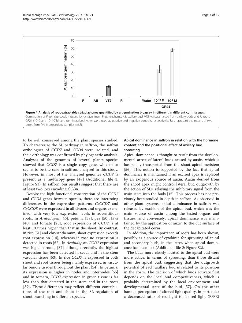
SLs in saffron cormsTo assess the presence of SLs in saffron, several partswere dissected and tested for the induction of germin-ation of Phelipanche ramosa seeds (Figure 4). Apicalbuds, axillary latent buds and sprouted axillary buds,external cover (the external surface of the corms withoutbuds), roots, basal plate, and vascular tissue from apical
buds, decapitated buds and axillary bud extracts wereapplied to P. ramosa seeds, and germination was scoredafter 7 d. The extracts of the main vascular tissue fromthe apical buds induced 11.5% germination, whereas ger-mination was not induced using extracts from the othertissues. The synthetic SL GR24 (10−9 to 10−10 M) wasused as a positive control and induced 58% and 34% ger-mination, respectively, whereas water (negative control)induced no germination (Figure 4).
Identification of the first saffron CCD7 and CCD8 genesSLs are a new class of plant hormones that have beenshown to be involved in the regulation of the outgrowthof preformed axillary buds. In order to study the rela-tionship between apical dominance and SLs in saffron,the CCD7 and CCD8 genes were isolated using a com-bination of degenerate primer PCR and gene walking.Saffron CCD7, hereafter designated as CsCCD7, containsfive introns (Figure 5A), and the 1912 bp of the codingsequence encodes a protein of 591 amino acids with apredicted pI of 6.6 and 66.17 kDa (GenBank accessionnumber KJ361477). The ChloroP 1.1 program predicteda 50-amino acid, N-terminal transit peptide, consistentwith plastid localization of CCD7 [44]. CsCCD7 showedthe highest homology with Solanum lycopersicum CCD7protein (67% identical). In the case of the CCD8, two.1pt?>different genes were isolated from saffron, desig-nated as CsCCD8a and CsCCD8b, which differ in thesequence of the first exon and intron. CsCCD8a was pre-dicted to have six exons (Figure 5B), 3151 nucleotidesand a coding sequence of 1533 nucleotides encoding aprotein of 511 amino acids with a predicted pI of 6.6 and57.17 kDa (GenBank accession number KJ361478).CsCCD8b was predicted to also have six exons, 3195nucleotides and a coding sequence of 1671 nucleotidesencoding a protein of 557 amino acids with a predictedpI of 6.1 and 62.03 kDa (GenBank accession numberKJ361479). Both CsCCD8 proteins showed 83% identitywith DAD1 (CCD8 from Petunia hybrida) and 76% toD10 (CCD8 from Oryza sativa), and were predicted tobe localized in plastids using the ChloroP 1.1 program,consistent with plastid localization of CCD8 [45].As part of the characterization of CsCCD7, CsCCD8a
and CsCCD8b, amino acid sequence alignments were
(See figure on previous page.)Figure 2 Dominance of the apical meristem in nondormant Crocus sativus corms. (A) removal of the apical bud allowed the sprouting ofthe axillary buds. a) corms with the apical meristems removed. b) intact corms. c) corms with sprouted secondary axillary bud 13 days after apicalmeristem removal. d) intact corms, as control, 13 days after the initiation of the decapitation experiment. (B) number of axillary buds sprouted orquiescent in each node after decapitation of the apical meristem. In each treatment, 25 corms were decapitated. Error bars represent SD of 25replicates. (C) removal of the apical bud with or without other corm parts and results in sprouting of axillary buds. In each treatment, 25 cormswere decapitated. Error bars represent SD of 25 replicates. (D) vascularization in the sprouted axillary buds of saffron corms (a-c). Absence ofvasculature in quiescent axillary buds (d-f). Buds were sectioned by hand and sections were stained for lignin with phloroglucinol-HCl. Ligninstaining is red. Pictures were taken under a dissection microscope.
Rubio-Moraga et al. BMC Plant Biology 2014, 14:171 Page 5 of 15http://www.biomedcentral.com/1471-2229/14/171

carried out, in order to build a phylogenetic tree usingthe CCD7 and CCD8 protein sequences from a varietyof plant species (Figure 5A and B). This analysis showedthat CsCCD7 was closer to the eudicot sequences thanto the grass sequences (Figure 5A) while CsCCD8a andCsCCD8b were in a cluster separate from the eudicots(Figure 5B and Additional file 3: Figure S3).
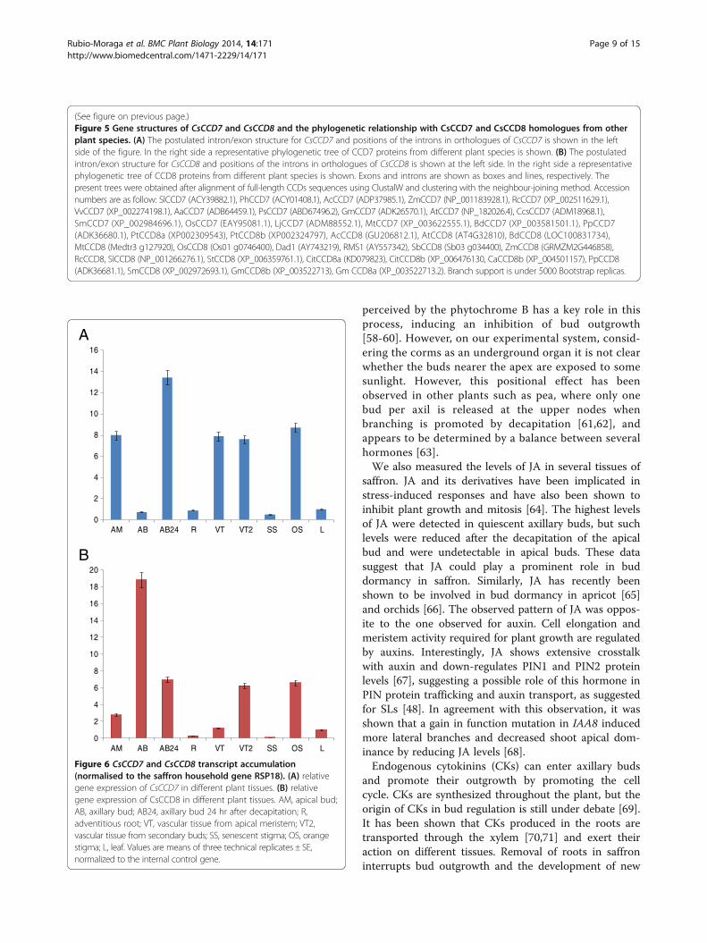
Gene expression of CsCCD7and CsCCD8To determine where CsCCD7 and CsCCD8 were expressed,the pattern of CsCCD7 and CsCCD8 transcript abundancewas determined in different tissues using quantitativereal-time RT-PCR (qPCR). CsCCD7 expression wasreadily detected in all the analyzed tissues, but showingdifferent expression levels. At the level of buds, the
highest expression was detected in the axillary buds24 h after removal of the apical bud, followed by thelevels of expression in the apical bud (Figure 6A). Thelevels in the vascular tissue from the apical buds or fromthe sprouted axillary buds were similar (Figure 6A).Interestingly, high expression levels were observed inthe orange stigma in contrast with the low levels de-tected in the senescent stigma. The expression levels werealso low in leaves and adventitious roots (Figure 6A). Thecombined levels of both CsCCD8 transcripts in mRNAextracted from different tissues were examined usingprimers that do not discriminate between the two differentcopies/alleles. The highest levels were detected in thequiescent axillary buds (Figure 6B). However, these levelswere drastically reduced in the axillary buds 24 h afterremoval of the apical bud. The expression levels in theapical bud were much reduced in comparison with theexpression levels in the quiescent axillary buds, suggestingthat the levels of CsCCD8 are reduced during the sprout-ing process (Figure 6B). The expression of CsCCD8 in thevascular tissue from the apical buds was lower thanthat in the newly developed vascular tissue of thesprouted axillary buds, but higher than that observed inadventitious roots (Figure 6B). Similarly to CsCCD7, arelatively high expression of CsCCD8 was detected inorange stigma, while this expression was reduced insenescent stigma (Figure 6B).
DiscussionAlmost all bulbous plant species are monocots, includ-ing economically important plants such as saffron, tulip,onion, garlic and lily. Their vegetative propagationconstitutes the most relevant process for agronomicalimprovements to markedly increase the potential numberof bulblets, while the control of dormancy is crucial tosolve many problems associated with the storage anddistribution of these crops. SLs play a key role in bothprocesses, development of new buds and sproutinginhibition by inhibition of bud outgrowth [20,21,46,47]and several genes involved in the biosynthesis andsignalling of SLs have been identified from a diverse rangeof species [48], although excluding bulbous plants. In thispaper, we describe and analyse the sprouting process insaffron induced by decapitation, as well as the involve-ment of SLs in this process through the isolation andcharacterization of two key genes in SL biosynthesis,CsCCD7 and CsCCD8.
Isolation of the saffron CCD7 and CCD8 genesThe genes so far identified that control branching arefrequently conserved between species. In particular,two carotenoid cleavage dioxygenase genes, CCD7(MAX3/RMS5/DAD3/D17-HTD1) and CCD8 (MAX4/RMS1/DAD1/D10), involved in SLs biosynthesis, appear
0
500
1000
1500
2000
Basal plate Epidermis Roots Apical bud Axillary bud Axillary bud 10 d. af ter
decapitation
Aux
in c
onte
nt (
ng/g
r dr
y w
eigh
t
A
B
C
JA c
onte
nt (
ng/g
r dr
y w
eigh
tS
A c
onte
nt (
ng/g
r dr
y w
eigh
t
0
10
20
30
40
50
60
Basal plate Epidermis Roots Apical bud Axillary bud Axillary bud 10 d. af ter
decapitation
0
50
100
150
200
250
300
Basal plate Epidermis Roots Apical bud Axillary bud Axillary bud 10 d. af ter
decapitation
Figure 3 Hormonal contents in different parts of saffron corms.Each column represents the mean ± of two to four replicates ofindependently harvested plant material. (A) Auxin content in thedifferent samples. (B) Jasmonic acid (JA) content in the differentsamples. (C) Salicylic acid (SA) content in the analyzed samples.
Rubio-Moraga et al. BMC Plant Biology 2014, 14:171 Page 6 of 15http://www.biomedcentral.com/1471-2229/14/171
to be well conserved among the plant species studied.To characterize the SL pathway in saffron, the saffronorthologues of CCD7 and CCD8 were isolated, andtheir orthology was confirmed by phylogenetic analysis.Analyses of the genomes of several plants speciesshowed that CCD7 is a single copy gene, which alsoseems to be the case in saffron, analysed in this study.However, in most of the analysed genomes CCD8 ispresent as a multicopy gene [49] (Additional file 3:Figure S3). In saffron, our results suggest that there areat least two loci encoding CCD8.Despite the high functional conservation of the CCD7
and CCD8 genes between species, there are interestingdifferences in the expression patterns. CsCCD7 andCsCCD8 were expressed in all tissues and organs exam-ined, with very low expression levels in adventitiousroots. In Arabidopsis [45], petunia [38], pea [50], kiwi[40] and tomato [25], root expression of CCD8 is atleast 10 times higher than that in the shoot. By contrast,in rice [51] and chrysanthemum, shoot expression exceedsroot expression [14], whereas in rose no expression isdetected in roots [52]. In Arabidopsis, CCD7 expressionwas high in roots, [37] although recently, the highestexpression has been detected in seeds and in the stemvascular tissue [53]. In rice CCD7 is expressed in bothshoot and root tissues being mainly expressed in vascu-lar bundle tissues throughout the plant [54]. In petunia,its expression is higher in nodes and internodes [55]and in tomato, CCD7 expression in green tissue is farless than that detected in the stem and in the roots[39]. These differences may reflect different contribu-tions of the root and shoot in the SL-regulation ofshoot branching in different species.
Apical dominance in saffron in relation with the hormonecontent and the positional effect of axillary budsproutingApical dominance is thought to result from the develop-mental arrest of lateral buds caused by auxin, which isbasipetally transported from the shoot apical meristem[56]. This notion is supported by the fact that apicaldominance is maintained if an excised apex is replacedby an exogenous source of auxin. Auxin derived fromthe shoot apex might control lateral bud outgrowth bythe action of SLs, relaying the inhibitory signal from themain stem into the buds [15]. This process has not pre-viously been studied in depth in saffron. As observed inother plant systems, apical dominance in saffron wasreleased by excision of the apical bud, which was themain source of auxin among the tested organs andtissues, and conversely, apical dominance was main-tained by the application of auxin to the cut surface ofthe decapitated corm.In addition, the importance of roots has been shown,
possibly as a source of cytokinin for sprouting of apicaland secondary buds, in the latter, when apical domin-ance has been lost (Additional file 2: Figure S2).The buds more closely located to the apical bud were
more active, in terms of sprouting, than those distantfrom the apical bud, suggesting that the outgrowthpotential of each axillary bud is related to its positionin the corm. The decision of which buds activate firstdepends on the local bud competitiveness, which isprobably determined by the local environment anddevelopmental state of the bud [57]. On the otherhand, a perception of altered light quality, in particulara decreased ratio of red light to far-red light (R/FR)
0
10
20
30
40
50
60
70
P AB VT2 R Water
P. r
amo
sa g
erm
inat
ion
(%
)
GR24
10-10 M 10-9 M
Figure 4 Analysis of root-extractable strigolactones quantified by a germination bioassay in different in different corm tissues.Germination of P. ramosa seeds induced by extracts from: P, parenchyma; AB, axillary bud; VT2, vascular tissue from axillary buds and R, roots.GR24 (10–9 and 10–10 M) and demineralized water were used as positive and negative controls, respectively. Bars represent the means of twopools from five independent samples (±SE).
Rubio-Moraga et al. BMC Plant Biology 2014, 14:171 Page 7 of 15http://www.biomedcentral.com/1471-2229/14/171

PtCCD7
MtCCD7
AaCCD7
MAX3
SbCCD7
RMS1
RcCCD7
VvCCD7
DAD3
SlCCD7
CsCCD7
CsCCD8a
CsCCD8b
RMS1
MtCCD8
CcsCCD8
CcCCD8
RcCCD8
SlCCD8
SbCCD8
MAX4
OsCCD8
ZmCCD8
A
B
Figure 5 (See legend on next page.)
Rubio-Moraga et al. BMC Plant Biology 2014, 14:171 Page 8 of 15http://www.biomedcentral.com/1471-2229/14/171
perceived by the phytochrome B has a key role in thisprocess, inducing an inhibition of bud outgrowth[58-60]. However, on our experimental system, consid-ering the corms as an underground organ it is not clearwhether the buds nearer the apex are exposed to somesunlight. However, this positional effect has beenobserved in other plants such as pea, where only onebud per axil is released at the upper nodes whenbranching is promoted by decapitation [61,62], andappears to be determined by a balance between severalhormones [63].We also measured the levels of JA in several tissues of
saffron. JA and its derivatives have been implicated instress-induced responses and have also been shown toinhibit plant growth and mitosis [64]. The highest levelsof JA were detected in quiescent axillary buds, but suchlevels were reduced after the decapitation of the apicalbud and were undetectable in apical buds. These datasuggest that JA could play a prominent role in buddormancy in saffron. Similarly, JA has recently beenshown to be involved in bud dormancy in apricot [65]and orchids [66]. The observed pattern of JA was oppos-ite to the one observed for auxin. Cell elongation andmeristem activity required for plant growth are regulatedby auxins. Interestingly, JA shows extensive crosstalkwith auxin and down-regulates PIN1 and PIN2 proteinlevels [67], suggesting a possible role of this hormone inPIN protein trafficking and auxin transport, as suggestedfor SLs [48]. In agreement with this observation, it wasshown that a gain in function mutation in IAA8 inducedmore lateral branches and decreased shoot apical dom-inance by reducing JA levels [68].Endogenous cytokinins (CKs) can enter axillary buds
and promote their outgrowth by promoting the cellcycle. CKs are synthesized throughout the plant, but theorigin of CKs in bud regulation is still under debate [69].It has been shown that CKs produced in the roots aretransported through the xylem [70,71] and exert theiraction on different tissues. Removal of roots in saffroninterrupts bud outgrowth and the development of new
(See figure on previous page.)Figure 5 Gene structures of CsCCD7 and CsCCD8 and the phylogenetic relationship with CsCCD7 and CsCCD8 homologues from otherplant species. (A) The postulated intron/exon structure for CsCCD7 and positions of the introns in orthologues of CsCCD7 is shown in the leftside of the figure. In the right side a representative phylogenetic tree of CCD7 proteins from different plant species is shown. (B) The postulatedintron/exon structure for CsCCD8 and positions of the introns in orthologues of CsCCD8 is shown at the left side. In the right side a representativephylogenetic tree of CCD8 proteins from different plant species is shown. Exons and introns are shown as boxes and lines, respectively. Thepresent trees were obtained after alignment of full-length CCDs sequences using ClustalW and clustering with the neighbour-joining method. Accessionnumbers are as follow: SlCCD7 (ACY39882.1), PhCCD7 (ACY01408.1), AcCCD7 (ADP37985.1), ZmCCD7 (NP_001183928.1), RcCCD7 (XP_002511629.1),VvCCD7 (XP_002274198.1), AaCCD7 (ADB64459.1), PsCCD7 (ABD67496.2), GmCCD7 (ADK26570.1), AtCCD7 (NP_182026.4), CcsCCD7 (ADM18968.1),SmCCD7 (XP_002984696.1), OsCCD7 (EAY95081.1), LjCCD7 (ADM88552.1), MtCCD7 (XP_003622555.1), BdCCD7 (XP_003581501.1), PpCCD7(ADK36680.1), PtCCD8a (XP002309543), PtCCD8b (XP002324797), AcCCD8 (GU206812.1), AtCCD8 (AT4G32810), BdCCD8 (LOC100831734),MtCCD8 (Medtr3 g127920), OsCCD8 (Os01 g0746400), Dad1 (AY743219), RMS1 (AY557342), SbCCD8 (Sb03 g034400), ZmCCD8 (GRMZM2G446858),RcCCD8, SlCCD8 (NP_001266276.1), StCCD8 (XP_006359761.1), CitCCD8a (KD079823), CitCCD8b (XP_006476130, CaCCD8b (XP_004501157), PpCCD8(ADK36681.1), SmCCD8 (XP_002972693.1), GmCCD8b (XP_003522713), Gm CCD8a (XP_003522713.2). Branch support is under 5000 Bootstrap replicas.
0
2
4
6
8
10
12
14
16
AM AB AB24 R VT VT2 SS OS L
0
2
4
6
8
10
12
14
16
18
20
AM AB AB24 R VT VT2 SS OS L
A
B
Figure 6 CsCCD7 and CsCCD8 transcript accumulation(normalised to the saffron household gene RSP18). (A) relativegene expression of CsCCD7 in different plant tissues. (B) relativegene expression of CsCCD8 in different plant tissues. AM, apical bud;AB, axillary bud; AB24, axillary bud 24 hr after decapitation; R,adventitious root; VT, vascular tissue from apical meristem; VT2,vascular tissue from secondary buds; SS, senescent stigma; OS, orangestigma; L, leaf. Values are means of three technical replicates ± SE,normalized to the internal control gene.
Rubio-Moraga et al. BMC Plant Biology 2014, 14:171 Page 9 of 15http://www.biomedcentral.com/1471-2229/14/171

roots restart the sprouting process. Due to the involve-ment of CKs in outgrowth bud promotion (Additionalfile 2: Figure S2), it is likely that CKs produced in theroots are responsible for this process in saffron as shownin other plant systems [72].SA levels have been shown to change between dor-
mant and waking saffron corms [73], suggesting its abil-ity to break dormancy. Therefore, we determined thelevels of SA in apical buds, axillary buds and in axillarybuds 10 d after removal of the apical bud. However, wedid not detect significant differences among the samples,suggesting that endogenous level of SA in buds is notinvolved in the control of paradormancy.
SLs in saffron corms and the roles of CsCCD8 andCsCCD7in sprouting and vascular tissue formationAnother major player in the control of shoot branchingare the SLs [74], originally identified as germinationstimulants for root parasitic plants [75]. SLs, togetherwith auxin, have been shown to have an inhibitory roleon shoot branching [20]. The auxin transport auto-inhibition hypothesis [76] proposed that organs remaindormant because they are not able to export their ownauxin into the stem polar auxin flow. Once axillary buddormancy is broken, bud outgrowth may depend on theestablishment of auxin transport from the bud via aprocess involving canalization, which is controlled bythe strength of the polar auxin transport stream and ormay require an auxin-regulated second messenger [16].The SLs were proposed to act as regulators of auxintransport by reducing the expression and/or plasmamembrane localization of auxin transporters [77,78].In the present work, the germination stimulatory ac-
tivity of P. ramosa seeds of different extracts was tested,and the stimulatory activity, albeit low, was only de-tected in the fractions from vascular tissue developedfrom the apical bud indicating the presence of SLs inthis tissue. The absence of activity in the extracts fromother tissues is most probably due to the presence ofSLs at extremely low levels. Grafting studies performedin several species showed that a wild-type rootstockgrafted to either a ccd7 or ccd8 mutant scion was able torestore wild-type branching patterns, indicating that SLwas produced in the roots [31,79]. However, wild-typeshoots on mutant roostocks also have near-wild-typebranching patterns [31,32,79]. In addition, wild-type epi-cotyl interstock grafts into rms1 and hypocotyl graftsinto Arabidopsis max3 are also able to reduce branching[80], indicating that biosynthesis of SLs is not limited tothe root system. Further, the expression of CsCCD7 andCsCCD8 in the vascular tissues connecting sproutingbuds with the mother corm, suggested that SLs are alsosynthesized in the stem vascular system in saffron. Inaddition, the SL profile found in tomato root exudates is
different from that found in xylem sap [81], suggestingthat different SLs could be produced in different tissuesin which they have different biological functionalities.The expression patterns of CsCCD7 and CsCCD8 were
analysed in apical and in axillary buds at two differentdevelopmental stages, quiescent and 24 h after elimin-ation of the main apical bud. For both genes expressionin apical buds was higher than in roots or leaves. How-ever, in axillary buds the expression patterns of bothgenes were clearly different. Although CsCCD8 showedthe highest levels of expression in the quiescent axillarybuds, the expression levels of CsCCD7 were very low inthis tissue. In Arabidopsis, expression of CCD8 has alsobeen reported to be relatively high in nodal tissue closeto the buds, while in rice, CCD7 was mainly found atthe node of the stem where the axillary meristem initi-ates [54]. In fact, the CsCCD8 transcript levels in theaxillary buds were rapidly down regulated by decapita-tion of the apical bud, although the expression ofCsCCD7 was up regulated, as has been observed inpotato [82]. Previous data on CCD7 and CCD8 expressionpatterns in other plant systems revealed that decapitationresults in decreased expression of these genes in thestem and in the axillary bud [14,15,32,51,83]. Eventhough this was the case for CsCCD8, CsCCD7 showedthe opposite behaviour. This result suggests that SLproduction related to bud dormancy is most probablycontrolled at CsCCD8 level.Moreover, bud auxin export is also a prerequisite for
the formation of vascular connections to the stem vascu-lature in inhibited buds [16,84]. In the quiescent axillarybuds of saffron the vasculature is not well developed,and the sprouting process is accompanied by the devel-opment of this system. The leaf primordia of thesequiescent axillary buds are not a source of auxins. How-ever, once the bud start to grow, buds synthesize auxin,as observed in other plants [85], and its export mayenhance vascular connections and nutrient flow to fur-ther stimulate the growing bud. Interestingly, it is in thevasculature of the axillary buds where the CsCCD8expression levels were enhanced, compared with thevasculature of apical buds, although CsCCD7 levelsremained practically unchanged, but high. Recently [86],it has been provide evidence that SLs positively regulatecambial activity. The expression pattern of the studiedgenes suggests that most probably SL or carlactone pro-duction in this vascular tissue is controlled at the levelof CsCCD8 but not CsCCD7. In agreement with this,several reports point out to the involvement of CCD7 inthe formation of other apocarotenoids [87,88]. BothCCD4 and CCD7 are currently candidates to deliver C27
intermediates for CCD1, which has been suggested toact preferentially over apocarotenoids [89]. Consistentwith this additional role for CCD7, strongly elevated
Rubio-Moraga et al. BMC Plant Biology 2014, 14:171 Page 10 of 15http://www.biomedcentral.com/1471-2229/14/171

levels of CCD7 can be found in green tomato fruits,from which SLs have not been detected [39] and inpanicles of rice [51]. Interestingly, expression of D27, aβ-carotene isomerase that converts all-trans-β-caroteneinto 9-cis-β-carotene, which is cleaved by CCD7, is alsohigh in panicle, but low in roots [90].
Involvement of CsCCD7 and CsCCD8 in stigma developmentUnexpectedly, CsCCD7 and CsCCD8 transcripts weredetected at relatively high levels in the stigma tissue.The abundance of both transcripts in immature orangestigmas exceeded that seen in vascular tissues, leavesand roots. CsCCD7 and CsCCD8 expression in the de-veloping stigma suggests potentially interesting novelfunction(s) for these enzymes and of SLs. The femaleorgans of C. sativus consist of a trilocular ovary, a verylong style, and 3 red stylar branches forming the stigmasfolded to give a trumpet-like structure [91]. This struc-ture is already present in the earlier developmentalstages of the stigma, which is approximately 2 mm inlength [4], and the cells continuously elongate until thestigma is fully developed reaching a final length of30 mm [92]. Auxins participated in the elongation of thefloral tube in Crocus [93] and they are probably involvedin the style elongation. Concomitant with cell elong-ation, the development of the vasculature of the stigmatakes place, and SLs could be actively participating inthis process, explaining the expression of CsCCD7 andCsCCD8 during the development of the stigma. Oncethe stigma is developed, the expression of both genesand probably the production of SLs drops in the senes-cent stigma. A putative function for SLs in flower devel-opment was already expected as the petunia ccd8/dad1mutant was reported to have smaller flowers [38] whilein SlCCD8 knock-down lines sepals, petals and antherswere smaller than in wild-type plants [25], suggestingthat SL deficiency affects flower development.
ConclusionsThe molecular and hormonal regulations on bud sprout-ing in bulbous plant species are largely unknown, butare fundamental for their propagation. We have deter-mined that the corm behaves as the stem of other higherplants and follows the same behaviour regarding apicaldominance. In this study, jasmonic acid, auxin and SLsare associated with the negative regulation of axillarybud outgrowth in saffron, while cytokinins positivelyregulates bud outgrowth after decapitation. Two keygenes in SLs biosynthesis, CCD7 and CCD8, were clonedfrom saffron. CsCCD8 may play an important role in thecontrol of apical dominance but also in the control ofvascular and stigma development. As the perception andsignalling mechanisms for SLs pathway are becomingunderstood in other plant species, more work needs to
be done to understand the mechanism of regulation ofthe sprouting process in saffron.
MethodsChemicals and plant materialsChemicals and reagents were obtained from Sigma-Aldrich unless otherwise stated. Diverse organs andplant tissues from C. sativus grown under field condi-tions in Tarazona de La Mancha, Spain, were usedthroughout the experiments. Corms, stigmas and budsat different developmental stages, and leaves werecollected for the experiments. All tissues were frozen inliquid nitrogen and stored at −80°C until required.
Sprout release assaySaffron corms of 15–20 g collected in September wereused for the sprouting experiments. Apical buds and otherplant tissues were excised with sterile surgical blades andthe outgrowth of the axillary buds in each corm was scoreddaily during a period of 30 days. 1-Naphthaleneacetic acid(NAA) was used at 50 μM concentration.
Histochemical staining of ligninHand-cut sections of corms were stained for lignin de-tection with phloroglucinol. Phloroglucinol-HCl reagentwas prepared by mixing 2 volumes of 2% (w/v) phloroglu-cinol in 95% ethanol with 1 volume of concentrated HCl.All photographs were taken within 30 min of staining.
Hormone levelsSaffron corms collected in September were dissected indifferent parts. Apical bud, secondary or axillary buds,roots, basal plate, nodes, nodes, external surface andparenchyma tissue were obtained (Figure 1), immediatelyfrozen in liquid nitrogen and lyophilized. Hormone ex-traction and analysis were carried out as follows: frozendry plant material was extracted in distilled water afterspiking with 100 ng of dihydrojasmonic acid, [2H4]-salicylicacid. After centrifugation at 4000 × g at 4°C, super-natants were recovered and pH adjusted to 3.0 with 30%acetic acid. The acidified water extract was partitionedtwice against 3 ml of di-ethyl ether. The organic layerwas recovered and evaporated under vacuum in a cen-trifuge concentrator (Speed Vac, Jouan, Saint HerblainCedex, France). The dry residue was then resuspendedin a 10% MeOH solution by gentle sonication. Theresulting solution was filtered through regeneratedcellulose 0.22 μm membrane syringe filters (Albet S.A.,Barcelona, Spain) and directly injected into a UPLCsystem (Acquity SDS, Waters Corp., Milford, MA,USA). Separations were carried out on a C18 column(Macherey-Nagel, 1.8 μm particle size, 50 × 2.1 mm,Scharlab, Barcelona, Spain) using a MeOH:H2O (bothsupplemented with 0.1% acetic acid) gradient at a flow
Rubio-Moraga et al. BMC Plant Biology 2014, 14:171 Page 11 of 15http://www.biomedcentral.com/1471-2229/14/171

rate of 300 μl min−1. Hormones were quantified witha Quattro LC triple quadrupole mass spectrometer(Micromass, Manchester, UK) connected online to theoutput of the column through an orthogonal Z-sprayelectrospray ion source.
Germination bioassay with P. ramosa seedsAs described above, SLs are germination stimulants ofroot parasitic plant seeds. Because of this germinatingactivity, bioassays based on seed germination of rootparasitic plants can be used as a reliable indirect way toquantify the levels of SLs produced by plant roots, espe-cially in plants where they have not been characterized,as in saffron. SLs from the different saffron corm tissueswere extracted as described [94]. Briefly, 0.3 g of eachcorm tissue were ground in a mortar with liquid nitro-gen and extracted twice with 0.3 mL of 50% acetone in a2 mL eppendorf tube. Tubes were vortexed for 2 minand centrifuged at 4ºC for 5 min at 8000 g in a table topcentrifuge. The organic phase was carefully transferredto 2 mL glass vials and stored at −20°C until use.Germination bioassays with P. ramosa seeds (kindlyprovided by Dr. Mauricio Vurro, Instituto di Scienzedelle Produzioni Alimentari, Bari, Italy) were precondi-tioned for 12 d at 21ºC. Then, aliquots of 50 μl ofextracts were added to two discs bearing approximately100 preconditioned seeds and incubated at 25°C. Thesynthetic germination stimulant GR24 and demine-ralised water were included as positive and negativecontrols in the bioassay. After 7 days, the germinatedand non-germinated seeds were counted using a bin-ocular microscope.
Cloning CsCCD7 and CsCCD8To facilitate genetic analysis of SL functioning in saffron,we focused on identifying steps in the saffron SL biosyn-thetic pathway. Partial coding sequences of saffronCCD7 and CCD8 were recovered from C. sativus gDNAusing degenerate primers (Table 1) corresponding toconserved protein sequence domains of the A. thaliana,Oryza sativa, and Zea mays CCD7 and CCD8 ortholo-gues. The CCD7 and CCD8 genomic loci were clonedusing the GenomeWalker Universal Kit (Clontech, http://www.clontech.com) as specified by the manufacturer andusing specific oligonucleotides (Table 1). The completeCCD7 and CCD8 coding sequences were PCR amplifiedfrom vascular tissue cDNA using specific oligonucleotides(Table 1) and High-Fidelity DNA Polymerase (NEB). DNAfragments were excised from agarose gels, isolated withthe Promega Gel Extraction Kit and ligated into thepGEM-T vector (Promega, http://www.promega.com).Plasmids containing the inserts were sequenced usingan automated DNA sequencer (ABI PRISM 3730xl,Perkin Elmer) from Macrogen Inc. (Seoul, Korea).
Computer-aided sequence similarity searches were madewith the BLAST suite of programs at the NationalCentre for Biotechnology Information (NCBI; http://www.ncbi.nlm.nih.gov) Motif searches were done using
Table 1 Primer sequences used for CsCCD7 and CsCCD8genes cloning and analysis
Sequences Primers (5′-3′)
Degenerate primers CCD8-F0 GTSGTGAGRATGGAASCHGG
CCD8-F1 GNCAYYTNTTYGYGGNTAYGC
CCD8-F2 CNCCNYTNTAYAARTTYARTGGCA
CCD8-F3 ATMCCAYTKGAYGGRAGC
CCD8-R0 CCATCATCYTCWTSGGTGC
CCD8-R1 CCAYTRAAYTTRTANARNGGNGT
CCD8-R2 TNGTNARNGTRTTNGGRAARCART
CCD8-R3 CCARCADCCRTGVARGCC
CCD7-F0 GACGAYCAYGGYTCCACSGTSCAC
CCD7-F1 CTCGACGGCCAYGGYTACCT
CCD7-R1 CYCKRTGCGTRAACCRCCACGA
Genome walker CCD8-F1 GGTGTGGTTATSTCTGTGATAAGTG
CCD8-F2 CCAACTTCGAGGAGTTGCACGA
CCD8-R1 ATGCTCATTCGGGTCGAGTGCT
CCD8-R2 CTTTGTCGCTCCCATCCAATGGT
CCD8-R3 TTGCATGGCCTCACAGCTCCA
CCD8-R4 CAACTCTCCTTTGTCGCTCCCATC
CCD8-R5 TGCCGTTGATGAAGTGGAAGGTCAG
CCD8-R6 TGTTGGCGTTGTCGCTGAGTGAC
CCD8-R7 AGAAGTTGTCGGTCTTCGGGACT
CCD8-R8 TACATGCTAGCCACAAGGGCTCCA
CCD8-R9 TGGTCTGGCGTTCGGTAGGTGCT
CCD7-F3 TCTCGGTGAACCCAAGCCAGCA
CCD7-F4 GCAGACATCACCCATATATCTGCT
CCD7-F5 TCTCGGTGAACCCAAGCCAGCA
CCD7-R1 CAAGACAAAAAGGAATGGCA
CCD7-R2 CAGTCGTGGATCATCAACTGGTCTG
CCD7-R3 TGTCTCCATCTGTTGTCTGGTTG
CCD7-R4 ATGATGGAGCTTATGTTGGGTCA
cDNA isolation CCD7-F ATGCACTCCATTTCTCACCGC
CCD7-R TAGTGATTTCTGCGAGACCA
CCD8a-F ATGGCAGACGTAGGGATACTGA
CCD8b-F CATGCAAACAAACTTAATAGCT
CCD8-R CTTGTCCTCTATTAAATTACTTC
QRT-PCR expressionanalysis
CCD7-F ACCTCCCCGTCATCCAAT
CCD7-R ATGACGGTTTCGGTCTCG
CCD8-F GAAGGGGTCGATCGAGGT
CCD8-R CGTCGCCGTACTCGAACT
Rubio-Moraga et al. BMC Plant Biology 2014, 14:171 Page 12 of 15http://www.biomedcentral.com/1471-2229/14/171

PROSITE (http://expasy.hcuge.ch/sprot/prosite.html),TMPRED (http://www.isrec.isb-sib.ch/sofware/sofware.html), SignalP (http://www.cbs.dtu.dk/services/SignalP)and PSORT II (http://psort.nibb.ac.jp).
Phylogenetic analysisTo construct the phylogenetic tree, the amino acid se-quences were aligned using the BLOSUM62 matrix withthe ClustalW (http://www.clustal.org) algorithm-basedAlignX module from MEGA Version 5.0 (http://www.megasoftware.net/mega.html). The alignments were savedand executed by MEGA Version 5.0 to generate aNeighbour Joining Tree with bootstrapping (5000 repli-cates) analysis and handling gaps with pairwise deletion.
Real-time quantitative RT-PCR (qPCR)Total RNA was isolated from apical buds, secondarybuds, vascular tissue from apical buds, vascular tissuefrom secondary buds, roots, stigmas and leaves by grind-ing the tissue in liquid nitrogen to a fine powder andextracting in 1 ml of Trizol reagent (Gibco-BRL) per100 mg of tissue fresh weight, according to the protocolof the manufacturer. The RNA was resuspended in100 μl of RNase-free water and treated with RQ1RNase-free DNase (Promega). The quantitative RT-PCRwas carried out on cDNA from 10 biological samples foreach analysed tissue; reactions were set up in GoTaq®qPCR Master Mix (Promega) according to manufacturer’sinstructions, with gene-specific primers (0.125 μM) in afinal volume of 25 μl. The primers were designed by usingPrimer3 program (http://frodo.wi.mit.edu/). Primer se-quences are listed in Table 1. The constitutive expressiongene 18SrRNA was used as a reference gene. The cyclingparameters of qPCR consisted in an initial denaturation at94°C for 5 min; 40 subsequent cycles of denaturation at94°C for 20 s, annealing at 58°C for 20 s and extension at72°C for 20 s; and finally extension at 72°C for 5 min.Assays were conducted with a StepOne™ Thermal Cycler(Applied Biosystems, California, USA) and analyzed usingStepOne software v2.0 (Applied Biosystems, California,USA). Following reactions, DNA melt curves were createdfor each primer combination to confirm the presence of asingle product.
Statistical analysisOne-way analysis of variance (ANOVA) was performedon all data sets by using GenStat for Windows. Whenneeded, data were also subjected to Student’s t-test.
Additional files
Additional file 1: Figure S1. Each sprouted axillary bud will form anew replacement corm. A) The developed new corms are formed from
apical buds. B) The developed new corms are formed from axillariesbuds, sprouted and developed after decapitation of the apical bud.
Additional file 2: Figure S2. Hormone treatments induced differenteffects on sprouting in saffron corms. Signal + refers to the removal ofthe apical bud. Signal – refers to intact corms with their apical bud. GA3,gibberellic acid; NAA, 1-naphthalene acetic acid; BAP, benzylaminopurine.Surface sterilize corms were grew in MS medium containing or not differenthormones at 100 μM final concentration. Picture was taken 10 days aftertreatment. The table shows the average length of the sprouted axyllary buds.
Additional file 3: Figure S3. Homologues of the CCD8 gene in differentplant species obtained by using CsCCD8a and b amino acid sequences inthe Phytozome v9.1 data base. Synteny of each gene is shown as well asthe exons distribution. Exon are shown in blue boxes and introns are shownas grey lines.
AbbreviationsABA: Abscisic acid; CCD: Carotenoid cleavage dioxygenase; GAs: Gibberellins;IAA: Indole acetic acid; PCR: Polymerase chain reaction; SLs: Strigolactones;qPCR: Real-time quantitative RT-PCR.
Competing interestsThe authors declare no competing interests.
Authors’ contributionsARM carried out the physiology studies and drafted the manuscript. OAcarried out the real-time PCR experiments and helped to draft the manuscript.RMCP and AGC carried out the determination of hormone levels. KY and JALRcarried out the determination of strigolactones and helped to draft themanuscript. RVM participated in the design of the study, performed thestatistical analysis and participated in the interpretation of data. LGG conceivedof the study, participated in its design and coordination and helped todraft the manuscript. All authors read and approved the final manuscript.
AcknowledgementsWe thank J. Argandoña (Instituto Botánico, Universidad de Castilla-La Mancha,Albacete, Spain) for excellent technical support, and K.A. Walsh for languagerevision. The laboratory is funded by the Spanish Ministerio de Ciencia eInnovación (BIO2009-07803) and participates in the IBERCAROT network(112RT0445). Dr. Ahrazem was funded by FPCYTA through the INCRECYTProgramme.
Author details1Departamento de Ciencia y Tecnología Agroforestal y Genética. Facultad deFarmacia, Instituto Botánico. Universidad de Castilla-La Mancha, CampusUniversitario s/n, 02071 Albacete, Spain. 2Fundación Parque Científico yTecnológico de Albacete. Campus Universitario s/n, 02071 Albacete, Spain.3Department of Agricultural and Environmental Sciences, Universitat Jaume I,12071 Castelló de la Plana, Spain. 4Weed Science Center, UtsunomiyaUniversity, 350 Mine-machi, Utsunomiya 321-8505, Japan. 5Department ofSoil Microbiology and Symbiotic Systems, Estación Experimental delZaidín-Consejo Superior de Investigaciones Científicas (EEZ-CSIC), Granada,Spain. 6Departamento de Biología Vegetal, Universidad Politécnica deValencia, 46071 Valencia, Spain.
Received: 7 May 2014 Accepted: 12 June 2014Published: 19 June 2014
References1. Rubio-Moraga A, Trapero A, Ahrazem O, Gomez-Gomez L: Crocins transport
in Crocus sativus: the long road from a senescent stigma to a newborncorm. Phytochemistry 2010, 71(13):1506–1513.
2. Hosseinzadeh H, Nassiri-Asl M: Avicenna’s (Ibn Sina) the canon of medicineand saffron (Crocus sativus): a review. Phytother Res 2013, 27(4):475–483.
3. Moraga AR, Rambla JL, Ahrazem O, Granell A, Gomez-Gomez L: Metaboliteand target transcript analyses during Crocus sativus stigma development.Phytochemistry 2009, 70(8):1009–1016.
4. Molina RV, Valero M, Navarro Y, Guardiola JL, García-Luis A: Temperatureeffects on flower formation in saffron (Crocus sativus L. Sci Hortic 2005,103:18.
Rubio-Moraga et al. BMC Plant Biology 2014, 14:171 Page 13 of 15http://www.biomedcentral.com/1471-2229/14/171
5. Renau-Morata B, Moyá L, Nebauer SG, Seguí-Simarro JM, Parra-Vega V,Gómez MD, Molina RV: The use of corms produced under storage at lowtemperatures as a source of explants for the in vitro propagation ofsaffron reduces contamination levels and increases multiplicationIndustrial. Crops Products 2013, 46:7.
6. Chrungoo NK: Concepts of dormancy regulation in vegetative plantpropagules: a review. Environ Exp Bot 1992, 32(4):9.
7. Ahrazem O, Rubio-Moraga A, Trapero A, Gomez-Gomez L: Developmentaland stress regulation of gene expression for a 9-cis-epoxycarotenoiddioxygenase, CstNCED, isolated from Crocus sativus stigmas. J Exp Bot2012, 63(2):681–694.
8. Farooq S, Koul KK: Changes in Gibberellin-like activity in corms of saffronplant (Crocus sativus L.) during dormancy and sprouting. Biochem PhysiolPflanz 1983, 178(8):5.
9. Aguilar-Martinez JA, Poza-Carrion C, Cubas P: Arabidopsis BRANCHED1 actsas an integrator of branching signals within axillary buds. Plant Cell 2007,19(2):458–472.
10. Shimizu-Sato S, Mori H: Control of outgrowth and dormancy in axillarybuds. Plant Physiol 2001, 127(4):1405–1413.
11. Brewer PB, Koltai H, Beveridge CA: Diverse roles of strigolactones in plantdevelopment. Mol Plant 2013, 6(1):18–28.
12. Bennett T, Sieberer T, Willett B, Booker J, Luschnig C, Leyser O: The ArabidopsisMAX pathway controls shoot branching by regulating auxin transport.Curr Biol 2006, 16(6):553–563.
13. Stirnberg P, van De Sande K, Leyser HM: MAX1 and MAX2 control shootlateral branching in Arabidopsis. Development 2002, 129(5):1131–1141.
14. Liang J, Zhao L, Challis R, Leyser O: Strigolactone regulation of shootbranching in chrysanthemum (Dendranthema grandiflorum). J Exp Bot2010, 61(11):3069–3078.
15. Brewer PB, Dun EA, Ferguson BJ, Rameau C, Beveridge CA: Strigolactoneacts downstream of auxin to regulate bud outgrowth in pea andArabidopsis. Plant Physiol 2009, 150(1):482–493.
16. Ferguson BJ, Beveridge CA: Roles for auxin, cytokinin, and strigolactone inregulating shoot branching. Plant Physiol 2009, 149(4):1929–1944.
17. Cook C, Coggon P, McPhail A, Wall M, Whichard L, Egley G, Luhan P:Germination stimulants. 2. Structure of strigol – potent seed-germinationstimulant for witchweed (Striga lutea Lour). J Am Chem Society 1972, 94:2.
18. Akiyama K, Matsuzaki K, Hayashi H: Plant sesquiterpenes induce hyphalbranching in arbuscular mycorrhizal fungi. Nature 2005, 435(7043):824–827.
19. Yoneyama K, Xie X, Takeuchi Y: Strigolactones: structures and biologicalactivities. Pest Manag Sci 2009, 65(5):467–470.
20. Gomez-Roldan V, Fermas S, Brewer PB, Puech-Pages V, Dun EA, Pillot JP,Letisse F, Matusova R, Danoun S, Portais JC, Bouwmeester H, Bécard G,Beveridge CA, Rameau C, Rochange SF: Strigolactone inhibition of shootbranching. Nature 2008, 455(7210):189–194.
21. Umehara M, Hanada A, Yoshida S, Akiyama K, Arite T, Takeda-Kamiya N,Magome H, Kamiya Y, Shirasu K, Yoneyama K, Kyozuka J, Yamaguchi S:Inhibition of shoot branching by new terpenoid plant hormones. Nature2008, 455(7210):195–200.
22. Ruyter-Spira C, Al-Babili S, van der Krol S, Bouwmeester H: The biology ofstrigolactones. Trends Plant Sci 2013, 18(2):72–83.
23. Kapulnik Y, Delaux PM, Resnick N, Mayzlish-Gati E, Wininger S, BhattacharyaC, Sejalon-Delmas N, Combier JP, Becard G, Belausov E, Beeckman T, Dor E,Hershenhorn J, Koltai H: Strigolactones affect lateral root formation androot-hair elongation in Arabidopsis. Planta 2011, 233(1):209–216.
24. Rasmussen A, Mason MG, De Cuyper C, Brewer PB, Herold S, Agusti J,Geelen D, Greb T, Goormachtig S, Beeckman T, Beveridge CA:Strigolactones suppress adventitious rooting in Arabidopsis and pea.Plant Physiol 2012, 158(4):1976–1987.
25. Kohlen W, Charnikhova T, Lammers M, Pollina T, Toth P, Haider I, Pozo MJ,de Maagd RA, Ruyter-Spira C, Bouwmeester HJ, López-Ráez JA: The tomatoCAROTENOID CLEAVAGE DIOXYGENASE8 (SlCCD8) regulates rhizospheresignaling, plant architecture and affects reproductive developmentthrough strigolactone biosynthesis. New Phytol 2012, 196(2):535–547.
26. Yoneyama K, Takeuchi Y, Sekimoto H: Phosphorus deficiency in redclover promotes exudation of orobanchol, the signal for mycorrhizalsymbionts and germination stimulant for root parasites. Planta 2007,225(4):1031–1038.
27. Lopez-Raez JA, Bouwmeester H: Fine-tuning regulation of strigolactonebiosynthesis under phosphate starvation. Plant Signal Behav 2008,3(11):963–965.
28. Ha CV, Leyva-Gonzalez MA, Osakabe Y, Tran UT, Nishiyama R, Watanabe Y,Tanaka M, Seki M, Yamaguchi S, Dong NV, Yamaguchi-Shinozaki K, Shinozaki K,Herrera-Estrella L, Tran LS: Positive regulatory role of strigolactone inplant responses to drought and salt stress. Proc Natl Acad Sci U S A2013, 111(2):851–856.
29. Aroca R, Ruiz-Lozano JM, Zamarreno AM, Paz JA, Garcia-Mina JM, Pozo MJ,Lopez-Raez JA: Arbuscular mycorrhizal symbiosis influences strigolactoneproduction under salinity and alleviates salt stress in lettuce plants.J Plant Physiol 2013, 170(1):47–55.
30. Torres-Vera R, Garcia JM, Pozo MJ, Lopez-Raez JA: Do strigolactonescontribute to plant defence? Mol Plant Pathol 2013, 15(2):211–216.
31. Beveridge CA, Ross JJ, Murfet IC: Branching Mutant rms-2 in Pisumsativum (Grafting Studies and Endogenous Indole-3-Acetic Acid Levels).Plant Physiol 1994, 104(3):953–959.
32. Sorefan K, Booker J, Haurogne K, Goussot M, Bainbridge K, Foo E, Chatfield S,Ward S, Beveridge C, Rameau C, Leyser O: MAX4 and RMS1 are orthologousdioxygenase-like genes that regulate shoot branching in Arabidopsis andpea. Genes Dev 2003, 17(12):1469–1474.
33. Alder A, Jamil M, Marzorati M, Bruno M, Vermathen M, Bigler P, Ghisla S,Bouwmeester H, Beyer P, Al-Babili S: The path from beta-carotene tocarlactone, a strigolactone-like plant hormone. Science 2012,335(6074):1348–1351.
34. Challis RJ, Hepworth J, Mouchel C, Waites R, Leyser O: A role for moreaxillary growth1 (MAX1) in evolutionary diversity in strigolactonesignaling upstream of MAX2. Plant Physiol 2013, 161(4):1885–1902.
35. Seto Y, Sado A, Asami K, Hanada A, Umehara M, Akiyama K, Yamaguchi S:Carlactone is an endogenous biosynthetic precursor for strigolactones.Proc Natl Acad Sci U S A 2014, 111(4):1640–1645.
36. Morris SE, Turnbull CG, Murfet IC, Beveridge CA: Mutational analysis ofbranching in pea. Evidence that Rms1 and Rms5 regulate the samenovel signal. Plant Physiol 2001, 126(3):1205–1213.
37. Booker J, Auldridge M, Wills S, McCarty D, Klee H, Leyser O: MAX3/CCD7 isa carotenoid cleavage dioxygenase required for the synthesis of a novelplant signaling molecule. Curr Biol 2004, 14(14):1232–1238.
38. Snowden KC, Simkin AJ, Janssen BJ, Templeton KR, Loucas HM, Simons JL,Karunairetnam S, Gleave AP, Clark DG, Klee HJ: The Decreased apicaldominance1/Petunia hybrida CAROTENOID CLEAVAGE DIOXYGENASE8gene affects branch production and plays a role in leaf senescence, rootgrowth, and flower development. Plant Cell 2005, 17(3):746–759.
39. Vogel JT, Walter MH, Giavalisco P, Lytovchenko A, Kohlen W, Charnikhova T,Simkin AJ, Goulet C, Strack D, Bouwmeester HJ, Fernie AR, Klee HJ:SlCCD7 controls strigolactone biosynthesis, shoot branching andmycorrhiza-induced apocarotenoid formation in tomato. Plant J 2010,61(2):300–311.
40. Ledger SE, Janssen BJ, Karunairetnam S, Wang T, Snowden KC: ModifiedCAROTENOID CLEAVAGE DIOXYGENASE8 expression correlates withaltered branching in kiwifruit (Actinidia chinensis). New Phytol 2010,188(3):803–813.
41. Proust H, Hoffmann B, Xie X, Yoneyama K, Schaefer DG, Nogue F, Rameau C:Strigolactones regulate protonema branching and act as a quorumsensing-like signal in the moss Physcomitrella patens. Development 2011,138(8):1531–1539.
42. Kepinski S, Leyser O: Plant development: an axis of auxin. Nature 2003,426(6963):132–135.
43. Leyser O: Regulation of shoot branching by auxin. Trends Plant Sci 2003,8(11):541–545.
44. Auldridge ME, McCarty DR, Klee HJ: Plant carotenoid cleavageoxygenases and their apocarotenoid products. Curr Opin Plant Biol2006, 9(3):315–321.
45. Auldridge ME, Block A, Vogel JT, Dabney-Smith C, Mila I, Bouzayen M,Magallanes-Lundback M, DellaPenna D, McCarty DR, Klee HJ:Characterization of three members of the Arabidopsis carotenoidcleavage dioxygenase family demonstrates the divergent roles of thismultifunctional enzyme family. Plant J 2006, 45(6):982–993.
46. Umehara M, Hanada A, Magome H, Takeda-Kamiya N, Yamaguchi S:Contribution of strigolactones to the inhibition of tiller bud outgrowthunder phosphate deficiency in rice. Plant Cell Physiol 2010,51(7):1118–1126.
47. Tsuchiya Y, Vidaurre D, Toh S, Hanada A, Nambara E, Kamiya Y, Yamaguchi S,McCourt P: A small-molecule screen identifies new functions for the planthormone strigolactone. Nat Chem Biol 2010, 6(10):741–749.
Rubio-Moraga et al. BMC Plant Biology 2014, 14:171 Page 14 of 15http://www.biomedcentral.com/1471-2229/14/171

48. Cheng X, Ruyter-Spira C, Bouwmeester H: The interaction betweenstrigolactones and other plant hormones in the regulation of plantdevelopment. Front Plant Sci 2013, 4:199.
49. Vallabhaneni R, Bradbury LM, Wurtzel ET: The carotenoid dioxygenasegene family in maize, sorghum, and rice. Arch Biochem Biophys 2010,504(1):104–111.
50. Foo E, Bullier E, Goussot M, Foucher F, Rameau C, Beveridge CA: Thebranching gene RAMOSUS1 mediates interactions among two novelsignals and auxin in pea. Plant Cell 2005, 17(2):464–474.
51. Arite T, Iwata H, Ohshima K, Maekawa M, Nakajima M, Kojima M, SakakibaraH, Kyozuka J: DWARF10, an RMS1/MAX4/DAD1 ortholog, controls lateralbud outgrowth in rice. Plant J 2007, 51(6):1019–1029.
52. Djennane S, Hibrand-Saint Oyant L, Kawamura K, Lalanne D, Laffaire M,Thouroude T, Chalain S, Sakr S, Boumaza R, Foucher F, Leduc N: Impacts oflight and temperature on shoot branching gradient and expression ofstrigolactone synthesis and signalling genes in rose. Plant Cell Environ2013, 37(3):742–757.
53. Liang YS, Jeon YA, Lim SH, Kim JK, Lee JY, Kim YM, Lee YH, Ha SH:Vascular-specific activity of the Arabidopsis carotenoid cleavagedioxygenase 7 gene promoter. Plant Cell Rep 2011, 30(6):973–980.
54. Zou J, Zhang S, Zhang W, Li G, Chen Z, Zhai W, Zhao X, Pan X, Xie Q, Zhu L:The rice HIGH-TILLERING DWARF1 encoding an ortholog of ArabidopsisMAX3 is required for negative regulation of the outgrowth of axillarybuds. Plant J 2006, 48(5):687–698.
55. Drummond RS, Martinez-Sanchez NM, Janssen BJ, Templeton KR, Simons JL,Quinn BD, Karunairetnam S, Snowden KC: Petunia hybrida CAROTENOIDCLEAVAGE DIOXYGENASE7 is involved in the production of negative andpositive branching signals in petunia. Plant Physiol 2009, 151(4):1867–1877.
56. Thimann KV, Skoog F: Studies on the Growth Hormone of Plants: III TheInhibiting Action of the Growth Substance on Bud Development.Proc Natl Acad Sci U S A 1933, 19(7):714–716.
57. Domagalska MA, Leyser O: Signal integration in the control of shootbranching. Nat Rev Mol Cell Biol 2011, 12(4):211–221.
58. Franklin KA: Light and temperature signal crosstalk in plant development.Curr Opin Plant Biol 2009, 12(1):63–68.
59. Kebrom TH, Burson BL, Finlayson SA: Phytochrome B represses TeosinteBranched1 expression and induces sorghum axillary bud outgrowth inresponse to light signals. Plant Physiol 2006, 140(3):1109–1117.
60. Finlayson SA, Krishnareddy SR, Kebrom TH, Casal JJ: Phytochrome regulation ofbranching in Arabidopsis. Plant Physiol 2010, 152(4):1914–1927.
61. Arumingtyas E, Floyd R, Gregory M, Murfet I: Branching in Pisum: inheritanceand allelism tests with 17 ramosus mutants. Pisum Genet 1992, 24:14.
62. Beveridge CA, Ross JJ, Murfet IC: Branching in Pea (Action of Genes Rms3and Rms4). Plant Physiol 1996, 110(3):859–865.
63. Sussex IM, Kerk NM: The evolution of plant architecture. Curr Opin PlantBiol 2001, 4(1):33–37.
64. Wasternack C, Hause B: Jasmonates: biosynthesis, perception, signaltransduction and action in plant stress response, growth anddevelopment. Ann Botany 2013, 111(6):37.
65. Zhong W, Gao Z, Zhuang W, Shi T, Zhang Z, Ni Z: Genome-wideexpression profiles of seasonal bud dormancy at four critical stages inJapanese apricot. Plant Mol Biol 2013, 83(3):247–264.
66. Qin Q, Kaas Q, Zhang C, Zhou L, Luo X, Zhou M, Sun X, Zhang L, Paek K-Y,Cui Y: The cold awakening of doritaenopsis ‘tinny Tender’ orchid flowers:the role of leaves in cold-induced Bud dormancy release. J Plant GrowthRegul 2012, 31(2):139–155.
67. Sun J, Chen Q, Qi L, Jiang H, Li S, Xu Y, Liu F, Zhou W, Pan J, Li X, Palme K, Li C:Jasmonate modulates endocytosis and plasma membrane accumulation ofthe Arabidopsis PIN2 protein. New Phytol 2011, 191(2):360–375.
68. Wang J, Yan DW, Yuan TT, Gao X, Lu YT: A gain-of-function mutation inIAA8 alters Arabidopsis floral organ development by change of jasmonicacid level. Plant Mol Biol 2013, 82(1–2):71–83.
69. Muller D, Leyser O: Auxin, cytokinin and the control of shoot branching.Ann Bot 2011, 107(7):1203–1212.
70. Hartung W, Sauter A, Hose E: Abscisic acid in the xylem: where does itcome from, where does it go to? J Exp Bot 2002, 53(366):27–32.
71. Sakakibara H, Takei K, Hirose N: Interactions between nitrogen andcytokinin in the regulation of metabolism and development. Trends PlantSci 2006, 11(9):440–448.
72. Shimizu-Sato S, Tanaka M, Mori H: Auxin-cytokinin interactions in thecontrol of shoot branching. Plant Mol Biol 2009, 69(4):429–435.
73. Esmaeili N, Ebrahimzadeh H, Abdi K, Safarian S: Determination of somephenolic compounds in Crocus sativus L. corms and its antioxidantactivities study. Pharmacogn Mag 2011, 7(25):74–80.
74. Hayward A, Stirnberg P, Beveridge C, Leyser O: Interactions betweenauxin and strigolactone in shoot branching control. Plant Physiol 2009,151(1):400–412.
75. Bouwmeester HJ, Matusova R, Zhongkui S, Beale MH: Secondarymetabolite signalling in host-parasitic plant interactions. Curr Opin PlantBiol 2003, 6(4):358–364.
76. Li CJ, Bangerth F: Autoinhibition of indoleacetic acid transport in theshoots of two-branched pea (Pisum sativum) plants and its relationshipto correlative dominance. Physiol Plant 1999, 106:5.
77. Dun EA, Brewer PB, Beveridge CA: Strigolactones: discovery of the elusiveshoot branching hormone. Trends Plant Sci 2009, 14(7):364–372.
78. Shinohara N, Taylor C, Leyser O: Strigolactone can promote or inhibitshoot branching by triggering rapid depletion of the auxin effluxprotein PIN1 from the plasma membrane. PLoS Biol 2013, 11(1):e1001474.
79. Napoli CA, Beveridge CA, Snowden KC: Reevaluating concepts of apicaldominance and the control of axillary bud outgrowth. Curr Top Dev Biol1999, 44:127–169.
80. Foo E, Turnbull CG, Beveridge CA: Long-distance signaling and the control ofbranching in the rms1 mutant of pea. Plant Physiol 2001, 126(1):203–209.
81. Kohlen W, Charnikhova T, Liu Q, Bours R, Domagalska MA, Beguerie S,Verstappen F, Leyser O, Bouwmeester H, Ruyter-Spira C: Strigolactones aretransported through the xylem and play a key role in shoot architecturalresponse to phosphate deficiency in nonarbuscular mycorrhizal hostArabidopsis. Plant Physiol 2011, 155(2):974–987.
82. Pasare SA, Ducreux LJ, Morris WL, Campbell R, Sharma SK, Roumeliotis E,Kohlen W, van der Krol S, Bramley PM, Roberts AG, Fraser PD, Taylor MA:The role of the potato (Solanum tuberosum) CCD8 gene in stolon andtuber development. New Phytol 2013, 198(4):1108–1120.
83. Johnson X, Brcich T, Dun EA, Goussot M, Haurogne K, Beveridge CA,Rameau C: Branching genes are conserved across species Genescontrolling a novel signal in pea are coregulated by other long-distancesignals. Plant Physiol 2006, 142(3):1014–1026.
84. Thimann KV: Fifty years of plant hormone research. Plant Physiol 1974,54(4):450–453.
85. Gocal GF, Pharis RP, Yeung EC, Pearce D: Changes after Decapitation inConcentrations of Indole-3-Acetic Acid and Abscisic Acid in the Larger AxillaryBud of Phaseolus vulgaris L. cv Tender Green. Plant Physiol 1991, 95(2):344–350.
86. Agusti J, Herold S, Schwarz M, Sanchez P, Ljung K, Dun EA, Brewer PB,Beveridge CA, Sieberer T, Sehr EM, Greb T: Strigolactone signaling isrequired for auxin-dependent stimulation of secondary growth in plants.Proc Natl Acad Sci U S A 2011, 108(50):20242–20247.
87. Walter MH, Floss DS, Strack D: Apocarotenoids: hormones, mycorrhizalmetabolites and aroma volatiles. Planta 2010, 232(1):1–17.
88. Floss DS, Walter MH: Role of carotenoid cleavage dioxygenase 1 (CCD1) inapocarotenoid biogenesis revisited. Plant Signal Behav 2009, 4(3):172–175.
89. Ilg A, Yu Q, Schaub P, Beyer P, Al-Babili S: Overexpression of the ricecarotenoid cleavage dioxygenase 1 gene in Golden Rice endosperm suggestsapocarotenoids as substrates in planta. Planta 2010, 232(3):691–699.
90. Lin H, Wang R, Qian Q, Yan M, Meng X, Fu Z, Yan C, Jiang B, Su Z, Li J,Wang Y: DWARF27, an iron-containing protein required for thebiosynthesis of strigolactones, regulates rice tiller bud outgrowth.Plant Cell 2009, 21(5):1512–1525.
91. Grilli-Caiola M: Saffron reproductive biology. Acta Horticult 2004, 650:12.92. Rubio A, Rambla JL, Santaella M, Gomez MD, Orzaez D, Granell A,
Gomez-Gomez L: Cytosolic and plastoglobule-targeted carotenoiddioxygenases from Crocus sativus are both involved in beta-iononerelease. J Biol Chem 2008, 283(36):24816–24825.
93. Stark D: Anatomical and physiological studies of floral tube elongation ofCrocus vernus iridaceae. Am J Bot 1982, 69(9):1476–1482.
94. Lopez-Raez JA, Charnikhova T, Gomez-Roldan V, Matusova R, Kohlen W,De Vos R, Verstappen F, Puech-Pages V, Becard G, Mulder P, Bouwmeester H:Tomato strigolactones are derived from carotenoids and their biosynthesis ispromoted by phosphate starvation. New Phytol 2008, 178(4):863–874.
doi:10.1186/1471-2229-14-171Cite this article as: Rubio-Moraga et al.: Apical dominance in saffron andthe involvement of the branching enzymes CCD7 and CCD8 in thecontrol of bud sprouting. BMC Plant Biology 2014 14:171.
Rubio-Moraga et al. BMC Plant Biology 2014, 14:171 Page 15 of 15http://www.biomedcentral.com/1471-2229/14/171