Research Article Etiologic Agents of Bacterial Sepsis and ... · Research Article Etiologic Agents...
9
Research Article Etiologic Agents of Bacterial Sepsis and Their Antibiotic Susceptibility Patterns among Patients Living with Human Immunodeficiency Virus at Gondar University Teaching Hospital, Northwest Ethiopia Gelila Alebachew, 1 Brhanu Teka, 2 Mengistu Endris, 1 Yitayal Shiferaw, 2,3 and Belay Tessema 1 1 Department of Medical Microbiology, School of Biomedical and Laboratory Sciences, College of Medicine and Health Sciences, University of Gondar, Gondar, Ethiopia 2 Department of Microbiology, Immunology and Parasitology, Addis Ababa University, Addis Ababa, Ethiopia 3 Department of Medical Biotechnology, School of Medicine, Flinders University, Bedford Park, SA 5042, Australia Correspondence should be addressed to Brhanu Teka; [email protected] Received 30 December 2015; Revised 20 April 2016; Accepted 3 May 2016 Academic Editor: David A. Hildeman Copyright © 2016 Gelila Alebachew et al. is is an open access article distributed under the Creative Commons Attribution License, which permits unrestricted use, distribution, and reproduction in any medium, provided the original work is properly cited. Background. Bacterial sepsis is a major cause of illness in human immunodeficiency virus infected patients. ere is scarce evidence about sepsis among HIV patients in Ethiopia. is study aimed to determine the etiologic agents of bacterial sepsis and their antibiotic susceptibility patterns among HIV infected patients. Methods. A cross-sectional study was carried out from March 1 to May 2, 2013. One hundred patients infected with HIV and suspected of having sepsis were included. Sociodemographic data were collected by interview and blood sample was aseptically collected from study participants. All blood cultures were incubated aerobically at 35 ∘ C and inspected daily for 7 days. e positive blood cultures were identified following the standard procedures and antimicrobial susceptibility testing was performed using disk diffusion technique. Data was entered by Epi-info version 3.5.1 and analysis was done using SPSS version 20. Results. Of the study participants, 31 (31%) confirmed bacterial sepsis. e major isolates were 13 (13%) Staphylococcus aureus, 8 (8%) coagulates negative staphylococci, and 3 (3%) viridans streptococci. Majority of the isolates, 25 (80.6%), were multidrug resistant to two or more antimicrobial agents. Conclusions. Bacterial sepsis was a major cause of admission for HIV infected patients predominated by Staphylococcus aureus and coagulase negative staphylococci species and most of the isolates were multidrug resistant. 1. Introduction Bacterial sepsis constitutes a significant public health prob- lem and represents an important cause of morbidity and mortality in HIV/AIDS patients in the world. Epidemiologic studies have shown that 1% to 10% of the sepsis patients are individuals with HIV/AIDS in ICU [1]. Sepsis is systemic illness caused by microbial invasion of normally sterile parts of the body and commonly defined as the presence of infection with the systemic inflammatory response syndrome (SIRS); it is the presence of two or more of the following conditions: abnormal body temperature (<36 ∘ C (96.8 ∘ F) or >38 ∘ C (100.4 ∘ F)); heart rate (>90 beats/min); respiratory rate (>20 breaths/min); and white blood cell (WBC) count (<4000/mm 3 or >12,000/mm 3 ) [2]. e source of infection can be lung, genitourinary tract, gastrointestinal tract, skin or soſt tissue, and so on [3]. Pathogenesis of bacteria due to the structural components of the Gram negative and Gram positive bacteria is responsible for initiating sepsis [4]. Sepsis can be caused by different types of bacteria. Gram negative bacteria are the most commonly isolated in septic Hindawi Publishing Corporation BioMed Research International Volume 2016, Article ID 5371875, 8 pages http://dx.doi.org/10.1155/2016/5371875
Transcript of Research Article Etiologic Agents of Bacterial Sepsis and ... · Research Article Etiologic Agents...
Research ArticleEtiologic Agents of Bacterial Sepsis and Their AntibioticSusceptibility Patterns among Patients Living with HumanImmunodeficiency Virus at Gondar University TeachingHospital, Northwest Ethiopia
Gelila Alebachew,1 Brhanu Teka,2 Mengistu Endris,1
Yitayal Shiferaw,2,3 and Belay Tessema1
1Department of Medical Microbiology, School of Biomedical and Laboratory Sciences,College of Medicine and Health Sciences, University of Gondar, Gondar, Ethiopia2Department of Microbiology, Immunology and Parasitology, Addis Ababa University, Addis Ababa, Ethiopia3Department of Medical Biotechnology, School of Medicine, Flinders University, Bedford Park, SA 5042, Australia
Correspondence should be addressed to Brhanu Teka; [email protected]
Received 30 December 2015; Revised 20 April 2016; Accepted 3 May 2016
Academic Editor: David A. Hildeman
Copyright © 2016 Gelila Alebachew et al. This is an open access article distributed under the Creative Commons AttributionLicense, which permits unrestricted use, distribution, and reproduction in any medium, provided the original work is properlycited.
Background. Bacterial sepsis is amajor cause of illness in human immunodeficiency virus infected patients.There is scarce evidenceabout sepsis among HIV patients in Ethiopia. This study aimed to determine the etiologic agents of bacterial sepsis and theirantibiotic susceptibility patterns among HIV infected patients. Methods. A cross-sectional study was carried out from March 1to May 2, 2013. One hundred patients infected with HIV and suspected of having sepsis were included. Sociodemographic datawere collected by interview and blood sample was aseptically collected from study participants. All blood cultures were incubatedaerobically at 35∘C and inspected daily for 7 days.The positive blood cultures were identified following the standard procedures andantimicrobial susceptibility testing was performed using disk diffusion technique. Data was entered by Epi-info version 3.5.1 andanalysis was done using SPSS version 20. Results. Of the study participants, 31 (31%) confirmed bacterial sepsis. The major isolateswere 13 (13%) Staphylococcus aureus, 8 (8%) coagulates negative staphylococci, and 3 (3%) viridans streptococci. Majority of theisolates, 25 (80.6%), were multidrug resistant to two or more antimicrobial agents. Conclusions. Bacterial sepsis was a major causeof admission for HIV infected patients predominated by Staphylococcus aureus and coagulase negative staphylococci species andmost of the isolates were multidrug resistant.
1. Introduction
Bacterial sepsis constitutes a significant public health prob-lem and represents an important cause of morbidity andmortality in HIV/AIDS patients in the world. Epidemiologicstudies have shown that 1% to 10% of the sepsis patients areindividuals with HIV/AIDS in ICU [1]. Sepsis is systemicillness caused by microbial invasion of normally sterile partsof the body and commonly defined as the presence ofinfectionwith the systemic inflammatory response syndrome(SIRS); it is the presence of two or more of the following
conditions: abnormal body temperature (<36∘C (96.8∘F) or>38∘C (100.4∘F)); heart rate (>90 beats/min); respiratoryrate (>20 breaths/min); and white blood cell (WBC) count(<4000/mm3 or >12,000/mm3) [2].
The source of infection can be lung, genitourinary tract,gastrointestinal tract, skin or soft tissue, and so on [3].Pathogenesis of bacteria due to the structural components ofthe Gram negative and Gram positive bacteria is responsiblefor initiating sepsis [4].
Sepsis can be caused by different types of bacteria. Gramnegative bacteria are the most commonly isolated in septic
Hindawi Publishing CorporationBioMed Research InternationalVolume 2016, Article ID 5371875, 8 pageshttp://dx.doi.org/10.1155/2016/5371875
2 BioMed Research International
patients and have lipopolysaccharide or endotoxin structure[5]. The percentage of patients with severe infections causedby Gram positive bacteria has increased in recent years,accounting for almost half of the incidents of septicemiaand severe systemic infections [6, 7]. Gram positive bacteriahave no endotoxin, but they cause sepsis indistinguishablefrom Gram negative bacteria by two distinct mechanisms, byreleasing exotoxins that act as super antigens (e.g., staphy-lococci or streptococci) and cell wall components of Grampositive bacteria (both peptidoglycan and lipoteichoic acid)[7].
The clinical syndrome is caused bymicroorganism and itsvirulence factorswith the host and its inflammatory response.Bacteria with special relevance for sepsis include Grampositive: Streptococcus pneumonia, Streptococcus progenies,Streptococcus agalactiae, and Staphylococcus auras; and Gramnegative: Neisseria meningitides, enteric (Escherichia coli,Klebsiella, Proteus, Enterobacter, Serratia, Citrobacter, andSalmonella), and nonenteric (Pseudomonas aeruginosa andAcinetobacter) [5].
People living with HIV are at greater risk for sepsisbecause of immune suppression. In areas of high HIVprevalence, sepsis might contribute substantially to overallmortality [8, 9].
Emerging of multidrug resistant bacteria can reducethe management of bacterial infections. Bacterial infectionto the HIV infected cases can mostly be caused by themultidrug resistant organism. Multidrug resistant organismis a bacterium that is resistant to more than one antibiotic. Ifbacteria are resistant to an antibiotic, certain drug treatmentswill not work [10].
Like in many other developing countries, bacterial sepsisand antibiotic resistant organism in Ethiopia are the mostcommon disease for mortality and morbidity. But there isno evidence for bacterial sepsis among HIV/AIDS patientsand their interaction. Hence, this study aimed to analyze theetiologic agents of bacterial sepsis and susceptibility patternof bacterial isolates among HIV infected patients.
2. Materials and Methods
2.1. Study Area. The study was conducted at University ofGondar Teaching Hospital which is located in Gondar town,Northwest Ethiopia, 737 km away from Addis Ababa. TheUniversity Hospital was purposefully selected with the factthat it is one of the biggest tertiary level referral and teachinghospitals in the region visited by about six million peoplefrom the surrounding zones and nearby regions both forinpatient and for outpatient treatment. The hospital consistsof ART pharmacy, ART laboratory, ART clinic, ICU with 16beds, 13 wards with 327 beds, and outpatient departments.
2.2. Study Design and Population. Hospital based cross-sectional study was conducted among HIV/AIDS patientsattending at ART clinic fromMarch 1 to May 2, 2013.
2.3. Study Participants. All HIV/AIDS patients who weresuspected of having sepsis and attended the University of
Gondar Teaching Hospital during the study period wereenrolled in the study.
2.4. Inclusion Criteria. All confirmed HIV patients that weresuspected of having sepsis by clinicians were included in thestudy.
2.5. Exclusion Criteria. Patients receiving antibiotic treat-ment and neonates were excluded from the study.
2.6. Operational Definitions
(i) Sepsis suspect is a patient who fulfils definition ofSIRS.
(ii) SIRS is the presence of 2 or more of the following:
(a) Temperature <36∘C (96.8∘F) (hypothermia) or>38∘C (100.4∘F).
(b) Heart rate >90 beats/min (tachycardia).(c) Respiratory rate >20 breaths/min (tachypnea).(d) WBC count <4000/mm3 (leucopenia) or>12,000/mm3 (leukocytosis).
2.7. Sample Size and Sampling Technique. During the two-month study period, 100 study subjects were enrolled usingconvenient sampling technique from the study populationvisiting University of Gondar Teaching Hospital.
2.8. Data Collection and Laboratory Methods. A pretested,structured questionnaire was used to collect sociodemo-graphic information, clinical signs, or symptoms by medicaldoctor. The pretest of the questionnaire was conducted in10 patients at University of Gondar Teaching Hospital andpretest results were not included in the final results of theresearch but we used it to validate the questionnaire beforeconducting data collection.
2.8.1. Sample Collection and Handling. Blood samples werecollected from patients before antibiotic therapy by usingvenipuncture technique. Careful skin cleaning using tinctureof iodine and 70% alcohol was done before drawing blood toprevent contamination. Ten mL of blood sample from adultsand 2mL of blood sample from children were asepticallycollected following the request of the clinician. Then, it wasinoculated into a blood culture bottle each containing 45mLof tryptone soy broth (TSB) (Oxoid) [11].
2.8.2. Culture and Identification. Blood cultures were incu-bated aerobically at 35∘C and inspected daily for 7 days forthe presence of visible microbial growth by observing anyone of the following: turbidity, haemolysis, gas production,and coagulation of broth. For blood cultures that showedsigns of microbial growth, subcultures were done onto blood,chocolate, andMacConkey agar (Oxoid).Theblood andMac-Conkey agar plates were incubated in aerobic and chocolateagar inmicroaerophilic atmosphere using a candle jar at 35∘Cfor 24–48 hours [11].
BioMed Research International 3
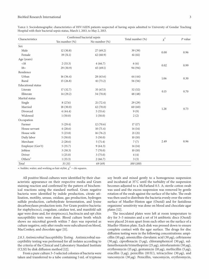
Table 1: Sociodemographic characteristics of HIV/AIDS patients suspected of having sepsis admitted to University of Gondar TeachingHospital with their bacterial sepsis status, March 1, 2013, to May 2, 2013.
Characteristics Confirmed bacterial sepsis Total number (%) 𝜒2
𝑃 valueYes number (%) No number (%)
SexMale 12 (30.8) 27 (69.2) 39 (39) 0.00 0.96Female 19 (31.1) 42 (68.9) 61 (61)
Age (years)<18 2 (33.3) 4 (66.7) 6 (6) 0.02 0.9918+ 29 (30.9) 65 (69.1) 94 (94)
ResidenceUrban 16 (36.4) 28 (63.6) 44 (44) 1.06 0.30Rural 15 (26.8) 41 (73.2) 56 (56)
Educational statusLiterate 17 (32.7) 35 (67.3) 52 (52) 0.15 0.70Illiterate 14 (29.2) 34 (70.8) 48 (48)
Marital statusSingle 8 (27.6) 21 (72.4) 29 (29)
1.28 0.73Married 18 (30.0) 42 (70.0) 60 (60)Divorced 4 (44.4) 5 (55.6) 9 (9)Widowed 1 (50.0) 1 (50.0) 2 (2)
OccupationFarmer 5 (29.4) 12 (70.6) 17 (17)
2.49 0.96
House servant 4 (28.6) 10 (71.4) 14 (14)House wife 5 (23.8) 16 (76.2) 21 (21)Daily labor 5 (50.0) 5 (50.0) 10 (10)Merchant 2 (28.6) 5 (71.4) 7 (7)Employer (Gov’t) 5 (35.7) 9 (64.3) 14 (14)Jobless 3 (30.3) 7 (70.0) 10 (10)Driver 1 (25.0) 3 (75.0) 4 (4)Others∗ 1 (33.3) 2 (66.7) 3 (3)
Total 31 (31) 69 (69) 100 (100)∗: builder, waiter, and working as hair stylist, 𝜒2 = chi-squares.
All positive blood cultures were identified by their char-acteristic appearance on their respective media and Gramstaining reaction and confirmed by the pattern of biochem-ical reactions using the standard method. Gram negativebacteria were identified by indole production, citrate uti-lization, motility, urease, oxidase, gas production, hydrogensulfide production, carbohydrate fermentation, and lysinedecarboxylases production tests. For Gram positive bacteria:for staphylococci, coagulase, catalase test, and manifold saltagar were done and, for streptococci, bacitracin and opt chinsusceptibility tests were done. Blood culture broth whichshows no microbial growth within 7 days was reported asculture negative, only after results were subcultured on blood,MacConkey, and chocolate agar [11].
2.8.3. Antimicrobial Susceptibility Testing. Antimicrobial sus-ceptibility testing was performed for all isolates according tothe criteria of the Clinical and Laboratory Standard Institute(CLSI) by disk diffusion method [12].
From a pure culture 3–5 selected colonies of bacteria weretaken and transferred to a tube containing 5mL of tryptone
soy broth and mixed gently to a homogenous suspensionand incubated at 35∘C until the turbidity of the suspensionbecomes adjusted to a McFarland 0.5. A; sterile cotton swabwas used and the excess suspension was removed by gentlerotation of the swab against the surface of the tube. The swabwas then used to distribute the bacteria evenly over the entiresurface of Mueller-Hinton agar (Oxoid) and for fastidiousorganisms’ sensitivity was done on blood and chocolate agarplates [12].
The inoculated plates were left at room temperature todry for 3–5 minutes and a set of 14 antibiotic discs (Oxoid)were placed 24mm apart from each other on the surface of aMueller-Hinton plate. Each disk was pressed down to ensurecomplete contact with the agar surface. The drugs for discdiffusion testing were in the following concentrations: ampi-cillin (10 𝜇g), amoxicillin-clavulanic acid (30 𝜇g), ceftriaxone(30 𝜇g), ciprofloxacin (5 𝜇g), chloramphenicol (30 𝜇g), sul-famethoxazole/trimethoprim (25𝜇g), nitrofurantoin (50𝜇g),erythromycin (15 𝜇g), gentamicin (10 𝜇g), methicillin (5𝜇g),oxacillin (1 𝜇g), penicillin (10 IU), tetracycline (30 𝜇g), andvancomycin (30𝜇g). Penicillin, vancomycin, erythromycin,
4 BioMed Research International
oxacillin, and methicillin were tested only for Gram positivebacteria. The plates were then incubated at 35∘C for 24–48hours. Diameters of the zone of inhibition around the discwere measured to the nearest millimeter using the ruler, andthe isolates were classified as sensitive and resistant accordingto the standardized table supplied by the CLSI [12].
2.8.4. Quality Control. Standard reference strains of Escher-ichia coli (American Type Culture Collection- (ATCC-)25922), Staphylococcus aureus (ATCC-25923), and Pseu-domonas aeruginosa (ATCC-27853) were used as a qualitycontrol throughout the study for culture and antimicrobialsusceptibility testing.
2.8.5. Data Analysis and Interpretation. Data entry was doneby Epi-info version 3.5.1 and analysis was done using statis-tical package for social science (SPSS) software version 20.Descriptive statistical methods were employed to describemagnitude and percentage of sepsis. Chi-square and oddsratio tests with 95% confidence interval were used to deter-mine presence and strength of association. In all cases𝑃 valueless than 0.05 was considered as statistically significant.
2.8.6. Ethical Consideration. This study was approved by theResearch and Ethical Committee of the School of Biomedicaland Laboratory Sciences, College of Medicine and HealthSciences, University of Gondar. Participation was voluntaryand informed written consent was obtained from each studyparticipant/guardian.The laboratory result was sent to physi-cians for appropriate treatment of the study participants. Allinformation about the patients was kept confidential.
3. Results
One hundred HIV positive patients suspected of havingsepsis were included in this study. Out of this, 39 (39%) weremales and 61 (61%) were females. The age range of partici-pants was 5–53 years with mean and SD 33.2 ± 9.8. Therewas no significant association between the sociodemographiccharacteristics of participants and bacterial sepsis (Table 1).
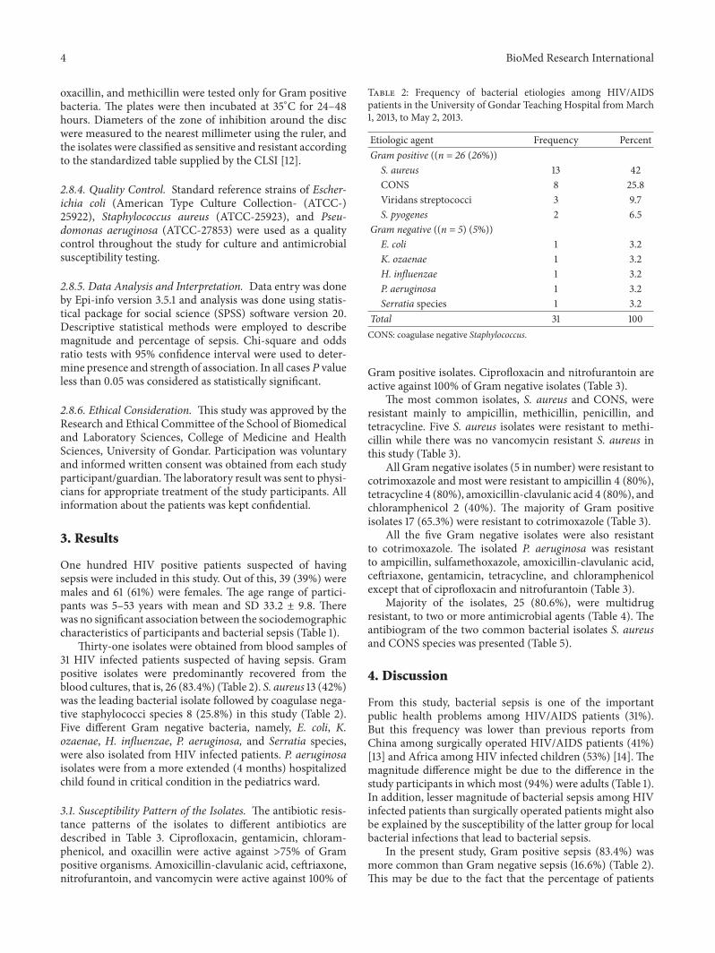
Thirty-one isolates were obtained from blood samples of31 HIV infected patients suspected of having sepsis. Grampositive isolates were predominantly recovered from theblood cultures, that is, 26 (83.4%) (Table 2). S. aureus 13 (42%)was the leading bacterial isolate followed by coagulase nega-tive staphylococci species 8 (25.8%) in this study (Table 2).Five different Gram negative bacteria, namely, E. coli, K.ozaenae, H. influenzae, P. aeruginosa, and Serratia species,were also isolated from HIV infected patients. P. aeruginosaisolates were from a more extended (4 months) hospitalizedchild found in critical condition in the pediatrics ward.
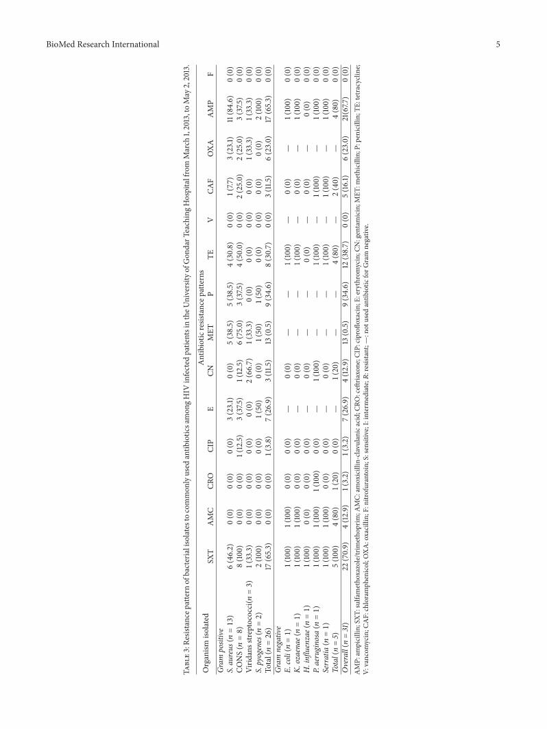
3.1. Susceptibility Pattern of the Isolates. The antibiotic resis-tance patterns of the isolates to different antibiotics aredescribed in Table 3. Ciprofloxacin, gentamicin, chloram-phenicol, and oxacillin were active against >75% of Grampositive organisms. Amoxicillin-clavulanic acid, ceftriaxone,nitrofurantoin, and vancomycin were active against 100% of
Table 2: Frequency of bacterial etiologies among HIV/AIDSpatients in the University of Gondar Teaching Hospital fromMarch1, 2013, to May 2, 2013.
Etiologic agent Frequency PercentGram positive ((n = 26 (26%))S. aureus 13 42CONS 8 25.8Viridans streptococci 3 9.7S. pyogenes 2 6.5
Gram negative ((n = 5) (5%))E. coli 1 3.2K. ozaenae 1 3.2H. influenzae 1 3.2P. aeruginosa 1 3.2Serratia species 1 3.2
Total 31 100CONS: coagulase negative Staphylococcus.
Gram positive isolates. Ciprofloxacin and nitrofurantoin areactive against 100% of Gram negative isolates (Table 3).
The most common isolates, S. aureus and CONS, wereresistant mainly to ampicillin, methicillin, penicillin, andtetracycline. Five S. aureus isolates were resistant to methi-cillin while there was no vancomycin resistant S. aureus inthis study (Table 3).
All Gram negative isolates (5 in number) were resistant tocotrimoxazole and most were resistant to ampicillin 4 (80%),tetracycline 4 (80%), amoxicillin-clavulanic acid 4 (80%), andchloramphenicol 2 (40%). The majority of Gram positiveisolates 17 (65.3%) were resistant to cotrimoxazole (Table 3).
All the five Gram negative isolates were also resistantto cotrimoxazole. The isolated P. aeruginosa was resistantto ampicillin, sulfamethoxazole, amoxicillin-clavulanic acid,ceftriaxone, gentamicin, tetracycline, and chloramphenicolexcept that of ciprofloxacin and nitrofurantoin (Table 3).
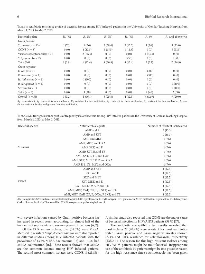
Majority of the isolates, 25 (80.6%), were multidrugresistant, to two or more antimicrobial agents (Table 4). Theantibiogram of the two common bacterial isolates S. aureusand CONS species was presented (Table 5).
4. Discussion
From this study, bacterial sepsis is one of the importantpublic health problems among HIV/AIDS patients (31%).But this frequency was lower than previous reports fromChina among surgically operated HIV/AIDS patients (41%)[13] and Africa among HIV infected children (53%) [14]. Themagnitude difference might be due to the difference in thestudy participants in which most (94%) were adults (Table 1).In addition, lesser magnitude of bacterial sepsis among HIVinfected patients than surgically operated patients might alsobe explained by the susceptibility of the latter group for localbacterial infections that lead to bacterial sepsis.
In the present study, Gram positive sepsis (83.4%) wasmore common than Gram negative sepsis (16.6%) (Table 2).This may be due to the fact that the percentage of patients
BioMed Research International 5
Table3:Re
sistancep
attern
ofbacterialisolatesto
common
lyused
antib
iotic
samon
gHIV
infected
patie
ntsintheU
niversity
ofGon
darT
eachingH
ospitalfrom
March
1,2013,toMay
2,2013.
Organism
isolated
Antibiotic
resistancep
atterns
SXT
AMC
CRO
CIP
ECN
MET
PTE
VCA
FOXA
AMP
FGr
ampositive
S.aureus
(𝑛=13)
6(46.2)
0(0)
0(0)
0(0)
3(23.1)
0(0)
5(38.5)
5(38.5)
4(30.8)
0(0)
1(7.7
)3(23.1)
11(84.6)
0(0)
CONS(𝑛=8)
8(100)
0(0)
0(0)
1(12.5)
3(37.5
)1(12.5)
6(75.0)
3(37.5
)4(50.0)
0(0)
2(25.0)
2(25.0)
3(37.5
)0(0)
Virid
ansstre
ptococci(𝑛=3)
1(33.3)
0(0)
0(0)
0(0)
0(0)
2(66.7)
1(33.3)
0(0)
0(0)
0(0)
0(0)
1(33.3)
1(33.3)
0(0)
S.pyogenes(𝑛=2)
2(100)
0(0)
0(0)
0(0)
1(50)
0(0)
1(50)
1(50)
0(0)
0(0)
0(0)
0(0)
2(100)
0(0)
Total(𝑛=26)
17(65.3)
0(0)
0(0)
1(3.8)
7(26.9)
3(11.5
)13
(0.5)
9(34.6)
8(30.7)
0(0)
3(11.5
)6(23.0)
17(65.3)
0(0)
Gram
negative
E.coli(𝑛=1)
1(100)
1(100)
0(0)
0(0)
—0(0)
——
1(100)
—0(0)
—1(100)
0(0)
K.ozaena
e(𝑛=1)
1(100)
1(100)
0(0)
0(0)
—0(0)
——
1(100)
—0(0)
—1(100)
0(0)
H.infl
uenzae
(𝑛=1)
1(100)
0(0)
0(0)
0(0)
—0(0)
——
0(0)
—0(0)
—0(0)
0(0)
P.aeruginosa
(𝑛=1)
1(100)
1(100)
1(100)
0(0)
—1(100)
——
1(100)
—1(100)
—1(100)
0(0)
Serratia(𝑛=1)
1(100)
1(100)
0(0)
0(0)
—0(0)
——
1(100)
—1(100)
—1(100)
0(0)
Total(n=5)
5(100)
4(80)
1(20)
0(0)
—1(20)
——
4(80)
—2(40)
—4(80)
0(0)
Overall(n
=31)
22(70.9)
4(12.9)
1(3.2)
1(3.2)
7(26.9)
4(12.9)
13(0.5)
9(34.6)
12(38.7)
0(0)
5(16.1)
6(23.0)
21(67.7
)0(0)
AMP:
ampicillin;SX
T:sulfametho
xazole/tr
imetho
prim
;AMC:
amoxicillin-clav
ulanicacid;C
RO:ceft
riaxone;C
IP:ciproflo
xacin;E:
erythrom
ycin;C
N:gentamicin;M
ET:m
ethicillin;P:penicillin;TE
:tetracycline;
V:vancom
ycin;C
AF:chloramph
enicol;O
XA:oxacillin;
F:nitro
furantoin;
S:sensitive;I:intermediate;𝑅
:resistant;—:not
used
antib
iotic
forG
ram
negativ
e.
6 BioMed Research International
Table 4: Antibiotic resistance profile of bacterial isolate among HIV infected patients in the University of Gondar Teaching Hospital fromMarch 1, 2013, to May 2, 2013.
Bacterial isolate 𝑅0
(%) 𝑅1
(%) 𝑅2
(%) 𝑅3
(%) 𝑅4
(%) 𝑅5
and above (%)Gram positiveS. aureus (𝑛 = 13) 1 (7.6) 1 (7.6) 5 (38.4) 2 (15.3) 1 (7.6) 3 (23.0)CONS (𝑛 = 8) 0 (0) 1 (12.5) 3 (37.5) 1 (12.5) 0 (0) 3 (37.5)Viridans streptococci(𝑛 = 3) 0 (0) 2 (66.6) 0 (0) 0 (0) 1 (33.3) 0 (0)S. pyogenes (𝑛 = 2) 0 (0) 0 (0) 0 (0) 1 (50) 0 (0) 1 (50)Total (26) 1 (3.8) 4 (15.4) 8 (30.8) 4 (15.4) 2 (7.7) 7 (26.9)Gram negativeE. coli (𝑛 = 1) 0 (0) 0 (0) 0 (0) 0 (0) 1 (100) 0 (0)K. ozaenae (𝑛 = 1) 0 (0) 0 (0) 0 (0) 0 (0) 1 (100) 0 (0)H. influenzae (𝑛 = 1) 0 (0) 1 (100) 0 (0) 0 (0) 0 (0) 0 (0)P. aeruginosa (𝑛 = 1) 0 (0) 0 (0) 0 (0) 0 (0) 0 (0) 1 (100)Serratia (𝑛 = 1) 0 (0) 0 (0) 0 (0) 0 (0) 0 (0) 1 (100)Total (n = 5) 0 (0) 1 (20) 0 (0) 0 (0) 2 (40) 2 (00)Overall (n = 31) 1 (3.2) 5 (16.1) 8 (25.8) 4 (12.9) 4 (12.9) 9 (29.0)𝑅0: nonresistant; 𝑅
1: resistant for one antibiotic; 𝑅
2: resistant for two antibiotics; 𝑅
3: resistant for three antibiotics; 𝑅
4: resistant for four antibiotics; 𝑅
5and
above: resistant for five and greater than five antibiotics.
Table 5:Multidrug resistance profile of frequently isolate bacteria amongHIV infected patients in theUniversity ofGondar TeachingHospitalfromMarch 1, 2013, to May 2, 2013.
Bacterial species Antimicrobial agents Number of resistant isolates (%)
S. aureus
AMP and P 2 (15.3)AMP and SXT 2 (15.3)AMP and MET 1 (7.6)
AMP, MET, and OXA 1 (7.6)AMP, SXT, and P 1 (7.6)
AMP, SXT, E, and TE 1 (7.6)AMP, SXT, E, TE, and CAF 1 (7.6)
AMP, SXT, MET, TE, P, and OXA 1 (7.6)AMP, P, E, TE, MET, and OXA 1 (7.6)
CONS
AMP and SXT 1 (12.5)SXT and E 1 (12.5)
SXT and MET 1 (12.5)SXT, MET, and E 1 (12.5)
SXT, MET, OXA, P, and TE 1 (12.5)AMP, MET, CAF, CIP, E, P, SXT, and TE 1 (12.5)
AMP, MET, CAF, CN, E, OXA, P, SXT, and TE 1 (12.5)AMP: ampicillin; SXT: sulfamethoxazole/trimethoprim; CIP: ciprofloxacin; E: erythromycin; CN: gentamicin; MET: methicillin; P: penicillin; TE: tetracycline;CAF: chloramphenicol; OXA: oxacillin; CONS: coagulase negative staphylococci.
with severe infections caused by Gram positive bacteria hasincreased in recent years, accounting for almost half of theincidents of septicemia and severe systemic infections [6, 7].
Of the 13 S. aureus isolates, five (38.5%) were MRSA.Methicillin resistant Staphylococcus aureuswere also reportedin different studies among HIV infected patients with theprevalence of 43.5% MRSA bacteraemia [15] and 10.3% hadMRSA colonization [16]. These results showed that MRSAare the common isolates among HIV infected patients.The second most common isolates were CONS, 8 (25.8%).
A similar study also reported that CONS are the major causeof bacterial infection in HIV/AIDS patients (58%) [17].
The antibiotic susceptibility test results revealed thatmost isolates 22 (70.9%) were resistant for most antibioticstested. Gram positive and Gram negative isolates showed65.3% and 100% resistance for cotrimoxazole, respectively(Table 3). The reason for this high resistant isolates amongHIV/AIDS patients might be multifactorial. Inappropriateuse of the antibiotic by patients might be one possible reasonfor the high resistance since cotrimoxazole has been given
BioMed Research International 7
as prophylaxis for HIV/AIDS patients. This action mightalso enhance the resistance to the antimicrobial agent. It isalso very difficult to generalize with this sample size aboutthe cotrimoxazole resistance among isolates of HIV/AIDSpatients. Large scale comparative study might answer thequestion. From previous retrospective cohort study amongHIV infected patients, most of the isolates resist cotri-moxazole widespread use for prophylaxis may exacerbateantimicrobial resistance [14].
Gram positive bacteria also showed good sensitivitypatterns to amoxicillin-clavulanic acid, ceftriaxone, nitrofu-rantoin, and vancomycin.
Gram negative bacteria showed good sensitivity patternonly for ciprofloxacin and nitrofurantoin (Table 3). Thismight be due to the use of this specific antibiotic which is notwidespread.
One P. aeruginosa isolate was from a more extended(4 months) hospitalized child found in critical conditionin the pediatrics ward. P. aeruginosa have also been morefrequently recognized as nosocomial bacteraemia in HIVinfected patients [17].
5. Conclusions
The prevalence of culture that confirmed bacterial sepsisamongHIV infected patients was high in this study (31%) andthe two most common causative agents for bacterial sepsisare S. aureus and CONS. Amoxicillin-clavulanic acid, cef-triaxone, nitrofurantoin, and vancomycin were 100% activeagainst Gram positive bacteria. Ciprofloxacin and nitrofu-rantoin were 100% active against Gram negative bacteria.Most of the isolates were multidrug resistant includingcotrimoxazole.
6. Recommendations
Based on the findings of this study, we recommend thatblood culture should be done for HIV infected patientssuspected of having sepsis. Amoxicillin-clavulanic acid, cef-triaxone, nitrofurantoin, and vancomycin can be used againstGram positives if the laboratory setups are incomplete.Continuous antimicrobial susceptibility surveillance shall beimplemented in the study area with large scale study amongHIV/AIDS patients focusing on cotrimoxazole.
Competing Interests
The authors declare that they have no competing interests.
Acknowledgments
The authors acknowledge the Department of Medical Micro-biology, University of Gondar, for allowing them reagents andmaterials during this study. The authors’ special appreciationalso goes to Mr. Aschalew Gelaw for his great contributionand guidance in the laboratory procedures of this study.Finally, the authors would like to thank all the study partici-pants.
References
[1] D. C. Angus, W. T. Linde-Zwirble, J. Lidicker, G. Clermont, J.Carcillo, and M. R. Pinsky, “Epidemiology of severe sepsis intheUnited States: analysis of incidence, outcome, and associatedcosts of care,”Critical CareMedicine, vol. 29, no. 7, pp. 1303–1310,2001.
[2] A. Lever and I. Mackenzie, “Sepsis: definition, epidemiology,and diagnosis,” The British Medical Journal, vol. 335, no. 7625,pp. 879–883, 2007.
[3] B. A. Cunha, “Sepsis and septic shock: selection of empiricantimicrobial therapy,” Critical Care Clinics, vol. 24, no. 2, pp.313–334, 2008.
[4] E. Jean-Baptiste, “Cellular mechanisms in sepsis,” Journal ofIntensive Care Medicine, vol. 22, no. 2, pp. 63–72, 2007.
[5] S. Latif, M. S. Anwar, and I. Ahmad, “Bacterial pathogensresponsible for blood stream infection and pattern of drugresistance in a tertiary care hospital of Lahore,” Biomedica, vol.25, pp. 101–105, 2009.
[6] T. van der Poll and S. M. Opal, “Host-pathogen interactions insepsis,” The Lancet Infectious Diseases, vol. 8, no. 1, pp. 32–43,2008.
[7] D. Sun, B. Raisley, M. Langer et al., “Anti-peptidoglycan anti-bodies and Fc𝛾 receptors are the keymediators of inflammationin gram-positive sepsis,” Journal of Immunology, vol. 189, no. 5,pp. 2423–2431, 2012.
[8] A. C. Cheng, T. E.West, D. Limmathurotsakul, and S. J. Peacock,“Strategies to reduce mortality from bacterial sepsis in adults indeveloping countries,” PLoS Medicine, vol. 5, no. 8, article e175,2008.
[9] A. Bane, A. G. Yohannes, and D. Fekade, “Morbidity andmortality of adult patients with HIV/AIDS at Tikur AnbessaTeaching Hospital, Addis Ababa, Ethiopia,” Ethiopian MedicalJournal, vol. 41, no. 2, pp. 131–140, 2003.
[10] K. D. Waikhom and K. S. Devi, “Emergence of multidrugresistant bacterial infection in HIV/AIDS cases,” The Health,vol. 3, pp. 49–52, 2012.
[11] Clinical and Laboratory Standard Institution, “Principles andprocedures for blood culture; approved guideline,” Clinical andLaboratory Standard Institution Document M47-A, Clinicaland Laboratory Standard Institution, Wayne, Pa, USA, 2007.
[12] Clinical and Laboratory Standards Institute, Performance Stan-dards for Antimicrobial Disk Susceptibility Tests, Clinical andLaboratory Standards Institute Document M2-A9, Clinical andLaboratory Standards Institute, Wayne, Pa, USA, 9th edition,2006.
[13] B. Liu, L. Zhang, R. Guo, J. Su, L. Li, and Y. Si, “Anti-infective treatment in HIV-infected patients during periopera-tive period,” AIDS Research andTherapy, vol. 9, article 36, 2012.
[14] H. B. Jaspan, L. C. Huang, M. F. Cotton, A. Whitelaw, andL. Myer, “Bacterial disease and antimicrobial susceptibilitypatterns in HIV-infected, hospitalized children: a retrospectivecohort study,” PLoS ONE, vol. 3, no. 9, article e3260, 2008.
[15] M. D. Burkey, L. E.Wilson, R. D.Moore, G.M. Lucas, J. Francis,and K. A. Gebo, “The incidence of and risk factors for MRSAbacteraemia in anHIV-infected cohort in theHAART era,”HIVMedicine, vol. 9, no. 10, pp. 858–862, 2008.
[16] M. J. Cenizal, R. D. Hardy, M. Anderson, K. Katz, and D. J.Skiest, “Prevalence of and risk factors for methicillin-resistant
8 BioMed Research International
Staphylococcus aureus (MRSA) nasal colonization in HIV-infected ambulatory patients,” Journal of Acquired ImmuneDeficiency Syndromes, vol. 48, no. 5, pp. 567–571, 2008.
[17] A. I. Adeyemi, A. A. Sulaiman, B. B. Solomon, O. A. Chinedu,and I. A. Victor, “Bacterial bloodstream infections in HIV-infected adults attending a lagos teaching hospital,” Journal ofHealth, Population and Nutrition, vol. 28, no. 4, pp. 318–326,2010.

Submit your manuscripts athttp://www.hindawi.com
Hindawi Publishing Corporationhttp://www.hindawi.com Volume 2014
Anatomy Research International
PeptidesInternational Journal of
Hindawi Publishing Corporationhttp://www.hindawi.com Volume 2014
Hindawi Publishing Corporation http://www.hindawi.com
International Journal of
Volume 2014
Zoology
Hindawi Publishing Corporationhttp://www.hindawi.com Volume 2014
Molecular Biology International
GenomicsInternational Journal of
Hindawi Publishing Corporationhttp://www.hindawi.com Volume 2014
The Scientific World JournalHindawi Publishing Corporation http://www.hindawi.com Volume 2014
Hindawi Publishing Corporationhttp://www.hindawi.com Volume 2014
BioinformaticsAdvances in
Marine BiologyJournal of
Hindawi Publishing Corporationhttp://www.hindawi.com Volume 2014
Hindawi Publishing Corporationhttp://www.hindawi.com Volume 2014
Signal TransductionJournal of
Hindawi Publishing Corporationhttp://www.hindawi.com Volume 2014
BioMed Research International
Evolutionary BiologyInternational Journal of
Hindawi Publishing Corporationhttp://www.hindawi.com Volume 2014
Hindawi Publishing Corporationhttp://www.hindawi.com Volume 2014
Biochemistry Research International
ArchaeaHindawi Publishing Corporationhttp://www.hindawi.com Volume 2014
Hindawi Publishing Corporationhttp://www.hindawi.com Volume 2014
Genetics Research International
Hindawi Publishing Corporationhttp://www.hindawi.com Volume 2014
Advances in
Virolog y
Hindawi Publishing Corporationhttp://www.hindawi.com
Nucleic AcidsJournal of
Volume 2014
Stem CellsInternational
Hindawi Publishing Corporationhttp://www.hindawi.com Volume 2014
Hindawi Publishing Corporationhttp://www.hindawi.com Volume 2014
Enzyme Research
Hindawi Publishing Corporationhttp://www.hindawi.com Volume 2014
International Journal of
Microbiology