Reprogramming Tumor-Associated Dendritic Cells In Vivo...
12
Microenvironment and Immunology Reprogramming Tumor-Associated Dendritic Cells In Vivo Using miRNA Mimetics Triggers Protective Immunity against Ovarian Cancer Juan R. Cubillos-Ruiz 1 , Jason R. Baird 1,2 , Amelia J. Tesone 5 , Melanie R. Rutkowski 5 , Uciane K. Scarlett 5 , Ana L. Camposeco-Jacobs 1 , Jorge Anadon-Arnillas 1 , Noah M. Harwood 1 , Murray Korc 3,4 , Steven N. Fiering 1,2 , Lorenzo F. Sempere 3 , and Jose R. Conejo-Garcia 5 Abstract Modulating the activity of miRNAs provides opportunities for novel cancer interventions. However, low bioavailability and poor cellular uptake are major challenges for delivering miRNA mimetics specifically to tumor cells. Here, we took advantage of the spontaneous enhanced endocytic activity of ovarian cancer-associated dendritic cells (DC) to selectively supplement the immunostimulatory miRNA miR-155. In vivo processing of nanoparticles carrying oligonucleotide duplexes mimicking the bulged structure of endogenous pre-miRNA (but not siRNA-like oligonucleotides) dramatically augmented miR-155 activity without saturating the RNA-induced silencing complex. Endogenous processing of synthetic miR-155 favored Ago2 and, to a lesser extent, Ago4 loading, resulting in genome-wide transcriptional changes that included silencing of multiple immunosuppres- sive mediators. Correspondingly, tumor-infiltrating DCs were transformed from immunosuppressive to highly immunostimulatory cells capable of triggering potent antitumor responses that abrogated the progression of established ovarian cancers. Our results show both the feasibility and therapeutic potential of supplementing/ replenishing miRNAs in vivo using nonviral approaches to boost protective immunity against lethal tumors. Thus, we provide a platform, an optimized design, and a mechanistic rationale for the clinical testing of nonviral miRNA mimetics. Cancer Res; 72(7); 1683–93. Ó2012 AACR. Introduction miRNAs are small endogenous noncoding RNAs implicated in the posttranscriptional control of gene expression in devel- opmental, physiologic, and pathologic processes. Biologically active/mature miRNAs bind to partially complementary sequences [miRNA recognition element (MRE)] in hundreds of mRNAs, which diminish protein production via mRNA degradation and/or translational repression. miRNA-mediated regulation therefore constitutes a major mechanism to control global gene expression patterns (1–3). miRNAs are quickly challenging our understanding of genet- ic regulation in health and disease, including cancer etiology (4) and the generation and inhibition of antitumor immune responses (5–9). Biologically active miRNAs bind to MREs on multiple mRNAs and simultaneously silence multiple target genes. This process can directly or indirectly modulate global gene expression and eventually determines transcriptional programs associated with a specific phenotype. Because immune responses–including those against tumor antigens–depend on rapid phenotypic changes, it is not sur- prising that miRNAs have emerged as critical regulators of virtually all immune cell types (5, 7). miR-155 epitomizes the role of miRNAs in the immune system. miR-155 is basally expressed at low levels in B cells (7, 8), T cells (10), macrophages (11), dendritic cells (DC; ref. 7), and progenitor/stem cell populations (10). Activation signals such as antigen, toll-like receptor (TLR) stimulation and inflammatory cytokines, rap- idly increase miR-155 expression in various leukocytic subsets, including bone marrow DCs (BMDC) and macrophages (7, 8, 11). Interestingly, BMDCs matured in the absence of miR-155 upregulate MHC-II and costimulatory molecules but are inca- pable of effectively activating T cells (7). We previously showed that leukocytes with predominant phenotypic attributes of regulatory DCs (including the expres- sion of CD11c and DEC205) home to perivascular locations in the ovarian cancer microenvironment, where they express multiple immunosuppressive mediators (12–14). From their position around blood vessels, these regulatory DCs inhibit the protective function of antitumor T cells infiltrating the tumor from the blood. Although specific delivery of RNA oligo- nucleotides to cancer cells is challenging because of low Authors' Affiliations: Departments of 1 Microbiology and Immunology, 2 Genetics, 3 Medicine and 4 Pharmacology and Toxicology, Dartmouth Medical School, Lebanon, New Hampshire; and 5 Tumor Microenvironment and Metastasis Program, The Wistar Institute, Philadelphia, Pennsylvania Note: Supplementary data for this article are available at Cancer Research Online (http://cancerres.aacrjournals.org/). Current address for M. Korc, IU Simon Cancer Center, 980 W. Walnut Street, Walther Hall, R3 C528E, Indianapolis, IN 46202. Corresponding Author: Jose R. Conejo-Garcia, Tumor Microenviron- ment and Metastasis Program, The Wistar Institute, 3601 Spruce Street, Philadelphia, PA 19104. Phone: 215-495-6825; Fax: 215-495-6817; E-mail: [email protected] doi: 10.1158/0008-5472.CAN-11-3160 Ó2012 American Association for Cancer Research. Cancer Research www.aacrjournals.org 1683 on May 4, 2018. © 2012 American Association for Cancer Research. cancerres.aacrjournals.org Downloaded from Published OnlineFirst February 3, 2012; DOI: 10.1158/0008-5472.CAN-11-3160
Transcript of Reprogramming Tumor-Associated Dendritic Cells In Vivo...
Microenvironment and Immunology
Reprogramming Tumor-Associated Dendritic Cells In VivoUsingmiRNAMimetics Triggers Protective Immunity againstOvarian Cancer
Juan R. Cubillos-Ruiz1, Jason R. Baird1,2, Amelia J. Tesone5, Melanie R. Rutkowski5, Uciane K. Scarlett5,Ana L. Camposeco-Jacobs1, Jorge Anadon-Arnillas1, Noah M. Harwood1, Murray Korc3,4,Steven N. Fiering1,2, Lorenzo F. Sempere3, and Jose R. Conejo-Garcia5
AbstractModulating the activity of miRNAs provides opportunities for novel cancer interventions. However, low
bioavailability and poor cellular uptake aremajor challenges for deliveringmiRNAmimetics specifically to tumorcells. Here, we took advantage of the spontaneous enhanced endocytic activity of ovarian cancer-associateddendritic cells (DC) to selectively supplement the immunostimulatory miRNA miR-155. In vivo processing ofnanoparticles carrying oligonucleotide duplexes mimicking the bulged structure of endogenous pre-miRNA (butnot siRNA-like oligonucleotides) dramatically augmented miR-155 activity without saturating the RNA-inducedsilencing complex. Endogenous processing of synthetic miR-155 favored Ago2 and, to a lesser extent, Ago4loading, resulting in genome-wide transcriptional changes that included silencing of multiple immunosuppres-sive mediators. Correspondingly, tumor-infiltrating DCs were transformed from immunosuppressive to highlyimmunostimulatory cells capable of triggering potent antitumor responses that abrogated the progression ofestablished ovarian cancers. Our results show both the feasibility and therapeutic potential of supplementing/replenishingmiRNAs in vivo using nonviral approaches to boost protective immunity against lethal tumors. Thus,we provide a platform, an optimized design, and amechanistic rationale for the clinical testing of nonviralmiRNAmimetics. Cancer Res; 72(7); 1683–93. �2012 AACR.
IntroductionmiRNAs are small endogenous noncoding RNAs implicated
in the posttranscriptional control of gene expression in devel-opmental, physiologic, and pathologic processes. Biologicallyactive/mature miRNAs bind to partially complementarysequences [miRNA recognition element (MRE)] in hundredsof mRNAs, which diminish protein production via mRNAdegradation and/or translational repression.miRNA-mediatedregulation therefore constitutes amajormechanism to controlglobal gene expression patterns (1–3).miRNAs are quickly challenging our understanding of genet-
ic regulation in health and disease, including cancer etiology(4) and the generation and inhibition of antitumor immune
responses (5–9). Biologically active miRNAs bind to MREs onmultiple mRNAs and simultaneously silence multiple targetgenes. This process can directly or indirectly modulate globalgene expression and eventually determines transcriptionalprograms associated with a specific phenotype.
Because immune responses–including those against tumorantigens–depend on rapid phenotypic changes, it is not sur-prising that miRNAs have emerged as critical regulators ofvirtually all immune cell types (5, 7). miR-155 epitomizes therole of miRNAs in the immune system. miR-155 is basallyexpressed at low levels in B cells (7, 8), T cells (10),macrophages(11), dendritic cells (DC; ref. 7), and progenitor/stem cellpopulations (10). Activation signals such as antigen, toll-likereceptor (TLR) stimulation and inflammatory cytokines, rap-idly increase miR-155 expression in various leukocytic subsets,including bone marrow DCs (BMDC) and macrophages (7,8, 11). Interestingly, BMDCsmatured in the absence ofmiR-155upregulate MHC-II and costimulatory molecules but are inca-pable of effectively activating T cells (7).
We previously showed that leukocytes with predominantphenotypic attributes of regulatory DCs (including the expres-sion of CD11c and DEC205) home to perivascular locations inthe ovarian cancer microenvironment, where they expressmultiple immunosuppressive mediators (12–14). From theirposition around blood vessels, these regulatory DCs inhibit theprotective function of antitumor T cells infiltrating the tumorfrom the blood. Although specific delivery of RNA oligo-nucleotides to cancer cells is challenging because of low
Authors' Affiliations: Departments of 1Microbiology and Immunology,2Genetics, 3Medicine and 4Pharmacology and Toxicology, DartmouthMedical School, Lebanon, NewHampshire; and 5TumorMicroenvironmentand Metastasis Program, The Wistar Institute, Philadelphia, Pennsylvania
Note: Supplementary data for this article are available at Cancer ResearchOnline (http://cancerres.aacrjournals.org/).
Current address for M. Korc, IU Simon Cancer Center, 980 W. WalnutStreet, Walther Hall, R3 C528E, Indianapolis, IN 46202.
Corresponding Author: Jose R. Conejo-Garcia, Tumor Microenviron-ment and Metastasis Program, The Wistar Institute, 3601 Spruce Street,Philadelphia, PA 19104. Phone: 215-495-6825; Fax: 215-495-6817;E-mail: [email protected]
doi: 10.1158/0008-5472.CAN-11-3160
�2012 American Association for Cancer Research.
CancerResearch
www.aacrjournals.org 1683
on May 4, 2018. © 2012 American Association for Cancer Research. cancerres.aacrjournals.org Downloaded from
Published OnlineFirst February 3, 2012; DOI: 10.1158/0008-5472.CAN-11-3160
bioavailability, poor cellular uptake, and abundant phagocyticactivity of other cell types in the tumormicroenvironment (15),the enhanced endocytic pathways and relative accessibility ofovarian cancer–associated myeloid leukocytes makes themideal targets for nanocomplex-mediated delivery. Thus, wepreviously showed that polyethylenimine (PEI)-based nano-complexes are selectively and avidly taken up byDCs at ovariancancer locations, in the absence of any targeting motif (12).Using this optimized system, we now show that activity ofmature miR-155 can be augmented in tumor-associated DCsby delivering novel Dicer substrate RNA duplexes that mimicthe structure of endogenous precursor miR-155 hairpin(Dmi155) and that are efficiently processed in vivo. Replenish-ing miR-155 levels in tumor-associated DCs reprogrammedtheir immunosuppressive phenotype by modulating theexpression of nearly half of the mRNAs in their transcriptome.Synthetic enhancement of miR-155 signaling in DCs boostedpotent antitumor immune responses that abrogated the pro-gression of established ovarian cancers. Our results show thefeasibility of supplementing/replenishing miRNAs in vivo toboost antitumor immunity against aggressive, advanced, andfrequently lethal tumors.
Materials and MethodsProductionof PEI-basednanoparticles encapsulatingDSRNA duplexes
Endotoxin-free PEI for in vivo experiments "in vivo-jetPEI"was purchased from PolyPlus Transfection. Dicer substrates(Dsi) were synthesized at Integrated DNA Technologies (IDT)using the following chimeric sequences:
Control GFP-specific Dicer substrate (GFP Dsi):Plus: 50 rUrGrCrArGrArUrGrArArCrUrUrCrArGrGrGrUrCrAr-
GrCTT 30Minus: 50 rArArGrCrU rGrArC rCrCrU rGrArA rGrUrU rCrArU-
rCrUrG rCrArUrU 30
Control GFP-specific "bulged" Dicer substrate:Plus: 50 rUrGrCrArGrArUrGrArArCrUrUrCrArGrGrGrUrCrAr-
GrCTT 30Minus: 50 rArArGrCrU rGrArC rCrCrU rG rGrUrU rCrArU rCr-
UrGrCrArUrU 30
siRNA-like miR-155 Dicer substrate (Dsi155):Plus: 50 rUrUrA rArUrG rCrUrA rArUrU rGrUrG rArUrA rGrGr-
GrGrUT T 30Minus: 50 rArArA rCrCrC rCrUrA rUrCrA rCrArArUrUrArGrCr-
ArUrUrA rArUrU 30
miRNA-like bulged miR-155 Dicer substrate (Dmi155):Plus: 50 rUrUrA rArUrG rCrUrA rArUrU rGrUrG rArUrA rGrGr-
GrGrUT T 30Minus: 50 rArArA rCrCrC rCrUrA rUrCrA rA rUrUrA rGrCrA-
rUrUrA rArUrU 30
In all cases, "r" represents a ribonucleotide and the absenceof an "r" indicates a deoxynucleotide. The "plus" strand contains2 terminal deoxynucleotides that resemble the loop of endog-enous pre-miRNA and that function as cleavage signal for
Dicer. The "plus" strand refers to the strand that will give riseto the mature miRNA after Dicer processing and preferentialincorporation into the RNA-induced silencing complex (RISC).
To generate PEI-based nanoparticles encapsulating Dsi, 50to 100 mg of each annealed duplex were complexed with "invivo-jetPEI" at an N/P ratio of 6, following the recommenda-tions of the manufacturer (PolyPlus Transfection). For biodis-tribution experiments, Dsi were fluorescently labeled in the 30
end of the plus strand using Cy3 (IDT). Biotinylated Dsi werealso chemically synthesized at IDT and include a Biotin groupin the 50 end of the "plus" strand. Thus, after intracellularprocessing of the Dsi, the mature form of the miRNA remainsbiotinylated in vivo.
Transfection and in vivo delivery of DsiLipofectamine 2000 (Invitrogen) was used for in vitro trans-
fection of Dsi into HEK293 cells in 96-well plates, following therecommendations of the manufacturer. For in vivo biodistri-bution, phenotypic and gene silencing experiments, micebearing ID8-Defb29/Vegf-A tumors (12) for 3 to 4 weeks wereintraperitoneally injected with PEI-Dsi nanoparticles (50 mg ofDsi complexed with "in vivo-jetPEI" at N/P 6, per mouse). In allphenotypic and functional experiments, tumor-associatedDCsfrommice injectedwith nanoparticleswere sorted fromascitesor peritoneal wash samples by flow cytometry on the basis ofCD45, CD11c, and MHC-II positive expression.
Tumor progression experimentsWild-type C57BL/6 mice were intraperitoneally injected
with 2 � 106 parental ID8 (kindly provided by K. Robby, Uni-versity of Kansas Medical Center, Kansas City, KS; ref. 16) andtreatments started 15 days posttumor injection. A total of 2 �106 aggressive ID8-Defb29/Vegf-A ovarian carcinoma cells wereinjected intraperitoneally and treatments started after 8 days.In all cases mice received 50 mg of Dsi complexed with "in vivo-jetPEI" at N/P 6 in glucose 5% at the indicated time points.Some experimental groups were also intraperitoneally injectedwith 50 mg anti-CD40 antibody (clone FGK4.5) 3 hours beforeadministration of PEI-based nanoparticles containing Dsi.
For tumor rechallenge protection experiments, 3 � 106
CD3þ T cells negatively immunopurified from the spleens oftumor-bearing mice treated with PBS (day 32 after tumorchallenge) or aCD40 Ab plus Dmi155-PEI nanoparticles (day61 after tumor challenge; treatments at days 8, 13, 18, 23, 27, and60) were intravenously transferred into naive C57BL/6 micepreviously irradiated with 300g (5 mice per group). Twenty-four hours later mice were challenged in the flank with ID8-Defb29/Vegf-A ovarian carcinoma cells, as described (14).Tumor pictures were taken 25 days later. Tumor volumes werecalculated by the formula V¼ 0.5 (L�W2), in which L is lengthand W is width.
ResultsDicer substrate RNA duplexes generate functionallyactive mature miR-155
miR-155 plays an important role in oncogenesis (9) but isalso required for optimal antigen presentation and T-cell
Cubillos-Ruiz et al.
Cancer Res; 72(7) April 1, 2012 Cancer Research1684
on May 4, 2018. © 2012 American Association for Cancer Research. cancerres.aacrjournals.org Downloaded from
Published OnlineFirst February 3, 2012; DOI: 10.1158/0008-5472.CAN-11-3160
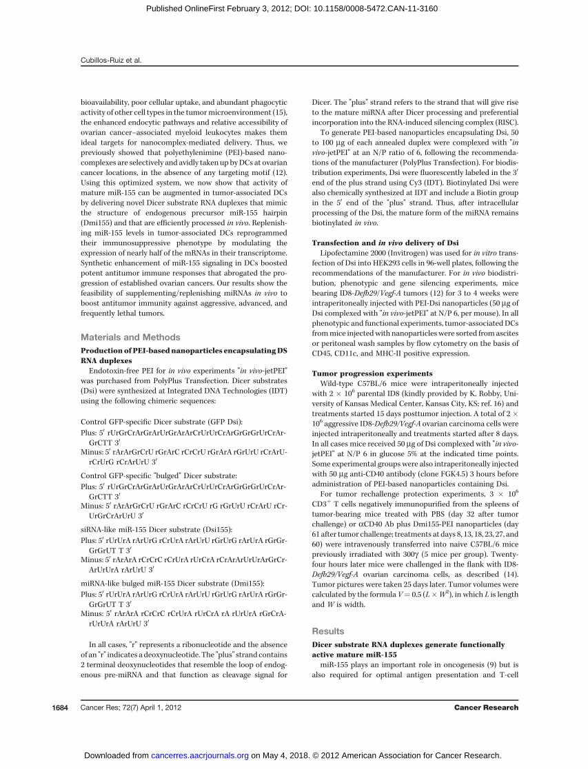
activation by mature DCs (7). We found that immunosuppres-sive CD45þCD11cþMHC-IIþ DCs (12, 13, 17–19) sorted fromadvanced orthotopic ID8-Defb29/Vegf-A tumors, an aggressivemodel of ovarian cancer that recapitulates the inflammatorymicroenvironment of human ovarian carcinomas (13, 14, 20,21), showed significantly reduced levels of mature miR-155(Fig. 1A). However, in vivo administration of CD40 plus TLR3agonists, which synergistically transform tumor-associatedDCs from immunosuppressive to immunostimulatory (13),induced a dramatic upregulation of mature miR-155 (Fig.1B). We therefore hypothesized that miR-155 upregulation inDCs in vivo at tumor locations could be the crucial eventpromoting their capacity to elicit therapeutic antitumorimmunity.To augment miR-155 activity, we generated novel synthetic
Dicer substrate (Dsi) RNA duplexes. To become functionally
active, Dsi require processing by Dicer, the same RNAse type IIIenzyme that processes endogenous miRNA precursors andexogenous siRNAs. In addition, Dsi exhibit markedly enhancedsilencing efficiency comparedwith conventional 21-mer siRNAoligonucleotides (22, 23). In all cases, we designed a forward(sense) RNA strand containing the sequence of endogenousmature miR-155 followed by 2 terminal deoxynucleotides inthe 30 end.We then generated 2 structural versions formiR-155mimetic compounds by using different passenger (antisense)strands: An internally bulged complementary strand thatrecapitulates the precursor miRNA hairpin (Dmi155) and aperfectly matching, siRNA-like, complementary strand(Dsi155; Fig. 1C). Control irrelevant bulged or siRNA-like Dsidesigned to target GFP were also produced in parallel.
Transfection of HEK293 cells with either Dsi155 or Dmi155led to a dramatic dose-dependent increase in the intracellular
A B C
**
*
G
tDCs
BMDCs
BMDCs +
LPS
0
50
100
150
200
Rela
tive m
iR-1
55 e
xpre
ssio
n
D
*
αCD40
+
poly(I:
C)
poly(I:
C)
αCD40
PBS
10
20
30
Rela
tive m
iR-1
55 e
xpre
ssio
n
Lu
cife
rase
activity
(%)
Untr
ansf
ecte
d
pluc-
Sfpi1
+ GFP D
si
+ Dsi
155
+ Dm
i155
0
20
40
60
80
100
120
293
+ GFP D
si
+
Dsi
155
+Dm
i155
0.1
1
10
100
1,000
10,000
100,000
Untr
ansf
ecte
dplu
c
+ GFPD
si
+ Dsi
155
+ Dm
i155
0
20
40
60
80
100
120
***
*
Rela
tive m
iR-1
55 e
xpre
ssio
n
Lu
cife
rase
activity
(%)
F
293
293
+ Dsi
155
293
+ Dm
i155
Dsi
155
Dm
i155
1
10
100
1,000
10,000
100,000
Rela
tive m
iR-1
55 e
xpre
ssio
n
E
Figure 1. miR-155 expression by tumor-associatedDCs and activity of RNAmimicking pre-miR-155. A, CD45þCD11cþMHC-IIþ tumor-associatedDCs (tDCs)were sorted from ID8-Defb29/Vegf-A tumor ascites. BMDCs, generated as described (19), were stimulatedwith 1 mg/mL of LPS for 6 hours. Representative of3 independent experiments. B, ID8-Defb29/Vegf-A tumor-bearing mice (n ¼ 3) received intraperitoneal PBS, aCD40 (50 mg), poly(I:C) (100 mg), oraCD40 in combinationwith poly(I:C). CD11cþMHCIIþDCswere sorted from peritoneal wash after 48 hours. Representative of 2 independent experiments. C,top, endogenous pre–miR-155; middle, siRNA-like Dsi155; bottom, bulgedmiRNA-like Dmi155. Underlined, deoxynucleotides. Framed, mature miR-155. D,HEK293 cells in 96-well plates were transfected with 5, 10, or 25 pmol of miR-155 mimicking or control GFP-specific Dsi and 18 hours later maturemiR-155 was quantified. Representative of 5 independent experiments. E, HEK293 cells in 96-well plates were transfected with 50 pmol of Dsi155 or Dmi155andRNAwas isolated 18 hours later. Fifty pmol of Dsi155 or Dmi155were directly used as template as control. Representative of 3 independent experiments.In all cases, mature miR-155 was quantified by stem-loop qRT-PCR and data were normalized to U6 snRNA. F, a luciferase reporter vector harboringthe MRE of miR-155 on Sfpi1 was cotransfected into HEK293 cells together with different RNA duplexes. Luciferase activity in whole cell lysates wasmeasured 24 hours later. Representative of 4 independent experiments. G, experiments were conducted as in E but using a luciferase-expressing constructwithout Sfpi1 MRE. Representative of 2 independent experiments. �, P < 0.05; ��, P < 0.01 (Mann–Whitney in all cases).
www.aacrjournals.org Cancer Res; 72(7) April 1, 2012 1685
Synthetic miRNA Elicits Protective Antitumor Immunity
on May 4, 2018. © 2012 American Association for Cancer Research. cancerres.aacrjournals.org Downloaded from
Published OnlineFirst February 3, 2012; DOI: 10.1158/0008-5472.CAN-11-3160
levels of processed miR-155, as detected by mature miRNA-specific stem-loop quantitative reverse transcriptase PCR(qRT-PCR; Fig. 1D). Confirming the selective detection ofprocessed miRNAs by the cellular machinery, negligible signalwas detected when synthetic Dsi155 or Dmi155 were directlyreversed transcribed and amplified before transfection (Fig.1E). To determine the functionality of processed miR-155 RNAgenerated from synthetic RNA, we cotransfected HEK293 cellswith a luciferase reporter construct containing the miR-155MRE of Sfpi1, an experimentally validated target gene of miR-155 (24). As expected, Dmi155 and, to a significantly lesserextent, Dsi155, rapidly silenced luciferase protein expression,whereas control (GFP specific) Dsi had no effect (Fig. 1F).Importantly, duplexes did not alter luciferase expression whenthe reporter constructs lacked the cognate miR-155 MRE (Fig.1G). Together, these data showed that synthetic Dsi RNAduplexes can be used to effectively generate functionally activemature miR-155 in the cell, and suggest that a bulged structuremay be important for the functionality of the miRNAgenerated.
Functional miR-155 delivered to tumor-associated DCsvia PEI-Dsi nanocomplexes is preferentially loaded ontoAgo2
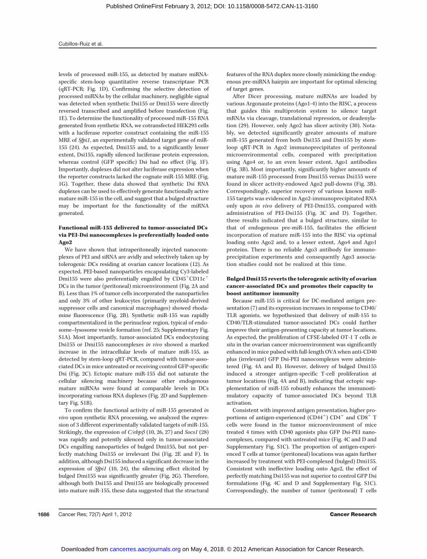
We have shown that intraperitoneally injected nanocom-plexes of PEI and siRNA are avidly and selectively taken up bytolerogenic DCs residing at ovarian cancer locations (12). Asexpected, PEI-based nanoparticles encapsulating Cy3-labeledDmi155 were also preferentially engulfed by CD45þCD11cþ
DCs in the tumor (peritoneal) microenvironment (Fig. 2A andB). Less than 1% of tumor cells incorporated the nanoparticlesand only 3% of other leukocytes (primarily myeloid-derivedsuppressor cells and canonical macrophages) showed rhoda-mine fluorescence (Fig. 2B). Synthetic miR-155 was rapidlycompartmentalized in the perinuclear region, typical of endo-some–lysosome vesicle formation (ref. 25; Supplementary Fig.S1A). Most importantly, tumor-associated DCs endocytozingDsi155 or Dmi155 nanocomplexes in vivo showed a markedincrease in the intracellular levels of mature miR-155, asdetected by stem-loop qRT-PCR, compared with tumor-asso-ciated DCs inmice untreated or receiving control GFP-specificDsi (Fig. 2C). Ectopic mature miR-155 did not saturate thecellular silencing machinery because other endogenousmature miRNAs were found at comparable levels in DCsincorporating various RNA duplexes (Fig. 2D and Supplemen-tary Fig. S1B).
To confirm the functional activity of miR-155 generated invivo upon synthetic RNA processing, we analyzed the expres-sion of 3 different experimentally validated targets of miR-155.Strikingly, the expression of C/ebpb (10, 26, 27) and Socs1 (28)was rapidly and potently silenced only in tumor-associatedDCs engulfing nanoparticles of bulged Dmi155, but not per-fectly matching Dsi155 or irrelevant Dsi (Fig. 2E and F). Inaddition, although Dsi155 induced a significant decrease in theexpression of Sfpi1 (10, 24), the silencing effect elicited bybulged Dmi155 was significantly greater (Fig. 2G). Therefore,although both Dsi155 and Dmi155 are biologically processedinto mature miR-155, these data suggested that the structural
features of the RNA duplex more closely mimicking the endog-enous pre-miRNA hairpin are important for optimal silencingof target genes.
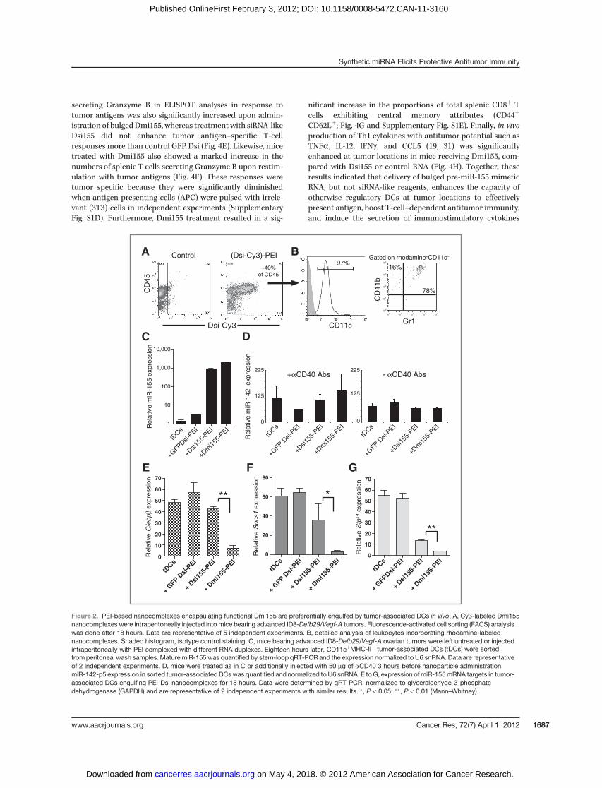
After Dicer processing, mature miRNAs are loaded byvarious Argonaute proteins (Ago1-4) into the RISC, a processthat guides this multiprotein system to silence targetmRNAs via cleavage, translational repression, or deadenyla-tion (29). However, only Ago2 has slicer activity (30). Nota-bly, we detected significantly greater amounts of maturemiR-155 generated from both Dsi155 and Dmi155 by stem-loop qRT-PCR in Ago2 immunoprecipitates of peritonealmicroenvironmental cells, compared with precipitationusing Ago4 or, to an even lesser extent, Ago1 antibodies(Fig. 3B). Most importantly, significantly higher amounts ofmature miR-155 processed from Dmi155 versus Dsi155 werefound in slicer activity-endowed Ago2 pull-downs (Fig. 3B).Correspondingly, superior recovery of various known miR-155 targets was evidenced in Ago2-immunoprecipitated RNAonly upon in vivo delivery of PEI-Dmi155, compared withadministration of PEI-Dsi155 (Fig. 3C and D). Together,these results indicated that a bulged structure, similar tothat of endogenous pre-miR-155, facilitates the efficientincorporation of mature miR-155 into the RISC via optimalloading onto Ago2 and, to a lesser extent, Ago4 and Ago1proteins. There is no reliable Ago3 antibody for immuno-precipitation experiments and consequently Ago3 associa-tion studies could not be realized at this time.
BulgedDmi155 reverts the tolerogenic activity of ovariancancer-associated DCs and promotes their capacity toboost antitumor immunity
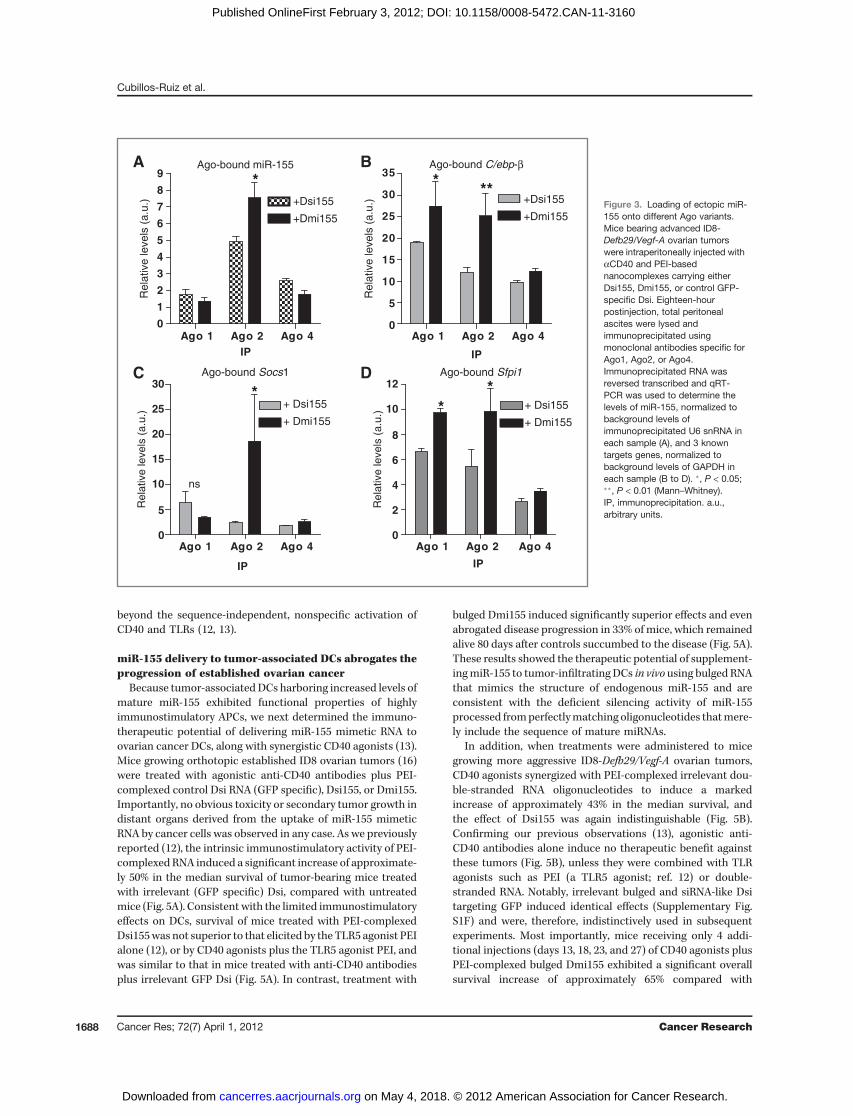
Because miR-155 is critical for DC-mediated antigen pre-sentation (7) and its expression increases in response to CD40/TLR agonists, we hypothesized that delivery of miR-155 toCD40/TLR-stimulated tumor-associated DCs could furtherimprove their antigen-presenting capacity at tumor locations.As expected, the proliferation of CFSE-labeled OT-1 T cells insitu in the ovarian cancer microenvironment was significantlyenhanced inmice pulsed with full-length OVAwhen anti-CD40plus (irrelevant) GFP Dsi-PEI nanocomplexes were adminis-tered (Fig. 4A and B). However, delivery of bulged Dmi155induced a stronger antigen-specific T-cell proliferation attumor locations (Fig. 4A and B), indicating that ectopic sup-plementation of miR-155 robustly enhances the immunosti-mulatory capacity of tumor-associated DCs beyond TLRactivation.
Consistent with improved antigen presentation, higher pro-portions of antigen-experienced (CD44þ) CD4þ and CD8þ Tcells were found in the tumor microenvironment of micetreated 4 times with CD40 agonists plus GFP Dsi-PEI nano-complexes, compared with untreated mice (Fig. 4C and D andSupplementary Fig. S1C). The proportion of antigen-experi-enced T cells at tumor (peritoneal) locations was again furtherincreased by treatment with PEI-complexed (bulged) Dmi155.Consistent with ineffective loading onto Ago2, the effect ofperfectly matching Dsi155 was not superior to control GFP Dsiformulations (Fig. 4C and D and Supplementary Fig. S1C).Correspondingly, the number of tumor (peritoneal) T cells
Cancer Res; 72(7) April 1, 2012 Cancer Research1686
Cubillos-Ruiz et al.
on May 4, 2018. © 2012 American Association for Cancer Research. cancerres.aacrjournals.org Downloaded from
Published OnlineFirst February 3, 2012; DOI: 10.1158/0008-5472.CAN-11-3160
secreting Granzyme B in ELISPOT analyses in response totumor antigens was also significantly increased upon admin-istration of bulged Dmi155, whereas treatment with siRNA-likeDsi155 did not enhance tumor antigen–specific T-cellresponses more than control GFP Dsi (Fig. 4E). Likewise, micetreated with Dmi155 also showed a marked increase in thenumbers of splenic T cells secreting Granzyme B upon restim-ulation with tumor antigens (Fig. 4F). These responses weretumor specific because they were significantly diminishedwhen antigen-presenting cells (APC) were pulsed with irrele-vant (3T3) cells in independent experiments (SupplementaryFig. S1D). Furthermore, Dmi155 treatment resulted in a sig-
nificant increase in the proportions of total splenic CD8þ Tcells exhibiting central memory attributes (CD44þ
CD62Lþ; Fig. 4G and Supplementary Fig. S1E). Finally, in vivoproduction of Th1 cytokines with antitumor potential such asTNFa, IL-12, IFNg , and CCL5 (19, 31) was significantlyenhanced at tumor locations in mice receiving Dmi155, com-pared with Dsi155 or control RNA (Fig. 4H). Together, theseresults indicated that delivery of bulged pre-miR-155 mimeticRNA, but not siRNA-like reagents, enhances the capacity ofotherwise regulatory DCs at tumor locations to effectivelypresent antigen, boost T-cell–dependent antitumor immunity,and induce the secretion of immunostimulatory cytokines
A
CD
45
Control (Dsi-Cy3)-PEI
Dsi-Cy3 CD11c
97%~40%
of CD45
C
F GE
tDCs
+ GFP D
si-P
EI
+ Dsi
155-
PEI
+ Dm
i155
-PEI
0
10
20
30
40
50
60
70
Rela
tive C
/ebp
β expre
ssio
n
**
tDCs
+ GFP
Dsi
-PEI
+ Dsi
155-
PEI
+ Dm
i155
-PEI
0
10
20
30
40
50
60
70
Re
lative
Sfp
i1 e
xp
ressio
n
tDCs
+ GFP
Dsi
-PEI
+ Dsi
155-
PEI
+ Dm
i155
-PEI
0
20
40
60
80
Rela
tive S
ocs1
expre
ssio
n
***
tDCs
+GFPD
si-P
EI
+Dsi15
5-PEI
+Dm
i155
-PEI
1
10
100
1,000
10,000
Rela
tive m
iR-1
55 e
xpre
ssio
n
D
Rela
tive m
iR-1
42
expre
ssio
n
tDCs
+Dm
i155
-PEI
+Dsi15
5-PEI
+GFP D
si-P
EI
- αCD40 Abs
0
125
225
0
125
225+αCD40 Abs
tDCs
+GFP D
si-P
EI
+Dsi15
5-PEI
+Dm
i155
-PEI
B
100
101
102
103
104
100
101
102
103
104
Gr1
CD
11b
78%
16%
Gated on rhodamine+CD11c–
Figure 2. PEI-based nanocomplexes encapsulating functional Dmi155 are preferentially engulfed by tumor-associated DCs in vivo. A, Cy3-labeled Dmi155nanocomplexes were intraperitoneally injected into mice bearing advanced ID8-Defb29/Vegf-A tumors. Fluorescence-activated cell sorting (FACS) analysiswas done after 18 hours. Data are representative of 5 independent experiments. B, detailed analysis of leukocytes incorporating rhodamine-labelednanocomplexes. Shaded histogram, isotype control staining. C, mice bearing advanced ID8-Defb29/Vegf-A ovarian tumors were left untreated or injectedintraperitoneally with PEI complexed with different RNA duplexes. Eighteen hours later, CD11cþMHC-IIþ tumor-associated DCs (tDCs) were sortedfrom peritoneal wash samples. Mature miR-155 was quantified by stem-loop qRT-PCR and the expression normalized to U6 snRNA. Data are representativeof 2 independent experiments. D, mice were treated as in C or additionally injected with 50 mg of aCD40 3 hours before nanoparticle administration.miR-142-p5 expression in sorted tumor-associated DCswas quantified and normalized to U6 snRNA. E to G, expression of miR-155mRNA targets in tumor-associated DCs engulfing PEI-Dsi nanocomplexes for 18 hours. Data were determined by qRT-PCR, normalized to glyceraldehyde-3-phosphatedehydrogenase (GAPDH) and are representative of 2 independent experiments with similar results. �, P < 0.05; ��, P < 0.01 (Mann–Whitney).
www.aacrjournals.org Cancer Res; 72(7) April 1, 2012 1687
Synthetic miRNA Elicits Protective Antitumor Immunity
on May 4, 2018. © 2012 American Association for Cancer Research. cancerres.aacrjournals.org Downloaded from
Published OnlineFirst February 3, 2012; DOI: 10.1158/0008-5472.CAN-11-3160
beyond the sequence-independent, nonspecific activation ofCD40 and TLRs (12, 13).
miR-155 delivery to tumor-associated DCs abrogates theprogression of established ovarian cancer
Because tumor-associated DCs harboring increased levels ofmature miR-155 exhibited functional properties of highlyimmunostimulatory APCs, we next determined the immuno-therapeutic potential of delivering miR-155 mimetic RNA toovarian cancer DCs, along with synergistic CD40 agonists (13).Mice growing orthotopic established ID8 ovarian tumors (16)were treated with agonistic anti-CD40 antibodies plus PEI-complexed control Dsi RNA (GFP specific), Dsi155, or Dmi155.Importantly, no obvious toxicity or secondary tumor growth indistant organs derived from the uptake of miR-155 mimeticRNA by cancer cells was observed in any case. As we previouslyreported (12), the intrinsic immunostimulatory activity of PEI-complexed RNA induced a significant increase of approximate-ly 50% in the median survival of tumor-bearing mice treatedwith irrelevant (GFP specific) Dsi, compared with untreatedmice (Fig. 5A). Consistent with the limited immunostimulatoryeffects on DCs, survival of mice treated with PEI-complexedDsi155was not superior to that elicited by theTLR5 agonist PEIalone (12), or by CD40 agonists plus the TLR5 agonist PEI, andwas similar to that in mice treated with anti-CD40 antibodiesplus irrelevant GFP Dsi (Fig. 5A). In contrast, treatment with
bulged Dmi155 induced significantly superior effects and evenabrogated disease progression in 33% of mice, which remainedalive 80 days after controls succumbed to the disease (Fig. 5A).These results showed the therapeutic potential of supplement-ingmiR-155 to tumor-infiltrating DCs in vivo using bulged RNAthat mimics the structure of endogenous miR-155 and areconsistent with the deficient silencing activity of miR-155processed fromperfectlymatching oligonucleotides thatmere-ly include the sequence of mature miRNAs.
In addition, when treatments were administered to micegrowing more aggressive ID8-Defb29/Vegf-A ovarian tumors,CD40 agonists synergized with PEI-complexed irrelevant dou-ble-stranded RNA oligonucleotides to induce a markedincrease of approximately 43% in the median survival, andthe effect of Dsi155 was again indistinguishable (Fig. 5B).Confirming our previous observations (13), agonistic anti-CD40 antibodies alone induce no therapeutic benefit againstthese tumors (Fig. 5B), unless they were combined with TLRagonists such as PEI (a TLR5 agonist; ref. 12) or double-stranded RNA. Notably, irrelevant bulged and siRNA-like Dsitargeting GFP induced identical effects (Supplementary Fig.S1F) and were, therefore, indistinctively used in subsequentexperiments. Most importantly, mice receiving only 4 addi-tional injections (days 13, 18, 23, and 27) of CD40 agonists plusPEI-complexed bulged Dmi155 exhibited a significant overallsurvival increase of approximately 65% compared with
A Ago-bound C/ebp-β
Ago 1 Ago 2 Ago 40
5
10
15
20
25
30
35
+Dsi155
+Dmi155
IPR
ela
tive levels
(a.u
.)
***
C Ago-bound Sfpi1
Ago 1 Ago 2 Ago 40
2
4
6
8
10
12
+ Dsi155
+ Dmi155
IP
Re
lative
le
ve
ls (
a.u
.) *
*DAgo-bound Socs1
Ago 1 Ago 2 Ago 40
5
10
15
20
25
30
+ Dsi155
+ Dmi155
IP
Re
lative
le
ve
ls (
a.u
.)
*
ns
Ago-bound miR-155
Ago 1 Ago 2 Ago 40
1
2
3
4
5
6
7
8
9
+Dsi155
+Dmi155
IP
Rela
tive levels
(a.u
.)
*B
Figure 3. Loading of ectopic miR-155 onto different Ago variants.Mice bearing advanced ID8-Defb29/Vegf-A ovarian tumorswere intraperitoneally injected withaCD40 and PEI-basednanocomplexes carrying eitherDsi155, Dmi155, or control GFP-specific Dsi. Eighteen-hourpostinjection, total peritonealascites were lysed andimmunoprecipitated usingmonoclonal antibodies specific forAgo1, Ago2, or Ago4.Immunoprecipitated RNA wasreversed transcribed and qRT-PCR was used to determine thelevels of miR-155, normalized tobackground levels ofimmunoprecipitated U6 snRNA ineach sample (A), and 3 knowntargets genes, normalized tobackground levels of GAPDH ineach sample (B to D). �, P < 0.05;��, P < 0.01 (Mann–Whitney).IP, immunoprecipitation. a.u.,arbitrary units.
Cubillos-Ruiz et al.
Cancer Res; 72(7) April 1, 2012 Cancer Research1688
on May 4, 2018. © 2012 American Association for Cancer Research. cancerres.aacrjournals.org Downloaded from
Published OnlineFirst February 3, 2012; DOI: 10.1158/0008-5472.CAN-11-3160
untreatedmice. This therapeutic effect was significantly stron-ger than that induced by an identical schedule of GFP Dsi-PEIor Dsi155-PEI treatments (Fig. 5B).
To confirm the antitumor effects ofmiR-155mimetics in theabsence of the mRNAs upregulated by CD40 activation, wefinally treated aggressive tumor-bearingmice with an identical
A
Untreated αCD40 + GFP Dsi αCD40 + Dmi155
42% 65% 93%
CFSE
B
Unt
reat
ed
αCD40
+ G
FP Dsi
αCD40
+ D
mi155
0
20
40
60
80
100
Cells
div
ided (
%)
C D
Unt
reat
ed
αCD40
+ G
FP Dsi
αCD40
+ D
si15
5
αCD40
+ D
mi155
0
10
20
30
40
50
60
70
CD
4+C
D44
+ T
cells
(%
)
E F
G*
**
Unt
reat
ed0
10
20
30
40
50
60 **
*
IL-12
0
40
80
120
160 IFNγ
0
5
10
15
20
25
30
35 CCL5
0
100
200
300
400TNFα
0
200
400
600
800
1000 **
Concentr
ation (
pg/m
L)
Unt
reat
ed
αCD40
+ G
FP Dsi
αCD40
+ D
si15
5
αCD40
+ D
mi155
** ** **
Unt
reat
ed
αCD40
+ G
FP Dsi
αCD40
+ D
si15
5
αCD40
+ D
mi155
Unt
reat
ed
Unt
reat
ed
H
Unt
reat
ed
αCD40
+ G
FP Dsi
αCD40
+ D
si15
5
αCD40
+ D
mi155
0
25
50
75
100
125
150
# G
rzB
spots
/10
5 c
ells
**
**
*
*
Unt
reat
ed
αCD40
+ G
FP Dsi
αCD40
+ D
si15
5
αCD40
+ D
mi155
0
5
10
15
20
25**
*
CD
8+C
D44
+ T
cells
(%
)
# G
rzB
spots
/10
5 c
ells
Unt
reat
ed0
2
4
6
8
10
12
CD
8+C
D62L
+C
D44
+ T
cells
(%
)
Unt
reat
ed
αCD40
+ G
FP Dsi
αCD40
+ D
mi155
0.0
0.5
1.0
1.5
2.0
Div
isio
n index
αCD40
+ G
FP Dsi
αCD40
+ D
si15
5
αCD40
+ D
mi155
αCD40
+ G
FP Dsi
αCD40
+ D
si15
5
αCD40
+ D
mi155
αCD40
+ G
FP Dsi
αCD40
+ D
si15
5
αCD40
+ D
mi155
αCD40
+ G
FP Dsi
αCD40
+ D
si15
5
αCD40
+ D
mi155
Figure 4. miR-155 delivery to tumor-associated DCs enhances antigen presentation and triggers antitumor immunity. A, mice growing ID8-Defb29/Vegf-Aovarian tumors for 3 weeks received 0.6 mg full-length endotoxin-free OVA (SIGMA, grade VII) intraperitoneally. Three hours later, mice were left untreated orinjected with 50 mg anti-CD40 followed by 50 mg PEI-Dsi (N/P 6). Eighteen hours later, mice received 2 � 106 CFSE-labeled OVA-specific CD3þ T cellsnegatively purified from OT-1 transgenic mice (intraperitoneally). Peritoneal wash samples (10 mL) were collected 48 hours later and T-cell proliferation wasanalyzed by FACS on the basis of CFSE dilution. B, left, percentage of cells divided in duplicate for each sample; right, division index of proliferating cells(FlowJo). Data are representative of 2 different mice per group. C to H, enhanced antitumor immune responses in mice treated with aCD40 plus Dmi155-PEInanocomplexes. ID8-Defb29/Vegf-A tumor-bearing mice (n ¼ 3 per group, 2 independent experiments) were treated at days 8, 13, 18, and 23 post tumorinjection and peritoneal wash samples were analyzed at day 27. The proportion of antigen-experienced CD4þ (C) and CD8þ (D) T cells infiltrating tumorlocationswasdeterminedbyFACS (gatedonCD3þcells). E andF, representative ELISPOTanalysis showing increasednumbers of tumor-reactive,GranzymeB–secreting T cells in the peritoneal cavity (E) or spleens (F) of mice treated with a-CD40 and Dmi155-PEI nanoparticles. GrzB, Granzyme B. G, proportion ofCD8þ T cells exhibiting central memory-like markers in the spleen of treated mice. H, total ascites supernatants were collected 18 hours after theadministration of each indicated treatment and cytokinesweremeasuredbyBio-plex. Data are representative of 2 experiments. �,P <0.05; ��,P<0.01 (Mann–Whitney in all cases).
www.aacrjournals.org Cancer Res; 72(7) April 1, 2012 1689
Synthetic miRNA Elicits Protective Antitumor Immunity
on May 4, 2018. © 2012 American Association for Cancer Research. cancerres.aacrjournals.org Downloaded from
Published OnlineFirst February 3, 2012; DOI: 10.1158/0008-5472.CAN-11-3160

regimen of only control or miR-155 mimicking compounds. Asshown in Fig. 5C, corresponding, although obviously weakereffects were observed. Notably, survival increases resultingfrom miR-155 supplementation were associated with T-cell–dependent protection because T cells from CD40/Dmi155-treated mice restrained tumor growth upon rechallenge, com-pared with T cells from untreated mice (Fig. 5D). Together,these results showed that only Dmi155 mimicking the bulgedstructure of endogenous pre-miR-155 is able to induce ther-apeutic benefits and synergize with the in situ activation ofCD40 to extend survival in hosts bearing established aggressiveovarian carcinomas.
In vivo delivery of miR-155mimetic RNA reprograms thetranscriptome of tumor-associated DCs
To understand how mature miR-155 processed from deliv-ered Dmi155 promotes the immunostimulatory phenotype oftumor-associated DCs in such striking manner, we next ana-lyzed transcriptional changes in treated mice. Strikingly, deepsequencing analysis of the transcriptome of tumor-associatedCD45þCD11cþMHC-IIþ DCs revealed that Dmi155, directly orindirectly, induced the silencing of thousands of transcripts,including multiple genes associated with an immunosuppres-sive phenotype (Supplementary File S1). Overall, 48% of totalgenes detected in tumor-associated DCs were downregulated2-fold or more at the mRNA level by Dmi155 treatment. Thoseincluded known immunosuppressive targets of miR-155 suchas C/epbb, recently described as critical regulator of theimmunosuppressive environment created by growing cancers(32); multiple mediators of Tgf-b signaling pathway, includingTgfb1, Smad1, Smad6, and Smad7; and Ccl22, which recruitsregulatory T cells to the tumor microenvironment (18). Unex-pectedly, we also found that Satb1, a master genomic organizer(33), is expressed in tumor-associated DCs and silenced bymiR-155.
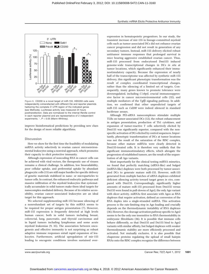
In addition, we found downregulation of multiple tran-scripts not previously associated with miR-155. We focusedon Cd200, a know mediator of DC-induced tolerance (34).Supporting that Cd200 is indeed a bona fide immunosup-pressive target of miR-155, luciferase activity was silencedby Dmi155, but not by irrelevant Dsi, in the presence of the30-untranslated region (30-UTR) of Cd200 (Fig. 6). The spec-ificity of the analysis is supported by the parallel silencing ofSatb1, recently confirmed as a target of miR-155, but notof Pdcd4, the expression of which is not significantly alteredin vivo (Fig. 6).
Interestingly, Cd200 is not a predicted target of miR-155 inany major databases. This is not surprising because 56% ofpublished targets of miR-155 are also not contained in anymajor databases, including Miranda, Targetscan, DianaMT,miRDB, Mirwalk, PITA, RNA22, and PicTar.
Together, these data indicated that the transformation ofplastic DCs at tumor locations into immunostimulatory cellsby synthetic miR-155 is the result of complex genome-widetranscriptional changes rather than the silencing of a limitedset of targets. In addition, our optimization ofmiRNAmimeticsand delivery system provides multiple experimental hints fornew targets of individual miRNAs, which should help to
00
20
40
60
80
100
GFP Dsi-PEIDsi155-PEI
Dmi155-PEI
35 40 45 50 55 60 65
00
20
40
60
80
100
Untreated
αCD40
αCD40 + GFP Dsi-PEIαCD40 + Dsi155-PEI
35 40 45 50 55 60 65
αCD40 + Dmi155-PEI
days
B
A
**
*
**
0 70 80 90 100 110 120 130 140 1500
25
50
75
100
Untreated
PEIαCD40 + PEI
αCD40 + GFP Dsi-PEI
αCD40 + Dsi155-PEIαCD40 + Dmi155-PEI
**
**
Surv
ival (%
)
Untreated
C
Surv
ival (%
)
days
Surv
ival (%
)
days
D
0
25
50
75
100
125
150
175
200
225
αCD40
+
Dm
i155
-PEI
Unt
reat
ed
Tu
mor
volu
me (
mm
3)
**
Figure 5. miR-155 delivery to tumor-associated DCs abrogatesprogression of established ovarian cancers. A,mice growing parental ID8tumors (6 per group) received aCD40 antibodies and PEI-complexed Dsiat days 15, 21, 27, 28, 33, 48, and 63. Dmi155-treated mice received 2more injections at days114 and129, after Dsi155-treatedmice havedied.ID8-Defb29/Vegf-A tumor-bearingmice (6 per group) were treated after 8days with PEI–Dsi nanocomplexes in the presence (B) or absence (C) ofa-CD40 agonistic antibodies. Additional treatments were given at days13, 18, 23, and 27. D, sublethally irradiated healthy C57BL/6 micereceived T cells negatively purified from the spleens of ID8-Defb29/Vegf-A tumor-bearing mice treated with PBS or a-CD40 agonistic antibodiesplusDmi155-PEI nanoparticles andwere then challenged in the flankwiththe same ovarian carcinoma cells. Tumor growth in both groups wasmonitored 26 days later. Left, side-by-side comparison of resectedtumors. Right, average tumor size in both groups.�, P < 0.05; ��, P < 0.01(log-rank or Student t test).
Cancer Res; 72(7) April 1, 2012 Cancer Research1690
Cubillos-Ruiz et al.
on May 4, 2018. © 2012 American Association for Cancer Research. cancerres.aacrjournals.org Downloaded from
Published OnlineFirst February 3, 2012; DOI: 10.1158/0008-5472.CAN-11-3160
improve bioinformatical predictions by providing new cluesfor the design of more reliable algorithms.
DiscussionHere we show for the first time the feasibility of modulating
miRNA activity selectively in ovarian cancer microenviron-mental leukocytes using a nonviral approach, which promotestheir capacity to elicit protective immunity.Although expression of noncoding RNA in cancer cells can
be achieved with viral vectors, the therapeutic use of virusesremains a clinical challenge. In addition, low bioavailability,poor cellular uptake, and preferential uptake by abundantphagocytic cells (15) are still major hurdles for specific deliveryof genetic materials stabilized in nano- or microparticles totumor cells. In contrast, the enhanced endocytic pathways andmassive infiltration of the myeloid leukocytes that systemat-ically accumulate in solid tumors make them ideal targets fornanocomplex-mediated delivery. Because of its relative acces-sibility, ovarian cancer–associated leukocytes are an idealtarget for this approach.We selected supplementing miR-155 because silencing of
a nonredundant set of targets by this miRNA seems tobe required for proper antigen presentation (7). However,miR-155 expression is frequently detected at high levelshuman cancer, both in solid tumors including breast,colorectal, lung, pancreatic, and thyroid carcinomas andin liquid tumors including lymphomas and some acutemyeloid leukemias (9, 35). The association between onco-genesis and effective immunity is not surprising as robustadaptive immune responses entail rapid expansion of leu-kocytes. Furthermore, artificial upregulation of mir-155leading to oncogenic conditions involves sustained over-
expression in hematopoietic progenitors. In our study, thetransient increase of mir-155 in lineage-committed myeloidcells such as tumor-associated DCs did not enhance ovariancancer progression and did not result in generation of anysecondary tumors. Instead, miR-155 delivery elicited robustantitumor immune responses that prolonged survival inmice bearing aggressive established ovarian cancer. Thus,miR-155 processed from endocytosed Dmi155 inducedgenome-wide transcriptional changes in DCs in situ attumor locations, which significantly enhanced their immu-nostimulatory capacity. Because the expression of nearlyhalf of the transcriptome was affected by synthetic miR-155delivery, this significant phenotypic transformation was theresult of complex coordinated transcriptional changes,rather than the silencing of a limited set of targets. Con-sequently, many genes known to promote tolerance weredownregulated, including C/ebpb, crucial immunosuppres-sive factor in cancer microenvironmental cells (32), andmultiple mediators of the Tgfb signaling pathway. In addi-tion, we confirmed that other unpredicted targets ofmiR-155 such as Cd200 were indeed silenced in standardluciferase assays.
Although PEI-siRNA nanocomplexes stimulate multipleTLRs on tumor-associated DCs (12), the robust enhancementin antigen presentation, production of Th1 cytokines, andexpansion of tumor-reactive T cells selectively elicited byDmi155 was significantly superior, compared with the non-specific activation of DCs elicited by control sequences. Impor-tantly, phenotypic transformation of DCs at tumor locationswas not the result of the saturation of the RISC complex,because other mature miRNAs were clearly detected inDmi155-treated cells. It is therefore very unlikely that thesignificant immunostimulatory effects, which abrogate theprogression of established tumors, are the result of the seques-tration of all Ago variants.
Most importantly for the clinical testing miRNA mimetics,we found that perfectly matching (siRNA-like) and bulged(miRNA-like) duplexes were both processed by tumor-associ-ated DCs to generate mature miR-155. However, miR-155generated from multiple batches of siRNA duplexes exhibiteddeficient silencing activity toward target genes in vivo, com-pared with Dmi155. Correspondingly, significantly higheramounts of mature miR-155 processed from Dmi155 versusDsi155 were found in pull-downs of Ago2, the only Ago variantwith slicer activity. miRNAs first associate with Agos as RNAduplexes that require activation, defined as conversion of theRNA duplex into a single-stranded miRNA. This activationprocess is the rate-limiting step in Ago loading and cruciallydepends on the thermodynamic instability of RNA duplexes(36). However, the cleavage activation pathway specific toAgo2seems to be the only one insensitive to RNA thermostability inembryonic fibroblasts (36). It is possible that immune cellsbehave differently, so that Dmi155 and Dsi155 bind to Ago2variants with similar affinity, but bulged duplexes with weakerthermodynamic stability are more efficiently processed andactivated. Not mutually exclusive, it is also possible thatchaperone proteins regulating the upload of small hairpinRNAs onto the RISC complex recognize the difference between
0
25
50
75
100
125
None
Pdcd4
Satb1
CD200
3′ -UTR:
*
*
Luci
fera
se a
ctiv
ity
(%)
No
Dsi
Dm
i GFP
Dm
i155
No
Dsi
Dm
i GFP
Dm
i155
No
Dsi
Dm
i GFP
Dm
i155
No
Dsi
Dm
i GFP
Dm
i155
Figure 6. CD200 is a novel target of miR-155. HEK293 cells wereindependently cotransfected with different Dsi and reporter plasmidsharboring the complete 30-UTR region of the indicated genes(see Methods). Luciferase activity was measured 24 hoursposttransfection. Data are normalized to the internal Renilla controlin each reporter plasmid and are representative of 2 independentexperiments. �, P < 0.05 (Mann–Whitney).
www.aacrjournals.org Cancer Res; 72(7) April 1, 2012 1691
Synthetic miRNA Elicits Protective Antitumor Immunity
on May 4, 2018. © 2012 American Association for Cancer Research. cancerres.aacrjournals.org Downloaded from
Published OnlineFirst February 3, 2012; DOI: 10.1158/0008-5472.CAN-11-3160

a bulged versus a matching structure in DCs, so that differentcompositions are incorporated with distinct efficiency.
In summary, our results show the feasibility of deliveringsynthetic miRNAs to tumor microenvironmental cells as anovel cancer intervention and provide fundamental clues forthe optimization of this approach.
Disclosure of Potential Conflicts of InterestNo potential conflicts of interest were disclosed.
Authors' ContributionsConception and design: J.R. Cubillos-Ruiz, J.R. Baird, S.N. Fiering, L.F. Sempere,and J.R. Conejo-Garcia.Development of methodology: J.R. Cubillos-Ruiz, J.R. Baird, S.N. Fiering, L.F.Sempere, and J.R. Conejo-Garcia.Acquisition of data (provided animals, acquired and managed patients,provided facilities, etc.): J.R. Cubillos-Ruiz, J.R. Baird, A.J. Tesone, M.R.Rutkowski, A.L. Camposeco-Jacobs, J. Anadon-Arnillas, N.M. Harwood, M. Korc,and J.R. Conejo-Garcia.Analysis and interpretation of data (e.g., statistical analysis, biostatistics,computational analysis): J.R. Cubillos-Ruiz, J.R. Baird, A.J. Tesone, M.R.Rutkowski, J. Anadon-Arnillas, N.M. Harwood, M. Korc, S.N. Fiering, and J.R.Conejo-Garcia.
Writing, review, and/or revision of the manuscript: J.R. Cubillos-Ruiz, A.J.Tesone, M.R. Rutkowski, U.K. Scarlett, S.N. Fiering, L.F. Sempere, and J.R. Conejo-Garcia.Administrative, technical, or material support (i.e., reporting or orga-nizing data, constructing databases): J.R. Baird.Generated data: U.K. Scarlett.Study supervision: S.N. Fiering and J.R. Conejo-Garcia.
AcknowledgmentsThe authors thank the Bioinformatics, Genomics, Flow Cytometry, and
Animal Facilities at The Wistar Institute and the Genomics and MicroarrayLaboratory at Dartmouth.
Grant SupportThis study was supported by NCI grants CA157664, CA124515, CA124515S,
CA132026, CA141017 and P30CA010815, and by DoD grant OC100059 (to J.R.Conejo-Garcia). J.R. Cubillos-Ruiz was supported by the 2009–2010 John H.Copenhaver, Jr. and William H. Thomas, MD 1952 Fellowship.
The costs of publication of this article were defrayed in part by thepayment of page charges. This article must therefore be hereby markedadvertisement in accordance with 18 U.S.C. Section 1734 solely to indicate thisfact.
Received September 20, 2011; revised January 16, 2012; accepted January 23,2012; published OnlineFirst February 3, 2012.
References1. Ambros V. The functions of animal microRNAs. Nature 2004;431:350–
5.2. Bartel DP. MicroRNAs: target recognition and regulatory functions.
Cell 2009;136:215–33.3. Chi SW, Zang JB, Mele A, Darnell RB. Argonaute HITS-CLIP decodes
microRNA-mRNA interaction maps. Nature 2009;460:479–86.4. Sempere LF, Kauppinen KS. Translational implications of microRNAs
in clinical diagnostics and therapeutics. 2nd edition. ed. Oxford:Academic Press.; 2009.
5. Li QJ, Chau J, Ebert PJ, Sylvester G,Min H, Liu G, et al. miR-181a is anintrinsic modulator of T cell sensitivity and selection. Cell 2007;129:147–61.
6. O'Connell RM, Rao DS, Chaudhuri AA, Baltimore D. Physiological andpathological roles for microRNAs in the immune system. Nat RevImmunol 2010;10;111–22.
7. Rodriguez A, Vigorito E, Clare S, Warren MV, Couttet P, Soond DR,et al. Requirement of bic/microRNA-155 for normal immune function.Science 2007;316:608–11.
8. Thai TH, Calado DP, Casola S, Ansel KM, Xiao C, Xue Y, et al.Regulation of the germinal center response by microRNA-155. Sci-ence 2007;316:604–8.
9. Xiao C, Rajewsky K. MicroRNA control in the immune system: basicprinciples. Cell 2009;136:26–36.
10. O'Connell RM, Rao DS, Chaudhuri AA, Boldin MP, Taganov KD, NicollJ, et al. Sustained expression of microRNA-155 in hematopoietic stemcells causes a myeloproliferative disorder. J Exp Med 2008;205:585–94.
11. O'Connell RM, Taganov KD, Boldin MP, Cheng G, Baltimore D.MicroRNA-155 is induced during the macrophage inflammatoryresponse. Proc Natl Acad Sci U S A 2007;104:1604–9.
12. Cubillos-Ruiz JR, Engle X, Scarlett UK, Martinez D, Barber A, ElguetaR, et al. Polyethylenimine-based siRNA nanocomplexes reprogramtumor-associated dendritic cells via TLR5 to elicit therapeutic antitu-mor immunity. J Clin Invest 2009;119:2231–44.
13. Scarlett UK, Cubillos-Ruiz JR, Nesbeth YC, Martinez DG, Engle X,Gewirtz AT, et al. In situ stimulation of CD40 and toll-like receptor 3transforms ovarian cancer-infiltrating dendritic cells from immuno-suppressive to immunostimulatory cells. Cancer Res 2009;69:7329–37.
14. Conejo-Garcia JR, Benencia F, Courreges MC, Kang E, Mohamed-Hadley A, Buckanovich RJ, et al. Tumor-infiltrating dendritic cell
precursors recruited by a beta-defensin contribute to vasculogenesisunder the influence of Vegf-A. Nat Med 2004;10:950–8.
15. Garzon R, Marcucci G, Croce CM. Targeting microRNAs in cancer:rationale, strategies and challenges. Nat Rev Drug Discov 2010;9:775–89.
16. RobyKF, Taylor CC, Sweetwood JP,ChengY, Pace JL, TawfikO, et al.Development of a syngeneicmousemodel for events related toovariancancer. Carcinogenesis 2000;21:585–91.
17. Cubillos-Ruiz JR, Martinez D, Scarlett UK, Rutkowski MR, NesbethYC, Camposeco-Jacobs AL, et al. CD277 is a negative co-stimulatorymolecule universally expressed by ovarian cancermicroenvironmentalcells. Oncotarget 2010;1:329–8.
18. Curiel TJ, Coukos G, Zou L, Alvarez X, Cheng P, Mottram P, et al.Specific recruitment of regulatory T cells in ovarian carcinoma fostersimmune privilege and predicts reduced survival. Nat Med2004;10:942–9.
19. Nesbeth Y, Scarlett U, Cubillos-Ruiz J, Martinez D, Engle X, Turk MJ,et al. CCL5-mediated endogenous antitumor immunity elicited byadoptively transferred lymphocytes and dendritic cell depletion. Can-cer Res 2009;69:6331–8.
20. Conejo-Garcia JR,BuckanovichRJ,BenenciaF,CourregesMC,RubinSC, Carroll RG, et al. Vascular leukocytes contribute to tumor vascu-larization. Blood 2005;105:679–81.
21. Huarte E, Cubillos-Ruiz JR, Nesbeth YC, Scarlett UK, Martinez DG,Buckanovich RJ, et al. Depletion of dendritic cells delays ovariancancer progression by boosting antitumor immunity. Cancer Res2008;68:7684–91.
22. Kim DH, Behlke MA, Rose SD, Chang MS, Choi S, Rossi JJ. SyntheticdsRNA Dicer substrates enhance RNAi potency and efficacy. NatBiotechnol 2005;23:222–6.
23. Rose SD, Kim DH, Amarzguioui M, Heidel JD, Collingwood MA, DavisME, et al. Functional polarity is introduced byDicer processing of shortsubstrate RNAs. Nucleic Acids Res 2005;33:4140–56.
24. Martinez-NunezRT, LouafiF, FriedmannPS,Sanchez-Elsner T.Micro-RNA-155 modulates the pathogen binding ability of dendritic cells(DCs) by down-regulation of DC-specific intercellular adhesion mol-ecule-3 grabbing non-integrin (DC-SIGN). J Biol Chem 2009;284:16334–42.
25. Chiu YL, Ali A, Chu CY, Cao H, Rana TM. Visualizing a correlationbetween siRNA localization, cellular uptake, and RNAi in living cells.Chem Biol 2004;11:1165–75.
Cancer Res; 72(7) April 1, 2012 Cancer Research1692
Cubillos-Ruiz et al.
on May 4, 2018. © 2012 American Association for Cancer Research. cancerres.aacrjournals.org Downloaded from
Published OnlineFirst February 3, 2012; DOI: 10.1158/0008-5472.CAN-11-3160
26. He M, Xu Z, Ding T, Kuang DM, Zheng L. MicroRNA-155 regulatesinflammatory cytokine production in tumor-associated macrophagesvia targeting C/EBPbeta. Cell Mol Immunol 2009;6:343–52.
27. Costinean S, Sandhu SK, Pedersen IM, Tili E, Trotta R, Perrotti D,et al. Src homology 2 domain-containing inositol-5-phosphataseand CCAAT enhancer-binding protein beta are targeted by miR-155in B cells of Emicro-MiR-155 transgenic mice. Blood 2009;114:1374–82.
28. Lu LF, Thai TH, Calado DP, Chaudhry A, Kubo M, Tanaka K, et al.Foxp3-dependent microRNA155 confers competitive fitness to regu-latory T cells by targeting SOCS1 protein. Immunity 2009;30:80–91.
29. Winter J, Jung S, Keller S, Gregory RI, Diederichs S. Many roads tomaturity: microRNAbiogenesis pathways and their regulation. NatCellBiol 2009;11:228–34.
30. Grimm D, Wang L, Lee JS, Schurmann N, Gu S, Borner K, et al.Argonaute proteins are key determinants of RNAi efficacy, toxicity,and persistence in the adult mouse liver. J Clin Invest 2010;120:3106–19.
31. Nesbeth YC, Martinez DG, Toraya S, Scarlett UK, Cubillos-Ruiz JR,Rutkowski MR, et al. CD4 þT cells elicit host immune responsesto MHC class II- ovarian cancer through CCL5 secretion and
CD40-mediated licensing of dendritic cells. J Immunol 2010;184:5654–62.
32. Marigo I, Bosio E, Solito S, Mesa C, Fernandez A, Dolcetti L, et al.Tumor-induced tolerance and immune suppression depend on the C/EBPbeta transcription factor. Immunity 2010;32:790–802.
33. Cai S, Lee CC, Kohwi-Shigematsu T. SATB1 packages denselylooped, transcriptionally active chromatin for coordinated expressionof cytokine genes. Nat Genet 2006;38:1278–88.
34. Clark DA, Gorczynski RM, Blajchman MA. Transfusion-related immu-nomodulation due to peripheral blood dendritic cells expressing theCD200 tolerance signaling molecule and alloantigen. Transfusion2008;48:814–21.
35. Sempere LF, Preis M, Yezefski T, Ouyang H, Suriawinata AA,Silahtaroglu A, et al. Fluorescence-based codetection with proteinmarkers reveals distinct cellular compartments for altered Micro-RNA expression in solid tumors. Clin Cancer Res 2010;16:4246–55.
36. Gu S, Jin L, Zhang F, Huang Y, Grimm D, Rossi JJ, et al. Thermody-namic stability of small hairpin RNAs highly influences the loadingprocess of different mammalian Argonautes. Proc Natl Acad Sci U S A2011;108;9208–13.
www.aacrjournals.org Cancer Res; 72(7) April 1, 2012 1693
Synthetic miRNA Elicits Protective Antitumor Immunity
on May 4, 2018. © 2012 American Association for Cancer Research. cancerres.aacrjournals.org Downloaded from
Published OnlineFirst February 3, 2012; DOI: 10.1158/0008-5472.CAN-11-3160
2012;72:1683-1693. Published OnlineFirst February 3, 2012.Cancer Res Juan R. Cubillos-Ruiz, Jason R. Baird, Amelia J. Tesone, et al. CancermiRNA Mimetics Triggers Protective Immunity against Ovarian
UsingIn VivoReprogramming Tumor-Associated Dendritic Cells
Updated version
10.1158/0008-5472.CAN-11-3160doi:
Access the most recent version of this article at:
Material
Supplementary
http://cancerres.aacrjournals.org/content/suppl/2012/02/03/0008-5472.CAN-11-3160.DC1
Access the most recent supplemental material at:
Cited articles
http://cancerres.aacrjournals.org/content/72/7/1683.full#ref-list-1
This article cites 35 articles, 13 of which you can access for free at:
Citing articles
http://cancerres.aacrjournals.org/content/72/7/1683.full#related-urls
This article has been cited by 8 HighWire-hosted articles. Access the articles at:
E-mail alerts related to this article or journal.Sign up to receive free email-alerts
Subscriptions
Reprints and
To order reprints of this article or to subscribe to the journal, contact the AACR Publications Department at
Permissions
Rightslink site. Click on "Request Permissions" which will take you to the Copyright Clearance Center's (CCC)
.http://cancerres.aacrjournals.org/content/72/7/1683To request permission to re-use all or part of this article, use this link
on May 4, 2018. © 2012 American Association for Cancer Research. cancerres.aacrjournals.org Downloaded from
Published OnlineFirst February 3, 2012; DOI: 10.1158/0008-5472.CAN-11-3160