Reprinted with permission from DENTAL ... - Schick by...
4
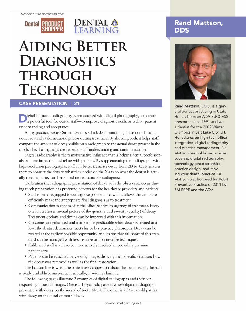
www.dentallearning.net CASE PRESENTATION | 21 Aiding Better Diagnostics through Technology D igital intraoral radiography, when coupled with digital photography, can create a powerful tool for dental staff—to improve diagnostic skills, as well as patient understanding and acceptance. At my practice, we use Sirona Dental’s Schick 33 intraoral digital sensors. In addi- tion, I routinely take intraoral photos during treatment. By showing both, it helps staff compare the amount of decay visible on a radiograph to the actual decay present in the tooth. This sharing helps create better staff understanding and communication. Digital radiography is the transformative influence that is helping dental profession- als be more impactful and relate with patients. By supplementing the radiographs with high-resolution photographs, staff can better translate decay from 2D to 3D. It enables them to connect the dots to what they notice on the X-ray to what the dentist is actu- ally treating—they can better and more accurately codiagnose. Calibrating the radiographic presentation of decay with the observable decay dur- ing tooth preparation has profound benefits for the healthcare providers and patients: • Staff is better equipped to codiagnose problem areas. This allows the dentist to efficiently make the appropriate final diagnosis as to treatment. • Communication is enhanced in the office relative to urgency of treatment. Every- one has a clearer mental picture of the quantity and severity (quality) of decay. Treatment options and timing can be improved with this information. • Outcomes are enhanced and made more predictable when decay is treated at a level the dentist determines meets his or her practice philosophy. Decay can be treated at the earliest possible opportunity and lesions that fall short of this stan- dard can be managed with less invasive or non invasive techniques. • Calibrated staff is able to be more actively involved in providing premium patient care. • Patients can be educated by viewing images showing their specific situation; how the decay was removed as well as the final restoration. The bottom line is when the patient asks a question about their oral health, the staff is ready and able to answer academically, as well as clinically. The following pages illustrate 2 examples of digital radiographs and their cor- responding intraoral images. One is a 17-year-old patient whose digital radiographs presented with decay on the mesial of tooth No. 4. The other is a 24-year-old patient with decay on the distal of tooth No. 4. Rand Mattson, DDS, is a gen- eral dentist practicing in Utah. He has been an ADA SUCCESS presenter since 1991 and was a dentist for the 2002 Winter Olympics in Salt Lake City, UT. He lectures on high-tech office integration, digital radiography, and practice management. Dr. Mattson has published articles covering digital radiography, technology, practice ethics, practice design, and mov- ing your dental practice. Dr. Mattson was honored for Adult Preventive Practice of 2011 by 3M ESPE and the ADA. Reprinted with permission from D ENTAL LEARNING Rand Mattson, DDS
Transcript of Reprinted with permission from DENTAL ... - Schick by...
www.dentallearning.net
CASE PRESENTATION | 21
Aiding Better Diagnostics throughTechnology
Digital intraoral radiography, when coupled with digital photography, can create a powerful tool for dental staff—to improve diagnostic skills, as well as patient
understanding and acceptance. At my practice, we use Sirona Dental’s Schick 33 intraoral digital sensors. In addi-
tion, I routinely take intraoral photos during treatment. By showing both, it helps staff compare the amount of decay visible on a radiograph to the actual decay present in the tooth. This sharing helps create better staff understanding and communication.
Digital radiography is the transformative in� uence that is helping dental profession-als be more impactful and relate with patients. By supplementing the radiographs with high-resolution photographs, staff can better translate decay from 2D to 3D. It enables them to connect the dots to what they notice on the X-ray to what the dentist is actu-ally treating—they can better and more accurately codiagnose.
Calibrating the radiographic presentation of decay with the observable decay dur-ing tooth preparation has profound bene� ts for the healthcare providers and patients:
• Staff is better equipped to codiagnose problem areas. This allows the dentist to ef� ciently make the appropriate � nal diagnosis as to treatment.
• Communication is enhanced in the of� ce relative to urgency of treatment. Every-one has a clearer mental picture of the quantity and severity (quality) of decay. Treatment options and timing can be improved with this information.
• Outcomes are enhanced and made more predictable when decay is treated at a level the dentist determines meets his or her practice philosophy. Decay can be treated at the earliest possible opportunity and lesions that fall short of this stan-dard can be managed with less invasive or non invasive techniques.
• Calibrated staff is able to be more actively involved in providing premium patient care.
• Patients can be educated by viewing images showing their speci� c situation; how the decay was removed as well as the � nal restoration.
The bottom line is when the patient asks a question about their oral health, the staff is ready and able to answer academically, as well as clinically.
The following pages illustrate 2 examples of digital radiographs and their cor-responding intraoral images. One is a 17-year-old patient whose digital radiographs presented with decay on the mesial of tooth No. 4. The other is a 24-year-old patient with decay on the distal of tooth No. 4.
Rand Mattson, DDS, is a gen-eral dentist practicing in Utah. He has been an ADA SUCCESS presenter since 1991 and was a dentist for the 2002 Winter Olympics in Salt Lake City, UT. He lectures on high-tech of� ce integration, digital radiography, and practice management. Dr. Mattson has published articles covering digital radiography, technology, practice ethics, practice design, and mov-ing your dental practice. Dr. Mattson was honored for Adult Preventive Practice of 2011 by 3M ESPE and the ADA.
Reprinted with permission from
DENTAL LEARNING
Rand Mattson, DDS

Figure 3—The decay has invaded the enamel and has progressed into the dentin. Note the color change.
Figure 2—Preoperative view of tooth No. 4. The image was taken with an Iris intraoral camera (Digital Doc) during the preparation of the tooth before re�ning the preparation, isolation, and placement of the �nal restoration.
Figure 1—A 17-year-old male presented with radiographic decay on the mesial of tooth No. 4. The radiograph was taken with a Schick 33 digital sensor (Sirona Dental), using the AimRight parallel positioning device (Sirona Dental).
www.dentallearning.net

Figure 7—Decay in the � oor of the box. Note the color of the decaying dentin.Figure 6—Decay as it passes the dentoenamel junction.
Figure 5—A pre-op view of tooth No. 4, taken during the preparation of the tooth before re� ning the prep, isolation, and placement of the � nal restoration.
Figure 4—A 24-year-old male presented with decay on the distal of tooth No. 4.
GO-TO PRODUCTS USED IN THIS CASE
EAGLESOFTEaglesoft practice management software allows the clinician to manage the entire practice with 1 system and customize it for the way they work. Eliminating the need for paper charts, the all-digital Eaglesoft system is both powerful and intuitive, allowing the clinician to focus on patient care while it operates in the background.
PATTERSON DENTAL800.690.1826 ext. 99068www.pattersondental.com
PDCARE WIPESPatterson Dental’s pdCARE Wipes are disposable disinfecting towelettes that are intended as a surface cleaner and disinfectant on hard, nonporous surfaces in health care settings such as dental of� ces, hospitals, laboratories and clinics. They are proven effective as a tuberculocide, bactericide, virucide and fungicide in a 2-3 minute window.
PATTERSON DENTAL800.690.1826 ext. 99068www.pattersondental.com
SCHICK 33Schick 33 is an advanced digital imaging sensor that delivers a combination of high-resolution images and dynamic image management while easily integrating with the Schick Elite Platform. Schick 33’s patented imaging technology offers a theoretical resolution limit of 33 line pairs per millimeter, the highest in the industry.
SIRONA DENTAL, INC800.690.1826 ext. 99083www.sirona.com
SCHICK AIMRIGHT SENSOR HOLDERLike Sirona Dental adhesive-style holders, the AimRight system is color-coded for easy, intuitive placement. Rounded edges and corners around the holders signi� cantly increase comfort for patients during positioning, so the clinician can work ef� ciently and with greater satisfaction.
SIRONA DENTAL, INC800.690.1826 ext. 99083www.sirona.com
www.dentallearning.net

Schick 71991 Sponsorship Ad DPS-Dr Kahn— Single page, T: 7.75”w x 10”h, B: 8.75”w x 11”h
GENERAL DENTISTRY ENDODONTIC PERIODONTIC RESTORATIVEGENERAL DENTISTRY ENDODONTIC PERIODONTIC RESTORATIVE
DR. MITCHELL L. KAHN, DMD – YORKTOWN HEIGHTS, NY
WITH MORE WITH MORE DIAGNOSTIC CONTROL, DIAGNOSTIC CONTROL, I HAVE MORE WAYS TO I HAVE MORE WAYS TO MAKE A DIFFERENCE.MAKE A DIFFERENCE.
INTRODUCING SCHICK 33: THE HIGHEST
RESOLUTION SENSOR + POWERFULLY SIMPLE
IMAGE MANAGEMENT SOFTWARE.
With its user-friendly dynamic image enhancer,
image preference presets and industry-leading
resolution, the Schick 33 Digital Intraoral Sensor
and Image Management System raises the level of
care by meeting the needs of every dental practice.
Unprecedented control. Unmatched image quality.
Unequaled diagnostics. Schick 33 changes digital
diagnostics so you can change your world.
Learn more at 33.schickbysirona.com/DPS or call
1-800-873-7683 for a free in-offi ce demonstration.
THAT’S INSPIRING.THAT’S INSPIRING.
OUR PARTNER IN PROVIDING YOU UNPARALLELED SERVICE + SUPPORT. 100% SATISFACTION GUARANTEED.
GLOBALNUMBER
71911_Schick_33SpnsrshpAD_DrKahn_DPS.indd 1 8/13/13 3:14 PM