Regulatory Cohesion of Cell Cycle and Cell Differentiation ...
12
Regulatory Cohesion of Cell Cycle and Cell Differentiation through Interlinked Phosphorylation and Second Messenger Networks The MIT Faculty has made this article openly available. Please share how this access benefits you. Your story matters. Citation Abel, Soren, Peter Chien, Paul Wassmann, Tilman Schirmer, Volkhard Kaever, Michael T. Laub, Tania A. Baker, and Urs Jenal. “Regulatory Cohesion of Cell Cycle and Cell Differentiation through Interlinked Phosphorylation and Second Messenger Networks.” Molecular Cell 43, no. 4 (August 2011): 550-560. © 2011 Elsevier Inc. As Published http://dx.doi.org/10.1016/j.molcel.2011.07.018 Publisher Elsevier B.V. Version Final published version Citable link http://hdl.handle.net/1721.1/82942 Terms of Use Article is made available in accordance with the publisher's policy and may be subject to US copyright law. Please refer to the publisher's site for terms of use.
Transcript of Regulatory Cohesion of Cell Cycle and Cell Differentiation ...
Regulatory Cohesion of Cell Cycle and Cell Differentiation throughInterlinked Phosphorylation and Second Messenger Networks
The MIT Faculty has made this article openly available. Please share how this access benefits you. Your story matters.
Citation Abel, Soren, Peter Chien, Paul Wassmann, Tilman Schirmer,Volkhard Kaever, Michael T. Laub, Tania A. Baker, and Urs Jenal.“Regulatory Cohesion of Cell Cycle and Cell Differentiation throughInterlinked Phosphorylation and Second Messenger Networks.”Molecular Cell 43, no. 4 (August 2011): 550-560. © 2011 Elsevier Inc.
As Published http://dx.doi.org/10.1016/j.molcel.2011.07.018
Publisher Elsevier B.V.
Version Final published version
Citable link http://hdl.handle.net/1721.1/82942
Terms of Use Article is made available in accordance with the publisher'spolicy and may be subject to US copyright law. Please refer to thepublisher's site for terms of use.
Molecular Cell
Article
Regulatory Cohesion of Cell Cycle and CellDifferentiation through InterlinkedPhosphorylation and Second Messenger NetworksSoren Abel,1,7 Peter Chien,2,5,7 Paul Wassmann,1,6 Tilman Schirmer,1 Volkhard Kaever,4 Michael T. Laub,2,3
Tania A. Baker,2,3 and Urs Jenal1,*1Biozentrum of the University of Basel, Klingelbergstrasse 50, CH-4054 Basel, Switzerland2Department of Biology3Howard Hughes Medical Institute
Massachusetts Institute of Technology, 77 Massachusetts Avenue, Cambridge, MA 02139, USA4Institute of Pharmacology, Hannover Medical School, 30625 Hannover, Germany5Present address: Department of Biochemistry and Molecular Biology, University of Massachusetts Amherst, Amherst, MA 01003, USA6Present address: Glenmark Pharmaceuticals S.A., 2300 La Chaux-de-Fonds, Switzerland7These authors contributed equally to this work
*Correspondence: [email protected]
DOI 10.1016/j.molcel.2011.07.018
SUMMARY
In Caulobacter crescentus, phosphorylation of keyregulators is coordinatedwith the secondmessengercyclic di-GMP to drive cell-cycle progression anddifferentiation. The diguanylate cyclase PleD directspole morphogenesis, while the c-di-GMP effectorPopA initiates degradation of the replication inhibitorCtrA by the AAA+ protease ClpXP to license S phaseentry. Here, we establish a direct link between PleDand PopA reliant on the phosphodiesterase PdeAand thediguanylate cyclaseDgcB.PdeAantagonizesDgcB activity until the G1-S transition, when PdeA isdegraded by the ClpXP protease. The unopposedDgcB activity, together with PleD activation, upshiftsc-di-GMP to drive PopA-dependent CtrA degrada-tion and S phase entry. PdeA degradation requiresCpdR, a response regulator that delivers PdeA tothe ClpXP protease in a phosphorylation-dependentmanner. Thus, CpdR serves as a crucial link betweenphosphorylation pathways and c-di-GMP metabo-lism to mediate protein degradation events that irre-versibly and coordinately drive bacterial cell-cycleprogression and development.
INTRODUCTION
The development of all living organisms depends on the genera-
tion of specialized cells in appropriate numbers. This requires
tight regulation of proliferation-differentiation decisions by inte-
grating cell-fate determination processes with replication and
cell division. For example, embryonic development and tissue
homeostasis requires a strict balance between cells in amultipo-
tent, self-renewing state and differentiated nonreplicative cells
(Yadirgi and Marino, 2009). Just how differentiation and prolifer-
ation processes are coordinated remains poorly understood
(Bateman and McNeill, 2004; Caro et al., 2007). Many bacteria
use complex developmental strategies to optimize their survival.
Like their eukaryotic counterparts, bacteria tightly coordinate
morphogenetic programs with growth and division, be this to
facilitate the transition between a replicative and a terminally
differentiated cell form (Errington, 2003; Flardh and Buttner,
2009; Kaiser, 2008) or to couple cell differentiation to cell prolif-
eration (Curtis and Brun, 2010). In the gram-negative bacterium
Caulobacter crescentus an asymmetric division produces two
daughters with distinct morphologies, behavior, and replicative
potential, a motile swarmer cell and a sessile stalked cell.
Whereas the newborn stalked cell immediately enters S phase
to initiate chromosome replication, the swarmer cell inherits
a chromosome that remains quiescent for an extended period
termed the G1 phase. Concurrent with the morphological transi-
tion of the swarmer cell into a stalked cell, the replication block is
suspended and cells proceed first into S phase to double their
chromosomes and then into G2 phase to undergo cell division.
C. crescentus uses two-component phosphorylation systems
to integrate cellular asymmetry with the processes that deter-
mine temporal progression through the division cycle. One of
the key components of C. crescentus development and cell-
cycle control is the essential response regulator CtrA. CtrA func-
tions as a transcription factor for more than 100 cell-cycle genes
and acts as repressor of replication initiation by directly binding
to the chromosomal origin of replication (Cori) where it occludes
replication initiation factors (Laub et al., 2002; Laub et al., 2000;
Quon et al., 1998). Upon S phase entry, activated CtrA�P is elim-
inated by two redundant mechanisms, dephosphorylation, and
proteolysis (Domian et al., 1997; Domian et al., 1999), thereby
freeing the Cori and enabling replication to start. Cell cycle-
dependent degradation of CtrA (Chien et al., 2007; Jenal and
Fuchs, 1998) entails a specific spatial arrangement of the proteo-
lytic machinery. During the G1-S transition, CtrA and the ClpXP
protease both transiently localize to the incipient stalked cell
pole where CtrA is degraded (McGrath et al., 2006; Ryan et al.,
2004). Polar localization of ClpXP and CtrA are highly regulated
550 Molecular Cell 43, 550–560, August 19, 2011 ª2011 Elsevier Inc.

processes, each of which is governed by dedicated localization
factors. ClpXP localization requires CpdR, a single domain
response regulator that itself sequesters to the incipient stalked
pole subject to its phosphorylation state (Iniesta et al., 2006).
CpdR is kept in an inactive, phosphorylated form by the CckA-
ChpT phosphorylation cascade that also activates CtrA, thereby
coupling CtrA activity and stability (Biondi et al., 2006). Delivery
of CtrA to the same pole requires PopA, a protein that sequesters
to the incipient stalked pole upon binding of the second
messenger cyclic di-GMP (Duerig et al., 2009). PopA activation
and localization coincides with an upshift of c-di-GMP during
G1-S transition (Christen et al., 2010; Paul et al., 2008), arguing
that periodic fluctuations of the second messenger are an inte-
gral part of the C. crescentus cell-cycle clock. The only diguany-
late cyclase (DGC) known to modulate c-di-GMP levels during
the cell cycle is the response regulator PleD, which is inactive
in swarmer cells but is phosphorylated during differentiation to
set off pole morphogenesis (Paul et al., 2008; Paul et al., 2004).
Although PleD and PopA are activated simultaneously during
the cell cycle, genetic analysis failed to demonstrate PleD inter-
ference with PopA-mediated CtrA degradation (Duerig et al.,
2009). Hence, although c-di-GMP controls both S phase entry
and pole development it is unclear how the second messenger
serves to connect these processes and how it integrates with
the CckA-ChpT-CpdR phosphorelay, implicated in modulating
ClpXP localization.
Here, we isolate critical components of the c-di-GMP regula-
tory network in C. crescentus and uncover the molecular princi-
ples that serve to coordinate development and cell-cycle
progression. In particular, we identify the phosphodiesterase
PdeA as a key regulator of the swarmer cell program. PdeA is
limited to the swarmer cell where it antagonizes the activity of
the diguanylate cyclase DgcB and blocks differentiation. During
the G1-S transition PdeA degradation by ClpXP unleashes DgcB
activity coincident with PleD activation. DgcB and PleD together
establish the sessile stalked cell program, driving PopA-medi-
ated CtrA degradation to orchestrate pole morphogenesis with
the underlying cell cycle. Finally, we demonstrate that CpdR
directly controls PdeA degradation by acting as a phosphoryla-
tion-dependent adaptor protein for the ClpXP protease. These
experiments reveal CpdR as a central component of an inter-
woven network of phosphorylation, protein degradation, and
c-di-GMP-mediated reactions that is designed to coordinately
drive cell differentiation and cell-cycle progression.
RESULTS
The Phosphodiesterase PdeA and the DiguanylateCyclases PleD and DgcB Coordinately ControlC. crescentus DevelopmentThe DGC PleD and the c-di-GMP effector protein PopA
have been identified as regulatory components involved in
C. crescentus pole morphogenesis and cell-cycle progression,
respectively (Duerig et al., 2009; Paul et al., 2004). To identify
additional proteins of the c-di-GMP network involved in
C. crescentus cell-cycle control, we individually deleted genes
encoding potential diguanylate cyclases and phosphodiester-
ases and analyzed the resulting mutants with respect to their
morphological features and cell type-specific behavior. Deletion
of one of the selected genes, pdeA (CC3396), resulted in a
severe loss of motility and an increased propensity to attach to
surfaces, a phenotype generally associated with increased
levels of c-di-GMP (Hengge, 2009) (Figure 1A). Consistent with
these observations, the pdeA gene codes for an active EAL
phosphodiesterase (Christen et al., 2005) and cells lacking
PdeA had increased concentrations of c-di-GMP (Figure 1A).
Although pdeA mutants were unable to spread on semisolid
agar plates, they assembled a flagellum and were motile in liquid
media (data not shown). This is reminiscent of c-di-GMP medi-
ated flagellar motility control in Escherichia coli (Boehm et al.,
2010) and argued that PdeA reduces c-di-GMP to modulate
the cell-cycle interval reserved for motility and chemotaxis. The
increased surface attachment of mutants lacking PdeA corre-
lateswith an increased number of cells exhibiting a polar holdfast
(Figure 1A). In agreement with this, pdeA mutants assembled
holdfast prematurely resulting in newborn swarmer cells that
ectopically express an adhesive organelle (Figure 1B). These
results suggested that PdeA is a phosphodiesterase specifically
required for the motile swarmer cell program and that it acts by
delaying the transition to the sessile cell type.
To identify components interacting with PdeA, we isolated
motile suppressors of the pdeA mutant. One suppressor allele
mapped to gene CC1850, which codes for a protein with an
N-terminal coiled coil and a C-terminal GGDEF domain.
Biochemical studies demonstrated that CC1850 exhibits Mg2+-
dependent and Ca2+-sensitive diguanylate cyclase activity and
that the protein is a constitutive dimer in vitro (Figure S1 available
online). We thus renamed this protein DgcB. A dgcB deletion
strain showed increased motility and reduced attachment levels
(Figure 1A). Deletion of dgcB in a pdeA background restored
motility, attachment and holdfast biogenesis indicating that the
two proteins functionally interact to coordinate pole develop-
ment (Figure 1A). Importantly, dgcB pdeA double mutants
restored the proper cell-cycle timing of holdfast formation (Fig-
ure 1B) indicating that DgcB is active in G1 swarmer cells and
responsible for premature holdfast synthesis observed in
a pdeA mutant. In contrast, PdeA does not seem to functionally
interact with the DGCPleD as pleD pdeA double and pdeA single
mutants showed the same motility and holdfast timing defect
and attachment of cells lacking both the PdeA and PleD
remained as low as observed for pleD single mutants (Figure 1).
Since PleD is only active in sessile stalked cells (Paul et al.,
2008), this raised the possibility that PdeA activity is limited to
swarmer cells.
PleD and DgcB are both required for optimal attachment and
holdfast biogenesis. Whereas single mutants showed delayed
holdfast formation, an intermediate level of surface attachment
and intermediate stalk length, strains lacking both DGCs showed
markedly reduced c-di-GMP levels and a complete failure to
elongate stalks, synthesize holdfast, and attach to surfaces
(Figure 1 and Figure S2). Importantly, deleting pdeA in mutants
lacking both cyclases did not restore attachment, holdfast
formation, and c-di-GMP levels (Figure 1A). Together this indi-
cated that C. crescentus cell fate determination is governed by
the coordinated action of the phosphodiesterase PdeA and the
diguanylate cyclases PleD and DgcB.
Molecular Cell
Cohesion of Cell Cycle and Cell Differentiation
Molecular Cell 43, 550–560, August 19, 2011 ª2011 Elsevier Inc. 551
Regulated Proteolysis Limits PdeA to the MotileSwarmer Cell TypeThe functional analysis discussed above suggested that DgcB
is active both in swarmer and in stalked cells, while PdeA seems
to act exclusively in swarmer cells. With the help of polyclonal
antibodies raised against purified PdeA and DgcB, we analyzed
the distribution of both proteins. Whereas DgcB is present
throughout the cell cycle, PdeA was only observed at the very
beginning and end of the cell cycle (Figure 2A). This indicated
that PdeA is present in swarmer cells but is specifically removed
prior to S phase entry, reminiscent of the cell cycle-dependent
distribution of CtrA (Domian et al., 1997) (Figure 2A). This was
confirmed by the analysis of the cellular distribution of fluores-
cently tagged PdeA, which transiently localized to the ClpXP
occupied stalked cell pole before being cleared (Figure 2B). To
identify the protease responsible for PdeA degradation, several
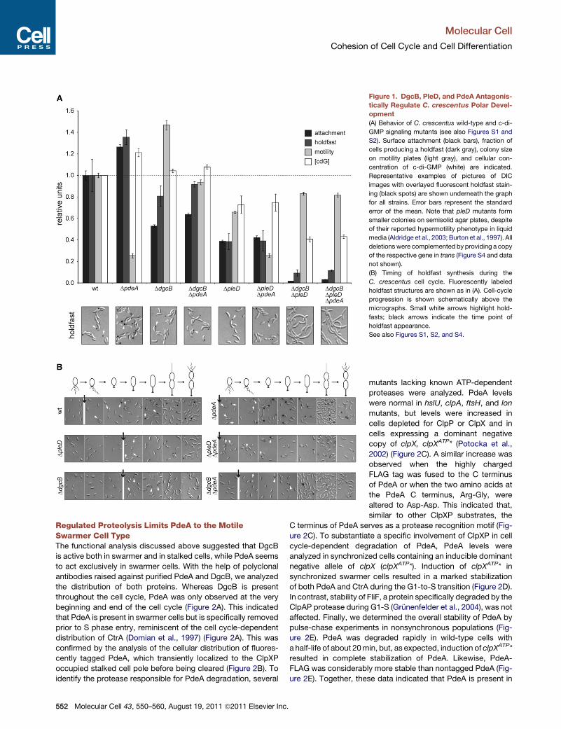
Figure 1. DgcB, PleD, and PdeA Antagonis-
tically Regulate C. crescentus Polar Devel-
opment
(A) Behavior of C. crescentus wild-type and c-di-
GMP signaling mutants (see also Figures S1 and
S2). Surface attachment (black bars), fraction of
cells producing a holdfast (dark gray), colony size
on motility plates (light gray), and cellular con-
centration of c-di-GMP (white) are indicated.
Representative examples of pictures of DIC
images with overlayed fluorescent holdfast stain-
ing (black spots) are shown underneath the graph
for all strains. Error bars represent the standard
error of the mean. Note that pleD mutants form
smaller colonies on semisolid agar plates, despite
of their reported hypermotility phenotype in liquid
media (Aldridge et al., 2003; Burton et al., 1997). All
deletionswere complemented by providing a copy
of the respective gene in trans (Figure S4 and data
not shown).
(B) Timing of holdfast synthesis during the
C. crescentus cell cycle. Fluorescently labeled
holdfast structures are shown as in (A). Cell-cycle
progression is shown schematically above the
micrographs. Small white arrows highlight hold-
fasts; black arrows indicate the time point of
holdfast appearance.
See also Figures S1, S2, and S4.
mutants lacking known ATP-dependent
proteases were analyzed. PdeA levels
were normal in hslU, clpA, ftsH, and lon
mutants, but levels were increased in
cells depleted for ClpP or ClpX and in
cells expressing a dominant negative
copy of clpX, clpXATP* (Potocka et al.,
2002) (Figure 2C). A similar increase was
observed when the highly charged
FLAG tag was fused to the C terminus
of PdeA or when the two amino acids at
the PdeA C terminus, Arg-Gly, were
altered to Asp-Asp. This indicated that,
similar to other ClpXP substrates, the
C terminus of PdeA serves as a protease recognition motif (Fig-
ure 2C). To substantiate a specific involvement of ClpXP in cell
cycle-dependent degradation of PdeA, PdeA levels were
analyzed in synchronized cells containing an inducible dominant
negative allele of clpX (clpXATP*). Induction of clpXATP* in
synchronized swarmer cells resulted in a marked stabilization
of both PdeA and CtrA during the G1-to-S transition (Figure 2D).
In contrast, stability of FliF, a protein specifically degraded by the
ClpAP protease during G1-S (Grunenfelder et al., 2004), was not
affected. Finally, we determined the overall stability of PdeA by
pulse-chase experiments in nonsynchronous populations (Fig-
ure 2E). PdeA was degraded rapidly in wild-type cells with
a half-life of about 20min, but, as expected, induction of clpXATP*
resulted in complete stabilization of PdeA. Likewise, PdeA-
FLAG was considerably more stable than nontagged PdeA (Fig-
ure 2E). Together, these data indicated that PdeA is present in
Molecular Cell
Cohesion of Cell Cycle and Cell Differentiation
552 Molecular Cell 43, 550–560, August 19, 2011 ª2011 Elsevier Inc.
swarmer cells but is specifically degraded during the G1-to-S
transition by the ClpXP protease complex.
CpdR, but Not PopA and RcdA, Is Required for CellCycle-Dependent Proteolysis of PdeATemporal control of CtrA degradation involves several factors
controlling the coordinated spatial organization of the ClpXP
protease and its substrate. This includes CpdR,which is involved
in recruiting ClpXP to the old cell pole upon S phase entry (Iniesta
et al., 2006) as well as PopA and RcdA, which are required for the
localization of CtrA to the same subcellular site (Duerig et al.,
2009; McGrath et al., 2006). To test whether ClpXP degrades
PdeA via the same pathway, we analyzed the dynamic distribu-
tion of PdeA throughout the cell cycle. An N-terminal fusion of
PdeA to the fluorescent protein Venus was found at the flagel-
lated pole of the newborn swarmer cell (Figure 2B, Figure S3A,
and Movie S1). During the swarmer-to-stalked cell transition,
VEN-PdeA disappeared from the pole coincident with its proteo-
lytic removal (Figures 2A and 2B, Figure S3A, and Movie S1).
VEN-PdeA reappeared in the predivisional cell but upon cell
constriction quickly localized to the pole in the stalked compart-
ment, while retaining a dispersed distribution in the swarmer
compartment (Figure 2B, Figure S3A, and Movie S1). This sug-
gested that VEN-PdeA redistributes to the ClpXP occupied old
cell pole to be degraded upon entry of cells into S phase. Consis-
tent with this, fusion of YFP to the C terminus of PdeA resulted
in a protein that mimicked the spatial behavior of VEN-PdeA
but persisted at the old cell pole due to shielding of the
C-terminal ClpXP degradation motif (Figure S3B).
To test whether any of the regulators involved in CtrA degrada-
tion are required for PdeA turnover, we analyzed PdeA levels
during the cell cycle in the respective mutant strains. PdeA
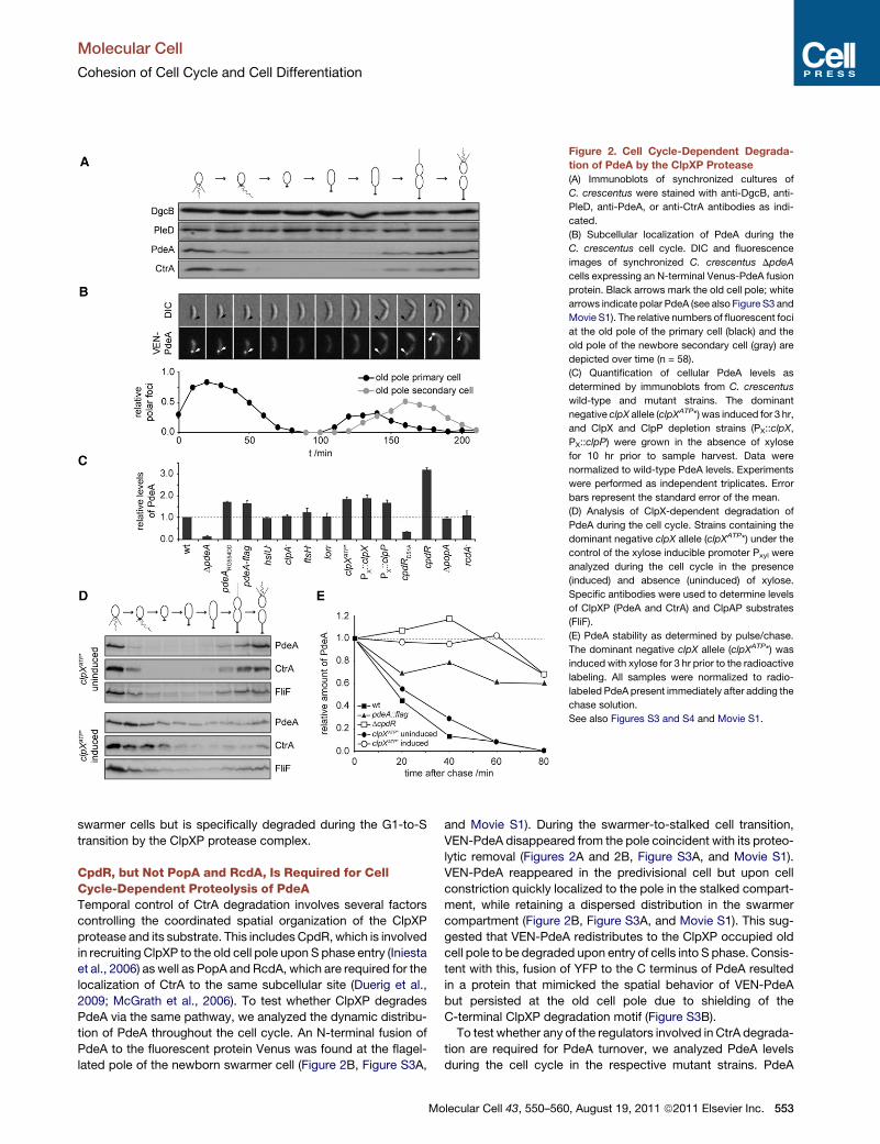
Figure 2. Cell Cycle-Dependent Degrada-
tion of PdeA by the ClpXP Protease
(A) Immunoblots of synchronized cultures of
C. crescentus were stained with anti-DgcB, anti-
PleD, anti-PdeA, or anti-CtrA antibodies as indi-
cated.
(B) Subcellular localization of PdeA during the
C. crescentus cell cycle. DIC and fluorescence
images of synchronized C. crescentus DpdeA
cells expressing an N-terminal Venus-PdeA fusion
protein. Black arrows mark the old cell pole; white
arrows indicate polar PdeA (see also Figure S3 and
Movie S1). The relative numbers of fluorescent foci
at the old pole of the primary cell (black) and the
old pole of the newbore secondary cell (gray) are
depicted over time (n = 58).
(C) Quantification of cellular PdeA levels as
determined by immunoblots from C. crescentus
wild-type and mutant strains. The dominant
negative clpX allele (clpXATP*) was induced for 3 hr,
and ClpX and ClpP depletion strains (PX::clpX,
PX::clpP) were grown in the absence of xylose
for 10 hr prior to sample harvest. Data were
normalized to wild-type PdeA levels. Experiments
were performed as independent triplicates. Error
bars represent the standard error of the mean.
(D) Analysis of ClpX-dependent degradation of
PdeA during the cell cycle. Strains containing the
dominant negative clpX allele (clpXATP*) under the
control of the xylose inducible promoter Pxyl were
analyzed during the cell cycle in the presence
(induced) and absence (uninduced) of xylose.
Specific antibodies were used to determine levels
of ClpXP (PdeA and CtrA) and ClpAP substrates
(FliF).
(E) PdeA stability as determined by pulse/chase.
The dominant negative clpX allele (clpXATP*) was
induced with xylose for 3 hr prior to the radioactive
labeling. All samples were normalized to radio-
labeled PdeA present immediately after adding the
chase solution.
See also Figures S3 and S4 and Movie S1.
Molecular Cell
Cohesion of Cell Cycle and Cell Differentiation
Molecular Cell 43, 550–560, August 19, 2011 ª2011 Elsevier Inc. 553
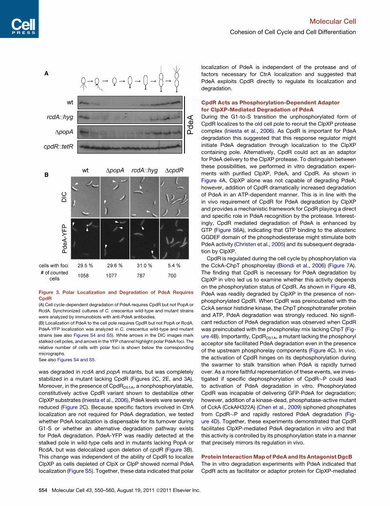
was degraded in rcdA and popA mutants, but was completely
stabilized in a mutant lacking CpdR (Figures 2C, 2E, and 3A).
Moreover, in the presence of CpdRD51A, a nonphosphorylatable,
constitutively active CpdR variant shown to destabilize other
ClpXP substrates (Iniesta et al., 2006), PdeA levels were severely
reduced (Figure 2C). Because specific factors involved in CtrA
localization are not required for PdeA degradation, we tested
whether PdeA localization is dispensable for its turnover during
G1-S or whether an alternative degradation pathway exists
for PdeA degradation. PdeA-YFP was readily detected at the
stalked pole in wild-type cells and in mutants lacking PopA or
RcdA, but was delocalized upon deletion of cpdR (Figure 3B).
This change was independent of the ability of CpdR to localize
ClpXP as cells depleted of ClpX or ClpP showed normal PdeA
localization (Figure S5). Together, these data indicated that polar
localization of PdeA is independent of the protease and of
factors necessary for CtrA localization and suggested that
PdeA exploits CpdR directly to regulate its localization and
degradation.
CpdR Acts as Phosphorylation-Dependent Adaptorfor ClpXP-Mediated Degradation of PdeADuring the G1-to-S transition the unphosphorylated form of
CpdR localizes to the old cell pole to recruit the ClpXP protease
complex (Iniesta et al., 2006). As CpdR is important for PdeA
degradation this suggested that this response regulator might
initiate PdeA degradation through localization to the ClpXP
containing pole. Alternatively, CpdR could act as an adaptor
for PdeA delivery to the ClpXP protease. To distinguish between
these possibilities, we performed in vitro degradation experi-
ments with purified ClpXP, PdeA, and CpdR. As shown in
Figure 4A, ClpXP alone was not capable of degrading PdeA;
however, addition of CpdR dramatically increased degradation
of PdeA in an ATP-dependent manner. This is in line with the
in vivo requirement of CpdR for PdeA degradation by ClpXP
and provides a mechanistic framework for CpdR playing a direct
and specific role in PdeA recognition by the protease. Interest-
ingly, CpdR mediated degradation of PdeA is enhanced by
GTP (Figure S6A), indicating that GTP binding to the allosteric
GGDEF domain of the phosphodiesterase might stimulate both
PdeA activity (Christen et al., 2005) and its subsequent degrada-
tion by ClpXP.
CpdR is regulated during the cell cycle by phosphorylation via
the CckA-ChpT phosphorelay (Biondi et al., 2006) (Figure 7A).
The finding that CpdR is necessary for PdeA degradation by
ClpXP in vitro led us to examine whether this activity depends
on the phosphorylation status of CpdR. As shown in Figure 4B,
PdeA was readily degraded by ClpXP in the presence of non-
phosphorylated CpdR. When CpdR was preincubated with the
CckA sensor histidine kinase, the ChpT phosphotransfer protein
and ATP, PdeA degradation was strongly reduced. No signifi-
cant reduction of PdeA degradation was observed when CpdR
was preincubated with the phosphorelay mix lacking ChpT (Fig-
ure 4B). Importantly, CpdRD51A, a mutant lacking the phosphoryl
acceptor site facilitated PdeA degradation even in the presence
of the upstream phosphorelay components (Figure 4C). In vivo,
the activation of CpdR hinges on its dephosphorylation during
the swarmer to stalk transition when PdeA is rapidly turned
over. As amore faithful representation of these events, we inves-
tigated if specific dephosphorylation of CpdR�P could lead
to activation of PdeA degradation in vitro. Phosphorylated
CpdR was incapable of delivering GFP-PdeA for degradation;
however, addition of a kinase-dead, phosphatase-active mutant
of CckA (CckAH322A) (Chen et al., 2009) siphoned phosphates
from CpdR�P and rapidly restored PdeA degradation (Fig-
ure 4D). Together, these experiments demonstrated that CpdR
facilitates ClpXP-mediated PdeA degradation in vitro and that
this activity is controlled by its phosphorylation state in a manner
that precisely mirrors its regulation in vivo.
Protein InteractionMapof PdeAand Its Antagonist DgcBThe in vitro degradation experiments with PdeA indicated that
CpdR acts as facilitator or adaptor protein for ClpXP-mediated
Figure 3. Polar Localization and Degradation of PdeA Requires
CpdR
(A) Cell cycle-dependent degradation of PdeA requires CpdR but not PopA or
RcdA. Synchronized cultures of C. crescentus wild-type and mutant strains
were analyzed by immunoblots with anti-PdeA antibodies.
(B) Localization of PdeA to the cell pole requires CpdR but not PopA or RcdA.
PdeA-YFP localization was analyzed in C. crescentus wild-type and mutant
strains (see also Figures S4 and S5). White arrows in the DIC images mark
stalked cell poles, and arrows in the YFP channel highlight polar PdeA foci. The
relative number of cells with polar foci is shown below the corresponding
micrographs.
See also Figures S4 and S5.
Molecular Cell
Cohesion of Cell Cycle and Cell Differentiation
554 Molecular Cell 43, 550–560, August 19, 2011 ª2011 Elsevier Inc.
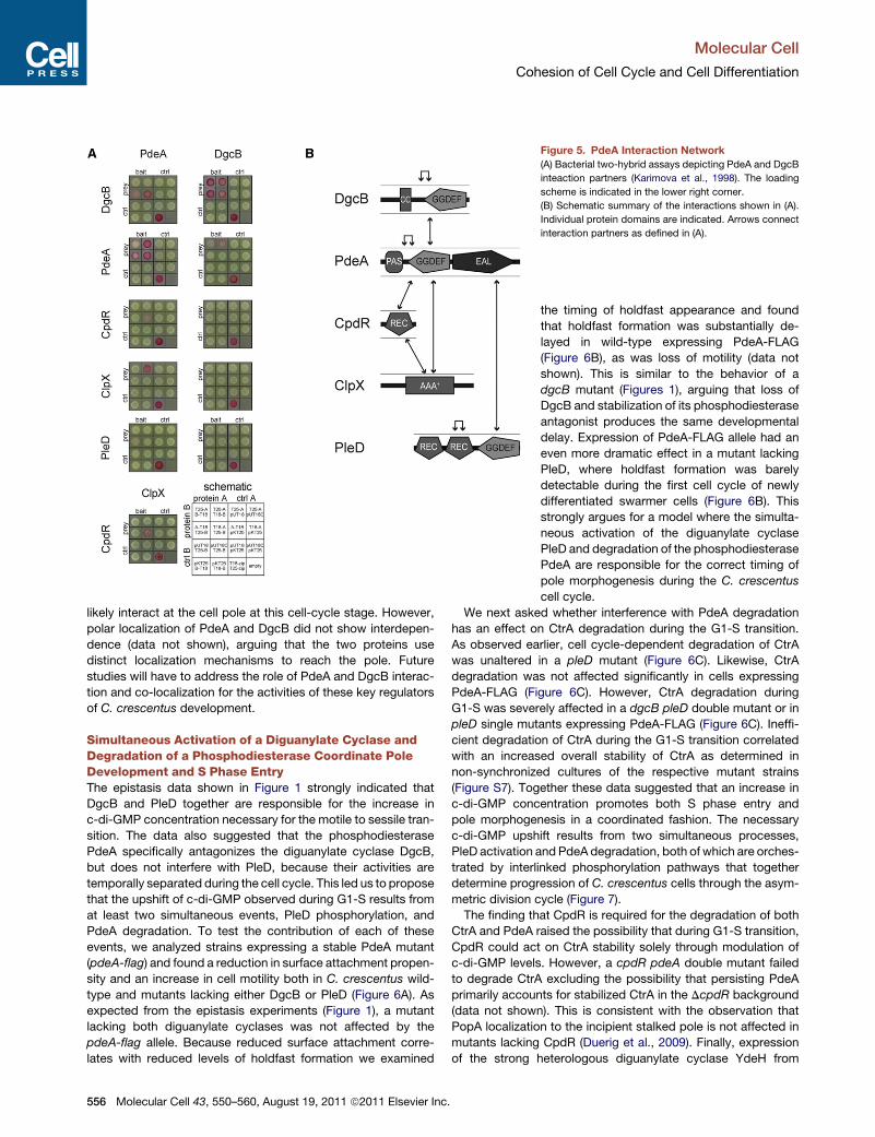
degradation of PdeA. Such a role implies that PdeA directly
interacts with both CpdR and the chaperone subunit of the
protease. To examine the direct interactions between CpdR,
PdeA, and ClpX we made use of the bacterial two-hybrid
system (BacTH) that exploits complementary fragments (T18
and T25) of Bordetella pertussis adenylate cyclase (Karimova
et al., 1998). As shown in Figure 5A, at least one combination
of fusions scored positive for PdeA interaction with ClpX
and CpdR, respectively. PdeA also strongly interacted with
itself, indicating an oligomerization-dependent activation mech-
anism. Interestingly, the BacTH assay revealed a strong inter-
action between PdeA and its functional counterpart DgcB
(Figure 5A). Since DgcB failed to interact with any of the other
components tested, this interaction appears to be specific.
PdeA-DgcB interaction was validated in vitro with purified
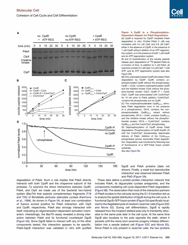
Figure 4. CpdR Is a Phosphorylation-
Dependent Adaptor for PdeA Degradation
(A) CpdR is required for ClpXP mediated PdeA
degradation in vitro. Purifed PdeA (1 mM) was
incubated with 0.4 mM ClpX and 0.8 mM ClpP,
either in the absence of CpdR, in the presence of
1 mM CpdR without addition of an ATP regenera-
tion system, or in the presence of both 1 mMCpdR
and an ATP regeneration system.
(B and C) Quantification of the soluble peptide
release upon degradation of 35S labeled PdeA as
a function of time. In addition to 2 mM PdeA, all
reactions contain 0.2 mMClpX, 0.4 mMClpP, 1 mM
GTP, and an ATP regeneration system (see also
Figure S6).
(B) Only unphosphorylated CpdR stimulates PdeA
degradation by ClpXP. CpdR, contains un-
phosphorylated CpdR without the phosphorelay;
CpdR + CckA, contains unphosphorylated CpdR
and the histidine kinase CckA without the phos-
phor-transfer protein ChpT; CpdR�P + CckA/
ChpT, CpdR was preincubated with CckA/ChpT
for 10 min prior to PdeA addition; CckA/ChpT,
contains the phosphorelay but no CpdR.
(C) The nonphosphorylateable CpdRD51A stimu-
lates PdeA degradation even in the presence
of a phosphodonor. D51A, contains the non-
phosphorylatable CpdRD51A variant without
phosphorelay; D51A + CckA, contains CpdRD51A
and the histidine kinase without the phosphor-
transfer protein; D51A + CckA/ChpT, contains
CpdRD51A and the CckA/ChpT phosphorelay.
(D) Dephosphorylation of CpdR drives PdeA
degradation. Phosphorylation of CpdR (CpdR�P)
with the CckA/ChpT phosphorelay deactivates
delivery of PdeA. Addition of the CckAH322A
phosphatase (arrow) reactivates PdeA degrada-
tion. Degradation was monitored by following loss
of fluorescence of a GFP-PdeA fusion protein
substrate.
See also Figure S6.
DgcB and PdeA proteins (data not
shown). Finally, a weak but reproducible
interaction was observed between PdeA
and PleD (Figure 5A).
These data define a protein-protein interaction network that
includes PdeA, its diguanylate cyclase antagonists, and the
components mediating cell cycle-dependent PdeA degradation
(Figure 5B). The observation that most of the interaction partners
of PdeA localize to the cell pole during the G1-S transition led us
to analyze the spatial distribution of DgcB during the cell cycle. A
functional DgcB-GFP fusion protein (Figure S4) specifically local-
ized to the flagellated pole of newborn swarmer cells (Figure S3C
and Movie S2). During cell differentiation, DgcB is rapidly
released from the incipient stalked pole only to transiently reloc-
alize to the same pole later in the cell cycle. At the same time
DgcB also localizes to the pole opposite the stalk, where it
persists until the newly born swarmer progeny initiates differen-
tiation into a sessile stalked cell (Figure S3C and Movie S2).
Since PdeA is only present in swarmer cells, the two proteins
Molecular Cell
Cohesion of Cell Cycle and Cell Differentiation
Molecular Cell 43, 550–560, August 19, 2011 ª2011 Elsevier Inc. 555
likely interact at the cell pole at this cell-cycle stage. However,
polar localization of PdeA and DgcB did not show interdepen-
dence (data not shown), arguing that the two proteins use
distinct localization mechanisms to reach the pole. Future
studies will have to address the role of PdeA and DgcB interac-
tion and co-localization for the activities of these key regulators
of C. crescentus development.
Simultaneous Activation of a Diguanylate Cyclase andDegradation of a Phosphodiesterase Coordinate PoleDevelopment and S Phase EntryThe epistasis data shown in Figure 1 strongly indicated that
DgcB and PleD together are responsible for the increase in
c-di-GMP concentration necessary for the motile to sessile tran-
sition. The data also suggested that the phosphodiesterase
PdeA specifically antagonizes the diguanylate cyclase DgcB,
but does not interfere with PleD, because their activities are
temporally separated during the cell cycle. This led us to propose
that the upshift of c-di-GMP observed during G1-S results from
at least two simultaneous events, PleD phosphorylation, and
PdeA degradation. To test the contribution of each of these
events, we analyzed strains expressing a stable PdeA mutant
(pdeA-flag) and found a reduction in surface attachment propen-
sity and an increase in cell motility both in C. crescentus wild-
type and mutants lacking either DgcB or PleD (Figure 6A). As
expected from the epistasis experiments (Figure 1), a mutant
lacking both diguanylate cyclases was not affected by the
pdeA-flag allele. Because reduced surface attachment corre-
lates with reduced levels of holdfast formation we examined
Figure 5. PdeA Interaction Network
(A) Bacterial two-hybrid assays depicting PdeA and DgcB
inteaction partners (Karimova et al., 1998). The loading
scheme is indicated in the lower right corner.
(B) Schematic summary of the interactions shown in (A).
Individual protein domains are indicated. Arrows connect
interaction partners as defined in (A).
the timing of holdfast appearance and found
that holdfast formation was substantially de-
layed in wild-type expressing PdeA-FLAG
(Figure 6B), as was loss of motility (data not
shown). This is similar to the behavior of a
dgcB mutant (Figures 1), arguing that loss of
DgcB and stabilization of its phosphodiesterase
antagonist produces the same developmental
delay. Expression of PdeA-FLAG allele had an
even more dramatic effect in a mutant lacking
PleD, where holdfast formation was barely
detectable during the first cell cycle of newly
differentiated swarmer cells (Figure 6B). This
strongly argues for a model where the simulta-
neous activation of the diguanylate cyclase
PleD and degradation of the phosphodiesterase
PdeA are responsible for the correct timing of
pole morphogenesis during the C. crescentus
cell cycle.
We next asked whether interference with PdeA degradation
has an effect on CtrA degradation during the G1-S transition.
As observed earlier, cell cycle-dependent degradation of CtrA
was unaltered in a pleD mutant (Figure 6C). Likewise, CtrA
degradation was not affected significantly in cells expressing
PdeA-FLAG (Figure 6C). However, CtrA degradation during
G1-S was severely affected in a dgcB pleD double mutant or in
pleD single mutants expressing PdeA-FLAG (Figure 6C). Ineffi-
cient degradation of CtrA during the G1-S transition correlated
with an increased overall stability of CtrA as determined in
non-synchronized cultures of the respective mutant strains
(Figure S7). Together these data suggested that an increase in
c-di-GMP concentration promotes both S phase entry and
pole morphogenesis in a coordinated fashion. The necessary
c-di-GMP upshift results from two simultaneous processes,
PleD activation and PdeA degradation, both of which are orches-
trated by interlinked phosphorylation pathways that together
determine progression of C. crescentus cells through the asym-
metric division cycle (Figure 7).
The finding that CpdR is required for the degradation of both
CtrA and PdeA raised the possibility that during G1-S transition,
CpdR could act on CtrA stability solely through modulation of
c-di-GMP levels. However, a cpdR pdeA double mutant failed
to degrade CtrA excluding the possibility that persisting PdeA
primarily accounts for stabilized CtrA in the DcpdR background
(data not shown). This is consistent with the observation that
PopA localization to the incipient stalked pole is not affected in
mutants lacking CpdR (Duerig et al., 2009). Finally, expression
of the strong heterologous diguanylate cyclase YdeH from
Molecular Cell
Cohesion of Cell Cycle and Cell Differentiation
556 Molecular Cell 43, 550–560, August 19, 2011 ª2011 Elsevier Inc.

E. coli (Boehm et al., 2009) in the cpdR mutant failed to restore
CtrA degradation (data not shown). From this we conclude that
the single domain response regulator CpdR is a bifunctional
protein that operates as protease localization factor and at the
same time acts as specific adaptor protein for certain ClpXP
substrates like PdeA.
DISCUSSION
The coupling of cell morphogenesis and proliferation allows
C. crescentus to generate specialized cell types to optimize
survival. Our results uncover how differentiation and cell-cycle
progression are coordinated in this organism by tight cohesion
of phosphorylation and c-di-GMP signaling networks. Signaling
through this network culminates in successive protein degrada-
tion events that robustly and irreversibly commit cells to S phase
and to a sessile lifestyle. At the top of this regulatory cascade are
sensor histidine kinases, which drive andmaintain the oscillatory
cell-cycle program. In particular, the CckA/ChpT phosphorelay
is responsible for the activation and stabilization of CtrA in
swarmer and predivisional cells (Biondi et al., 2006) (Figure 7).
Figure 6. Pole Development and Cell-Cycle
Progression Requires PdeA Degradation
(A) Attachment and motility of C. crescentuswild-type and
mutant strains expressing a stabilized form of PdeA. The
mean of eight (attachment) and four (motility) independent
colonies is depicted. Data are presented as relative values
of the wild-type. Error bars represent the standard error of
the mean.
(B) Cell cycle-dependent holdfast formation in strains ex-
pressing a stabilized form of PdeA. Small white arrows
highlight labeled holdfasts; black arrows indicate the time
point of holdfast appearance. Distribution of stabilized
PdeA-FLAG during the cell cycle is indicated in the
immunoblot stained with anti-PdeA antibodies.
(C) Cell cycle-dependent degradation of CtrA in strains
with altered c-di-GMP metabolism. Synchronized
swarmer cells of wild-type and mutants were followed
throughout the cell cycle. CtrA protein levels were
analyzed in immunoblots. Immunoblots with an anti-CcrM
antibody are shown as control for cell-cycle progression.
See also Figure S7.
CtrA stability control is governed through phos-
phorylation-mediated inactivation of CpdR,
which maintains the ClpXP protease in a delo-
calized state in these cell types (Iniesta et al.,
2006). We show here that in addition to stabi-
lizing CtrA, the CckA pathway also stabilizes
the PdeA phosphodiesterase via CpdR phos-
phorylation. Our data demonstrate that CpdR
in its nonphosphorylated form directly facili-
tates ClpXP-dependent degradation of PdeA.
Thus, the accumulation of non-phosphorylated
CpdR during the G1-to-S transition mediates
the rapid degradation of the replication initiation
inhibitor CtrA by distinct mechanisms. First,
CpdR affects polar localization of the ClpXP
protease at the G1-S transition; second,
CpdR-mediated delivery of PdeA to the polar ClpXP complex
contributes to the upshift in c-di-GMP, the activation of PopA,
and thus the recruitment of CtrA to the ClpXP-occupied cell
pole (Figure 7).
The phosphate flux through the CckA-ChpT pathway reverses
prior to S phase entry, contributing to CtrA and CpdR dephos-
phorylation and, ultimately, replication initiation. This activity is
coordinated with a second phosphorylation pathway involved
in G1-S transition that triggers the synthesis of c-di-GMP
through phosphorylation of the PleD diguanylate cyclase
(Aldridge et al., 2003; Paul et al., 2004) (Figure 7). The cell
type-specific activity of this pathway relies on the spatial
dynamic behavior of two sensor histidine kinases, PleC and
DivJ, which position to opposite poles of the Caulobacter
predivisional cell and differentially segregate into the daughter
progeny (McAdams and Shapiro, 2003). PleC is a phosphatase
in swarmer cells but during cell differentiation adopts strong
kinase activity and, together with the newly synthesized DivJ
kinase, promotes a rapid upshift of c-di-GMP through the activa-
tion of PleD (Aldridge et al., 2003; Paul et al., 2004). Reversal of
PleC activity is implemented by the essential single domain
Molecular Cell
Cohesion of Cell Cycle and Cell Differentiation
Molecular Cell 43, 550–560, August 19, 2011 ª2011 Elsevier Inc. 557
response regulator DivK that acts as an allosteric activator of
both PleC and DivJ autokinase activity in sessile stalked cells
(Paul et al., 2008). At the same time, activated DivK downregu-
lates the CckA-ChpT pathway to contribute to the removal of
active CtrA during G1-S transition (Biondi et al., 2006; Tsokos
et al., 2011) and through this mechanism helps to adjust the
activity of the two cell type-specific phosphorylation pathways
during the C. crescentus cell cycle (Figure 7).
While DivK links the two phosphorylation networks at the level
of the kinase activities, our work reveals CpdR as an additional
key component coordinating these two pathways. Although
CpdR was originally identified as a polar recruitment factor for
the ClpXP protease, we show here that CpdR also serves as
a specific adaptor to deliver the PdeA phosphodiesterase to
ClpXP prior to S phase entry. Adaptor proteins for AAA+ prote-
ases increase the stringency of substrate selection and alter
priorities of target degradation (Kirstein et al., 2009). Before
this work, the only characterized adaptor in C. crescentus was
SspB that delivers ssrA-tagged substrates to the ClpXP protease
(Chien et al., 2007). However, in contrast to the SspB adaptor
case, delivery of PdeA by CpdR is dependent on the phosphor-
ylation status of the adaptor. Although cell cycle-regulated acti-
vation is unprecedented for ClpXP adaptors, phosphorylation
dependent changes in adaptor function have been described
before (Kirstein et al., 2006; Mika and Hengge, 2005). Thus, it
appears that coupling of adaptor phosphorylation with adaptor
activity is a conserved mechanism to control specific substrate
delivery. The observation that CpdR itself is degraded by the
ClpXP protease (Iniesta and Shapiro, 2008) suggests that
the ClpXP pathway can be rapidly inactivated in S phase by
the simultaneous removal of adaptor and substrate protein.
Several lines of evidence argue that DgcB and PdeA function
as antagonists and that PdeA, due to its dominance over DgcB,
establishes themotile program in swarmer cells. Howdoes PdeA
‘‘neutralize’’ DgcB in swarmer cells? A simple explanation would
be that PdeA is catalytically more active than its antagonist. The
direct physical coupling of PdeA and DgcB could enhance this
effect. Similar to the concept of ‘‘metabolic channeling’’ (Con-
rado et al., 2008), such an arrangement could increase the
efficiency of PdeA control over DgcB by preventing diffusion of
c-di-GMP into the surrounding cytoplasm. This ‘‘futile cycle’’
mechanism, although seemingly wasteful, may provide for a
rapid response to environmental signals that can override the
internal cell-cycle control. Alternatively, PdeA could directly
control DgcB activity through allosteric or inhibitory effects
resulting from the simple physical interaction between the two
proteins. The observation that a PdeA active site mutation
shows the same phenotype as a pdeA deletion mutation argues
against such a scenario (data not shown). We have shown that
PdeA activity is allosterically stimulated by GTP binding to its
regulatory GGDEF domain (Christen et al., 2005). While the
kinetic parameters suggest that PdeA is fully induced under
physiological conditions, certain conditions could lead to a (local)
drop in GTP that would be readily transduced into an increase in
c-di-GMP by downregulating PdeA. Clearly, we are just begin-
ning to understand how CpdR, PdeA, PleD, PopA and ClpXP
collaborate to drive development and cell-cycle progression.
Understanding the dynamic nature of these complexes at the
cell pole (Figure 7) will be the aim of future work.
EXPERIMENTAL PROCEDURES
More-detailed descriptions of experimental procedures and a list of all plas-
mids and strains (Table S1) are provided in the Supplemental Experimental
Procedures.
Microscopy
Fluorescence and differential interference contrast (DIC) microscopy were
performed on a DeltaVision Core (Applied Precision, USA)/Olympus IX71
microscope equipped with an UPlanSApo 1003/1.40 Oil objective (Olympus,
Japan) and a coolSNAP HQ-2 (Photometrics, USA) CCD camera. Cells were
placed on a PYE pad solidified with 1% agarose (Sigma, USA). Images were
processed and analyzed with softWoRx version 5.0.0 (Applied Precision,
USA) and Photoshop CS3 (Adobe, USA) software.
Bacterial Two-Hybrid Experiments
Bacterial two hybrid screens were performed according to Karimova et al.
(1998). Full open reading frames or gene fragments were fused to the 30 endof the T25 (pKT25), the 30 end of the T18 (pUT18C) or the 50 end of the T18
(pUT18) fragment of the gene coding for Bordetella pertussis adenylate
cyclase. Two microliter of a MG1655 cyaA::frt culture containing the relevant
plasmids were spotted on a MacConkey Agar Base plate supplemented
with kanamycin, ampicilin and maltose, incubated at 30�C.
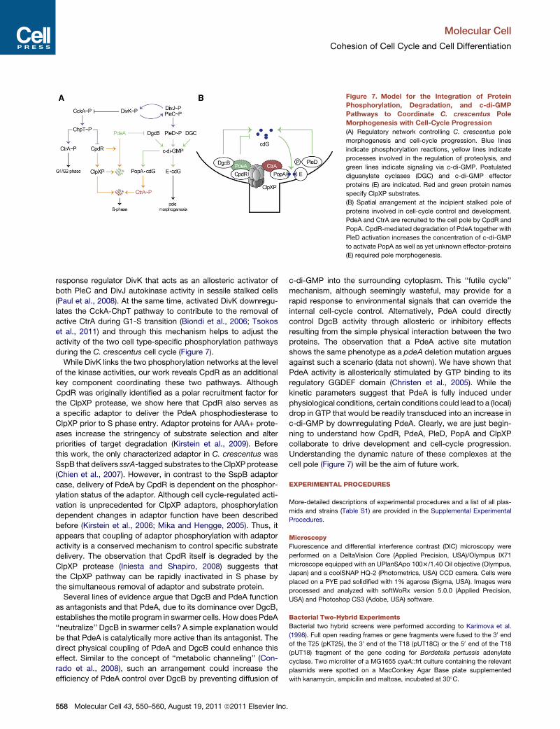
Figure 7. Model for the Integration of Protein
Phosphorylation, Degradation, and c-di-GMP
Pathways to Coordinate C. crescentus Pole
Morphogenesis with Cell-Cycle Progression
(A) Regulatory network controlling C. crescentus pole
morphogenesis and cell-cycle progression. Blue lines
indicate phosphorylation reactions, yellow lines indicate
processes involved in the regulation of proteolysis, and
green lines indicate signaling via c-di-GMP. Postulated
diguanylate cyclases (DGC) and c-di-GMP effector
proteins (E) are indicated. Red and green protein names
specify ClpXP substrates.
(B) Spatial arrangement at the incipient stalked pole of
proteins involved in cell-cycle control and development.
PdeA and CtrA are recruited to the cell pole by CpdR and
PopA. CpdR-mediated degradation of PdeA together with
PleD activation increases the concentration of c-di-GMP
to activate PopA as well as yet unknown effector-proteins
(E) required pole morphogenesis.
Molecular Cell
Cohesion of Cell Cycle and Cell Differentiation
558 Molecular Cell 43, 550–560, August 19, 2011 ª2011 Elsevier Inc.
Protein Expression and Purification
E. coli BL21 (DE3) pLys (Stratagene, USA) carrying the dgcB expression
plasmid were grown in LB medium to an OD600 of 0.5 before expression
was induced by adding isopropyl 1-thio-b-D-galactopyranoside (IPTG) to
a final concentration of 0.5 mM. Cells were harvested by centrifugation, resus-
pended in 20 mM HEPES (pH 7.5), 100 mM KCl, 20 mM imidazole running
buffer and lysed by passage through a French pressure cell. Clarified crude
extract was loaded on a HP HisTrap column (GE Healthcare, UK) attached
to an AKTApurifier (GE Healthcare, UK). Protein was eluted by raising the
imidazole concentration to 500 mM in running buffer. Further purification
and buffer exchange were performed by size-exclusion chromatography on
a Superdex200 HR26/60 column (GE Healthcare, UK) with 20 mM HEPES
(pH 7.5), 100 mM KCl as running buffer. PdeA and CpdR were expressed as
C-terminal fusions to a his-tagged SUMO domain in BL21DE3 plysS cells.
Purification of the fusions, cleavage of the SUMO domain, and separation of
the cleaved protein were performed as before (Wang et al., 2007). GFP-
PdeA was purified via standard Ni-NTA protocols (QIAGEN). Subsequent puri-
fication was performed via size-exclusion chromatography (Sephacryl S-300
for PdeA and GFP-PdeA; Superdex-75 for CpdR) with 20 mM HEPES,
100 mM KCl, 5 mM MgCl2, 5 mM Beta-Mercaptoethanol, 10% Glycerol
(pH 7.4) used as the buffer (H-buffer). Monomeric fractions were concentrated
and stored at �80�C. Radiolabeled PdeA was produced by labeling with [35S]
labeled L-methionine (Wang et al., 2007) and purification as above, with the
exception of the gel-filtration step. ClpX and ClpP were purified as before
(Chien et al., 2007).
Protease Assay
PdeA degradation was assayed at 30�C in H-buffer (20 mM HEPES, 100 mM
KCl, 5 mM MgCl2, 5 mM Beta-Mercaptoethanol, 10% Glycerol [pH 7.4]). For
typical qualitative reactions, 1 mM PdeA was incubated with 1 mM GTP,
0.4 mM ClpX6, 0.8 mM ClpP14, 1 mM CpdR, and an ATP regeneration system
(Chien et al., 2007). Aliquots were removed at indicated times and quenched
by addition of SDS loading dye and immediately frozen. Samples were
separated by SDS-PAGE and stained with Coomassie for visualization. For
quantitative assays, degradation of radiolabeled PdeA was monitored by
measurement of TCA soluble peptides (Wang et al., 2007). Degradation of
GFP-PdeA was performed in the same conditions as above with the exception
that GFP fluorescence was continuously monitored in 384-well plates with
a Spectramax M5 (Molecular Devices). Phosphorylation of CpdR was per-
formed as before (Chen et al., 2009).
SUPPLEMENTAL INFORMATION
Supplemental Information includes Supplemental Experimental Procedures,
seven figures, one table, and two movies and can be found with this article
online at doi:10.1016/j.molcel.2011.07.018.
ACKNOWLEDGMENTS
We thank Anna Duerig and Fabienne Hamburger for strains and plasmids,
Samuel Steiner for strain E. coli MG1655 DcyaA::frt, and Pia Abel zur Wiesch
for help with data analysis and for critical reading of the manuscript. This
work was supported by Swiss National Science Foundation grants 31-
108186 and 31003A_130469 to U.J., National Institutes of Health grants
GM-082899 to M.T.L., GM-049224 to T.A.B., and GM-084157 to P.C.;
T.A.B. and M.T.L. are employees of the Howard Hughes Medical Institute.
Received: January 6, 2011
Revised: May 27, 2011
Accepted: July 25, 2011
Published: August 18, 2011
REFERENCES
Aldridge, P., Paul, R., Goymer, P., Rainey, P., and Jenal, U. (2003). Role of
the GGDEF regulator PleD in polar development of Caulobacter crescentus.
Mol. Microbiol. 47, 1695–1708.
Bateman, J.M., and McNeill, H. (2004). Temporal control of differentiation by
the insulin receptor/tor pathway in Drosophila. Cell 119, 87–96.
Biondi, E.G., Reisinger, S.J., Skerker, J.M., Arif, M., Perchuk, B.S., Ryan, K.R.,
and Laub, M.T. (2006). Regulation of the bacterial cell cycle by an integrated
genetic circuit. Nature 444, 899–904.
Boehm, A., Steiner, S., Zaehringer, F., Casanova, A., Hamburger, F., Ritz, D.,
Keck, W., Ackermann, M., Schirmer, T., and Jenal, U. (2009). Second
messenger signalling governs Escherichia coli biofilm induction upon ribo-
somal stress. Mol. Microbiol. 72, 1500–1516.
Boehm, A., Kaiser, M., Li, H., Spangler, C., Kasper, C.A., Ackermann, M.,
Kaever, V., Sourjik, V., Roth, V., and Jenal, U. (2010). Second messenger-
mediated adjustment of bacterial swimming velocity. Cell 141, 107–116.
Burton, G.J., Hecht, G.B., and Newton, A. (1997). Roles of the histidine protein
kinase pleC in Caulobacter crescentus motility and chemotaxis. J. Bacteriol.
179, 5849–5853.
Caro, E., Castellano, M.M., and Gutierrez, C. (2007). A chromatin link that
couples cell division to root epidermis patterning in Arabidopsis. Nature 447,
213–217.
Chen, Y.E., Tsokos, C.G., Biondi, E.G., Perchuk, B.S., and Laub, M.T. (2009).
Dynamics of two Phosphorelays controlling cell cycle progression in
Caulobacter crescentus. J. Bacteriol. 191, 7417–7429.
Chien, P., Perchuk, B.S., Laub,M.T., Sauer, R.T., andBaker, T.A. (2007). Direct
and adaptor-mediated substrate recognition by an essential AAA+ protease.
Proc. Natl. Acad. Sci. USA 104, 6590–6595.
Christen, M., Christen, B., Folcher, M., Schauerte, A., and Jenal, U. (2005).
Identification and characterization of a cyclic di-GMP-specific phosphodies-
terase and its allosteric control by GTP. J. Biol. Chem. 280, 30829–30837.
Christen, M., Kulasekara, H.D., Christen, B., Kulasekara, B.R., Hoffman, L.R.,
and Miller, S.I. (2010). Asymmetrical distribution of the second messenger
c-di-GMP upon bacterial cell division. Science 328, 1295–1297.
Conrado, R.J., Varner, J.D., and DeLisa, M.P. (2008). Engineering the spatial
organization of metabolic enzymes: mimicking nature’s synergy. Curr. Opin.
Biotechnol. 19, 492–499.
Curtis, P.D., and Brun, Y.V. (2010). Getting in the loop: regulation of develop-
ment in Caulobacter crescentus. Microbiol. Mol. Biol. Rev. 74, 13–41.
Domian, I.J., Quon, K.C., and Shapiro, L. (1997). Cell type-specific phosphor-
ylation and proteolysis of a transcriptional regulator controls the G1-to-S tran-
sition in a bacterial cell cycle. Cell 90, 415–424.
Domian, I.J., Reisenauer, A., and Shapiro, L. (1999). Feedback control of
a master bacterial cell-cycle regulator. Proc. Natl. Acad. Sci. USA 96, 6648–
6653.
Duerig, A., Abel, S., Folcher, M., Nicollier, M., Schwede, T., Amiot, N., Giese,
B., and Jenal, U. (2009). Second messenger-mediated spatiotemporal control
of protein degradation regulates bacterial cell cycle progression. Genes Dev.
23, 93–104.
Errington, J. (2003). Regulation of endospore formation in Bacillus subtilis. Nat.
Rev. Microbiol. 1, 117–126.
Flardh, K., and Buttner, M.J. (2009). Streptomyces morphogenetics: dissect-
ing differentiation in a filamentous bacterium. Nat. Rev. Microbiol. 7, 36–49.
Grunenfelder, B., Tawfilis, S., Gehrig, S., ØSteras, M., Eglin, D., and Jenal, U.
(2004). Identification of the protease and the turnover signal responsible for
cell cycle-dependent degradation of the Caulobacter FliF motor protein.
J. Bacteriol. 186, 4960–4971.
Hengge, R. (2009). Principles of c-di-GMP signalling in bacteria. Nat. Rev.
Microbiol. 7, 263–273.
Iniesta, A.A., and Shapiro, L. (2008). A bacterial control circuit integrates polar
localization and proteolysis of key regulatory proteins with a phospho-
signaling cascade. Proc. Natl. Acad. Sci. USA 105, 16602–16607.
Iniesta, A.A., McGrath, P.T., Reisenauer, A., McAdams, H.H., and Shapiro, L.
(2006). A phospho-signaling pathway controls the localization and activity of
a protease complex critical for bacterial cell cycle progression. Proc. Natl.
Acad. Sci. USA 103, 10935–10940.
Molecular Cell
Cohesion of Cell Cycle and Cell Differentiation
Molecular Cell 43, 550–560, August 19, 2011 ª2011 Elsevier Inc. 559

Jenal, U., and Fuchs, T. (1998). An essential protease involved in bacterial cell-
cycle control. EMBO J. 17, 5658–5669.
Kaiser, D. (2008). Myxococcus-from single-cell polarity to complex multicel-
lular patterns. Annu. Rev. Genet. 42, 109–130.
Karimova, G., Pidoux, J., Ullmann, A., and Ladant, D. (1998). A bacterial two-
hybrid system based on a reconstituted signal transduction pathway. Proc.
Natl. Acad. Sci. USA 95, 5752–5756.
Kirstein, J., Schlothauer, T., Dougan, D.A., Lilie, H., Tischendorf, G., Mogk, A.,
Bukau, B., and Turgay, K. (2006). Adaptor protein controlled oligomerization
activates the AAA+ protein ClpC. EMBO J. 25, 1481–1491.
Kirstein, J., Moliere, N., Dougan, D.A., and Turgay, K. (2009). Adapting the
machine: adaptor proteins for Hsp100/Clp and AAA+ proteases. Nat. Rev.
Microbiol. 7, 589–599.
Laub, M.T., McAdams, H.H., Feldblyum, T., Fraser, C.M., and Shapiro, L.
(2000). Global analysis of the genetic network controlling a bacterial cell cycle.
Science 290, 2144–2148.
Laub, M.T., Chen, S.L., Shapiro, L., andMcAdams, H.H. (2002). Genes directly
controlled by CtrA, a master regulator of the Caulobacter cell cycle. Proc. Natl.
Acad. Sci. USA 99, 4632–4637.
McAdams, H.H., and Shapiro, L. (2003). A bacterial cell-cycle regulatory
network operating in time and space. Science 301, 1874–1877.
McGrath, P.T., Iniesta, A.A., Ryan, K.R., Shapiro, L., and McAdams, H.H.
(2006). A dynamically localized protease complex and a polar specificity factor
control a cell cycle master regulator. Cell 124, 535–547.
Mika, F., and Hengge, R. (2005). A two-component phosphotransfer network
involving ArcB, ArcA, and RssB coordinates synthesis and proteolysis of
sigmaS (RpoS) in E. coli. Genes Dev. 19, 2770–2781.
Paul, R., Weiser, S., Amiot, N.C., Chan, C., Schirmer, T., Giese, B., and Jenal,
U. (2004). Cell cycle-dependent dynamic localization of a bacterial response
regulator with a novel di-guanylate cyclase output domain. Genes Dev. 18,
715–727.
Paul, R., Jaeger, T., Abel, S., Wiederkehr, I., Folcher, M., Biondi, E.G., Laub,
M.T., and Jenal, U. (2008). Allosteric regulation of histidine kinases by their
cognate response regulator determines cell fate. Cell 133, 452–461.
Potocka, I., Thein, M., ØSteras, M., Jenal, U., and Alley, M.R. (2002).
Degradation of a Caulobacter soluble cytoplasmic chemoreceptor is ClpX
dependent. J. Bacteriol. 184, 6635–6641.
Quon, K.C., Yang, B., Domian, I.J., Shapiro, L., and Marczynski, G.T. (1998).
Negative control of bacterial DNA replication by a cell cycle regulatory protein
that binds at the chromosome origin. Proc. Natl. Acad. Sci. USA 95, 120–125.
Ryan, K.R., Huntwork, S., and Shapiro, L. (2004). Recruitment of a cytoplasmic
response regulator to the cell pole is linked to its cell cycle-regulated proteol-
ysis. Proc. Natl. Acad. Sci. USA 101, 7415–7420.
Tsokos, C.G., Perchuk, B.S., and Laub, M.T. (2011). A dynamic complex of
signaling proteins uses polar localization to regulate cell-fate asymmetry in
Caulobacter crescentus. Dev. Cell 20, 329–341.
Wang, K.H., Sauer, R.T., and Baker, T.A. (2007). ClpS modulates but is not
essential for bacterial N-end rule degradation. Genes Dev. 21, 403–408.
Yadirgi, G., and Marino, S. (2009). Adult neural stem cells and their role in brain
pathology. J. Pathol. 217, 242–253.
Molecular Cell
Cohesion of Cell Cycle and Cell Differentiation
560 Molecular Cell 43, 550–560, August 19, 2011 ª2011 Elsevier Inc.