regulate intestinal tumor immunity Natural killer T (NKT ... · regulate intestinal tumor immunity...
80
Natural killer T (NKT) lymphocytes regulate intestinal tumor immunity Ying Wang Department of Microbiology and Immunology Institute of Biomedicine Sahlgrenska Academy at University of Gothenburg Gothenburg 2017
Transcript of regulate intestinal tumor immunity Natural killer T (NKT ... · regulate intestinal tumor immunity...

Natural killer T (NKT) lymphocytes regulate intestinal tumor immunity
Ying Wang
Department of Microbiology and Immunology Institute of Biomedicine
Sahlgrenska Academy at University of Gothenburg
Gothenburg 2017

Cover illustration: Invariant natural killer T (iNKT) cells suppressed T-helper 1 (TH1) immunity and promoted an immunoregulatory microenvironment in polyps.
Natural killer T (NKT) lymphocytes regulate intestinal tumor immunity © Ying Wang 2017 [email protected] ISBN 978-91-629-0272-8 ISBN 978-91-629-0273-5 (e-pub) Printed by Ineko AB, Kållered, Sweden 2017

人生没有白走的路,每一步都算数


Natural killer T (NKT) lymphocytes regulate intestinal tumor immunity
Ying Wang
Department of Microbiology and Immunology, Institute of Biomedicine Sahlgrenska Academy at University of Gothenburg
Göteborg, Sweden
ABSTRACT
CD1d-restricted natural killer T (NKT) lymphocytes are known as potent early regulatory cells of immune responses, acting as a bridge between innate and adaptive immunity. While invariant NKT (iNKT) cells have a protective role in many tumor models, their ability to promote intestinal inflammation, known to enhance intestinal cancer, raised the question if they would be protective in intestinal tumor development. In this thesis we aimed to define the regulatory role of iNKT lymphocytes in the immune response to intestinal tumors, and explore iNKT cell directed immunotherapy in this disease. In the first section we have investigated the natural regulation by iNKT cells of intestinal tumor formation. ApcMin/+ mice were used as a mouse model for colorectal cancer (CRC) in these studies. By crossing ApcMin/+ mice with two different iNKT cell deficient mouse strains, we demonstrated that the absence of iNKT cells markedly decreased the total number of intestinal polyps in ApcMin/+ mice. Results from mechanistic studies suggest that iNKT cells promote intestinal polyps by enhancing the activity of regulatory T cells specifically in polyps, promoting a switch to a suppressive (M2) macrophage phenotype, and suppressing antitumor TH1 immunity. In the second section we performed preclinical therapeutic studies with different iNKT cell ligands to determine whether this treatment could subvert the tumor enhancing function of iNKT cells and result in suppressed tumor development. We demonstrate that iNKT cell directed immunotherapy prevented the tumor enhancing function of NKT cells leading to a reduction of tumor growth. Further, a treatment combining the iNKT ligand α-GalCer with PD-1/PD-L1/2 immune checkpoint blockade succeeded to further reduce polyp development.
In summary, this thesis demonstrates that iNKT cells naturally promote intestinal tumor development, by enhancing immunoregulation and suppressing TH1 anti-tumor immunity. In contrast, iNKT cell directed immunotherapy combined with immune checkpoint blockade led to a reduction of tumors. This prompts further exploration of iNKT cell directed immunotherapy in intestinal cancer.
Keywords: NKT lymphocyte, CD1d, intestinal tumor, colorectal cancer, immunoregulation, α-galactosylceramide, PD-1
ISBN: 978-91-629-0272-8 ISBN: 978-91-629-0273-5 (e-pub)
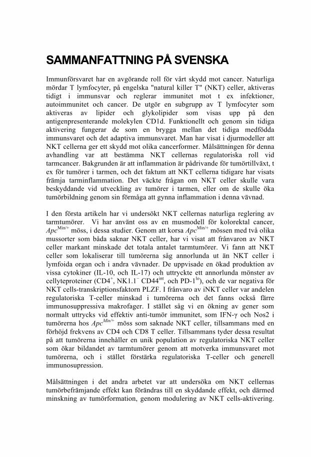
SAMMANFATTNING PÅ SVENSKA Immunförsvaret har en avgörande roll för vårt skydd mot cancer. Naturliga mördar T lymfocyter, på engelska "natural killer T" (NKT) celler, aktiveras tidigt i immunsvar och reglerar immunitet mot t ex infektioner, autoimmunitet och cancer. De utgör en subgrupp av T lymfocyter som aktiveras av lipider och glykolipider som visas upp på den antigenpresenterande molekylen CD1d. Funktionellt och genom sin tidiga aktivering fungerar de som en brygga mellan det tidiga medfödda immunsvaret och det adaptiva immunsvaret. Man har visat i djurmodeller att NKT cellerna ger ett skydd mot olika cancerformer. Målsättningen för denna avhandling var att bestämma NKT cellernas regulatoriska roll vid tarmcancer. Bakgrunden är att inflammation är pådrivande för tumörtillväxt, t ex för tumörer i tarmen, och det faktum att NKT cellerna tidigare har visats främja tarminflammation. Det väckte frågan om NKT celler skulle vara beskyddande vid utveckling av tumörer i tarmen, eller om de skulle öka tumörbildning genom sin förmåga att gynna inflammation i denna vävnad.
I den första artikeln har vi undersökt NKT cellernas naturliga reglering av tarmtumörer. Vi har använt oss av en musmodell för kolorektal cancer, ApcMin/+ möss, i dessa studier. Genom att korsa ApcMin/+ mössen med två olika mussorter som båda saknar NKT celler, har vi visat att frånvaron av NKT celler markant minskade det totala antalet tarmtumörer. Vi fann att NKT celler som lokaliserar till tumörerna såg annorlunda ut än NKT celler i lymfoida organ och i andra vävnader. De uppvisade en ökad produktion av vissa cytokiner (IL-10, och IL-17) och uttryckte ett annorlunda mönster av cellyteproteiner (CD4+, NK1.1− CD44int, och PD-1lo), och de var negativa för NKT cells-transkriptionsfaktorn PLZF. I frånvaro av iNKT celler var andelen regulatoriska T-celler minskad i tumörerna och det fanns också färre immunosuppressiva makrofager. I stället såg vi en ökning av gener som normalt uttrycks vid effektiv anti-tumör immunitet, som IFN-γ och Nos2 i tumörerna hos ApcMin/+ möss som saknade NKT celler, tillsammans med en förhöjd frekvens av CD4 och CD8 T celler. Tillsammans tyder dessa resultat på att tumörerna innehåller en unik population av regulatoriska NKT celler som ökar bildandet av tarmtumörer genom att motverka immunsvaret mot tumörerna, och i stället förstärka regulatoriska T-celler och generell immunosupression.
Målsättningen i det andra arbetet var att undersöka om NKT cellernas tumörbefrämjande effekt kan förändras till en skyddande effekt, och därmed minskning av tumörformation, genom modulering av NKT cells-aktivering.

Vi behandlade ApcMin/+ möss med olika lipid-ligander för NKT celler, som tidigare visats ha olika effekt på NKT celler i andra sjukdomsmodeller. ApcMin/+ möss blev behandlade under den tidiga fasen (5 - 15 veckors ålder) eller den sena fasen (12 - 15 veckors ålder) av tumörtillväxt. Möss behandlade i tidig fas med liganden C26:0 visade en signifikant minskning i antal och storlek på tumörer, medan behandling i sen fas reducerade tumörstorlek men inte tumörantal. I motsats till detta ledde behandling med liganden C20:2 i tidig fas till förhöjd tumörtillväxt, medan behandling i sen fas resulterade i minskning av tumörer. Dessa resultat visar att NKT cellsaktiverande immunterapi kan ändra funktionen hos NKT celler från tumörbefrämjande till tumörbekämpande. Resultaten visar också att olika NKT cellsaktiverande ligander har motsatt effekt och tidpunkten för behandling var central betydelse.
För kraftig aktivering av NKT celler leder till en sorts förlamning, anergi, som gör att de inte längre kan aktiveras effektivt. Detta beror på uppreglerad ytexpression av den inhibitoriska receptorn PD-1. Denna receptor finna också högt uttryckt på T celler i tumörer, och förhindrar deras förmåga att attackera tumören. I det tredje arbetet utförde vi därför en ny behandling av ApcMin/+ möss där vi kombinerade NKT cells-liganden C26:0 och blockering av PD-1 receptorn. Vi fann att kombinationen lyckades reducera polyptillväxt ytterligare. Vi kunde visa att blockering av PD-1 förhindrade anergi hos NKT celler i ApcMin/+ mössen som behandlats med C26:0. Vi såg också att kombinationsbehandlingen ökade aktivering av tumör-infiltrerande T-celler. Detta tyder på att kombinationsbehandlingen med NKT cells- aktiverande ligand och blockad av den inhibitoriska PD-1 receptorn förhöjer effekten av NKT cellsaktivering, och ökar immunsvaret mot tumörerna vilket leder till minskad tumörtillväxt.
Sammanfattningsvis visar denna avhandling att den naturliga funktionen av NKT celler i tarmen är att främja tumörutveckling genom att bekämpa ett effektivt tumör-immunsvar. Denna funktion hos NKT celler kan motverkas genom NKT cellsaktiverade immunterapi och minska tumörtillväxt. Det visar att NKT celler har avgörande betydelse för immunsvar mot tarmtumörer, och bör utforskas vidare för utveckling av NKT cellsaktiverande immunoterapi vid tarmcancer.


Ying Wang
1
LIST OF PAPERS This thesis is based on the following papers, referred to in the text by their Roman numerals.
I. Wang Y, Sedimbi S, Löfbom L, Singh A K, Porcelli S A, and Cardell S L. Unique invariant natural killer T cells promote intestinal polyps by suppressing TH1 immunity and promoting regulatory T cells. Mucosal Immunology. doi: 10.1038/mi.2017.34
II. Wang Y, Sedimbi S, Löfbom L, Porcelli S A, and Cardell S L. Modulation of intestinal tumor development by natural killer (NK) T cell directed immunotherapy. Manuscript.
III. Wang Y, Sedimbi S, Löfbom L, Porcelli S A, Yagita H, and Cardell S L. Natural killer T cell agonist and PD-1 blockade cooperate to reduce intestinal tumor development. Manuscript.

Natural killer T (NKT) lymphocytes regulate intestinal tumor immunity
2

Ying Wang
3
CONTENT ABSTRACTSAMMANFATNINGPÅSVENSKA
LISTOFPAPERS................................................................................1ABBREVIATIONS..............................................................................5
INTRODUCTION...............................................................................7NaturalkillerT(NKT)cells.....................................................................7
TheCD1dmolecule.................................................................................7NKTcellclassification..............................................................................8NKTcelldevelopment...........................................................................10
LigandsofiNKTcells............................................................................11Immunitytotumors............................................................................13
Innateimmuneresponsestotumors....................................................13Adaptiveimmuneresponsestotumors................................................14Suppressionoftheimmuneresponsetotumors..................................14NKTcellsintumorimmunity.................................................................15Immunotherapyagainstcancer............................................................16
Colorectalcancer(CRC)........................................................................19Mousemodelsofcolorectalcancer.....................................................20
Geneticallymanipulatedmice...............................................................20Chemicallyinducedcolorectalcancer...................................................21
AIMS.............................................................................................23
METHODLOGICALCONSEDERATIONS.............................................24TheApcMin/+mousemodelforCRC.......................................................24NKTcelldeficientmice........................................................................24Geneexpressionanalysis.....................................................................25CharacterizationofiNKTcellsandtheirfunctions................................27iNKTtargetingimmunotherapyinApcMin/+mice...................................28CombinationofcheckpointblockadeandiNKTcelldirectedtherapy...29Statisticalanalysis...............................................................................30
RESULTSANDDISCUSSIONS...........................................................32CharacterizationofApcMin/+mice.........................................................32iNKTcellsnaturallypromotedintestinaltumordevelopmentinApcMin/+mice....................................................................................................35

Natural killer T (NKT) lymphocytes regulate intestinal tumor immunity
4
PolypiNKTcellsinApcMin/+micedemonstratedauniquephenotypeandfunction...............................................................................................36ThemechanismsunderlyingthenaturalpromotionbyiNKTcellsofintestinaltumordevelopmentinApcMin/+mice....................................39iNKTcelldirectedimmunotherapymodulatedtumordevelopmentinApcMin/+mice........................................................................................42iNKTcelldirectedtherapycooperatedwithPD-1checkpointblockadeandreducedintestinaltumordevelopmentinApcMin/+mice................45
CONCLUDINGREMARKSANDFUTUREPERSPECTIVES.....................48
ACKNOWLEDGEMENTS..................................................................51REFERENCES...................................................................................55

Ying Wang
5
ABBREVIATIONS α-GalCer α-galactosylceramide ANOVA analysis of variance AOM azoxymethane Apc/APC adenomatous polyposis coli (gene, mouse/human) APC adenomatous polyposis coli (protein) APCs antigen presenting cells COX-2 cyclooxygenase-2 CTLs cytotoxic T lymphocytes DCs dendritic cells DMSO dimethyl sulfoxide DP double positive FAP familial adenomatous polyposis FoxP3 forkhead box P3 HNPCC hereditary non-polyposis colorectal cancer IBD inflammatory bowl disease iGb3 isoglobotriosylceramide IL interleukin iNOS inducible nitric oxide synthase i.p. intraperitoneal i.v. intravenous KO knock out LP lamina propria MDSC myeloid derived suppressor cell M-MDSC monocytic-MDSC MHC major histocompatibility complex Min multiple intestinal neoplasia MLN mesenteric lymph node Mø macrophage NK cells natural killer cells NKT cells natural killer T cells PCR polymerase chain reaction PD-1 programmed cell death protein 1

Natural killer T (NKT) lymphocytes regulate intestinal tumor immunity
6
PD-L1 programmed death-ligand 1 PLZF promyelocytic leukaemia zinc finger PMN-MDSC polymorphonuclear-MDSC TAM tumor associated macrophages TCR T cell receptor TLR Toll-like receptor TH T helper TNF-α tumor necrosis factor alpha Treg regulatory T cells

Ying Wang
7
INTRODUCTION
Natural killer T (NKT) cells The term "NKT cells" was first used to define a subset of αβ T cells that expressed the natural killer (NK) cell marker NK1.1 (CD161) in C57Bl/6 mice [1]. Further studies showed that a majority of these cells are CD1d-restricted [2, 3]. Therefore, it is now generally accepted that the term NKT cells refers to CD1d-restricted T cells. As compared to the conventional major histocompatibility complex (MHC) restricted CD4+ and CD8+ T cells, NKT cells express an intermediate level of T cell receptors (TCR). Following activation, NKT cells respond rapidly and produce large amounts of cytokines, including TH1, TH2 and TH17 type cytokines such as IFN-γ, IL-4 and IL-17 [4-7]. They can thereby regulate diverse immune responses before the adaptive T- and B-lymphocytes become effector cells in an immune response. Also, NKT cells respond to innate activating signals through toll-like receptors (TLR) [8], and in turn influence the downstream adaptive immune response. Due to their innate-like properties and functions, NKT cells are sometimes referred to as "innate-like T cells", and are seen as a bridge of the innate and adaptive immune system.
The CD1d molecule
The CD1-family is a group of glycoproteins expressed on most professional antigen presenting cells (APCs). Antigens presented by CD1 molecules are not peptides, as for MHC class I- and -II molecules, but lipids and glycolipids [9-11]. There are five isoforms of CD1 molecules that have been classified into three groups according to the differences in the mode of lipid presentation. Group 1 (CD1a CD1b and CD1c) and group 2 (CD1d) involved in lipid presentation to T cells, while group 3 (CD1e) facilitates intracellular lipid processing and trafficking [12]. CD1 genes are found in all mammalian species, while different numbers of CD1 isoforms are expressed in different

Natural killer T (NKT) lymphocytes regulate intestinal tumor immunity
8
species. CD1a-e are expressed in human while only CD1d is expressed in rat and mouse [13]. Murine and human CD1d and CD1d-restricted NKT cells are highly homologous. Structurally, the CD1d molecule is an MHC class I-like molecule, consisting of a CD1d heavy chain that associates with β2-microglobulin. The professional APCs such as dendritic cells (DCs), B cells and macrophages (Mø) are the major CD1d expressing cells. Unlike classical MHC molecules, CD1 molecules are non-polymorphic, a feature of importance for potential applications such as vaccine development and therapeutic treatments that target NKT cells. Various structures of lipids can bind to CD1d, such as glycosphingolipids (GSLs) [14, 15] and phospholipids [16].
NKT cell classification
According to the type of TCR expressed, NKT cells can be divided into type I NKT and type II NKT cells. The most frequent NKT cells in mice are type I NKT cells, while type II NKT cells may be more common in human [17, 18].
The type I NKT cell, also called invariant NKT (iNKT) cell, is an evolutionarily conserved category of NKT cells. It uses a semi-invariant TCR containing a unique TCR α-chain. In mice, iNKT cells express an invariant Vα14-Jα18 TCR α-chain paired with a limited set of TCR β-chains including Vβ8.2, Vβ7 and Vβ2 [19]. Homologous to the murine iNKT cells, human iNKT cells express an invariant Vα24-Jα18 TCR α-chain paired with Vβ11 [20, 21]. Phenotypically, murine iNKT cells are either CD4+CD8- or CD4/CD8 double-negative whereas also CD8+ iNKT cell can be found in human. All iNKT cells are activated by the artificial lipid ligand α-galactosylceramide (α-GalCer) [22]. Therefore these cells can be identified and quantitated using α-GalCer loaded CD1d tetramers [23-26]. iNKT cells can also be divided into three functional subsets according to their expression of the transcription factors promyelocytic leukaemia zinc finger (PLZF), T-bet and RORγt [27]. PLZFloT-bet+ iNKT1, PLZFhi iNKT2, and
Ying Wang
9
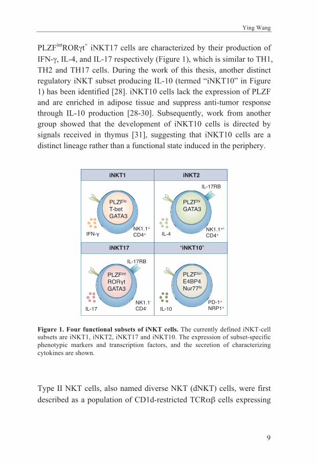
PLZFintRORγt+ iNKT17 cells are characterized by their production of IFN-γ, IL-4, and IL-17 respectively (Figure 1), which is similar to TH1, TH2 and TH17 cells. During the work of this thesis, another distinct regulatory iNKT subset producing IL-10 (termed “iNKT10” in Figure 1) has been identified [28]. iNKT10 cells lack the expression of PLZF and are enriched in adipose tissue and suppress anti-tumor response through IL-10 production [28-30]. Subsequently, work from another group showed that the development of iNKT10 cells is directed by signals received in thymus [31], suggesting that iNKT10 cells are a distinct lineage rather than a functional state induced in the periphery.
Figure 1. Four functional subsets of iNKT cells. The currently defined iNKT-cell subsets are iNKT1, iNKT2, iNKT17 and iNKT10. The expression of subset-specific phenotypic markers and transcription factors, and the secretion of characterizing cytokines are shown.
Type II NKT cells, also named diverse NKT (dNKT) cells, were first described as a population of CD1d-restricted TCRαβ cells expressing
iNKT1 iNKT2
iNKT17 “iNKT10”
PLZFlo
T-betGATA3
PLZFhi
GATA3
PLZFint
RORγtGATA3
PLZFlo/-
E4BP4Nur77hi
IL-17RB
IL-17RB
NK1.1+
CD4+NK1.1+/-
CD4+
NK1.1-
CD4-PD-1+
NRP1+
IFN-γ IL-4
IL-17 IL-10

Natural killer T (NKT) lymphocytes regulate intestinal tumor immunity
10
a diversity of TCR α- and TCR β-chains in mice lacking MHC II [3]. These cells have no response to α-GalCer [32] and therefore cannot be detected by α-GalCer loaded CD1d tetramers. Studies have shown that a fraction of dNKT cells respond to sulfatide [33]. Due to the poor stability and high background staining, the sulfatide-loaded CD1d tetramers, however, have not been were commonly used for identifying dNKT cells. Further, a human dNKT population isolated from from the plasma of myeloma patients responds to lysophosphatidylcholin (LPC) and binds to LPC-CD1d dimer [34]. Nonetheless, the lack of unique reagent, the diversity of ligands recognized and the limitation of the techniques to detect these cells make dNKT cells less well studied.
NKT cell development
The use of α-GalCer-CD1d tetramers has enabled studies of iNKT cell development, whereas there is no common ligand that could be used to detect dNKT cells. This makes most of the knowledge of NKT cell development based on studies of iNKT cells. NKT cells arise in the thymus from the same CD4+CD8+ double-positive (DP) precursor as that of conventional T cells. At this stage, DP thymocytes that will enter the NKT cell lineage are positively selected by binding to self-lipid or glycolipid presented on CD1d on the surface of other DP cells. This is in contrast to the selection of conventional CD4+ T cells or CD8+ T cells, which are selected by binding of TCR to self-peptide presented on MHC II or MHC I on thymic epithelial cells. Once selected, NKT cell precursors undergo a series of developmental stages (illustrated in Figure 2). At least four distinct NKT cell developmental stages have been defined through differences in expression of CD24, CD44 and NK1.1; these are controlled by a series of transcription factors. PLZF plays a role as a master regulator and controls the NKT development [35, 36]. Most NKT cells migrate from the thymus at stage 2 and progress to stage 3 in the periphery. The transcription factor T-bet is essential in this step. Some stage 3 NKT cells remain in the thymus as long-term thymus-resident cells [37]. Evidence suggests that CD4− NKT cells branch from CD4+ NKT cells at approximately

Ying Wang
11
stage 1 of development. A separate pathway of NKT cell development gives rise to an IL-17-producing subset that seems to be regulated by the transcription factor RORγt [7].
Figure 2. NKT cell development. CD4+CD8+ DP cells are selected by TCR-CD1d ligation. A costimulatory signal SLAM-SLAM is necessary for the positive selection. Further development of NKT cells is divided into different stages according to the surface expression of CD24, CD44 and NK1.1. Most NKT cells leave thymus at stage 2 and complete the development in periphery, whereas some NKT cells stay in thymus. This figure is simplified from Godfrey et al., Nature Immunology, 2010 [38].
Ligands of iNKT cells Invariant NKT cells can bind to a variety of lipid-based antigens presented on CD1d molecules, including α-GalCer, exogenous microbial ligands and a list of endogenous self-antigens [38, 39].
Thymus
Periphery
Stage 0 Stage 1 Stage 2 Stage 3
Stage 2 Stage 3
NKT NKT NKT
NKT
NKT
NKT
CD4+
CD8+
CD4+
CD8+ CD24+
CD44-
NK1.1-
CD1d
self-lipid
Vα14Jα18TCR
SLAM-SLAM
CD24-
CD44-
NK1.1-
CD24-
CD44+
NK1.1-
CD24-
CD44+
NK1.1+
NK1.1
NK1.1
CD24-
CD44+
NK1.1-
CD24-
CD44+
NK1.1+

Natural killer T (NKT) lymphocytes regulate intestinal tumor immunity
12
α-GalCer was originally derived from a marine sponge [40]. It has been described as a compound that has strong anti-tumor properties, identified in a broad screen for molecules that could prevent murine lung metastasis [41]. Several studies applying α-GalCer in different disease models found that α-GalCer potently activate iNKT cells associated with rapid TH1/TH2 cytokine secretion. Subsequently the stimulatory effects of alternative synthetic α-GalCer analogues was investigated (reviewed in Venkataswamy and Porcelli [42]; Tyznik et al.[43]). The aims of these investigations were to identify synthetic α-GalCer analogues that were skewing either a TH1 or a TH2 cytokine response and find the ones which could be applied on the treating conditions where polarized cytokine responses were implicated in pathogenesis, such as cancer, allergy and autoimmunity. These synthetic ligands include the sphingosine-based truncated derivative of α-GalCer OCH [42], which induces a Th2 response during activation of iNKT cells in mice as defined by rapid IL-4 production with no detectable IFN-γ [44]. Other variants of α-GalCer have been made by altering the length and the degree of unsaturation of the fatty acyl chain, including the C20:2 analogue and α-C-GalCer, which are ligands skewing the iNKT cell response towards TH2- and TH1 cytokines, respectively [42, 45]. The list of synthetic lipid antigens for iNKT cells is growing and their capacity to induce biased immune responses holds great promise therapeutically [46].
Besides α-GalCer, a number of exogenous ligands such as microbial glycolipids have been identified to stimulate the activation of iNKT cells. The first identified microbial glycolipid is GSLs from Sphingomonas spp., a Gram-negative member of α-proteobacteria. GSLs have been shown to induce strong CD1d-dependent iNKT activation [47-50], and their function related to clearing of microbial infections [51].
During the thymic development of iNKT cells, an endogenous self-lipid ligand and iNKT autoreactivity is necessary for the positive selection [52]. This autoreactivity is also required for the TLR

Ying Wang
13
triggered immune response against bacterial infection [51, 53]. Isoglobotriosylceramide (iGb3) was thought to be a possible endogenous antigen in both mice and human. iGb3 was reported as an iNKT cell activating ligand [54]. However, a study of the distribution of iGb3 with high-pressure liquid chromatography analysis demonstrated that iGb3 was not detected in either the mouse or human thymus [55]. Further, the iGb3 synthase knockout (iGb3S-/-) mice did not show decreased number of iNKT cells in the thymus, spleen, or liver, and showed a similar cytokine response to α-GalCer administration as compared to iGb3S+/- mice [56]. Taken together, the results strongly suggested that iGb3 is unlikely to be the endogenous ligand required for iNKT cell selection in the thymus.
Immunity to tumors The effector mechanisms of both innate and adaptive immunity have been shown to attack tumor cells. Despite this, immunity to tumors is often under immunoregulatory control, leading to tumor escape from immune destruction. To determine the mechanisms that underpin immunoregulation of tumor immunity will be essential as a basis for the development of novel therapies that contribute to improved immune protection against tumors.
Innate immune responses to tumors
The main killers of tumor cells in the innate immune response are natural killer (NK) cells and Mø. NK cells kill many types of tumor cells. Their tumor killing activity is termed natural because they do not require activation to kill cells. NK cells carry several activating receptors, the ligands of some are upregulated on tumor cells. Classical studies have shown that MHC I molecules on the surface of normal cells inhibit NK cells and prevent lysis [57]. Thus, the decreased level of MHC I molecule expression characteristic of many tumor cells may allow activation of NK cells and subsequent tumor killing.

Natural killer T (NKT) lymphocytes regulate intestinal tumor immunity
14
Mø can kill tumor cells when activated by a combination of factors, including cytokines. They are less effective than T cell-mediated cytotoxic mechanisms. Under certain circumstances, Mø may present tumor antigens to T cells and stimulate tumor-specific immune responses. Classically activated M1 Mø display various anti-tumor functions. They produce large amounts of proinflammatory cytokines, such as IL-6, IL-1 and TNF-α, and are involved in the killing of tumor cells [58]. They also express inducible nitric oxide synthase (iNOS). In contrast, activated alternatively M2 Mø produce IL-10 and transforming growth factor-β (TGF-β), and are thought to be associated with tissue repair [59, 60]. Tumor associated Mø (TAM) are normally of M2-like phenotype and evidence suggests that they are part of inflammatory circuits that promote tumor progression [61-63].
Adaptive immune responses to tumors
The principal mechanism of adaptive tumor immunity is killing of tumor cells by CD8+ cytotoxic T lymphocytes (CTLs). Tumor-specific CTLs have been found in a diversity of cancers including neuroblastomas; malignant melanomas; sarcomas; and carcinomas [64]. CTLs recognize peptide antigens presented on MHC I on target tumor cells and lyse these cells. CD4+ helper T cells stimulated by peptides presented by MHC II on APCs produce diverse cytokines, which provide the help for CTLs and activate other cells with tumor killing capacity, such as NK cells and Mø.
Suppression of the immune response to tumors
Regulatory T cells (Treg) are MHC II restricted CD4+ T cells that express the master transcription factor FoxP3. They develop in the thymus and are normally present in the body and function to prevent autoimmune reactions. They are also induced peripherally during the active phase of immune responses to pathogens and limit the strong immune response that could damage the host. Studies of Treg cells in

Ying Wang
15
mouse models and cancer patients have shown that Treg accumulate in tumor-bearing individuals, especially at the tumor site [65]. These cells secrete IL-10 and TGF-β and result in an inhibited CTL response and suppression of tumor immunity [66, 67]. Depletion of Treg cells in tumor-bearing mice has been shown to induce T cell infiltration, enhance anti-tumor immunity by increasing TH1 cell proliferation and thereby reduce tumor growth [68-71].
Myeloid-derived suppressor cells (MDSCs) are a diverse set of cells that accumulate in cancer patients [72, 73]. They consist of immature myeloid cells and their precursors, lacking the surface markers specific for monocytes, macrophages or DCs [74]. In mice, MDSCs are defined by co-expression of the myeloid lineage markers CD11b and Ly6G [75]. In humans, MDSCs are characterized as CD14-CD11b+ [76] or CD33+HLA-DR- cells [77]. MDSCs are classified into two subtypes: monocytic MDSC (M-MDSC) and polymorphonuclear MDSC (PMN-MDSC) according to their surface marker expression. In mice, M-MDSC express high level of Ly6C and low or no Ly6G, while PMN-MDSC express intermediate Ly6C and positive for Ly6G [74, 78]. These cells accumulate in large numbers in cancers and potently suppress anti-tumor innate and T cell responses by mechanisms that include IL-10 secretion [79]. MDSCs also indirectly impair anti-tumor T cell responses by enhancing Tregs and skewing CD4 helper T cell differentiation to TH2 cells [79, 80].
NKT cells in tumor immunity
The role of NKT cells in tumor immunity has been displayed in many studies. It has been shown that NKT cells play important roles in tumor surveillance and the control of tumor metastasis [81]. It has been shown in several experiment mouse models that iNKT cells promote tumor immunity and protect against tumors [82, 83]. The IFN-γ production by iNKT cells was identified as a key component of the iNKT anti-tumor effect [84]. Further, NK cells were activated by NKT

Natural killer T (NKT) lymphocytes regulate intestinal tumor immunity
16
cells in an IFN-γ and IL-2 dependent manner and suppressed tumor cell growth [85]. Although many studies have shown that iNKT cells protect against tumors, in some models, iNKT cells have instead been shown to suppress immune-surveillance by producing TH2 cytokines, such as IL-13, IL-4 and IL-5 [86, 87]. Regulation of tumor immunity by dNKT has also been demonstrated. An IL-13 producing dNKT population was increased in peripheral blood from myeloma patients, and these cells were activated by an inflammation-associated lysophospholipid presented on CD1d [88]. Moreover, a series of elegant studies by Berzofsky and co-workers have demonstrated that dNKT cells suppress CD8+ T cell mediated tumor immunity through IL-13 and TGF-β production [89].
Immunotherapy against cancer
Immunothreapy is the treatment that takes advantages of the ability of immune system to fight against disease such as cancer. The main advantage of immunotherapy compared to traditional drug therapies is that the immune response is specific for tumor antigens and will not injure most of the normal cells, whereas the drugs often have severe side effects on normal proliferating cells. The main types of immunotherapy used to treat cancers include monoclonal antibodies, adoptive cell transfer, cancer vaccines and immune checkpoint blockade.
Antibody therapy
Antibodies are a key component of the adaptive immune system. Man-made monoclonal antibodies are designed to bind to tumor-specific antigens, and thereby cause an immune response to attack the tumor cells. Currently there are more than 100 monoclonal antibodies that have been explored for cancer therapy [90, 91]. Some of them have been applied in experimental animal models, some tested in human clinical trials and some have been approved for clinical use [92]. One of the most successful anti-tumor antibodies is the humanized mouse

Ying Wang
17
monoclonal anti-CD20, which has been used for treating B-cell lymphoma patients [93]. The mechanisms of tumor cell elimination by antibodies include opsonization, activation of the complement system, and antibody-dependent cell-mediated cytotoxicity. Further, another type of antibody may directly activate apoptosis in tumor cells such as anti-CD30 used to treat lymphomas [94, 95]. Adoptive cellular immunotherapy
The approach called adoptive cell transfer collects and utilizes the patient's own cells to treat the cancer. Adoptive cell transfers have been applied in small clinical trials to the patients in different types of cancer [96-98]. There are different forms of adoptive cell transfer treatments. In CAR T cell therapy, T cells from the patients are collected and are genetically engineered to produce specific receptors called chimeric antigen receptor (CARs) [99]. CARs allow T cells to recognize specific tumor antigens. The CAR T cells are cultured in vitro and the expanded population is then transferred back to the patient. The transferred CAR T cells multiply in vivo and recognize and kill cancer cells by the guidance of the introduced receptors. Tumor vaccines
The use of vaccination with tumor antigens is another approach to immunotherapy. These vaccines are usually made from tumor cells from cancer patients produced by tumor cells [100]. They are designed to treat cancers by enhancing the immune response against the tumor. Two types of tumor vaccines have been shown to be effective in clinical trails and experimental animal models [101]. One approach is to vaccinate with patient derived DCs that have been incubated with tumor antigens or transfected with genes encoding these antigens [102, 103]. An alternative approach in clinical trails is the use of DNA vaccines composed plasmids of viral vectors encoding tumor antigens [104-107]. As the encoded antigens are synthesized in cytoplasm and then enter the MHC I antigen presentation pathway, the cell-based and

Natural killer T (NKT) lymphocytes regulate intestinal tumor immunity
18
DNA vaccines may provide the best ways to induce CTL responses [108-110]. Immune checkpoint blockade
Checkpoint blockade is an immunotherapy approach to block the ability of certain proteins, called immune checkpoint proteins, which limit the strength and duration of immune responses [111]. It is been clearly shown that tumors can make use of certain checkpoint pathways to resist T cell immunity [112]. Checkpoint pathways can be blocked by administration of specific monocolonal antibodies, since many of them are initiated by ligand-receptor interactions. The first checkpoint blockades target, cytotoxic T-lymphocyte-associated antigen 4 (CTLA-4), is a receptor that down regulates immune response. It is highly expressed on Treg cells and is up regulated after T cell activation. CTLA-4 share exact ligands, CD80 and CD86 (also known as B7-1 and B7-2), with CD28 [113-115] and has much higher affinity for both ligands. Binding of CTLA-4 to CD80/CD86 blocks the co-stimulatory signals of T cells through CD28, and delivers inhibitory signals to T cells [116-120]. The CTLA-4 specific checkpoint inhibitors enhance the strength of immune responses by preventing inhibitory signals by binding to and blocking CTLA-4.
Another immune checkpoint is the receptor programmed death protein 1 (PD-1) pathway. PD-1 ligation has been reported to promote self-tolerance and to limit autoimmunity by down regulating the T cell activity [121-127]. Expression of PD-1 is induced when T cells become activated [121]. Ligation of PD-1 with its ligands PD-L1/2 inhibits kinases involved in T cell activation [122]. Similar to CTLA-4, PD-1 is also highly expressed on Treg cells [128]. Tumor associated T cells often have high PD-1 expression that inhibits their anti-tumor activity. Moreover, PD-L1 is often found upregulated on tumor cells. Administration of antibodies to PD-1 or its ligands releases T cell activation towards the tumor, allowing T cell mediated tumor eradication. During recent years, check-point blockade treatments have

Ying Wang
19
led to important clinical advances, resulting in durable clinical responses and long-term remission in a fraction of treated patients.
iNKT cell targeting cancer therapy
A number of studies have reported the anti-tumor effects by iNKT cells targeting therapies in animal models (see reviews [129-131]). There are three main iNKT cells-based anti-tumor therapies have been applied in animal models and clinical trails. Systemic administration of α-GalCer was found to control tumor metastasis and increase the survival in different models [132-134]. In addition, modified analogues of α-GalCer that induce an enhanced TH1-skewed cytokine response in iNKT cells were found to be superior to α-GalCer in inducing anti-tumor immunity [135]. Further, injections of α-GalCer pulsed DCs led to an enhanced iNKT and downstream NK cell response and reduced tumor formation in a B16 melanoma model [134]. Alternatively, adoptive transfer of ex vivo expanded iNKT cells into non-small-cell lung cancer patients resulted in downstream NK cell activation and IFN-γ production [136]. Interestingly, the combination of iNKT cell adoptive transfer and injections of α-GalCer pulsed DCs has been report to enhance the anti-tumor response in patients with head and neck carcinoma [137, 138].
Colorectal cancer (CRC) Colorectal cancer (CRC), is the second most common cancer worldwide, after lung cancer. The risk for developing CRC is influenced by environmental and genetic factors. The sporadic form of CRC increases with age with 90% of the cases occurring after 50 years of age. Sporadic CRC is in most cases initiated by a mutation in the adenomatous polyposis coli (APC) gene, followed by additional mutations in oncogenes, tumor suppressor genes and genes encoding DNA repair proteins. Besides a diet that is high in red meat, smoking and heavy alcohol use can also raise the CRC risk. Genetic mutations are the main risk factor for hereditary CRC. The most common

Natural killer T (NKT) lymphocytes regulate intestinal tumor immunity
20
inherited syndromes linked with colorectal cancers are familial adenomatous polyposis (FAP) and Lynch syndrome (hereditary non-polyposis colorectal cancer, or HNPCC). FAP is caused by an inherited mutation in the adenomatous polyposis coli (APC) gene and accounts for around 1% of all CRC. In contrast to the sporadic CRC, FAP patients present with high numbers of colorectal adenomatous polyps as early as age 20 and almost all individuals with FAP will have colon cancer by the age of 40 unless their colon has been removed. Persons with FAP also have an increased risk for cancers of the stomach, small intestines, and some other organs. Lynch syndrome accounts for about 2% to 4% of all colorectal cancers. In most cases, this disorder is caused by an inherited defect in either the MLH1 or MSH2 genes, which play an important role in DNA mismatch repair. The most common colorectal cancer treatment currently is surgery as there is no other efficient treatment available.
Mouse models of colorectal cancer Mouse models of colorectal cancer and intestinal cancer are experimental systems in which mice are genetically manipulated, fed a modified diet or challenged with chemicals to develop malignancies in the gastrointestinal tract. These models enable researchers to study the onset, progression of the disease, and understand in depth the molecular events that contribute to the development and spread of colorectal cancer.
Genetically manipulated mice
Common genetic mouse models for CRC are mutant mice carrying a heterozygous mutation in the Apc gene. The Apc gene is defined as a tumor suppressor gene, which is involved in the Wnt/β-catenin signaling pathway. Deficiency of the APC protein will lead to nuclear accumulation of β-catenin that results in a dysregulated cell division, and thereby initiating cancer formation. The first mouse described that contained a mutation in the Apc gene was designated multiple

Ying Wang
21
intestinal neoplasia (Min) [139]. This mouse model is called the ApcMin mouse, which carries a truncation mutation at codon 850 of the Apc gene. The ApcMin mouse can develop more than 100 polyps in the small intestine and colon. Two years later, a new mutant of the Apc gene with a truncation mutation at codon 716 (ApcΔ716)[140] was engineered. It results in a mouse that develops more than 300 polyps in the small intestine. More recently a novel Apc mutation mouse model having multiple polyps form in the distal colon was constructed [141]. In this model an additional mutation in the Cdx2 gene on the ApcΔ716 background shifted the formation of polyps from the entire intestine to the colon, resembling human CRC. In addition, a mouse model carrying mutations in ApcΔ716 and Smad4 is characterized with development of invasive adenocarcinomas [142].
Since heterozygous gene deletions were less successful for constructing the mouse model for HNPCC, mice carrying homozygous deletions of the mismatch repair genes such as Msh2, Mlh1, Msh6, Msh3, Pms2, and Pms1have been used as disease models for HNPCC-like cancers [143]. Although such mice are more susceptible to tumor formation, the tumor spectrum observed consists of various lymphomas that are almost never encountered in HNPCC affected patients. Also, deficiency of Msh2 and Pms2 promoted APC-mediated intestinal tumorigenesis [144].
Chemically induced colorectal cancer
Carcinogen-induced colon cancer in rodents can recapitulate in a reliable way the phases of initiation and progression of tumors that occurs in humans. Such models are frequently used to assess activity of chemo-preventive compounds and to identify risk factors. These models are highly reproducible, they can be readily tested on animals with different genetic backgrounds, and the pathogenesis recapitulates human CRC.

Natural killer T (NKT) lymphocytes regulate intestinal tumor immunity
22
A variety of chemicals have been used for inducing colon tumors in animals. Azoxymethane (AOM) is a genotoxic carcinogen and is routinely used to induce colon tumors in mice [145, 146]. The AOM-induced tumors locate in the distal colon whereas a p21 knock out mouse treated with AOM shows tumor distribution throughout the colon [147]. They share many histopathological characteristics with human CRC and frequently carry K-Ras mutations, whereas AOM induced APC mutations are less frequent in rodents and the tendency to metastasize is low [144]. An inflammation-related mouse model of colorectal carcinogenesis induces colon lesions with combination of AOM and dextran sodium sulphate (DSS). AOM/DSS induced adenocarcinoma showed positive staining for nuclear β-catenin, cyclooxygenase-2 (COX-2) and iNOS [146]

Ying Wang
23
AIMS NKT cells have been shown to play important roles in tumor surveillance and the control of tumor metastasis [148, 149]. Activation of iNKT cells provides protection against tumor growth and metastasis in various experimental models [40, 150, 151]. However, it has been reported that NKT cells inhibit tumor immunity, as examplified in a murine lymphoma model [86]. Cytokines produced by inflammatory cells directly or indirectly promote cancer cell growth [152-156]. Inflammation plays a critical role in the development of IBD-associated CRC [157, 158]. Significantly, NKT cells have been found to promote intestinal inflammation in a mouse model for IBD through IL-13 production [159]. The pro-inflammatory role of NKT cells and the dual role of inflammation in CRC raised the question whether NKT lymphocytes may promote the inflammation driven tumorigenesis in the intestine. In this thesis we have investigated the natural effect of iNKT cells in tumor regulation in the intestine, and performed preclinical studies of therapeutic treatments to suppress tumor development through iNKT cell activation.
Specific aims: - To investigate whether NKT cells naturally modulate tumor development in the ApcMin/+ mouse model for CRC
- To identify the mechanism underlying the natural promotion of intestinal tumors by iNKT cells in ApcMin/+ mice
- To apply iNKT cell directed immunotherapy to investigate whether treatment with iNKT cell agonists can prevent tumor development in ApcMin/+ mice
- To apply PD-1 checkpoint blockade together with iNKT cell directed immunotherapy to investigate whether this would improve suppression of tumor development in ApcMin/+ mice

Natural killer T (NKT) lymphocytes regulate intestinal tumor immunity
24
METHODLOGICAL CONSEDERATIONS
The ApcMin/+ mouse model for CRC The ApcMin/+ mouse was established on the C57BL/6 genetic background and when used as a model for CRC, carries a heterozygous mutation of the Apc gene. While homozygous mutant mice (ApcMin/Min) are not viable, a heterozygous mutation results in spontaneous polyp formation in the small intestine and colon. Tumor immunity can be studied after transplantation of tumor cell lines, such as the MC38 cell line, which is derived from C57BL/6 mouse adenocarcinoma. However, ApcMin/+ mice developing spontaneous polyp formation allow us to investigate the regulation of tumor immunity in a proper tumor microenvironment in vivo. In addition, due to the same gene mutation as in human CRC, these mice are recapitulating early events in human colorectal carcinogenesis, which provide a natural process of tumor growth rather than in chemically induced CRC models. Therefore, we took the advantages of ApcMin/+ mouse and used the mice as a colorectal cancer model to investigate iNKT cell regulation of intestinal tumor development. Female ApcMin/+ mice and Apc+/+ littermates were used for studies of the natural effect of iNKT cells (Paper I) and both male and female ApcMin/+ mice were used for the preclinical immunotherapeutic study (Paper II and III).
NKT cell deficient mice In order to investigate the role of iNKT cells in intestinal tumor development in the ApcMin/+ mouse model, we crossed ApcMin/+ mice with Jα18-/- mice to generate ApcMin/+Jα18-/- and ApcMin/+Jα18+/- littermate controls (Paper I). Jα18-/- mice lack a TCR α-segment, which is required to form the iNKT cell TCR, so that ApcMin/+Jα18-/- mice are completely devoid of iNKT cells.

Ying Wang
25
Since a recent study showed that Jα18-/- mice have impaired diversity of the TCR repertoire due to suppressed rearrangement to Traj (Jα) gene segments upstream of Traj18 (Jα18) [160]. Consequently, the effects on tumor development in these mice might be caused by a decreased repertoire of T cells. Concerning the validity of the experiment, we also introduced another NKT deficient mouse model, the CD1d-/- mouse, on the ApcMin/+ background. The CD1d-/- mouse lacks the CD1d molecule, which results in a loss of the TCR ligand for positive selection of NKT cells in the thymus, and the mice consequently lack all NKT cells. Thus, we crossed ApcMin/+ mice with CD1d-/- mice to confirm the effect of iNKT cells in intestinal tumor formation in the ApcMin/+ mouse model (Paper I).
Gene expression analysis To identify gene expression regulated by iNKT cells, we performed real time PCR (RT PCR) for analysis of gene expression in the presence and absence of iNKT cells. RT PCR also called quantitative PCR (qPCR) or real time quantitative PCR (RT-qPCR), is a common method to quantify gene expression at the transcriptional level. Due to the property of high throughput and sensitivity, in this thesis, we first designed and applied custom RT2 profiler PCR arrays,to determine the mRNA expression levels of selected genes in tissues from iNKT deficient mice (Paper I). We selected genes relevant for immune responses, tumor growth and apoptosis. The genes have been grouped according to the encoded protein types, such as cytokines, chemokines and chemokine receptors, cell linage markers, and are listed in Table 1. The arrays allowed us to have a fast and broad peek into the iNKT cell dependent regulation of the tumor microenvironment; thereby providing us with a reasonable hypothesis for further experimentation. RT PCR was used as an important supportive and complementary method to screen and confirm the regulation of certain genes such as those encoding chemokines, cytokines and transcription factors. The

Natural killer T (NKT) lymphocytes regulate intestinal tumor immunity
26
most regulated gene expressions we obtained from the gene arrays were confirmed with RT PCR (Paper I).
Table 1. List of genes that were analyzed in the gene expression array. Cytokine Cytokinereceptor Immuneresponse Tumorgrowth
Il1b IL-1β Il4ra IL-4RA Rorc RORγt Myc c-MYCIl1a IL-1α Il13ra1 IL-13Rα1 Tbx21 T-bet Mapk1 ERKIl2 IL-2 Il13ra2 IL-13Rα2 Gata3 GATA3 Mmp9 MMP9Il4 IL-4 Il22ra1 IL22Rα1 Stat1 STAT1 Mmp3 MMP3Il5 IL-5 Il22ra2 IL22Rα2 Stat3 STAT3 Mmp1a MMP1Il6 IL-6 Chemokine Stat6 STAT6 Egf EGFIl8 IL-8 Ccl20 CCL20 Nfkb1 NF-κB Egfr ErbB1Il9 IL-9 Cxcl1 CXCL1 Klrk1 KLRK1 Vegfa VEGFIl10 IL-10 Cxcl10 IP10 Rae1 RAE1 Fgf2 bFGFIl11 IL-11 Cxcl9 CXCL9 H60a H-60 Tgm2 TGM2Il12a IL-12A Cxcl11 CXCL11 Ido1 INDO ApoptosisIl12b IL-12B Chemokinereceptor Celllinagemarker Cd274 PD-L1Il13 IL-13 Cxcr2 CXCR2 Cd4 CD4 Pdcd1lg2 PD-L2Il15 IL-15 Cxcr3 CXCR3 Cd8b1 CD8β Pdcd1 PD-1Il17a IL-17A Ccr2 CCR2 Cd19 CD19 Pdgfb PDGFBIl17f IL-17F Ccr6 CCR6 Foxp3 FoxP3 Gzma GramzymeAIl18 IL-18 Zbtb16 PLZF Gzmb GramzymeBIl21 IL-21 Arg1 ARG1 Bcl2l1 BCL-XLIl22 IL-22 Nos2 NOS2 Xiap IAP3Il23a IL-23A Chi3l3 YM1 Il25 IL-25 Mrc1 MRC1
Il27p28 IL-27A Ly6g Ly6G Il33 IL-33 Mpo MPO Ifng IFN-γ Retnla Fizz1 Tnf TNF Ptgs2 COX2
Tgfb1 TGF-β1 Ifnb IFN-β Tslp TSLP
Tnfsf15 TL1A

Ying Wang
27
Characterization of iNKT cells and their functions To determine the expression of extracellular and intracellular markers on defined cell populations, we performed flow cytometry on cells from spleen, MLN, intestine, polyps (Paper I, II and III) and liver (Paper II). There are alternatives to this method, such as immunohistology. The advantage of flow cytometry compared to other options is that flow cytometry can process thousands of cells per second; it is fast and directly showing the protein expression levels on the cells. Also, flow cytometry enabled us to do subpopulation analysis, so that the phenotypes and function of specific immune cells can be determined. In this thesis, cell surface and intracellular antigen expression was detected by using fluorochrome-conjugated anti-mouse antibodies. To investigate the cytokine production by iNKT cells, we stimulated iNKT cells in vitro with phorbol myristate acetate (PMA) plus ionomycin, in the presence of brefeldin A. Followed by intracellular staining, the cytokine production was determined by flow cytometry.
We applied adoptive transfer of iNKT cells into ApcMin/+Jα18-/- mice, to determine whether adding back iNKT cells to ApcMin/+Jα18-/- mice lacking these cells could switch back the macrophage phenotype from M1 to M2, (Paper I). Since iNKT cells are enriched in the liver providing a rich source of iNKT cells, we isolated mouse hepatic iNKT cells by fluorescence-activated cell sorting (FACS) and then the isolated cells were i.v. injected to 12-week old ApcMin/+Jα18-/- mice. Although cell sorting is more time consuming than cell enrichment through immunomagnetic separation, sorting results in a more pure preparation of iNKT cells.
In addition, we performed cytometric bead array (CBA) analysis, a flow cytometry based method, to determine the dynamic cytokine

Natural killer T (NKT) lymphocytes regulate intestinal tumor immunity
28
production in mouse serum (Paper I). Because of the high sensitivity of this method, it significantly reduces sample volume requirements and time to results in comparison with traditional ELISA and Western blot techniques.
iNKT targeting immunotherapy in ApcMin/+ mice In this thesis, we performed preclinical immunotherapeutic studies to investigate whether α-GalCer therapy is beneficial in the ApcMin/+ model of intestinal tumors (Paper II). An important finding in the field is that while α-GalCer stimulation of iNKT cells results in a mixed IFN-γ and IL-4 cytokine production, certain analogues of α-GalCer can skew the iNKT cells cytokine production towards a IFN-γ dominated Th1 profile [161] or a TH2 profile with high amounts of IL-4 [45, 162]. These iNKT ligands provide more sophisticated tools for iNKT cell target immunotherapy, for situations when a specific cytokine profile is desired. Thus we also included groups treated with either the TH1 skewing α-GalCer analogue α-C-galactosylceramide (referred to C-glycoside below), or the TH2 skewing analogue C20:2 (Figure 3). Here we treated ApcMin/+ mice from 5 weeks of age with α-GalCer C26:0, or vehicle, to evaluate the effect of treatment on the early-phase of tumor formation (for treatment schedule see Figure 4).
To investigate whether α-GalCer based treatment could modulate the late phase of polyp growth, we also performed a three-week treatment schedule of ApcMin/+ mice starting at 12 weeks, and sacrificed the mice for analysis at 15 weeks of age (see treatment schedule in Figure 4). This time, we compared mice treated with α-GalCer C26:0 and mice treated with C20:2, the two treatments that had resulted in the most significant and opposite results using the long-term early-phase treatment protocol.

Ying Wang
29
Figure 3. Chemical structures of αα-GalCer and analogues used in this thesis.
Figure 4. Treatment schedule for early- and late-phase iNKT cell directed therapy. For early-phase treatment with glycolipid, 5 week old ApcMin/+ mice were injected i. p. with 4 μg of glycolipid in 200 μl of vehicle, or vehicle control. Mice were injected on day 1, 2, 7, 14, 21, 28 and 60, and sacrificed at 15 weeks of age. For late-phase treatment with glycolipid, 12 week-old ApcMin/+ mice were injected i.p. with 4 μg of glycolipid in 200 μl vehicle solution or vehicle control only on day 1, 7, 14, and the mice were sacrificed at 15 weeks of age. Arrows indicate the injections of glycolipid.
Combination of checkpoint blockade and iNKT cell directed therapy Ligation of the PD-1 on T cells results in an inhibitory signal, and is therefore referred to as an immune checkpoint for T cells.
α-GalCer C26:0 (Th1/Th2)
α-GalCer C20:2 (Th2)
α-C-Glycoside (Th1)
5 6 7 8 9 10 11 12 13 14 15
Late phase treatment with glycolipid
Early phase treatment with glycolipid

Natural killer T (NKT) lymphocytes regulate intestinal tumor immunity
30
Accumulated evidence has revealed that blockade of the PD-1/PD-L1 pathway can enhance the anti-tumor response [163]. In colorectal cancer, checkpoint blockade with PD-1 targeting antibody was effective in a subset of patients [164]. With the aim to achieve improved effects, the PD-1/PD-L1 checkpoint blockade has been combined with other immunotherapies. For example, an improved clinical response has been shown after treatment with the combination of anti-PD-L1 and anti-CTLA-4 in melanoma patients [165, 166]. Here we hypnotized that a combination treatment with the iNKT cell agonist α-GalCer together with PD-1 antibody might cooperate and enhance anti-tumor activities. In this thesis (Paper III), we performed the combination of iNKT directed therapy and checkpoint blockade in the ApcMin/+ mouse model in the late phase of tumor formation to investigate whether this treatment could enhance the anti-tumor response and reduce intestinal tumor development (treatment schedule in Figure 5).
Figure 5. Combination of checkpoint blockade and iNKT direct treatment schedule. 12 weeks ApcMin/+ mice were i.p. administrated with 0.25 mg anti-PD-1 antibody RMP1-14 twice a week, together with or without weekly 4µg α-GalCer in 200μl of PBS solution. The mice were sacrificed at 15 weeks of age. Arrows indicate the injections of RMP1-14 and α-GalCer.
Statistical analysis Since we had relatively small numbers of data points and non-normal distribution, nonparametric statistical tests were applied. To evaluate significant difference between genotypes (Paper I), Mann-Whitney test was used. Unpaired one-way ANOVA was used to evaluate the
12 13 14
Ab Ab Ab Ab Ab
α-GalCer α-GalCer α-GalCer
Ab
15

Ying Wang
31
significance between treatment groups (Paper II and III). p values < 0.05 were considered significant.

Natural killer T (NKT) lymphocytes regulate intestinal tumor immunity
32
RESULTS AND DISCUSSIONS The results from the studies presented in Paper I-III on the natural and induced effects of iNKT cells in intestinal tumor development will be summarized and discussed in this chapter following the aims.
Characterization of ApcMin/+ mice As a spontaneous model for CRC, ApcMin/+ mice develop a number of adenomas in the small and large intestine, as summarized in Figure 6. Early ileal lesions can be found in 1-month old ApcMin/+ mice [167]. In our breeding colony, 10–12-week old ApcMin/+ mice had no macroscopically visible intestinal polyps, but early polyp formation could be seen on hematoxylin-eosin stained sections using a microscope (Paper I, Figure 1a). By 15 weeks of age, an average of 20 polyps were found in the entire small intestine in ApcMin/+ mice (Figure 8). Based on these observations, we designed our experimental system according to these key time points. To investigate the role of NKT cells in the regulation of tumor formation, and the underlying mechanisms, we used 15-week old mice due to the presence of substantial polyp formation in the small intestine at this time (Paper I). We performed early-phase treatment (also referred to as long-term treatment in Paper II) of the mice with iNKT ligands starting at 5 weeks of age when only early lesions can be detected. In the late phase treatment protocol (also referred as short-term treatment in Paper II and III), we started from 12 weeks of age when small polyps had been established, to investigate whether a short treatment from 12 to 15 weeks could be effective at a time when the polyps were growing rapidly.

Ying Wang
33
Figure 6. The diagram shows tumor development in ApcMin/+ mice over time. Early lesions in ileum can be detected as early as at 4 weeks of age [167]. At 12 weeks of age, early polyp formation in small intestine is shown with hematoxylin and eosin staining on sections from paraffin embedded tissue. Macroscopic polyps in small intestine from a 15-week old ApcMin/+ mouse are indicated by red arrows.
Another interesting observation was that ApcMin/+ mice developed spenomegaly with age (Figure 7A and B). The ApcMin/+ spleen weight was around 3-fold increased and the cell number was twice as high as in the littermate Apc+/+ mice. We also determined the frequency of leukocytes in the spleen with flow cytometry (Figure 7C). The cell population that had increased most in ApcMin/+ mice among total CD45+ cells was negative for common cell surface linage markers. Research has shown that aged ApcMin/+ mice (around 26 weeks old) demonstrated neutrophilia and monocytosis with macrocytic anemia and anisopoikilocytosis [168]. The cell population, which increased in 15-week ApcMin/+ spleen, might include hematopoietic precursor cells.
Depending on the location of the truncating mutation, ApcMin/+ mice develop 3-300 adenomas/polyps in the intestine (see review [169]). In our specific pathogen-free (SPF) facility, the mice develop a relatively low number of polyps in the small intestine (with a median ∼20 per mouse) at 15 weeks of age, compared to several publications on

Natural killer T (NKT) lymphocytes regulate intestinal tumor immunity
34
ApcMin/+ mice (see for example [170-172]). Germ free ApcMin/+ mice have been shown to develop significantly reduced tumor load in both small intestine and colon than SPF mice, due to the gut microbiota [173]. Therefore, the possible reason for the low number of polyps in our breeding unit might be that the species and diversity of the gut microbiota in our unit is different from that of other mouse facilities, resulting in the numeric difference of polyps in different studies.
Figure 7. Spenomegaly and altered immune cell populations in ApcMin/+ spleen. (A) Photo shows representative spleens from 15 week old Apc+/+ and ApcMin/+ mice. (B) Spleen weight and absolute number of splenocytes in the same mice. Data indicate mean ± standard deviation (s.d.) of 13 mice. (C) Splenocytes from 15 week old Apc+/+ and ApcMin/+ mice were stained with linage markers for each cell population (Lin-: CD45+CD3-CD19-; T cell: CD45+CD3+; B cell: CD45+CD19+; NK cell: Lin-NK1.1+; MDSC: Lin-F4/80-CD11b+; DC: Lin-F4/80-CD11chi; Mø: Lin-
F4/80+; iNKT: CD45+CD19-CD3+CD1d-tet+). Pie chart shows the percentage of each cell population among CD45+ cells.
ApcMin/+ Apc+/+
cm
0
1
2
Apc+/+ ApcMin/+0
200
400
600
Sple
en w
eigh
t (m
g)
****
Apc+/+ ApcMin/+0
100
200
300
No.
of s
plee
n ce
lls (×
10-6
) ***A B
C Apc+/+ ApcMin/+

Ying Wang
35
We also compared the polyp numbers in 15-week old male and female ApcMin/+ mice. There was no significant difference in total polyp numbers between males and females (Figure 8). A published study using different CRC models demonstrated an enhanced tumorigenesis in colon but not small intestine of ApcMin/+ males, enhanced colon tumorigenesis shown in a rat model to depend on male hormones [174]. In our investigations, we used females for the study of the natural effect of iNKT cells on polyp development (Paper I), while we used both males and females for the preclinical therapeutic studies (Paper II and III).
iNKT cells naturally promoted intestinal tumor development in ApcMin/+ mice To investigate the natural regulation of polyp development in ApcMin/+ mice by NKT cells, we first crossed iNKT deficient Jα18-/- mice with ApcMin/+ mice. At 15 weeks of age, ApcMin/+Jα18-/- mice demonstrated ∼75% reduction of polyp numbers in the small intestine, and the splenomegaly was reduced compared with ApcMin/+Jα18-/- littermate controls, whereas there was no significant polyp reduction in colon (Paper I, Figure 1). We established another NKT deficient model, ApcMin/+CD1d-/- mice, that showed a similar reduction of polyp numbers at 20 weeks of age (Paper I, Figure 1). These results from two distinct NKT cell deficient mouse models demonstrated that iNKT cells promote intestinal polyp development. Moreover, the fact that lack of iNKT and lack of all NKT cells resulted in similar reduction of polyp
Male Female0
10
20
30
40
No.
of p
olyp
s / m
ouse
nsFigure 8. No difference in polyp numbers in male and female 15 week old ApcMin/+ mice. Male and female ApcMin/+ mice were sacrificed at 15 weeks of age and total polyp numbers were counted. Data are presented as mean ± s.d. of 9 mice.

Natural killer T (NKT) lymphocytes regulate intestinal tumor immunity
36
numbers, suggested that dNKT cells did not play a significant role in the regulation of polyps in ApcMin/+ mice. This was surprising, as this result is opposite to studies of other tumor models, in which iNKT cells usually associate with promotion of tumor immunity and suppression of tumors [82, 175, 176] while dNKT usually play immunosuppressive role to down-regulate tumor immunosurveillance [176-179].
Polyp iNKT cells in ApcMin/+ mice demonstrated a unique phenotype and function
Our finding that iNKT cells naturally promoted tumor development in ApcMin/+ mice, motivated us to investigate the mechanism underlying the tumor promotion. We first determined whether the numbers and functions of iNKT cells were influenced by the heterozygous Apc mutation in ApcMin/+ mice (Paper I, Figure 1). We found similar frequencies of iNKT cells in lymphoid organs and comparable levels of cytokine production after in vivo α-GalCer stimulation in ApcMin/+ and Apc+/+ mice at 12-week of age (Paper I, Figure 1). This suggested that iNKT cells in ApcMin/+ mice are not directly affected by the heterozygous ApcMin mutation, and have comparable functions as in Apc+/+ mice. Intestinal polyps were visible in all ApcMin/+ mice at 15 weeks of age. At this tumor-bearing stage, the frequencies of iNKT cells were comparable in lymphoid organs and small intestine lamina propria (LP) between ApcMin/+ and Apc+/+ mice. The percentage of polyp-infiltrating iNKT cells were similar to the levels found in LP (Paper I, Figure 2). Comparing iNKT from 12-week and 15-week old mice, the absolute number of iNKT cells was slightly but significantly increased in ApcMin/+ mice due to the splenomegaly (Figure 9 and Paper I, Figures 1 and 2). In vitro stimulation of splenocytes from 15-week old mice with PMA and ionomycin showed similar frequency of cytokine producing iNKT cells in ApcMin/+ and Apc+/+ mice (Figure 10), suggesting that iNKT cells were not systemically altered in polyp bearing mice at this age.

Ying Wang
37
Figure 9. iNKT cell frequencies and absolute numbers in spleens of ApcMin/+ and Apc+/+ mice. ApcMin/+ and Apc+/+ mice were sacrificed at 12 or 15 weeks of age. Splenocytes were stained with TCRβ and α-GalCer-loaded CD1d-tetramer to detect iNKT cells (n= 7-12).
Figure 10. Cytokine production by splenic iNKT cells from ApcMin/+ and Apc+/+ littermate mice. Spleen cells from 15 week old ApcMin/+ and Apc+/+ mice were stimulated in vitro with PMA and ionomycin for 4 hours in the presence of Brefeldin A. Cells were stained for surface markers and intracellular IFN-γ and IL-4. Panels have been gated for α-GalCer-loaded CD1d-tetramer positive cells (iNKT cells), FACS plots (A) are representative and the graphical representation of the data (B) show a pool of three experiments. Bars indicate the average values and data are presented as mean ± s.d. of 10 mice.
Apc+/+ ApcMin/+ Apc+/+ ApcMin/+0.0
0.5
1.0
1.5
2.0
% iN
KT
12 weeks 15 weeks
ns
ns
Apc+/+ ApcMin/+ Apc+/+ ApcMin/+0
1
2
3
# iN
KT c
ells
(x10
-6)
12 weeks 15 weeks
ns
***
Unstim Apc+/+ ApcMin/+
αGalCer-CD1d tet
IFNγ
IL-4
A B
0 13.1
87.2085.7
13.70
0
0 0.463
99.50
0 0.755
99.40
0 31.1
68.90
0 34.1
65.90
Apc+/+ ApcMin/+0
10
20
30
IL-4
+ of
CD
1d-te
t+ (%
)
Apc+/+ ApcMin/+0
10
20
30
40
IFNγ+
of C
D1d
-tet+
(%)

Natural killer T (NKT) lymphocytes regulate intestinal tumor immunity
38
To investigate putative regulatory functions of iNKT cells in tumor immunity in ApcMin/+ mice, we first perfomed a phenotypic study of iNKT cells in tumor-bearing mice. We found that polyp iNKT cells had a different surface phenotype compared to iNKT cells in other organs with high percentage of CD69+ cells, but lower percentage of CD4+, NK1.1+, CD44+, PD-1+ and CCR9+ cells (Figure 11, and Paper I, Figure 2). We next identified functional subsets of iNKT cells in different tissues by staining the cells for the relevant transcription factors. We surprisingly found that over 90% of polyp iNKT cells were negative for PLZF (Paper I, Figure 2), normally expressed by all iNKT cells [35]. Accordingly, we termed the PLZF- iNKT cells “iNKT-Pneg”. We found that iNKT-Pneg had a generally lower production of IFN-γ, IL-2, IL-4, IL-13 and TNF-α after in vivo α-GalCer activation compared to spleen and LP iNKT cells, however, they were enriched for IL-10 and IL-17 producing cells (Paper I Figure 2). These polyp iNKT cells share some features with adipose tissue iNKT cells as well as with IL-10 producing regulatory iNKT cells (so called iNKT10 cells) [28, 30]. Adipose tissue iNKT cells were negative for PLZF, and both activated adipose tissue iNKT and iNKT10 cells produce IL-10. Similar to ApcMin/+ iNKT cells, α-GalCer induced iNKT10 cells promote tumor growth in the B16 melanoma model via IL-10 production [28]. Taken together, this suggests that iNKT cells might enhance immunoregulation in ApcMin/+ mice through mechanisms that include IL-10 production.
Figure 11. Expression of phenotypic markers on iNKT cells from ApcMin/+ and Apc+/+ mice. Mice were sacrificed at 15 weeks of age. Phenotypes of iNKT cells were determined by flow cytometry. Data are presented as mean ± s.d. of 10 mice.
0
20
40
60
80
100
CD
69+
(%)
Spleen MLN LP Polyp0
20
40
60
80
100
CC
R9+
(%)
spleen mLN LP polyp
Apc+/+
ApcMin/+

Ying Wang
39
The mechanisms underlying the natural promotion by iNKT cells of intestinal tumor development in ApcMin/+ mice
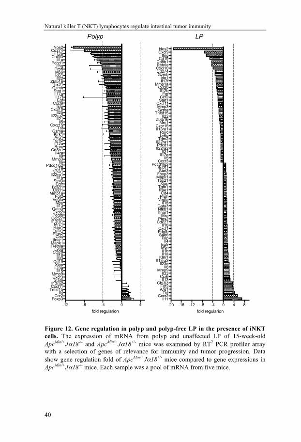
To identify the mechanisms underlying the polyp promotion of iNKT cells, we first performed a custom RT2 profiler PCR array to compare the inflammatory microenvironment in polyps and LP from ApcMin/+Jα18-/- and ApcMin/+Jα18+/- mice. Results from the array demonstrated an increased expression of proinflammatory genes in both LP and polyps, and down-regulated FoxP3 transcription in polyps, in the absence of iNKT cells (Figure 12 and Paper I, Figure 4). The expression of highly up-regulated genes in polyp and LP (Ifng, Nos2) and the most down-regulated gene in LP (Foxp3) was verified with RT qPCR. We also investigated IL17A expression by RT qPCR, as this cytokine had previously been shown to promote polyp formation in ApcMin/+ mice [180]. Absence of iNKT cells did not significantly alter IL-17A expression in polyp or LP, although levels of IL-17A were generally higher in polyp compared to LP (Paper I, Figure 4C). The decreased FoxP3 expression in polyps from iNKT deficient ApcMin/+Jα18-/- mice compared to ApcMin/+Jα18+/- mice suggested that Treg cells might be promoted in the presence of iNKT cells. The up-regulation of IFN-γ and NOS2 expression in iNKT cell deficient ApcMin/+ mice indicated that a Th1 immune response was inhibited in the presence iNKT cells. This strongly suggested iNKT cells promoted polyp development by suppressing Th1 immunity and enhancing immune regulation by Treg cells.
Natural killer T (NKT) lymphocytes regulate intestinal tumor immunity
40
Figure 12. Gene regulation in polyp and polyp-free LP in the presence of iNKT cells. The expression of mRNA from polyp and unaffected LP of 15-week-old ApcMin/+Jα18-/- and ApcMin/+Jα18+/- mice was examined by RT2 PCR profiler array with a selection of genes of relevance for immunity and tumor progression. Data show gene regulation fold of ApcMin/+Jα18+/- mice compared to gene expressions in ApcMin/+Jα18-/- mice. Each sample was a pool of mRNA from five mice.
-12 -8 -4 0 4Foxp3
Ccr2Il4
Tnfsf15Cxcl11Il13ra2
Stat6Cxcr3Mmp9
Il15Il23aXiap
Cd19Il33Il18
Ccl20Ly6g
RetnlaMapk1Ifnar1
Cd4Ptgs2Tgfb1Rae1Mrc1
Il13ra1Cdc27PdgfbCxcr2Gata3
Il13Myc
VegfaFgf2
Mmp1aCxcl1Bcl2l1
EgfrStat3
Il1bIl22ra1Nfkb1Ccr6
Pdcd1lg2Il6
Mmp3Il9
Il4raCd8b1
RorcIl12a
Tgm2Arg1Klrk1
GzmaTnf
Cxcr15Il5
Il21Il22ra2
Il25Cxcl10
TslpCxcl9
EgfIl17f
Il17aStat1
GzmbTbx21
Zbtb16Il22
Ido1MpoPrphIfng
Pdcd1Il1a
Chi3l3Il12b
Cd274Nos2
fold regularion-20 -16 -12 -8 -4 0 4 8
Il11Cxcr2
TnfArg1Fgf2
Chi3l3Il13
Ccr6Il33
Mmp9Il6
Il23aIl13ra2
Klrk1Il1aIl1bMycEgfr
Il4Tslp
Stat6PdgfbCxcl1
Il18Cdc27Ptgs2
Il4raIfnar1Nfkb1Gata3
Il15VegfaPrphCd4
Rae1Tgfb1Xiap
Tbx21Mapk1Foxp3Stat3
Bcl2l1Pdcd1lg2
Cxcr3Il5
Il17aIl21
Il22ra2Pdcd1Il22ra1Tgm2Ly6gRorc
Il13ra1Cxcr15
Mrc1Zbtb16
Il22Tnfsf15
GzmaMmp3Cxcl11
Stat1Ccr2
EgfIl12b
Ccl20Mmp1a
Il17fIdo1
GzmbCd274Cxcl10RetnlaCd8b1
Cd19Ifng
Cxcl9Nos2
fold regularion
Polyp LP

Ying Wang
41
Following this assumption, we explored the Treg cells in ApcMin/+Jα18-
/- and ApcMin/+Jα18+/- mice. As found by previous studies (see for example [65]), we confirmed that ApcMin/+ polyps contain high frequencies of Treg cells. Further, we found that only in polyp but not in other organs, both the frequency of FoxP3+ Treg cells and the levels of FoxP3 protein expression in Treg cells were decreased (Paper I, Figure 5). Notably, significantly lower expression of ST2, receptor for the alarmin IL-33, was shown on both polyp and LP Treg cells in the absence of iNKT cells. Recent studies have demonstrated that ST2 is highly expressed on colonic and adipose Treg cells [181-183], and TH2 biased ST2-expressing Treg cells are highly activated and suppress CD4 T cell proliferation through IL-10 and TGFβ production [184] Moreover, ST2 deficient mice were prevented from colonic tumor development [185]. Further, IL-33 promotes tissue-specific Treg accumulation and stability in colon [181]. This may indicate that iNKT cells promote the accumulation and maintenance of Treg cells in polyps through enhanced IL-33/ST2 signaling.
The absence of iNKT cells in ApcMin/+ mice was associated with increased expression of IFN-γ and NOS2 (also denoted iNOS) (Paper I, Figure 4). Although we did not determine the source of IFN-γ, CD8+ T cells are possibly the candidates for the increased expression of IFN-γ. Consistently, we defined differences in T cells infiltrating the polyp in ApcMin/+Jα18-/- compared to ApcMin/+Jα18+/- mice. In the absence of iNKT cells, the frequencies of conventional CD4+ T cells were increased in both MLN and polyps and the frequencies of CD8+ T cells were augmented in polyps (Paper I, Figure 3). This suggested that iNKT cells limited the polyp infiltration of conventional CD4+ T and CD8+ T cells. This maybe an indirect effect due to the reduction of Treg cells in ApcMin/+Jα18+/- mice. Indeed, it was recently shown that depletion of Treg in ApcMin/+ mice leads to the accumulation of CXCR3+ conventional T cells in intestinal tumors [68].
Different types of TAM have been described, and they can be either

Natural killer T (NKT) lymphocytes regulate intestinal tumor immunity
42
tumor killing (M1) or tumor promoting (M2) [186, 187]. iNOS is potentially expressed by M1 Mø, and absence of iNKT cells resulted in a systemically altered Mø phenotype from M2 towards M1 (Paper I, Figure 6c and d). Moreover, the phenotype shift was reversed by adoptively transferred iNKT cells to iNKT cell deficient ApcMin/+ mice (Paper I, Figure 6g). Therefore, we conclude that iNKT cells promote an anti-inflammtory environment and enhance M2 Mø in ApcMin/+ mice. Previous research has shown that ApcMin/+ mice lacking iNOS have significantly more adenomas than iNOS-positive littermate controls [188]. This is consistent with data from CRC patients showing that those that have tumors highly infiltrated with iNOS expressing Mø had a significantly improved prognosis [189]. In addition, we found that presence of PMN-MDSC was strongly reduced in polyps in the absence of iNKT cells (Paper I, Figure 6e and f). In conclusion, iNKT cell promotion of polyps was associated with an immunoregulatory polyp microenvironment, characterized by increased Treg and PMN-MDSC populations and an enhanced M2 macrophage phenotype, but suppressed Th1 immunity and reduced frequencies of tumor-infiltrating conventional CD4 and CD8 T cells.
iNKT cell directed immunotherapy modulated tumor development in ApcMin/+ mice The first identified NKT cell ligand, α-GalCer, was found to prevent tumor development and increased survival in several cancer models [41, 190, 191]. Positive results from clinical trials with cancer patients have shown the feasibility of the treatment [137, 192, 193]. The results in Paper I demonstrated that iNKT cells naturally promote polyp development associated with a suppression of Th1 immune response, and an increase in M2 macrophages and Treg cells in polyps. This could be an indication that further activation of iNKT cells would increase polyp development in ApcMin/+ mice. However, iNKT cell ligands have been described that preferentially induce Th1 cytokines,

Ying Wang
43
and we speculated that treatment with such ligands might reduce polyp development. We therefore investigated whether iNKT cell targeted immunotherapy could subvert the polyp promotion by iNKT cells and suppress polyp development in the ApcMin/+ mouse model. We performed one treatment schedule starting at the time of early-phase polyp initiation (long term, 5-15 weeks of age), and a late-phase treatment (short term, 12-15 weeks of age) during the period when the polyps expanded in size in the intestine (see treatment schedules in Figure 3).
In the early-phase treatment, we compared the glycolipid α-GalCer (C26:0), and the analogues of this ligand, C20:2 and C-glycoside, which enhance TH2 [45] or TH1[161] cytokine responses by iNKT cells, respectively. We hypothesized that the two analogues would give opposite results, that C20:2 would enhance the polyp development whereas C-glycoside would suppress the polyp growth in ApcMin/+ mice. As we expected, treatment with C20:2 increased polyp growth as evident by the increased numbers of large polyps in the small intestine compared to vehicle treated mice (Paper II, Figure 1). This was not the case for treatment with C26:0, which resulted in a significant decrease in number and size of polyps (Paper I, Figure 1 and Paper II, Figure 1). These results suggested that activation of iNKT cells by C26:0 treatment enhanced a tumor immune response. Surprisingly, instead of promoting an anti-polyp immune response, treatment with the Th1 skewing ligand C-glycoside did not show significant difference in polyp number and size. (Paper II, Figure 1). Therefore, we excluded C-glycoside in the late-phase treatment. We also a different lipid solvent in the late-phase treatment. The vehicle used to dissolve the glycolipids in the early-phase treatment contained dimethyl sulfoxide (DMSO), which is a commonly used solvent for α-GalCer. DMSO has been reported as having anti-inflammatory effects by blocking certain inflammatory pathways [194, 195]. To avoid anti-inflammatory effects that might be due to DMSO in the glycolipid solvent, for the late-phase treatment the vehicle for dissolving the glycolipids was changed to a

Natural killer T (NKT) lymphocytes regulate intestinal tumor immunity
44
solvent described in the protocol for α-GalCer in vivo assays recommended by Kirin Brewery Co., Ltd. [196], which contains no DMSO.
As in the early-phase treated animals, ApcMin/+ mice that had been treated late-phase with C26:0 exhibited positive effects with diminished polyp size although the effects were less pronounced than after early-phase treatment. In contrast, C20:2 that had significantly increased polyp growth in the early-phase treated ApcMin/+ mice produced the opposite result with the late-phase treatment schedule. Total polyp numbers were reduced, and there was a dramatic decrease in large polyps compared to vehicle treated mice (Paper II Figure 3).
Besides the polyp number and size, we also investigated the iNKT cells after treatments. Regardless of the type of iNKT ligand and treatment schedule, injections of the iNKT agonists resulted in a reduction of iNKT cell frequencies in the lymphoid organs (Paper II, Figure 2 and Figure 4). Moreover, there was a low responsiveness of splenocytes from C26:0 treated mice to in vitro α-GalCer restimulation (Paper II, Figure 2 and Figure 4). In contrast, after the late-phase treatment, splenocytes from the C20:2 treated mice still maintained the response to the in vitro α-GalCer activation. In accordance, the phenotypic marker for cell anergy, PD-1, was expressed at a lower level on remaining iNKT cells in spleen and MLN from C20:2 treated mice (Paper II, Figure 4). This indicated that a state of anergy was not fully reached with C20:2 during the short time period of treatment.
The results from the early- and late-phase treatments have been summarized in Table 2. The opposite results after early- and late-phase treatment with C20:2 triggered our speculation on possible causes. One contributing factor might be an age-related change of the polyp microenvironment. The polyp/intestinal microenvironment might be different during the different treatment periods (early versus late). This could be reflected in a difference in polyp infiltrating/LP immune cells, such as the frequency or phenotype/function of Treg cells [65], or a

Ying Wang
45
difference in phenotypes of iNKT cells (such as iNKT1 present in unaffected LP, versus iNKTpneg in polyps), etc. On the other hand, during the early-phase treatment, frequently injections of CD1d-restircted TCR ligand induced cytokine burst and systemic loss of iNKT cells, that also altered the polyp microenvironment. This demonstrates that the timing for the treatment is an important factor that needs to be considered.
From the early- and late-phase treatment experiments, we conclude that iNKT cell directed immunotherapy with α-GalCer could reduce tumor growth in ApcMin/+ mice. Surprisingly, however, effects with the Th1 and Th2 skewing analogues were not predictable, and indicated that the treatment timing could be essential.
Table 2. Summary of the iNKT directed treatments results. Early-phase Late-phase
Polyp#
Polypsize
iNKTloss/anergy
Polyp#
Polypsize
iNKTloss/anergy
C26:0 ↓ ↓ ++ → ↓ +++ C20:2 → ↑ ++ ↓ ↓ ++
C-Glycoside → → ++ ND ND NDArrows indicate increase or decrease of the polyp number and size. “+” was used to grade the iNKT loss/anergy. ND means not determined.
iNKT cell directed therapy cooperated with PD-1 checkpoint blockade and reduced intestinal tumor development in ApcMin/+ mice According to our results from the iNKT cell directed therapeutic study (Paper II), α-GalCer C26:0 (referred to α-GalCer below, as in Paper III) only succeeded to reduce the polyp size but not the number of

Natural killer T (NKT) lymphocytes regulate intestinal tumor immunity
46
polyps. The therapeutic effects might have been limited by an anergic state induced in iNKT cells by α-GalCer [197, 198], controlled by PD-1 [199]. Indeed, after the late phase treatment with α-GalCer, we found that iNKT cells showed an anergic phenotype with high level of PD-1 expression on the surface (Paper II, Figure 4). Further, PD-1 can be found on tumor-infiltrating T cells in different types of cancer [200-202]. Consistently, during our studies of immune cells in ApcMin/+ mice, we found that PD-1 was highly expressed on T cells in LP in these mice, but not on LP T cells from Apc+/+ littermates (Paper III, Figure 1). This suggested that interactions between PD-1 on T cells and PD-1 ligand(s) might suppress anti-tumor responses in ApcMin/+ mice, and that blocking this interaction could enhance tumor immunity. Based on these findings, we performed a new treatment of ApcMin/+ mice by combining the iNKT directed late-phase treatment with PD-1 checkpoint blockade. We predicted that blocking PD-1/PD-L1/2 signaling would firstly prevent iNKT loss and anergy after α-GalCer administration, and secondly, release conventional T cells from PD-1 mediated inhibition. We therefore expected that the α-GalCer anti-tumor effect could be improved in this way.
As we expected, we found that the combined treatment with anti-PD-1 and α-GalCer had an additive effect on polyp size resulting in a significant reduction of large polyps (Paper III, Figure 2). We investigated iNKT cells after the combined treatment schedule, and the results showed that iNKT cells could be detected in different organs from the treated mice including polyps. This revealed that the addition of PD-1 blockade to α-GalCer treatment prevented anergy induction and loss of iNKT cells. Further analysis of immune cells in treated mice demonstrated that the combination treatment resulted in increased activation of T cells, especially in the polyps. In addition, PD-1 blockade prevented α-GalCer induced accumulation of M-MDSC in spleen and PMN-MDSC in spleen and LP, while α-GalCer treatment promoted an M1 macrophage phenotype in the spleen both in the presence and absence of PD-1 blockade. The results from the
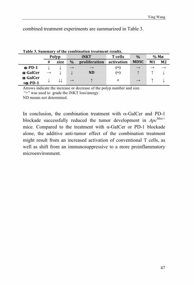
Ying Wang
47
combined treatment experiments are summarized in Table 3.
Table 3. Summary of the combination treatment results.
Polyp iNKT Tcells %MDSC
%Mø# size % proliferation activation M1 M2
α-PD-1 ↓ ↓ → → (+) → → → α-GalCer → ↓ ↓ ND (+) ↑ ↑ ↓ α-GalCer+α-PD-1
↓ ↓↓ → ↑ + → ↑ ↓
Arrows indicate the increase or decrease of the polyp number and size. “+” was used to grade the iNKT loss/anergy. ND means not determined.
In conclusion, the combination treatment with α-GalCer and PD-1 blockade successfully reduced the tumor development in ApcMin/+ mice. Compared to the treatment with α-GalCer or PD-1 blockade alone, the additive anti-tumor effect of the combination treatment might result from an increased activation of conventional T cells, as well as shift from an immunosuppressive to a more proinflammatory microenvironment.

Natural killer T (NKT) lymphocytes regulate intestinal tumor immunity
48
CONCLUDING REMARKS AND FUTURE PERSPECTIVES This thesis studied the regulation roles of iNKT cells in intestinal tumor development from two perspectives, natural and therapeutic. The results from these studies establish that NKT cells are potent regulators of intestinal tumor immunity, and should be further explored for the development of NKT cell directed immunotherapy in intestinal cancer. The main conclusions from the papers in the thesis are summarized below:
I. iNKT cells present a unique phenotype in polyps being PLZF- and enriched for IL-10 and IL-17 producing cells.
II. iNKT cells naturally promote intestinal tumor development by enhancing regulatory T cells and immunosuppression of antitumor TH1 immunity.
III. Preclinical therapeutic treatments with NKT cell
modulating ligands demostrated that NKT cell directed therapy could subvert the tumor enhancing function of NKT cells and suppress polyp growth.
IV. Treatments with NKT cell ligand with different starting
point give opposite results, indicates that the treatment timing is an ignorable consideration.
V. Combined treatment with the NKT cell agonist α-
GalCer and PD-1 immune checkpoint blockade succeeded to further reduce polyp growth.

Ying Wang
49
Although the current results explained the questions we asked from the beginning, some of our preliminary data still need to be further explored. Also, there are new questions popped up from what we have known, which also need be investigated.
First, we showed that the tumor promoting effect by iNKT cells, but if the effect is direct or indirect is unknown. We have set up a new mouse strain, ApcMin/+CD1dfloxVillinCre, which specifically lacking of NKT cells on epithelial cells. Investigation of these mice will enable us to understand if NKT cells directly interact with tumor cells and promote the tumor development.
Further, similar to iNKT10, polyp iNKT lacks PLZF expression and produce IL-10. Transfer of hepatic iNKT cells altered the Mø phenotype in ApcMin/+Jα18 -/- mice in polyps, even though hepatic iNKT cells are prominently iNKT1 cells, which are positive for PLZF and produce TH1 cytokines. This released a question that what makes the changes of iNKT1 behaving as iNKT10. The possible explanations can either be the iNKT cells changed phenotype in the location of polyps or only small population of iNKT10 expanded in polyp. Since it is hard to detect the transferred iNKT cells when we harvest the mice, we planed to transfer IL-10 KO iNKT cells to ApcMin/+Jα18 -/- mice to determine if iNKT10 plays an important role in Mø phenotype switch.
Moreover, the iNKT directed treatment with modulated ligands have shown to reduce the polyp formation, but the mechanisms are remaining unclear. We have done PCR arrays to screen the candidate genes that have been regulated by the treatments, and thereby more specific analysis could be performed to understand the mechanisms.
Last but not least, since ApcMin/+ mice develop most polyps in small intestine, all the results of this thesis are based on the studies in small intestine. Since the tumor microenvironment might be different in colon, the study of colon iNKT cannot be ignored eventually.

Natural killer T (NKT) lymphocytes regulate intestinal tumor immunity
50
Overall, we aim to further clarify the mechanisms of iNKT regulation in tumor immunity and hope the study can provide useful idea for the clinical treatment.

51
ACKNOWLEDGEMENTS
For more than eight years being in Sweden, Gothenburg has become by second hometown. During the years of my phD period, there are numbers of people gave me supports and I really grateful. I would like to express my gratitude to all the people who supported me, in and out of the department. Special thanks must go to the following people:
Susanna L. Cardell, my supervisor, thanks for bringing me into this fantastic project. You opened a door for me to touch the science of immunology. I learnt so much during the phD period. I do appreciate all your patience and knowledge. Instead of telling me what you know and what to do, you always try to inspire me and let me figure things out myself. Also you are the most energetic and knowledgeable person I have ever met, with all the enthusiasm to all the interesting points for the project. You are a real scientist, and I am so proud to be a student of you!
Marianne Quiding-Jarbrink, my co-supervisor, thank you for sharing your knowledges with me in fruitful discussions. I do appreciate the suggestions you gave!
Linda Löfbom, not only being technician but also my best friend and partner in the lab, I feel I have used all the words at the day you left. I really miss the time when you are in the lab! I could not survive without you in the long experiments. Our lab is so quiet and empty without you. However I also feel happy that you got the opportunity to try another way of life!
Saikiran Sedimbi, as the babysitter when I first arrived, you spent all the patience to teach me the knowledge and techniques needed in the project. Thanks for the nice guide from the beginning, so I could understand the project much better. Good luck with life in Stockholm!
The previous and present of members in our group: Sara Rhost, Prabhanshu Tripathi, Avadhesh Kumar Singh, Cristiana Rimniceanu and Urszula Chursa, thank you all for providing a nice circumstance in our lab with discussions on technical and experimental considerations!

Natural killer T (NKT) lymphocytes regulate intestinal tumor immunity
52
Staffs in EBM, especially Pia and Anders, thanks a lot for taking care of our mice and breedings. And thanks to all the mice bred for this project; nothing could be done without your suffering.
Paulina Akeus, you are the one know exactly how I feel when you open a mouse and see no polyps inside! Thanks for all the supports and advices according to the mice and cell preparations!
Samuel Alsén, thanks for staying late on a Friday evening to help me with the cell sorter!
My partners for the course teaching, Inta Gribonika, Filip Ahlmanner, Inga Rimkute, Astrid Von Mentzer, Samuel Alsén and Ratan Komban, it was a very nice experience to teach the students together with you!
My office-mates: Valentina bernasconi, Ratan Komban and Jocob Cervin. We share the most crowed office but we enjoy it. It makes us more closed to each other. Valentina, thanks for all the candies, snacks and trip tips you provided! Rathan and Jacob, you guys are so nice and warmhearted, always make me laugh and help me with everything I asked for. Also, I really love the tradition of Christmas decoration in our office!
Yu Fang, Zou Xiang and Jia-bin Sun, 你们是我的良师益友. It is my pleasure to know you in the department. You never hesitate to tell me what you know about immunology. Yu Fang helped me so much at the beginning of my phD; Zou always brought me nice stuffs from China; and my Chinese stomach were so satisfied with the 苹果、梨、韭菜 from Jia-bin’s garden.
Natalija Gerasimcik, my friend and collaborator, thanks for all the encouragements you gave during my thesis writing!
My family in Gothenburg, Cuicui Xie, Kai Zhou, Min Hu, Rong Ma, Yanling Wu and Zhiyuan Zou (Tony), We had so much nice memories being together! My life in Sweden is not complete without you guys!
Cuicui & Kai, we had a happy time together in Dalarna for the New Year and midsummer. Enjoy the happy life in Stockholm!
Yanling, not only my friend, but also my neighbor, thanks for all the dinners you made during I worked with the thesis! Enjoy your life at your new home! Let’s think of the trip in Rome this Christmas!!

53
Min, I will never forget the trip we went together in Italy and France, and the football match we watched together! You are like an elderly sister that thinks of every one in our “family”. I hope one day someone will take care of you like you did for us! Also, good luck with the visa application! I hope you get a bright future!
Rong, you are the first Chinese friend I got since I came to Sweden. I still remember the first day we met at the entrance of Zoology department. I guess you will also complete your phD soon. Good luck with everything!
Tony, the chef in the “family”, every party could not be perfect without the dishes you made! Thanks for all the suggestions you gave about the mice, as we use the same mouse model. I really hope to be your collaborator one day! Good luck with your paper submission! Also, 赶紧找个妹子嫁了!J
Peidi Liu, I probably wouldn’t apply for a phD position if you didn’t send me the link. It changed my life and career. Good luck with the job application! You and Dimitra are the perfect couple. Hope you two have a happy life!
Juan Li, even though you haven’t come here so long, and won’t stay here after this year, you are the witness of my last stage of my phD. Thanks for all the company when I was doing the thesis writing!
Edvin Vestin, thanks for the efforts you made for the Swedish translation! I really appreciate it!
Meng Chen (八爪 ), great thanks to bring my key back from China! I am so happy to see you have found Jingying as your soul mate. You are so nice couple! Wish to attend your wedding!
Yuan Zhang, my previous colleague, and also a cat lover, we have all the topics together about cat. Good luck with the Swedish learning and job application! I will visit you and Tiger for sure!
Carolina Nemethy (妹妹 ) and Alexander Stelmach (Patrick), Thanks Ecocycles let me know you and your family! Your whole family is so nice, especially your Mom, make me feel like home. 妹妹 , you will soon go to Copenhagen and start you new stage of life. Good luck with everything in there! Patrick, I can see your happiness from your face when you are together with your girl friend. I also feel happy for you!
Natural killer T (NKT) lymphocytes regulate intestinal tumor immunity
54
Bamse (小胖 =∩ω∩=), my lovely cat, you are always be with me when I was writing the thesis. Thanks for the company and all the happy time with you.
My parents, 父母恩重如山,女儿身在异国无以为报。唯有做更好的自己,让你们感到骄傲!I miss you so much!
My love, Yu Zhao, you are without a doubt the most importance person in my life besides by parents. Thanks for taking care of me these years. I remember all the late nights you were waiting for me when I was doing big experiments. And also thanks for bringing Bamse into our life; it was the best gift ever! 我愛你!❤

55
REFERENCES
1. Makino, Y., R. Kanno, T. Ito, K. Higashino, and M. Taniguchi, Predominant expression of invariant Vα14+ TCR α chain in NK1.1+ T cell populations. International Immunology, 1995. 7(7): p. 1157-1161.
2. Bendelac, A., O. Lantz, M.E. Quimby, J.W. Yewdell, Bennink, and R.R. Brutkiewicz, CD1 recognition by mouse NK1+ T lymphocytes. Science, 1995. 268(5212): p. 863.
3. Cardell, S., S. Tangri, S. Chan, M. Kronenberg, C. Benoist, and D. Mathis, CD1-restricted CD4+ T cells in major histocompatibility complex class II-deficient mice. The Journal of Experimental Medicine, 1995. 182(4): p. 993-1004.
4. Godfrey, D.I. and M. Kronenberg, Going both ways: Immune regulation via CD1d-dependent NKT cells. The Journal of Clinical Investigation, 2004. 114(10): p. 1379-1388.
5. Michel, M.-L., A.C. Keller, C. Paget, M. Fujio, F. Trottein, P.B. Savage, C.-H. Wong, E. Schneider, M. Dy, and M.C. Leite-de-Moraes, Identification of an IL-17–producing NK1.1neg iNKT cell population involved in airway neutrophilia. The Journal of Experimental Medicine, 2007. 204(5): p. 995.
6. Gumperz, J.E., S. Miyake, T. Yamamura, and M.B. Brenner, Functionally Distinct Subsets of CD1d-restricted Natural Killer T Cells Revealed by CD1d Tetramer Staining. The Journal of Experimental Medicine, 2002. 195(5): p. 625-636.
7. Coquet, J.M., S. Chakravarti, K. Kyparissoudis, F.W. McNab, L.A. Pitt, B.S. McKenzie, S.P. Berzins, M.J. Smyth, and D.I. Godfrey, Diverse cytokine production by NKT cell subsets and identification of an IL-17–producing CD4−NK1.1− NKT cell population. Proceedings of the National Academy of Sciences, 2008. 105(32): p. 11287-11292.
8. Godfrey, D.I. and J. Rossjohn, New ways to turn on NKT cells. The Journal of Experimental Medicine, 2011. 208(6): p. 1121-1125.
9. Porcelli, S., C.T. Morita, and M.B. Brenner, CDlb restricts the response of human CD4-8-T lymphocytes to a microbial antigen. Nature, 1992. 360(6404): p. 593-597.
10. Beckman, E.M., S.A. Porcelli, C.T. Morita, S.M. Behar, S.T. Furlong, and M.B. Brenner, Recognition of a lipid antigen by GDI-restricted [alpha][beta]+ T cells. Nature, 1994. 372(6507): p. 691-694.
11. M. Jackman, R., D. B Moody, and S.A. Porcelli, Mechanisms of Lipid Antigen Presentation by CD1. Vol. 19. 1999. 49-63.
12. Barral, D.C. and M.B. Brenner, CD1 antigen presentation: how it works. Nat Rev Immunol, 2007. 7(12): p. 929-941.
13. Kasmar, A., I.V. Rhijn, and D.B. Moody, The evolved functions of CD1 during infection. Current opinion in immunology, 2009. 21(4): p. 397-403.

Natural killer T (NKT) lymphocytes regulate intestinal tumor immunity
56
14. Naidenko, O.V., J.K. Maher, W.A. Ernst, T. Sakai, R.L. Modlin, and M. Kronenberg, Binding and Antigen Presentation of Ceramide-Containing Glycolipids by Soluble Mouse and Human Cd1d Molecules. The Journal of Experimental Medicine, 1999. 190(8): p. 1069.
15. Cantu, C., K. Benlagha, P.B. Savage, A. Bendelac, and L. Teyton, The Paradox of Immune Molecular Recognition of α-Galactosylceramide: Low Affinity, Low Specificity for CD1d, High Affinity for αβ TCRs. The Journal of Immunology, 2003. 170(9): p. 4673.
16. Cox, D., L. Fox, R. Tian, W. Bardet, M. Skaley, D. Mojsilovic, J. Gumperz, and W. Hildebrand, Determination of Cellular Lipids Bound to Human CD1d Molecules. PLOS ONE, 2009. 4(5): p. e5325.
17. Rhost, S., S. Sedimbi, N. Kadri, and S.L. Cardell, Immunomodulatory Type II Natural Killer T Lymphocytes in Health and Disease. Scandinavian Journal of Immunology, 2012. 76(3): p. 246-255.
18. Exley, M.A., S.M.A. Tahir, O. Cheng, A. Shaulov, R. Joyce, D. Avigan, R. Sackstein, and S.P. Balk, Cutting Edge: A Major Fraction of Human Bone Marrow Lymphocytes Are Th2-Like CD1d-Reactive T Cells That Can Suppress Mixed Lymphocyte Responses. The Journal of Immunology, 2001. 167(10): p. 5531.
19. Bendelac, A., O. Lantz, M.E. Quimby, J.W. Yewdell, J.R. Bennink, and R.R. Brutkiewicz, CD1 recognition by mouse NK1+ T lymphocytes. Science, 1995. 268(5212): p. 863-5.
20. Dellabona, P., E. Padovan, G. Casorati, M. Brockhaus, and A. Lanzavecchia, An invariant V alpha 24-J alpha Q/V beta 11 T cell receptor is expressed in all individuals by clonally expanded CD4-8- T cells. The Journal of Experimental Medicine, 1994. 180(3): p. 1171-1176.
21. Porcelli, S., C. Yockey, M. Brenner, and S. Balk, Analysis of T cell antigen receptor (TCR) expression by human peripheral blood CD4-8- alpha/beta T cells demonstrates preferential use of several V beta genes and an invariant TCR alpha chain. The Journal of Experimental Medicine, 1993. 178(1): p. 1-16.
22. Kawano, T., J. Cui, Y. Koezuka, I. Toura, Y. Kaneko, K. Motoki, H. Ueno, R. Nakagawa, H. Sato, E. Kondo, H. Koseki, and M. Taniguchi, CD1d-Restricted and TCR-Mediated Activation of V<sub>α</sub>14 NKT Cells by Glycosylceramides. Science, 1997. 278(5343): p. 1626.
23. Benlagha, K., A. Weiss, A. Beavis, L. Teyton, and A. Bendelac, In Vivo Identification of Glycolipid Antigen–Specific T Cells Using Fluorescent Cd1d Tetramers. The Journal of Experimental Medicine, 1999. 191(11): p. 1895.
24. Matsuda, J.L., O.V. Naidenko, L. Gapin, T. Nakayama, M. Taniguchi, C.-R. Wang, Y. Koezuka, and M. Kronenberg, Tracking the Response of Natural Killer T Cells to a Glycolipid Antigen Using Cd1d Tetramers. The Journal of Experimental Medicine, 2000. 192(5): p. 741-754.
25. Karadimitris, A., S. Gadola, M. Altamirano, D. Brown, A. Woolfson, P. Klenerman, J.-L. Chen, Y. Koezuka, I.A.G. Roberts, D.A. Price, G.
57
Dusheiko, C. Milstein, A. Fersht, L. Luzzatto, and V. Cerundolo, Human CD1d–glycolipid tetramers generated by in vitro oxidative refolding chromatography. Proceedings of the National Academy of Sciences of the United States of America, 2001. 98(6): p. 3294-3298.
26. Sidobre, S. and M. Kronenberg, CD1 tetramers: a powerful tool for the analysis of glycolipid-reactive T cells. Journal of Immunological Methods, 2002. 268(1): p. 107-121.
27. Lee, Y.J., K.L. Holzapfel, J. Zhu, S.C. Jameson, and K.A. Hogquist, Steady-state production of IL-4 modulates immunity in mouse strains and is determined by lineage diversity of iNKT cells. Nat Immunol, 2013. 14(11): p. 1146-1154.
28. Sag, D., P. Krause, C.C. Hedrick, M. Kronenberg, and G. Wingender, IL-10–producing NKT10 cells are a distinct regulatory invariant NKT cell subset. The Journal of Clinical Investigation, 2014. 124(9): p. 3725-3740.
29. Wingender, G., D. Sag, and M. Kronenberg, NKT10 cells: a novel iNKT cell subset. Oncotarget, 2015. 6(29): p. 26552-26553.
30. Lynch, L., X. Michelet, S. Zhang, P.J. Brennan, A. Moseman, C. Lester, G. Besra, E.E. Vomhof-Dekrey, M. Tighe, H.-F. Koay, D.I. Godfrey, E.A. Leadbetter, D.B. Sant'Angelo, U. von Andrian, and M.B. Brenner, Regulatory iNKT cells lack expression of the transcription factor PLZF and control the homeostasis of Treg cells and macrophages in adipose tissue. Nat Immunol, 2015. 16(1): p. 85-95.
31. Vieth, J.A., J. Das, F.M. Ranaivoson, D. Comoletti, L.K. Denzin, and D.B. Sant'Angelo, TCR[alpha]-TCR[beta] pairing controls recognition of CD1d and directs the development of adipose NKT cells. Nat Immunol, 2017. 18(1): p. 36-44.
32. Makowska, Kawano, Taniguchi, and Cardell, Differences in the Ligand Specificity between CD1d-Restricted T Cells with Limited and Diverse T-Cell Receptor Repertoire. Scandinavian Journal of Immunology, 2000. 52(1): p. 71-79.
33. Jahng, A., I. Maricic, C. Aguilera, S. Cardell, R.C. Halder, and V. Kumar, Prevention of Autoimmunity by Targeting a Distinct, Noninvariant CD1d-reactive T Cell Population Reactive to Sulfatide. J Exp Med, 2004. 199(7): p. 947-57.
34. Chang, D.H., H. Deng, P. Matthews, J. Krasovsky, G. Ragupathi, R. Spisek, A. Mazumder, D.H. Vesole, S. Jagannath, and M.V. Dhodapkar, Inflammation-associated lysophospholipids as ligands for CD1d-restricted T cells in human cancer. Blood, 2008. 112(4): p. 1308-1316.
35. Savage, A.K., M.G. Constantinides, J. Han, D. Picard, E. Martin, B. Li, O. Lantz, and A. Bendelac, The transcription factor PLZF (Zbtb16) directs the effector program of the NKT cell lineage. Immunity, 2008. 29(3): p. 391-403.
36. Kovalovsky, D., O.U. Uche, S. Eladad, R.M. Hobbs, W. Yi, E. Alonzo, K. Chua, M. Eidson, H.-J. Kim, J.S. Im, P.P. Pandolfi, and D.B. Sant'Angelo, The BTB-zinc finger transcriptional regulator, PLZF, controls the

Natural killer T (NKT) lymphocytes regulate intestinal tumor immunity
58
development of iNKT cell effector functions. Nature immunology, 2008. 9(9): p. 1055-1064.
37. Rossjohn, J., D.G. Pellicci, O. Patel, L. Gapin, and D.I. Godfrey, Recognition of CD1d-restricted antigens by natural killer T cells. Nat Rev Immunol, 2012. 12(12): p. 845-857.
38. Godfrey, D.I., S. Stankovic, and A.G. Baxter, Raising the NKT cell family. Nat Immunol, 2010. 11(3): p. 197-206.
39. Taniguchi, M., T. Tashiro, N. Dashtsoodol, N. Hongo, and H. Watarai, The specialized iNKT cell system recognizes glycolipid antigens and bridges the innate and acquired immune systems with potential applications for cancer therapy. International Immunology, 2010. 22(1): p. 1-6.
40. Kobayashi, E., K. Motoki, T. Uchida, H. Fukushima, and Y. Koezuka, KRN7000, a novel immunomodulator, and its antitumor activities. Oncol. Res., 1995. 7: p. 529-534.
41. Kobayashi, E., K. Motoki, T. Uchida, H. and Fukushima, and Y. Koezuka, KRN7000, a novel immunomodulator, and its antitumor activities. Vol. 7. 1995. 529-34.
42. Venkataswamy, M.M. and S.A. Porcelli, Lipid and glycolipid antigens of CD1d-restricted natural killer T cells. Seminars in immunology, 2010. 22(2): p. 68-78.
43. Tyznik, A.J., E. Farber, E. Girardi, A. Birkholz, Y. Li, S. Chitale, R. So, P. Arora, A. Khurana, J. Wang, S.A. Porcelli, D.M. Zajonc, M. Kronenberg, and A.R. Howell, Novel glycolipids that elicit IFN-γ-biased responses from natural killer T cells. Chemistry & biology, 2011. 18(12): p. 1620-1630.
44. Oki, S., A. Chiba, T. Yamamura, and S. Miyake, The clinical implication and molecular mechanism of preferential IL-4 production by modified glycolipid-stimulated NKT cells. The Journal of Clinical Investigation, 2004. 113(11): p. 1631-1640.
45. Yu, K.O.A., J.S. Im, A. Molano, Y. Dutronc, P.A. Illarionov, C. Forestier, N. Fujiwara, I. Arias, S. Miyake, T. Yamamura, Y.-T. Chang, G.S. Besra, and S.A. Porcelli, Modulation of CD1d-restricted NKT cell responses by using N-acyl variants of α-galactosylceramides. Proceedings of the National Academy of Sciences of the United States of America, 2005. 102(9): p. 3383-3388.
46. Schmieg, J., G. Yang, R.W. Franck, and M. Tsuji, Superior Protection against Malaria and Melanoma Metastases by a C-glycoside Analogue of the Natural Killer T Cell Ligand α-Galactosylceramide. The Journal of Experimental Medicine, 2003. 198(11): p. 1631.
47. Tupin, E., Y. Kinjo, and M. Kronenberg, The unique role of natural killer T cells in the response to microorganisms. Nat Rev Micro, 2007. 5(6): p. 405-417.
48. Kinjo, Y., N. Kitano, and M. Kronenberg, The role of invariant natural killer T cells in microbial immunity. Journal of infection and chemotherapy : official journal of the Japan Society of Chemotherapy, 2013. 19(4): p. 10.1007/s10156-013-0638-1.

59
49. Kinjo, Y., B. Pei, S. Bufali, R. Raju, S.K. Richardson, M. Imamura, M. Fujio, D. Wu, A. Khurana, K. Kawahara, C.-H. Wong, A.R. Howell, P.H. Seeberger, and M. Kronenberg, Natural Sphingomonas Glycolipids Vary Greatly in their Ability to Activate Natural Killer T Cells. Chemistry & biology, 2008. 15(7): p. 654-664.
50. Kinjo, Y., D. Wu, G. Kim, G.-W. Xing, M.A. Poles, D.D. Ho, M. Tsuji, K. Kawahara, C.-H. Wong, and M. Kronenberg, Recognition of bacterial glycosphingolipids by natural killer T cells. Nature, 2005. 434(7032): p. 520-525.
51. Mattner, J., K.L. DeBord, N. Ismail, R.D. Goff, C. Cantu, D. Zhou, P. Saint-Mezard, V. Wang, Y. Gao, N. Yin, K. Hoebe, O. Schneewind, D. Walker, B. Beutler, L. Teyton, P.B. Savage, and A. Bendelac, Exogenous and endogenous glycolipid antigens activate NKT cells during microbial infections. Nature, 2005. 434(7032): p. 525-529.
52. Bendelac, A., Positive selection of mouse NK1+ T cells by CD1-expressing cortical thymocytes. The Journal of Experimental Medicine, 1995. 182(6): p. 2091-2096.
53. Brigl, M., L. Bry, S.C. Kent, J.E. Gumperz, and M.B. Brenner, Mechanism of CD1d-restricted natural killer T cell activation during microbial infection. Nat Immunol, 2003. 4(12): p. 1230-1237.
54. Zhou, D., J. Mattner, C. Cantu, N. Schrantz, N. Yin, Y. Gao, Y. Sagiv, K. Hudspeth, Y.-P. Wu, T. Yamashita, S. Teneberg, D. Wang, R.L. Proia, S.B. Levery, P.B. Savage, L. Teyton, and A. Bendelac, Lysosomal Glycosphingolipid Recognition by NKT Cells. Science, 2004. 306(5702): p. 1786.
55. Speak, A.O., M. Salio, D.C.A. Neville, J. Fontaine, D.A. Priestman, N. Platt, T. Heare, T.D. Butters, R.A. Dwek, F. Trottein, M.A. Exley, V. Cerundolo, and F.M. Platt, Implications for invariant natural killer T cell ligands due to the restricted presence of isoglobotrihexosylceramide in mammals. Proceedings of the National Academy of Sciences, 2007. 104(14): p. 5971-5976.
56. Porubsky, S., A.O. Speak, B. Luckow, V. Cerundolo, F.M. Platt, and H.-J. Gröne, Normal development and function of invariant natural killer T cells in mice with isoglobotrihexosylceramide (iGb3) deficiency. Proceedings of the National Academy of Sciences, 2007. 104(14): p. 5977-5982.
57. Kärre, K., H.G. Ljunggren, G. Piontek, and R. Kiessling, Selective rejection of H-2-deficient lymphoma variants suggests alternative immune defence strategy. Nature, 1986. 319 6055: p. 675-678.
58. Gordon, S. and P.R. Taylor, Monocyte and macrophage heterogeneity. Nat Rev Immunol, 2005. 5(12): p. 953-964.
59. Murdoch, C., M. Muthana, S.B. Coffelt, and C.E. Lewis, The role of myeloid cells in the promotion of tumour angiogenesis. Nat Rev Cancer, 2008. 8(8): p. 618-631.
60. Mantovani, A., P. Allavena, A. Sica, and F. Balkwill, Cancer-related inflammation. Nature, 2008. 454(7203): p. 436-444.

Natural killer T (NKT) lymphocytes regulate intestinal tumor immunity
60
61. Mantovani, A., B. Bottazzi, F. Colotta, S. Sozzani, and L. Ruco, The origin and function of tumor-associated macrophages. Immunology Today, 1992. 13(7): p. 265-270.
62. Balkwill, F. and A. Mantovani, Inflammation and cancer: back to Virchow? The Lancet, 2001. 357(9255): p. 539-545.
63. Mantovani, A., S. Sozzani, M. Locati, P. Allavena, and A. Sica, Macrophage polarization: tumor-associated macrophages as a paradigm for polarized M2 mononuclear phagocytes. Trends in Immunology, 2002. 23(11): p. 549-555.
64. Hariharan, K., G. Braslawsky, A. Black, S. Raychaudhuri, and N. Hanna, The Induction of Cytotoxic T Cells and Tumor Regression by Soluble Antigen Formulation. Cancer Research, 1995. 55(16): p. 3486.
65. Akeus, P., V. Langenes, A. von Mentzer, U. Yrlid, Å. Sjöling, P. Saksena, S. Raghavan, and M. Quiding-Järbrink, Altered chemokine production and accumulation of regulatory T cells in intestinal adenomas of APCMin/+ mice. Cancer Immunology, Immunotherapy, 2014. 63(8): p. 807-819.
66. Ohkura, N. and S. Sakaguchi, Regulatory T cells: roles of T cell receptor for their development and function. Seminars in Immunopathology, 2010. 32(2): p. 95-106.
67. Hsu, P., B. Santner-Nanan, M. Hu, K. Skarratt, C.H. Lee, M. Stormon, M. Wong, S.J. Fuller, and R. Nanan, IL-10 Potentiates Differentiation of Human Induced Regulatory T Cells via STAT3 and Foxo1. The Journal of Immunology, 2015. 195(8): p. 3665.
68. Akeus, P., V. Langenes, J. Kristensen, A. von Mentzer, T. Sparwasser, S. Raghavan, and M. Quiding-Järbrink, Treg-cell depletion promotes chemokine production and accumulation of CXCR3+ conventional T cells in intestinal tumors. European Journal of Immunology, 2015. 45(6): p. 1654-1666.
69. Li, X., E. Kostareli, J. Suffner, N. Garbi, and G.J. Hämmerling, Efficient Treg depletion induces T-cell infiltration and rejection of large tumors. European Journal of Immunology, 2010. 40(12): p. 3325-3335.
70. Hindley, J.P., E. Jones, K. Smart, H. Bridgeman, S.N. Lauder, B. Ondondo, S. Cutting, K. Ladell, K.K. Wynn, D. Withers, D.A. Price, A. Ager, A.J. Godkin, and A.M. Gallimore, T Cell Trafficking Facilitated by High Endothelial Venules is Required for Tumor Control after Regulatory T Cell Depletion. Cancer research, 2012. 72(21): p. 5473-5482.
71. Teng, M.W.L., S.F. Ngiow, B. von Scheidt, N. McLaughlin, T. Sparwasser, and M.J. Smyth, Conditional Regulatory T-Cell Depletion Releases Adaptive Immunity Preventing Carcinogenesis and Suppressing Established Tumor Growth. Cancer Research, 2010. 70(20): p. 7800.
72. Kumar, V., S. Patel, E. Tcyganov, and D.I. Gabrilovich, The Nature of Myeloid-Derived Suppressor Cells in the Tumor Microenvironment. Trends in Immunology, 2016. 37(3): p. 208-220.
73. Gabrilovich, D.I. and S. Nagaraj, Myeloid-derived suppressor cells as regulators of the immune system. Nat Rev Immunol, 2009. 9(3): p. 162-174.

61
74. Youn, J.-I., S. Nagaraj, M. Collazo, and D.I. Gabrilovich, Subsets of Myeloid-Derived Suppressor Cells in Tumor Bearing Mice. Journal of immunology (Baltimore, Md. : 1950), 2008. 181(8): p. 5791-5802.
75. Kusmartsev, S., Y. Nefedova, D. Yoder, and D.I. Gabrilovich, Antigen-Specific Inhibition of CD8<sup>+</sup> T Cell Response by Immature Myeloid Cells in Cancer Is Mediated by Reactive Oxygen Species. The Journal of Immunology, 2004. 172(2): p. 989.
76. Ochoa, A.C., A.H. Zea, C. Hernandez, and P.C. Rodriguez, Arginase, Prostaglandins, and Myeloid-Derived Suppressor Cells in Renal Cell Carcinoma. Clinical Cancer Research, 2007. 13(2): p. 721s.
77. Almand, B., J.I. Clark, E. Nikitina, J. van Beynen, N.R. English, S.C. Knight, D.P. Carbone, and D.I. Gabrilovich, Increased Production of Immature Myeloid Cells in Cancer Patients: A Mechanism of Immunosuppression in Cancer. The Journal of Immunology, 2001. 166(1): p. 678.
78. Hestdal, K., F.W. Ruscetti, J.N. Ihle, S.E. Jacobsen, C.M. Dubois, W.C. Kopp, D.L. Longo, and J.R. Keller, Characterization and regulation of RB6-8C5 antigen expression on murine bone marrow cells. The Journal of Immunology, 1991. 147(1): p. 22.
79. Huang, B., P.-Y. Pan, Q. Li, A.I. Sato, D.E. Levy, J. Bromberg, C.M. Divino, and S.-H. Chen, Gr-1+CD115+ immature myeloid suppressor cells mediate the development of tumor-induced T regulatory cells and T-cell anergy in tumor-bearing host. Cancer Research, 2006. 66(2): p. 1123.
80. Yang, R., Z. Cai, Y. Zhang, W.H. Yutzy, K.F. Roby, and R.B.S. Roden, CD80 in Immune Suppression by Mouse Ovarian Carcinoma–Associated Gr-1<sup>+</sup>CD11b<sup>+</sup> Myeloid Cells. Cancer Research, 2006. 66(13): p. 6807.
81. Smyth, M.J., N.Y. Crowe, Y. Hayakawa, K. Takeda, H. Yagita, and D.I. Godfrey, NKT cells - conductors of tumor immunity? Curr Opin Immunol, 2002. 14(2): p. 165-71.
82. Bellone, M., M. Ceccon, M. Grioni, E. Jachetti, A. Calcinotto, A. Napolitano, M. Freschi, G. Casorati, and P. Dellabona, iNKT cells control mouse spontaneous carcinoma independently of tumor-specific cytotoxic T cells. PLoS One, 2010. 5(1): p. e8646.
83. Swann, J.B., A.P. Uldrich, S. van Dommelen, J. Sharkey, W.K. Murray, D.I. Godfrey, and M.J. Smyth, Type I natural killer T cells suppress tumors caused by p53 loss in mice. Blood, 2009. 113(25): p. 6382-5.
84. Crowe, N.Y., J.M. Coquet, S.P. Berzins, K. Kyparissoudis, R. Keating, D.G. Pellicci, Y. Hayakawa, D.I. Godfrey, and M.J. Smyth, Differential antitumor immunity mediated by NKT cell subsets in vivo. J Exp Med, 2005. 202(9): p. 1279-88.
85. Metelitsa, L.S., O.V. Naidenko, A. Kant, H.-W. Wu, M.J. Loza, B. Perussia, M. Kronenberg, and R.C. Seeger, Human NKT Cells Mediate Antitumor Cytotoxicity Directly by Recognizing Target Cell CD1d with Bound Ligand
Natural killer T (NKT) lymphocytes regulate intestinal tumor immunity
62
or Indirectly by Producing IL-2 to Activate NK Cells. The Journal of Immunology, 2001. 167(6): p. 3114.
86. Renukaradhya, G.J., V. Sriram, W. Du, J. Gervay-Hague, L. Van Kaer, and R.R. Brutkiewicz, Inhibition of antitumor immunity by invariant natural killer T cells in a T-cell lymphoma model in vivo. International Journal of Cancer, 2006. 118(12): p. 3045-3053.
87. Osada, T., M.A. Morse, H.K. Lyerly, and T.M. Clay, Ex vivo expanded human CD4+ regulatory NKT cells suppress expansion of tumor antigen-specific CTLs. International Immunology, 2005. 17(9): p. 1143-1155.
88. Chang, D.H., H. Deng, P. Matthews, J. Krasovsky, G. Ragupathi, R. Spisek, A. Mazumder, D.H. Vesole, S. Jagannath, and M.V. Dhodapkar, Inflammation-associated lysophospholipids as ligands for CD1d-restricted T cells in human cancer. Blood, 2008. 112(4): p. 1308-16.
89. Terabe, M. and J.A. Berzofsky, The role of NKT cells in tumor immunity. Adv Cancer Res, 2008. 101: p. 277-348.
90. Chan, A.C. and P.J. Carter, Therapeutic antibodies for autoimmunity and inflammation. Nat Rev Immunol, 2010. 10(5): p. 301-316.
91. Scott, A.M., J.D. Wolchok, and L.J. Old, Antibody therapy of cancer. Nat Rev Cancer, 2012. 12(4): p. 278-287.
92. Cheson, B.D. and J.P. Leonard, Monoclonal Antibody Therapy for B-Cell Non-Hodgkin's Lymphoma. New England Journal of Medicine, 2008. 359(6): p. 613-626.
93. Golay, J., L. Zaffaroni, T. Vaccari, M. Lazzari, G.-M. Borleri, S. Bernasconi, F. Tedesco, A. Rambaldi, and M. Introna, Biologic response of B lymphoma cells to anti-CD20 monoclonal antibody rituximab in vitro: CD55 and CD59 regulate complement-mediated cell lysis. Blood, 2000. 95(12): p. 3900.
94. Foyil, K.V. and N.L. Bartlett, Anti-CD30 Antibodies for Hodgkin Lymphoma. Current Hematologic Malignancy Reports, 2010. 5(3): p. 140-147.
95. Schirrmann, T., M. Steinwand, X. Wezler, A. ten Haaf, M.K. Tur, and S. Barth, CD30 as a Therapeutic Target for Lymphoma. BioDrugs, 2014. 28(2): p. 181-209.
96. institute, N.c. CAR T Cells: Engineering Patients’ Immune Cells to Treat Their Cancers. Available from: https://www.cancer.gov/about-cancer/treatment/research/car-t-cells.
97. institute, N.c. CAR T Cells: Expanding into Multiple Myeloma. Available from: https://www.cancer.gov/news-events/cancer-currents-blog/2017/car-t-cell-multiple-myeloma.
98. Rosenberg, S.A., N.P. Restifo, J.C. Yang, R.A. Morgan, and M.E. Dudley, Adoptive cell transfer: a clinical path to effective cancer immunotherapy. Nature reviews. Cancer, 2008. 8(4): p. 299-308.
99. Karpanen, T. and J. Olweus, T-cell receptor gene therapy — ready to go viral? Molecular Oncology, 2015. 9(10): p. 2019-2042.

63
100. Buonaguro, L., A. Petrizzo, M.L. Tornesello, and F.M. Buonaguro, Translating Tumor Antigens into Cancer Vaccines. Clinical and Vaccine Immunology : CVI, 2011. 18(1): p. 23-34.
101. Chiang, C.L.-L., G. Coukos, and L.E. Kandalaft, Whole Tumor Antigen Vaccines: Where Are We? Vaccines, 2015. 3(2): p. 344-372.
102. Tagliamonte, M., A. Petrizzo, M.L. Tornesello, F.M. Buonaguro, and L. Buonaguro, Antigen-specific vaccines for cancer treatment. Human Vaccines & Immunotherapeutics, 2014. 10(11): p. 3332-3346.
103. Butterfield, L.H., A. Ribas, V.B. Dissette, S.N. Amarnani, H.T. Vu, D. Oseguera, H.J. Wang, R.M. Elashoff, W.H. McBride, B. Mukherji, A.J. Cochran, J.A. Glaspy, and J.S. Economou, Determinant spreading associated with clinical response in dendritic cell-based immunotherapy for malignant melanoma. Clin Cancer Res, 2003. 9.
104. Akbari, O., N. Panjwani, S. Garcia, R. Tascon, D. Lowrie, and B. Stockinger, DNA vaccination: transfection and activation of dendritic cells as key events for immunity. J Exp Med, 1999. 189.
105. Bowne, W.B., J.D. Wolchok, W.G. Hawkins, R. Srinivasan, P. Gregor, N.E. Blachere, Y. Moroi, M.E. Engelhorn, A.N. Houghton, and J.J. Lewis, Injection of DNA encoding granulocyte-macrophage colony-stimulating factor recruits dendritic cells for immune adjuvant effects. Cytokines Cell Mol Ther, 1999. 5.
106. Conry, R.M., D.T. Curiel, T.V. Strong, S.E. Moore, K.O. Allen, D.L. Barlow, D.R. Shaw, and A.F. LoBuglio, Safety and immunogenicity of a DNA vaccine encoding carcinoembryonic antigen and hepatitis B surface antigen in colorectal carcinoma patients. Clin Cancer Res, 2002. 8.
107. Foy, T.M., J. Bannink, R.A. Sutherland, P.D. McNeill, G.G. Moulton, J. Smith, M.A. Cheever, and K. Grabstein, Vaccination with Her-2/neu DNA or protein subunits protects against growth of a Her-2/neu-expressing murine tumor. Vaccine, 2001. 19.
108. Boon, T., P.G. Coulie, and B. Van den Eynde, Tumor antigens recognized by T cells. Immunol Today, 1997. 18.
109. Fu, T.M., J.B. Ulmer, M.J. Caulfield, R.R. Deck, A. Friedman, S. Wang, X. Liu, J.J. Donnelly, and M.A. Liu, Priming of cytotoxic T lymphocytes by DNA vaccines: requirement for professional antigen presenting cells and evidence for antigen transfer from myocytes. Mol Med, 1997. 3.
110. Tsuboi, A., Y. Oka, H. Ogawa, O.A. Elisseeva, H. Li, K. Kawasaki, K. Aozasa, T. Kishimoto, K. Udaka, and H. Sugiyama, Cytotoxic T-lymphocyte responses elicited to Wilms' tumor gene WT1 product by DNA vaccination. J Clin Immunol, 2000. 20.
111. Sharma, P. and J.P. Allison, The future of immune checkpoint therapy. Science, 2015. 348(6230): p. 56.
112. Pardoll, D.M., The blockade of immune checkpoints in cancer immunotherapy. Nature reviews. Cancer, 2012. 12(4): p. 252-264.

Natural killer T (NKT) lymphocytes regulate intestinal tumor immunity
64
113. Schwartz, R.H., Costimulation of T lymphocytes: the role of CD28, CTLA-4, and B7/BB1 in interleukin-2 production and immunotherapy. Cell, 1992. 71(7): p. 1065-1068.
114. Lenschow, D.J., T.L. Walunas, and J.A. Bluestone, CD28/B7 SYSTEM OF T CELL COSTIMULATION. Annual Review of Immunology, 1996. 14(1): p. 233-258.
115. Rudd, C.E., A. Taylor, and H. Schneider, CD28 and CTLA-4 coreceptor expression and signal transduction. Immunological reviews, 2009. 229(1): p. 12-26.
116. Linsley, P.S., J.L. Greene, W. Brady, J. Bajorath, J.A. Ledbetter, and R. Peach, Human B7-1 (CD80) and B7-2 (CD86) bind with similar avidities but distinct kinetics to CD28 and CTLA-4 receptors. Immunity, 1994. 1(9): p. 793-801.
117. Riley, J.L., M. Mao, S. Kobayashi, M. Biery, J. Burchard, G. Cavet, B.P. Gregson, C.H. June, and P.S. Linsley, Modulation of TCR-induced transcriptional profiles by ligation of CD28, ICOS, and CTLA-4 receptors. Proceedings of the National Academy of Sciences of the United States of America, 2002. 99(18): p. 11790-11795.
118. Schneider, H., J. Downey, A. Smith, B.H. Zinselmeyer, C. Rush, J.M. Brewer, B. Wei, N. Hogg, P. Garside, and C.E. Rudd, Reversal of the TCR Stop Signal by CTLA-4. Science, 2006. 313(5795): p. 1972.
119. Egen, J.G. and J.P. Allison, Cytotoxic T Lymphocyte Antigen-4 Accumulation in the Immunological Synapse Is Regulated by TCR Signal Strength. Immunity, 2002. 16(1): p. 23-35.
120. Schneider, H., D.A. Mandelbrot, R.J. Greenwald, F. Ng, R. Lechler, A.H. Sharpe, and C.E. Rudd, Cutting Edge: CTLA-4 (CD152) Differentially Regulates Mitogen-Activated Protein Kinases (Extracellular Signal-Regulated Kinase and c-Jun N-Terminal Kinase) in CD4<sup>+</sup> T Cells from Receptor/Ligand-Deficient Mice. The Journal of Immunology, 2002. 169(7): p. 3475.
121. Ishida, Y., Y. Agata, K. Shibahara, and T. Honjo, Induced expression of PD-1, a novel member of the immunoglobulin gene superfamily, upon programmed cell death. The EMBO Journal, 1992. 11(11): p. 3887-3895.
122. Freeman, G.J., A.J. Long, Y. Iwai, K. Bourque, T. Chernova, H. Nishimura, L.J. Fitz, N. Malenkovich, T. Okazaki, M.C. Byrne, H.F. Horton, L. Fouser, L. Carter, V. Ling, M.R. Bowman, B.M. Carreno, M. Collins, C.R. Wood, and T. Honjo, Engagement of the Pd-1 Immunoinhibitory Receptor by a Novel B7 Family Member Leads to Negative Regulation of Lymphocyte Activation. The Journal of Experimental Medicine, 2000. 192(7): p. 1027-1034.
123. Keir, M.E., S.C. Liang, I. Guleria, Y.E. Latchman, A. Qipo, L.A. Albacker, M. Koulmanda, G.J. Freeman, M.H. Sayegh, and A.H. Sharpe, Tissue expression of PD-L1 mediates peripheral T cell tolerance. The Journal of Experimental Medicine, 2006. 203(4): p. 883-895.

65
124. Nishimura, H., T. Okazaki, Y. Tanaka, K. Nakatani, M. Hara, A. Matsumori, S. Sasayama, A. Mizoguchi, H. Hiai, N. Minato, and T. Honjo, Autoimmune Dilated Cardiomyopathy in PD-1 Receptor-Deficient Mice. Science, 2001. 291(5502): p. 319.
125. Nishimura, H., M. Nose, H. Hiai, N. Minato, and T. Honjo, Development of Lupus-like Autoimmune Diseases by Disruption of the PD-1 Gene Encoding an ITIM Motif-Carrying Immunoreceptor. Immunity, 1999. 11(2): p. 141-151.
126. Okazaki, T. and T. Honjo, PD-1 and PD-1 ligands: from discovery to clinical application. International Immunology, 2007. 19(7): p. 813-824.
127. Keir, M.E., M.J. Butte, G.J. Freeman, and A.H. Sharpe, PD-1 and Its Ligands in Tolerance and Immunity. Annual Review of Immunology, 2008. 26(1): p. 677-704.
128. Francisco, L.M., V.H. Salinas, K.E. Brown, V.K. Vanguri, G.J. Freeman, V.K. Kuchroo, and A.H. Sharpe, PD-L1 regulates the development, maintenance, and function of induced regulatory T cells. The Journal of Experimental Medicine, 2009. 206(13): p. 3015-3029.
129. van der Vliet, H.J.J., J.W. Molling, B.M.E. von Blomberg, N. Nishi, W. Kölgen, A.J.M. van den Eertwegh, H.M. Pinedo, G. Giaccone, and R.J. Scheper, The immunoregulatory role of CD1d-restricted natural killer T cells in disease. Clinical Immunology, 2004. 112(1): p. 8-23.
130. Bendelac, A., P.B. Savage, and L. Teyton, The Biology of NKT Cells. Annual Review of Immunology, 2007. 25(1): p. 297-336.
131. Terabe, M. and J.A. Berzofsky, NKT cells in immunoregulation of tumor immunity: a new immunoregulatory axis. Trends in Immunology, 2007. 28(11): p. 491-496.
132. Kawano, T., J. Cui, Y. Koezuka, I. Toura, Y. Kaneko, H. Sato, E. Kondo, M. Harada, H. Koseki, T. Nakayama, Y. Tanaka, and M. Taniguchi, Natural killer-like nonspecific tumor cell lysis mediated by specific ligand-activated Vα14 NKT cells. Proceedings of the National Academy of Sciences of the United States of America, 1998. 95(10): p. 5690-5693.
133. Hayakawa, Y., S. Rovero, G. Forni, and M.J. Smyth, α-Galactosylceramide (KRN7000) suppression of chemical- and oncogene-dependent carcinogenesis. Proceedings of the National Academy of Sciences of the United States of America, 2003. 100(16): p. 9464-9469.
134. Ambrosino, E., M. Terabe, R.C. Halder, J. Peng, S. Takaku, S. Miyake, T. Yamamura, V. Kumar, and J.A. Berzofsky, Cross-Regulation between Type I and Type II NKT Cells in Regulating Tumor Immunity: A New Immunoregulatory Axis. The Journal of Immunology, 2007. 179(8): p. 5126.
135. Carreño, L.J., N.A. Saavedra-Ávila, and S.A. Porcelli, Synthetic glycolipid activators of natural killer T cells as immunotherapeutic agents. Clinical & Translational Immunology, 2016. 5(4): p. e69.
136. Motohashi, S., A. Ishikawa, E. Ishikawa, M. Otsuji, T. Iizasa, H. Hanaoka, N. Shimizu, S. Horiguchi, Y. Okamoto, S.-i. Fujii, M. Taniguchi, T. Fujisawa, and T. Nakayama, A Phase I Study of <em>In

Natural killer T (NKT) lymphocytes regulate intestinal tumor immunity
66
vitro</em> Expanded Natural Killer T Cells in Patients with Advanced and Recurrent Non–Small Cell Lung Cancer. Clinical Cancer Research, 2006. 12(20): p. 6079.
137. Kunii, N., S. Horiguchi, S. Motohashi, H. Yamamoto, N. Ueno, S. Yamamoto, D. Sakurai, M. Taniguchi, T. Nakayama, and Y. Okamoto, Combination therapy of in vitro-expanded natural killer T cells and α-galactosylceramide-pulsed antigen-presenting cells in patients with recurrent head and neck carcinoma. Cancer Science, 2009. 100(6): p. 1092-1098.
138. Yamasaki, K., S. Horiguchi, M. Kurosaki, N. Kunii, K. Nagato, H. Hanaoka, N. Shimizu, N. Ueno, S. Yamamoto, M. Taniguchi, S. Motohashi, T. Nakayama, and Y. Okamoto, Induction of NKT cell-specific immune responses in cancer tissues after NKT cell-targeted adoptive immunotherapy. Clinical Immunology, 2011. 138(3): p. 255-265.
139. Su, L.K., K.W. Kinzler, B. Vogelstein, A.C. Preisinger, A.R. Moser, C. Luongo, K.A. Gould, and W.F. Dove, Multiple intestinal neoplasia caused by a mutation in the murine homolog of the APC gene. Science, 1992. 256(5057): p. 668.
140. Fodde, R., W. Edelmann, K. Yang, C. van Leeuwen, C. Carlson, B. Renault, C. Breukel, E. Alt, M. Lipkin, and P.M. Khan, A targeted chain-termination mutation in the mouse Apc gene results in multiple intestinal tumors. Proceedings of the National Academy of Sciences, 1994. 91(19): p. 8969-8973.
141. Aoki, K., Y. Tamai, S. Horiike, M. Oshima, and M.M. Taketo, Colonic polyposis caused by mTOR-mediated chromosomal instability in Apc+/[Delta]716Cdx2+/- compound mutant mice. Nat Genet, 2003. 35(4): p. 323-330.
142. Takaku, K., M. Oshima, H. Miyoshi, M. Matsui, M.F. Seldin, and M.M. Taketo, Intestinal Tumorigenesis in Compound Mutant Mice of both Dpc4(Smad4) and Apc Genes. Cell, 1998. 92(5): p. 645-656.
143. Joerg Heyer, K.Y., Martin Lipkin, Winfried Edelmann and Raju Kucherlapati, Mouse models for colorectal cancer. Oncogene, 1999. 18(38): p. 5325-5333.
144. Robertis, M.D., E. Massi, M.L. Poeta, S. Carotti, S. Morini, L. Cecchetelli, E. Signori, and V.M. Fazio, The AOM/DSS murine model for the study of colon carcinogenesis: From pathways to diagnosis and therapy studies. Journal of Carcinogenesis, 2011. 10: p. 9.
145. Neufert, C., C. Becker, and M.F. Neurath, An inducible mouse model of colon carcinogenesis for the analysis of sporadic and inflammation-driven tumor progression. Nat. Protocols, 2007. 2(8): p. 1998-2004.
146. Tanaka, T., H. Kohno, R. Suzuki, Y. Yamada, S. Sugie, and H. Mori, A novel inflammation-related mouse colon carcinogenesis model induced by azoxymethane and dextran sodium sulfate. Cancer Science, 2003. 94(11): p. 965-973.

67
147. Poole, A.J., D. Heap, R.E. Carroll, and A.L. Tyner, Tumor suppressor functions for the Cdk inhibitor p21 in the mouse colon. Oncogene, 2004. 23(49): p. 8128-8134.
148. Smyth, M.J., K.Y.T. Thia, S.E.A. Street, E. Cretney, J.A. Trapani, M. Taniguchi, T. Kawano, S.B. Pelikan, N.Y. Crowe, and D.I. Godfrey, Differential Tumor Surveillance by Natural Killer (Nk) and Nkt Cells. The Journal of Experimental Medicine, 2000. 191(4): p. 661-668.
149. Smyth, M.J., N.Y. Crowe, Y. Hayakawa, K. Takeda, H. Yagita, and D.I. Godfrey, NKT cells — conductors of tumor immunity? Current Opinion in Immunology, 2002. 14(2): p. 165-171.
150. Morita, M., K. Motoki, K. Akimoto, T. Natori, T. Sakai, E. Sawa, K. Yamaji, Y. Koezuka, E. Kobayashi, and H. Fukushima, Structure-activity relationship of alpha-galactosylceramides against B16-bearing mice. J Med Chem, 1995. 38: p. 2176–87.
151. Motoki, K., M. Morita, E. Kobayashi, T. Uchida, K. Akimoto, H. Fukushima, and Y. Koezuka, Immunostimulatory and Antitumor Activities of Monoglycosylceramides Having Various Sugar Moieties. Biological & Pharmaceutical Bulletin, 1995. 18(11): p. 1487-1491.
152. Bertazza, L. and S. Mocellin, The Dual Role of Tumor Necrosis Factor (TNF) in Cancer Biology. Current Medicinal Chemistry, 2010. 17(29): p. 3337-3352.
153. Duque, J., M.D. Díaz-Muñoz, M. Fresno, and M.A. Iñiguez, Up-regulation of cyclooxygenase-2 by interleukin-1β in colon carcinoma cells. Cellular Signalling, 2006. 18(8): p. 1262-1269.
154. Krelin, Y., E. Voronov, S. Dotan, M. Elkabets, E. Reich, M. Fogel, M. Huszar, Y. Iwakura, S. Segal, C.A. Dinarello, and R.N. Apte, Interleukin-1β–Driven Inflammation Promotes the Development and Invasiveness of Chemical Carcinogen–Induced Tumors. Cancer Research, 2007. 67(3): p. 1062.
155. Rizzo, A., F. Pallone, G. Monteleone, and M.C. Fantini, Intestinal inflammation and colorectal cancer: A double-edged sword? World Journal of Gastroenterology : WJG, 2011. 17(26): p. 3092-3100.
156. Erreni, M., A. Mantovani, and P. Allavena, Tumor-associated Macrophages (TAM) and Inflammation in Colorectal Cancer. Cancer Microenvironment, 2011. 4(2): p. 141-154.
157. Monteleone, G., F. Pallone, and C. Stolfi, The Dual Role of Inflammation in Colon Carcinogenesis. International Journal of Molecular Sciences, 2012. 13(9): p. 11071-11084.
158. Klampfer, L., CYTOKINES, INFLAMMATION AND COLON CANCER. Current cancer drug targets, 2011. 11(4): p. 451-464.
159. Heller, F., I.J. Fuss, E.E. Nieuwenhuis, R.S. Blumberg, and W. Strober, Oxazolone Colitis, a Th2 Colitis Model Resembling Ulcerative Colitis, Is Mediated by IL-13-Producing NK-T Cells. Immunity, 2002. 17(5): p. 629-638.

Natural killer T (NKT) lymphocytes regulate intestinal tumor immunity
68
160. Bedel, R., J. Matsuda, M. Brigl, J. White, J. Kappler, P. Marrack, and L. Gapin, Lower TCR repertoire diversity in TRAJ18-deficient mice. Nature immunology, 2012. 13(8): p. 705-706.
161. Ortaldo, J.R., H.A. Young, R.T. Winkler-Pickett, E.W. Bere, W.J. Murphy, and R.H. Wiltrout, Dissociation of NKT Stimulation, Cytokine Induction, and NK Activation In Vivo by the Use of Distinct TCR-Binding Ceramides. The Journal of Immunology, 2004. 172(2): p. 943.
162. Miyamoto, K., S. Miyake, and T. Yamamura, A synthetic glycolipid prevents autoimmune encephalomyelitis by inducing TH2 bias of natural killer T cells. Nature, 2001. 413(6855): p. 531-534.
163. Vrajesh V. Parekh, S.L., Sungjune Kim, Ramesh Halder, Miyuki Azuma, and V.K. Hideo Yagita, Lan Wu, and Luc Van Kaer2, PD-1/PD-L Blockade Prevents Anergy Induction and Enhances the Anti-Tumor Activities of Glycolipid-Activated Invariant NKT Cell. The Journal of Immunology, 2009. 182: p. 11.
164. Angelova, M., P. Charoentong, H. Hackl, and Z. Trajanoski, The colorectal cancer immune paradox revisited. Oncoimmunology, 2016. 5(2): p. e1078058.
165. Wolchok , J.D., H. Kluger , M.K. Callahan , M.A. Postow , N.A. Rizvi , A.M. Lesokhin , N.H. Segal , C.E. Ariyan , R.-A. Gordon , K. Reed , M.M. Burke , A. Caldwell , S.A. Kronenberg , B.U. Agunwamba , X. Zhang , I. Lowy , H.D. Inzunza , W. Feely , C.E. Horak , Q. Hong , A.J. Korman , J.M. Wigginton , A. Gupta , and M. Sznol Nivolumab plus Ipilimumab in Advanced Melanoma. New England Journal of Medicine, 2013. 369(2): p. 122-133.
166. Postow, M.A., J. Chesney, A.C. Pavlick, C. Robert, K. Grossmann, D. McDermott, G.P. Linette, N. Meyer, J.K. Giguere, S.S. Agarwala, M. Shaheen, M.S. Ernstoff, D. Minor, A.K. Salama, M. Taylor, P.A. Ott, L.M. Rollin, C. Horak, P. Gagnier, J.D. Wolchok, and F.S. Hodi, Nivolumab and Ipilimumab versus Ipilimumab in Untreated Melanoma. New England Journal of Medicine, 2015. 372(21): p. 2006-2017.
167. Roberts, R.B., L. Min, M.K. Washington, S.J. Olsen, S.H. Settle, R.J. Coffey, and D.W. Threadgill, Importance of epidermal growth factor receptor signaling in establishment of adenomas and maintenance of carcinomas during intestinal tumorigenesis. Proceedings of the National Academy of Sciences, 2002. 99(3): p. 1521-1526.
168. Lane, S.W., S.M. Sykes, F. Al-Shahrour, S. Shterental, M. Paktinat, C. Lo Celso, J.L. Jesneck, B.L. Ebert, D.A. Williams, and D.G. Gilliland, The Apc(min) mouse has altered hematopoietic stem cell function and provides a model for MPD/MDS. Blood, 2010. 115(17): p. 3489-3497.
169. Karim, B.O. and D.L. Huso, Mouse models for colorectal cancer. American Journal of Cancer Research, 2013. 3(3): p. 240-250.
170. Hull, M.A., R.J. Cuthbert, C.W.S. Ko, D.J. Scott, E.J. Cartwright, G. Hawcroft, S.L. Perry, N. Ingram, I.M. Carr, A.F. Markham, C. Bonifer, and P.L. Coletta, Paracrine cyclooxygenase-2 activity by macrophages drives

69
colorectal adenoma progression in the Apc Min/+ mouse model of intestinal tumorigenesis. Scientific Reports, 2017. 7(1): p. 6074.
171. Chae, W.-J. and A.L.M. Bothwell, Spontaneous Intestinal Tumorigenesis in Mice Requires Altered T Cell Development with IL-17A. Journal of Immunology Research, 2015. 2015: p. 11.
172. Liu, V., A. Dietrich, M.S. Kasparek, P. Benhaqi, M.R. Schneider, M. Schemann, H. Seeliger, and M.E. Kreis, Extrinsic intestinal denervation modulates tumor development in the small intestine of ApcMin/+ mice. Journal of Experimental & Clinical Cancer Research, 2015. 34(1): p. 39.
173. Li, Y., P. Kundu, S.W. Seow, C.T. de Matos, L. Aronsson, K.C. Chin, K. Kärre, S. Pettersson, and G. Greicius, Gut microbiota accelerate tumor growth via c-jun and STAT3 phosphorylation in APC Min/+ mice. Carcinogenesis, 2012. 33(6): p. 1231-1238.
174. Amos-Landgraf, J.M., J. Heijmans, M.C.B. Wielenga, E. Dunkin, K.J. Krentz, L. Clipson, A.G. Ederveen, P.G. Groothuis, S. Mosselman, V. Muncan, D.W. Hommes, A. Shedlovsky, W.F. Dove, and G.R. van den Brink, Sex disparity in colonic adenomagenesis involves promotion by male hormones, not protection by female hormones. Proceedings of the National Academy of Sciences of the United States of America, 2014. 111(46): p. 16514-16519.
175. Swann, J.B., A.P. Uldrich, S. van Dommelen, J. Sharkey, W.K. Murray, D.I. Godfrey, and M.J. Smyth, Type I natural killer T cells suppress tumors caused by p53 loss in mice. Blood, 2009. 113(25): p. 6382-6385.
176. Renukaradhya, G.J., M.A. Khan, M. Vieira, W. Du, J. Gervay-Hague, and R.R. Brutkiewicz, Type I NKT cells protect (and type II NKT cells suppress) the host's innate antitumor immune response to a B-cell lymphoma. Blood, 2008. 111(12): p. 5637-5645.
177. Robertson, F.C., J.A. Berzofsky, and M. Terabe, NKT Cell Networks in the Regulation of Tumor Immunity. Frontiers in Immunology, 2014. 5: p. 543.
178. Hix, L.M., Y.H. Shi, R.R. Brutkiewicz, P.L. Stein, C.-R. Wang, and M. Zhang, CD1d-Expressing Breast Cancer Cells Modulate NKT Cell-Mediated Antitumor Immunity in a Murine Model of Breast Cancer Metastasis. PLoS ONE, 2011. 6(6): p. e20702.
179. Izhak, L., E. Ambrosino, S. Kato, S.T. Parish, J.J. O’Konek, H. Weber, Z. Xia, D. Venzon, J.A. Berzofsky, and M. Terabe, Delicate balance among three types of T cells in concurrent regulation of tumor immunity. Cancer research, 2013. 73(5): p. 1514-1523.
180. Chae, W.-J., T.F. Gibson, D. Zelterman, L. Hao, O. Henegariu, and A.L.M. Bothwell, Ablation of IL-17A abrogates progression of spontaneous intestinal tumorigenesis. Proceedings of the National Academy of Sciences of the United States of America, 2010. 107(12): p. 5540-5544.
181. Schiering, C., T. Krausgruber, A. Chomka, A. Fröhlich, K. Adelmann, E.A. Wohlfert, J. Pott, T. Griseri, J. Bollrath, A.N. Hegazy, O.J. Harrison, B.M.J. Owens, M. Löhning, Y. Belkaid, P.G. Fallon, and F. Powrie, The Alarmin

Natural killer T (NKT) lymphocytes regulate intestinal tumor immunity
70
IL-33 Promotes Regulatory T Cell Function in the Intestine. Nature, 2014. 513(7519): p. 564-568.
182. Vasanthakumar, A., K. Moro, A. Xin, Y. Liao, R. Gloury, S. Kawamoto, S. Fagarasan, L.A. Mielke, S. Afshar-Sterle, S.L. Masters, S. Nakae, H. Saito, J.M. Wentworth, P. Li, W. Liao, W.J. Leonard, G.K. Smyth, W. Shi, S.L. Nutt, S. Koyasu, and A. Kallies, The transcriptional regulators IRF4, BATF and IL-33 orchestrate development and maintenance of adipose tissue-resident regulatory T cells. Nat Immunol, 2015. 16(3): p. 276-285.
183. Kolodin, D., N. van Panhuys, C. Li, Angela M. Magnuson, D. Cipolletta, Christine M. Miller, A. Wagers, Ronald N. Germain, C. Benoist, and D. Mathis, Antigen- and Cytokine-Driven Accumulation of Regulatory T Cells in Visceral Adipose Tissue of Lean Mice. Cell Metabolism. 21(4): p. 543-557.
184. Siede, J., A. Fröhlich, A. Datsi, A.N. Hegazy, D.V. Varga, V. Holecska, H. Saito, S. Nakae, and M. Löhning, IL-33 Receptor-Expressing Regulatory T Cells Are Highly Activated, Th2 Biased and Suppress CD4 T Cell Proliferation through IL-10 and TGFβ Release. PLOS ONE, 2016. 11(8): p. e0161507.
185. Mertz, K.D., L.F. Mager, M.-H. Wasmer, T. Thiesler, V.H. Koelzer, G. Ruzzante, S. Joller, J.R. Murdoch, T. Brümmendorf, V. Genitsch, A. Lugli, G. Cathomas, H. Moch, A. Weber, I. Zlobec, T. Junt, and P. Krebs, The IL-33/ST2 pathway contributes to intestinal tumorigenesis in humans and mice. Oncoimmunology, 2016. 5(1): p. e1062966.
186. Biswas, S.K., P. Allavena, and A. Mantovani, Tumor-associated macrophages: functional diversity, clinical significance, and open questions. Seminars in Immunopathology, 2013. 35(5): p. 585-600.
187. Sica, A., P. Larghi, A. Mancino, L. Rubino, C. Porta, M.G. Totaro, M. Rimoldi, S.K. Biswas, P. Allavena, and A. Mantovani, Macrophage polarization in tumour progression. Seminars in Cancer Biology, 2008. 18(5): p. 349-355.
188. Scott, D.J., M.A. Hull, E.J. Cartwright, W.K. Lam, A. Tisbury, R. Poulsom, A.F. Markham, C. Bonifer, and P.L. Coletta, Lack of inducible nitric oxide synthase promotes intestinal tumorigenesis in the ApcMin/+ mouse. Gastroenterology, 2001. 121(4): p. 889-899.
189. Edin, S., M.L. Wikberg, A.M. Dahlin, J. Rutegård, Å. Öberg, P.-A. Oldenborg, and R. Palmqvist, The Distribution of Macrophages with a M1 or M2 Phenotype in Relation to Prognosis and the Molecular Characteristics of Colorectal Cancer. PLOS ONE, 2012. 7(10): p. e47045.
190. Morita, M., K. Motoki, K. Akimoto, T. Natori, T. Sakai, E. Sawa, K. Yamaji, Y. Koezuka, E. Kobayashi, and H. Fukushima, Structure-Activity Relationship of .alpha.-Galactosylceramides against B16-Bearing Mice. Journal of Medicinal Chemistry, 1995. 38(12): p. 2176-2187.
191. Smyth, M.J., K.Y.T. Thia, S.E.A. Street, E. Cretney, J.A. Trapani, M. Taniguchi, T. Kawano, S.B. Pelikan, N.Y. Crowe, and D.I. Godfrey,

71
Differential Tumor Surveillance by Natural Killer (Nk) and Nkt Cells. The Journal of Experimental Medicine, 2000. 191(4): p. 661.
192. Nieda, M., M. Okai, A. Tazbirkova, H. Lin, A. Yamaura, K. Ide, R. Abraham, T. Juji, D.J. Macfarlane, and A.J. Nicol, Therapeutic activation of Vα24<sup>+</sup>Vβ11<sup>+</sup> NKT cells in human subjects results in highly coordinated secondary activation of acquired and innate immunity. Blood, 2003. 103(2): p. 383.
193. Richter, J., N. Neparidze, L. Zhang, S. Nair, T. Monesmith, R. Sundaram, F. Miesowicz, K.M. Dhodapkar, and M.V. Dhodapkar, Clinical regressions and broad immune activation following combination therapy targeting human NKT cells in myeloma. Blood, 2013. 121(3): p. 423.
194. Becker, K., S. Schroecksnadel, J. Gostner, C. Zaknun, H. Schennach, F. Überall, and D. Fuchs, Comparison of in vitro tests for antioxidant and immunomodulatory capacities of compounds. Phytomedicine, 2014. 21(2): p. 164-171.
195. Stølevik, S.B., U.C. Nygaard, E. Namork, B. Granum, A. Pellerud, D.M. van Leeuwen, H. Gmuender, J.H.M. van Delft, H. van Loveren, and M. Løvik, In vitro cytokine release from human peripheral blood mononuclear cells in the assessment of the immunotoxic potential of chemicals. Toxicology in Vitro, 2011. 25(2): p. 555-562.
196. http://www.alpha-galcer.net/protocols.html. 197. Parekh, V.V., M.T. Wilson, D. Olivares-Villagómez, A.K. Singh, L. Wu,
C.-R. Wang, S. Joyce, and L. Van Kaer, Glycolipid antigen induces long-term natural killer T cell anergy in mice. Journal of Clinical Investigation, 2005. 115(9): p. 2572-2583.
198. Uldrich, A.P., N.Y. Crowe, K. Kyparissoudis, D.G. Pellicci, Y. Zhan, A.M. Lew, P. Bouillet, A. Strasser, M.J. Smyth, and D.I. Godfrey, NKT cell stimulation with glycolipid antigen in vivo: co-stimulation-dependent expansion, Bim-dependent contraction, and hypo-responsiveness to further antigenic challenge. Journal of immunology (Baltimore, Md. : 1950), 2005. 175(5): p. 3092-3101.
199. Parekh, V.V., S. Lalani, S. Kim, R. Halder, M. Azuma, H. Yagita, V. Kumar, L. Wu, and L. Van Kaer, PD-1:PD-L blockade prevents anergy induction and enhances the anti-tumor activities of glycolipid-activated iNKT cells. Journal of immunology (Baltimore, Md. : 1950), 2009. 182(5): p. 2816-2826.
200. Wong, R.M., R.R. Scotland, R.L. Lau, C. Wang, A.J. Korman, W.M. Kast, and J.S. Weber, Programmed death-1 blockade enhances expansion and functional capacity of human melanoma antigen-specific CTLs. International Immunology, 2007. 19(10): p. 1223-1234.
201. Wu, K., I. Kryczek, L. Chen, W. Zou, and T.H. Welling, Kupffer Cell Suppression of CD8<sup>+</sup> T Cells in Human Hepatocellular Carcinoma Is Mediated by B7-H1/Programmed Death-1 Interactions. Cancer Research, 2009. 69(20): p. 8067.

Natural killer T (NKT) lymphocytes regulate intestinal tumor immunity
72
202. Ahmadzadeh, M., L.A. Johnson, B. Heemskerk, J.R. Wunderlich, M.E. Dudley, D.E. White, and S.A. Rosenberg, Tumor antigen–specific CD8 T cells infiltrating the tumor express high levels of PD-1 and are functionally impaired. Blood, 2009. 114(8): p. 1537-1544.