REFLECTIONS - The Journal of Biological Chemistry polymerase with its properties of extremely high...
21
1 REFLECTIONS Jounral of Biological Chemistry MY LIFE WITH BACTERIOPHAGE Ø29 Margarita Salas Centro de Biología Molecular Severo Ochoa (CSIC-UAM), Universidad Autónoma, Cantoblanco, 28049 Madrid, Spain __________________________________________________________________ Abstract In this article, I make a survey of my scientific work over 52 years. During my postdoctoral stay in Severo Ochoa’s lab, I determined the direction of reading of the genetic message, and I discovered two proteins that I showed to be involved in the initiation of protein synthesis. Back in Spain, the work on bacteriophage ø29 for 45 years has been very rewarding. I can say that I was lucky because I did not expect that ø29 was going to give so many interesting results. But I worked hard, with a lot of dedication and enthusiasm, and I was there when the luck arrived. I would like to stress our work on the control of ø29 DNA transcription but, in particular, on the finding for the first time of a protein covalently linked to the 5’ ends of ø29 DNA that we later showed to be the primer for the initiation of the phage DNA replication. Very relevant was the discovery of the ø29 DNA polymerase with its properties of extremely high processivity and strand displacement capacity, together with its high fidelity. The ø29 DNA polymerase has become an ideal enzyme for DNA amplification, both rolling circle and whole- genome linear amplification. I am also very proud of the many brilliant students and collaborators that I have had over the years that have become excellent scientists. This Reflections article is not intended to be the end of my scientific career. I expect to keep working for many years to come. Early years I was born in November 1938, in Canero, Spain, a small village in the north coast of Asturias, close to Luarca, where my teacher and friend Severo Ochoa, Nobel Prize winner, was born. When I was 1 year old, after the Spanish civil war finished, my parents moved to Gijón, a town also located in the north coast of Asturias. There, I attended the school and received the Baccalaureate title in 1954. My parents had three children, one boy and two girls, and they had made very clear that my sister and I would follow a university career, something not very common at that time. Before going to the university, I had to have the pre-university studies, and I had to choose whether I wanted to follow a scientific or a humanistic career. I decided to go into science. After I finished my pre- university studies I had to decide which scientific career I wanted to study. I was hesitant between chemistry and medicine. Since medicine was not available at the University of Oviedo, close to Gijón, I decided to go to Madrid University (now called Madrid Complutense University) to follow a so-called selective course, common for both careers. This gave me one more year to make a decision. Finally, I decided to study Chemistry. I think that was a good decision, because very soon I enjoyed the laboratory work, especially the one carried out in organic chemistry. We spent many hours for several months doing practical work at the laboratory. In the summer of 1958, when I had finished my third year of chemistry, I went to Gijón to spend the summer holidays, and I was very lucky to meet Severo Ochoa, which had an important influence on my future. That year Ochoa went to Gijón, the http://www.jbc.org/cgi/doi/10.1074/jbc.X112.433458 The latest version is at JBC Papers in Press. Published on November 2, 2012 as Manuscript X112.433458 Copyright 2012 by The American Society for Biochemistry and Molecular Biology, Inc. by guest on June 5, 2018 http://www.jbc.org/ Downloaded from
Transcript of REFLECTIONS - The Journal of Biological Chemistry polymerase with its properties of extremely high...

1
REFLECTIONS
Jounral of Biological Chemistry
MY LIFE WITH BACTERIOPHAGE Ø29
Margarita Salas
Centro de Biología Molecular Severo Ochoa (CSIC-UAM), Universidad Autónoma, Cantoblanco, 28049 Madrid, Spain
__________________________________________________________________ Abstract In this article, I make a survey of my scientific work over 52 years. During my postdoctoral stay in Severo Ochoa’s lab, I determined the direction of reading of the genetic message, and I discovered two proteins that I showed to be involved in the initiation of protein synthesis. Back in Spain, the work on bacteriophage ø29 for 45 years has been very rewarding. I can say that I was lucky because I did not expect that ø29 was going to give so many interesting results. But I worked hard, with a lot of dedication and enthusiasm, and I was there when the luck arrived. I would like to stress our work on the control of ø29 DNA transcription but, in particular, on the finding for the first time of a protein covalently linked to the 5’ ends of ø29 DNA that we later showed to be the primer for the initiation of the phage DNA replication. Very relevant was the discovery of the ø29 DNA polymerase with its properties of extremely high processivity and strand displacement capacity, together with its high fidelity. The ø29 DNA polymerase has become an ideal enzyme for DNA amplification, both rolling circle and whole-genome linear amplification. I am also very proud of the many brilliant students and collaborators that I have had over the years that have become excellent scientists. This Reflections article is not intended to be the end of my scientific career. I expect to keep working for many years to come. Early years I was born in November 1938, in Canero, Spain, a small village in the north coast of
Asturias, close to Luarca, where my teacher and friend Severo Ochoa, Nobel Prize winner, was born. When I was 1 year old, after the Spanish civil war finished, my parents moved to Gijón, a town also located in the north coast of Asturias. There, I attended the school and received the Baccalaureate title in 1954. My parents had three children, one boy and two girls, and they had made very clear that my sister and I would follow a university career, something not very common at that time. Before going to the university, I had to have the pre-university studies, and I had to choose whether I wanted to follow a scientific or a humanistic career. I decided to go into science. After I finished my pre-university studies I had to decide which scientific career I wanted to study. I was hesitant between chemistry and medicine. Since medicine was not available at the University of Oviedo, close to Gijón, I decided to go to Madrid University (now called Madrid Complutense University) to follow a so-called selective course, common for both careers. This gave me one more year to make a decision. Finally, I decided to study Chemistry. I think that was a good decision, because very soon I enjoyed the laboratory work, especially the one carried out in organic chemistry. We spent many hours for several months doing practical work at the laboratory. In the summer of 1958, when I had finished my third year of chemistry, I went to Gijón to spend the summer holidays, and I was very lucky to meet Severo Ochoa, which had an important influence on my future. That year Ochoa went to Gijón, the
http://www.jbc.org/cgi/doi/10.1074/jbc.X112.433458The latest version is at JBC Papers in Press. Published on November 2, 2012 as Manuscript X112.433458
Copyright 2012 by The American Society for Biochemistry and Molecular Biology, Inc.
by guest on June 5, 2018http://w
ww
.jbc.org/D
ownloaded from
2
hometown of his wife, Carmen, and came to our house to visit us. Severo and my father were good friends and they also had a family relationship. I had the opportunity to talk with Ochoa about my projects, and the next day I attended a conference he gave about his work. Severo Ochoa was a brilliant speaker, and I was fascinated by his talk. I had not studied yet biochemistry, which was taught in the next year, and Ochoa promised to send me a biochemistry book. Indeed, he sent me the book General Biochemistry by Joseph S. Fruton and Sofia Simmonds. I was very happy when I received the book dedicated by Severo Ochoa. When I finished my chemistry studies, I decided to dedicate myself to Biochemistry. Ochoa advised me to do a Ph.D. thesis in Spain with an excellent biochemist, Alberto Sols, who had been trained in the laboratory of Carl and Gerty Cori in Washington University School of Medicine in St. Louis. Then, I could go to Ochoa’s lab at the New York University School of Medicine for postdoctoral training. Ochoa wrote a reference letter for me to be accepted in Sols’ lab. Sols could not refuse something requested by Severo Ochoa, even if I was a woman, because at that time he had obtained already the Nobel Prize. During my Ph.D. thesis, I worked on carbohydrate metabolism, mainly on glucosephosphate isomerase from yeast, the enzyme that converts glucose-6-phosphate to fructose-6-phosphate. We found that the enzyme has an anomerase-like activity producing an intermediate product that seemed to be the open form of glucose-6-phosphate. This was the first finding in my scientific career, something that was very rewarding to me. The results of this work were published in the Journal of Biological Chemistry (1). At the end of my studies in chemistry, I became the fiancée of Eladio Viñuela, a brilliant student who also went to Alberto Sols’ lab for his Ph.D. thesis (Figure 1). Eladio’s work dealt with yeast phosphofructokinase and he demonstrated its allosteric properties. In addition, he discovered a new enzyme, liver glucokinase, that converts glucose into glucose-6-
phosphate in the liver and disappears in fasted and diabetic rats. I joined Eladio in this work, and we found that the enzyme reappears by refeeding and insulin administration, respectively. This work also was published in the Journal of Biological Chemistry (2,3). Publishing at that time from Spain in such a prestigious international journal was quite an accomplishment for us. Postdoctoral years In 1963, Eladio and I married, thanks to a fellowship from the Juan March Foundation that I obtained, and one year later, in August 1964, after finishing our Ph.D. theses, we went to New York to join Severo Ochoa’s lab in time to attend the International Congress of Biochemistry, where Philip Leder and Marshall Nirenberg presented their work on the use of trinucleotides of specific sequence for the binding of the different aminoacyl-tRNAS. This was the final step in the determination of the genetic code, completing the work carried out in the laboratories of Severo Ochoa, Marshall Nirenberg and Gobind Khorana. Ochoa decided that Eladio and I should work in different groups. He told us: “At least, you will learn English.” I think that he wanted each of us to develop our work independently. My initial research project was to determine the direction of reading of the genetic message --that is, whether the reading of the mRNA was in the 5’ to 3’ or in the 3’ to 5’ direction. I used a cell-free protein synthesis system that consisted in a high-speed supernatant of Lactobacillus arabinosus that had very low nuclease activity and ribosomes from Escherichia coli that had been washed with 0.5 M NH4Cl and further purified by DEAE-cellulose chromatography. Marvin Smith constructed synthetic polynucleotides that contained the AAC codon at the 3’ or 5’ end. When I used the polynucleotide 5’ AAAAAA….AAAAAC 3’, the amino acids lysine and asparagine were incorporated. Treatment with carboxypeptidase A released asparagine, which indicated that this amino acid was
by guest on June 5, 2018http://w
ww
.jbc.org/D
ownloaded from

3
located at the carboxyl end (4). When the polynucleotide 5’ AAAAACAAA…AAA 3 ’ was used, asparagine was incorporated at the amino end, as indicated by the fact that it was not released by treatment with carboxypeptidase A but with carboxypeptidase B, which hydrolyzes lysines from the carboxyl end (5). I should point out that the AAA triplet at the 5’ end is not translated. The AUG triplet is required to initiate translation at the 5’end (see below). Later on, I started a new project, which was the translation of a natural mRNA. I used as messenger the RNA from phage MS2 and a translation cell-free system obtained from E. coli with the ribosomes purified as described above. To my surprise, the system, that was active with polyA as messenger, giving rise to the incorporation of lysine, was completely inactive with the MS2-RNA. When I precipitated with ammonium sulphate the ribosomal wash and added this fraction to the purified ribosomes, I recovered the activity with MS2-RNA. I remember that, at that time, Walter (Wally) Gilbert came to NYU to give a seminar and Ochoa asked me to show him the results. He suggested that the fraction obtained from the ribosomal wash might have something to do with the termination of protein synthesis. It turned out not to be the case. When I used the polynucleotide 5’ AUG (A)24 3’ prepared by Wendell Stanley Jr., I found that, as in the case of the MS2-RNA, this messenger was not active with the purified ribosomes, but the activity was recovered when I added the ammonium sulphate fraction (6). Because the AUG triplet at the 5’ end of a messenger codes for formyl-methionine, it was likely that the fraction I was adding could be involved in the initiation of protein synthesis. I purified two proteins from the ribosomal wash, which I called F1 and F2 (later on called iF1 and iF2) and found that the two proteins were needed for the binding of formyl-methionyl-tRNA to the ribosomes in the presence of the triplet AUG. This result demonstrated that proteins F1 and F2 were required for the initiation of protein
synthesis (7). The work on initiation factors for protein synthesis became the future work in Severo Ochoa’s lab. I also collaborated with Jerry Last in the determination that UAA was a termination triplet. We used the cell-free system with the purified ribosomes and the initiation factors described above and the synthetic polynucleotides 5’ AUGUUUAAA…AAA 3’ and 5’ AUGUUUUAAAAA…AAA 3’ prepared by Wendell Stanley Jr. The first polynucleotide gave rise to the systhesis of a polypeptide consisting of formyl-methionyl-phenylalanyl-lysyl….. lysine, whereas the second polypeptide produced the peptide formyl-methionyl-phenylalanine, indicating that the UAA triplet is a polypeptide chain termination codon (8). After working for one year with Charles Weissmann on the replication of MS2-RNA, Eladio proposed to Ochoa to carry out a project on his own that consisted in the characterization of the proteins induced in E. coli after infection with phage MS2. For this project, Eladio developed the technique of polyacrylamide gel electrophoresis in the presence of sodium dodecyl sulphate (SDS) that separates proteins according to their molecular weight (9). In collaboration with Eladio, we showed that the E. coli cell-free system indicated above, using MS2-RNA as messenger, initiated with formyl-methionine the synthesis of two of the three proteins synthesized in vivo after infection of E. coli with phage MS2 (10). These results suggested that formyl-methionine was the initiator of each of the proteins encoded in a polycistronic mRNA. I have very good memories of my stay in Severo Ochoa’s lab. The three years I spent there were the best ones of my scientific life. Ochoa taught us (Eladio and me), not only the molecular biology that we would develop and teach in Spain but also his experimental rigor and enthusiasm for research. I would also like to mention some of the scientists from the Biochemistry Department headed by Ochoa: Bob Warner, Bob Chambers, Charles Weissmann, Albrecht Kleinschmidt and Dan Lane. With
by guest on June 5, 2018http://w
ww
.jbc.org/D
ownloaded from

4
Dane Lane and his wife, Pat, we became very good friends. Back to Spain After three years in Severo Ochoa’s lab, Eladio and I decided to return to Spain to try to develop in our country the molecular biology that we learned with Ochoa. We knew it would be difficult to do science in Spain, but we wanted to give a try. The first thing to decide was the project we would like to do that should be different from the projects we had carried out in Ochoa’s lab. We also decided to work together because we knew it would be difficult to start a research group in Spain and it would be easier if we joined our efforts. The previous summer (1966) we had followed the Bacteriophage Course at Cold Spring Harbor, where we learned how to work with phages. Thus, we decided to choose a phage as a model system to study at the molecular level, including the morphogenesis of the phage particle and the mechanisms of transfer of the genetic information such as replication and transcription. Our choice was a Bacillus subtilis phage called ø29 that Eladio found reading a paper from Dwight Anderson’s lab in which the morphology of the phage particle and the size of the DNA were described (11). This phage had been initially characterized by Bernie Reilly in John Spizizen’s lab and had the characteristics we were looking for: It had small size and a complex morphology (see Figure 2), and very little was known about it. We wrote a grant proposal to the Jane Coffin Childs Memorial Fund for Medical Research and obtained the funding, undoubtedly with the help of Severo Ochoa. Thanks to this grant we could start our work in Spain as at that time (1967) there was no funding to do research in Spain. After learning that we had been awarded the grant and having obtained phage ø29 from Dwight Anderson, we returned to Madrid in July 1967 to start our scientific adventure. We started the ø29 work in Madrid in September 1967 at the Center of Biological Research of the Spanish National Research Council (CSIC), of which we had been
appointed research scientists. The only characteristics known about phage ø29 at that time were the size of the DNA (a molecular mass of about 12 million Da) and its morphology from an electron micrograph published by Dwight Anderson’s lab (11). Thus, we had to start from the very basic knowledge of the phage: to do the genetics isolating conditional lethal mutants (temperature-sensitive and suppressor-sensitive) and to characterize the structural proteins of the phage as steps previous to the study of the morphogenesis of the phage particle, to isolate the phage DNA and to study its transcription and replication. Fortunately, a few months after our arrival to Spain, the first predoctoral fellowships were awarded, and we could have our first students. The chairman of the Department of Biochemistry of the Faculty of Chemistry at Madrid Complutense University had invited Eladio and me to teach a course on molecular genetics, something that we did. In this way, Molecular Genetics was taught for the first time at the Spanish University. Teaching of this course allowed us to select the very best students to do the Ph.D. thesis work in our lab. The first student, Enrique Méndez, characterized the structural proteins of the ø29 particle by using the SDS-polyacrylamide gel electrophoresis technique developed by Eladio at NYU. Then, Jesús Ávila joined to group and he isolated and characterized the B. subtilis RNA polymerase, needed for the transcription of ø29 DNA. He showed that the enzyme was composed of several subunits corresponding to the E. coli β’, β, σ and α. The paper describing these results was published in Nature (12), something that made us very happy. I was also very excited when I received a letter from Jim Watson inviting me to attend the Cold Spring Harbor Symposium on Transcription. There, I learned that Richard Losick had obtained results similar to ours. Later on, another Ph.D. student, Antonio Talavera, joined the group, and he isolated temperature-sensitive (ts) mutants that he mapped and characterized the mutants that
by guest on June 5, 2018http://w
ww
.jbc.org/D
ownloaded from

5
were affected in DNA synthesis. Felipe Moreno, a Spanish student who was working in Paris at that time, told me that he wanted to do the Ph.D. thesis on the genetics of ø29. He isolated suppressor-sensitive (sus) mutants that he also mapped. In parallel to our work, Bernie Reilly, in Dwight Anderson’s lab, also had isolated a collection of ts and sus mutants, and we decided to combine the two collections in a linear genetic map in which 17 genes were characterized (13). Genes 1 to 6 and 17, coding mainly for replication proteins, were transcribed early after infection by the B. subtilis RNA polymerase, and genes 7-16, coding for structural, morphogenetic and lysis proteins, were transcribed later and required, in addition to the host RNA polymerase, the early gene 4 product (see later). Using the sus mutants available, several students in the lab characterized the morphogenetic route for the construction of the ø29 particle. Our results were very similar to those obtained in Dwight Anderson’s lab. We also constructed a physical map relative to the genetic map by using marker rescue experiments with the restriction nuclease EcoRI. Because restriction nucleases were not commercially available at that time, José M. Lázaro had to purify the EcoRI enzyme. This was the first time that a restriction nuclease was used in Spain. Juan Ortín, another student who joined the group, was involved in the isolation of the phage DNA. To our surprise, the DNA was not obtained in a linear form, as it was expected to be, but as circular DNA and concatemers, as determined by electron microscopy by Cesar Vasquez, an Argentinian scientist who had been trained by Albrecht Kleinschmidt at NYU and who joined our lab for a few months. We further showed that the circles and concatemers were converted into unit-length linear DNA by treatment with a proteolytic enzyme like trypsin. This indicated that, somehow, protein was involved in the formation of the circular and concatemeric DNA. This paper was published in the journal, now defunct, Nature New Biology (14). Interestingly, two
years after our publication, Robinson and coworkers published a similar result with adenovirus DNA (15). We were very happy to learn that our finding was not restricted to phage ø29 but also occurred in an animal virus. Later on, Rekosh and coworkers characterized a protein covalently linked at the 5’ ends of adenovirus DNA and proposed a model for the initiation of replication in which a free molecule of the protein would act as a primer by forming a covalent linkage with the 5’ terminal nucleotide, dCMP, which would provide the 3’-OH group needed for elongation by the DNA polymerase (16). We also characterized a protein of 31,000 Da, the product of the viral gene 3, covalently linked to the 5’ ends of ø29 DNA (17), and later we showed that it was involved in the initiation of ø29 DNA replication. This protein was called terminal protein (TP). Figure 3 is an electron micrograph, taken by José M. Sogo, an excellent electron microscopist, showing a linear ø29 DNA molecule with the TP at the DNA ends. As I mentioned before, the product of the viral gene 4 was required for late transcription because infection with a sus4 mutant was impaired in the transcription of the late genes. Several possibilities were open, among them, that the gene 4 product was a transcriptional activator required, together with the host RNA polymerase, to transcribe the late genes. The next step was trying to characterize the protein product of gene 4. This was not easy because the protein was not synthesized in amounts high enough to be detected. We were lucky that at that time genetic engineering techniques became available and we could clone the gene to overproduce and characterize the protein. This was the case, not only for the gene 4 product, but for other ø29 proteins. This opened new possibilities for the ø29 work. Another change took place in the ø29 group in the early 1970s. Eladio decided to start a new project, the study of African Swine Fever virus, which was a plague in Spain in general, and in particular in Extremadura, his homeland. Eladio also had
by guest on June 5, 2018http://w
ww
.jbc.org/D
ownloaded from

6
in mind that, by leaving the ø29 work, I would be the only group leader in this project that we had started together. This would allow me to show to my colleagues whether I was able to develop research on my own. At that time, the scientific work of women was very little appreciated. I worked hard, I had very good students, and Eladio helped me continuously. I became a scientist in my own right. In 1980, I was fortunate enough to get a grant from the National Institutes of Health that was maintained for 24 years. This funding was crucial for the research of my group, complementing the funds I had obtained from Spanish and European sources. I also would like to mention that, from 1980 to 1996, every four years, I organized in Salamanca, Spain, the International Workshop on Bacteriophages, funded by the European Molecular Biology Organization (EMBO). We had the best phage workers from Europe and the United States. At the first workshop, I could show to my foreign colleagues that we were able to do good science in Spain. I think this was the beginning of my international recognition. Work at the Center of Molecular Biology “Severo Ochoa” In 1977, we moved to a new center, the Center of Molecular Biology “Severo Ochoa” (CBMSO), which was built with the idea of bringing Ochoa back to Spain. The scientific contribution of Eladio made possible the quality of the Center. The work carried out at the CBMSO by my group was mainly the study of the mechanisms of control of ø29 DNA transcription and, in particular, of TP-primed ø29 DNA replication. Control of ø29 DNA transcription The sequences of the four main early promoters, called A1, A2c, A2b and C2, and that of the late promoter A3 were determined. The early promoters have -10 and -35 hexamers and are recognized by the σA-RNA polymerase, whereas the late
promoter lacks the -35 hexamer and requires, in addition to the σA-RNA polymerase, the product of gene 4. We cloned gene 4 in E. coli under the control of the PL promoter of phage λ and the protein was overproduced and purified. The setup of an in vitro transcription assay allowed us to characterize p4 as a transcriptional activator of the late A3 promoter, stabilizing the σA RNA polymerase as a closed complex. In addition, protein p4 represses promoter A2b by displacing the RNA polymerase from it, as well as promoter A2c through a mechanism that implies the simultaneous binding of p4 and RNA polymerase to the promoter, preventing the escape of the RNA polymerase from it, as shown by María Monsalve and Mario Mencía, two Ph.D. students, and Fernando Rojo, who was trained as a postdoc in Ken Timmis’ lab and who joined my group after coming back to Spain. Interestingly, both promoter A3 activation and A2c repression require interaction between Arg120 of p4 and the carboxy-terminal domain of the α subunit of B. subtilis RNA polymerase (18). We also showed that, when the -35 hexamer was removed from the A2c promoter, protein p4 activated the promoter instead of repressing it, whereas introduction of a -35 hexamer at the A3 promoter led to its repression by p4 (19). Later, it was shown by Monserrat Elías-Arnanz, who joined my group after having been trained in Paul Berg’s lab as a postdoc, that expression of the A2b, A2c and A3 promoters is also regulated by the ø29 protein p6 (20). Protein p6 is a nucleoid-type protein that is involved in the initiation of ø29 DNA replication (see later). In addition, p6 promotes p4-mediated repression of the A2b promoter and activation of promoter A3 by enhancing binding of p4 to its recognition site at the A3 promoter. On the other hand, p4 promotes p6-mediated repression of the A2c promoter by favouring the formation of a p6-nucleoprotein complex that interferes with the binding of the RNA polymerase. The crystal structure of p4 alone and in complex with a 41-bp DNA, including the binding site of the A3 promoter, with the
by guest on June 5, 2018http://w
ww
.jbc.org/D
ownloaded from

7
target sequence 5’-AACTTTTT-15bp-AAAATGTT-3’, was determined in collaboration with Miquel Coll’s lab. Protein p4 has a unique α/β fold that contains a new DNA recognition motif consisting of two N-terminal β turn substructures, or N-hooks, located at the tips of an elongated protein homodimer. The two N-hooks, one of each monomer, enter the major groove of the double helix, establishing base-specific contacts. The relevance of the different residues for DNA binding was determined by site-directed mutagenesis. The results indicated that the only base-specific contacts are between Arg6 of p4 and the G residues at the inverted repeat of the target sequence, the rest of the contacts being with the phosphate backbone (21). More recently, Ana Camacho, a previous Ph.D. student who later on came back to my lab, showed that, in addition to the Arg6-G-specific contacts, there is DNA sequence-specific recognition through indirect readout of A-tracts. Altogether, the results obtained in the study of ø29 DNA transcription indicated that its control is very sophisticated in spite of the small size of the phage DNA. Protein-primed ø29 DNA replication The ends of ø29 DNA contain an inverted terminal repeat six nucleotides long (5’ AAAGTA 3’), as determined by Cristina Escarmís, who came to my lab after postdoctoral training, first with Bob Warner and then with Martin Billeter in Charles Weissmann’s lab. Cristina sequenced DNA for the first time in Spain. The TP, 266 amino acids long, is linked to the DNA ends by a phosphoester bond between the OH group of serine 232 and 5’dAMP. Electron microscopy analysis of the replicative intermediates synthesized in ø29-infected B. subtilis showed the presence of two types of replicating molecules, similar to those found in adenovirus-infected cells. Type I molecules are double-stranded DNA with single-stranded tails coming from one or the two DNA ends, and type II molecules are partially double-stranded and partially single-stranded. Analysis of these molecules
indicated that replication starts at either DNA end, non-simultaneously, and proceeds toward the other end by strand displacement. When Miguel A. Peñalva, a Ph.D. student, incubated extracts from ø29-infected B. subtilis with [α-32P]dATP and ø29 TP-DNA as template, a 32P-labeled protein with the electrophoretic mobility of the TP was obtained. Treatment of this product with piperidine released 5’dAMP, indicating the formation of a TP-dAMP covalent complex (22). Genes 2 and 3 were shown, both in vivo and in vitro, to be required for the initiation of ø29 DNA replication. They were initially cloned under the control of the PL promoter of phage λ to overproduce and purify the proteins. The purified protein p2 catalyzed the initiation reaction and, as shown by Luis Blanco, another Ph.D. student, had DNA polymerase activity. In addition, it has 3’-5’ exonuclease activity involved in proofreading. Similar results were obtained in Jun Ito’s lab. We showed that the purified system consisting of TP, DNA polymerase and TP-DNA as template produced the synthesis of full-length ø29 DNA in a very processive way (23). Thus, the ø29 DNA polymerase is a unique polymerase that is able to use not only the 3’OH group of a nucleotide but also the OH group of a specific serine residue of the TP. With primed M13-DNA as template, the ø29 DNA polymerase synthesized DNA chains greater than 70 kb (24), indicating that the polymerase has strand-displacement capacity. As a result of these two properties, processivity and strand-displacement capacity, in addition to its proofreading activity, the ø29 DNA polymerase has been used commercially for isothermal rolling-circle amplification (25) and whole-genome linear amplification (26) (see later). A sliding-back mechanism to initiate TP-primed DNA replication We showed that TP-free ø29 DNA terminal fragments are templates in vitro for the protein-primed initiation and
by guest on June 5, 2018http://w
ww
.jbc.org/D
ownloaded from

8
elongation of replication, although their activity was lower than that of TP-DNA. Single-stranded oligonucleotides with the sequence corresponding to the 3’ terminus of ø29 DNA also were active templates for TP-primed replication. Thus, Juan Méndez, a Ph.D. student, used as templates single-stranded oligonucleotides with changes in the first, second or third position relative to the wild-type 3’ terminal sequence. To our surprise, initiation did not take place at the 3’ terminal nucleotide but at the second position from the 3’ end. The DNA ends were recovered by a mechanism that we called sliding-back. The TP-dAMP complex formed directed by the second T at the 3’ end slides backward, locating the dAMP in front of the first 3’ terminal nucleotide of the template. Then, the next nucleotide, dAMP, is incorporated into the TP-dAMP initiation complex, again using the second T of the template as director (27). Internal initiation is not a peculiarity of the ø29 system. It also occurs in the ø29-related phages Nf and GA-1, in the Streptococcus pneumoniae phage Cp1 and in the E. coli phage PRD1, as determined in collaboration with Pedro García’s and Dennis Bamford’s labs, respectively. Internal initiation also was shown by Peter van der Vliet’s lab to take place in adenovirus (reviewed in 28). For the initiation of ø29 DNA replication, the phage DNA polymerase forms an heterodimer with a free molecule of the TP to prime DNA synthesis at each ø29 DNA end. The two proteins in the heterodimer do not dissociate immediately after initiation. There is a so-called transition stage in which the polymerase synthesizes a five-nucleotides-long DNA while still complexed with the primer TP. After some structural change during the incorporation of nucleotides 6 to 9, the DNA polymerase dissociates from the primer TP when nucleotide 10 is incorporated into the nascent DNA chain (29).
Structural-functional studies on the ø29 DNA polymerase and terminal protein At the time we started the work, the three-dimensional structure of the ø29 DNA polymerase was not available, so Luis Blanco, already a postdoc in the lab, and Antonio Bernad, a Ph.D. student, made a comparison of the amino acid sequences of prokaryotic and eukaryotic DNA-dependent DNA polymerases that led to the finding of a number of conserved motifs. We proposed that the 3’-5’ exonclease active site of prokaryotic and eukaryotic DNA polymerases is evolutionary conserved, formed by three N-terminal amino acid motifs that we called ExoI, ExoII and ExoIII, invariantly containing four carboxylate groups that bind two metal ions and one tyrosine that is involved in orienting the attacking water molecule (30). Mutants lacking the carboxylic groups of residues Asp12, Glu14, Asp66 or Asp169 showed a 100-fold reduction of the exonuclease activity and had lost the strand displacement capacity. Other residues were analysed by Miguel de Vega, a Ph.D. student, who showed that amino acids such as Asn62 and Phe65 are involved in single-stranded DNA binding and have a 10-fold reduction of the exonuclease activity but keep the strand displacement capacity. Site-directed mutagenesis of conserved amino acids at motifs DX2SLYP, KX3NSXYG, TX2GR, YXDTDS and KXY in the C-terminal domain of ø29 DNA polymerase showed that this domain is involved in both polymerization and protein-primed initiation. In addition, we identified amino acids involved in metal binding and catalysis, DNA binding, TP binding, and dNTP interaction (reviewed in 31). Several students contributed to this work, among them María A. Blasco, now director of the National Center of Oncological Research in Madrid. Later on, the crystal structure of ø29 DNA polymerase was determined in collaboration with Tom Steitz’s lab (32). The structure provided a topological basis for its intrinsic processivity and strand-
by guest on June 5, 2018http://w
ww
.jbc.org/D
ownloaded from

9
displacement capacity. The main difference between ø29 DNA polymerase and other family B DNA polymerases is the presence of two additional subdomains corresponding to sequence insertions that we had identified as specifically conserved in protein-primed DNA polymerases and called TPR1 and TPR2. When we made a deletion of the TPR2 region, the resulting DNA polymerase mutant showed a decreased DNA-binding capacity and a drastic impairment in its processivity. In addition, the strand-displacement capacity of the ø29 DNA polymerase was abolished in the TPR2-deletion mutant (33). The crystal structure of the ø29 DNA polymerase/TP heterodimer, also obtained in collaboration with Tom Steitz’s lab (34), showed three domains in the TP: the N-terminal domain making no interactions with the DNA polymerase; the intermediate domain, binding the DNA polymerase; and the priming domain that contains the serine 232 residue and occupies the same binding cleft in the polymerase as duplex DNA does during elongation. A model was proposed for the transition from initiation to elongation according to which the TP should dissociate from the polymerase after incorporation of approximately six nucleotides, which was in good agreement with existing biochemical data. The specificity of serine 232 in the TP was determined by a Ph.D. student, Cristina Garmendia, who has been recently minister of science and innovation of Spain, who showed that a change of this amino acid into threonine completely abolished the primer activity. The N-terminal domain of the TP was shown by Daniel Muñoz, then a postdoctoral student, to have sequence-independent DNA binding capacity and to be responsible for the nucleoid association of the TP. In its absence, the efficiency of TP-DNA replication is severely affected. Moreover, the TP recruits the ø29 DNA polymerase to the bacterial nucleoid, and both proteins later are redistributed to enlarged helix-like structures in an MreB cystoskeleton-dependent way (35).
Interestingly, we predicted nuclear localization signals (NLSs) within the TPs of bacteriophages from diverse families and hosts and, indeed, the TPs of ø29, Nf, PRD1, Bam35 and Cp-1, out of seven TPs tested, were found by Modesto Redrejo, who joined my group after a postdoctoral stay at the Pasteur Institute, to localize in the nucleus when expressed in mammalian cells. Analysis of ø29 TP led us to identify a bona fide NLS within residues 1-37. Importantly, gene delivery into the eukaryotic nucleus was enhanced by the presence of ø29 TP attached to the 5’ DNA ends (36). These findings show a common feature of TPs from diverse bacteriophages targeting the eukaryotic nucleus and suggest a possible common function by facilitating the horizontal transfer of genes between prokaryotes and eukaryotes. Viral proteins p6 and p5, essential for ø29 DNA replication The viral protein p6 is a histone-like protein essential for in vivo ø29 DNA initiation of replication and is highly expressed in ø29-infected cells, amounting to up to 700,000 molecules per cell. The purified protein p6 stimulates the in vitro formation of the TP-dAMP initiation complex by facilitating the opening of the DNA ends and decreasing the Km for dATP. Protein p6 binds as a dimer to ø29 DNA, preferentially to the DNA ends, every 24 nucleotides in a cooperative way, forming a nucleoprotein complex. Manuel Serrano, a Ph.D. student who is now group leader at the National Center of Oncological Research, showed that p6 binding to circular DNA restrains positive supercoiling, supporting a model in which a righthanded superhelix tightly wraps around a multimeric p6 core (37). Gene 5 of ø29 encodes the single-stranded DNA binding protein p5 (SSB), which is essential for elongation of replication in vivo and very abundant in ø29-infected cells. By electron microscopy, Crisanto Gutiérrez, who joined the group after a postdoctoral stay in Mel DePamphilis’ lab, showed in vitro binding of
by guest on June 5, 2018http://w
ww
.jbc.org/D
ownloaded from

10
ø29 SSB to ø29 DNA replication intermediates. Binding of the SSB to ø29 DNA stimulates dNTP incorporation and increases the elongation rate, mainly when ø29 DNA polymerase mutants impaired in strand displacement are used. This likely occurs because of the helix-destabilizing activity of the ø29 SSB. Phage-host interactions in ø29 development Although ø29 TP-DNA can be replicated efficiently in vitro with the four proteins already described (TP, DNA polymerase, protein p6 and SSB; see later), there are host proteins that play a role, positive or negative, in ø29 DNA replication. Early gene 56, located at the left side of gene 1, encodes p56, a small protein of 56 amino acids. We found that p56 binds to the B. subtilis uracil-DNA glycosylase (UDG), an enzyme involved in the base excision repair pathway. In addition, p56 inhibits the B. subtilis UDG activity. As shown by Gemma Serrano, a Ph.D. student in the lab, inhibition of the cellular UDG by protein p56 is a defense mechanism developed by ø29 to prevent the action of the base excision repair pathway if uracil residues arise in their single-stranded replicative intermediates (38). Protein p56 is the first case described of a UDG inhibitor encoded by a nonuracil-containing viral DNA. It is likely that other DNAs that replicate by a similar mechanism of strand displacement also encode a UDG inhibitor. On the other hand, the MreB cystoskeleton plays a crucial role in organizing ø29 DNA replication at the membrane. Thus, phage double-stranded DNA and components of the ø29 replication machinery localize in peripheral helix-like structures in a cytoskeleton-dependent way. We have shown that MreB interacts directly with the ø29 membrane protein p16.7, responsible for attaching the viral DNA at the cell membrane (39). For ø29 development we use B. subtilis SpoOA- because the phage does not develop efficiently in B. subtilis that is able to sporulate. The SpoOA protein is the master
regulator for the initiation of sporulation and, interestingly, we found that it has several binding sites in ø29 DNA. As shown by Wilfried Meijer, a postdoctoral fellow coming from Holland and later a research scientist of the Consejo Superior de Investigaciones Científicas (CSIC,) SpoOA suppresses ø29 development by repressing the early promoters A2c, A2b and C2 and by preventing activation of the late promoter A3. In addition, protein SpoOA inhibits ø29 DNA replication by binding near the ø29 DNA ends, preventing the formation of the protein p6-nucleoprotein complex and, thus, inhibiting the initiation of ø29 DNA replication. These results explain why we had to use for ø29 development B. subtilis that was unable to sporulate. From molecular biology to biotecnology The two intrinsic properties of the ø29 DNA polymerase, processivity and strand-displacement capacity, together with its high fidelity because of the combination of a high dNMP insertion discrimination and a strong proofreading activity, led us to envisage ø29 DNA polymerase as an ideal tool to obtain strand-displacement amplification. Using the four purified ø29 proteins described above (TP, DNA polymerase, protein p6 and SSB), Luis Blanco, already a postdoc in the lab and now group leader at the CBMSO, amplified in vitro small amounts (0.5 ng) of ø29 TP-DNA (19,285 bp long) by three orders of magnitude, obtaining 0.5 µg of DNA after one hour of incubation at 30° C. The fidelity of the amplified DNA was demonstrated by transfection experiments in which the infectivity of the amplified DNA, measured as the ability to produce phage particles, was identical to that of the natural ø29 DNA obtained from virions (40). Recently, Mario Mencía, a former Ph.D. student in the lab who came back after a postdoctoral stay in Kevin Struhl’s lab, obtained TP-primed amplification of heterologous DNA with a minimal replication system based on the phage ø29
by guest on June 5, 2018http://w
ww
.jbc.org/D
ownloaded from

11
DNA replication machinery, producing DNA with TP covalently attached to the 5’ ends. The amplification requires the four ø29 proteins mentioned above, TP, DNA polymerase, protein p6 and SSB. The DNA to be amplified is inserted between two sequences that are the ø29 DNA replication origins, consisting of 191 and 194 bp from the left and right ends of the phage genome, respectively (41). This method opens up possibilities in relation to amplification of DNA and the generation of hybrid protein-DNA molecules. On the other hand, the ability of ø29 DNA polymerase to use circular ssDNA as template allowed asymmetric rolling-circle DNA amplification, producing single-stranded concatemeric DNA containing more than 10 copies of the initial circular template (24). In a procedure developed by Amersham Biosciences/Molecular Staging, ø29 DNA polymerase is combined with random hexamer primers to achieve isothermal and faithful 104-to 106-fold amplification of either circular (25) or linear (26) genomes, yielding high-quality amplification products that can be either digested or sequenced directly without further purification. More recently, to enhance the amplification efficiency of ø29 DNA polymerase, Miguel de Vega, a former Ph.D. student and then a research scientist of the CSIC in my group, constructed chimerical DNA polymerases by fusing DNA binding domains to the C-terminus of the ø29 DNA polymerase (42). The addition of Helix-hairpin-Helix [(HhH)2] domains increases DNA binding of the hybrid polymerases without hindering their replication rate. In addition, the chimerical DNA polymerases display an improved and faithful multiplyprimed DNA amplification proficiency on both circular plasmids and genomic linear DNA and are unique ø29 DNA polymerase variants with enhanced amplification performance. These chimerical DNA polymerases will contribute to make ø29 DNA polymerase-based amplification technologies one of the most powerful tools for genomics. Our research on ø29 DNA polymerase, carried out for 30
years, has allowed us to exploit the potential of this small viral enzyme, a good example of basic research applied to biotechnology. Final comments I have dedicated 52 years of my life to research, seven of them as a predoctoral and postdoctoral student, and 45 of them as an independent investigator working on phage ø29. This work has been very rewarding to me. We have made relevant contributions to unravelling the mechanism of protein-primed ø29 DNA replication as well as to the control of ø29 DNA transcription. Although the aim of my work on ø29 has been basic research, I am very happy to say that this basic science led to an important biotechnological application: the use of phage ø29 DNA polymerase for DNA amplification. This is a good example of how basic research can lead to applications that were not foreseen. My 23 years of teaching molecular genetics at Madrid Complutense University were also very rewarding. I had brilliant students, some of whom came to my lab for a Ph.D. thesis and who are now group leaders doing excellent research. I would like to stress the fact that the work carried out in my laboratory is the result of the dedication and ideas of the many people who have worked in the ø29 group, as pre- and postdocts during these exciting 45 years, several of whom I have mentioned in this article. I am very grateful to all of them, especially to those who have helped me in the supervision of the work. Many thanks to José M. Lázaro, who has been working with me since 1972 and is the person who has maintained the lab over the years, and to my secretary, Angeles M. Villarraso, who has been with me for the past 16 years and helps me always with great efficiency. I also would like to thank the funding agencies that have supported my work: The Jane Coffin Childs Memorial Fund for Medical Research, thanks to whom we could start our work in Spain; the National Institutes of Health; the European Community; the Spanish Ministry of Education and Science; the Spanish
by guest on June 5, 2018http://w
ww
.jbc.org/D
ownloaded from

12
Ministry of Science and Innovation; the Madrid Autonomous Community; and the institutional grant of Fundación Ramón Areces to the Centro de Biología Molecular “Severo Ochoa.” Thanks to my teachers: Alberto Sol,s for his teaching of enzymology; to Severo Ochoa, to whom I owe my decision to do research in biochemistry and who taught us (Eladio and myself) the
molecular biology that we could teach and develop in Spain; and especially Eladio Viñuela, husband, friend, colleague and always a teacher to me. Eladio, who is no longer with us, has been the most important person in my life, both from the personal and scientific points of view. I dedicate these Reflections to Eladio.
by guest on June 5, 2018http://w
ww
.jbc.org/D
ownloaded from
13
REFERENCES
1. M. Salas, E. Viñuela and A. Sols (1965). Spontaneous and enzymatically catalyzed anomerization of glucose-6-P and anomeric specificity of related enzymes. J. Biol. Chem., 240, 561-568.
2. E. Viñuela, M. Salas and A. Sols (1963). Glucokinase and hexokinase in liver in relation to glycogen synthesis. J. Biol. Chem., 238, 1175-1177.
3. M. Salas, E. Viñuela and A. Sols (1963). Insulin-dependent synthesis of liver glucokinase in the rat. J. Biol. Chem., 238, 3535-3538.
4. M. Salas, M.A. Smith, W.M. Stanley,Jr., A.J. Wahba and S. Ochoa (1965). Direction of reading of the genetic message. J. Biol. Chem., 240, 3988-3995.
5. M.A. Smith, M. Salas, W.M. Stanley,Jr., A.J. Wahba and S. Ochoa (1966). Direction of reading of the genetic message. II. Proc. Natl. Acad. Sci. USA, 55, 141-147.
6. W.M. Stanley,Jr., M. Salas, A.J. Wahba and S. Ochoa (1966). Translation of the genetic message. I. Factors involved in the initiation of protein synthesis. Proc. Natl. Acad. Sci. USA, 56, 290-295.
7. M. Salas, M.B. Hille, J.A. Last, A.J. Wahba and S. Ochoa (1967). Translation of the genetic message. II. Effect of initiation factors on the binding of formyl-methionyl-tRNA to ribosomes. Proc. Natl. Acad. Sci. USA, 57, 387-394.
8. J.A. Last, W.M. Stanley,Jr., M. Salas, M.B. Hille, A.J. Wahba and S. Ochoa (1967). Translation of the genetic message. IV. UAA as a chain termination codon. Proc. Natl. Acad. Sci. USA, 57, 1062-1067.
9. A.L. Shapiro, E. Viñuela and H.V. Maizel, (1967). Molecular weight estimation of polypeptide chains by electrophoresis in SDS-polyacrylamide gels. Biochem. Biophys. Res. Commun. 28, 815-820.
10. E. Viñuela, M. Salas and S. Ochoa (1967). Translation of the genetic message. III. Formylmethionine as initiator of proteins programed by polycistronic messenger RNA. Proc. Natl. Acad. Sci. USA, 57, 729-734.
11. D.L. Anderson, D.D. Hickman and B.E. Reilly (1966). Structure of Bacillus subtilis bacteriophage ø29 and the length of ø29 deoxyribonucleic acid. J. Bacteriol. 91, 2081-2089.
12. J. Avila, J.M. Hermoso, E. Viñuela and M. Salas (1970). Subunit composition of B. subtilis RNA polymerase. Nature, 226, 1244-1245.
13. R.P. Mellado, F. Moreno, E. Viñuela, M. Salas, B.E. Reilly and D.L. Anderson (1976). Genetic analysis of bacteriophage ø29 of Bacillus subtilis: Integration and mapping of reference mutants of two collections. J. Virol. 19, 495-500.
14. J. Ortín, E. Viñuela, M. Salas and C. Vásquez (1971). DNA-protein complex in circular DNA from phage ø29. Nature New Biology, 234, 275-277.
15. A.J. Robinson, H.B. Younghusband and A.J. Bellet (1973). A circular DNA-protein from adenoviruses. Virology, 56, 54-69.
16. D.M. Rekosh, W.C. Russell, A.J. Bellet, and A.J.Robinson (1977). Identification of a protein linked to the ends of adenovirus DNA. Cell, 11, 283-295.
17. M. Salas, R.P. Mellado, E. Viñuela and J.M. Sogo (1978). Characterization of a protein covalently linked to the 5' termini of the DNA of Bacillus subtilis phage ø29. J. Mol. Biol. 119, 269-291.
18. F. Rojo, M. Mencía, M. Monsalve and M. Salas (1998) Transcription activation and repression by interaction of a regulator with the subun it of R phage ø29 protein p4. Progress Nucleic Acids Res. Mol. Biol. 60, 29-46.
by guest on June 5, 2018http://w
ww
.jbc.org/D
ownloaded from

14
19. M. Monsalve, B. Calles, M. Mencía, M. Salas and F. Rojo (1997) Transcription activation or repression by phage ø29 protein p4 depends on the strength of the RNA polymerase-promoter interactions. Molecular Cell, 1, 1-9.
20. M. Elías-Arnanz and M. Salas (1999) Functional interactions between a phage histone-like protein and a transcriptional factor in regulation of ø29 early-late transcriptional switch. Genes Dev. 13, 2502-2513.
21. D. Badía, A. Camacho, L. Pérez-Lago, C. Escandón, M. Salas and M. Coll. (2006). The structure of ø29 transcription regulator p4-DNA complex reveals a novel DNA binding motif. Mol. Cell. 22, 73-81.
22. M.A. Peñalva and M. Salas (1982). Initiation of phage ø29 DNA replication in vitro: Formation of a covalent complex between the terminal protein, p3, and 5'-dAMP. Proc. Natl. Acad. Sci. USA, 79, 5522-5526.
23. L. Blanco and M. Salas (1985). Replication of ø29 DNA with purified terminal protein and DNA polymerase: synthesis of full-length ø29 DNA. Proc. Natl. Acad. Sci. USA, 82, 6404-6408.
24. L. Blanco, A. Bernad, J.M. Lázaro, G. Martín, C. Garmendia and M. Salas (1989). Highly efficient DNA synthesis by the phage ø29 DNA polymerase. Symmetrical mode of DNA replication. J. Biol. Chem., 264, 8935-8940.
25. F.B. Dean, J.R. Nelson, T.L. Geisler and R.S. Lasken (2001). Rapid amplification of plasmid and phage DNA using ø29 DNA polymerase and multiply-primed rolling circle amplification. Genome Res. 11, 1095-1099.
26. F.B. Dean, S. Hosono, L. Fang, X. Wu, A.F. Faruqi, P. Bray-Ward, Z. Sun, Q, Zong, Y. Du, J. Du, M. Driscoll, W. Song, S.F. Kingsmore, M. Egholma and R.S. Lasken (2002). Comprehensive human genome amplification using multiple displacement amplification. Proc. Natl. Acad. Sci. USA, 99, 5261-5266.
27. J. Méndez, L. Blanco, J.A. Esteban, A. Bernad and M. Salas (1992). Initiation of ø29 DNA replication occurs at the second 3' nucleotide of the linear template: a sliding-back mechanism for protein-primed DNA replication. Proc. Natl. Acad Sci. USA, 89, 9579-9583.
28. M. Salas, J.T. Miller, J. Leis and M.L. DePamphilis (1996). Mechanisms for priming DNA synthesis. In DNA Replication in Eukaryotic Cells. M. DePamphilis, ed. Cold Spring Harbor Press, pp. 131-176.
29. J. Méndez, L. Blanco and M. Salas (1997). Protein-primed DNA replication: A transition between two modes of priming by a unique DNA polymerase. EMBO J., 16, 2519-2527.
30. A. Bernad, L. Blanco, J.M. Lázaro, G. Martín and M. Salas (1989). A conserved 3' 5' exonuclease active site in prokaryotic and eukaryotic DNA polymerases. Cell, 59, 219-228.
31. L. Blanco and M. Salas (1996). Relating structure to function in ø29 DNA polymerase. J. Biol. Chem. 271, 8509-8512.
32. S. Kamtekar, A. Berman , J. Wang, J.M. Lázaro, M. de Vega, L. Blanco, M. Salas and T. A. Steitz. (2004). Insights into strand displacement and processivity from the crystal structure of the protein-primed DNA polymerase of bacteriophage ø29. Mol. Cell 16, 609-618.
33. I. Rodríguez, J.M. Lázaro, L. Blanco, S. Kamtekar, A.J. Berman, J. Wang, T.A. Steitz, M. Salas and M. de Vega (2005). A specific subdomain in φ29 DNA polymerase confers both processivity and strand displacement capacity. Proc. Natl. Acad. Sci. USA 102, 6407-6412.
34. S. Kamtekar, A.J. Berman, J. Wang, J.M. Lázaro, M. de Vega, L. Blanco, M. Salas and T.A. Steitz. (2006). The ø29 DNA polymerase: protein-primed structure suggests a model for the initiation to elongation transition. EMBO J. 25, 1335-1343.
35. D. Muñóz-Espín, I. Holguera, D. Ballesteros-Plaza, R. Carballido-López and M. Salas (2010). Viral terminal protein directs early organization of phage DNA replication at the bacterial nucleoid. Proc. Natl. Acad. Sci. USA. 107, 16548-16553.
by guest on June 5, 2018http://w
ww
.jbc.org/D
ownloaded from

15
36. M. Redrejo-Rodríguez, D. Muñoz-Espín, I. Holguera, M. Mencía and M. Salas (2012). Functional eukaryotic nuclear localization signals are widespread in terminal proteins of bacteriophages. Proc. Natl. Acad. Sci. USA. In press.
37. M. Serrano, M. Salas and J.M. Hermoso (1990). A novel nucleoprotein complex at a replication origin. Science, 248, 1012-1016.
38. G. Serrano-Heras, A. Bravo and M. Salas (2008). Phage ø29 protein p56 prevents viral DNA replication impairment caused by uracil excision activity of uracil-DNA glycosylase. Proc. Natl. Acad. Sci. USA 205, 19044-19049.
39. D. Muñoz-Espín, R. Daniel, Y. Kawai, R. Carballido-López, V. Castilla-Llorente, J. Errington, W. J.J. Meijer and M. Salas (2009). The actin-like MreB cytoskeleton organizes viral DNA replication in bacteria. Proc. Natl. Acad. Sci. USA. 106, 13347-13352
40. L. Blanco, J.M. Lázaro, M. de Vega, A. Bonnin and M. Salas (1994) Terminal protein-primed DNA amplification. Proc. Natl. Acad. Sci. USA, 91, 12198-12202.
41. M. Mencía, P. Gella, A. Camacho, M. de Vega and M. Salas (2011). Terminal protein-primed amplification of heterologous DNA with a minimal replication system based on phage ø29. Proc. Natl. Acad. Sci. USA 108, 18655-18660.
42. M. de Vega, J.M. Lázaro, M. Mencía, L. Blanco and M. Salas (2010). Improvement of ø29 DNA polymerase amplification performance by fusion of DNA binding motifs. Proc. Natl. Acad. Sci. USA. 107, 16506-16511.
by guest on June 5, 2018http://w
ww
.jbc.org/D
ownloaded from

16
Figure 1. Margarita Salas (right) and Eladio Viñuela at the Center for Biological Research during their Ph.D. thesis work (1962).
by guest on June 5, 2018http://w
ww
.jbc.org/D
ownloaded from

17
Figure 2. Electron micrograph of bacteriophage ø29.
by guest on June 5, 2018http://w
ww
.jbc.org/D
ownloaded from

18
Figure 3. Electron micrograph of the protein-DNA complex of bacteriophage ø29 (photo taken by José M. Sogo).
by guest on June 5, 2018http://w
ww
.jbc.org/D
ownloaded from
19
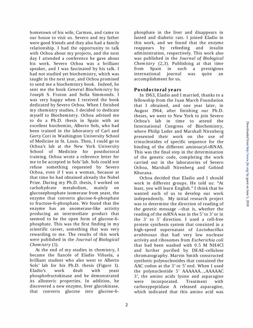
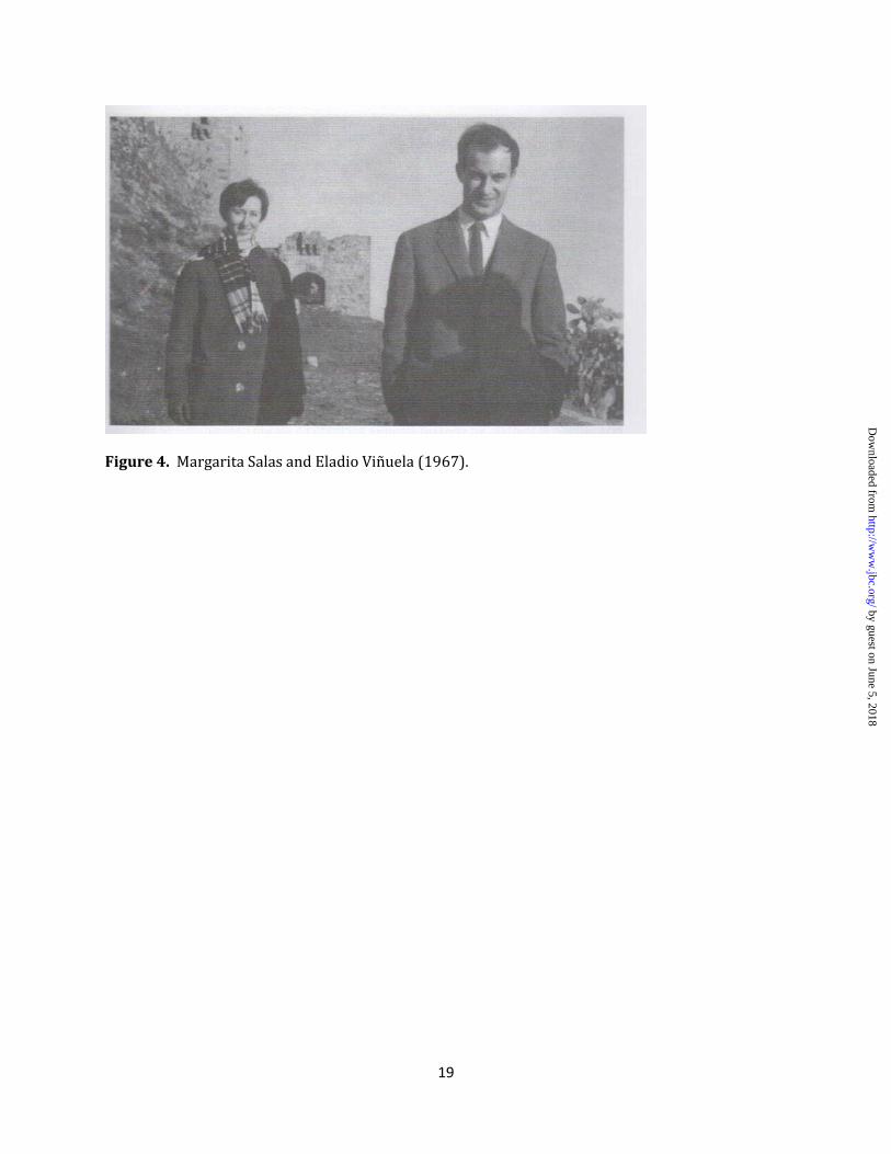
Figure 4. Margarita Salas and Eladio Viñuela (1967).
by guest on June 5, 2018http://w
ww
.jbc.org/D
ownloaded from

20
Figure 5. Severo Ochoa and Margarita Salas (1986).
by guest on June 5, 2018http://w
ww
.jbc.org/D
ownloaded from
Margarita SalasMy life with bacteriophage Ø29
published online November 2, 2012J. Biol. Chem.
10.1074/jbc.X112.433458Access the most updated version of this article at doi:
Alerts:
When a correction for this article is posted•
When this article is cited•
to choose from all of JBC's e-mail alertsClick here
by guest on June 5, 2018http://w
ww
.jbc.org/D
ownloaded from