
Realtime PCR Quality Control Guidelines.pdf
50
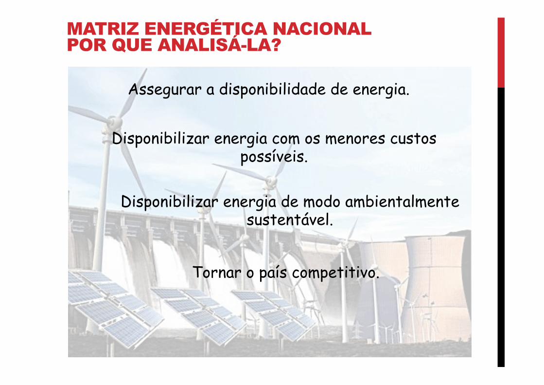
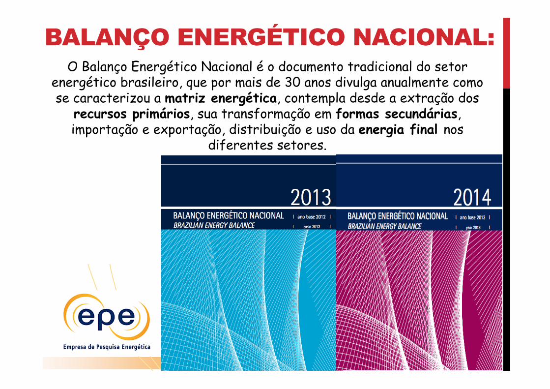
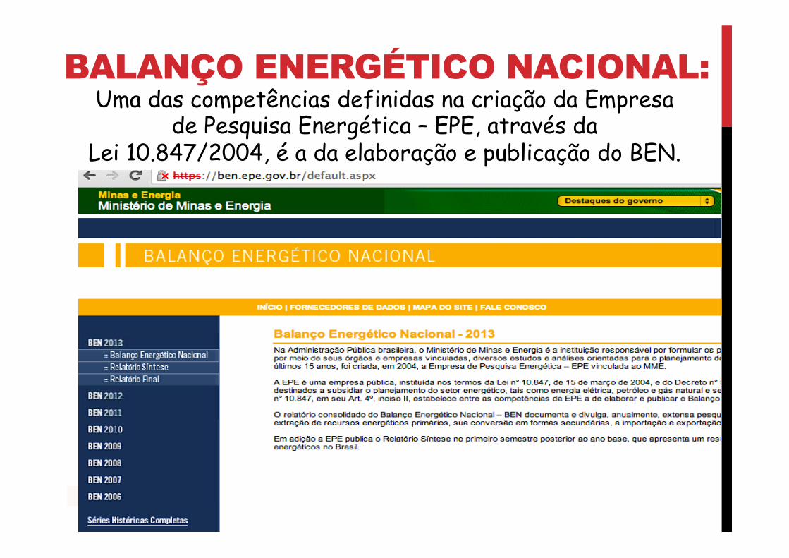
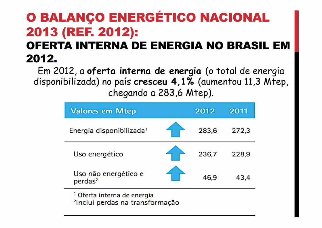
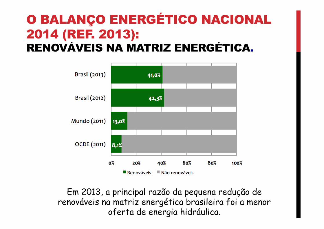
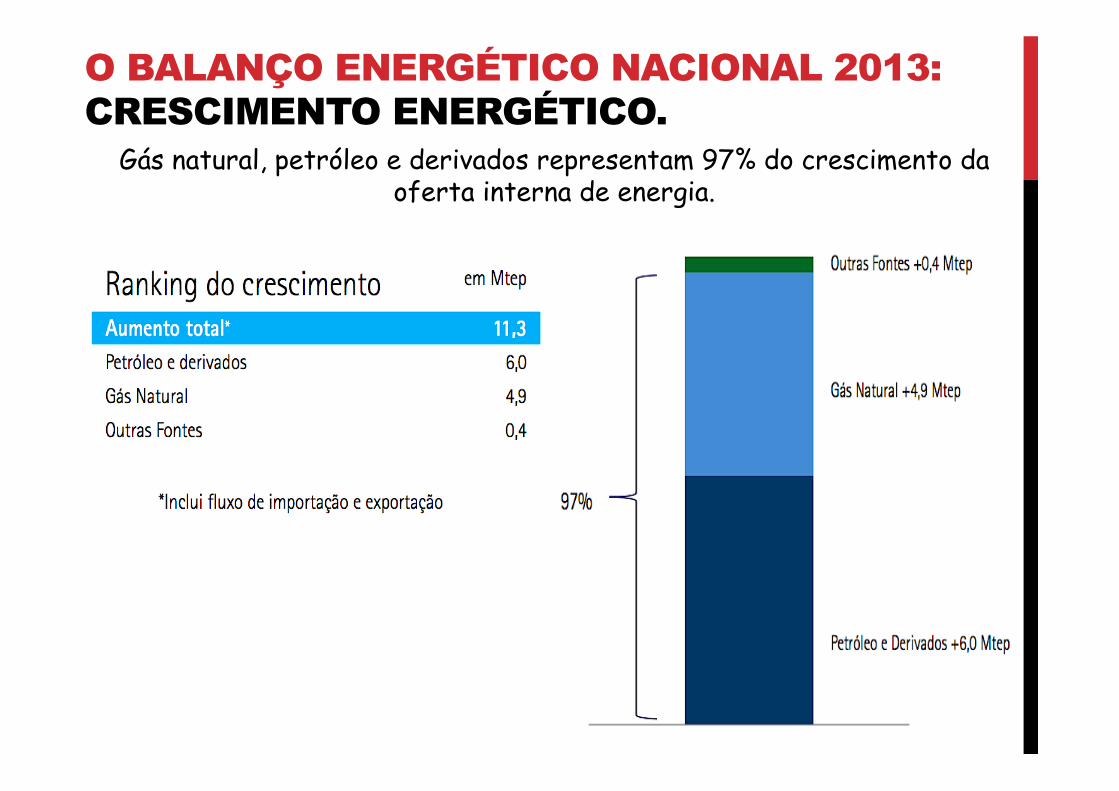
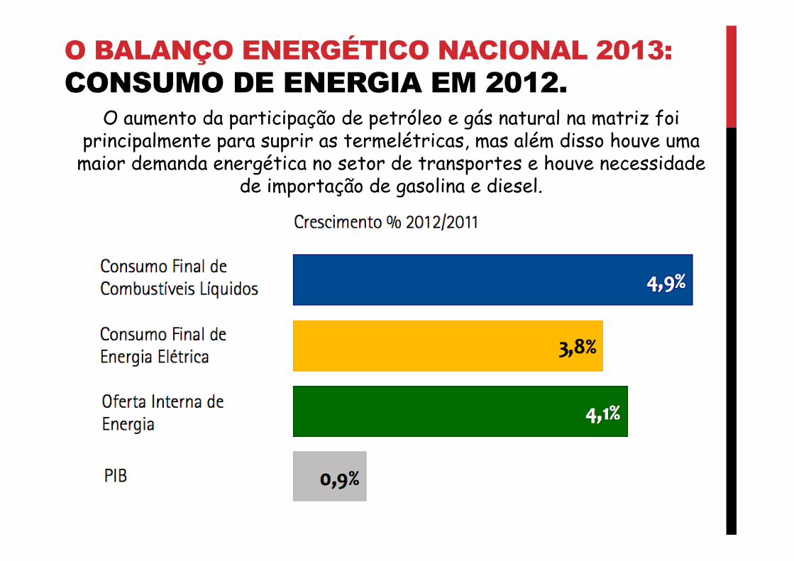
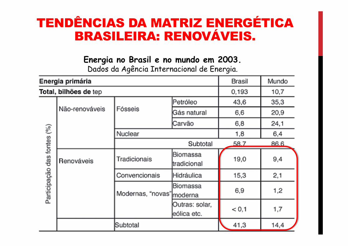
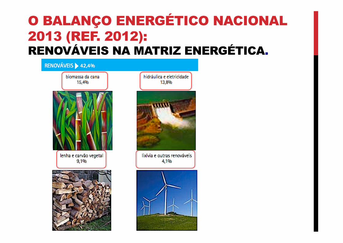
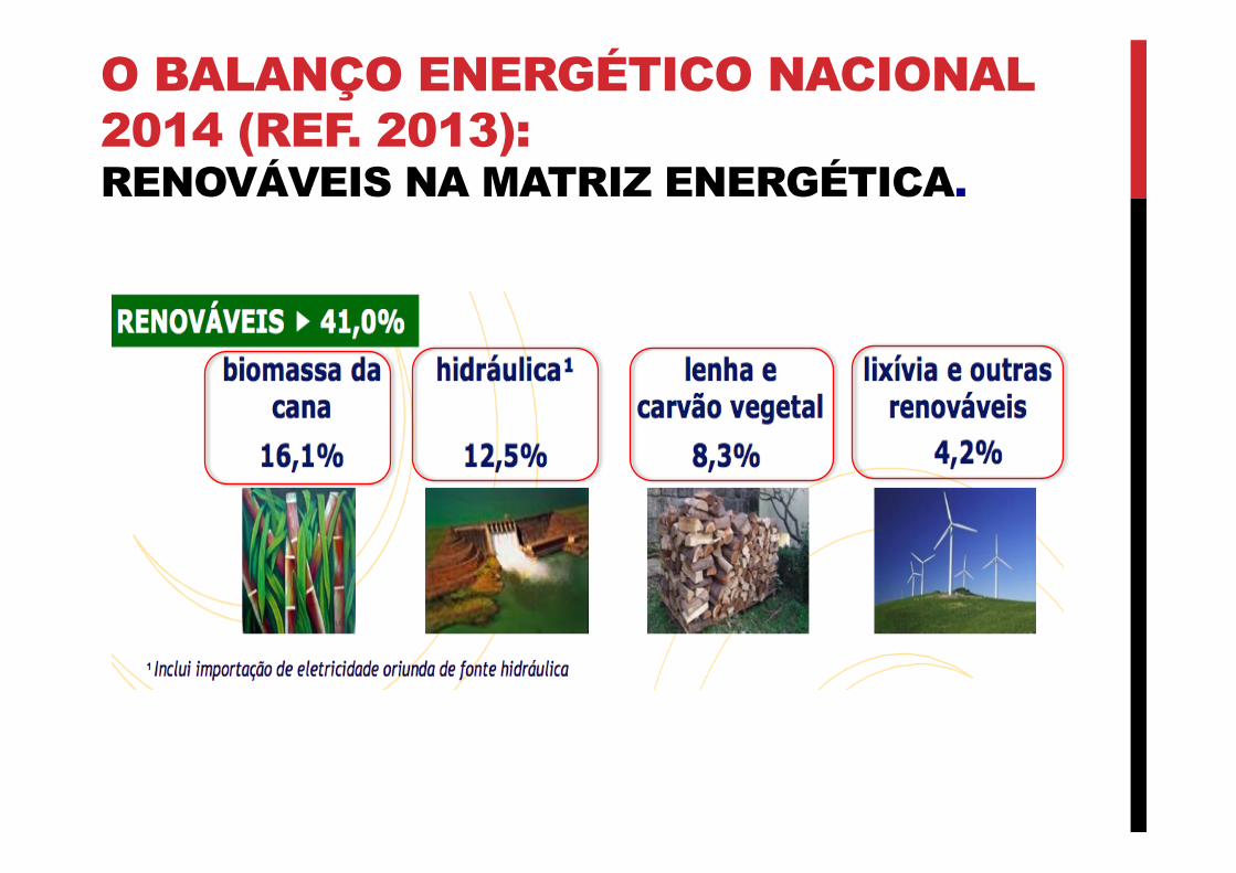
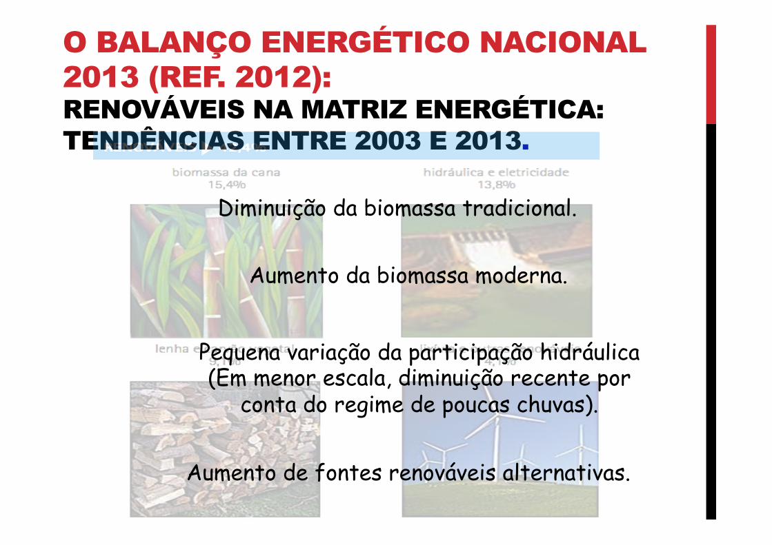
RECURSOS ENERGÉTICOS 03 O BALANÇO ENERGÉTICO NACIONAL. PROFESSOR RAFAEL FARIA
-
Upload
darwin-landicho -
Category
Documents
-
view
248 -
download
0
Transcript of Realtime PCR Quality Control Guidelines.pdf

Real-Time PCR Core Facility Quality Assurance/Quality Control Guidance for Real-time PCR Analysis
February 2011 Real-Time PCR Research and Diagnostic Core Facility Department of Medicine and Epidemiology School of Veterinary Medicine 3110 Tupper Hall University of California, Davis Davis, CA 95616 Phone: (530) 752-7991 Fax: (530) 754-6862 http://www.vetmed.ucdavis.edu/vme/taqmanservice/ Approved By:_____________________________________ Date:____________
Real-Time PCR Core Facility Director Approved By:_____________________________________ Date:____________
Real-Time PCR Core Facility Advisor

Real-Time PCR Research and Diagnostic Core Facility, UC Davis
2
This document was prepared by and for the Real-Time PCR Core Facility. Disclaimer This manual is not a regulation, only guidance for the facility while working with real-time polymerase chain reaction (qPCR) based-analyses on contaminants in diagnostic and research samples and for decision makers who need to judge the quality of PCR data. Any questions regarding this document should be addressed to:
- Real-Time PCR Core Facility Director - Real-Time PCR Core Facility Quality Assurance Manager - Real-Time PCR Core Facility Lab Manager
Real-Time PCR Research and Diagnostic Core Facility, UC Davis
3
TABLE OF CONTENTS 1. INTRODUCTION 5 1.1 Code of ethics 5 1.2 Purpose 6 1.3 Scope 6 2. LABORATORY QUALITY ASSURANCE 6 2.1 Personnel 6 2.1.1 Background and training 7 2.1.2 Outerwear 8 2.2 Facility design and workflow 8 2.2.1 Facility design 8 2.2.1.1 Reagent preparation room 8 2.2.1.2 Sample preparation room 8 2.2.1.3 Amplification room 9 2.2.2 Workflow 9 2.3 Sample acceptance procedure 9 2.4 Equipment 9 2.4.1 Power supplies 9 2.4.2 Laminar-flow hoods/PCR workstations 9
2.4.3 Nucleic acid extraction systems 10 2.4.3.1 Assessing RNA quality 11
2.4.4 Thermal cyclers 11 2.4.5 Centrifuges 11
2.4.6 Pipettes 11 2.4.7 Temperature-dependent equipment 12
2.5 Disposables 12 2.5.1 Pipette tips 12 2.5.2 Sample and PCR tubes 12 2.5.3 Gloves 12
2.6 Laboratory cleaning 12 3. REAGENTS, KITS, PRIMER SETS, AND ENZYMES 13
3.1 Reagents 13 3.2 Commercially available kits 13 3.3 Primer sets and hybridization probes 13
3.3.1 Storage 14 3.4 Enzymes 14
4. ANALYTICAL PROCEDURES AND ASSESSMENT 14 4.1 Biological sample collection and processing 15
4.1.1 Sample custody 15 4.2 Nucleic acid extraction 15 4.3 Polymerase chain reaction amplification 15
4.3.1 Reaction volumes 16 4.3.2 Primer and template concentrations 16 4.3.3 PCR reagents and master mix preparation 16 4.3.4 Selection of procedure parameters 17
4.4 Amplicon detection and confirmation 17 4.5 Method sensitivity, precision, and recovery 17
4.5.1 Detection limits 17 4.5.1.1 Detection limit of PCR 18 4.5.1.2 Detection limit of method 18

Real-Time PCR Research and Diagnostic Core Facility, UC Davis
4
4.5.2 Precision 18 4.5.3 Recovery 18
5. INTERNAL QUALITY CONTROL PROCEDURES 19 5.1 Positive controls 19
5.1.1 PCR Positive 20 5.1.2 PCR inhibition positive controls 20 5.1.3 Method positive control 21 5.1.4 Matrix spike 21
5.2 Negative controls 21 5.2.1 PCR negative control 22 5.2.2 Method blank 22
5.3 Reverse-transcriptase RT-qPCR quality control 22 5.3.1 Contamination of RNA samples 22
5.3.1.1 Sources of RNase contamination 22 5.3.2 Preventing RNase contamination 23 5.3.3 Quality-control samples for RT-qPCR analysis 23
5.4 Corrective actions 24 5.5 False-positive/false-negative prevention 24
5.5.1 False positive prevention and detection 24 5.5.1.1 Product carryover reduction 25
5.5.2 False negative prevention 25 6. DATA RECORDING, RECORD KEEPING, AND DATA EVALUATION 25
6.1 Data recording and record keeping 25 6.1.1 Equipment 25 6.1.2 Reagents, kits, primer sets, and enzymes 25 6.1.3 Sample processing and 26
6.1.4 Quantitative real-time PCR 26 7. REFERENCES 27

Real-Time PCR Research and Diagnostic Core Facility, UC Davis
5
1. INTRODUCTION
From the isolation of specific genes to the sequencing of entire genomes, the polymerase chain reaction (PCR) has become one of the most widely used technologies for conducting biological research. Advances have led to the development of specific and sensitive high-throughput PCR methods for the detection of a variety of microorganisms, and analysis of gene expression. Since the introduction of quantitative PCR, this laboratory method has witnessed rapid development and improvements.
Quantitation has been achieved by using internal standards such as competitors or mimics, by limiting dilution, and additive PCR. The new generation of PCR methods is automated and standardized, and is the springboard to the next-generation PCR. A unique feature of DNA polymerases allowed the development of hydrolysis probes that are labeled with an energy-adsorbing quencher on the 3' end and a reporter fluorescent dye at the 5' end. During the annealing/extension phase, the probe hybridizes to the amplicons and cleavage of the probe by the DNA polymerase results in increased reporter and decreased quencher fluorescence due to the loss of spatial proximity of the two dyes. Monitoring amplification by using a 7900HT Fast Real Time PCR System (Applied Biosystems, Foster City, CA), is possible in a real-time fashion. The increase of reporter fluorescence is proportional to the amount of amplicons and therefore proportional to the initial amount of targets. This system detects DNA and RNA targets with a sensitivity of 5 molecules. The successful application of PCR requires the proper use of techniques and interpretation of results.
Due to the ability to amplify small amounts of nucleic acid, PCR can be used to detect organisms that are difficult to culture in vitro or that cannot be cultured. However, the advantages of these techniques can be offset by the demanding assay protocols and the need to follow quality assurance/quality control (QA/QC) procedures carefully. These QA/QC procedures are necessary because the ability of PCR to produce many copies of target DNA creates the possibility of contamination by previously amplified products, which can lead to false-positive results. In addition, biological samples may contain PCR inhibitors, which can lead to false-negative results. PCR protocols are standardized for analyses of biological samples, so it is essential to standardized QA/QC procedures.
Real-Time PCR Research and Diagnostic Core Facility is specialized in the detection and quantitation of infectious agents, gene transcription, and the presence of genetic diseases (allelic discrimination). The Core Facility provides a platform for multifaceted, multidisciplinary research at UC Davis for the academic and private sector, and is available for interested groups to implement state-of-the-art laser-guided technology to micro-dissect tissue for sample collection, automated nucleic acid recovery and quantitation of nucleic acids using qPCR based on hydrolysis probe chemistry.
The main goal of the Real-Time Core Facility is to produce data that is scientifically valid, legally defensible, and of known and documented quality in accordance with nationally recognized standards. All analysis are performed using promulgated reference methods for which the facility has demonstrated competency prior to its use. 1.1 Code of ethics All employees are to adhere to the following code of ethical business practices: - All employees will perform all work in a manner that merits full confidence and trust. - Real-Time PCR Core Facility and employees thereof will not engage in illegal practices, or cooperate with anyone so engaged.

Real-Time PCR Research and Diagnostic Core Facility, UC Davis
6
- Real-Time PCR Core Facility and employees thereof will ensure the integrity of their data by complete adherence to the facility QA/QC guidance, and will be diligent to expose and correct any errors that may be brought to light. - Real-Time PCR Core Facility and employees thereof will work and act in a strict spirit of honesty and fairness to clients, and in a spirit of personal helpfulness and fraternity toward fellow employees. - Real-Time PCR Core Facility and employees thereof will not accept any of the following:
♦ Fabrication of data ♦ Misrepresentation of QC samples ♦ Non acceptable instrument calibration procedures ♦ Modification of samples to alter their characteristics ♦ Improper and unethical manual integrations ♦ Manipulation of analytical results ♦ Substitution of samples, files, or data ♦ Falsification of records or instrument readings ♦ Any other form of fraud
Real-Time PCR Core Facility and employees thereof will demonstrate the positive qualities of enthusiasm, diligence, responsibility, initiative, integrity, honesty, kindness, and patience in dealing with both clients and fellow employees.
Real-Time PCR Core Facility and employees thereof, will advise clients of the probability of success before undertaking a project, and will not accept work that would constitute conflict of interest.
Real-Time PCR Core Facility and employees thereof will ensure the confidentiality of all data and information provided by the clients.
Real-Time PCR Core Facility and employees thereof will only perform testing services for which they are consistently demonstrated full compliance with high quality. 1.2 Purpose
The guidance manual has been created as a resource for the Real-Time PCR Core Facility QA/QC practices for biological sample collection, stabilization, storage, DNA and RNA extraction, qPCR data analysis, and for developing of new methods. Also it is a basis for principal investigators, researchers, veterinary practitioners, and students to evaluate the quality of PCR data for research projects and technical papers. 1.3 Scope
This manual is a general guidance for the Real-Time PCR Core Facility and method specific QA/QC procedures for real-time PCR data analysis of biological samples. This document does not address federal, state, and local regulations governing waste management, hazardous materials, and radioactive material. Furthermore, this guidance does not address related safety issues. 2. LABORATORY QUALITY ASSURANCE
This section provides guidance on general laboratory practices on day-to-day routine and provides recommendations for personnel, workflow, equipment, disposables, cleaning, the laboratory organization, and line of authority. 2.1 Personnel The Real-Time PCR Core Facility director has the responsibility for the overall management and supervision of the laboratory and its personnel. She/he will interface with clients on all aspects of their projects including progress, problems, and recommended solutions. She/he will also work with the quality assurance manager,

Real-Time PCR Research and Diagnostic Core Facility, UC Davis
7
laboratory manager and laboratory personnel in reviewing progress reports, analytical reports, financial reports, and QC reports. She/he is responsible for the review of all data generated in the facility for accuracy and interpretation. She/he is primarily responsible to ensure that all employees have received the necessary level of training to make them capable of properly executing their duties. The Real-Time PCR Core Facility quality assurance manager (QAM) assists the facility director in assuring the production of accurate, valid, and reliable data by continuously monitoring the implementation of the laboratory quality assurance program. The QAM administers all inter-laboratory QA/QC efforts, schedules and reviews performance evaluation results, takes corrective actions, and prepares QA/QC reports. She/he also conducts annual audits of the overall laboratory operation. The Real-Time PCR Core Facility manager is responsible for each task identified in their scope of work. She/he is responsible for organizing and directing the technical activities within their assigned sections(s). She/he is involved in daily laboratory operations and is responsible for verifying that laboratory QC and analytical procedures are being followed as specified for each project. She/he is responsible for organizing, assembling, disseminating, and filing all documents pertinent to the analysis for each set of samples. She/he also advises the facility director of progress, needs, and potential problems of their assigned section(s). She/he is responsible for the ongoing training of each employee within their section.
The researchers are responsible for sample analysis, data processing and recording in accordance with the facility QA/QC guidance and established protocols. They are responsible for calibration and preventive maintenance of instrumentation, data reduction, data review, and reporting of all out-of-control situations, as well as for initial corrective actions whenever necessary. Well documented training records are kept on file for each researcher in order to provide proof of proper training for each method they perform. Personnel working in the laboratory performing qPCR analysis should meet background and training specifications outlined below and should follow the guidance provided concerning protective outerwear. 2.1.1 Background and training
Laboratory personnel should have undergraduate course work in molecular biology, biotechnology, biochemistry, molecular genetics, or other course work that covers PCR and recombinant DNA/RNA theory and practice. However, commensurate job-related training and experience may be substituted. Hands-on training, including the review of standard operating procedures or manuals, should be completed for each technique under the supervision of experienced personnel. A new employee will receive orientation and skills training. New or established employees may receive training on new methods given by the method developer.
Although the amount of time required for training will vary depending on the procedure and the technique, each researcher should demonstrate that they could successfully perform the assay through analyses of positive and negative control samples before being allowed to analyze biological samples without supervision. An initial demonstration of capability should include at least four replicate analyses of seeded reagent water, two replicates of seeded environmental water, and a method blank, as well as proficiency testing. These recommendations for the initial demonstration of capability are based on current requirements for conventional microbiological methods. Each researcher also is expected to be knowledgeable in laboratory safety and QA/QC procedures.
The Real-Time PCR Core Facility should maintain a training record for each researcher that documents the following:
♦ Dates and scope of PCR method training
Real-Time PCR Research and Diagnostic Core Facility, UC Davis
8
♦ Initial demonstration of capability ♦ Proficiency test results for each analysis type ♦ Dates and scope of laboratory QA/QC training ♦ Dates and scope of laboratory safety training
2.1.2 Outerwear Dedicated laboratory coats and powder-free gloves should be available in each
laboratory room. Laboratory coats should be removed and gloves discarded before leaving each room. Changing laboratory coats and gloves reduces the possibility of contamination with template. Gloves should be changed after working with seeded or environmental samples, after handling template or amplified nucleic acids, and after contact of the outside of the gloves with skin. The latter prevents introduction of enzymes prevalent on the skin, such as DNases and RNases that degrade nucleic acids.
Laboratory coats should be cleaned regularly to reduce the possibility of contamination of the designated workspace and the PCR reaction. Laboratory coats should be separated from non-laboratory clothing, and cleaned only with other laboratory coats that were in the same work area. The frequency of cleaning is dependant on the amount of PCR work the Real-Time PCR Core Facility is performing. 2.2 Facility design and workflow
The high sensitivity of PCR techniques requires that demanding assay conditions be followed. The Real-Time PCR Core Facility is designed and operated in a way that prevents contamination of reactions with amplified products from previous assays and cross-contamination between samples, both of which can lead to false-positive results. 2.2.1 Facility design
Contamination between samples and from previous qPCR amplicons generated in the Real-Time PCR Core Facility could be significant potential source of invalid PCR results. Thus, the Real-Time PCR Core Facility has physically separate rooms: • Reagent preparation (using positive pressure to prevent the introduction of contamination); • Sample preparation (using negative pressure to keep template nucleic acids in the room); • Amplification and analysis (using negative pressure to keep amplified nucleic acids in the room). Prior to PCR amplification, sample preparation, DNA extractions, and PCR setup are conducted at separate times. No materials, supplies, or equipment from the sample preparation room should be taken into the reagent preparation room. Nothing from the amplification and analysis room should be taken into the sample preparation room or the reagent preparation room. The equipment should not be moved between the rooms used for any PCR sample processing and analysis steps. 2.2.1.1 Reagent preparation room The reagent preparation room is dedicated for the preparation and storage of PCR reagents, including master mixes. Addition of master mixes to PCR tubes should be performed in this room. To prevent cross-contamination and to avoid repeated freezing and thawing, reagent-stock solutions should be aliquoted into smaller “working” volumes and stored in this room for later use. The reagent preparation room should have dedicated pipettes with plugged, aerosol-barrier tips, laboratory coats, and disposable gloves. Important: Materials from other rooms (including amplified template, and target nucleic acid or positive controls, supplies, or equipment) should not be brought into the reagent preparation room.
Real-Time PCR Research and Diagnostic Core Facility, UC Davis
9
2.2.1.2 Sample preparation room The sample preparation room is dedicated for sample processing and preparation of positive and negative controls. Sample processing includes extraction and purification of nucleic acids. The processed samples and controls should be added to tubes containing PCR master mix in this room. The sample preparation room should have dedicated adjustable pipettes with plugged, aerosol-barrier tips. Fresh gloves and laboratory coats should be worn at all times to control contamination from this room to any other location. Biological safety cabinets should be used within the room—one for sample and negative control preparation, and the other for positive control preparation to protect the samples from cross-contamination Important: Nothing from this room should be taken to the reagent preparation room. 2.2.1.3 Amplification room A thermocycler and qPCR instrument(s) are located in this room, which is designated for activities associated with PCR amplification and post-PCR analyses. Gloves and laboratory coats should be worn at all times and removed before leaving the room to control amplicon contamination of other locations. Important: Nothing from this room should be taken into either the sample preparation room or the reagent preparation room. 2.2.2 Workflow A unidirectional workflow should be used to reduce the potential for contamination. Laboratory manuals and notebooks also should not be moved from room to room. 2.3 Sample acceptance procedure The Real-Time PCR Core Facility should assessed the sample when it is received at the laboratory to verify that the sample volume was adequate, the sample was handled and preserved appropriately, the holding time requirement was met, and that all required sample collection information was recorded by the sample collector. After the sample is assessed, information on the date and time of sample receipt and sample condition should be recorded. The sample should be marked, logged, and tracked with a unique identifier. 2.4 Equipment The Real-Time PCR Core Facility should use equipments suitable for the methods employed that function properly in order to generate reliable data. New instruments and equipment, or instruments and equipment that have undergone repair or maintenance, shall be calibrated before being used in casework analysis. To verify that equipment is functioning properly, the facility should have a schedule for maintaining equipments, which include the setup, calibration, repair, record keeping, and normal operation of all equipment used in sample analysis. The results of all tests should be documented in an electronic database. 2.4.1 Power supplies Power supplies should provide voltage and current readings. Before use, electrodes should be checked to ensure that they fit snugly. Electrical cables, electrode wires, and cable connections should be checked for breaks, fraying, corrosion, or looseness, and replaced if needed. 2.4.2 Laminar-flow hoods / PCR workstations The Real-Time PCR Core Facility has three types of hoods: 1) three laminar-flow hoods, 2) two hazardous-waste fume hoods, and 3) three PCR workstations. The operation of all hoods are checked and certified by a qualified inspector annually and recorded in the LIMS. The laminar flow hoods have magnehelic pressure gauges (MAG) that are used to monitor operation of the hoods. When using these hoods, check to make sure the pressure gauge is reading at a level approximately equal to the annually
Real-Time PCR Research and Diagnostic Core Facility, UC Davis
10
recorded MAG level on the calibration sticker. A significant increase in pressure indicates that the filters are dirty whereas a significant decrease in pressure indicates an electrical problem. The laminar-flow hoods and PCR workstation must be free from contamination by live organisms. The working surfaces of the laminar-flow hoods and the PCR workstations should be wiped down with 70 % ethanol before and after general use. The laminar-flow hoods and PCR workstation have ultraviolet bulbs designed to decrease biological and nucleic acid contamination by cross-linking nucleic acids. Before use, hoods should be decontaminated using UV light for at least 15 minutes. The UV bulb should be wiped with a wet cloth to remove dust every week. A bulb that is dull in the center needs to be replaced. Biannually, the working surfaces of the laminar-flow hoods and the PCR workstations should be swiped with sterile swabs and examined by PCR for contamination of the most common used systems. The hazardous-waste fume hood must be checked to ensure that it is operating properly. Check the operation of the hazardous-waste fume hood quarterly. 2.4.3 Nucleic acid extraction systems The Real-Time PCR Core Facility uses the ABI PRISM™ 6100 nucleic acid PrepStation and the Corbett X-tractor Gene™ automated nucleic acid extraction, including total RNA and genomic DNA from a variety of biological samples. Automated platforms are compact, simpler to use, and provide the highest quality nucleic acid product.
For nucleic acid extraction by using the ABI PRISM™ 6100 Nucleic Acid PrepStation biological samples should be stored in lyses solution (an RNA preservative). A variety of tissue sample types (for example, heart, lung, skeletal muscle, vein/artery, or plant tissue) can contain large amounts of collagenous/fibrous material, mucopolysaccharides, or polyphenols/chlorophyll. These materials can block the purification tray membrane and potentially contaminate other samples in the purification run by causing the purification tray wells to overflow during washing procedures. These tissues may also have low intrinsic RNA content (<1 µg/mg of tissue) and require larger amounts of tissue to be processed to return acceptable amounts of RNA.
To overcome this problem the Real-Time PCR Core Facility uses the SPEX SamplePrep Model 2000 Geno/Grinder that is specifically designed for vigorous up-and-down shaking to prepare tissue as well as plant materials for extractions of nucleic acid (DNA/RNA) in deep-well titer plates. Two safety interlocks, a lockdown latch and a pneumatic cylinder stabilize the lid for safe operation. The clamping mechanism latches have secondary locking tabs for additional safety. Poor homogenization techniques can also leave large particles in the lysates, clogging the pipette tips or the purification tray membrane. In these cases, pass the biological sample through the pre-filter or treat with proteinase K, before adding it to the total RNA purification tray. Clean all instruments thoroughly by washing with the following items in the specified order: a) 10% bleach b) 70% ethanol c) Molecular biology grade water.
A maximum of 200 µL of liquid sample may be processed using the Corbett X-tractor Gene™ automated nucleic acid extraction. Volumes less than 200 µL can be processed by bringing the sample volume up to 200 µL with phosphate buffered saline (PBS). After run dispose of plastic ware and liquid waste in accordance with the facility guidelines for the sample type and reagent hazard. Transfer carriages, waste sink and tip chute thoroughly rinse under cold tap water and allow drying. Do not apply hot water to these components. Bleaching is not generally recommended, so use 1% sodium

Real-Time PCR Research and Diagnostic Core Facility, UC Davis
11
hypochlorite for < 30 minutes and not more often than once a week. The separator plate must be washed to ensure it is RNA/DNA and RNase/DNase-free. Ensure it is dry before re-using. For processing biological samples the same or next working day, the homogenized lysate may be readily stored at 4 °C for up to 24 hours with little degradation. For longer storage times, freeze the homogenized lysate at –20 to –80 °C. 2.4.3.1 Assessing RNA quality Because mRNA comprises only 1-3% of total RNA samples it is not readily detectable even with the most sensitive of methods. Ribosomal RNA, on the other hand, makes up >80% of total RNA samples, with the majority of that comprised by the 28S and 18S rRNA species (in mammalian systems). Mammalian 28S and 18S rRNAs are approximately 5 kb and 2 kb in size, the theoretical 28S:18S ratio is approximately 2.7:1; but a 2:1 ratio has long been considered the benchmark for intact RNA. The Real-Time PCR Core Facility uses an Agilent 2100 bioanalyzer. The bioanalyzer uses a combination of microfluidics, capillary electrophoresis, and fluorescent dyes that bind to nucleic acid to simultaneously evaluate both RNA concentration and integrity. To ensure quality of purified RNA the facility assesses 5 to 10% of each sample batch.
Figure 1. Intact RNA Figure 2. Degraded RNA Note: To ensure that extracted RNA is free of contaminants that can compromise integrity, a simple stability test by incubating a small amount of RNA at 37°C for several hours to overnight and compare it to a duplicate sample stored at -20°C could be performed. The sample stored at 37°C should show a minimal decrease in the 28S:18S ratio relative to the one stored at -20°C. In general, samples with greater than a 20% change in rRNA ratio over time may not perform well in downstream applications. 2.4.4 Thermal cyclers Thermal cyclers are essential to all PCR methods, and great care should be taken to ensure that they are well-maintained and reliable. The Real-Time PCR Core Facility uses two thermal cyclers: 1) End-point PCR thermal cycler (the temperature should be monitored twice a year); 2) QPCR thermal cycler (background calibration should be performed monthly, a pure-spectra assay is done twice a year, an RNase verification run is done annually). The manufacturers of the instruments have developed recommended procedures to test and maintain the instrument. 2.4.5 Centrifuges Separate centrifuges should be used for pre- and post-PCR procedures. The manufacturers’ instructions for calibration should be followed. The centrifuge should be balanced before use to increase bearing life and minimize vibrations that can unsettle concentrates.

Real-Time PCR Research and Diagnostic Core Facility, UC Davis
12
2.4.6 Pipettes Due to the numerous small volume transfers involved in PCR methods,
automatic pipettes, fixed-volume, adjustable, positive-displacement, and/or micropipettes should be used, and should be calibrated by the researcher using the tips commonly used in the laboratory. Each pipette should be sterilized according to manufacturers’ recommendation on a regular basis or whenever contamination is suspected. Calibration of the pipette should be performed after sterilization. 2.4.7 Temperature-dependent equipment
For equipment used in PCR analysis, the following temperature ranges should be applied:
♦ Incubators, water baths, and heating blocks: ± 0.5 °C of the temperature required by the protocol
♦ Refrigerators: 1°C to 5°C ♦ Standard laboratory freezers: -20°C ± 5°C ♦ Ultra-low freezers: -70°C ± 10°C
Temperatures of equipment should be monitored at least once a day for each workday in use. Separate refrigerators and freezers for samples, reagents, and final amplification products should be maintained in the appropriately designated laboratory room. Amplified product should always be kept in a separate freezer from reagents (such as master mix), samples, and sample concentrates. 2.5 Disposables
Disposable materials used in PCR analysis include pipette tips, sample tubes, PCR tubes, and gloves. To reduce the contamination and degradation of the target nucleic acids, disposable materials should meet the standards discussed in sections 2.5.1 to 2.5.3. 2.5.1 Pipette tips
Special tips for PCR analysis include barrier tips and aerosol-resistant tips, both of which minimize cross-contamination of samples during pipetting. Pipette tips for PCR analyses should be lot-certified, RNase-free, DNase-free, and pyrogen-free. 2.5.2 Sample and PCR tubes
Laboratories should use polypropylene tubes and plates that are lot-certified DNase-, RNase-, and pyrogen-free. Thin-walled tubes provide the best heat transfer, ensuring that the reaction volume reaches its specified temperature in the shortest amount of time, thereby improving specificity and reproducibility. Tubes and plates containing stored samples and reagents should be centrifuged briefly before opening to ensure that all liquids are at the bottom. 2.5.3 Gloves
Disposable gloves should be available in each section of laboratories used for PCR analysis. Gloves should be changed before leaving and entering each section of the laboratory and each time contaminating DNA is potentially encountered. In addition to reducing potential contamination from samples, wearing gloves may protect the researcher from potential chemical exposure and prevent sample contamination due to human DNases and RNase. 2.6 Laboratory cleaning
All work surfaces should be cleaned after each use with 0.6% sodium hypochlorite (NaOCl). The NaOCl solution should be prepared fresh daily by diluting commercial bleach 1:10 in water and adjusting the pH to 7. This solution inactivates pathogenic agents and destroys nucleic acids. Residual bleach, which may cause pitting in stainless steel counter tops and hoods, may be removed with a 70% ethanol rinse or equivalent. Commercial products that are specifically designed for removing nucleic acids and nucleases also can be used for surface cleaning. Thermocyclers and

Real-Time PCR Research and Diagnostic Core Facility, UC Davis
13
centrifuges should be cleaned with the diluted bleach solution whenever contamination is suspected. Pipettes should be cleaned according to manufacturers’ instructions. 3. REAGENTS, KITS, PRIMER SETS, AND ENZYMES 3.1 Reagents
The reagents used in PCR amplification can be purchased, and should be molecular grade. These reagents should be stored according to the manufacturers’ recommendations. All reagents from new lots should be tested to ensure that they work properly by running a PCR positive control. Care should be taken to ensure the reagents are maintained contamination-free. All reagents should be clearly labeled with name, expiration date, and relevant safety information. Reagents from different lot numbers should not be interchanged without prior functional validation.
Molecular-grade water or its equivalent from commercial sources (DNase/RNase-free) should be used for all assays. Diethylpyrocarbonate (DEPC) treatment can be used to eliminate RNase from water used in RNA analysis. Reagent water is treated with a solution of 0.1% DEPC for several hours and then autoclaved to degrade the DEPC completely. Proper autoclaving is necessary, because trace amounts of DEPC in a solution will lead to the modification of the purine residues in RNA by carboxymethylation. This leads to downstream effects in RNA experimentation. 3.2 Commercially available kits
Many types of commercial kits are available for isolation of DNA and RNA and the purification of nucleic acids to remove contaminants. A copy of the manufacturers’ specifications and procedures should be kept in the facility’s binder. The effectiveness of each new kit should be evaluated before use for the analysis of field samples by running an appropriate positive control. 3.3 Primer sets and hybridization probes
Primer sets should be designed specifically to prime the amplification of a portion of a target nucleic acid of interest. Hybridization probes are specific for nucleotide sequences that are internal to the sequences of the primers and which are used to confirm the amplification of the target. As a general rule, primers are characterized by the following:
♦ Length of 18 to 27 base pairs ♦ No homology within or between primers, especially at the 3'end to avoid primer-
dimer formation ♦ No guanine-cytosine (GC) stretches greater than two base pairs at the last five
nucleotides at the 3' end ♦ GC content of 30% to 80% ♦ Melting temperatures (Tm) of 58-600 C, and probes with a Tm value of 100 C
higher ♦ No hairpin loops with an energy of -0.5 kcal/mol or less ♦ It is preferable to have primers spanning exon-exon junctions in the cDNA
sequence ♦ Maximum amplicon size should not exceed 400 bp (ideally 50-150 bases) ♦ The probes should not have runs of identical nucleotides (especially four or more
consecutive Gs), there should be more Cs than Gs, and not a G at the 5' end. The choice of probe should be made first.
The Real-Time PCR Core Facility uses the Primer Express software for primer design. New primers and probes should always be tested experimentally for. The specificity of a chosen sequence should be evaluated using BLAST. Specificity testing should be conducted as described for the PCR positive control by substituting the
Real-Time PCR Research and Diagnostic Core Facility, UC Davis
14
heterologous sample in place of the target organism or nucleic acid. Primers and/or probes should be redesigned if any heterologous sample is positive. Primers and probes should be selected based on the best theoretical considerations, and negative features should be documented, and results interpreted accordingly. Every lot of new oligos should be checked for contamination by being used in a PCR negative control. No positive results should be found. Primers and probes should be added to the PCR master mix in the reagent preparation area. Functional validation also should be performed on new lots of primers and probes by comparing their performance against older sets of known quality. The PCR efficiency (E) is calculated from the slope of a standard curve (S) using the formula, E=10-1/slope. The samples used for generating standard curves should be carefully prepared dilutions of a stock solution containing a known number of purified PCR template nucleic acids or total nucleic acids extracted from the target organism. A PCR efficiency of 1 (or 100%) is obtained when each target sequence present in the PCR reaction doubles during each round of amplification. A high efficiency is needed to obtain accurate and reproducible results. A low efficiency indicates that primers and/or probes are performing poorly. No single efficiency (as a percentage) has been set as a limit for data to be acceptable, although 90% or greater is an accepted norm. 3.3.1 Storage Most oligos and DNA templates should be stored at -20°C or -70°C in either TE buffer (10 mM Tris-HCl and 0.1mM EDTA, pH 8.0) or molecular grade water. PCR products may also be stored at -20°C or -70°C. RNA templates should be aliquoted and stored at -70°C. The pure, concentrated oligos should be stored in the original tube from the manufacturer and labeled with the primer name and concentration. To minimize the chance of contamination and degradation, these concentrated stocks should not be used on a regular basis. Diluted working stocks should be made for each oligo, and these working stocks should be used for all experiments. The facility should check the oligos sensitivity when they are a year old and then on a regular basis to see if any degradation has occurred. The sensitivity of primers can be checked by running a PCR positive control using the old and new primers. If degradation has occurred, new primers and probes should be prepared. 3.4 Enzymes Enzymes should be purchased from a commercial source to ensure purity. The Real-Time PCR Core Facility selects vendors that provide QA information with the enzymes. After receipt from commercial sources, each new lot of enzyme should be compared with old lots using known controls. The new enzymes should be rejected or the concentration adjusted, if performance is significantly below that of the proven lot. The researcher should have only one container open at a time when working with enzymes. Pipette tips should be discarded after each dispenses to prevent cross-contamination. Enzymes typically are stored at -20°C, and should never be left at room temperature in the laboratory. Insulated bench-top coolers or ice can be used to keep the enzyme cold in the laboratory, when used on the bench top. 4. ANALYTICAL PROCEDURES AND ASSESSMENT
The Real-Time PCR Core Facility maintains standard operating procedures that accurately reflect all laboratory activities such as assessing data integrity, corrective actions, handling customer complaints, and all test methods. When developing a new PCR system, the facility should design and select the individual components of the
Real-Time PCR Research and Diagnostic Core Facility, UC Davis
15
system, to optimize its ability to recover and detect the target genes. The facility should consider how samples will be collected and processed for the system, and assess the performance of the individual system components and the entire analytical process before using the system for biological sample analyses.
The facility shall have and follow written analytical procedures, and have a standard operating protocol for each analytical technique used. The procedures shall include reagents, sample preparation, extraction, equipment, and controls that are standard for DNA/RNA analysis and data interpretation. 4.1 Biological sample collection and processing
Sample collection and transport conditions impact the results of PCR analysis. So, the Real-Time PCR Core Facility should advise principal investigators, researchers, veterinary practitioners, and students to the collection, storage, and transportation of samples to be analyzed. Acceptable sample volumes, sample handling, and the time samples can be held before being processed should be described, in order to preserve the integrity of the target nucleic acid sequence. The sample processing is performed to extract the nucleic acid of interest, while minimizing co-purification of potential contaminants and components of the matrix that may inhibit PCR. The extraction procedure also should stabilize the target nucleic acid from nucleases and reduce the sample to a volume small enough to be analyzed by PCR. Positive controls that demonstrate that the target organism or nucleic acid has been successfully isolated and that the PCR inhibitors have been removed are discussed in section 5. 4.1.1 Sample custody The Real-Time PCR Core Facility has a sample acceptance policy that outlines the circumstances under which samples will be accepted. The policy requires complete documentation, including the sample identification, the location, date and time of collection, collector's name, preservation type, sample type and source, and any special remarks concerning the sample. If a sample discrepancy, such as a broken or missing sample is observed at check-in, a statement to that effect is written into the data base remarks section. The client must be notified and approve of any changes made. At this time, the laboratory manager is notified so that the problem can be addressed. All samples received are recorded into the data base with the following information: client name, client address, analytical parameter requested, and laboratory work order number. The laboratory work order number is a sequential number that is unique to each sample. Samples are processed through the laboratory by their unique laboratory work order number. Special instructions about the samples are written and are entered into the data base in the comments section. Samples which specifically require thermal preservation are stored under refrigeration. Samples are stored in a manner that prevents cross contamination. All samples, digestates and extracts or other sample preparation products are disposed of in accordance with Federal and State laws and regulations. 4.2 Nucleic acid extraction Some methods require the lysis of the biological samples and the isolation of its nucleic acid before proceeding to PCR. Extracted nucleic acids should be assessed as part of the entire analytical process. The efficiency of nucleic acid extraction varies with sample type and extraction procedure. Ideally, an isolation technique should meet the following goals:
♦ Demonstrate a high efficiency of target nucleic acid recovery ♦ Maintain nucleic acid integrity and minimize fragmentation ♦ Provide sufficiently pure nucleic acid, free from PCR inhibitors ♦ Minimize the use of dangerous chemicals

Real-Time PCR Research and Diagnostic Core Facility, UC Davis
16
♦ Be repeatable 4.3 Polymerase chain reaction amplification To analyze biological samples using PCR, the first step is to select the type of PCR that is most appropriate for the analysis being conducted. The performance of the PCR amplification step should be assessed separately first, then as part of the entire analytical method. Aspects that should be considered in the selection of the appropriate technique include PCR type, enzyme type, primer and probe design, and reaction parameters, such as thermal cycling temperatures and reagent concentrations. The Real-Time PCR Core Facility employs following PCR:
♦ Conventional PCR uses a thermostable DNA polymerase to amplify a region of target DNA defined at each end by a specific primer. The exponential replication of the same target sequence produces enough amplicons for use in subsequent analyses.
♦ qPCR detects and measures the amplification of target nucleic acids as they are produced. qPCR requires the use of primers and an oligonucleotide probe labeled with fluorescent dyes or analternative fluorescent detection chemistry. A computer is used to monitor the fluorescence increase and to calculate a quantification cycle (Cq) value. qPCR allows quantification of template by absolute standard curve method, relative standard method, or comparative Cq method. There are several different fluorescent detection chemistries used for real-time PCR, including the following:
-Dual hybridization probes -Hydrolysis probes -Molecular beacon probes -Scorpion probes -SYBR® Green I
♦ Reverse transcriptase-qPCR is used to amplify RNA target sequences, such as messenger RNA and viral RNA genomes. This type of PCR involves an initial incubation of the biological sample or control RNA with a reverse transcriptase enzyme and a DNA primer. DNA primers that are used commonly include oligos dT (an oligos consisting of only thymidine residues), random hexamers (primers made of six random nucleotides), or a specific primer.
♦ Nested PCR or semi-nested PCR is used to increase both the specificity and sensitivity of target sequence detection. The use of a second amplification step with the "nested" primer set results in a reduced background and increased amount of amplicon. However, there is an increased chance of carryover or cross-contamination when taking product from the first round of PCR and putting it in the tubes for the second round of PCR. Additional steps and precautions may need to be taken to reduce the chance of sample contamination and false-positives, including the following:
-Never opening more than one tube at a time -Using a separate thermocycler -Adding additional negative control 4.3.1 Reaction volumes PCR volumes typically range from 10 to 100 µL and may be dependent on the type of thermal cycling instrument and method being used. Increasing the volume of sample added to a reaction may increase the probability of a positive result at low target concentrations because more samples can be tested per reaction, but may also increase inhibition.
Real-Time PCR Research and Diagnostic Core Facility, UC Davis
17
4.3.2 Primer and template concentrations The primer concentrations used in each newly developed PCR assay should be optimized to obtain maximum amplification efficiency. The addition of too much DNA (either template or nontemplate) to the reaction may be inhibitory. 4.3.3 PCR reagents and master mix preparation PCR kits containing all reagents, except primers and templates, are purchased from commercial sources and used by the Real-Time PCR Core Facility after being optimized for the particular assay. In addition to required reagents, enhancers that act on different aspects of the reaction may be added to the PCR master mix, and these include dimethyl sulfoxide (DMSO), betaine, tetramethylammonium chloride (TMAC), and bovine serum albumin (BSA). Other proprietary enhancers and additives are available from various manufactures. Each additive should be empirically tested with each system. A master mix containing optimized reagents should be prepared in the reagent preparation room. During master mix preparation, pipette tips should be changed after handling each reagent. The master mix should be prepared on ice, unless the manufacturers’ instructions specifically state that the reagents can be mixed at room temperature. The master mix should be aliquoted into reaction tubes in the reagent preparation room. The facility can prepare a large quantity of master mix and store aliquots at -20°C after demonstrating that the freeze/thawing of aliquots does not reduce sensitivity. 4.3.4 Selection of procedure parameters The exact parameters of the PCR should be selected based on the target nucleic acids that are to be amplified. Parameters that need to be evaluated for successful PCR amplification include thermocycling conditions, reaction volumes, template concentration and the concentration of PCR reagents, including that of primers. After optimal conditions are established, they should be documented for the method and followed thereafter. 4.4 Amplicon detection and confirmation PCR results must be confirmed. Probe-based qPCR is a method employed by the facility, which confirms the presence of a specific product by detection of the fluorescence dye. Note: Due to the small size of some amplicons in qPCR detections, confirmation by sequencing may not be possible. 4.5 Method sensitivity, precision, and recovery After the individual components of the method have been optimized, the entire method should be evaluated to determine the method’s detection limits (sensitivity), precision, and recovery. 4.5.1 Detection limits Target organisms are often present at low numbers in the environment, and some of these may be able to produce infection at low concentrations. Also, expression of selected genes while monitoring their changes can be at very low levels. Therefore, an acceptable PCR method should be applied to detect a level of organisms or genes targeted that is biologically significant. The level may be based upon acceptable risk levels, predicted environmental concentrations, or concentrations that produce an infectious dose. In order to know whether an acceptable level can be detected, a detection limit must be determined for each system. The systems employed by the facility should have the detection limit of at least 5 molecules of DNA and RNA targets with the confidence level typically from 95% to 99%. For presence/absence methods, the detection limit is the minimum concentration of a

Real-Time PCR Research and Diagnostic Core Facility, UC Davis
18
target that produces a positive response with a given level of confidence. There are many uncertainties that can affect the detection limit. Some of them are:
♦ The type of target nucleic acid being detected (e.g., DNA, mRNA, tRNA, rRNA, etc.)
♦ The secondary structure and the GC content of the nucleic acid target molecule ♦ The matrix from which the organism is isolated ♦ The detection of microbes that are inactivated by physical and chemical
disinfectants The detection limits can be determined by analysis of replicate, seeded samples containing increasingly lower levels of the target organism or target nucleic acid. The number of replicates that should be analyzed at each dilution level will depend on the variability of the procedure, the accuracy and variability of the spike enumerations, the desired level of confidence in the detection limit, and the application of the method. Errors associated with serial dilutions should be minimized by using appropriate volumes that can be accurately measured and by thoroughly homogenizing stocks immediately before removing an aliquot for the next dilution. Multiple replicates of each dilution should be enumerated independently of the PCR analysis to assess the accuracy of the dilutions. 4.5.1.1 Detection limit of qPCR The PCR detection limit reflects the sensitivity of the PCR procedure, which includes the sensitivity of the primers and probes as well as the preparation of the master mix and the optimization of thermocycling conditions. It does not include reductions in sensitivity by any procedures used to concentrate organisms in the sample and isolate nucleic acids. For this reason should be determined on the basis of nucleic acid concentrations rather than organism concentrations. 4.5.1.2 Detection limit of method The method detection limit is the lowest amount of target that can be reproducibly detected. It reflects not only the sensitivity of the qPCR, but the efficiency of the procedures used to detect target from the biological sample and the efficiency of the procedures for recovering nucleic acid. Thus, a method’s detection limit will depend on a range of factors, including the efficiency of the collection, concentration, lysis of sample, and extraction of nucleic acid from the biological sample, in addition to the detection limit of the PCR itself. The method detection limit can be determined by processing standardized matrix, or biological samples that have been spiked with different concentrations of the target organism prior to any sample processing. 4.5.2 Precision Good PCR methods will have a high precision. Precision is a measure of how closely values from replicate measurements of a sample agree with each other. For quantitative PCR analyses on biological samples, method precision can be expressed as the relative standard deviation (RSD) or the relative percent difference (RPD). The RSD is the standard deviation from three or more replicate samples divided by the mean and multiplied by 100%. The RPD is the absolute difference of duplicate samples divided by the mean and multiplied by 100%. The RSD or RPD decreases with increasing precision and increases with increased variability. Variability (i.e., low precision) may result from differences in performance, reagents or equipment, or sample characteristics. 4.5.3 Recovery In addition to high precision, good analytical methods will have low bias. Bias is a measure of disagreement between the concentration of a DNA or RNA template as measured by a method and the true concentration in the sample that was tested. A PCR method’s recovery of the target sequence of interest is an important factor for

Real-Time PCR Research and Diagnostic Core Facility, UC Davis
19
characterizing method bias at levels higher than the detection limit. Bias can occur at any component of a PCR method. If a method is designed to detect a group of related organisms, members of the group may have different recoveries and thus different biases through the sampling protocol, nucleic acid extraction, and the PCR steps. Bias at the PCR level can occur from the favoring of the amplification of certain target sequences due to properties of the target, the flanking sequences, or the overall genome. PCR bias may be seen for mixed-template PCRs, including reactions using internal controls, and may make interpretation of results difficult, because the final results do not reflect the original makeup of the target templates. Bias usually results when the amplification efficiencies of the target sequences are not the same. Target sequences may amplify at different efficiencies because the sequences are not equally accessible to primer hybridization after denaturation, primer-template hybrids do not form with equal efficiency for all templates, and/or the polymerase acts on the templates with different efficiencies. One factor that may effect primer-template hybridization is the GC content of the template. PCR bias may be reduced by running a low number of PCR cycles and by using high template concentrations, when possible. Recovery of the PCR analysis and the entire method can be assessed by the analysis of seeded samples. For qPCR analyses, recovery is determined as the total amount of the template found in a spiked sample minus the background (i.e., amount detected in the non spiked sample) divided by the amount of the template spiked into the sample. To determine recovery for presence/absence methods, serial dilutions of the spiked sample should be performed and five replicates at each dilution analyzed. The amount of template in the spiked and non spiked samples should then be calculated. Results that are obtained using methods that have low recoveries should be qualified as potentially biased. Also the recovery of target organisms that are endogenous to a matrix (e.g., enteric pathogens associated with fecal maters) may not be the same as that measured by spiked samples (e.g., where the organisms are not associated with other material). Unless it can be shown that the recoveries of endogenous and spiked organisms are similar, sample results should be recorded as biased. 5. INTERNAL QUALITY CONTROL PROCEDURES The Real-Time PCR Core Facility employs quality control samples to assess the validity of the analytical results of all samples. Determination of the validity of all sample results is based on the acceptance criteria being met by the control sample. The control samples are analyzed in the same manner as the samples. They are interspersed with the samples at frequencies that are specified by the appropriate standard operating procedure. The frequencies may be altered to comply with client-specific requirements. The data acquired from QC procedures is used to estimate the quality of analytical data, to determine the need for corrective action in response to identified deficiencies, and to interpret results after corrective action procedures are implemented. The facility should analyze positive and negative QC samples on a routine basis to demonstrate adequate performance of PCR based methods. 5.1 Positive controls Positive controls are analyzed to verify that the method is capable of adequately recovering and amplifying the target. The concentration of the sequence of interest in these positive controls should be 10 to 100 times higher than the defined detection limit of the PCR. A positive control is considered to be acceptable if the DNA/cDNA of interest was amplified by PCR. Precautions should be taken to avoid contamination of biological samples with the positive control template. Positive control preparation should be
Real-Time PCR Research and Diagnostic Core Facility, UC Davis
20
physically separated from biological sample and negative control preparation, and positive control samples should be handled last. A summary of the recommended positive QC samples is as follow:
Description Purpose Frequency
PCR positive control
Verify that the PCR master mix and reagents were prepared correctly to produce amplification of the target nucleic acid
One or more per PCR batch
Inhibition positive control
Verify that interfering constituents from an environmental matrix carried over from the isolation of the organism or nucleic acids do not inhibit the PCR
With every sample batch
Method positive control
Verify that the entire method is performing properly and is capable of amplifying the target nucleic acid from the organism of interest
With every sample batch
Matrix spike
Determine the effect of the matrix on the overall method's recovery and verify it does not have an inhibitory effect on the PCR
Per matrix
5.1.1 PCR positive control PCR positive controls are used to verify that the PCR master mix and reagents were prepared correctly in order to produce amplification of the target nucleic acid. This type of positive control is run with each PCR batch. A PCR batch is defined as a group of samples that are processed and amplified at the same time under the same conditions, using the same PCR master mix, and in the same thermocycler. PCR positive controls are prepared by the addition of an exogenous control to the master mix. Exogenous controls can be any of the following:
♦ A purified total nucleic acid extract from the organism or the biological sample containing the sequence of interest.
♦ The whole organism, which may be used when the nucleic acid target of interest can be released from the seeded organism by heating before or during PCR.
♦ A specific nucleic acid fragment containing the entire sequence to be amplified, including primer binding sites.
♦ A cloned DNA fragment containing a modified form of the target sequence. ♦ A heterologous sequence that has been previously shown to be amplified with
efficiency that is comparable to the target sequence. ♦ RNA transcribed from a cloned DNA fragment containing the target sequence or
a modified target sequence (for use in RT-qPCR applications). For quantitative PCR methods, the PCR positive control is evaluated by determining the total amount of the target nucleic acid or organism in the control divided by the amount added to the reaction as a spike. 5.1.2 PCR inhibition positive controls Inhibition positive controls are used to verify that interfering constituents from an environmental matrix, which may be carried over during isolation of nucleic acids or
Real-Time PCR Research and Diagnostic Core Facility, UC Davis
21
organisms during sample processing, do not inhibit the PCR. Inhibition positive control templates can be prepared by adding any of the exogenous controls from section 5.1.1 to a processed sample or by using an endogenous control. Endogenous controls are target sequences that are expected to always be present in the sample (e.g., ribosomal DNA or RNA). Exogenous controls can be used to analyze for PCR inhibition in several ways:
♦ In separate aliquots of the same nucleic acid extract from the same sample ♦ In extracts from separate, replicate samples processed in parallel ♦ In the same aliquot of the same sample using modified internal controls
The absence of detectable PCR product from this control signals PCR inhibition. The facility employs a heterologous control template assay, but it is particularly important to demonstrate that the susceptibility of this assay to PCR inhibition is comparable to that of the target sequence in all samples. Because environmental matrices are constantly changing, inhibition positive controls should be performed for every sample batch and every target. If no matrix effects on the targets of interest are detected, then the frequency with which this control is performed can be reduced to a periodic check to assess potential changes in the matrix over time. 5.1.3 Method positive control The method positive control is used to verify that the entire method is performing properly. This control should be performed by analyzing a reagent biological sample spiked with known quantities of the target organisms prior the start of sample processing. For quantitative PCR methods, recovery of the method positive control is determined as the total amount of the analyte found in the sample divided by the amount of the control analyte added into the sample as a spike. The recovery should be expressed in terms of the units used to measure the concentration of the spike. At a minimum, method positive controls should be performed for each sample batch. A sample batch is defined as a set of test samples setup and processed together through all steps of the method leading to PCR. 5.1.4 Matrix spike The matrix spike is used to determine the effect of the matrix on the overall method recovery. This control can be performed by the analysis of a duplicate sample collected at the same time and spiked with known amounts of the same or different target organism prior to sample processing. The spiked sample should be processed at the same time and in the same manner as the non spiked biological sample. Warning. Use caution when processing spiked matrix samples at the same time as non spiked biological samples to prevent cross-contamination. The matrix spike recovery is determined as the total amount of the analyte found in the sample minus the background divided by the amount of the target analyte added into the sample as a spike. 5.2 Negative controls Negative controls using each primer set and a probe should be analyzed to verify that no contaminating nucleic acid has been introduced into the master mix or into samples during sample processing. These negative controls are considered acceptable if no amplification of nucleic acids is detected. Guidance on the two major types of negative controls is provided in sections 5.2.1 and 5.2.2. The negative controls are as follow:
Real-Time PCR Research and Diagnostic Core Facility, UC Davis
22
Description Purpose Frequency PCR negative control
Verify that no contaminating nucleic acid has been introduced into the master mix
At least 1-5% of the number of field samples analyzed per PCR batch
Method blank Verify that no contamination has been introduced throughout the entire sample processing
With every sample batch
5.2.1 PCR negative control
PCR negative controls are used to verify that no contaminating nucleic acid has been introduced into the master mix. These controls are prepared when template is added to the master mix. They are prepared as separate samples to which aliquots of molecular-grade water or buffer are added to the master mix in place of target nucleic acid or sample. A negative result with this control indicates that the master mix and final processing reagents are not contaminated. The number of PCR negative controls should be 1-5% of the field samples analyzed per primer set per PCR batch. A PCR batch is defined as a group of samples set-up and amplified at the same time under the same conditions, using the same PCR master mix, and in the same thermocycler. 5.2.2 Method blank
The method blank is designed to check for contamination throughout sample processing and PCR analysis. This control is performed on a sterile reagent water sample that is processed with the test samples using the same preparation, extraction, sample transfer, and PCR procedures as the test samples. At a minimum, method blank samples should be performed once per batch. A sample batch is defined as a set of test samples processed together through all steps of the method leading to PCR. Method blank samples may need to be run more frequently, depending on the data quality needs or method and monitoring program requirements. 5.3 Reverse-transcriptase-qPCR quality control Reverse transcriptase PCR reaction is a process in which single-stranded RNA is reverse transcribed into complementary DNA (cDNA) by using total cellular RNA or poly(A) RNA, a reverse transcriptase enzyme, a primer, dNTPs and an RNase inhibitor. The resulting cDNA can be used in RT-qPCR reaction. Three types of primers can be used for RT reaction: oligo (dT) primers, random (hexamer) primers and gene specific primers. For a RT reaction, 1-2 µg of RNA is typically used. As molecular analysis plays an increasing role in research and in clinical trials, standardization and external QC programs become mandatory. 5.3.1 Contamination of RNA samples Working with RNA, prevention, detection and elimination of nuclease contamination, particularly ribonuclease (RNase) contamination, should be constant. RNase plays an important role in nucleic acid metabolism, and is found in both prokaryotes and eukaryotes, and in practically every cell type. RNases tend to regain their native structure and partial function after being cooled to room temperature in the absence of a denaturant, and consequently can retain activity after freeze-thaw cycles and even after autoclaving. The robust nature of these enzymes makes them refractory to many methods of decontamination. 5.3.1.1 Sources of RNase contamination RNA extracted from biological samples can be contaminated by RNase from several sources:
♦ Bodily fluids such as perspiration
Real-Time PCR Research and Diagnostic Core Facility, UC Davis
23
♦ Tips & tubes (always use tips and tubes that have been tested and certified RNase-free)
♦ Water and buffers ♦ Laboratory surfaces ♦ Endogenous RNases ♦ RNA samples ♦ RNA storage ♦ Chemical nucleases ♦ Enzymes
5.3.2 Preventing RNase contamination Some basic precautions need to be taken when working with RNA. These include:
♦ Wearing gloves throughout experiments and changing them after touching skin, door knobs, and common surfaces
♦ Having a dedicated set of pipettors that are used solely for RNA work ♦ Using tips and tubes that are tested and guaranteed to be RNase-free ♦ Using RNase-free chemicals and reagents ♦ Designating a "low-traffic" area as an "RNase-free zone"
Eliminating contamination from laboratory surfaces, glassware, metal ware, and pipettors by wiping it down with a commercially available RNase decontamination solution such as Ambion's RNaseZap, 10% solution of bleach, or NaOH. Spray or wipe the inside of biological safety cabinet or clean area used for sample processing with RNaseZap, or 10% bleach. In hoods, turn on the UV light and wait about 15 minutes before proceeding. WARNING: Do not process samples in areas where RT-qPCR products have been handled! Do not process samples after working in rooms where RT-qPCR products have been handled! Typically, each batch of samples and a negative control should be processed. Fewer samples can be processed, but each run must include a negative process control. 5.3.3 Quality-control samples for RT-qPCR analysis It is widely known that extracted RNA can subject of degradation in various samples. RNA degradation especially taking place in paraffin-embedded tissues, but degraded RNA to various degrees has been reported in freshly collected biological samples. However, when using state of the art technologies, such as immediate transfer of biological samples into a solution that inhibits RNases and stabilizes RNA, degraded RNA is usually, but not always, absent after RNA isolation. To correct false gene expression measurements in potentially degraded samples, it is necessary to quantify the height of degradation. The Real-Time PCR Core Facility uses an adequate house-keeping gene with almost constant copy number in tissues of different origin. The RT-qPCR is a complex assay and all physical and chemical components of the reaction are interdependent. They must be considered carefully when optimizing the specificity, sensitivity, reproducibility or fidelity of the reaction. To assess every step following controls should be employ:
SAMPLE TYPE DESCRIPTION PURPOSE Negative-process control
Sterile water analyzed with each batch of samples beginning at inhibitor removal
Cross-contamination of samples during inhibitor removal, RT-qPCR
Real-Time PCR Research and Diagnostic Core Facility, UC Davis
24
Spiked negative-process control (SPUD assay)
Total RNA added to a duplicate negative process control with each batch of samples before RT-qPCR
Verify proper completion of RT-qPCR reaction
Matrix spikes Total RNA added to duplicate biological samples before RT-qPCR
Indicates whether inhibitors in environmental samples were present
PCR positive controls
RT-qPCR products from positive control added to second step reaction
Verify the proper completion of the RT-qPCR
PCR negative controls
Sterile water added at the RT-qPCR step
Contamination during RT-qPCR
5.4 Corrective actions If any positive control failures occur, all samples associated with the control should be considered invalid, and negative field samples should be listed as potentially false-negative samples. If amplification of a positive control fails to produce the specific amplification product, the integrity of the control and the PCR design should be examined to determine the reason for the failure. When determined, the reason for the failure should be documented and the controls and samples re-run. Samples associated with matrix spike or inhibition positive controls that have low or no recovery should be qualified as potentially biased or false negatives, as appropriate, and the matrix should be evaluated to determine the cause of the interference, if possible. For some samples, it may be possible to correct for bias in test results based upon the recovery as determined by the matrix spike. Corrections cannot be made if the PCR inhibition positive control shows evidence of inhibition. Corrections also cannot be extrapolated to samples for which matrix spike data are not available. If PCR negative controls or method blanks produce specific amplification products, all samples associated with the failed controls should be considered invalid, and all positive samples should be listed as potentially false-positive samples. The source of contamination should be identified and eliminated. If the source of the contamination cannot be identified, additional types of negative controls should be added at various steps in the method to determine where the contamination is being introduced. Three such controls are as follow:
♦ Equipment blank. This control is performed by passing sterile reagent grade water or buffer through the equipment and processing the water as if it were a PCR negative control. If this control is found to be positive, all analysis should cease until the source of the problem is identified. Equipment blanks should then be run more frequently until it is shown that the problem has been corrected.
♦ Wipe test. This control is performed by wiping an area, pipette, or other equipment with a sterile, nucleic acid-free, gauze or q-tip, then suspending the wipe in molecular-grade water and processing it as a PCR negative control. To determine whether nucleic acids detected are part of the normal background or the source of sample contamination, wipe tests should be performed routinely to establish a baseline for comparison.
♦ Room QC controls. This control is prepared by adding sterile water in place of template to a master mix in the reagent preparation room. These tubes are placed in working areas in the reagent preparation and sample preparation rooms and opened for 15 minutes. The tubes are then closed and run as a PCR negative control. These controls may be run on a monthly basis. If any of these
Real-Time PCR Research and Diagnostic Core Facility, UC Davis
25
controls are positive, all work should cease until the source of the contamination can be identified and corrected.
5.5 False-positive/false-negative prevention Specific procedures should be implemented to prevent false-positive and false-negative reactions from the techniques used. Details on preventing false positives and false negatives are provided in sections 5.5.1 and 5.5.2. 5.5.1 False positive prevention and detection False positives can occur from contamination and non-specific amplification. Contamination introduced by personnel, positive controls, or positive samples (i.e., cross-sample contamination) can be minimized by the guidance provided in sections 2 and 3, and especially by physically separating work areas. 5.5.1.1 Product carryover reduction Product carryover (contamination with DNA that has been previously isolated and/or amplified) can result in contamination of reagents or samples. Several approaches are available to prevent false-positive reactions through carryover contamination including the following:
♦ Enzymatic inactivation ♦ Photochemical inactivation (addition of psoralen after the PCR links the double-
stranded DNA and prevents amplification in subsequent PCR) ♦ Hydroxylamine treatment (hydroxylamine is added after the PCR and prevents
cytosine from bonding with guanine in subsequent PCR) The most widely used approach, enzymatic inactivation, involves the introduction of dUTP (the nucleotide deoxyuradine) in lieu of TTP (the nucleotide thymine), along with the enzyme uracil N–glycosylase (UNG, also known as uracil DNA glycosylase, UDG), into the qPCR master mix. As a result of this substitution, all amplified product will contain deoxyuradine instead of thymidine. 5.5.2 False negative prevention False negatives can result from inhibitors from the environment, poor experimental design of the qPCR, poor primer design, the variability of the environmental processing, or contaminants like DNase and RNase that destroy the nucleic acid. There are several procedures that are reported to reduce inhibition, including the incorporation of BSA (bovine serum albumin), DMSO (dimethyl sulfoxide), betaine (N,N,N-trimethylglycine= [carboxymethyl]trimethylammonium), formamide, non-ionic detergents (e.g. Triton X-100, Tween 20 or Nonidet P-40), TMAC (tetramethylammonium chloride), 7-deaza-2'-deoxyguanosine (dC7GTP), or T4 gene 32 protein, to the PCR. However, there are no procedures that work with all applications. 6. DATA RECORDING, RECORD KEEPING, AND DATA EVALUATION The Real-Time PCR Core Facility keeps all generated data should on the server, so a third party can assess the results. 6.1 Data recording and record keeping The Real-Time PCR Core Facility should retain copies of all data records for a minimum of five years after the project is completed. Electronically maintained data should be backed up on a regular basis and stored in a separate location from the original data. All data recording should be checked by the facility and QA/QC managers for correctness and completeness, and each entry should be checked for accuracy of transcription.
Real-Time PCR Research and Diagnostic Core Facility, UC Davis
26
6.1.1 Equipment All equipment calibration and maintenance should be documented in appropriate logbooks. A schedule of equipment calibration and maintenance should be established with the procurement of each new piece of equipment, and a copy of the schedule should be posted in the laboratory near the equipment. 6.1.2 Reagents, kits, primer sets, and enzymes Logbooks should be maintained for all reagents, kits, primer sets, and enzymes, and should document all pertinent information needed to identify possible sources of contamination, including the following:
♦ Product name or name of the primer ♦ Manufacturer ♦ Product number ♦ Formulations (reagents) ♦ Sequence (primers and probes) ♦ Receipt or preparation analysts’ name and initials ♦ Storage location and location of components ♦ Location of the commercially available kit specifications and procedure ♦ Concentration of original primer stocks and working solutions ♦ Storage buffer ♦ Number of units (enzyme) ♦ Associated buffer (enzyme)
The reagent logbooks should be centrally located and readily accessible. 6.1.3 Sample processing and analysis The Real-Time PCR Core Facility documents all steps involved in the analysis of biological samples, including the handling, processing, and examination of the samples. Information (date, investigator, source, location) that identifies the sample are recorded, and the sample condition upon receipt. Detailed information should be recorded concerning the analysis of the sample; including the date and time each analytical step was performed, as well as the researcher. For all procedures, data should be presented in a clear and concise way to ensure that results are interpreted properly. All laboratory QA/QC procedures for the method should be followed. Before being reported to the end user, all data should be reviewed and eventually analyzed to assure that appropriate controls were used. Any information that impacts the quality or validity of the data should be noted on the data report. 6.1.4 Quantitative real-time PCR Raw data, including fluorescence growth curves, the fluorescence threshold values, should be maintained. It should be saved and backed up by paper copy or in an electronic form other than the real-time instrument.

Real-Time PCR Research and Diagnostic Core Facility, UC Davis
27
7. REFERENCES 1. Applied Biosystems. Designing TaqMan® MBG probe and primer sets for gene expression using Primer express software version 2.0. 2002. 2. Ambion. The Basics: RNase Control. Ambion, Inc.®. [Online] Accessed January 2008. 3. Bartolini, B., A.R. Garbuglia, D. Horejsh, F. Martini, F. Carletti, and M.R. Capobianchi. Simultaneous control of DNA and RNA processing efficiency using a nucleic acid calibration set. BioTechniques 42:452-456, 2007. 4. Basic Laboratory Incorporated. Quality Assurance Plan, revision 11.0. Basic Laboratory Inc.[Online.] Accessed January 2008. 5. Becker, S., P. Böger, R. Oehlmann, and A. Ernstet. PCR bias in ecological analysis: A case study for quantitative Taq nuclease assays in analyses of microbial communities. Appl. Environ. Microb. 66(11):4945-4953, 2000. 6. Bustin, S.A. Absolute quantification of mRNA using real-time reverse transcription polymerase chain reaction assays. Journal of Molecular Endocrinology 25: 169–193, 2000. 7. Dorak, M.T. 1996. Real-time PCR. Updated November 2007. [Online] Accessed December 2007. 8. Eurofins GeneScan. Quality control and quality assurance. [Online] Accessed January 2008. 9. Federal Bureau of Investigation. Quality Assurance Standards for Forensic DNA Testing Laboratories. Forensic Science Communication 2(3), 2000, [Online] Accessed December 2007. 10. Fleige, S. and M.W. Pfaffl. RNA integrity and the effect on the real-time qRT-PCR performance. Molecular Aspects of Medicine 27: 126–139, 2006. 11. Francy, D.S., R.N. Bushon, E.E. Bertke, A.M. G. Brady, C.M. Kephart, C.A. Likirdopulos, and D.M. Stoeckel. Quality Assurance/Quality Control Manual: Ohio Water Microbiology Laboratory. Updated February 2007. [Online] Accessed January 2008. 12. Gabert, J., E. Beillard, V.H.J. van der Velden, W. Bi, D. Grimwade, N. Pallisgaard, G. Barbany, G. Cazzaniga, J.M. Cayuela, H. Cave, F. Pane, J.L.E. Aerts, D. De Micheli, X. Thirion, V. Pradel, M. Gonzalez, S. Viehmann, M. Malec, G. Saglio and J.J.M. van Dongen. Standardization and quality control studies of ‘real-time’ quantitative reverse transcriptase polymerase chain reaction of fusion gene transcripts for residual disease detection in leukemia – A Europe Against Cancer Program. Leukemia 17: 2318–2357, 2003. 13. Heuvel, J. V. Quantitative Reverse Transcription Polymerase Chain Reaction (RT-PCR) and Other PCR Procedures. Penn State University. [Online.] Accessed December 2007. 14. Johannes Gutenberg-Universität Mainz. PCR Additives. [Online] Accessed December 2007. 15. Keya, S., G.S. Fout, R. Haugland, C. Moulton, A. Grimm, G.Di Giovanni, M.A. Feige, J.B. Best, G. Lott, J. Scheller, E. Reilly, K. Connell, and M. Marshall. Quality Assurance/Quality Control Guidance for Laboratories Performing PCR Analyses on Environmental Samples. U.S. Environmental Protection Agency (EPA) Office of Ground Water and Drinking Water and the Office of Research and Development. [Online] Accessed January 2008. 16. Kontanis, E.J. and F.A. Reed. Evaluation of Real-Time PCR Amplification Efficiencies to Detect PCR Inhibitors. J Forensic Sci, 51, 4:795-804, 2006. 17. Kube, D.M., C.D. Savci-Heijink, A.F. Lamblin, Kosari, F., G. Vasmatzis, J.C.

Real-Time PCR Research and Diagnostic Core Facility, UC Davis
28
Cheville, D.P. Connelly and G.G. Klee. Optimization of laser capture microdissection and RNA amplification for gene expression profiling of prostate cancer. BMC Molecular Biology 8:25-39, 2007. 18. Kubista, M., J.M. Andrade, M. Bengtsson, A. Forootan, J. Jonak, K. Lind, R.Sindelka, R. Sjoback, B. Sjogreen, L. Strombom, A. Stahlberg , and N. Zoric. The real-time polymerase chain reaction. Mol Aspects Med. 27(2-3):95-125, 2006. 19. Mueller, M., S. Lightfoot and A. Schroeder. RNA Integrity Number (RIN) – Standardization of RNA Quality Control Pacific Laboratory Products and Agilent Technologies E-newsletter [Online] Accessed June 2006. 20. Nolan, T., R.E. Hands, W. Ogunkolade, and S.A. Bustin. SPUD: a quantitative PCR assay for the detection of inhibitors in nucleic acid preparations. Anal Biochem. 351(2):308-310, 2006. 21. Ou, C., J. L. Moore, and G. Schochetman. Use of UV irradiation to reduce false positivity in polymerase chain reaction. BioTechniques. 10(4):442, 444, 446, 1991. 22. Palmer, M. and E. Prediger. Assessing RNA Quality. Ambion TechNotes 11:1, 2004 23. Pérez-Novo, C.A., C. Claeys, F. Speleman, P.V. Cauwenberge, C. Bachert, and J. Vandesompele. Impact of RNA quality on reference gene expression stability. BioTechniques 39:52-56, 2005. 24. Pfaffl M. W. A new mathematical model for relative quantification in real-time RT-PCR. Nucleic Acids Res. 29(9):2002-2007, 2001. 25. Port, M., H.U. Schmelz, T. Stassen, K. Mueller, M. Stockinger, R. Obermair and M. Abend: Correcting false gene expression measurements from degraded RNA using RT-qPCR. Diagn Mol Pathol 16:38–49, 2007. 26. Reynisson, E., M.H. Josefsen, M. Krause, and J. Hoorfar. Evaluation of probe chemistries and platforms to improve the detection limit of real-time PCR. Journal of Microbiological Methods 66(2):206-216, 2006. 27. Rybicki. E.P. A manual of online molecular biology techniques. University of Cape Town. [Online.] Accessed December 2007. 28. University of Iowa. "Real-Time" or Kinetic PCR. The University of Iowa Homepage. [Online] Accessed December 2007.

![PCR & RealTime [modalità compatibilità] di Biotecnologie... · duplicazione del DNA, ... Quantitative RealQuantitative Real--time PCR time PCR Tecnica che consente la simultanea](https://static.fdocuments.net/doc/165x107/5bb16d9b09d3f2057e8da8b0/pcr-realtime-modalita-compatibilita-di-biotecnologie-duplicazione-del.jpg)