Real-time quantitative imaging of failure events in ... · Real-time quantitative imaging of...
7
ARTICLES PUBLISHED ONLINE: 9 DECEMBER 2012 | DOI: 10.1038/NMAT3497 Real-time quantitative imaging of failure events in materials under load at temperatures above 1,600 ◦ C Hrishikesh A. Bale 1 , Abdel Haboub 2 , Alastair A. MacDowell 2 , James R. Nasiatka 2 , Dilworth Y. Parkinson 2 , Brian N. Cox 3 , David B. Marshall 3 and Robert O. Ritchie 1,2 * Ceramic matrix composites are the emerging material of choice for structures that will see temperatures above ∼1,500 ◦ C in hostile environments, as for example in next-generation gas turbines and hypersonic-flight applications. The safe operation of applications depends on how small cracks forming inside the material are restrained by its microstructure. As with natural tissue such as bone and seashells, the tailored microstructural complexity of ceramic matrix composites imparts them with mechanical toughness, which is essential to avoiding failure. Yet gathering three-dimensional observations of damage evolution in extreme environments has been a challenge. Using synchrotron X-ray computed microtomography, we have fully resolved sequences of microcrack damage as cracks grow under load at temperatures up to 1,750 ◦ C. Our observations are key ingredients for the high-fidelity simulations used to compute failure risks under extreme operating conditions. X -ray computed microtomography (μ-CT), using the high fluxes from synchrotron sources, has evolved into a powerful imaging tool in the fields of physical and biological sciences from its ability to image structure in three dimensions with high spatial resolution at macroscopic to submicroscopic scales 1,2 . With the development of increasingly complex structural materials, such as fibre-reinforced ceramic composites and polymer-matrix composites, that are finding increasing use in cutting-edge aerospace applications 3–7 , three-dimensional (3D) characterization of both structure and damage processes is essential, for it is the geometry, scale and nature of these structures in all three dimensions that control their properties. The past decade has witnessed the emergence of strong and tough ceramic matrix composites, within which various design strategies are used on different spatial scales to overcome the brittleness that is inherent in materials that are able to survive extreme temperatures and chemically corrosive environments. Strong continuous fibre bundles (scale 0.1–1 mm) are woven in custom-designed 3D patterns, with individual bundles oriented in space so that they will follow the primary load paths expected in a given component to maximize its strength, and interlocked with one another to prevent catastrophic separation when damaged 3 . Larger interstices between the fibre bundles may be partially filled with randomly oriented fine reinforcing rods (scale, 1–10 μm), inhibiting local cracking under thermal shock. Coatings applied to individual fibres (scale, 0.1–1 μm) inhibit chemical reactions and ensure that the interfaces between the fibres and the matrix remain weak, allowing a ductile response through matrix cracking and frictional pullout of crack-bridging fibres. The remaining space between coated fibres, fibre bundles, and reinforcing rods is filled with a ceramic matrix material, which itself may be a hybrid containing, for example, graphitic sheets that inhibit oxygen ingress (scale, 1–100 nm; ref. 8). Thus, like many natural materials, these 1 Department of Materials Science and Engineering, University of California, Berkeley, California 94720, USA, 2 Lawrence Berkeley National Laboratory, Berkeley, California 94720, USA, 3 Teledyne Scientific Company, Thousand Oaks, California 91360, USA. *e-mail: [email protected]. new ceramic composites achieve robustness through complexity: their hierarchical, hybrid microstructure impedes the growth of local damage and prevents the large fatal cracks that are characteristic of brittle materials. However, complexity in composition brings complexity in safe use. Most engineering structures, airframes, ships, buildings and so on, are designed to tolerate quite large cracks, which can be safely left monitored but unattended if they are less than a critical length; for example, 10 mm or more in an airframe. Such cracks are large compared with the internal microstructural heterogeneity of a con- ventional material, which makes the prediction of their growth rela- tively easy; the effects of heterogeneity on crack growth tend to aver- age out and therefore need not be included explicitly in engineering safety codes 9 . For ceramic composites in ultrahigh-temperature applications, especially where corrosive species in the environment must be kept out of the material, relatively small cracks, on the order of the thickness of a fibre bundle (∼1 mm), can be unacceptable. These new ceramic materials thus violate the simplifying maxim of most traditional materials, that they be considered homogeneous on the scale at which damage becomes critical. Exactly how microcracks are restrained by such a tailored microstructure becomes the central question for the materials scientist, who seeks to find the optimal composition or architecture, and the design engineer, who must predict the failure envelope. These questions raise many challenges: the conditions of interest are extreme. Observational methods based on direct imaging of the surface are complicated by high thermal noise (however, see a recent breakthrough in optical digital-image correlation performed at 1,500 ◦ C; ref. 10). The properties (strength, and so on) of the composite’s constituent materials and their interfaces are generally unknown at high temperature; they are also impossible to calibrate by independent tests, because the strength of different phases combined at nanometre and micrometre scales is not 40 NATURE MATERIALS | VOL 12 | JANUARY 2013 | www.nature.com/naturematerials
Transcript of Real-time quantitative imaging of failure events in ... · Real-time quantitative imaging of...

ARTICLESPUBLISHED ONLINE: 9 DECEMBER 2012 | DOI: 10.1038/NMAT3497
Real-time quantitative imaging of failure eventsin materials under load at temperaturesabove 1,600 ◦CHrishikesh A. Bale1, Abdel Haboub2, Alastair A. MacDowell2, James R. Nasiatka2,Dilworth Y. Parkinson2, Brian N. Cox3, David B. Marshall3 and Robert O. Ritchie1,2*
Ceramic matrix composites are the emerging material of choice for structures that will see temperatures above ∼1,500 ◦C inhostile environments, as for example in next-generation gas turbines and hypersonic-flight applications. The safe operationof applications depends on how small cracks forming inside the material are restrained by its microstructure. As withnatural tissue such as bone and seashells, the tailored microstructural complexity of ceramic matrix composites imparts themwith mechanical toughness, which is essential to avoiding failure. Yet gathering three-dimensional observations of damageevolution in extreme environments has been a challenge. Using synchrotron X-ray computed microtomography, we have fullyresolved sequences of microcrack damage as cracks grow under load at temperatures up to 1,750 ◦C. Our observations are keyingredients for the high-fidelity simulations used to compute failure risks under extreme operating conditions.
X-ray computed microtomography (µ-CT), using the highfluxes from synchrotron sources, has evolved into a powerfulimaging tool in the fields of physical and biological sciences
from its ability to image structure in three dimensions with highspatial resolution at macroscopic to submicroscopic scales1,2. Withthe development of increasingly complex structural materials,such as fibre-reinforced ceramic composites and polymer-matrixcomposites, that are finding increasing use in cutting-edgeaerospace applications3–7, three-dimensional (3D) characterizationof both structure and damage processes is essential, for it isthe geometry, scale and nature of these structures in all threedimensions that control their properties.
The past decade has witnessed the emergence of strong andtough ceramic matrix composites, within which various designstrategies are used on different spatial scales to overcome thebrittleness that is inherent in materials that are able to surviveextreme temperatures and chemically corrosive environments.Strong continuous fibre bundles (scale 0.1–1mm) are woven incustom-designed 3D patterns, with individual bundles oriented inspace so that they will follow the primary load paths expected ina given component to maximize its strength, and interlocked withone another to prevent catastrophic separation when damaged3.Larger interstices between the fibre bundles may be partially filledwith randomly oriented fine reinforcing rods (scale, 1–10 µm),inhibiting local cracking under thermal shock. Coatings appliedto individual fibres (scale, 0.1–1 µm) inhibit chemical reactionsand ensure that the interfaces between the fibres and the matrixremain weak, allowing a ductile response through matrix crackingand frictional pullout of crack-bridging fibres. The remaining spacebetween coated fibres, fibre bundles, and reinforcing rods is filledwith a ceramic matrix material, which itself may be a hybridcontaining, for example, graphitic sheets that inhibit oxygen ingress(scale, 1–100 nm; ref. 8). Thus, like many natural materials, these
1Department of Materials Science and Engineering, University of California, Berkeley, California 94720, USA, 2Lawrence Berkeley National Laboratory,Berkeley, California 94720, USA, 3Teledyne Scientific Company, Thousand Oaks, California 91360, USA. *e-mail: [email protected].
new ceramic composites achieve robustness through complexity:their hierarchical, hybrid microstructure impedes the growthof local damage and prevents the large fatal cracks that arecharacteristic of brittle materials.
However, complexity in composition brings complexity in safeuse. Most engineering structures, airframes, ships, buildings and soon, are designed to tolerate quite large cracks, which can be safelyleft monitored but unattended if they are less than a critical length;for example, 10mm or more in an airframe. Such cracks are largecompared with the internal microstructural heterogeneity of a con-ventionalmaterial, whichmakes the prediction of their growth rela-tively easy; the effects of heterogeneity on crack growth tend to aver-age out and therefore need not be included explicitly in engineeringsafety codes9. For ceramic composites in ultrahigh-temperatureapplications, especially where corrosive species in the environmentmust be kept out of thematerial, relatively small cracks, on the orderof the thickness of a fibre bundle (∼1mm), can be unacceptable.These new ceramic materials thus violate the simplifying maxim ofmost traditional materials, that they be considered homogeneouson the scale at which damage becomes critical.
Exactly how microcracks are restrained by such a tailoredmicrostructure becomes the central question for the materialsscientist, who seeks to find the optimal composition or architecture,and the design engineer, who must predict the failure envelope.These questions raise many challenges: the conditions of interestare extreme. Observational methods based on direct imaging ofthe surface are complicated by high thermal noise (however, see arecent breakthrough in optical digital-image correlation performedat 1,500 ◦C; ref. 10). The properties (strength, and so on) ofthe composite’s constituent materials and their interfaces aregenerally unknown at high temperature; they are also impossibleto calibrate by independent tests, because the strength of differentphases combined at nanometre and micrometre scales is not
40 NATURE MATERIALS | VOL 12 | JANUARY 2013 | www.nature.com/naturematerials

NATURE MATERIALS DOI: 10.1038/NMAT3497 ARTICLES
Mirror
Vacuum
X-rays
X-rays
Inert gas
30 mm
Harmonicmotor drive
Linearactuatorstage
Loadfeedbackmodule
Controlled ultrahightemperature samplechamber undervacuum/inert gasenvironment
30 mm
Sample
Water cooledtensile grips
Infraredheat lamps
Aluminum X-raytransmissionwindow
SynchrotronX-rays
Schematic not to scale
Rotational/translationstage
Scintillator
Visible light
Optics and highresolution
camera
a b
c
Figure 1 | In situ ultrahigh temperature tensile test rig. a, Schematic illustration of in situ ultrahigh temperature tensile test rig for synchrotron X-raycomputed microtomography (Beamline 8.3.2 of Advanced Light Source). b, Sectional view of the heating chamber illustrates X-ray transmission paththrough the heating chamber and sample. We have used this chamber to test materials at temperatures as high as 1,750 ◦C. c, Schematic of the rig intransmission mode for X-ray computed tomography.
represented by tests on large specimens of the phase isolated asa monolithic material.
Measurements made at high temperature are the only faithfulsource of the details of failure. If a test specimen is cooled to anexperimentally convenient 25 ◦C for examination, the very act ofcooling introduces thermal strains, of the order of at least 0.1–0.5%depending on composition and cooling rate, which can completelychange the cracking patterns present before such cracks can bemeasured. In situ tomographic observations of deformation in analuminium alloy have been made at temperatures as high as 555 ◦C(ref. 11). However, this temperature is much lower than the rangeof interest for ceramic composites.
In this work, we describe research to enable the mechanical and3D structural characterization of ceramic composites in situ, that is,under load at ultrahigh temperatures.
In situX-rayµ-CT imaging at high temperature under loadWe have developed a unique facility that permits real-time µ-CTunder tensile or compressive loads at very high temperaturesfor the 3D imaging of materials using synchrotron X-rays. Thenotable features of the set-up are the ability to maintain an in situtemperature environment of up to 1,750 ◦C in inert or oxidizingatmospheres with a controlled load applied to the sample, whilesimultaneously imaging in real time with X-rays. The system iscapable of generating 3D tomograms at a sufficiently high spatialresolution of 0.65 µm per voxel to image structural details at themicroscale and to resolve the opening displacements of internal mi-crocracks and other forms of internal damage as a function of load.
The schematics of the in situ rig and its working principles areshown in Fig. 1. Detailed procedures are given in theMethods, with
further information available in the Supplementary Information.The sample is held by water-cooled grips in the centre of avacuum-sealed cell of diameter ∼170mm, which can be evacuatedand backfilled with a selected gas. Heating is provided by a hexapolearrangement of 150W halogen lamps, each with an ellipsoidalreflector aimed at the centre of the cell (Fig. 1b), giving a sphericalhot zone of diameter ∼5mm. Temperatures of test samples in thehot zone were determined from separate calibration of lamp powerwith thermocouples (see Supplementary Information). The waistof the cell consists of a cylindrical aluminium window (300 µmthickness, 7mm height), which allows X-rays to illuminate thesample and pass through to an X-ray imaging system (Fig. 1c)consisting of a scintillator coupled with microscope optics to adigital camera12. For each scan, a set of 1,200 radiographs arecollected and converted to a reconstructed 3D tomographic imageusing inverse Radon transforms.
Images formed with the coherent synchrotron X-ray sourcecontain a mixture of phase and absorption contrast13, whichemphasizes edges and can make quantitative measurement ofcrack openings difficult. To minimize phase-contrast effects weused the modified Bronnikov algorithm (MBA) and filteredback-projection to obtain a 3D tomographic reconstruction ofthe phase signal, enabling more accurate quantitative struc-tural measurements14.
Ceramic matrix composite materials for in situ observationsWe chose to examine two SiC-based composites to illustrate thecapability for 3D imaging under load at ultrahigh temperaturesand the potential for revealing microscopic damage mechanismsthat control strength and toughness. These materials were selected
NATURE MATERIALS | VOL 12 | JANUARY 2013 | www.nature.com/naturematerials 41

ARTICLES NATURE MATERIALS DOI: 10.1038/NMAT3497
ReconstructedCT slice
SEM
50 μm
500 μm
500 μm
500 μm 200 μm
50 μm
10 N 127 N* 98 N*
10 N 143 N* 115 N*
Load at 45 N Load at 127 N
25 °C, 127 N
Fibre breakIntact fibre
1,750 °C, 127 N
a
d
e
b c f
g
Figure 2 | In situ testing of single-tow SiC–SiC composite specimens at room (25 ◦C) and ultrahigh (1,750 ◦C) temperatures. Force–displacement curvesare given in Fig. 3. a,b, Scanning electron micrographs (SEM) of room-temperature specimen after testing. Arrows in a indicate multiple matrix cracksnormal to the applied tensile load. The image in b was taken after complete separation ex situ. Failure is associated with pullout of fibres from the matrix.c, Comparison of image resolution in a reconstructed µ-CT slice from the specimen in a before testing and a cross-sectional SEM image from anothersample of the same composite. d, Longitudinal µ-CT slices from tests at 25 and 1,750 ◦C (both under an applied load of 127 N), showing a single planarcrack in the former and bifurcated crack with two fibre breaks (indicated by arrows) in the latter. e, Cross-section µ-CT slices from the 1,750 ◦C specimenin d at two stages of loading (45 and 127 N). Red circles indicate a fibre that is intact at 45 N and broken at 127 N. f,g, 3D volume-rendered µ-CT imagesfrom specimens tested at room temperature (f) and at 1,750 ◦C (g) at several applied tensile loads, as indicated. False colours were applied in g to highlightthe different test temperatures. * Load reading after first matrix crack initiated.
because of their exceptional combination of strength-to-weightratio and high-temperature capability, which make them the mostimportant candidate materials for future structural and engineapplications at extreme temperatures.
One composite was in the form of rods (∼1mm diameter,55mm length) consisting of a single tow of SiC fibres (Nip-pon Carbon Hi–Nicalon Type-S, 500 fibres/tow, fibre diameter10 µm) embedded within a matrix of SiC formed by chemical-vapour infiltration (CVI). The other was in the shape of beams(∼2×3×55mm) cut from a plate of textile-based compositeconsisting of a woven preform of carbon fibre tows (angle interlockweave structure, T300 carbon fibres, 6,000 fibres/tow, fibre diame-ter 7 µm) embedded in a matrix of SiC formed by a combination ofCVI and infiltration/pyrolysis of a slurry of SiC particles in a poly-mer precursor for SiC (ref. 15). The fibres within each compositewere surrounded by thin weak interphases of thickness∼0.5–2 µm;boron nitride in SiCf–SiCm and pyrolytic carbon in Cf–SiCm.Further details on thesematerials are given in theMethods.
Test specimens with 15mm gauge length were bonded usinghigh-temperature material similar to the matrix of the Cf–SiCmcomposite into threaded molybdenum grips (molybdenum wasselected because its thermal expansion coefficient is similar to thatof SiC over the temperature range 25–1,750 ◦C.), which were thenmounted in a self-aligning ball-and-socket loading fixture in the testchamber. Tomography data for each specimen were collected whiletensile forces were applied in steps by a displacement-controlled
loading system, increasing monotonically from 10N until the peakload carrying capacity was exceeded. Each image consists of severaltiled scans to increase the field of view. After each load step, an initialsmall load relaxation (<5%) was observed, followed by a period ofconstant load for the duration of the image acquisition (∼20minfor each scan plus additional time for mechanical stabilizationafter moving the stage, giving a total period up to 90min forthree tiled scans). The absence of load relaxation during imageacquisition at high temperature indicated that deformation dueto time-dependent mechanisms such as creep were negligiblein these experiments.
Observation of damage in composite rodsThe single-tow specimens were imaged with a magnificationcorresponding to 0.65 µm per voxel over a 5mm length of testsection, in tests carried out at both ambient temperature and1,750 ◦C. The individual fibres and BN coatings are readily resolvedin cross-sectional slices normal to the fibre tows (Fig. 2a) withno discernible difference in resolution between low- and high-temperature images. At both temperatures cracks developed in thematrix during loading, and from the 3D images they can be seento extend continuously in the matrix through the cross-sectionof the composite, whereas the entire load was carried by intactfibres bridging these cracks. In these and other tests, the paths ofthe matrix cracks showed some variability. Most were close to asingle planar surface normal to the applied load. However, some
42 NATURE MATERIALS | VOL 12 | JANUARY 2013 | www.nature.com/naturematerials

NATURE MATERIALS DOI: 10.1038/NMAT3497 ARTICLESa
127 N 126 N* 143 N*
70
0
35
At 25 °C
b
106 N 112 N 133 N
100
0
50
At 1,750 °C
c
d
e
0.5 mm
0.5 mm
0
3
6
9
12
111
21Distance from crack (mm) 31
25 °C
1,750 °C
10
106112
127 133
98
10
127 126
143
115
0
40
80
120
160
0 150 300 450 600
For
ce (
N)
Displacement (µm)
1,750 °C25 °C
0.0
0.5
1.0
1.5
0 50 100 150 200 250
Pullout length (µm)
25 °C1,750 °C
CO
D (10
¬3 m
m)
CO
D (10
¬3 m
m)
Dis
tanc
e fr
om c
rack
(m
m)
Num
ber
of fi
bres
brea
king
at p
eak
load
Figure 3 |Quantification of cracks in matrix and fibres of single-tow SiC–SiC composite specimens from Fig. 2. a,b, 3D rendering from µ-CT data showsmatrix cracks and individual fibre breaks in specimens tested at 25 ◦C (a) and 1,750 ◦C (b). The red–blue colour scheme indicates opening displacementsof matrix cracks, quantified by the processing of 3D tomography data. Yellow arrows indicate cylindrical holes remaining after relaxation of broken fibres.The fibre and matrix materials have been set transparent to reveal the cracks. * Load reading after first matrix crack initiated. c, Force–displacement curvesfrom in situ tests in a,b. Red curve offset by 70 µm for visual clarity. Hollow circles indicate acquired µ-CT data at that load; blue and red solid circlesindicate loads corresponding to images in a,b, respectively. d, Comparison of statistical data on fibre fracture at the peak loads in the specimens in a,b:each symbol indicates the distance of a fibre fracture from the nearest matrix crack and the separation of the fractured fibre ends after relaxation by sliding;red circles correspond to the 1,750 ◦C test, black circles to the 25 ◦C test. e, Histograms of the number of broken fibres as a function of distance from theclosest matrix cracks at peak load. Further detailed animations of the complete sets of µ-CT data from these two tests are included in the SupplementaryMovies S1 and S2.
followed a helical path around the fibre axis, with ends joined bya crack segment parallel to the axis (top crack in Fig. 3), similarto earlier µ-CT observations in room-temperature tests on similarcomposite specimens16, whereas others were branched, as in thehigh-temperature specimen in Fig. 2. Beginning at ∼100N tensileload (below peak load), broken fibres were detected at locationsdistributed throughout the volume of the composite. The numberof broken fibres increased with increasing load, with up to 20%of the fibres breaking before peak load. Eventually, the fibrespulled out completely from the matrix (Fig. 2). The 3D imagesreveal a wealth of information on the events occurring in theinterior of the composite during this failure process: the loadsand locations at which individual fibres broke, the distances thefibres relaxed after breaking, the opening displacements of thematrix cracks, and the 3D surfaces of the matrix cracks. This canbe seen in Fig. 3; further details are given in the SupplementaryMovies S1 and S2 corresponding, respectively, to the ambient andhigh-temperature experiments.
The occurrence of crack bridging, which is essential to thetoughening of thesematerials, is enabled by theweakBN interphase,which allows debonding and sliding between the fibres and matrix.
The magnitude of the sliding resistance dictates how multiplecracks form in the matrix17, which in turn dictates the macroscopicstress–strain response of the composite. Specifically, the frictionstress determines a limiting lower-bound for the applied tensilestress at which matrix cracking can occur18–20 (which relates to theonset of inelastic deformation), as well as a characteristic lengthover which sliding occurs and stress is transferred between the fibresand the matrix. The sliding distance determines the spacing andthe opening displacements of matrix cracks as well as the relaxationof fibres that are broken within the matrix. The crack spacing andopening displacements are responsible for the nonlinear stress–strain response after the onset of inelastic behaviour.
Quantitative analysis of µ-CT dataAlthough the tomography images confirm similarity in thetoughening mechanism at 25 ◦C and at 1,750 ◦C, they alsoreveal differences that indicate a change in the sliding resistanceat high temperature.
At a given load, the matrix crack-opening displacement waslarger at 1,750 ◦C than at 25 ◦C (Fig. 2). As long as the sliding zonesassociated with adjacent cracks do not overlap and a significant
NATURE MATERIALS | VOL 12 | JANUARY 2013 | www.nature.com/naturematerials 43
ARTICLES NATURE MATERIALS DOI: 10.1038/NMAT3497
1 mm
Section 1
Section 1
Section 2
Section 2
135 N
130 N
1 mm
At 1,750 °C
45
135
120
10
105 130
0
40
80
120
160
0 200 400 600 800
Forc
e (N
)
Displacement (µm)
25 °C
1,750 °C
a
c
d
b
At 25 °C
45 N 135 N 120 N
10 N 105 N 130 N
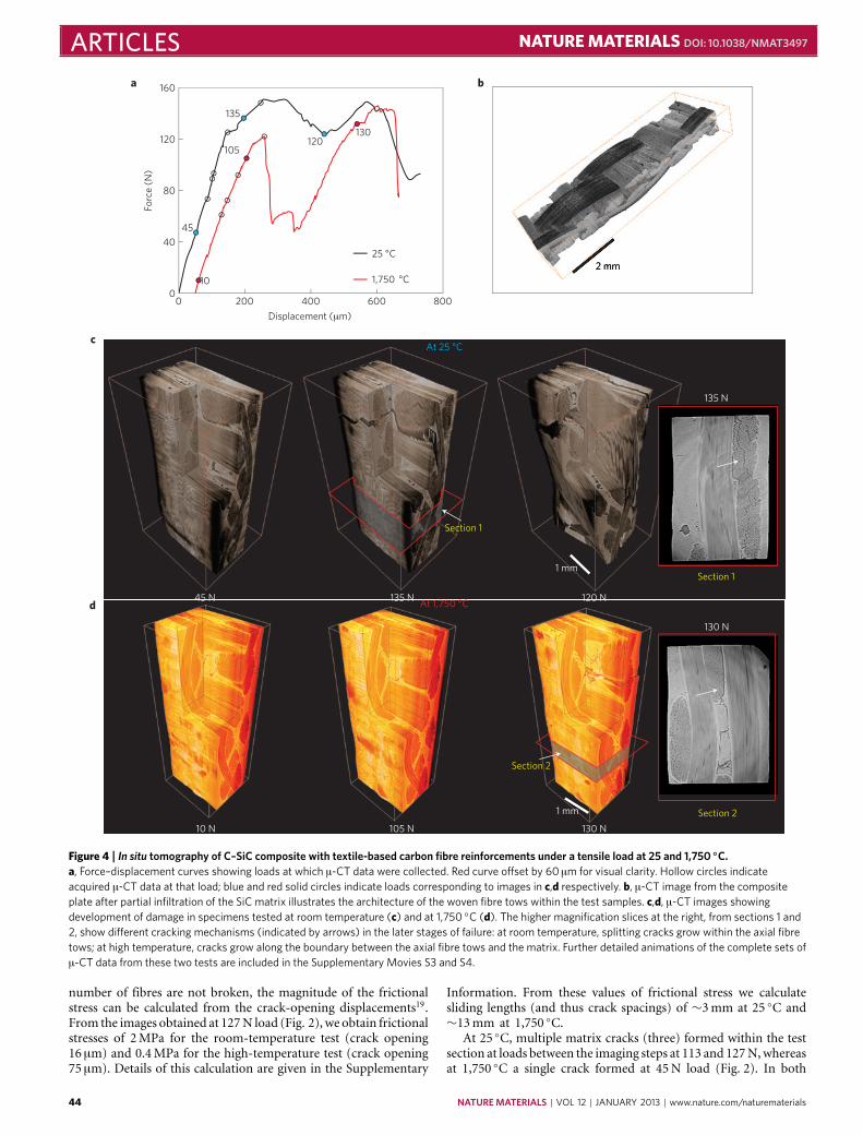
Figure 4 | In situ tomography of C–SiC composite with textile-based carbon fibre reinforcements under a tensile load at 25 and 1,750 ◦C.a, Force–displacement curves showing loads at which µ-CT data were collected. Red curve offset by 60 µm for visual clarity. Hollow circles indicateacquired µ-CT data at that load; blue and red solid circles indicate loads corresponding to images in c,d respectively. b, µ-CT image from the compositeplate after partial infiltration of the SiC matrix illustrates the architecture of the woven fibre tows within the test samples. c,d, µ-CT images showingdevelopment of damage in specimens tested at room temperature (c) and at 1,750 ◦C (d). The higher magnification slices at the right, from sections 1 and2, show different cracking mechanisms (indicated by arrows) in the later stages of failure: at room temperature, splitting cracks grow within the axial fibretows; at high temperature, cracks grow along the boundary between the axial fibre tows and the matrix. Further detailed animations of the complete sets ofµ-CT data from these two tests are included in the Supplementary Movies S3 and S4.
number of fibres are not broken, the magnitude of the frictionalstress can be calculated from the crack-opening displacements19.From the images obtained at 127N load (Fig. 2), we obtain frictionalstresses of 2MPa for the room-temperature test (crack opening16 µm) and 0.4MPa for the high-temperature test (crack opening75 µm). Details of this calculation are given in the Supplementary
Information. From these values of frictional stress we calculatesliding lengths (and thus crack spacings) of ∼3mm at 25 ◦C and∼13mm at 1,750 ◦C.
At 25 ◦C, multiple matrix cracks (three) formed within the testsection at loads between the imaging steps at 113 and 127N,whereasat 1,750 ◦C a single crack formed at 45N load (Fig. 2). In both
44 NATURE MATERIALS | VOL 12 | JANUARY 2013 | www.nature.com/naturematerials

NATURE MATERIALS DOI: 10.1038/NMAT3497 ARTICLEScases, the crack-opening displacements continued to increase withfurther loading, while no new cracks were formed (confirmedalso by scanning electron microscope (SEM) observations afterloading to failure). The room-temperature results are consistentwith observations from separate ex situ tests using samples withlonger test sections (60mm), which showed a distribution ofcracks separated by distances between 1 and 4mm (average 2mm),which is consistent with calculated sliding lengths (above). In thehigh-temperature specimen, the observation of a single crack isalso consistent with the calculated sliding length, which in this caseexceeds half the gauge length.
Differences were seen in the statistical distributions of fibrefailure sites and relaxation lengths at 25 and 1,750 ◦C (seen inFig. 3 and Supplementary Movies S1 and S2). At high temperature,failure sites were distributed over larger distances from the matrixcracks and the relaxation distances were larger than at 25 ◦C.These observations are also consistent with the sliding resistancebeing smaller at 1,750 ◦C than at 25 ◦C. The distributed loadsand positions of fibre fractures relate directly to the statisticaldistribution of fibre strengths21,22 (if the fibre strengths weresingle-valued, all failures would occur at the plane of the matrixcrack) and can be used to evaluate the parameters characterizingthis distribution. There is currently no other way to access thisinformation, which is critical for detecting and quantifying changesin fibre properties due to high-temperature heat treatments andpossible chemical reactions during processing of the composite.
These results from in situ tomography provide new insightinto how the 3D behaviour in SiCf–SiCm composites under tensileload differs at 25 ◦C and at 1,750 ◦C. The observed reduction infibre sliding resistance at high temperature could potentially becaused by changes in residual stresses due to thermal expansionmismatch.However, residual stresses in this composite are expectedto be small. Another possible mechanism is a change in the shearproperties of the boron nitride layer between the fibres and matrixat high temperature.
Damage evolution in textile compositesResults obtained from similar loading experiments using Cf–SiCmtextile composite specimens, with a larger length scale and morecomplex architecture than the single-tow samples, are shown inFig. 4a–c (detailed views of cracks initiating and propagating in theroom- and high-temperature specimens are shown, respectively,in the Supplementary Movies S3 and S4). Once more, differencescan be seen in the development of cracks at 25 and 1,750 ◦C.Images here were recorded at lower resolution (1.3 µm per voxel)to allow a larger field of view. The individual carbon fibresin this composite were not visible in the sections normal orparallel to the fibre tows, because of this lower resolutionand the minimal density difference between the fibres and thepyrolytic-carbon coatings. Nevertheless, the fibre tows are clearlydistinguished from the denser matrix, which consists of two layers:one is a thin brighter layer of CVI SiC surrounding all of thefibre tows and the other the polymer-derived SiC that fills theremaining space. Images obtained at 25 and 1,750 ◦C were ofidentical quality. During initial loading at both temperatures,cracks formed in the matrix normal to the loading directionat positions where the matrix lays over a transverse fibre tow.With increasing load, the cracks grew through the transverse towsuntil they met an underlying axial tow (at loads in the range∼40–70N), where they were deflected. At 25 ◦C this deflectioninvolved formation of multiple splitting cracks (Fig. 4), whichprogressed incrementally along the centres of the axial tows asthe load was increased to the peak value of 150N. At 1,750 ◦C,the deflection of the crack at each tow involved a single crackthat grew along the edge of the axial fibre tows as the loadincreased to 120N, whereupon there was a large load drop. By
influencing the access of ambient gas to the internal reinforcingfibres, differences in crack paths such as these could potentiallyhave a large effect on subsequent high-temperature oxidationdamage. Further experiments are needed to determine the reasonfor the difference in crack paths and to assess whether similardifferences in cracking behaviour are consistently observed inother loading directions.
OutlookIn this exploratory study only a small fraction of the quantitativeand statistical information potentially available in theµ-CTdata setswas used. Theµ-CTdata contain complete quantitative informationon crack paths, crack surface areas and orientations, spatialvariations in the crack-opening displacements, statistics of relativespatial location of cracks and microstructural heterogeneitieswithin the sample volume; all these parameters are critical inany analysis of fracture as they govern the toughness of thematerial. The challenge is to extract this information from thereconstructed 3D images in a form that can be readily used forvalidation of computational models and to provide calibrationof material constitutive laws as input for the models. Successrests on efficient methods for processing the 3D image data withtechniques such as segmentation for automated identification andrepresentation of cracks and microstructural features. The imagesin Fig. 3 (and the Supplementary Movies S1 and S2) are examplesof such representations obtained from the image data in Fig. 2through the use of multiple image post-processing steps (seeSupplementary Information).
We have demonstrated that in situ X-ray microtomography cannow be used for studying the advanced materials that are being de-signed for future ultrahigh temperature environments. The meansto acquire real-time high-resolution (up to 0.65 µm per voxel) 3Dstructural data for ceramic matrix composites has been presentedwith the ability to observe microstructure and damage under loadat different hierarchical length scales (from a micrometre to severalmillimetres) at unprecedented temperatures (as high as 1,750 ◦C).The results contain vital information pertaining to the underlyingfailure mechanisms within ceramic composites that can be usedto optimize their performance. An extension of this study wouldinvolve rigorous analysis and interpretations of the different physi-cal phenomena occurring in situ under the complex environmentof load and temperature. The study of time-dependent damagemechanisms involving high-temperature creep would be facilitatedby reducing the scan time for image acquisition. Scan times atthe Advanced Light Source (ALS) facility, where this work wasperformed, have recently been reduced to several minutes withoutloss of image quality through the use of a high-frame-rate cameraand an improved high-efficiency scintillator. Even shorter scantimes can be achieved at the expense of image quality by limitingthe number of radiographs collected in each scan. These and otheradvances in synchrotron facilities show promise for enabling insitu tomography under near-continuous loading conditions. Thecapacity of validating the models developed for virtual testingthrough direct, real-time, non-invasive experimental observationswill greatly advance our understanding and help promote innova-tion on these technologically important materials.
MethodsMaterials. The single-tow SiCf–SiCm composite specimens were fabricated atHypertherm (Huntington Beach) by winding a SiC fibre tow on a carbon frameand by processing through CVI in two steps: the first to deposit a thin layer ofboron nitride, which was doped with silicon to improve its oxidation resistance,and the second to deposit the matrix of SiC. The textile-based Cf–SiCm compositewas fabricated by first infiltrating a woven fabric of carbon fibre tows with athin fibre coating of pyrolytic carbon followed by a thin layer of SiC by CVI (atHypertherm), then forming the remaining matrix by multiple cycles of infiltrationand pyrolysis of a slurry consisting of submicrometre particles of silicon carbide in
NATURE MATERIALS | VOL 12 | JANUARY 2013 | www.nature.com/naturematerials 45

ARTICLES NATURE MATERIALS DOI: 10.1038/NMAT3497
a polymer (Allylhydridopolycarbosilane) that decomposes at high temperature toform SiC. The textile fabric was a three-layer angle-interlock structure, consistingof three layers of relatively straight weft fibre tows linked together with side-by-sidewarp fibre tows that follow almost sinusoidal paths through the thickness of thecomposite. The test samples were cut from a large plate with the warp fibre towsalong the axial lengths of the beams.
µ-CT characterization. We used the hard X-ray beamline BL 8.3.2 at the AdvancedLight Source (Lawrence Berkeley National Laboratory) for the µ-CT tomographyexperiments. The ultrahigh-temperature tensile rig was mounted on an air-bearingrotation stage which positioned the beam by means of translation stages. In the caseof the high-temperature experiments, a non-oxidizing environment is maintainedby admitting a low flow of high-purity nitrogen while pumping to 10−3 torr. Loadis applied to the sample by a stepper motor, while force and displacement aremeasured using an in-line load cell and a linear variable differential transformer(LVDT) sensor. A 300 µm-thick aluminiumwindow allows tensile forces up to 2 kNto be applied to the test sample while obtaining an X-ray transmission of ∼90%with the filtered white light (2.5mm aluminium filter) used for sample illuminationat a sample-to-detector distance of 150mm. Tomography data for each specimenwere collected while tensile forces were applied in steps, increasing monotonicallyfrom 10N until the peak load carrying capacity was exceeded. Each image consistsof several tiled scans to increase the field of view.
For the smaller, single tow specimens, a magnification corresponding to0.65 µm per voxel was used, giving a vertical field of view of approximately 1.4mm.The full 3D dataset for this sample consists of four adjacent scans ‘tiled’ along thevertical direction, covering over 5.5mm of the sample; for the textile compositespecimen the data were collected in two vertical tiles with a magnificationcorresponding to 1.3 µm per voxel, covering nearly 5mm total of the specimen.Each scan consisted of multiple exposures, each of 100ms, collected at 0.125◦angular steps over a 180◦ rotation of the sample.
Tomographic slices were generated using a commercial reconstructionalgorithm (Octopus v8; IIC UGent, Zwijnaarde). Data were processed using imageprocessing tools in ImageJ (Rasband, W.S., ImageJ, US National Institutes ofHealth, Bethesda, http://imagej.nih.gov/ij/, 1997–2011.) and visualized in Avizo6.1 (VSG, Visualization Sciences Group). For crack segmentation, the wavelet-FFTalgorithm (referred to as xStripes)23 was adapted for use in ImageJ. Transverseµ-CT slices parallel to the axis of the fibres produced striped patterns of the fibreswhich are eliminated by the wavelet-FFT filter. Filtered slices contained greyscaleinformation of cracks alone, which were binarized and further segmented to obtainthe crack-opening measurements.
The magnitudes of the frictional sliding stresses were calculated from thecrack-opening displacements19; details are given in the Supplementary Information.
Temperaturemeasurement. Measurement of high temperatures in this test set-upis challenging as the small size of the test samples precludes directly attachingor embedding thermocouples. In these experiments, the sample temperatures inthe hot zone were estimated from separate calibration tests, in which a C-typetungsten-rhenium thermocouple (maximum measurable temperature of 2,300 ◦C)was mounted in the top grips and translated across the field of view using themotorized loading stage, with X-ray transmission images being used to determinethe thermocouple position. The variation within the central 5mm of the field ofview is approximately 150 ◦C at the set temperature of 1,750 ◦C. The operatingtemperature range of the furnace can be adjusted for a particular sample usingcalibrated voltage and current settings.
Further information on temperature measurements can be found in theSupplementary Information.
Received 24 August 2012; accepted 22 October 2012;published online 9 December 2012
References1. Stock, S. R. Recent advances in X-ray microtomography applied to materials.
Int. Mater. Rev. 53, 129–181 (2008).2. Sakdinawat, A. & Attwood, D. Nanoscale X-ray imaging. Nature Photon. 4,
840–848 (2010).3. Marshall, D. B. & Cox, B. N. Integral textile ceramic structures. Annu. Rev.
Mater. Res. 38, 425–443 (2008).4. Schmidt, S. et al. Ceramic matrix composites: A challenge in space-
propulsion technology applications. Int. J. Appl. Ceram. Technol. 2,85–96 (2005).
5. Mouritz, A. P., Bannister, M. K., Falzon, P. J. & Leong, K. H. Review ofapplications for advanced three-dimensional fibre textile composites.Composite A 30, 1445–1461 (1999).
6. Morscher, G. N. & Pujar, V. V. Design guidelines for in-plane mechanicalproperties of SiC fiber-reinforced melt-infiltrated SiC composites. Int. J. Appl.Ceram. Technol. 6, 151–163 (2009).
7. Zhao, J. C. &Westbrook, J. H. Ultrahigh-temperature materials for jet engines.Mater. Res. Soc. Bull. 28, 622–630 (2003).
8. Raj, R., Scarmi, A. & Soraru, G. D. The role of carbon in unexpectedvisco(an)elastic behavior of amorphous silicon oxycarbide above 1273K.J. Non-Cryst. Solids 351, 2238–2243 (2005).
9. Zhuang, W., Barter, S. & Molent, L. Flight-by-flight fatigue crack growth lifeassessment. Int. J. Fatigue 29, 1647–1657 (2007).
10. Novak, M. D. & Zok, F. W. High-temperature materials testing with full-fieldstrain measurement: Experimental design and practice. Rev. Sci. Instrum. 82,115101 (2011).
11. Terzi, S. et al. In situ X-ray tomography observation of inhomogeneousdeformation in semi-solid aluminium alloys. Scr. Mater. 61, 449–452 (2009).
12. Kinney, J. H. & Nichols, M. C. X-ray tomographic microscopy (XTM) usingsynchrotron radiation. Annu. Rev. Mater. Sci. 22, 121–152 (1992).
13. Langer, M., Cloetens, P., Guigay, J-P. & Peyrin, F. Quantitative comparison ofdirect phase retrieval algorithms in in-line phase tomography.Med. Phys. 35,4556–4566 (2008).
14. Groso, A., Abela, R. & Stampanoni, M. Implementation of a fast method forhigh resolution phase contrast tomography.Opt. Express 14, 8103–8110 (2006).
15. Berbon, Z., Rugg, K. L., Dadkhah, M. S. & Marshall, D. B. Effect of weavearchitecture on tensile properties and local strain heterogeneity in thin-sheetC-SiC composites. J. Am. Ceram. Soc. 85, 2039–2048 (2002).
16. Chateau, C. et al. In situ X-ray microtomography characterization of damagein SiCf/SiC minicomposites. Compos. Sci. Technol. 71, 916–924 (2011).
17. Budiansky, B., Evans, A. G. & Hutchinson, J. W. Fiber–matrix debondingeffects on cracking in aligned fiber ceramic composites. Int. J. Solids Struct. 32,315–328 (1995).
18. Morscher, G. N. Tensile stress rupture of SiCf/SiCm minicomposites withcarbon and boron nitride interphases at elevated temperatures in air. J. Am.Ceram. Soc. 80, 2029–2042 (1997).
19. Marshall, D. B., Cox, B. N. & Evans, A. G. The mechanics of matrixcracking in brittle–matrix fiber composites. Acta Metall. Mater. 33,2013–2021 (1985).
20. Morscher, G. N., Martinez-Fernandez, J. & Purdy, M. J. Determination ofinterfacial properties using a single-fiber microcomposite test. J. Am. Ceram.Soc. 79, 1083–1091 (1996).
21. Okabe, T., Nishikawa, M. & Curtin, W. A. Estimation of statistical strengthdistribution of carborundum polycrystalline SiC fiber using the single fibercomposite with consideration of the matrix hardening. Compos. Sci. Technol.68, 3067–3072 (2008).
22. Cox, B. N., Marshall, D. B. & Thouless, M. D. Influence of statistical fiberstrength distribution on matrix cracking in fiber composites. Acta Metall.Mater. 37, 1933–1943 (1989).
23. Muench, B., Trtik, P., Marone, F. & Stampanoni, M. Stripe and ringartifact removal with combined wavelet—fourier filtering. Opt. Express 17,8567–8591 (2009).
AcknowledgementsWork supported by the Air Force Office of Scientific Research (A. Sayir) and NASA(A. Calomino) under the National Hypersonics Science Center for Materials andStructures (AFOSR Contract No. FA9550-09-1-0477). We acknowledge the use of theX-ray synchrotron micro-tomography beam line (8.3.2) at the Advanced Light Source(ALS) at the Lawrence Berkeley National Laboratory, which is supported by the Office ofScience of theUSDepartment of Energy under contract no.DE-AC02-05CH11231.
Author contributionsB.N.C., D.B.M. and R.O.R. conceived the project, J.R.N. and A.A.M. designed theequipment and A.H. and H.A.B. built it. D.B.M. prepared the composite samples,H.A.B. performed the experiments and analysis with assistance from A.H., A.A.M.,D.L.P. and D.B.M., and H.A.B., B.N.C., D.B.M. and R.O.R. wrote the manuscript withcontributions from A.A.M.
Additional informationSupplementary information is available in the online version of the paper. Reprints andpermissions information is available online at www.nature.com/reprints. Correspondenceand requests for materials should be addressed to R.O.R.
Competing financial interestsThe authors declare no competing financial interests.
46 NATURE MATERIALS | VOL 12 | JANUARY 2013 | www.nature.com/naturematerials


![Quantitative Magnetic Particle Imaging Monitors the ...Magnetic particle imaging (MPI) [16–21], an imaging modality distinct from MRI, produces line-arly quantitative images of iron](https://static.fdocuments.net/doc/165x107/6124fca4732f0c68d25dc95d/quantitative-magnetic-particle-imaging-monitors-the-magnetic-particle-imaging.jpg)