Rapid Separation of 25-OH- Vitamin D3 and 3-Epi-25- …phx.phenomenex.com/lib/po91591211_L_1.pdf ·...
16
PO91591211_L_1 Rapid Separation of 25-OH- Vitamin D3 and 3-Epi-25- OH-Vitamin D3 in Human Serum using Tandem Mass Spectrometry Detection Jeff Layne, Michael McCoy, and Sky Countryman Phenomenex, Inc., 411 Madrid Ave., Torrance, CA 90501 USA
Transcript of Rapid Separation of 25-OH- Vitamin D3 and 3-Epi-25- …phx.phenomenex.com/lib/po91591211_L_1.pdf ·...

PO
9159
1211
_L_1
Rapid Separation of 25-OH-Vitamin D3 and 3-Epi-25-OH-Vitamin D3 in Human Serum using Tandem Mass Spectrometry Detection
Jeff Layne, Michael McCoy, and Sky Countryman
Phenomenex, Inc., 411 Madrid Ave., Torrance, CA 90501 USA

Faulty vitamin D metabolism in children less than 12 months of age can lead to formation of the inactive 3-epi-25 monohydroxy form. The resolution of 3-epimer from the active monohydroxy form by tandem mass spectrometry is not possible due to mostly identical fragmentation pattern of the two species. As a result, the two isomers should be separated chromatographically. The method described here resolves the critical pair within a short run time.
Serum/plasma samples were treated with acetonitrile to precipitate the protein, followed by centrifugation. A small volume of the supernatant was injected on the LC column. The chromatographic separation is carried out by a high efficiency media that allowed for separation of the monohydroxy vitamin D3 isomers
as well as separation of the 3-epi-25 monohydroxy epimer. A typical methanol and formic acid mobile phase combination starting with high organic concentration is used. The column is maintained at ambient temperature, ~22 °C. The signal detection is carried out by a triple quadrupole mass spectrometer operating in multiple reactions monitoring (MRM) function. An atmospheric pressure ionization source operating in positive polarity and using high purity nitrogen gas produced the [M+H+-H2O]+ precursor ions. The LOD for both 25-OH-Vit D3 and its 3-epimer were similar at 2.5 ng/mL.
The method prescribed here provides excellent resolution of the monohydroxy vitamin D3 isomers within a short run time.
Abstract

In recent years, vitamin D (Ergocalciferol, D2 and Cholecalciferol, D3) has been subject to increasing investigation for a range of potentially beneficial health effects. The measurement of Vitamin D metabolites, 25-hydroxy (25-OH) and 1α, 25-DiOH vitamin D (Vit D), is used as marker to determine vitamin D deficiency.
Isomerization of 25-OH-Vit D produces 3-epi Vit D3 (conversion of α-OH to ß-OH), a diasteromeric form. The presence of the epimer was first reported in 2006 by Singh et al. In infants, a significant portion of the 25-OH-
Vit D may be present as the epimeric form. Thus, in order to determine the accurate vitamin D status of such patients, it is necessary to be able to distinguish between the two diastereomeric forms.
Historically, analysis of Vit D and its metabolites has been performed via immunoassays. However, there is some question as to the ability of immunoassays to discriminate between 25-OH-D3 and its epimer. Thus, the development of an LC/MS/MS analysis that can distinguish the 25-OH-Vit D metabolite from its epimeric form is greatly desired.
Introduction

Instrumentation and Conditions
LC System:
Agilent® 1260 UPLC System with binary LC pumps
HPLC conditions: As specified on the chromatogram
MS System:
AB SCIEX API 5000™ operating under Pos polarity APCI
MS Parameters:
Gas 1 (GS1) 40
Curtain Gas (CUR) 25
Temperature (TEM) 350 °C
Nebulizer Current (NC) 5 μA
Collision Gas (CAD) 8
DP 100 V
Entrance Potential (EP) 10 V
CXP 10 V
Dwell 150 msec

Table 1. MRM Transitions Table
Compound ID Q1, Da Q3, Da CE, V
OH-Vit D2 395.3 209.3 30
OH-Vit D3/Epi-D3 383.2 257.2 25
Int Std (OH-D3-2H3) 386.2 257.2 25
OH-Vit D3 (Sec Trans) 383.2 229.1 30
OH-Vit D2 (Sec Trans) 395.3 269.2 22

Chromatographic Media
• The final, optimized LC/MS/MS method for the separation and analysis of 25-OH-Vit D and its epimer was performed using a core-shell column - Kinetex® 2.6 µm PFP.
• The core-shell Kinetex particle consists of a solid inner core surrounded by a layer of porous silica material.
• This unique core-shell structure can provide exceptionally high efficiency at relatively modest backpressure (compatible with a conventional HPLC system).
2.6 µm Core-Shell Particle
Performance equivalent to or better
than fully porous
Can be used on conventional HPLC systems and UH-
0.35 µm Porous Shell
1.9 µm Solid Core
™

Results and Discussion
Figure 1. Separation using a fully porous silica bonded with C18 chemistry
min
25-OH-D2
25-OH-D3 and 3-Epi-25-OH-D3
Column: Kinetex 2.6 µm C18 Dimensions: 30 x 3.0 mm
Part No.: 00A-4462-Y0Mobile Phase: A. 0.1 % Formic Acid in DI water
B. 0.1% Formic Acid in Acetonitrile
Time (min) % B 0 55 0.3 80 1 80 1.1 95 1.5 95 1.51 55 2 55
Flow Rate: 400 µLTemperature: Ambient
Detection: Tandem Mass Spec (MS-MS) Instrument: API 5000
Gradient:
• Although the majority of reversed phase HPLC methods are developed using C18 bonded phases, the C18 stationary phase chemistry lacks the ability to adequately resolve the 25-OH-Vit D3 from its epimer.
Ap
p ID
20
00
5

Figure 2. Separation using a fully porous silica bonded with phenyl chemistry
• A phenyl-based stationary phase bonded to conventional fully-porous silica (Synergi™ Polar-RP) was able to adequately resolve 25-OH-Vit D2 from 25-OH-Vit D2, but it did not display any resolution of the 25-OH-Vit D3 epimeric form.
min
25-OH-D2
Ap
p ID
20
00
2
Column: Synergi™ 2.5 µm Polar-RP Dimensions: 100 x 2.0 mm
Part No.: 00D-4371-B0Mobile Phase: A. 0.1 % Formic Acid in DI water
B. 0.1% Formic Acid in Methanol
Time (min) % B 0 75 2 80 3.8 80 3.81 75 6 75
Flow Rate: 400 µLTemperature: Ambient
Detection: Tandem Mass Spec (MS-MS) (ambient)Instrument: API 5000
Gradient:
25-OH-D3 and 3-Epi-25-OH-D3

Ap
p ID
20
00
4
min
• Using the core-shell Kinetex PFP column in a water/acetonitrile/formic acid mobile phase, it is possible to separate 25-OH-D3 from its epimer, and also to separate out the 25-OH-D2 in a run time of about 10 minutes.
Figure 3. Separation using the Kinetex PFP and Acetonitrile
Column: Kinetex 2.6 µm PFP Dimensions: 100 x 2.1mm
Part No.: 00D-4477-ANMobile Phase: A.0.1% Formic Acid in DI water
B 0.1% Formic Acid in Acetonitrile (55:45)Flow Rate: 400 µL
Temperature: AmbientDetection: Tandem Mass Spec (MS-MS)
(ambient)3-Epi-25-OH-D3
25-OH-D3
25-OH-D2

0.5 1.0 1.5 2.0 2.5 3.0 3.5 4.0 4.5 5.00.0
2.0e5
4.0e5
6.0e5
8.0e5
1.0e6
1.2e6
1.4e6
1.6e6
1.8e6
2.0e6
2.2e6
2.4e6
2.5e63.66
3.884.20
Inte
nsity
, cp
s
min
• By switching to a mobile phase containing methanol rather than acetonitrile, we can take advantage of the unique PFP selectivity to separate 25-OH-D2 from 25-OH-D3 and also to fully-resolve the epimeric 25-OH-D3 metabolite with a total analysis time less than 5 minutes.
25-OH-D2
Figure 4. Separation using the Kinetex PFP and Methanol
Ap
p ID
20
00
3
Column: Kinetex 2.6 µm PFP Dimensions: 100 x 2.1 mm
Part No.: 00D-4477-ANMobile Phase: A. 0.1 % Formic Acid in DI water
B. 0.1% Formic Acid in Methanol
Time (min) % B 0 75 2 80 3.8 80 3.81 75 6 75
Flow Rate: 400 µLTemperature: Ambient
Detection: Tandem Mass Spec (MS-MS)Instrument: API 5000
Gradient:
3-Epi-25-OH-D3
25-OH-D3

min
3-Epi-25-OH-D3
• Commercially-available human serum contains relatively high levels of both 25-OH-D3 and its epimer, making it unsuitable for use in making a calibration curve. • Because of this, we used double charcoal-stripped human serum, which was found to have significantly lower levels of these components.
Figure 5. Chromatogram of a commercially available human serum
Column: Kinetex 2.6 µm PFP Dimensions: 100 x 2.1 mm
Part No.: 00D-4477-ANMobile Phase: A. 0.1 % Formic Acid in DI water
B. 0.1% Formic Acid in Methanol
Time (min) % B 0 75 2 80 3.8 80 3.81 75 6 75
Flow Rate: 400 µLTemperature: Ambient
Detection: Tandem Mass Spec (MS-MS) (ambient)Instrument: API 5000
Gradient:
25-OH-D3
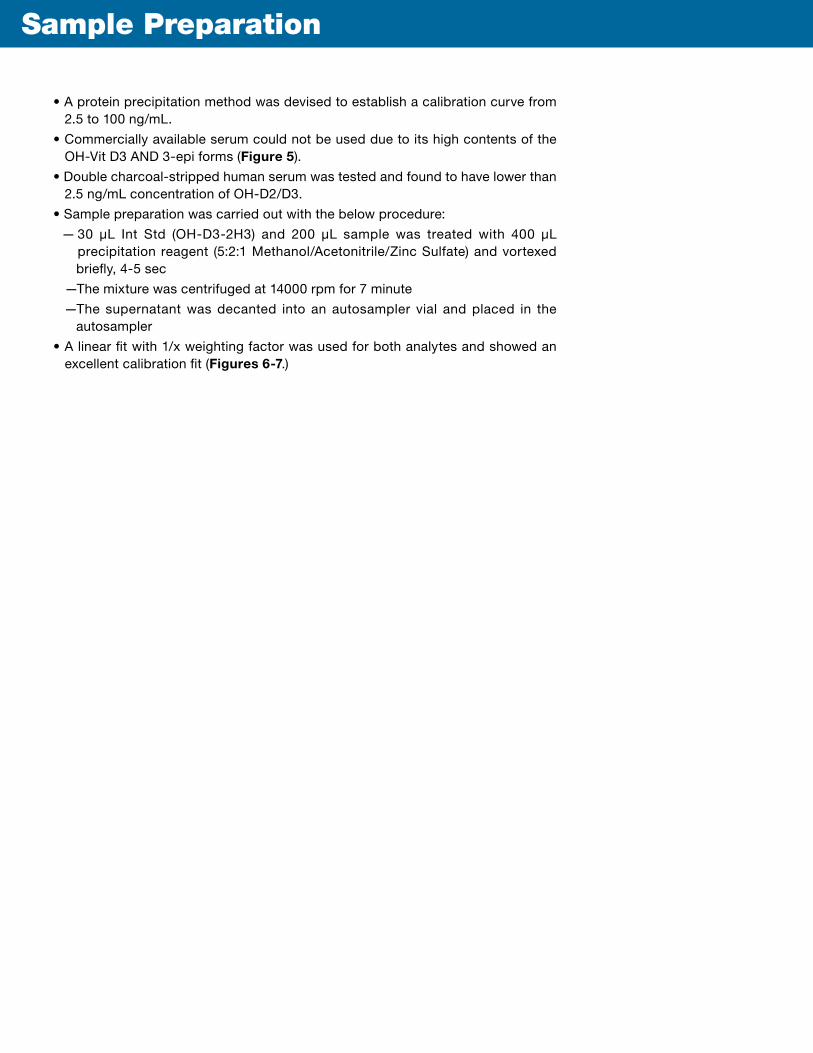
Sample Preparation
• A protein precipitation method was devised to establish a calibration curve from 2.5 to 100 ng/mL.
• Commercially available serum could not be used due to its high contents of the OH-Vit D3 AND 3-epi forms (Figure 5).
• Double charcoal-stripped human serum was tested and found to have lower than 2.5 ng/mL concentration of OH-D2/D3.
• Sample preparation was carried out with the below procedure:
— 30 µL Int Std (OH-D3-2H3) and 200 µL sample was treated with 400 µL precipitation reagent (5:2:1 Methanol/Acetonitrile/Zinc Sulfate) and vortexed briefly, 4-5 sec
—The mixture was centrifuged at 14000 rpm for 7 minute
—The supernatant was decanted into an autosampler vial and placed in the autosampler
• A linear fit with 1/x weighting factor was used for both analytes and showed an excellent calibration fit (Figures 6-7.)

Figure 6. OH-Vit D3 calibration curve from 2.5 to 100 ng/mL
• Calibration curve for OH-Vit D3 from 2.5 to 100 ng/mL, r=0.9984
5 10
1.20
1.10
1.00
0.90
0.80
0.70
0.60
0.50
0.40
0.30
0.20
0.10
0.0015 20 25 30 35 40 45 50 55 60 5 70 75 80 85 90 95 100
Analyte Conc. / IS Conc.
Ana
lyte
Are
a /
IS A
rea
Ap
p ID
20
007

Figure 7. OH-Vit D2 calibration curve from 2.5 to 100 ng/mL
• Calibration curve for OH-Vit D2 from 2.5 to 100 ng/mL, r=0.9994
5 10
0.34
0.32
0.28
0.30
0.260.24
0.22
0.20
0.180.16
0.140.12
0.100.08
0.06
0.04
0.02
0.0015 20 25 30 35 40 45 50 55 60 5 70 75 80 85 90 95 100
Analyte Conc. / IS Conc.
Ana
lyte
Are
a /
IS A
rea
Ap
p ID
20
00
6

W e h a v e d e v e l o p e d a n a s s a y u s i n g t h e K i n e tex 2 .6 µ m PFP c o l u m n t h a t c a n a c c u r a te l y q u a n t i t a te 25 - O H -V i t D 3 in the presence of i ts epimer ic form using a s i m p l e w a t e r / m e t h a n o l / f o r m i c a c i d mobile phase.
This assay can also be used to quanti tate 25 - O H - D 2 , 25 - O H - D 3 , a n d t h e 25 - O H - D3 epimer with a total analysis time of less than 5 minutes.
Conclusions

References
1. Singh et al, J Clin Endocrinol Metab 2006; 91:3055–61
2. Hoofnagle et al, Clinica Chimica Acta 2012; 413:203–206
3. Schlelcher et al, Clinica Chimica Acta 2012; 412:1549–1599
TrademarksKinetex is a registered trademark and Synergi is a trademark of Phenomenex. AB SCIEX and API 5000 are trademarks of AB SCIEX Pte. Ltd. Agilent is a registered trademark of Agilent Technologies, Inc. UPLC is a registered trademark of Waters Corporation.
© 2012 Phenomenex, Inc. All rights reserved.