Quadruplex DNA-guided ligand selection from dynamic ...
10
HAL Id: hal-03186431 https://hal.archives-ouvertes.fr/hal-03186431 Submitted on 26 Jul 2021 HAL is a multi-disciplinary open access archive for the deposit and dissemination of sci- entific research documents, whether they are pub- lished or not. The documents may come from teaching and research institutions in France or abroad, or from public or private research centers. L’archive ouverte pluridisciplinaire HAL, est destinée au dépôt et à la diffusion de documents scientifiques de niveau recherche, publiés ou non, émanant des établissements d’enseignement et de recherche français ou étrangers, des laboratoires publics ou privés. Quadruplex DNA-guided ligand selection from dynamic combinatorial libraries of acylhydrazones Oksana Reznichenko, Anne Cucchiarini, Valérie Gabelica, Anton Granzhan To cite this version: Oksana Reznichenko, Anne Cucchiarini, Valérie Gabelica, Anton Granzhan. Quadruplex DNA-guided ligand selection from dynamic combinatorial libraries of acylhydrazones. Organic and Biomolecu- lar Chemistry, Royal Society of Chemistry, 2021, 19 (2), pp.379-386. 10.1039/D0OB01908A. hal- 03186431
Transcript of Quadruplex DNA-guided ligand selection from dynamic ...
HAL Id: hal-03186431https://hal.archives-ouvertes.fr/hal-03186431
Submitted on 26 Jul 2021
HAL is a multi-disciplinary open accessarchive for the deposit and dissemination of sci-entific research documents, whether they are pub-lished or not. The documents may come fromteaching and research institutions in France orabroad, or from public or private research centers.
L’archive ouverte pluridisciplinaire HAL, estdestinée au dépôt et à la diffusion de documentsscientifiques de niveau recherche, publiés ou non,émanant des établissements d’enseignement et derecherche français ou étrangers, des laboratoirespublics ou privés.
Quadruplex DNA-guided ligand selection from dynamiccombinatorial libraries of acylhydrazones
Oksana Reznichenko, Anne Cucchiarini, Valérie Gabelica, Anton Granzhan
To cite this version:Oksana Reznichenko, Anne Cucchiarini, Valérie Gabelica, Anton Granzhan. Quadruplex DNA-guidedligand selection from dynamic combinatorial libraries of acylhydrazones. Organic and Biomolecu-lar Chemistry, Royal Society of Chemistry, 2021, 19 (2), pp.379-386. �10.1039/D0OB01908A�. �hal-03186431�
ARTICLE
Please do not adjust margins
Please do not adjust margins
a. CNRS UMR9187, Inserm U1196, Institut Curie, PSL Research University, 91405 Orsay, France. E-mail: [email protected]
b. CNRS UMR9187, Inserm U1196, Université Paris Saclay, 91405 Orsay, France.
c. Univ. Bordeaux, CNRS, INSERM, ARNA, UMR 5320, U1212, IECB, 33600 Pessac, France.
† Electronic Supplementary Information (ESI) available. See DOI: 10.1039/x0xx00000x
Received 00th January 20xx,
Accepted 00th January 20xx
DOI: 10.1039/x0xx00000x
Quadruplex DNA-Guided Ligand Selection from Dynamic Combinatorial Libraries of Acylhydrazones†
Oksana Reznichenko, a,b
Anne Cucchiarini, a,b
Valérie Gabelica c and Anton Granzhan *
a,b
Dynamic combinatorial libraries of acylhydrazones were prepared from diacylhydrazides and several cationic or neutral
aldehydes in the presence of 5-methoxyanthranilic acid catalyst. Pull-down experiments with magnetic beads
functionalized with a G-quadruplex (G4)-forming oligonucleotide led to the identification of putative ligands, which were
resynthesized or emulated by close structural analogues. G4-binding properties of novel derivatives were assessed by
fluorimetric titrations, mass spectrometry and thermal denaturation experiments, giving evidence of strong binding (Kd <
10 nM) for two compounds.
Introduction
G-quadruplex (G4) structures of DNA and RNA are involved in
numerous cellular processes such as DNA replication, trans-
cription, telomere maintenance, and RNA translation.1,2
Targeting of these structures with small molecules (ligands)
paves a way for therapeutic control of biological processes
that involve G4-mediated regulation.3–5
However, considering
the large number of putative G4-forming sequences in the
human genome (over 700 000),6 G4 ligands need to acquire
selectivity to certain G4 structures over the others, in order to
minimize possible off-target effects. Despite more than 20
years of active research, only moderate progress has been
achieved in terms of intra-G4 selectivity of ligands.5,7,8
Clearly,
the conventional strategies that rely on the synthesis of small
libraries of putative ligands and subsequent assessment of
their binding to a panel of G4 structures are insufficient to
generate ligands with better affinity and selectivity, and novel
approaches allowing tailor-made ligand design are urgently
needed. Several strategies have been explored towards this
end, including fragment-based screening,9,10
small-molecule
microarrays,11
kinetic target-guided synthesis,12
and peptide
libraries,13,14
resulting in identifications of novel G4 ligands
with intriguing properties.
Dynamic combinatorial chemistry (DCC) is a
supramolecular approach that exploits reversible chemical
reactions to generate dynamic combinatorial libraries (DCL) of
products under thermodynamic control.15,16
This method is
particularly well-suited for the discovery of ligands and
receptors, since the addition of an external species (a “target”,
or a “template”) shifts the dynamic equilibrium towards the
formation of the product(s) having the highest affinity to the
target.17
While DCC has now been well established for protein
targeting,18,19
its application in the field of nucleic acids is
much more restricted.20,21
Along these lines, Bala-
subramanian22–24
and Ulven25
labs exploited DCC for the
discovery (or optimization) of G4-DNA ligands; of note, all
these approaches relied on the reaction of disulfide exchange
and involved only small number of components, typically a
central scaffold possessing a G4 affinity and a set of side
chains. More recently, Dash et al. utilized a DCC approach
based on the reversible formation of imines followed by pull-
down and reduction, to identify novel carbazole-based G4-
DNA ligands.26
However, a serious drawback of imine-based
DCC is that it requires the conversion of imines, identified from
DCL analysis, into stable amine analogues whose affinity and
selectivity to the target may be significantly different.
Therefore, there is a need for the development of alternative
chemistries suitable for nucleic acid-targeted DCC.
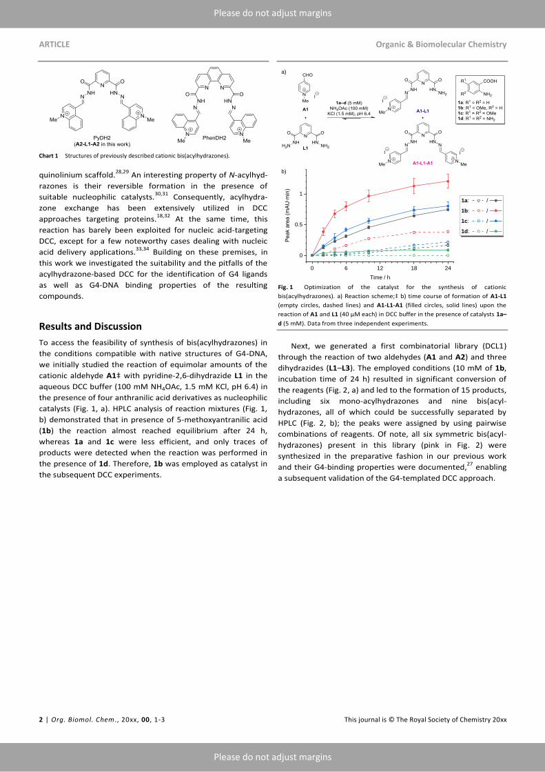
In this context, we have recently described cationic
bis(acylhydrazones), such as PyDH2 and PhenDH2 (Chart 1),27
as novel, biologically active G4 ligands based on the well-
established bis-
ARTICLE Organic & Biomolecular Chemistry
2 | Org. Biomol. Chem ., 20xx, 00, 1-3 This journal is © The Royal Society of Chemistry 20xx
Please do not adjust margins
Please do not adjust margins
Chart 1 Structures of previously described cationic bis(acylhydrazones).
quinolinium scaffold.28,29
An interesting property of N-acylhyd-
razones is their reversible formation in the presence of
suitable nucleophilic catalysts.30,31
Consequently, acylhydra-
zone exchange has been extensively utilized in DCC
approaches targeting proteins.18,32
At the same time, this
reaction has barely been exploited for nucleic acid-targeting
DCC, except for a few noteworthy cases dealing with nucleic
acid delivery applications.33,34
Building on these premises, in
this work we investigated the suitability and the pitfalls of the
acylhydrazone-based DCC for the identification of G4 ligands
as well as G4-DNA binding properties of the resulting
compounds.
Results and Discussion
To access the feasibility of synthesis of bis(acylhydrazones) in
the conditions compatible with native structures of G4-DNA,
we initially studied the reaction of equimolar amounts of the
cationic aldehyde A1‡ with pyridine-2,6-dihydrazide L1 in the
aqueous DCC buffer (100 mM NH4OAc, 1.5 mM KCl, pH 6.4) in
the presence of four anthranilic acid derivatives as nucleophilic
catalysts (Fig. 1, a). HPLC analysis of reaction mixtures (Fig. 1,
b) demonstrated that in presence of 5-methoxyantranilic acid
(1b) the reaction almost reached equilibrium after 24 h,
whereas 1a and 1c were less efficient, and only traces of
products were detected when the reaction was performed in
the presence of 1d. Therefore, 1b was employed as catalyst in
the subsequent DCC experiments.
Fig. 1 Optimization of the catalyst for the synthesis of cationic
bis(acylhydrazones). a) Reaction scheme;‡ b) time course of formation of A1-L1
(empty circles, dashed lines) and A1-L1-A1 (filled circles, solid lines) upon the
reaction of A1 and L1 (40 µM each) in DCC buffer in the presence of catalysts 1a–
d (5 mM). Data from three independent experiments.
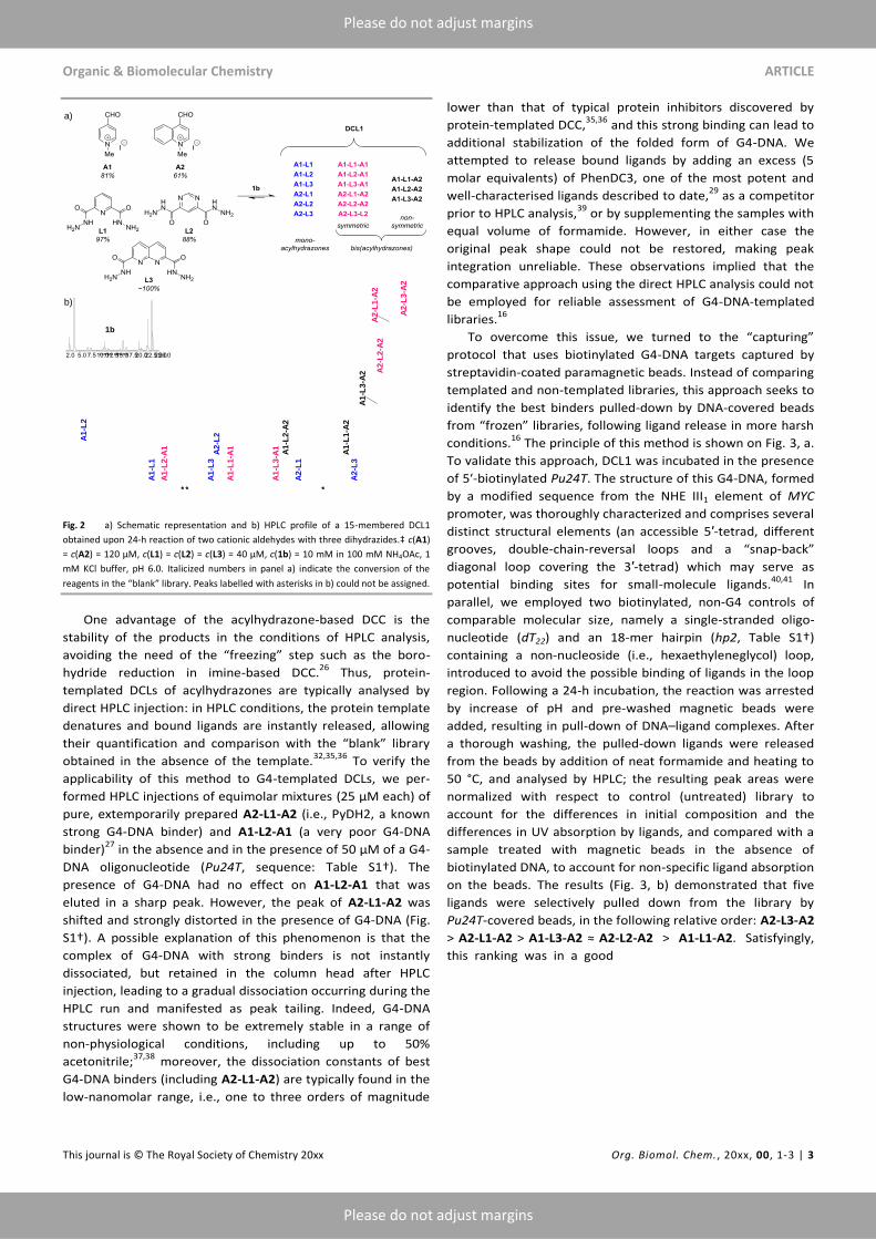
Next, we generated a first combinatorial library (DCL1)
through the reaction of two aldehydes (A1 and A2) and three
dihydrazides (L1–L3). The employed conditions (10 mM of 1b,
incubation time of 24 h) resulted in significant conversion of
the reagents (Fig. 2, a) and led to the formation of 15 products,
including six mono-acylhydrazones and nine bis(acyl-
hydrazones, all of which could be successfully separated by
HPLC (Fig. 2, b); the peaks were assigned by using pairwise
combinations of reagents. Of note, all six symmetric bis(acyl-
hydrazones) present in this library (pink in Fig. 2) were
synthesized in the preparative fashion in our previous work
and their G4-binding properties were documented,27
enabling
a subsequent validation of the G4-templated DCC approach.
b)
0 6 12 18 24
0
0.5
1
Peak area (mAU∙min)
Time / h
1a: /
1b: /
1c: /
1d: /
a)
Organic & Biomolecular Chemistry ARTICLE
This journal is © The Royal Society of Chemistry 20xx Org. Biomol. Chem. , 20xx, 00, 1-3 | 3
Please do not adjust margins
Please do not adjust margins
Fig. 2 a) Schematic representation and b) HPLC profile of a 15-membered DCL1
obtained upon 24-h reaction of two cationic aldehydes with three dihydrazides.‡ c(A1)
= c(A2) = 120 µM, c(L1) = c(L2) = c(L3) = 40 µM, c(1b) = 10 mM in 100 mM NH4OAc, 1
mM KCl buffer, pH 6.0. Italicized numbers in panel a) indicate the conversion of the
reagents in the “blank” library. Peaks labelled with asterisks in b) could not be assigned.
One advantage of the acylhydrazone-based DCC is the
stability of the products in the conditions of HPLC analysis,
avoiding the need of the “freezing” step such as the boro-
hydride reduction in imine-based DCC.26
Thus, protein-
templated DCLs of acylhydrazones are typically analysed by
direct HPLC injection: in HPLC conditions, the protein template
denatures and bound ligands are instantly released, allowing
their quantification and comparison with the “blank” library
obtained in the absence of the template.32,35,36
To verify the
applicability of this method to G4-templated DCLs, we per-
formed HPLC injections of equimolar mixtures (25 µM each) of
pure, extemporarily prepared A2-L1-A2 (i.e., PyDH2, a known
strong G4-DNA binder) and A1-L2-A1 (a very poor G4-DNA
binder)27
in the absence and in the presence of 50 µM of a G4-
DNA oligonucleotide (Pu24T, sequence: Table S1†). The
presence of G4-DNA had no effect on A1-L2-A1 that was
eluted in a sharp peak. However, the peak of A2-L1-A2 was
shifted and strongly distorted in the presence of G4-DNA (Fig.
S1†). A possible explanation of this phenomenon is that the
complex of G4-DNA with strong binders is not instantly
dissociated, but retained in the column head after HPLC
injection, leading to a gradual dissociation occurring during the
HPLC run and manifested as peak tailing. Indeed, G4-DNA
structures were shown to be extremely stable in a range of
non-physiological conditions, including up to 50%
acetonitrile;37,38
moreover, the dissociation constants of best
G4-DNA binders (including A2-L1-A2) are typically found in the
low-nanomolar range, i.e., one to three orders of magnitude
lower than that of typical protein inhibitors discovered by
protein-templated DCC,35,36
and this strong binding can lead to
additional stabilization of the folded form of G4-DNA. We
attempted to release bound ligands by adding an excess (5
molar equivalents) of PhenDC3, one of the most potent and
well-characterised ligands described to date,29
as a competitor
prior to HPLC analysis,39
or by supplementing the samples with
equal volume of formamide. However, in either case the
original peak shape could not be restored, making peak
integration unreliable. These observations implied that the
comparative approach using the direct HPLC analysis could not
be employed for reliable assessment of G4-DNA-templated
libraries.16
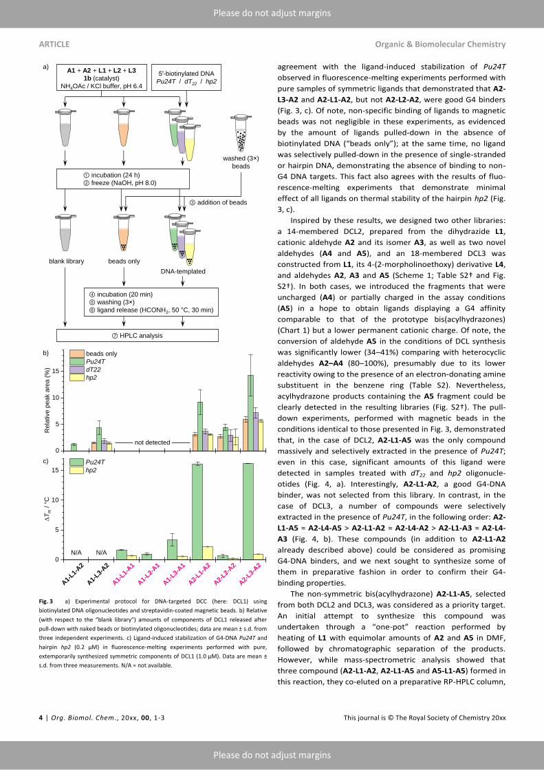
To overcome this issue, we turned to the “capturing”
protocol that uses biotinylated G4-DNA targets captured by
streptavidin-coated paramagnetic beads. Instead of comparing
templated and non-templated libraries, this approach seeks to
identify the best binders pulled-down by DNA-covered beads
from “frozen” libraries, following ligand release in more harsh
conditions.16
The principle of this method is shown on Fig. 3, a.
To validate this approach, DCL1 was incubated in the presence
of 5′-biotinylated Pu24T. The structure of this G4-DNA, formed
by a modified sequence from the NHE III1 element of MYC
promoter, was thoroughly characterized and comprises several
distinct structural elements (an accessible 5′-tetrad, different
grooves, double-chain-reversal loops and a “snap-back”
diagonal loop covering the 3′-tetrad) which may serve as
potential binding sites for small-molecule ligands.40,41
In
parallel, we employed two biotinylated, non-G4 controls of
comparable molecular size, namely a single-stranded oligo-
nucleotide (dT22) and an 18-mer hairpin (hp2, Table S1†)
containing a non-nucleoside (i.e., hexaethyleneglycol) loop,
introduced to avoid the possible binding of ligands in the loop
region. Following a 24-h incubation, the reaction was arrested
by increase of pH and pre-washed magnetic beads were
added, resulting in pull-down of DNA–ligand complexes. After
a thorough washing, the pulled-down ligands were released
from the beads by addition of neat formamide and heating to
50 °C, and analysed by HPLC; the resulting peak areas were
normalized with respect to control (untreated) library to
account for the differences in initial composition and the
differences in UV absorption by ligands, and compared with a
sample treated with magnetic beads in the absence of
biotinylated DNA, to account for non-specific ligand absorption
on the beads. The results (Fig. 3, b) demonstrated that five
ligands were selectively pulled down from the library by
Pu24T-covered beads, in the following relative order: A2-L3-A2
> A2-L1-A2 > A1-L3-A2 ≈ A2-L2-A2 > A1-L1-A2. Satisfyingly,
this ranking was in a good
a)
b)
A1
-L1
A1
-L2
* * *1b
A1
-L2
-A1
A1
-L2
-A2
A1
-L3
A2
-L1
A2
-L3
A2
-L2
A1
-L1
-A1
A1
-L3
-A1
A1
-L1
-A2
A1
-L3
-A2 A
2-L
2-A
2
A2
-L3
-A2
A2
-L1
-A2
ARTICLE Organic & Biomolecular Chemistry
4 | Org. Biomol. Chem ., 20xx, 00, 1-3 This journal is © The Royal Society of Chemistry 20xx
Please do not adjust margins
Please do not adjust margins
Fig. 3 a) Experimental protocol for DNA-targeted DCC (here: DCL1) using
biotinylated DNA oligonucleotides and streptavidin-coated magnetic beads. b) Relative
(with respect to the “blank library”) amounts of components of DCL1 released after
pull-down with naked beads or biotinylated oligonucleotides; data are mean ± s.d. from
three independent experiments. c) Ligand-induced stabilization of G4-DNA Pu24T and
hairpin hp2 (0.2 µM) in fluorescence-melting experiments performed with pure,
extemporarily synthesized symmetric components of DCL1 (1.0 µM). Data are mean ±
s.d. from three measurements. N/A = not available.
agreement with the ligand-induced stabilization of Pu24T
observed in fluorescence-melting experiments performed with
pure samples of symmetric ligands that demonstrated that A2-
L3-A2 and A2-L1-A2, but not A2-L2-A2, were good G4 binders
(Fig. 3, c). Of note, non-specific binding of ligands to magnetic
beads was not negligible in these experiments, as evidenced
by the amount of ligands pulled-down in the absence of
biotinylated DNA (“beads only”); at the same time, no ligand
was selectively pulled-down in the presence of single-stranded
or hairpin DNA, demonstrating the absence of binding to non-
G4 DNA targets. This fact also agrees with the results of fluo-
rescence-melting experiments that demonstrate minimal
effect of all ligands on thermal stability of the hairpin hp2 (Fig.
3, c).
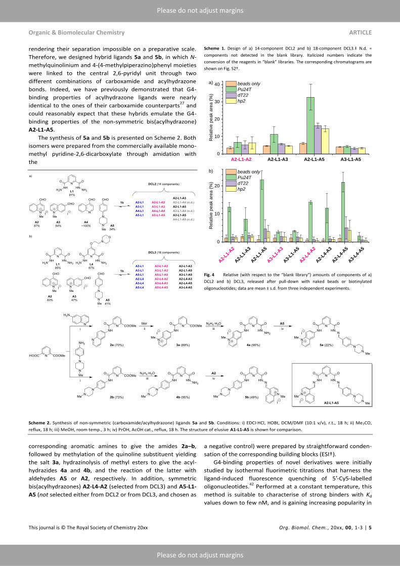
Inspired by these results, we designed two other libraries:
a 14-membered DCL2, prepared from the dihydrazide L1,
cationic aldehyde A2 and its isomer A3, as well as two novel
aldehydes (A4 and A5), and an 18-membered DCL3 was
constructed from L1, its 4-(2-morpholinoethoxy) derivative L4,
and aldehydes A2, A3 and A5 (Scheme 1; Table S2† and Fig.
S2†). In both cases, we introduced the fragments that were
uncharged (A4) or partially charged in the assay conditions
(A5) in a hope to obtain ligands displaying a G4 affinity
comparable to that of the prototype bis(acylhydrazones)
(Chart 1) but a lower permanent cationic charge. Of note, the
conversion of aldehyde A5 in the conditions of DCL synthesis
was significantly lower (34–41%) comparing with heterocyclic
aldehydes A2–A4 (80–100%), presumably due to its lower
reactivity owing to the presence of an electron-donating amine
substituent in the benzene ring (Table S2). Nevertheless,
acylhydrazone products containing the A5 fragment could be
clearly detected in the resulting libraries (Fig. S2†). The pull-
down experiments, performed with magnetic beads in the
conditions identical to those presented in Fig. 3, demonstrated
that, in the case of DCL2, A2-L1-A5 was the only compound
massively and selectively extracted in the presence of Pu24T;
even in this case, significant amounts of this ligand were
detected in samples treated with dT22 and hp2 oligonucle-
otides (Fig. 4, a). Interestingly, A2-L1-A2, a good G4-DNA
binder, was not selected from this library. In contrast, in the
case of DCL3, a number of compounds were selectively
extracted in the presence of Pu24T, in the following order: A2-
L1-A5 ≈ A2-L4-A5 > A2-L1-A2 ≈ A2-L4-A2 > A2-L1-A3 ≈ A2-L4-
A3 (Fig. 4, b). These compounds (in addition to A2-L1-A2
already described above) could be considered as promising
G4-DNA binders, and we next sought to synthesize some of
them in preparative fashion in order to confirm their G4-
binding properties.
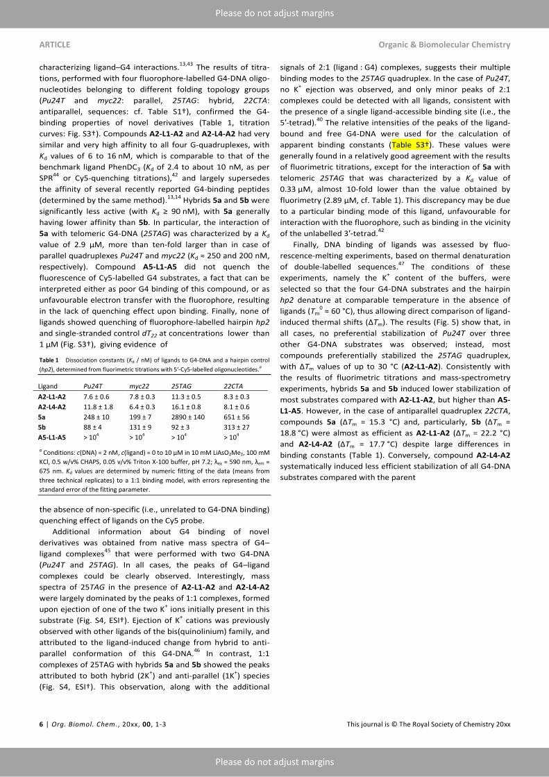
The non-symmetric bis(acylhydrazone) A2-L1-A5, selected
from both DCL2 and DCL3, was considered as a priority target.
An initial attempt to synthesize this compound was
undertaken through a “one-pot” reaction performed by
heating of L1 with equimolar amounts of A2 and A5 in DMF,
followed by chromatographic separation of the products.
However, while mass-spectrometric analysis showed that
three compound (A2-L1-A2, A2-L1-A5 and A5-L1-A5) formed in
this reaction, they co-eluted on a preparative RP-HPLC column,
A1 + A2 + L1 + L2 + L3
1b (catalyst)
NH4OAc / KCl buffer, pH 6.4
5′-biotinylated DNA
Pu24T / dT22 / hp2
blank library beads only
DNA-templated
① incubation (24 h)
② freeze (NaOH, pH 8.0)
washed (3 )
beads
③ addition of beads
④ incubation (20 min)
⑤ washing (3×)
⑥ ligand release (HCONH2, 50 °C, 30 min)
a)
⑦ HPLC analysis
A1-
L1-A2
A1-
L3-A2
A1-
L1-A1
A1-
L2-A1
A1-
L3-A1
A2-
L1-A2
A2-
L2-A2
A2-
L3-A2
0
5
10
15
DT
m /
°C
Pu24T
hp2
N/A N/A
0
5
10
15
Re
lative
pe
ak a
rea
(%
)
beads only
Pu24T
dT22
hp2
not detected
b)
c)
Organic & Biomolecular Chemistry ARTICLE
This journal is © The Royal Society of Chemistry 20xx Org. Biomol. Chem. , 20xx, 00, 1-3 | 5
Please do not adjust margins
Please do not adjust margins
rendering their separation impossible on a preparative scale.
Therefore, we designed hybrid ligands 5a and 5b, in which N-
methylquinolinium and 4-(4-methylpiperazino)phenyl moieties
were linked to the central 2,6-pyridyl unit through two
different combinations of carboxamide and acylhydrazone
bonds. Indeed, we have previously demonstrated that G4-
binding properties of acylhydrazone ligands were nearly
identical to the ones of their carboxamide counterparts27
and
could reasonably expect that these hybrids emulate the G4-
binding properties of the non-symmetric bis(acylhydrazone)
A2-L1-A5.
The synthesis of 5a and 5b is presented on Scheme 2. Both
isomers were prepared from the commercially available mono-
methyl pyridine-2,6-dicarboxylate through amidation with
the
Scheme 1. Design of a) 14-component DCL2 and b) 18-component DCL3.‡ N.d. =
components not detected in the blank library. Italicized numbers indicate the
conversion of the reagents in “blank” libraries. The corresponding chromatograms are
shown on Fig. S2†.
Fig. 4 Relative (with respect to the “blank library”) amounts of components of a)
DCL2 and b) DCL3, released after pull-down with naked beads or biotinylated
oligonucleotides; data are mean ± s.d. from three independent experiments.
Scheme 2. Synthesis of non-symmetric (carboxamide/acylhydrazone) ligands 5a and 5b. Conditions: i) EDCI·HCl, HOBt, DCM/DMF (10:1 v/v), r.t., 18 h; ii) Me2CO,
reflux, 18 h; iii) MeOH, room temp., 3 h; iv) PrOH, AcOH cat., reflux, 18 h. The structure of elusive A1-L1-A5 is shown for comparison.
corresponding aromatic amines to give the amides 2a–b,
followed by methylation of the quinoline substituent yielding
the salt 3a, hydrazinolysis of methyl esters to give the acyl-
hydrazides 4a and 4b, and the reaction of the latter with
aldehydes A5 or A2, respectively. In addition, symmetric
bis(acylhydrazones) A2-L4-A2 (selected from DCL3) and A5-L1-
A5 (not selected either from DCL2 or from DCL3, and chosen as
a negative control) were prepared by straightforward conden-
sation of the corresponding building blocks (ESI†).
G4-binding properties of novel derivatives were initially
studied by isothermal fluorimetric titrations that harness the
ligand-induced fluorescence quenching of 5′-Cy5-labelled
oligonucleotides.42
Performed at a constant temperature, this
method is suitable to characterise of strong binders with Kd
values down to few nM, and is gaining increasing popularity in
A2-L1-A2 A2-L1-A3 A2-L1-A5 A3-L1-A5
0
10
20
30
40
Rela
tive p
eak a
rea (
%)
beads only
Pu24T
dT22
hp2
A2-
L1-A2
A2-
L1-A3
A2-
L1-A5
A3-
L1-A3
A3-
L1-A5
A2-
L4-A2
A2-
L4-A3
A2-
L4-A5
A3-
L4-A5
0
10
20
Rela
tive p
eak a
rea (
%)
beads only
Pu24T
dT22
hp2
a)
b)
ARTICLE Organic & Biomolecular Chemistry
6 | Org. Biomol. Chem ., 20xx, 00, 1-3 This journal is © The Royal Society of Chemistry 20xx
Please do not adjust margins
Please do not adjust margins
characterizing ligand–G4 interactions.13,43
The results of titra-
tions, performed with four fluorophore-labelled G4-DNA oligo-
nucleotides belonging to different folding topology groups
(Pu24T and myc22: parallel, 25TAG: hybrid, 22CTA:
antiparallel, sequences: cf. Table S1†), confirmed the G4-
binding properties of novel derivatives (Table 1, titration
curves: Fig. S3†). Compounds A2-L1-A2 and A2-L4-A2 had very
similar and very high affinity to all four G-quadruplexes, with
Kd values of 6 to 16 nM, which is comparable to that of the
benchmark ligand PhenDC3 (Kd of 2.4 to about 10 nM, as per
SPR44
or Cy5-quenching titrations),42
and largely supersedes
the affinity of several recently reported G4-binding peptides
(determined by the same method).13,14
Hybrids 5a and 5b were
significantly less active (with Kd ≥ 90 nM), with 5a generally
having lower affinity than 5b. In particular, the interaction of
5a with telomeric G4-DNA (25TAG) was characterized by a Kd
value of 2.9 µM, more than ten-fold larger than in case of
parallel quadruplexes Pu24T and myc22 (Kd ≈ 250 and 200 nM,
respectively). Compound A5-L1-A5 did not quench the
fluorescence of Cy5-labelled G4 substrates, a fact that can be
interpreted either as poor G4 binding of this compound, or as
unfavourable electron transfer with the fluorophore, resulting
in the lack of quenching effect upon binding. Finally, none of
ligands showed quenching of fluorophore-labelled hairpin hp2
and single-stranded control dT22 at concentrations lower than
1 µM (Fig. S3†), giving evidence of
Table 1 Dissociation constants (Kd / nM) of ligands to G4-DNA and a hairpin control
(hp2), determined from fluorimetric titrations with 5′-Cy5-labelled oligonucleotides.a
Ligand Pu24T myc22 25TAG 22CTA
A2-L1-A2 7.6 ± 0.6 7.8 ± 0.3 11.3 ± 0.5 8.3 ± 0.3
A2-L4-A2 11.8 ± 1.8 6.4 ± 0.3 16.1 ± 0.8 8.1 ± 0.6
5a 248 ± 10 199 ± 7 2890 ± 140 651 ± 56
5b 88 ± 4 131 ± 9 92 ± 3 313 ± 27
A5-L1-A5 > 104 > 104 > 104 > 104
a Conditions: c(DNA) = 2 nM, c(ligand) = 0 to 10 µM in 10 mM LiAsO2Me2, 100 mM
KCl, 0.5 w/v% CHAPS, 0.05 v/v% Triton X-100 buffer, pH 7.2; λex = 590 nm, λem =
675 nm. Kd values are determined by numeric fitting of the data (means from
three technical replicates) to a 1:1 binding model, with errors representing the
standard error of the fitting parameter.
the absence of non-specific (i.e., unrelated to G4-DNA binding)
quenching effect of ligands on the Cy5 probe.
Additional information about G4 binding of novel
derivatives was obtained from native mass spectra of G4–
ligand complexes45
that were performed with two G4-DNA
(Pu24T and 25TAG). In all cases, the peaks of G4–ligand
complexes could be clearly observed. Interestingly, mass
spectra of 25TAG in the presence of A2-L1-A2 and A2-L4-A2
were largely dominated by the peaks of 1:1 complexes, formed
upon ejection of one of the two K+ ions initially present in this
substrate (Fig. S4, ESI†). Ejection of K+ cations was previously
observed with other ligands of the bis(quinolinium) family, and
attributed to the ligand-induced change from hybrid to anti-
parallel conformation of this G4-DNA.46
In contrast, 1:1
complexes of 25TAG with hybrids 5a and 5b showed the peaks
attributed to both hybrid (2K+) and anti-parallel (1K
+) species
(Fig. S4, ESI†). This observation, along with the additional
signals of 2:1 (ligand : G4) complexes, suggests their multiple
binding modes to the 25TAG quadruplex. In the case of Pu24T,
no K+ ejection was observed, and only minor peaks of 2:1
complexes could be detected with all ligands, consistent with
the presence of a single ligand-accessible binding site (i.e., the
5′-tetrad).40
The relative intensities of the peaks of the ligand-
bound and free G4-DNA were used for the calculation of
apparent binding constants (Table S3†). These values were
generally found in a relatively good agreement with the results
of fluorimetric titrations, except for the interaction of 5a with
telomeric 25TAG that was characterized by a Kd value of
0.33 µM, almost 10-fold lower than the value obtained by
fluorimetry (2.89 µM, cf. Table 1). This discrepancy may be due
to a particular binding mode of this ligand, unfavourable for
interaction with the fluorophore, such as binding in the vicinity
of the unlabelled 3′-tetrad.42
Finally, DNA binding of ligands was assessed by fluo-
rescence-melting experiments, based on thermal denaturation
of double-labelled sequences.47
The conditions of these
experiments, namely the K+ content of the buffers, were
selected so that the four G4-DNA substrates and the hairpin
hp2 denature at comparable temperature in the absence of
ligands (Tm0 ≈ 60 °C), thus allowing direct comparison of ligand-
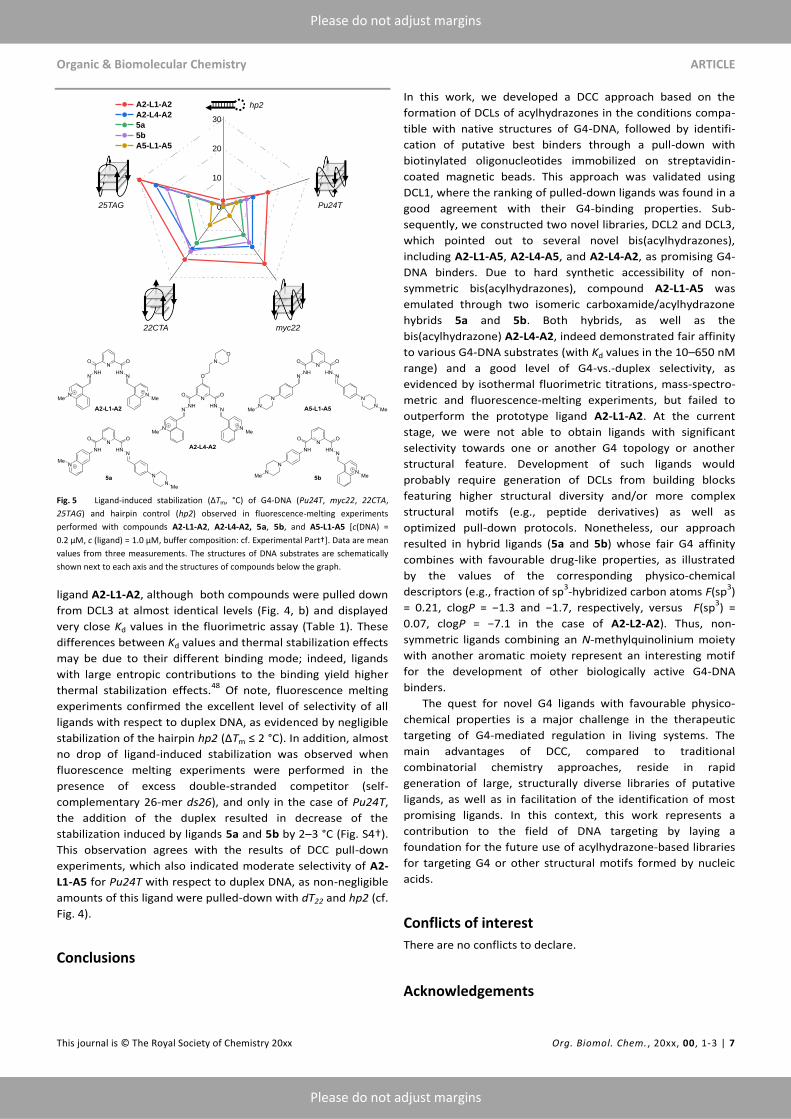
induced thermal shifts (∆Tm). The results (Fig. 5) show that, in
all cases, no preferential stabilization of Pu24T over three
other G4-DNA substrates was observed; instead, most
compounds preferentially stabilized the 25TAG quadruplex,
with ∆Tm values of up to 30 °C (A2-L1-A2). Consistently with
the results of fluorimetric titrations and mass-spectrometry
experiments, hybrids 5a and 5b induced lower stabilization of
most substrates compared with A2-L1-A2, but higher than A5-
L1-A5. However, in the case of antiparallel quadruplex 22CTA,
compounds 5a (∆Tm = 15.3 °C) and, particularly, 5b (∆Tm =
18.8 °C) were almost as efficient as A2-L1-A2 (∆Tm = 22.2 °C)
and A2-L4-A2 (∆Tm = 17.7 °C) despite large differences in
binding constants (Table 1). Conversely, compound A2-L4-A2
systematically induced less efficient stabilization of all G4-DNA
substrates compared with the parent
Organic & Biomolecular Chemistry ARTICLE
This journal is © The Royal Society of Chemistry 20xx Org. Biomol. Chem. , 20xx, 00, 1-3 | 7
Please do not adjust margins
Please do not adjust margins
Fig. 5 Ligand-induced stabilization (∆Tm, °C) of G4-DNA (Pu24T, myc22, 22CTA,
25TAG) and hairpin control (hp2) observed in fluorescence-melting experiments
performed with compounds A2-L1-A2, A2-L4-A2, 5a, 5b, and A5-L1-A5 [c(DNA) =
0.2 µM, c (ligand) = 1.0 µM, buffer composition: cf. Experimental Part†]. Data are mean
values from three measurements. The structures of DNA substrates are schematically
shown next to each axis and the structures of compounds below the graph.
ligand A2-L1-A2, although both compounds were pulled down
from DCL3 at almost identical levels (Fig. 4, b) and displayed
very close Kd values in the fluorimetric assay (Table 1). These
differences between Kd values and thermal stabilization effects
may be due to their different binding mode; indeed, ligands
with large entropic contributions to the binding yield higher
thermal stabilization effects.48
Of note, fluorescence melting
experiments confirmed the excellent level of selectivity of all
ligands with respect to duplex DNA, as evidenced by negligible
stabilization of the hairpin hp2 (∆Tm ≤ 2 °C). In addition, almost
no drop of ligand-induced stabilization was observed when
fluorescence melting experiments were performed in the
presence of excess double-stranded competitor (self-
complementary 26-mer ds26), and only in the case of Pu24T,
the addition of the duplex resulted in decrease of the
stabilization induced by ligands 5a and 5b by 2–3 °C (Fig. S4†).
This observation agrees with the results of DCC pull-down
experiments, which also indicated moderate selectivity of A2-
L1-A5 for Pu24T with respect to duplex DNA, as non-negligible
amounts of this ligand were pulled-down with dT22 and hp2 (cf.
Fig. 4).
Conclusions
In this work, we developed a DCC approach based on the
formation of DCLs of acylhydrazones in the conditions compa-
tible with native structures of G4-DNA, followed by identifi-
cation of putative best binders through a pull-down with
biotinylated oligonucleotides immobilized on streptavidin-
coated magnetic beads. This approach was validated using
DCL1, where the ranking of pulled-down ligands was found in a
good agreement with their G4-binding properties. Sub-
sequently, we constructed two novel libraries, DCL2 and DCL3,
which pointed out to several novel bis(acylhydrazones),
including A2-L1-A5, A2-L4-A5, and A2-L4-A2, as promising G4-
DNA binders. Due to hard synthetic accessibility of non-
symmetric bis(acylhydrazones), compound A2-L1-A5 was
emulated through two isomeric carboxamide/acylhydrazone
hybrids 5a and 5b. Both hybrids, as well as the
bis(acylhydrazone) A2-L4-A2, indeed demonstrated fair affinity
to various G4-DNA substrates (with Kd values in the 10–650 nM
range) and a good level of G4-vs.-duplex selectivity, as
evidenced by isothermal fluorimetric titrations, mass-spectro-
metric and fluorescence-melting experiments, but failed to
outperform the prototype ligand A2-L1-A2. At the current
stage, we were not able to obtain ligands with significant
selectivity towards one or another G4 topology or another
structural feature. Development of such ligands would
probably require generation of DCLs from building blocks
featuring higher structural diversity and/or more complex
structural motifs (e.g., peptide derivatives) as well as
optimized pull-down protocols. Nonetheless, our approach
resulted in hybrid ligands (5a and 5b) whose fair G4 affinity
combines with favourable drug-like properties, as illustrated
by the values of the corresponding physico-chemical
descriptors (e.g., fraction of sp3-hybridized carbon atoms F(sp
3)
= 0.21, clogP = −1.3 and −1.7, respectively, versus F(sp3) =
0.07, clogP = −7.1 in the case of A2-L2-A2). Thus, non-
symmetric ligands combining an N-methylquinolinium moiety
with another aromatic moiety represent an interesting motif
for the development of other biologically active G4-DNA
binders.
The quest for novel G4 ligands with favourable physico-
chemical properties is a major challenge in the therapeutic
targeting of G4-mediated regulation in living systems. The
main advantages of DCC, compared to traditional
combinatorial chemistry approaches, reside in rapid
generation of large, structurally diverse libraries of putative
ligands, as well as in facilitation of the identification of most
promising ligands. In this context, this work represents a
contribution to the field of DNA targeting by laying a
foundation for the future use of acylhydrazone-based libraries
for targeting G4 or other structural motifs formed by nucleic
acids.
Conflicts of interest
There are no conflicts to declare.
Acknowledgements
0
10
20
30
Pu24T25TAG
22CTA myc22
hp2 A2-L1-A2
A2-L4-A2
5a
5b
A5-L1-A5
hp2
Pu24T25TAG
22CTA myc22
ARTICLE Organic & Biomolecular Chemistry
8 | Org. Biomol. Chem ., 20xx, 00, 1-3 This journal is © The Royal Society of Chemistry 20xx
Please do not adjust margins
Please do not adjust margins
The authors thank Dr. Marie-Paule Teulade-Fichou (Institut
Curie) for a sample of PhenDC3 and helpful discussions. Dr.
Jorge González García and Ms. Hang Kang are acknowledged
for the preliminary experiments related to this project, and Dr.
Eric Largy (ARNA) and Dr. Frédéric Rosu (IECB) for assistance
and discussion regarding mass-spectrometry experiments. The
project benefited from access to the Plateforme de BioPhysico-
Chimie Structurale of the IECB (Univ. Bordeaux, CNRS
UMS3033, Inserm US001) for native mass spectrometry. This
work was supported by Agence Nationale de la Recherche
(grant-in aid ANR-17-CE07-0004-01 to AG), French Ministry of
Higher Education, Research and Innovation (PhD fellowship to
OR), and Institut Curie.
Notes and references
‡ Note that, while A1 and A2 are shown in the aldehyde form, both aldehydes are fully hydrated in aqueous solutions.
1 J. Spiegel, S. Adhikari and S. Balasubramanian, Trends Chem.,
2020, 2, 123–136.
2 D. Varshney, J. Spiegel, K. Zyner, D. Tannahill and S.
Balasubramanian, Nat. Rev. Mol. Cell Biol., 2020, 21, 459–474.
3 S. A. Ohnmacht and S. Neidle, Bioorg. Med. Chem. Lett., 2014,
24, 2602–2612.
4 S. Neidle, Nat. Rev. Chem., 2017, 1, 1–10.
5 S. Neidle, J. Med. Chem., 2016, 59, 5987–6011.
6 V. S. Chambers, G. Marsico, J. M. Boutell, M. Di Antonio, G. P.
Smith and S. Balasubramanian, Nat. Biotechnol., 2015, 33, 877–
881.
7 S. P. P. Pany, P. Bommisetti, K. V. Diveshkumar and P. I.
Pradeepkumar, Org. Biomol. Chem., 2016, 14, 5779–5793.
8 S. Asamitsu, T. Bando and H. Sugiyama, Chem. Eur. J., 2019, 25,
417–430.
9 M. Garavís, B. López-Méndez, A. Somoza, J. Oyarzabal, C. Dalvit,
A. Villasante, R. Campos-Olivas and C. González, ACS Chem.
Biol., 2014, 9, 1559–66.
10 M. Tassinari, A. Lena, E. Butovskaya, V. Pirota, M. Nadai, M.
Freccero, F. Doria and S. Richter, Molecules, 2018, 23, 1874.
11 K. M. Felsenstein, L. B. Saunders, J. K. Simmons, E. Leon, D. R.
Calabrese, S. Zhang, A. Michalowski, P. Gareiss, B. A. Mock, J. S.
Schneekloth and J. S. Schneekloth, Jr., ACS Chem. Biol., 2015,
11, 151013220852007.
12 M. Di Antonio, G. Biffi, A. Mariani, E. A. Raiber, R. Rodriguez and
S. Balasubramanian, Angew. Chem. Int. Ed., 2012, 51, 11073–
11078.
13 K. C. Liu, K. Röder, C. Mayer, S. Adhikari, D. J. Wales and S.
Balasubramanian, J. Am. Chem. Soc., 2020, 142, 8367–8373.
14 A. Minard, D. Morgan, F. Raguseo, A. Di Porzio, D. Liano, A. G.
Jamieson and M. Di Antonio, Chem. Commun., 2020, 56, 8940–
8943.
15 S. Ladame, Org. Biomol. Chem., 2008, 6, 219–226.
16 P. Frei, R. Hevey and B. Ernst, Chem. Eur. J., 2019, 25, 60–73.
17 O. Ramström and J.-M. Lehn, Nat. Rev. Drug Discov., 2002, 1,
26–36.
18 M. Mondal and A. K. H. Hirsch, Chem. Soc. Rev., 2015, 44, 2455–
2488.
19 A. M. Hartman, R. M. Gierse and A. K. H. Hirsch, European J.
Org. Chem., 2019, 2019, 3581–3590.
20 C. R. S. Durai and M. M. Harding, Aust. J. Chem., 2011, 64, 671–
680.
21 B. L. Miller, Top. Curr. Chem., 2011, 322, 107–137.
22 A. M. Whitney, S. Ladame and S. Balasubramanian, Angew.
Chem. Int. Ed., 2004, 43, 1143–1146.
23 S. Ladame, A. M. Whitney and S. Balasubramanian, Angew.
Chem. Int. Ed., 2005, 44, 5736–5739.
24 A. Bugaut, K. Jantos, J.-L. Wietor, R. Rodriguez, J. K. M. Sanders
and S. Balasubramanian, Angew. Chem. Int. Ed., 2008, 47, 2677–
2680.
25 M. C. Nielsen and T. Ulven, Chem. Eur. J., 2008, 14, 9487–9490.
26 S. ana, D. Panda, P. Saha, . D. Pantos and . Dash, J. Med.
Chem., 2019, 62, 762–773.
27 O. Reznichenko, A. Quillévéré, R. P. Martins, N. Loaëc, H. Kang,
M. J. Lista, C. Beauvineau, J. González-García, R. Guillot, C.
Voisset, C. Daskalogianni, R. Fåhraeus, M.-P. Teulade-Fichou, M.
Blondel and A. Granzhan, Eur. J. Med. Chem., 2019, 178, 13–29.
28 G. Pennarun, C. Granotier, L. R. Gauthier, D. Gomez, F.
Hoffschir, E. Mandine, J.-F. Riou, J.-L. Mergny, P. Mailliet and F.
D. Boussin, Oncogene, 2005, 24, 2917–2928.
29 A. De Cian, E. DeLemos, J.-L. Mergny, M.-P. Teulade-Fichou and
D. Monchaud, J. Am. Chem. Soc., 2007, 129, 1856–1857.
30 E. T. Kool, P. Crisalli and K. M. Chan, Org. Lett., 2014, 16, 1454–
1457.
31 D. Larsen, M. Pittelkow, S. Karmakar and E. T. Kool, Org. Lett.,
2015, 17, 274–277.
32 V. T. Bhat, A. M. Caniard, T. Luksch, R. Brenk, D. J. Campopiano
and M. F. Greaney, Nat. Chem., 2010, 2, 490–497.
33 E. Bartolami, Y. Bessin, V. Gervais, P. Dumy and S. Ulrich,
Angew. Chem. Int. Ed., 2015, 54, 10183–10187.
34 E. Bartolami, Y. Bessin, N. Bettache, M. Gary-Bobo, M. Garcia, P.
Dumy and S. Ulrich, Org. Biomol. Chem., 2015, 13, 9427–9438.
35 A. J. Clipson, V. T. Bhat, I. McNae, A. M. Caniard, D. J.
Campopiano and M. F. Greaney, Chem. Eur. J., 2012, 18, 10562–
10570.
36 A. M. Hartman, W. A. M. Elgaher, N. Hertrich, S. A. Andrei, C.
Ottmann and A. K. H. Hirsch, ACS Med. Chem. Lett., 2020, 11,
1041–1046.
37 M. C. Miller, R. Buscaglia, J. B. Chaires, A. N. Lane and J. O.
Trent, J. Am. Chem. Soc., 2010, 132, 17105–17107.
38 B. Heddi and A. T. Phan, J. Am. Chem. Soc., 2011, 133, 9824–
9833.
39 P. Frei, L. Pang, M. Silbermann, D. Eriş, T. Mühlethaler, O.
Schwardt and B. Ernst, Chem. Eur. J., 2017, 23, 11570–11577.
40 A. T. Phan, V. Kuryavyi, H. Y. Gaw and D. J. Patel, Nat. Chem.
Biol., 2005, 1, 167–173.
41 W. J. Chung, B. Heddi, F. Hamon, M.-P. Teulade-Fichou and A. T.
Phan, Angew. Chem. Int. Ed., 2014, 53, 999–1002.
42 D. D. Le, M. Di Antonio, L. K. M. Chan and S. Balasubramanian,
Chem. Commun., 2015, 51, 8048–8050.
43 R. N. Das, M. Andréasson, R. Kumar and E. Chorell, Chem. Sci.,
2020, 10529–10537.
44 L. Bonnat, L. Bar, B. Génnaro, H. Bonnet, O. Jarjayes, F. Thomas,
J. Dejeu, E. Defrancq and T. Lavergne, Chem. Eur. J., 2017, 23,
5602–5613.
Organic & Biomolecular Chemistry ARTICLE
This journal is © The Royal Society of Chemistry 20xx Org. Biomol. Chem. , 20xx, 00, 1-3 | 9
Please do not adjust margins
Please do not adjust margins
45 F. Rosu, E. De Pauw and V. Gabelica, Biochimie, 2008, 90, 1074–
1087.
46 A. Marchand, A. Granzhan, K. Iida, Y. Tsushima, Y. Ma, K.
Nagasawa, M. P. Teulade-Fichou and V. V. Gabelica, J. Am.
Chem. Soc., 2015, 137, 750–756.
47 A. De Rache and J.-L. Mergny, Biochimie, 2015, 115, 194–202.
48 A. Marchand, F. Rosu, R. Zenobi and V. Gabelica, J. Am. Chem.
Soc., 2018, 140, 12553–12565.
TOC entry
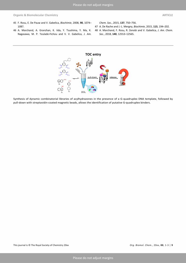
Synthesis of dynamic combinatorial libraries of acylhydrazones in the presence of a G-quadruplex DNA template, followed by
pull-down with streptavidin-coated magnetic beads, allows the identification of putative G-quadruplex binders.