Purification, characterization, and hypoglycemic ...
8
Purification, characterization, and hypoglycemic properties of eurocristatine from Eurotium cristatum spores in Fuzhuan brick tea† Gang Liu, abc Zhiguang Duan, abc Pan Wang, abc Daidi Fan abc and Chenhui Zhu * abc Fuzhuan brick tea (FBT) is a Chinese dark tea that is famous for its significant health benefits, in which Eurotium cristatum (E. cristatum) strains play a vital role in its postfermentation process. In this study, eurocristatine with hypoglycemic activity was discovered for the first time and purified from the spores of E. cristatum growing in FBT. Eurocristatine (98%) was obtained by D-101 macroporous resin-based column chromatography and preparative high performance liquid chromatography (HPLC) with a C 18 column as the stationary phase and 35% acetonitrile in ultrapure water as the mobile phase. Hypoglycemic activity in a Hep-G2 cell hypoglycemic model was used as a screening indicator during purification. The chemical structure of eurocristatine was characterized by ESI/MS, 1 H NMR and 13 C NMR analyses. The antidiabetic effects of eurocristatine were verified in high-fat diet/streptozocin-induced type 2 diabetes mellitus (T2DM) rats. The results showed that eurocristatine significantly reduced fasting blood glucose. Our study demonstrated that eurocristatine, as a newly discovered hypoglycemic active substance, could be considered a potential candidate for the treatment of diabetes and its complications. 1. Introduction Fuzhuan brick tea (FBT) is widely studied and loved by people due to its unique pharmacological health benets and patho- logical preventive effects, which has close ties with the growth of the dominant strain E. cristatum in the FBT postfermentation process. E. cristatum, a kind of probiotic, is nontoxic and safe, and the spores of FBT E. cristatum are capable of improving blood lipid metabolism, strengthening the immune system and have antioxidant and anticancer activity. 1–4 The spores can secrete multiple active metabolites during the process of golden ower blossoming. 5–7 In recent years, biologically active metabolites have been isolated from E. cristatum spores of FBT, including avonoids, proteases, alkaloids, diketopiperazines and polysaccharides. 8–14 These compounds show satisfactory biological activities, such as antibacterial activity, antiviral activity, antioxidant activity, and anticancer activity. 15–17 FBT extract can effectively prevent obesity in humans and experimental animals. 18 Studies have shown that FBT has the ability to alleviate obesity and regulate intestinal ora in C57BL/6J mice fed a high-fat diet (HFD). 19 The effect of E. cristatum-containing black tea fermentation broth on the activities of digestive enzymes such as amylase, protease and lipase showed that black tea fermentation broth containing E. cristatum could signicantly increase the activity of b-amylase and proteases and effectively reduce the activity of lipase. 20 However, most research has been limited to the extract or fermentation broth of FBT. It is not clear which specic substance plays a role in the above activities. E. cristatum was isolated from FBT and recultured separately to determine whether E. cristatum retained the above activities. Therefore, this research focused on the active metabolites of E. cristatum and their functions. Previous studies in our laboratory found that blood glucose levels in diabetic mice were signicantly lower than those in model mice aer the feeding of spores of the FBT E. cristatum for 4 consecutive weeks by intragastric administration. More- over, high doses of spores of the FBT E. cristatum showed no signicant adverse reactions. It is necessary to investigate the active hypoglycemic substance in spore metabolites. In this research, we investigated methods to separate hypoglycemic active substances from the spores of FBT E. cristatum by mac- roporous resin and preparative high-performance liquid chro- matography (HPLC), characterized the structure, veried the hypoglycemic activity of the active hypoglycemic substances, and solved the material basis of hypoglycemic activity from the spores of FBT E. cristatum. The quantitative analysis method using the identied hypoglycemic active substances as the a Shaanxi Key Laboratory of Degradable Biomedical Materials, School of Chemical Engineering, Northwest University, Taibai North Road 229, Xi'an, Shaanxi, 710069, China. E-mail: [email protected]; Fax: +86-29-88305118; Tel: +86-29-88305118 b Shaanxi R&D Center of Biomaterials and Fermentation Engineering, School of Chemical Engineering, Northwest University, Taibai North Road 229, Xi'an, Shaanxi, 710069, China c Biotech & Biomed Research Institute, Northwest University, Taibai North Road 229, Xi'an, Shaanxi, 710069, China † Electronic supplementary information (ESI) available. See DOI: 10.1039/d0ra03423a Cite this: RSC Adv. , 2020, 10, 22234 Received 17th April 2020 Accepted 2nd June 2020 DOI: 10.1039/d0ra03423a rsc.li/rsc-advances 22234 | RSC Adv. , 2020, 10, 22234–22241 This journal is © The Royal Society of Chemistry 2020 RSC Advances PAPER Open Access Article. Published on 10 June 2020. Downloaded on 3/11/2022 3:01:04 PM. This article is licensed under a Creative Commons Attribution-NonCommercial 3.0 Unported Licence. View Article Online View Journal | View Issue
Transcript of Purification, characterization, and hypoglycemic ...
RSC Advances
PAPER
Ope
n A
cces
s A
rtic
le. P
ublis
hed
on 1
0 Ju
ne 2
020.
Dow
nloa
ded
on 3
/11/
2022
3:0
1:04
PM
. T
his
artic
le is
lice
nsed
und
er a
Cre
ativ
e C
omm
ons
Attr
ibut
ion-
Non
Com
mer
cial
3.0
Unp
orte
d L
icen
ce.
View Article OnlineView Journal | View Issue
Purification, char
aShaanxi Key Laboratory of Degradable Bi
Engineering, Northwest University, Taibai N
China. E-mail: [email protected]; Fax:bShaanxi R&D Center of Biomaterials an
Chemical Engineering, Northwest Univer
Shaanxi, 710069, ChinacBiotech & Biomed Research Institute, North
Xi'an, Shaanxi, 710069, China
† Electronic supplementary informa10.1039/d0ra03423a
Cite this: RSC Adv., 2020, 10, 22234
Received 17th April 2020Accepted 2nd June 2020
DOI: 10.1039/d0ra03423a
rsc.li/rsc-advances
22234 | RSC Adv., 2020, 10, 22234–
acterization, and hypoglycemicproperties of eurocristatine from Eurotiumcristatum spores in Fuzhuan brick tea†
Gang Liu,abc Zhiguang Duan,abc Pan Wang,abc Daidi Fan abc and Chenhui Zhu *abc
Fuzhuan brick tea (FBT) is a Chinese dark tea that is famous for its significant health benefits, in which
Eurotium cristatum (E. cristatum) strains play a vital role in its postfermentation process. In this study,
eurocristatine with hypoglycemic activity was discovered for the first time and purified from the spores
of E. cristatum growing in FBT. Eurocristatine (98%) was obtained by D-101 macroporous resin-based
column chromatography and preparative high performance liquid chromatography (HPLC) with a C18
column as the stationary phase and 35% acetonitrile in ultrapure water as the mobile phase.
Hypoglycemic activity in a Hep-G2 cell hypoglycemic model was used as a screening indicator during
purification. The chemical structure of eurocristatine was characterized by ESI/MS, 1H NMR and 13C NMR
analyses. The antidiabetic effects of eurocristatine were verified in high-fat diet/streptozocin-induced
type 2 diabetes mellitus (T2DM) rats. The results showed that eurocristatine significantly reduced fasting
blood glucose. Our study demonstrated that eurocristatine, as a newly discovered hypoglycemic active
substance, could be considered a potential candidate for the treatment of diabetes and its complications.
1. Introduction
Fuzhuan brick tea (FBT) is widely studied and loved by peopledue to its unique pharmacological health benets and patho-logical preventive effects, which has close ties with the growth ofthe dominant strain E. cristatum in the FBT postfermentationprocess. E. cristatum, a kind of probiotic, is nontoxic and safe,and the spores of FBT E. cristatum are capable of improvingblood lipid metabolism, strengthening the immune system andhave antioxidant and anticancer activity.1–4 The spores cansecrete multiple active metabolites during the process of goldenower blossoming.5–7
In recent years, biologically active metabolites have beenisolated from E. cristatum spores of FBT, including avonoids,proteases, alkaloids, diketopiperazines and polysaccharides.8–14
These compounds show satisfactory biological activities, suchas antibacterial activity, antiviral activity, antioxidant activity,and anticancer activity.15–17 FBT extract can effectively preventobesity in humans and experimental animals.18 Studies have
omedical Materials, School of Chemical
orth Road 229, Xi'an, Shaanxi, 710069,
+86-29-88305118; Tel: +86-29-88305118
d Fermentation Engineering, School of
sity, Taibai North Road 229, Xi'an,
west University, Taibai North Road 229,
tion (ESI) available. See DOI:
22241
shown that FBT has the ability to alleviate obesity and regulateintestinal ora in C57BL/6J mice fed a high-fat diet (HFD).19 Theeffect of E. cristatum-containing black tea fermentation broth onthe activities of digestive enzymes such as amylase, proteaseand lipase showed that black tea fermentation broth containingE. cristatum could signicantly increase the activity of b-amylaseand proteases and effectively reduce the activity of lipase.20
However, most research has been limited to the extract orfermentation broth of FBT. It is not clear which specicsubstance plays a role in the above activities. E. cristatum wasisolated from FBT and recultured separately to determinewhether E. cristatum retained the above activities. Therefore,this research focused on the active metabolites of E. cristatumand their functions.
Previous studies in our laboratory found that blood glucoselevels in diabetic mice were signicantly lower than those inmodel mice aer the feeding of spores of the FBT E. cristatumfor 4 consecutive weeks by intragastric administration. More-over, high doses of spores of the FBT E. cristatum showed nosignicant adverse reactions. It is necessary to investigate theactive hypoglycemic substance in spore metabolites. In thisresearch, we investigated methods to separate hypoglycemicactive substances from the spores of FBT E. cristatum by mac-roporous resin and preparative high-performance liquid chro-matography (HPLC), characterized the structure, veried thehypoglycemic activity of the active hypoglycemic substances,and solved the material basis of hypoglycemic activity from thespores of FBT E. cristatum. The quantitative analysis methodusing the identied hypoglycemic active substances as the
This journal is © The Royal Society of Chemistry 2020
Paper RSC Advances
Ope
n A
cces
s A
rtic
le. P
ublis
hed
on 1
0 Ju
ne 2
020.
Dow
nloa
ded
on 3
/11/
2022
3:0
1:04
PM
. T
his
artic
le is
lice
nsed
und
er a
Cre
ativ
e C
omm
ons
Attr
ibut
ion-
Non
Com
mer
cial
3.0
Unp
orte
d L
icen
ce.
View Article Online
indicator was established using analytical HPLC. This study willprovide a scientic basis for the exploration and utilization ofclinical hypoglycemic medicines.
2. Materials and methods2.1. General experimental procedures
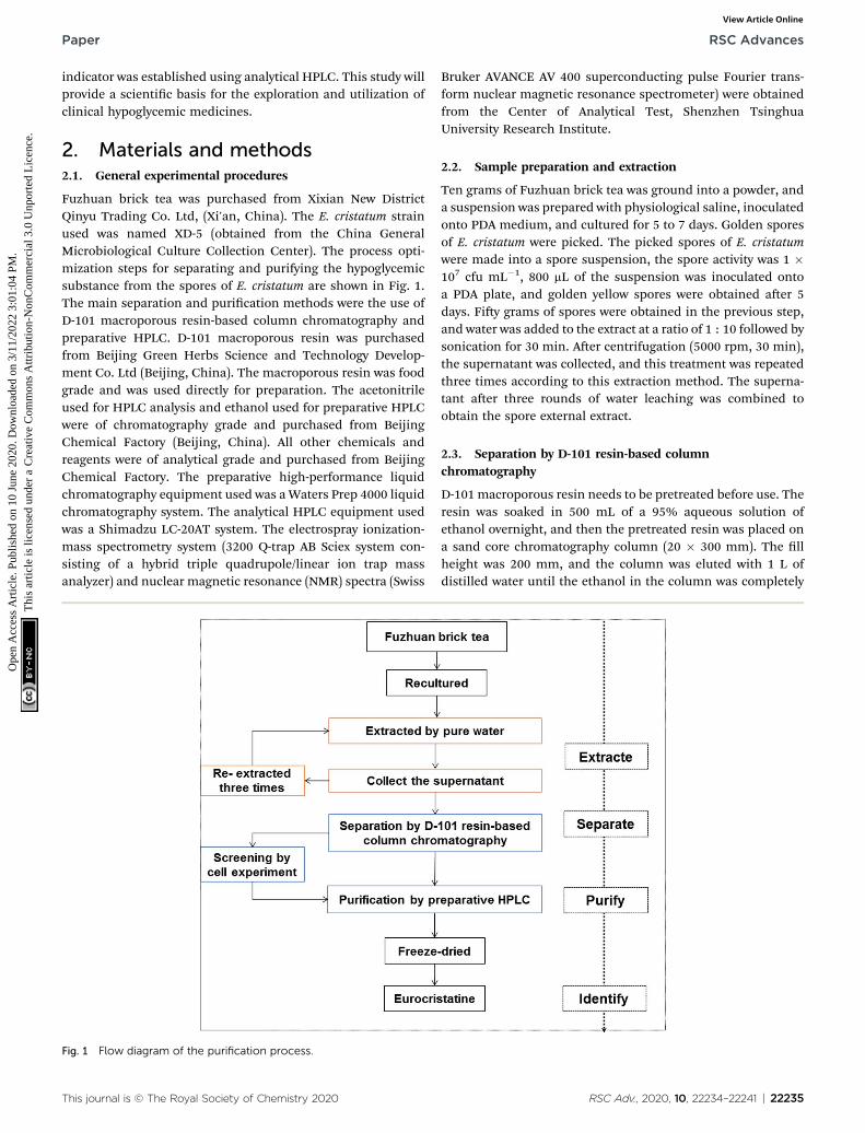
Fuzhuan brick tea was purchased from Xixian New DistrictQinyu Trading Co. Ltd, (Xi'an, China). The E. cristatum strainused was named XD-5 (obtained from the China GeneralMicrobiological Culture Collection Center). The process opti-mization steps for separating and purifying the hypoglycemicsubstance from the spores of E. cristatum are shown in Fig. 1.The main separation and purication methods were the use ofD-101 macroporous resin-based column chromatography andpreparative HPLC. D-101 macroporous resin was purchasedfrom Beijing Green Herbs Science and Technology Develop-ment Co. Ltd (Beijing, China). The macroporous resin was foodgrade and was used directly for preparation. The acetonitrileused for HPLC analysis and ethanol used for preparative HPLCwere of chromatography grade and purchased from BeijingChemical Factory (Beijing, China). All other chemicals andreagents were of analytical grade and purchased from BeijingChemical Factory. The preparative high-performance liquidchromatography equipment used was aWaters Prep 4000 liquidchromatography system. The analytical HPLC equipment usedwas a Shimadzu LC-20AT system. The electrospray ionization-mass spectrometry system (3200 Q-trap AB Sciex system con-sisting of a hybrid triple quadrupole/linear ion trap massanalyzer) and nuclear magnetic resonance (NMR) spectra (Swiss
Fig. 1 Flow diagram of the purification process.
This journal is © The Royal Society of Chemistry 2020
Bruker AVANCE AV 400 superconducting pulse Fourier trans-form nuclear magnetic resonance spectrometer) were obtainedfrom the Center of Analytical Test, Shenzhen TsinghuaUniversity Research Institute.
2.2. Sample preparation and extraction
Ten grams of Fuzhuan brick tea was ground into a powder, anda suspension was prepared with physiological saline, inoculatedonto PDA medium, and cultured for 5 to 7 days. Golden sporesof E. cristatum were picked. The picked spores of E. cristatumwere made into a spore suspension, the spore activity was 1 �107 cfu mL�1, 800 mL of the suspension was inoculated ontoa PDA plate, and golden yellow spores were obtained aer 5days. Fiy grams of spores were obtained in the previous step,and water was added to the extract at a ratio of 1 : 10 followed bysonication for 30 min. Aer centrifugation (5000 rpm, 30 min),the supernatant was collected, and this treatment was repeatedthree times according to this extraction method. The superna-tant aer three rounds of water leaching was combined toobtain the spore external extract.
2.3. Separation by D-101 resin-based columnchromatography
D-101 macroporous resin needs to be pretreated before use. Theresin was soaked in 500 mL of a 95% aqueous solution ofethanol overnight, and then the pretreated resin was placed ona sand core chromatography column (20 � 300 mm). The llheight was 200 mm, and the column was eluted with 1 L ofdistilled water until the ethanol in the column was completely
RSC Adv., 2020, 10, 22234–22241 | 22235
RSC Advances Paper
Ope
n A
cces
s A
rtic
le. P
ublis
hed
on 1
0 Ju
ne 2
020.
Dow
nloa
ded
on 3
/11/
2022
3:0
1:04
PM
. T
his
artic
le is
lice
nsed
und
er a
Cre
ativ
e C
omm
ons
Attr
ibut
ion-
Non
Com
mer
cial
3.0
Unp
orte
d L
icen
ce.
View Article Online
eluted. The spore external extract (200 mL, 50 mg mL�1) wasloaded onto the pretreated chromatography column andadsorbed at a ow rate of 1 BV min�1. Aer adsorption wascomplete, 6 column volumes of deionized water at a ow rate of2 BV min�1 were used for equilibration, and then 0%, 10%,20%, 30%, 40%, 50%, 60%, 70%, 80%, 90%, and 100% aqueousethanol solutions were eluted at a ow rate of 1 BV min�1 for 5column volumes to obtain 11 different eluents. The obtained 11eluents were concentrated and dried under vacuum at 50 �Cusing a rotary evaporator to obtain different extracts. Thehypoglycemic activity of the 11 components was veried by theHep-G2 cell model. The maximum dose concentration was setto 2 mg mL�1 based on the crude aqueous extract concentra-tion. The eluent of ethanol and water with the most signicanthypoglycemic activity was selected for further separation andpurication by preparative HPLC.
2.4. Purication by preparative HPLC
In order to obtain a high purity product, a preparative HPLCpurication method was performed. The ow rate was xed at30 mL min�1, the detection wavelength for monitoring was235 nm, and 35% ethanol in ultrapure water was used as themobile phase. The C18 column (4.6 � 250 mm, 3.5 mm, Waters)served as the stationary phase for the preparative liquid phase.The sample solution was injected through a sample port ontothe preparative HPLC column (20 mL). The maximum peakfraction was manually collected according to preparative HPLCchromatography and then concentrated and dried undervacuum at 40 �C using a rotary evaporator to obtain a monomer.Further verication of its hypoglycemic activity was carried outwith the Hep-G2 cell model and animal experiments.
2.5. Cell culture and treatments
Hep-G2 cells were cultured in medium supplemented with 10%bovine serum, 100 IU mL�1 penicillin and 100 mg mL�1 strep-tomycin at 37 �C in a 5% CO2 atmosphere. When the cells grewto occupy 80% of the surface of the medium, the cells werewashed with phosphate-buffered saline (PBS) twice, nitriedwith trypsin and the number of cells were counted. Cells wereseeded into a 96-well plate at a density of 1 � 104 cells per wellwith ve wells le as blanks. Cells were incubated for 24 h, andthe culture broth was decanted. Cells were incubated withnormal glucose (5.5 mM) or high glucose (30 mM) plus insulin(100 nM) in the absence or presence of the extract at differentconcentrations for 24 h. Cells treated with normal glucose (5.5mM) were used as blank controls, while cells treated withmetformin (2 mM) were used as positive controls. The mediumsolution was removed 24 h later, and glucose consumption wascalculated by the glucose concentrations of the blank wellsminus the glucose concentrations in the plated wells. Cellviability was measured using the MTT method: MTT dilution(diluted with PBS to a nal concentration of 5 mg mL�1) wasadded to each well at a ratio of MTT dilution : culture solution¼ 1 : 2. The cell pellet was dissolved in 150 mL of dimethylsulfoxide for 10 min, and the absorbance was measured at570 nm using a spectrophotometer.21
22236 | RSC Adv., 2020, 10, 22234–22241
2.6. Animal experiments
A total of 60 male 6 week-old specic pathogen-free SD rats(weighing 120 � 20 g) were purchased from the ExperimentalAnimal Center of the Fourth Military Medical University. Ratswere given the rst week to acclimate to their new environmentand were given a normal diet. Following the acclimation period,rats were randomly divided into 2 groups: the Con group (ratsfed basal diets, n ¼ 10) and the HF group (rats fed high-sugarand high-fat diets, n ¼ 50). Aer thirty days, the HF grouprats received an intraperitoneal injection of fresh STZ solution(60 mg per kg body weight), and the fasting blood glucose levelswere measured aer 7 days. Rats with fasting blood glucosevalues $11.1 mmol L�1 were considered diabetic rats and wererandomly divided into 4 groups: the Mod group (rats fed high-sugar and high-fat diets, n ¼ 10), Met group (rats fed high-sugar and high-fat diets and treated with 50 mg per kg bodyweight metformin, n ¼ 10), ET-L group (rats fed high-sugar andhigh-fat diets and treated with 15 mg per kg body weighteurocristatine, n¼ 10), and ET-H group (rats fed high-sugar andhigh-fat diets and treated with 30 mg per kg body weighteurocristatine, n ¼ 10). During the experiment, the fastingblood glucose levels of the rats were monitored and recordedweekly with a blood glucose meter (TD-3213A, Xiamen RaidmaxMedical Devices Co., Ltd). The initial and nal body weights ofthe rats were measured and recorded. The animals wereeuthanized and the livers were carefully removed. Liver sectionswere xed with 10% neutral buffered formalin and embeddedin paraffin.22 Then, the samples was cut into 5 mm sections forhematoxylin and eosin (H&E) staining (Solarbio, Beijing,China). All images were captured under a TE 2000 uorescencemicroscope (Nikon, Japan). All animal procedures were per-formed in accordance with the Guidelines for Care and Use ofLaboratory Animals of Northwest University and approved bythe Animal Ethics Committee of Northwest University (NWU-AWC-20190704M). All experiments met the requirements ofthe Laboratory Animal Act of the People's Republic of China.
2.7. Identication of compound structure
Using DMSO-d6 as the solvent, the prepared sample was dilutedto a concentration of 500 mg mL�1. Identication of the puriedcompound was carried out by an electrospray ionization massspectrometry (ESI/MS) system (3200 Q-trap AB Sciex systemconsisting of a hybrid triple quadrupole/linear ion trap massanalyzer) for quality and fragment pattern recognition. Thespray voltage of the electrospray ion source was set to 4 kV, theion source temperature was set to 300 �C, and nitrogen was usedas the drying gas at a ow rate of 10 mL min�1.23,24 Nuclearmagnetic resonance (1H and 13C NMR) spectra were obtainedon a Swiss Bruker AVANCE AV 400 superconducting pulseFourier transform nuclear magnetic resonance spectrometer.The chemical shis of the protons and carbons were recordedon the d scale of ppm. Hydrogen spectrum width: 6410.256 Hz(�16 ppm); carbon spectrum width 24 154.59 Hz (�240 ppm).All these identication results were obtained from analysts atthe Analytical Testing Center of the Research Institute ofTsinghua University in Shenzhen, analysis method JY/T 007-
This journal is © The Royal Society of Chemistry 2020
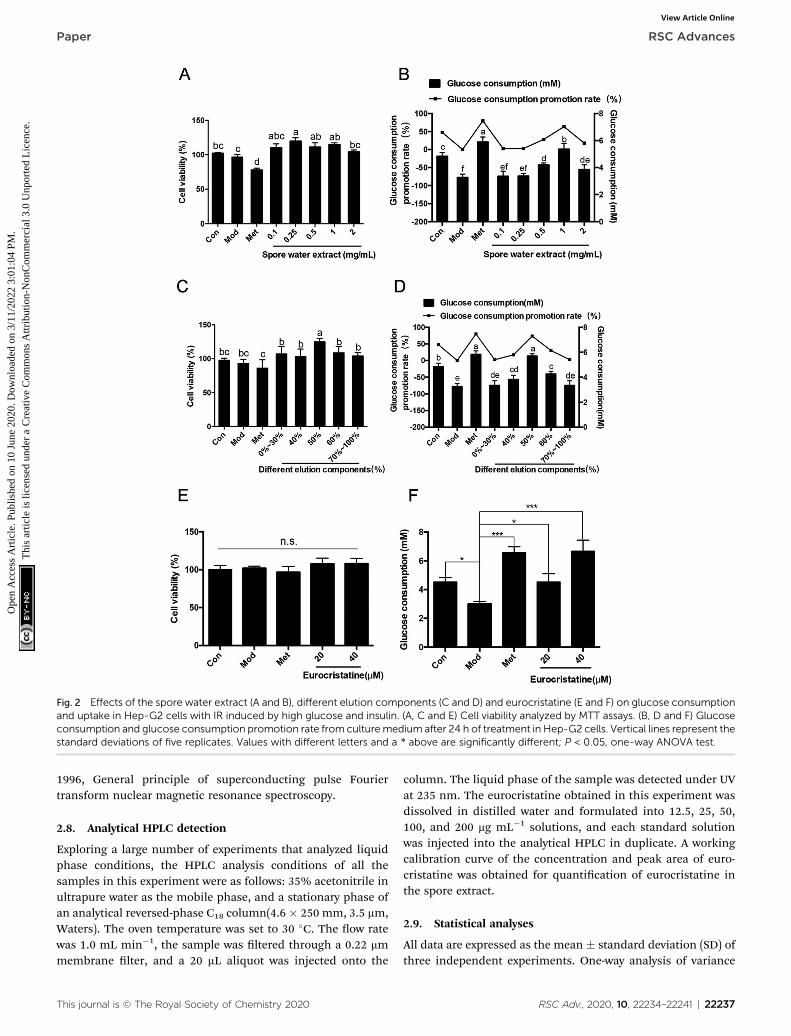
Fig. 2 Effects of the spore water extract (A and B), different elution components (C and D) and eurocristatine (E and F) on glucose consumptionand uptake in Hep-G2 cells with IR induced by high glucose and insulin. (A, C and E) Cell viability analyzed by MTT assays. (B, D and F) Glucoseconsumption and glucose consumption promotion rate from culturemedium after 24 h of treatment in Hep-G2 cells. Vertical lines represent thestandard deviations of five replicates. Values with different letters and a * above are significantly different; P < 0.05, one-way ANOVA test.
Paper RSC Advances
Ope
n A
cces
s A
rtic
le. P
ublis
hed
on 1
0 Ju
ne 2
020.
Dow
nloa
ded
on 3
/11/
2022
3:0
1:04
PM
. T
his
artic
le is
lice
nsed
und
er a
Cre
ativ
e C
omm
ons
Attr
ibut
ion-
Non
Com
mer
cial
3.0
Unp
orte
d L
icen
ce.
View Article Online
1996, General principle of superconducting pulse Fouriertransform nuclear magnetic resonance spectroscopy.
2.8. Analytical HPLC detection
Exploring a large number of experiments that analyzed liquidphase conditions, the HPLC analysis conditions of all thesamples in this experiment were as follows: 35% acetonitrile inultrapure water as the mobile phase, and a stationary phase ofan analytical reversed-phase C18 column(4.6 � 250 mm, 3.5 mm,Waters). The oven temperature was set to 30 �C. The ow ratewas 1.0 mL min�1, the sample was ltered through a 0.22 mmmembrane lter, and a 20 mL aliquot was injected onto the
This journal is © The Royal Society of Chemistry 2020
column. The liquid phase of the sample was detected under UVat 235 nm. The eurocristatine obtained in this experiment wasdissolved in distilled water and formulated into 12.5, 25, 50,100, and 200 mg mL�1 solutions, and each standard solutionwas injected into the analytical HPLC in duplicate. A workingcalibration curve of the concentration and peak area of euro-cristatine was obtained for quantication of eurocristatine inthe spore extract.
2.9. Statistical analyses
All data are expressed as the mean � standard deviation (SD) ofthree independent experiments. One-way analysis of variance
RSC Adv., 2020, 10, 22234–22241 | 22237
RSC Advances Paper
Ope
n A
cces
s A
rtic
le. P
ublis
hed
on 1
0 Ju
ne 2
020.
Dow
nloa
ded
on 3
/11/
2022
3:0
1:04
PM
. T
his
artic
le is
lice
nsed
und
er a
Cre
ativ
e C
omm
ons
Attr
ibut
ion-
Non
Com
mer
cial
3.0
Unp
orte
d L
icen
ce.
View Article Online
(ANOVA) followed by Duncan's test was performed using SPSSversion 19.0 soware (SPSS Inc, IL, USA). n ¼ 10 per group ratswere used in our study, statistical signicance was considered atp < 0.05.
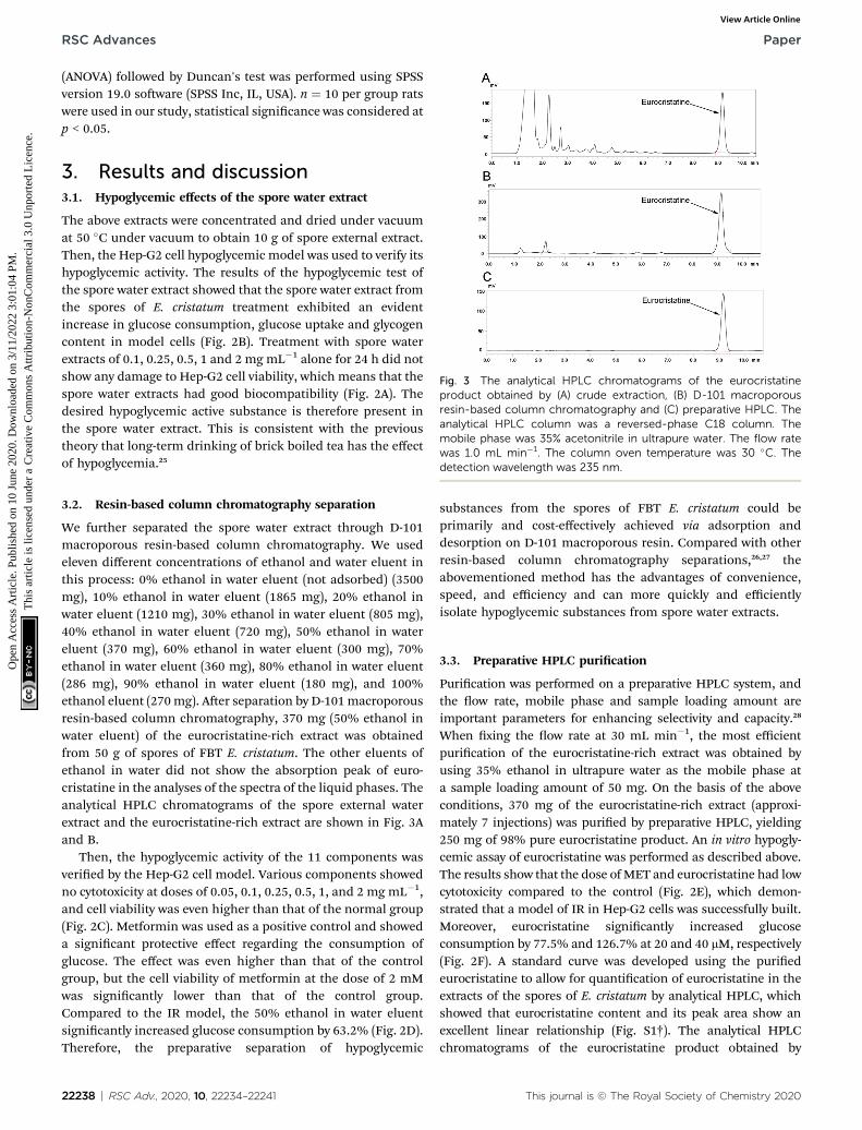
Fig. 3 The analytical HPLC chromatograms of the eurocristatineproduct obtained by (A) crude extraction, (B) D-101 macroporousresin-based column chromatography and (C) preparative HPLC. Theanalytical HPLC column was a reversed-phase C18 column. Themobile phase was 35% acetonitrile in ultrapure water. The flow ratewas 1.0 mL min�1. The column oven temperature was 30 �C. Thedetection wavelength was 235 nm.
3. Results and discussion3.1. Hypoglycemic effects of the spore water extract
The above extracts were concentrated and dried under vacuumat 50 �C under vacuum to obtain 10 g of spore external extract.Then, the Hep-G2 cell hypoglycemic model was used to verify itshypoglycemic activity. The results of the hypoglycemic test ofthe spore water extract showed that the spore water extract fromthe spores of E. cristatum treatment exhibited an evidentincrease in glucose consumption, glucose uptake and glycogencontent in model cells (Fig. 2B). Treatment with spore waterextracts of 0.1, 0.25, 0.5, 1 and 2 mg mL�1 alone for 24 h did notshow any damage to Hep-G2 cell viability, which means that thespore water extracts had good biocompatibility (Fig. 2A). Thedesired hypoglycemic active substance is therefore present inthe spore water extract. This is consistent with the previoustheory that long-term drinking of brick boiled tea has the effectof hypoglycemia.25
3.2. Resin-based column chromatography separation
We further separated the spore water extract through D-101macroporous resin-based column chromatography. We usedeleven different concentrations of ethanol and water eluent inthis process: 0% ethanol in water eluent (not adsorbed) (3500mg), 10% ethanol in water eluent (1865 mg), 20% ethanol inwater eluent (1210 mg), 30% ethanol in water eluent (805 mg),40% ethanol in water eluent (720 mg), 50% ethanol in watereluent (370 mg), 60% ethanol in water eluent (300 mg), 70%ethanol in water eluent (360 mg), 80% ethanol in water eluent(286 mg), 90% ethanol in water eluent (180 mg), and 100%ethanol eluent (270mg). Aer separation by D-101macroporousresin-based column chromatography, 370 mg (50% ethanol inwater eluent) of the eurocristatine-rich extract was obtainedfrom 50 g of spores of FBT E. cristatum. The other eluents ofethanol in water did not show the absorption peak of euro-cristatine in the analyses of the spectra of the liquid phases. Theanalytical HPLC chromatograms of the spore external waterextract and the eurocristatine-rich extract are shown in Fig. 3Aand B.
Then, the hypoglycemic activity of the 11 components wasveried by the Hep-G2 cell model. Various components showedno cytotoxicity at doses of 0.05, 0.1, 0.25, 0.5, 1, and 2 mg mL�1,and cell viability was even higher than that of the normal group(Fig. 2C). Metformin was used as a positive control and showeda signicant protective effect regarding the consumption ofglucose. The effect was even higher than that of the controlgroup, but the cell viability of metformin at the dose of 2 mMwas signicantly lower than that of the control group.Compared to the IR model, the 50% ethanol in water eluentsignicantly increased glucose consumption by 63.2% (Fig. 2D).Therefore, the preparative separation of hypoglycemic
22238 | RSC Adv., 2020, 10, 22234–22241
substances from the spores of FBT E. cristatum could beprimarily and cost-effectively achieved via adsorption anddesorption on D-101 macroporous resin. Compared with otherresin-based column chromatography separations,26,27 theabovementioned method has the advantages of convenience,speed, and efficiency and can more quickly and efficientlyisolate hypoglycemic substances from spore water extracts.
3.3. Preparative HPLC purication
Purication was performed on a preparative HPLC system, andthe ow rate, mobile phase and sample loading amount areimportant parameters for enhancing selectivity and capacity.28
When xing the ow rate at 30 mL min�1, the most efficientpurication of the eurocristatine-rich extract was obtained byusing 35% ethanol in ultrapure water as the mobile phase ata sample loading amount of 50 mg. On the basis of the aboveconditions, 370 mg of the eurocristatine-rich extract (approxi-mately 7 injections) was puried by preparative HPLC, yielding250 mg of 98% pure eurocristatine product. An in vitro hypogly-cemic assay of eurocristatine was performed as described above.The results show that the dose of MET and eurocristatine had lowcytotoxicity compared to the control (Fig. 2E), which demon-strated that a model of IR in Hep-G2 cells was successfully built.Moreover, eurocristatine signicantly increased glucoseconsumption by 77.5% and 126.7% at 20 and 40 mM, respectively(Fig. 2F). A standard curve was developed using the puriedeurocristatine to allow for quantication of eurocristatine in theextracts of the spores of E. cristatum by analytical HPLC, whichshowed that eurocristatine content and its peak area show anexcellent linear relationship (Fig. S1†). The analytical HPLCchromatograms of the eurocristatine product obtained by
This journal is © The Royal Society of Chemistry 2020
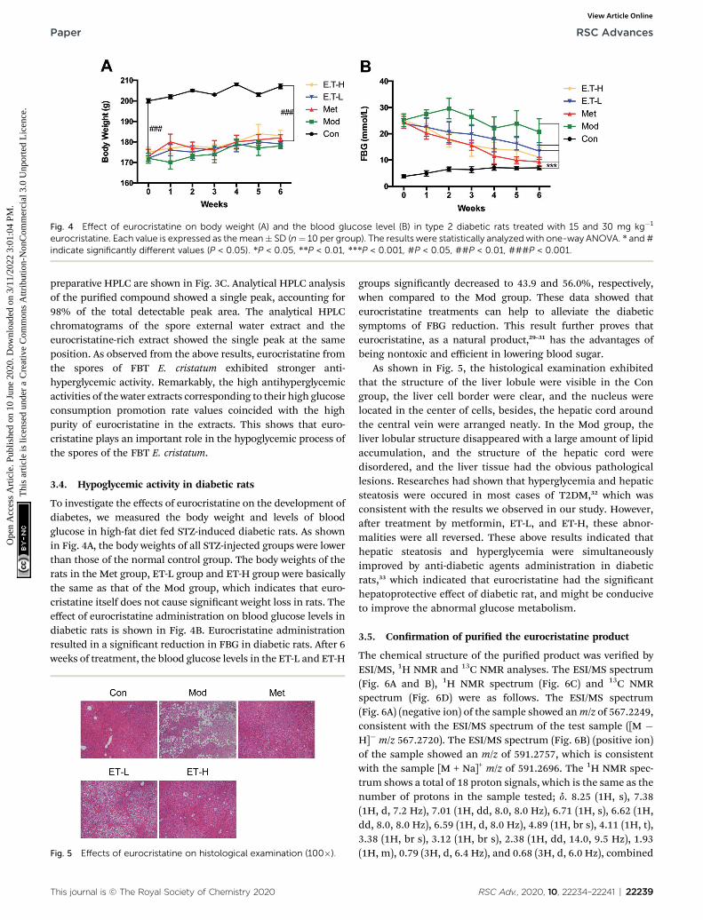
Fig. 4 Effect of eurocristatine on body weight (A) and the blood glucose level (B) in type 2 diabetic rats treated with 15 and 30 mg kg�1
eurocristatine. Each value is expressed as themean� SD (n¼ 10 per group). The results were statistically analyzed with one-way ANOVA. * and #indicate significantly different values (P < 0.05). *P < 0.05, **P < 0.01, ***P < 0.001, #P < 0.05, ##P < 0.01, ###P < 0.001.
Paper RSC Advances
Ope
n A
cces
s A
rtic
le. P
ublis
hed
on 1
0 Ju
ne 2
020.
Dow
nloa
ded
on 3
/11/
2022
3:0
1:04
PM
. T
his
artic
le is
lice
nsed
und
er a
Cre
ativ
e C
omm
ons
Attr
ibut
ion-
Non
Com
mer
cial
3.0
Unp
orte
d L
icen
ce.
View Article Online
preparative HPLC are shown in Fig. 3C. Analytical HPLC analysisof the puried compound showed a single peak, accounting for98% of the total detectable peak area. The analytical HPLCchromatograms of the spore external water extract and theeurocristatine-rich extract showed the single peak at the sameposition. As observed from the above results, eurocristatine fromthe spores of FBT E. cristatum exhibited stronger anti-hyperglycemic activity. Remarkably, the high antihyperglycemicactivities of the water extracts corresponding to their high glucoseconsumption promotion rate values coincided with the highpurity of eurocristatine in the extracts. This shows that euro-cristatine plays an important role in the hypoglycemic process ofthe spores of the FBT E. cristatum.
3.4. Hypoglycemic activity in diabetic rats
To investigate the effects of eurocristatine on the development ofdiabetes, we measured the body weight and levels of bloodglucose in high-fat diet fed STZ-induced diabetic rats. As shownin Fig. 4A, the body weights of all STZ-injected groups were lowerthan those of the normal control group. The body weights of therats in the Met group, ET-L group and ET-H group were basicallythe same as that of the Mod group, which indicates that euro-cristatine itself does not cause signicant weight loss in rats. Theeffect of eurocristatine administration on blood glucose levels indiabetic rats is shown in Fig. 4B. Eurocristatine administrationresulted in a signicant reduction in FBG in diabetic rats. Aer 6weeks of treatment, the blood glucose levels in the ET-L and ET-H
Fig. 5 Effects of eurocristatine on histological examination (100�).
This journal is © The Royal Society of Chemistry 2020
groups signicantly decreased to 43.9 and 56.0%, respectively,when compared to the Mod group. These data showed thateurocristatine treatments can help to alleviate the diabeticsymptoms of FBG reduction. This result further proves thateurocristatine, as a natural product,29–31 has the advantages ofbeing nontoxic and efficient in lowering blood sugar.
As shown in Fig. 5, the histological examination exhibitedthat the structure of the liver lobule were visible in the Congroup, the liver cell border were clear, and the nucleus werelocated in the center of cells, besides, the hepatic cord aroundthe central vein were arranged neatly. In the Mod group, theliver lobular structure disappeared with a large amount of lipidaccumulation, and the structure of the hepatic cord weredisordered, and the liver tissue had the obvious pathologicallesions. Researches had shown that hyperglycemia and hepaticsteatosis were occured in most cases of T2DM,32 which wasconsistent with the results we observed in our study. However,aer treatment by metformin, ET-L, and ET-H, these abnor-malities were all reversed. These above results indicated thathepatic steatosis and hyperglycemia were simultaneouslyimproved by anti-diabetic agents administration in diabeticrats,33 which indicated that eurocristatine had the signicanthepatoprotective effect of diabetic rat, and might be conduciveto improve the abnormal glucose metabolism.
3.5. Conrmation of puried the eurocristatine product
The chemical structure of the puried product was veried byESI/MS, 1H NMR and 13C NMR analyses. The ESI/MS spectrum(Fig. 6A and B), 1H NMR spectrum (Fig. 6C) and 13C NMRspectrum (Fig. 6D) were as follows. The ESI/MS spectrum(Fig. 6A) (negative ion) of the sample showed anm/z of 567.2249,consistent with the ESI/MS spectrum of the test sample ([M �H]� m/z 567.2720). The ESI/MS spectrum (Fig. 6B) (positive ion)of the sample showed an m/z of 591.2757, which is consistentwith the sample [M + Na]+ m/z of 591.2696. The 1H NMR spec-trum shows a total of 18 proton signals, which is the same as thenumber of protons in the sample tested; d. 8.25 (1H, s), 7.38(1H, d, 7.2 Hz), 7.01 (1H, dd, 8.0, 8.0 Hz), 6.71 (1H, s), 6.62 (1H,dd, 8.0, 8.0 Hz), 6.59 (1H, d, 8.0 Hz), 4.89 (1H, br s), 4.11 (1H, t),3.38 (1H, br s), 3.12 (1H, br s), 2.38 (1H, dd, 14.0, 9.5 Hz), 1.93(1H, m), 0.79 (3H, d, 6.4 Hz), and 0.68 (3H, d, 6.0 Hz), combined
RSC Adv., 2020, 10, 22234–22241 | 22239
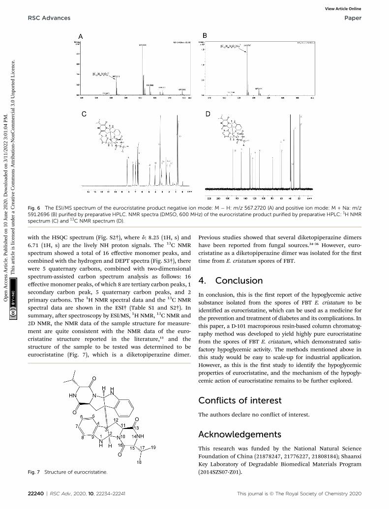
Fig. 6 The ESI/MS spectrum of the eurocristatine product negative ion mode: M � H: m/z 567.2720 (A) and positive ion mode: M + Na: m/z591.2696 (B) purified by preparative HPLC. NMR spectra (DMSO, 600 MHz) of the eurocristatine product purified by preparative HPLC: 1H NMRspectrum (C) and 13C NMR spectrum (D).
RSC Advances Paper
Ope
n A
cces
s A
rtic
le. P
ublis
hed
on 1
0 Ju
ne 2
020.
Dow
nloa
ded
on 3
/11/
2022
3:0
1:04
PM
. T
his
artic
le is
lice
nsed
und
er a
Cre
ativ
e C
omm
ons
Attr
ibut
ion-
Non
Com
mer
cial
3.0
Unp
orte
d L
icen
ce.
View Article Online
with the HSQC spectrum (Fig. S2†), where d: 8.25 (1H, s) and6.71 (1H, s) are the lively NH proton signals. The 13C NMRspectrum showed a total of 16 effective monomer peaks, andcombined with the hydrogen and DEPT spectra (Fig. S3†), therewere 5 quaternary carbons, combined with two-dimensionalspectrum-assisted carbon spectrum analysis as follows: 16effective monomer peaks, of which 8 are tertiary carbon peaks, 1secondary carbon peak, 5 quaternary carbon peaks, and 2primary carbons. The 1H NMR spectral data and the 13C NMRspectral data are shown in the ESI† (Table S1 and S2†). Insummary, aer spectroscopy by ESI/MS, 1H NMR, 13C NMR and2D NMR, the NMR data of the sample structure for measure-ment are quite consistent with the NMR data of the euro-cristatine structure reported in the literature,13 and thestructure of the sample to be tested was determined to beeurocristatine (Fig. 7), which is a diketopiperazine dimer.
Fig. 7 Structure of eurocristatine.
22240 | RSC Adv., 2020, 10, 22234–22241
Previous studies showed that several diketopiperazine dimershave been reported from fungal sources.34–36 However, euro-cristatine as a diketopiperazine dimer was isolated for the rsttime from E. cristatum spores of FBT.
4. Conclusion
In conclusion, this is the rst report of the hypoglycemic activesubstance isolated from the spores of FBT E. cristatum to beidentied as eurocristatine, which can be used as a medicine forthe prevention and treatment of diabetes and its complications. Inthis paper, a D-101 macroporous resin-based column chromatog-raphy method was developed to yield highly pure eurocristatinefrom the spores of FBT E. cristatum, which demonstrated satis-factory hypoglycemic activity. The methods mentioned above inthis study would be easy to scale-up for industrial application.However, as this is the rst study to identify the hypoglycemicproperties of eurocristatine, and the mechanism of the hypogly-cemic action of eurocristatine remains to be further explored.
Conflicts of interest
The authors declare no conict of interest.
Acknowledgements
This research was funded by the National Natural ScienceFoundation of China (21878247, 21776227, 21808184); ShaanxiKey Laboratory of Degradable Biomedical Materials Program(2014SZS07-Z01).
This journal is © The Royal Society of Chemistry 2020
Paper RSC Advances
Ope
n A
cces
s A
rtic
le. P
ublis
hed
on 1
0 Ju
ne 2
020.
Dow
nloa
ded
on 3
/11/
2022
3:0
1:04
PM
. T
his
artic
le is
lice
nsed
und
er a
Cre
ativ
e C
omm
ons
Attr
ibut
ion-
Non
Com
mer
cial
3.0
Unp
orte
d L
icen
ce.
View Article Online
References
1 W. W. May Zin, S. Buttachon, T. Dethoup, J. A. Pereira,L. Gales, A. Inacio, P. M. Costa, M. Lee, N. Sekeroglu,A. M. S. Silva, M. M. M. Pinto and A. Kijjoa, Phytochemistry,2017, 141, 86–97.
2 A. P. Almeida, T. Dethoup, N. Singburaudom, R. Lima,M. H. Vasconcelos, M. Pinto and A. Kijjoa, J. Nat. Pharm.,2010, 1, 25–29.
3 Y. Ishikawa, K. Morimoto and T. Hamasaki, J. Am. Oil Chem.Soc., 1984, 61, 1864–1868.
4 J. Gao, M. M. Radwan, F. Leon, X. Wang, M. R. Jacob,B. L. Tekwani, S. I. Khan, S. Lupien, R. A. Hill andF. M. Dugan, Med. Chem. Res., 2012, 21, 3080–3086.
5 H.-H. Li, L.-Y. Luo, J. Wang, D.-H. Fu and L. Zeng, Food Res.Int., 2019, 120, 275–284.
6 Y. Rui, P. Wan, G. Chen, M. Xie, Y. Sun, X. Zeng and Z. Liu,Lebensm.-Wiss. Technol., 2019, 110, 168–174.
7 A. Xu, Y. Wang, J. Wen, P. Liu, Z. Liu and Z. Li, Int. J. FoodMicrobiol., 2011, 146, 14–22.
8 X. Xu, H. Mo, M. Yan and Y. Zhu, J. Sci. Food Agric., 2007, 87,1502–1504.
9 Y. Xiao, K. Zhong, J.-R. Bai, Y.-P. Wu, J.-Q. Zhang and H. Gao,Lebensm.-Wiss. Technol., 2020, 117, 108629.
10 G. J. Slack, E. Puniani, J. C. Frisvad, R. A. Samson andJ. D. Miller, Mycol. Res., 2009, 113, 480–490.
11 M. Podojil, P. Sedmera, J. Vokoun, V. Betina, H. Barathova,Z. Durackova, K. Horakova and P. Nemec, Folia Microbiol.,1978, 23, 438–443.
12 O. Smetanina, A. Kalinovskii, Y. V. Khudyakova, N. Slinkina,M. Pivkin and T. Kuznetsova, Chem. Nat. Compd., 2007, 43,395–398.
13 N. M. Gomes, T. Dethoup, N. Singburaudom, L. Gales,A. M. S. Silva and A. Kijjoa, Phytochem. Lett., 2012, 5, 717–720.
14 X. Zou, Y. Li, X. Zhang, Q. Li, X. Liu, Y. Huang, T. Tang,S. Zheng, W. Wang and J. Tang, Molecules, 2014, 19,17839–17847.
15 F. Y. Du, X. M. Li, C. S. Li, S. Zhuo and B. G. Wang, Bioorg.Med. Chem. Lett., 2015, 43, 4650–4653.
16 F. Y. Du, L. Xin, L. Xiao-Ming, Z. Li-Wei andW. Bin-Gui,Mar.Drugs, 2017, 15, 24.
17 Y. Li, K. L. Sun, Y. Wang, P. Fu, P. P. Liu, C. Wang andW. M. Zhu, Chin. Chem. Lett., 2013, 24, 1049–1052.
This journal is © The Royal Society of Chemistry 2020
18 D. Kang, M. Su, Y. Duan and Y. Huang, Food Funct., 2019,10(8), 5032–5045.
19 G. Chen, M. Xie, Z. Dai, P. Wan and Y. Sun, Mol. Nutr. FoodRes., 2018, 62, 1700485.
20 Q. Huang, Microbiology, 2007, 34, 917–920.21 F. Yan, G. Dai and X. Zheng, J. Nutr. Biochem., 2016, 36, 68–
80.22 H. Yin, L. Huang, T. Ouyang and L. Chen, Int.
Immunopharmacol., 2018, 55, 55–62.23 K. Sichilongo, Z. G. Keolopile, S. Ndlovu, E. Mwando,
C. Shaba and A. Massele, Int. J. Mass Spectrom., 2016, 410,1–11.
24 R. Chera, A. Zaiter, S. Akkal, P. Chaimbault,A. B. Abdelwahab, G. Kirsch and N. Kacem Chaouche,Bioorg. Chem., 2020, 96, 103535.
25 Q. Li, Z. Liu, J. Huang, G. Luo and J. Hu, J. Sci. Food Agric.,2013, 93, 1310–1316.
26 L. Sun, Y. Guo, C. Fu, J. Li and Z. Li, Food Chem., 2013, 136,1022–1029.
27 G. Wang, W. Chen, J. Hu, B. Fan, J. Shi and J. Xu, J.Chromatogr. B: Anal. Technol. Biomed. Life Sci., 2019, 1110–1111, 43–50.
28 P. Kuang, D. Song, Q. Yuan, R. Yi, X. Lv and H. Liang, FoodChem., 2013, 136, 342–347.
29 S. Deshaware, S. Gupta, R. S. Singhal, M. Joshi andP. S. Variyar, Food Chem., 2018, 262, 78–85.
30 B. Arumugam, T. Manaharan, C. K. Heng, U. R. Kuppusamyand U. D. Palanisamy, LWT–Food Sci. Technol., 2014, 59, 707–712.
31 S. R. Naik, J. M. B. Filho, J. N. Dhuley and V. Deshmukh, J.Ethnopharmacol., 1991, 33, 37–44.
32 Z. Sun and M. A. Lazar, Trends Endocrinol. Metab., 2013, 24,4–12.
33 M.-S. Kim, S.-H. Kim, S.-J. Park, M. J. Sung, J. Park,J.-T. Hwang, H. J. Yang, S. Kim, D. Seo, S. S. Shin andH. J. Hur, J. Funct. Foods, 2017, 35, 295–302.
34 S. Cai, X. Kong, W. Wang, H. Zhou, T. Zhu, D. Li and Q. Gu,Cheminform, 2012, 53, 2615–2617.
35 R. Raju, A. M. Piggott, M. Conte, W. G. L. Aalbersberg andR. J. Capon, Org. Lett., 2009, 11, 3862–3865.
36 G. Y. Li, T. Yang, Y.-G. Luo, X.-Z. Chen, D.-M. Fang andG.-L. Zhang, Org. Lett., 2009, 11, 3714–3717.
RSC Adv., 2020, 10, 22234–22241 | 22241