Original Article Isolation, Extraction, Purification, and ...

www.ejbps.com
Chakraborty et al. European Journal of Biomedical and Pharmaceutical Sciences
116
ISOLATION, PARTIAL PURIFICATION AND CHARACTERIZATION OF A NOVEL
THERMOSTABLE LIPASE FROM SERRATIA MARCESCENS SCL1
Shaikh Rajesh Ali#, Syeda Sagufta Sultana
1, Sisir Rajak
# and Sibani Sen Chakraborty
1*
1Department of Microbiology, West Bengal State University, Kolkata, West Bengal, India.
#Department of Microbiology, Acharya Prafulla Chandra College, Kolkata, West Bengal, India.
Article Received on 26/05/2019 Article Revised on 16/06/2019 Article Accepted on 06/07/2019
1. INTRODUCTION
Extracellular microbial enzyme is now a potential need
marker for different industry due to its ability to improve
the products. These enzymes have recently found wide
industrial application globally. The hydrolytic enzymes
with their potentiality include amylase[1]
, protease[2]
,
cellulose[3]
, pectinase[4]
, xylanase[5]
, esterase[6]
,
lipase[7,8,9]
etc. have become an emerging field in applied
and industrial microbiological as well as enzyme
engineering sector. Lipases (Triacylglycerol lipase,
EC3.1.1.3) are the water soluble versatile hydrolytic
enzymes responsible for hydrolysis of triacylglycerol to
diaceyl glycerol, monoacyl glycerol, fatty acid and
glycerol in both aqueous and non-aqueous media
including their interface, and are also involved various
important reactions such as trans-esterification[10]
, inter-
esterification, aminolysis[11]
, alcoholysis.[12]
Since
discovery of lipase by Claude Berbard in 1856, scientists
focused on the isolation and characterization of lipase
producing microorganisms due to immense importance
of the extracellular enzyme in industrial field.[13,14]
Lipases can be extracted from bacteria[15,16]
, fungi[17]
,
plant[18]
and animals.[19]
Among them the bacterial
lipases have received more interest due to its consistency
in supply by easy cultivation in inexpensive media
without seasonal fluctuation. As also improvement in
product yield by genetic manipulation is possible more
over they are non-toxic, eco-friendly and green synthesis
is possible.[20,21,22]
Bacterial lipase is a multi-faceted
enzyme with lipolytic as well as esterolytic activity and
show versatility with respect to wide range of substrates,
regio-specificity[23]
, enantio-selectivity[24]
, chiral
selectivity[25]
and stability over a wide range of pH[26]
and temperature[27,28]
than the corresponding animal and
plant lipases. The bacteria of genus Bacillus[29]
,
Pseudomonas[30]
, Staphylococcus[31]
,
Chromobacterium[32]
, Achromobacter[33]
,
Burkholderia[34]
, Alacaligens[35]
, Arthobacter[36]
etc. are
widely used for production of industrially important
lipases are found to be ubiquitous and are isolated from
diverse habitats especially from industrial wastes of oil
industry[37]
, food and vegetable industry[38]
, oilseed[39]
,
oil contaminated soil[40,41]
, garbage of petroleum[42]
and
coal industry.[43]
The worldwide demand of microbial
lipases is increasing especially in developing countries
like India, China and Brazil.[44]
Microbial lipases have a
vast application in industrial field especially in waste
water treatment (detoxification and degradation of
contaminant)[45]
, food (flavour modifying enzyme)[46]
,
pharmaceutical (digestive enzyme)[47]
, cosmetic (for
removal of lipids)[48]
, leather (animal skin fat
elimination), pulp & paper industry (to remove the pitch
SJIF Impact Factor 4.918 Research Article ejbps, 2019, Volume 6, Issue 8, 116-130.
European Journal of Biomedical AND Pharmaceutical sciences
http://www.ejbps.com
ISSN 2349-8870
Volume: 6
Issue: 8
116-130
Year: 2019
*Corresponding Author: Dr. Sibani Sen Chakraborty
Department of Microbiology, West Bengal State University, Kolkata, West Bengal, India.
ABSTRACT
Thermostable lipases have become challenge for wide application in modern industrialization. The present study
aims in the isolation, partial purification and characterization of a thermostable lipase producing bacterial strain,
Serratia marcescens scl1 from medicinal waste. The bacteria isolated on Tributyrin agar plates was characterized
by different biochemical tests and confirmed by 16s-rDNA sequencing. The growth parameters of bacteria showed
best growth at 350C and pH 7.0 with 1% olive oil as carbon source after 48 hours of incubation. The bacterial
growth curve was analyzed and lipase partially purified taking 24 hour old cultures. The lipase production
analyzed with Tween 20 and Tween 80 agar plates and quantitative assay of lipase activity carried out using 2.0
mM pNPP as substrate in 50.0mM Tris-HCl, measuring absorbance at 410nm. The optimum temperature and pH
of extracellular lipase produced by the bacteria occurred at 750C and pH 8.0. The substrate saturation kinetics
showed maximum at 1.3mM [S] and activity 5.43±0.05 X10⁻²
unit/ml. The protein concentration determined is 240
µg/ml and specific activity of the enzyme is 22.58 unit/mg. The results indicate the isolate are able to produce low
cost high profile enzyme which can satisfy the requirements of future goal to modern industrialization.
KEYWORD: Thermostable lipase; 16s rDNA; KT877002; para Nitrophenyl palmitate.
www.ejbps.com
Chakraborty et al. European Journal of Biomedical and Pharmaceutical Sciences
117
from the pulp)[49]
detergent industry (hydrolysis of oil
and fats)[50]
, production of fine chemicals[51]
,
fermentation of vegetable and meat, synthesis of
surfactant and polymer, production of biodiesel[52]
etc.
The microbial lipase market is estimated to be USD
425.0 million in 2018. It is projected to reach USD 590.2
million by 2023, growing at a CAGR of 6.8% from 2018,
in terms of value (cicion prNewswire). Approximately
13 billion tons of detergent is produced each year using
1000 tons of lipases. With modernization of society, the
need for thermostable and alkali resistant lipase is in high
demand for detergent production used in machine wash
in laundry.
The bacterial lipase production is greatly influenced by
medium composition and physiological factors such as
temperature, pH, inducer etc. During bacterial lipase
production different lipidic carbon and organic nitrogen
sources are used as major controller. Considering the
ever increasing demand for extracellular industrially
important bacterial lipase with novel character, the
present study focuses on the isolation and
characterization of a thermostable lipase producing
bacterial strainSerratia marcescens scl1 andPartial
purification followed by activity analysis of lipase under
different physiological condition.
2. MATERIALS AND METHODS
2.1. Isolation and characterization of lipase producing
Strain
The lipase producing bacterial strain was isolated from
medicinal waste of a pharmaceutical industry in West
Bengal, India. Serially diluting up to 10-8
with 1 gm of
soil sample dissolved in 100 ml of saline water (0.8%)
and incubated for 30 minute at 100 rpm in laboratory
condition. 0.1ml sample of last three dilutions were
plated in nutrient agar plates and incubated for 48 hours
at 370C. The average no. of bacteria are counted using
formula as demonstrated by Niemela, S. 1983 with some
modification.[53]
Where, N is the total no. of bacteria (in terms of CFU)
per ml of isolated sample; ∑C is sum of total colonies on
all plates counted (last three plates); a is the number of
bacteria from third countable plate (10-8
dilution); b is the
number of bacteria from second countable plate (10-
7dilution); d is the number of bacteria from first
countable plate (10-6
dilution).
A replica was prepared taking 33 colonies from 10-7
diluted plate on Tributirin agar (TBA) media (containing
0.5% (w/v) peptone, 0.3% (w/v) yeastextract, 1% (v/v)
Tributyrin and 2% agar, pH 7.0) to analyze lipase
production by appearance of a clear zone after incubation
at 370C for 48 hours. The suspected colonies are further
screened by replating in TBA media and confirmed by
the appearance of clear zone on incubation. The bacterial
strain with maximum lipolytic activity was reconfirmed
by collecting the supernatant of bacteria growing in
nutrient broth under same conditions with shaking at 150
rpm and tested for zone of clearance in Tween 20 and
Tween 80 agar plates (1% Tween 20/Tween 80 with
0.02% methyl red in 2% agar).[54,55]
2.2. Identification of lipolytic strain
2.2.1. Morphological and biochemical identification
Taxonomic status of isolated bacteria was identified
according to Bergey’s Manual of Determinative
Bacteriology (Holt et al.,1994) through morphological
(Gram staining), biochemical (H2S production, Nitrate
Reduction, Catalase, Oxidase, Urease, Indole, MR
reduction, VP Test and Citrate test), fermentative
(Glucose, Sucrose, Maltose, Lactose, Mannitol,
Inositol)[56,57]
and enzyme production (Starch hydrolysis,
Lipid hydrolysis, Casine hydrolysis, Gelatin hydrolysis)
tests. The strain was further characterized by growing in
Mac Conkey agar plate, Shigella-Salmonella agar (SSA),
Hekteon entric agar (HEA) and Triple-Sugar-Iron Agar
(TSI) media. The strain was tested for different antibiotic
susceptibility.
2.2.2. Scanning Electron microscopy (SEM) study
The Scanning Electron microscopy (SEM) of isolated
strain grown in Nutrient broth for 48 hours at 370C
under 150 rpm with 1% seeding culture was
performed at Centre for Research in Nanoscience and
Nanotechnology (CRNN), Kolkata, India.
2.2.3. Identification and phylogenetic tree by 16s
rDNA sequencing
Identification of the isolated strain was confirmed by
extracting the genomic DNA of bacteria using DNA
extraction kit (Eurofinscat. no. 5224700305). The
fragment of 16S rDNA was selectively amplified from
genomic DNA by PCR using universal oligonucleotide
primers and amplicon was confirmed by single band of
1500 bp on 1% agarose gel. Forward and reverse DNA
sequencing reaction of PCR amplicon was carried out
with 16SF and 16SR primers using BDT v3.1 Cycle
sequencing kit on ABI 3730xl Genetic Analyzer (16SF
primer-CGGACGGGTGAGTAATGTCT and 16SR
primer-CTCAGACCAGCTAGGGATCG). Consensus
sequence of 16S rDNA gene was generated from forward
and reverse sequence data using aligner software. The
16S rDNA gene sequence was used to carry out BLAST
with the nrdatabase of NCBI genbank database. Based on
maximum identity score first ten sequences were selected
and aligned using multiple alignment software program
Clustal W. Distance matrix was generated using RDP
database and the phylogenetic tree was constructed using
MEGA 4.[58, 59]
2.3. Medium formulation for optimum growth and
growth kinetics
2.3.1. Growth medium formulation
The isolated strains were grown in different media and
physical environment to optimize and formulate the
www.ejbps.com
Chakraborty et al. European Journal of Biomedical and Pharmaceutical Sciences
118
growth kinetics. Three types of media with varying
composition were used for this purpose viz. nutrient
broth (Peptone 5gm, Beef extract 3 gram, NaCl 5gm,
Distilled Water 1000ml. pH-7.0), standard medium
(Peptone 5gm, Yeast extract 5gm, Glucose 5gm, NaCl
0.25 gm and MgSO4. 7H2O 0.5gm, Olive oil 5%,
Distilled Water 1000ml. pH-7.0) and production medium
(Peptone 5gm, Yeast extract 10 gram, NaCl 5gm, Oilve
oil- 1%, Distilled Water 1000ml. pH-7.0).[60]
All tests
were performed in triplicate in 250ml conical flask with
100ml medium containing 1% inoculum at 370C under
150 rpm shaking condition for different time duration
(viz. 24 hours, 48 hours and 72 hours). The production
medium (PM) showed best result and was used for
further experiment.
2.3.2. Optimum temperature and pH for bacteria
The optimum physical environment viz. temperature and
pH for isolate was analyzed in production medium (PM)
at different temperature (250C, 35
0C, 45
0C and 55
0C) and
at different pH (4, 5, 6, 7, 8, and 9) under 150rpm
shaking condition for 24hours. All growth measurements
were done by recording absorbance values at 600nm by
UV-Visible spectrophotometer (Systronic-105).
2.3.3. Growth kinetics of bacteria
The growth kinetics of bacteria was performed at
optimum temperature and pH in production medium
containing 1% inoculum at 370C under 150 rpm shaking
condition for about 32 hours recording absorbance values
at an interval of 4 hours. The generation time and growth
rate of the bacteria for the formulated medium is
calculated from exponential stage of growth.
2.4 Partial purification of lipase
2.4.1 Preparation of crude lipase enzyme
Production of lipase enzyme was carried out by growing
the bacteria Serratia marcescens scl1 in the desired
medium under optimum conditions of growth. The
culture supernatant containing crude enzyme lipase was
obtained by centrifugation of the culture broth of
Serratia marcescens scl1 at 8,000rpm for 15min at 37 0C. The protein estimation and lipase assay was
performed as described previously.
2.4.2 Ammonium sulphate fractionation
The cell free supernatant obtained in the previous step
was saturated with 30% ammonium sulphate at 40C with
continuous stirring. The ammonium sulphate fraction
was the dialysed against 50mM Tris-HCL buffer, pH 8.0
for 24hrs at 40C in a dialysis bag. The buffer used during
dialysis was changed three times during dialysis. The
concentrated partially purified enzyme obtained after
dialysis was checked for protein estimation and lipases
assay as described previously.
2.5. Medium optimization for lipase production
2.5.1. Medium and time course for lipase production
The lipase production ability of the bacteria was
analyzed in three different media viz. nutrient broth
(NB), standard media (SM) and production medium
(PM) with respect to different time course to standardize
the time required for optimum lipase production. The
best medium and suitable time for optimum lipase
production was analyzed by culturing the isolate in three
different media at optimum physical environment under
shaking condition for different time course of 24 hours,
48 hours and 72 hours. The lipase production ability was
analyzed by extracting supernatant of grown culture and
production of clear zone in Tween-agar plates.
2.5.2. Incubation temperature for lipase production
To evaluate the effect of incubation temperature on
lipase production, 1% isolate was inoculated in
production medium and incubated for 24 hours at
different temperatures viz. 250C, 35
0C, 45
0C and 55
0C
with shaking condition and then assayed for lipase
production.
2.5.3. Incubation pH for lipase production
Investigations on effect of medium pH on lipase
production were carried out by growing the bacteria in
production medium for 24 hours in different pH. The pH
of the medium was adjusted to 4, 5, 6, 7, 8 and 9 with
0.1N HCl or 0.1N NaOH and then assayed for lipase
production by spectroscopic method.
2.6. Partial purification of lipase and analysis of
lipase activity
2.6.1. Titrimetric assay of lipase
Extracellular lipase, an inducible enzyme is partially
extracted from 1% olive oil supplemented production
medium by centrifugation at 8000rpm for 5 min at room
temperature. The supernatant collected and lipolytic
activity analyzed by titrimetric method and spectroscopic
method.[61, 62]
In titration 1% olive oil was used as substrate.[63]
The
reaction mixture contained 1% olive oil, 0.1 ml
supernatant, 1.8 ml 50mM Tris-HCl buffer, pH8.0 and
was incubated at 750C for 20 min. The reaction was
terminated with ethanol- acetone (1:1) mixture and
liberated free fatty acid analysed by titration using 0.05N
NaOH with phenophathelin as indicator. The lipase
activity is calculated by free fatty acid liberated (in
micromole) per ml of supernatant (crude lipase) using
equation described by Manickam etal.
Where, Vs is amount of NaOH solution needed for
titration (ml); VB is volume of NaOH (ml) needed to
titrate control (without enzyme); N is the strength of
NaOH used (0.05N); S is the volume of total reaction
mixture (2 ml); T is the time of incubation (20 minute).
One unit of enzyme is the amount of enzyme required to
liberate 1 µmol of free fatty acid from the substrate
(olive oil) under optimal condition in one minute.
www.ejbps.com
Chakraborty et al. European Journal of Biomedical and Pharmaceutical Sciences
119
2.6.2. Spectroscopic assay of lipase
In spectroscopic method pNPP (para Nitrophenyl
palmitate) [Sigma-Aldrich/CAS Number 1492-30-4] was
used as substrate.[64]
20mM substrate stock of pNPP was
prepared in HPLC grade isopropanol. In this assay a
cocktail was prepared using 1.8ml of Tris-HCl buffer,
pH8.0; 0.1ml pNPP stock and 0.1 ml crude enzyme mix.
The resultant mixtures are incubated in water bath for 20
minute at 750C. The reaction was terminated
withethanol-acetone (1:1) mixture and analyzed amount
of para Nitrophenol (pNP) released due to enzymatic
activity analysed using spectrophotometer at 410nm. The
activity of lipase is calculated using standard curve of
pNP (2-20 mg/ml in 50mM Tris-HCl buffer, pH 8.0).
One unit (IU) of lipase activity is amount of enzyme able
to liberate 1 µmol of pNP in one minute under standard
condition.
2.6.3. Total protein assay
The total protein content of the supernatant was
determined using Bradford method (1976). 100µl of
extracted supernatant was mixed with 700µl of buffer
(Tris-HCl buffer, pH8.0) and 2000µl of commercial
Bradford reagent was added. Absorbance values were
recorded at 595nm. The standard curve is prepared using
known concentration of bovine serum albumin (BSA).
2.7. Characterization of lipase
To investigate extracellular lipase, cell was separated by
centrifugation and supernatant stored at 40C for further
use. Kinetics of enzyme was analyzed under optimal
condition. Each experiment was performed in triplicate
and mean taken for interpretation of the result.
2.7.1. Substrate saturation kinetics
The concentration of substrate is one of most important
parameter affecting rate of enzymatic reaction. During
enzymatic reaction the velocity of reaction increase as
the substrate concentration increases until saturation is
reached. To measure optimum substrate utilization,
enzyme concentration (0.1ml crude extract) and buffer
volume (1.8ml Tris-HCl, pH 8.0) was retained constant
while varying substrate concentration (pNPP in
isopropanol) from 0.1mM to 2.0mM. The reaction was
performed in ideal condition (temp. 750C) and absorption
taken at 410nm followed by termination of reaction.[65, 66]
2.7.2. Effect of temperature and temperature
optimum
The extracellular lipase activities of most bacteria are in
between 300- 50
0C. For determination of optimum
temperature, the effect of temperature on enzymatic
activity are determined at pH 8.0 at various temperatures,
i.e. 150C, 25
0C, 35
0C, 45
0C, 55
0C, 65
0C, 75
0C and 85
0C
under optimal assay condition and data compared with
standard curve.[67, 68]
2.7.3. Effect of pH on activity
The lipolytic activity of crude enzyme was determined at
pH range of 5.0-10.0 using 50mM of various buffer
solutions i.e. citrate buffer (pH 5.0-6.0); phosphate buffer
(pH 7.0); Tris-HCl buffer (8.0-9.0), Glycine-NaOH
buffer (10.0-11.0). The assays were performed in
triplicate manner at 750C for 20 minute using assay
mixture with different buffer.[69,70]
2.7.4. Determination of km and Vmax
The effect of para-nitrophenyl-palmitate (pNPP)
concentration on enzymetic activity was measured at
different concentration. The absorption data for each
pNPP concentration was used to calculate enzymatic
activity. The Lineweaver-Burk plot of above data was
used in determination of the Km and Vmax of isolated
enzyme. Km of the enzyme interprets affinity for the
substrate and a low value indicates high affinity. It is
reported that most of the industrial enzymes have Km
ranges 10-1
to 10-5
M. To determine Km and Vmax of
isolated enzyme each value of pNPP concentration and
corresponding activity is plotted in reciprocal manner to
generate to linear plot.[71, 72]
3. RESULTS AND DISCUSSION
3.1. Isolation and characterization of lipase producing
strain
The bacteria from medicinal waste of pharmaceutical
industry of West Bengal was screened quantitatively and
qualitatively by plating in nutrient agar followed by
tributyrin agar plates and selected colonies screened for
lipase production by replica plating in TBA plates. The
microbial load was 3.5 x10⁸/gm in the collecting soil
sample as calculated using the formula demonstrated by
Niemela, S. 1983. Among total 33 colonies of 10-7
diluted nutrient agar plate, 3 colonies were able to
produce clear zone on TBA media as analysed by
replating. The colonies which produced maximum zone
of clearance was screened and used for further study.
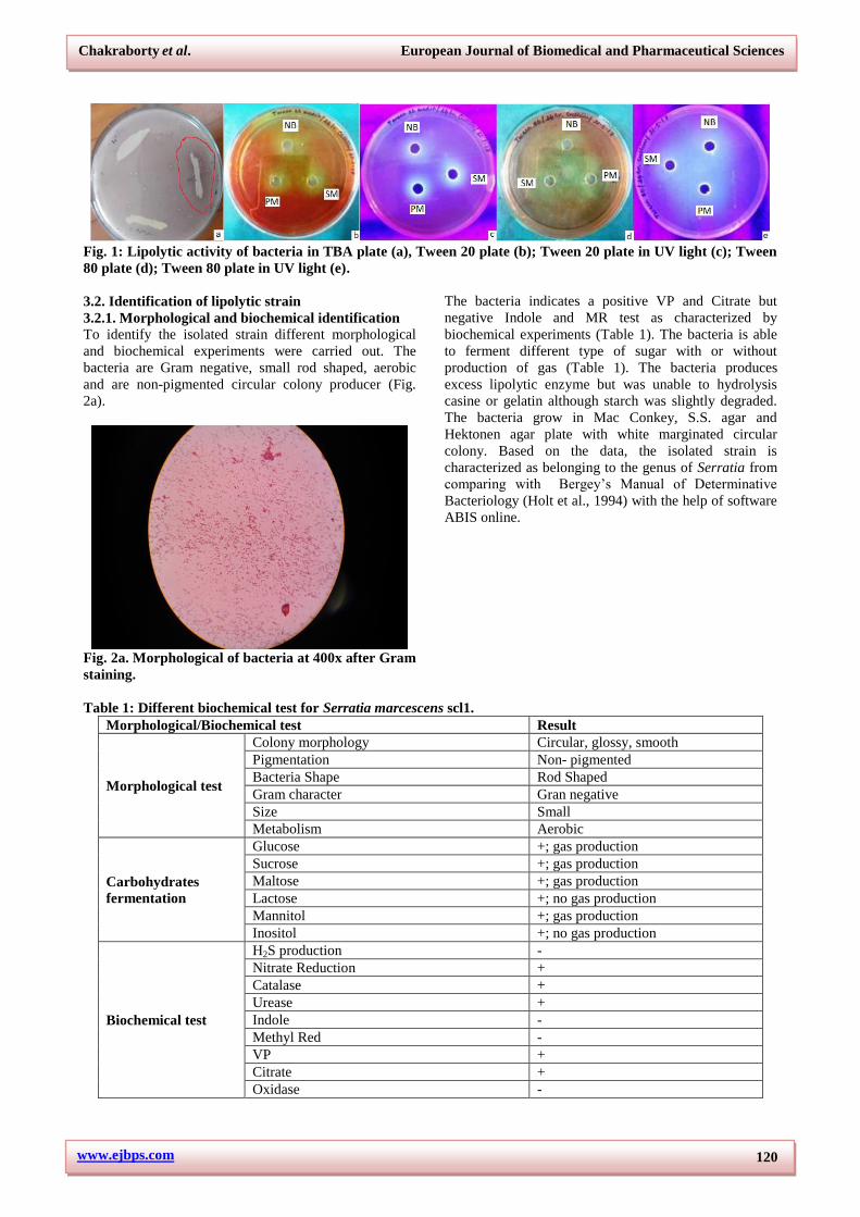
The confirmation of lipolytic/esterolytic activity of
isolated strain was performed by cup-plate method in
Tween 20 and Tween 80 agar plates using methyl red as
indicator. The supernatant collected from 48 hours
agitated culture in nutrient broth by centrifugation at
8000rpm was added to the well (100 µl) of Twee-agar
plates and incubated for 24hrs at 370C. Lipase, a
hydrolytic enzyme is responsible for the breakdown of
Tween 20/80 (fatty acid esters of polyoxyethylene
sorbitan) and results in a change in pH of the medium as
detected by change in colour of methyl red and
appearance of a halo zone under UV light (Fig.
1a,b,c,d,e).
www.ejbps.com
Chakraborty et al. European Journal of Biomedical and Pharmaceutical Sciences
120
Fig. 1: Lipolytic activity of bacteria in TBA plate (a), Tween 20 plate (b); Tween 20 plate in UV light (c); Tween
80 plate (d); Tween 80 plate in UV light (e).
3.2. Identification of lipolytic strain
3.2.1. Morphological and biochemical identification
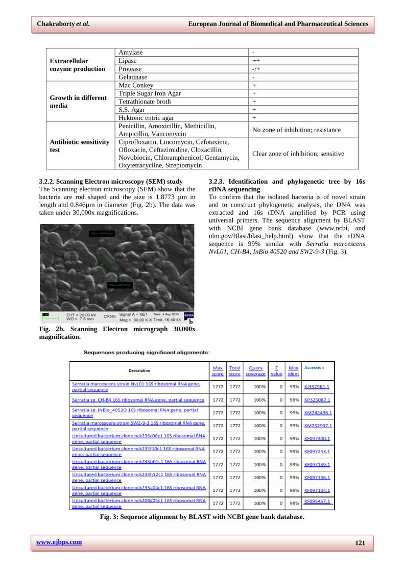
To identify the isolated strain different morphological
and biochemical experiments were carried out. The
bacteria are Gram negative, small rod shaped, aerobic
and are non-pigmented circular colony producer (Fig.
2a).
Fig. 2a. Morphological of bacteria at 400x after Gram
staining.
The bacteria indicates a positive VP and Citrate but
negative Indole and MR test as characterized by
biochemical experiments (Table 1). The bacteria is able
to ferment different type of sugar with or without
production of gas (Table 1). The bacteria produces
excess lipolytic enzyme but was unable to hydrolysis
casine or gelatin although starch was slightly degraded.
The bacteria grow in Mac Conkey, S.S. agar and
Hektonen agar plate with white marginated circular
colony. Based on the data, the isolated strain is
characterized as belonging to the genus of Serratia from
comparing with Bergey’s Manual of Determinative
Bacteriology (Holt et al., 1994) with the help of software
ABIS online.
Table 1: Different biochemical test for Serratia marcescens scl1.
Morphological/Biochemical test Result
Morphological test
Colony morphology Circular, glossy, smooth Pigmentation Non- pigmented Bacteria Shape Rod Shaped Gram character Gran negative Size Small Metabolism Aerobic
Carbohydrates
fermentation
Glucose +; gas production Sucrose +; gas production Maltose +; gas production Lactose +; no gas production Mannitol +; gas production Inositol +; no gas production
Biochemical test
H2S production - Nitrate Reduction + Catalase + Urease + Indole - Methyl Red - VP + Citrate + Oxidase -
www.ejbps.com
Chakraborty et al. European Journal of Biomedical and Pharmaceutical Sciences
121
Extracellular
enzyme production
Amylase - Lipase ++ Protease -/+ Gelatinase -
Growth in different
media
Mac Conkey + Triple Sugar Iron Agar + Tetrathionate broth + S.S. Agar + Hektonic entric agar +
Antibiotic sensitivity
test
Penicillin, Amoxicillin, Methicillin,
Ampicillin, Vancomycin No zone of inhibition; resistance
Ciprofloxacin, Lincomycin, Cefotaxime,
Ofloxacin, Ceftazimidine, Cloxacillin,
Novobiocin, Chloramphenicol, Gentamycin,
Oxytetracycline, Streptomycin
Clear zone of inhibition; sensitive
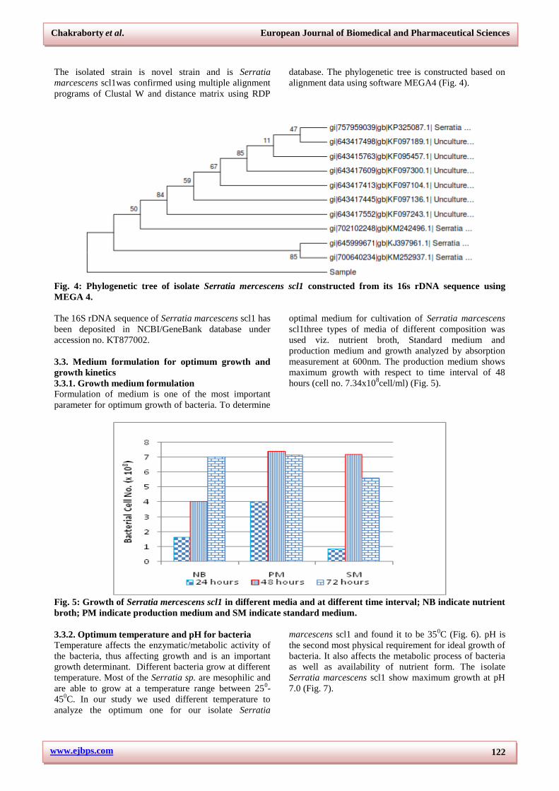
3.2.2. Scanning Electron microscopy (SEM) study
The Scanning electron microscopy (SEM) show that the
bacteria are rod shaped and the size is 1.8773 µm in
length and 0.846µm in diameter (Fig. 2b). The data was
taken under 30,000x magnifications.
Fig. 2b. Scanning Electron micrograph 30,000x
magnification.
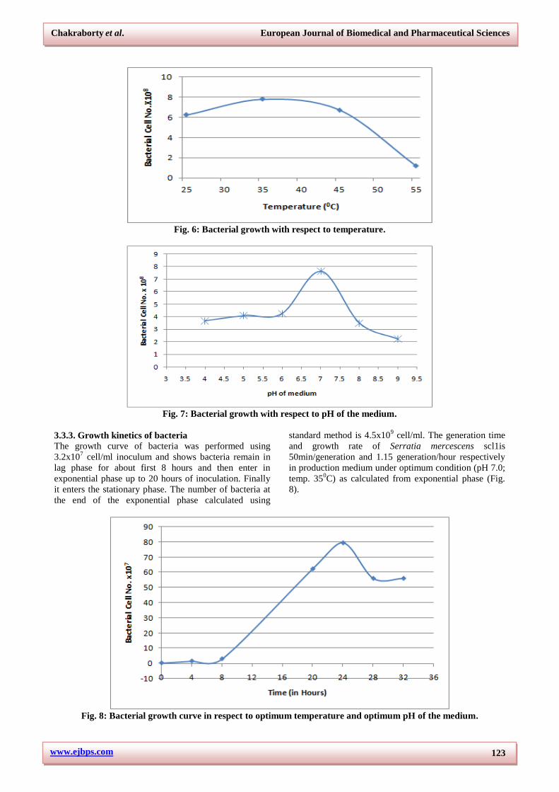
3.2.3. Identification and phylogenetic tree by 16s
rDNA sequencing
To confirm that the isolated bacteria is of novel strain
and to construct phylogenetic analysis, the DNA was
extracted and 16s rDNA amplified by PCR using
universal primers. The sequence alignment by BLAST
with NCBI gene bank database (www.ncbi, and
nlm.gov/Blast/blast_help.html) show that the rDNA
sequence is 99% similar with Serratia marcescens
NvL01, CH-B4, InBio 40520 and SW2-9-3 (Fig. 3).
Fig. 3: Sequence alignment by BLAST with NCBI gene bank database.
www.ejbps.com
Chakraborty et al. European Journal of Biomedical and Pharmaceutical Sciences
122
The isolated strain is novel strain and is Serratia
marcescens scl1was confirmed using multiple alignment
programs of Clustal W and distance matrix using RDP
database. The phylogenetic tree is constructed based on
alignment data using software MEGA4 (Fig. 4).
Fig. 4: Phylogenetic tree of isolate Serratia mercescens scl1 constructed from its 16s rDNA sequence using
MEGA 4.
The 16S rDNA sequence of Serratia marcescens scl1 has
been deposited in NCBI/GeneBank database under
accession no. KT877002.
3.3. Medium formulation for optimum growth and
growth kinetics
3.3.1. Growth medium formulation
Formulation of medium is one of the most important
parameter for optimum growth of bacteria. To determine
optimal medium for cultivation of Serratia marcescens
scl1three types of media of different composition was
used viz. nutrient broth, Standard medium and
production medium and growth analyzed by absorption
measurement at 600nm. The production medium shows
maximum growth with respect to time interval of 48
hours (cell no. 7.34x10⁸cell/ml) (Fig. 5).
Fig. 5: Growth of Serratia mercescens scl1 in different media and at different time interval; NB indicate nutrient
broth; PM indicate production medium and SM indicate standard medium.
3.3.2. Optimum temperature and pH for bacteria
Temperature affects the enzymatic/metabolic activity of
the bacteria, thus affecting growth and is an important
growth determinant. Different bacteria grow at different
temperature. Most of the Serratia sp. are mesophilic and
are able to grow at a temperature range between 250-
450C. In our study we used different temperature to
analyze the optimum one for our isolate Serratia
marcescens scl1 and found it to be 350C (Fig. 6). pH is
the second most physical requirement for ideal growth of
bacteria. It also affects the metabolic process of bacteria
as well as availability of nutrient form. The isolate
Serratia marcescens scl1 show maximum growth at pH
7.0 (Fig. 7).
www.ejbps.com
Chakraborty et al. European Journal of Biomedical and Pharmaceutical Sciences
123
Fig. 6: Bacterial growth with respect to temperature.
Fig. 7: Bacterial growth with respect to pH of the medium.
3.3.3. Growth kinetics of bacteria
The growth curve of bacteria was performed using
3.2x10⁷ cell/ml inoculum and shows bacteria remain in
lag phase for about first 8 hours and then enter in
exponential phase up to 20 hours of inoculation. Finally
it enters the stationary phase. The number of bacteria at
the end of the exponential phase calculated using
standard method is 4.5x10⁹ cell/ml. The generation time
and growth rate of Serratia mercescens scl1is
50min/generation and 1.15 generation/hour respectively
in production medium under optimum condition (pH 7.0;
temp. 350C) as calculated from exponential phase (Fig.
8).
Fig. 8: Bacterial growth curve in respect to optimum temperature and optimum pH of the medium.
www.ejbps.com
Chakraborty et al. European Journal of Biomedical and Pharmaceutical Sciences
124
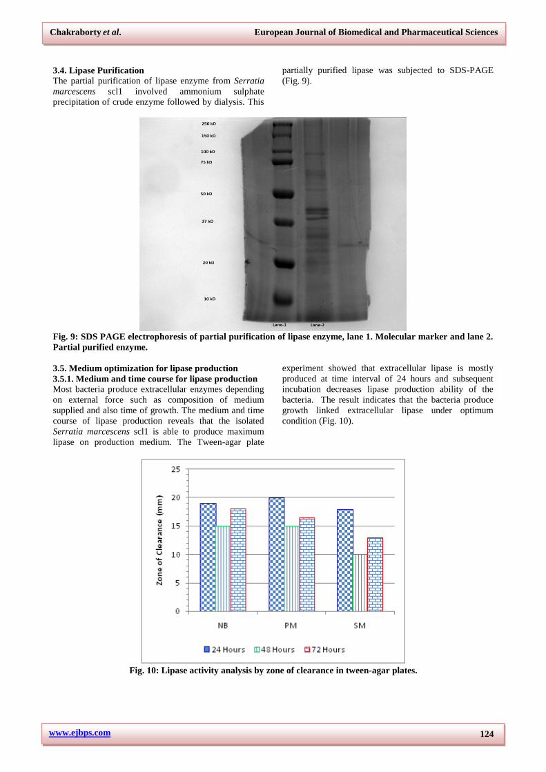
3.4. Lipase Purification
The partial purification of lipase enzyme from Serratia
marcescens scl1 involved ammonium sulphate
precipitation of crude enzyme followed by dialysis. This
partially purified lipase was subjected to SDS-PAGE
(Fig. 9).
Fig. 9: SDS PAGE electrophoresis of partial purification of lipase enzyme, lane 1. Molecular marker and lane 2.
Partial purified enzyme.
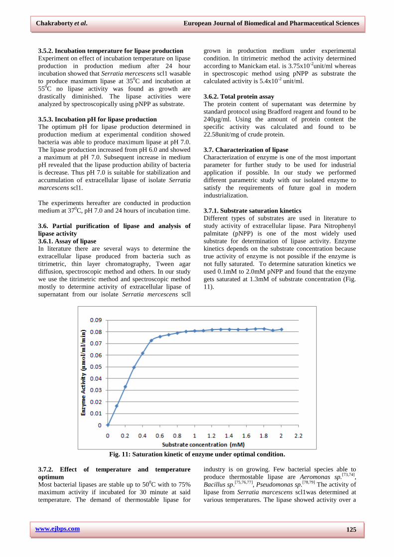
3.5. Medium optimization for lipase production
3.5.1. Medium and time course for lipase production
Most bacteria produce extracellular enzymes depending
on external force such as composition of medium
supplied and also time of growth. The medium and time
course of lipase production reveals that the isolated
Serratia marcescens scl1 is able to produce maximum
lipase on production medium. The Tween-agar plate
experiment showed that extracellular lipase is mostly
produced at time interval of 24 hours and subsequent
incubation decreases lipase production ability of the
bacteria. The result indicates that the bacteria produce
growth linked extracellular lipase under optimum
condition (Fig. 10).
Fig. 10: Lipase activity analysis by zone of clearance in tween-agar plates.
www.ejbps.com
Chakraborty et al. European Journal of Biomedical and Pharmaceutical Sciences
125
3.5.2. Incubation temperature for lipase production
Experiment on effect of incubation temperature on lipase
production in production medium after 24 hour
incubation showed that Serratia mercescens scl1 wasable
to produce maximum lipase at 350C and incubation at
550C no lipase activity was found as growth are
drastically diminished. The lipase activities were
analyzed by spectroscopically using pNPP as substrate.
3.5.3. Incubation pH for lipase production
The optimum pH for lipase production determined in
production medium at experimental condition showed
bacteria was able to produce maximum lipase at pH 7.0.
The lipase production increased from pH 6.0 and showed
a maximum at pH 7.0. Subsequent increase in medium
pH revealed that the lipase production ability of bacteria
is decrease. Thus pH 7.0 is suitable for stabilization and
accumulation of extracellular lipase of isolate Serratia
marcescens scl1.
The experiments hereafter are conducted in production
medium at 370C, pH 7.0 and 24 hours of incubation time.
3.6. Partial purification of lipase and analysis of
lipase activity
3.6.1. Assay of lipase
In literature there are several ways to determine the
extracellular lipase produced from bacteria such as
titrimetric, thin layer chromatography, Tween agar
diffusion, spectroscopic method and others. In our study
we use the titrimetric method and spectroscopic method
mostly to determine activity of extracellular lipase of
supernatant from our isolate Serratia mercescens scll
grown in production medium under experimental
condition. In titrimetric method the activity determined
according to Manickam etal. is 3.75x10⁻²
unit/ml whereas
in spectroscopic method using pNPP as substrate the
calculated activity is 5.4x10⁻²
unit/ml.
3.6.2. Total protein assay
The protein content of supernatant was determine by
standard protocol using Bradford reagent and found to be
240µg/ml. Using the amount of protein content the
specific activity was calculated and found to be
22.58unit/mg of crude protein.
3.7. Characterization of lipase
Characterization of enzyme is one of the most important
parameter for further study to be used for industrial
application if possible. In our study we performed
different parametric study with our isolated enzyme to
satisfy the requirements of future goal in modern
industrialization.
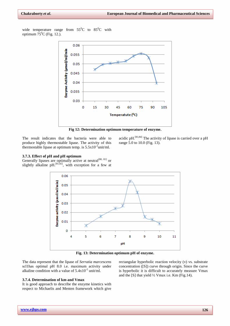
3.7.1. Substrate saturation kinetics
Different types of substrates are used in literature to
study activity of extracellular lipase. Para Nitrophenyl
palmitate (pNPP) is one of the most widely used
substrate for determination of lipase activity. Enzyme
kinetics depends on the substrate concentration because
true activity of enzyme is not possible if the enzyme is
not fully saturated. To determine saturation kinetics we
used 0.1mM to 2.0mM pNPP and found that the enzyme
gets saturated at 1.3mM of substrate concentration (Fig.
11).
Fig. 11: Saturation kinetic of enzyme under optimal condition.
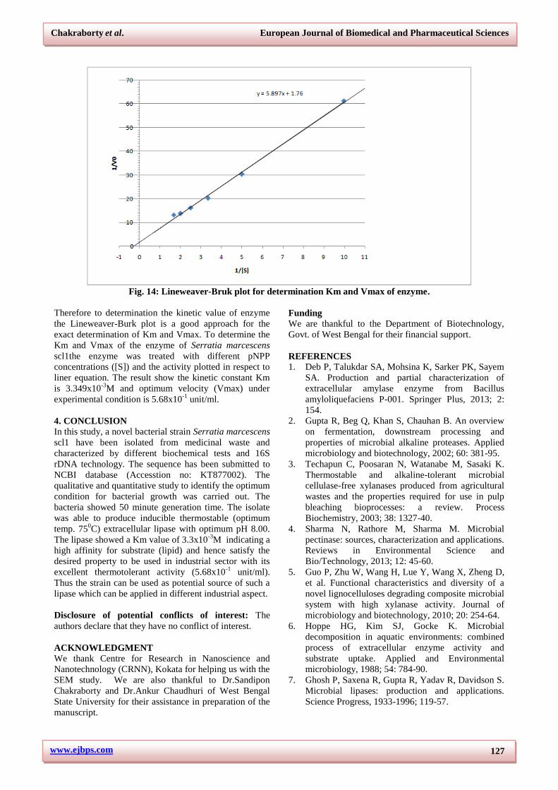
3.7.2. Effect of temperature and temperature
optimum
Most bacterial lipases are stable up to 500C with to 75%
maximum activity if incubated for 30 minute at said
temperature. The demand of thermostable lipase for
industry is on growing. Few bacterial species able to
produce thermostable lipase are Aeromonas sp.[73,74]
,
Bacillus sp.[75,76,77]
, Pseudomonas sp.[78,79]
The activity of
lipase from Serratia marcescens scl1was determined at
various temperatures. The lipase showed activity over a
www.ejbps.com
Chakraborty et al. European Journal of Biomedical and Pharmaceutical Sciences
126
wide temperature range from 550C to 85
0C with
optimum 750C (Fig. 12.).
Fig 12: Determination optimum temperature of enzyme.
The result indicates that the bacteria were able to
produce highly thermostable lipase. The activity of this
thermostable lipase at optimum temp. is 5.5x10⁻²
unit/ml.
3.7.3. Effect of pH and pH optimum
Generally lipases are optimally active at neutral[80, 81]
or
slightly alkaline pH.[81,82]
, with exception for a few at
acidic pH.[83,84]
The activity of lipase is carried over a pH
range 5.0 to 10.0 (Fig. 13).
Fig. 13: Determination optimum pH of enzyme.
The data represent that the lipase of Serratia marcescens
scl1has optimal pH 8.0 i.e. maximum activity under
alkaline condition with a value of 5.4x10⁻²
unit/ml.
3.7.4. Determination of km and Vmax
It is good approach to describe the enzyme kinetics with
respect to Michaelis and Menten framework which give
rectangular hyperbolic reaction velocity (v) vs. substrate
concentration ([S]) curve through origin. Since the curve
is hyperbolic it is difficult to accurately measure Vmax
and the [S] that yield ½ Vmax i.e. Km (Fig.14).
www.ejbps.com
Chakraborty et al. European Journal of Biomedical and Pharmaceutical Sciences
127
Fig. 14: Lineweaver-Bruk plot for determination Km and Vmax of enzyme.
Therefore to determination the kinetic value of enzyme
the Lineweaver-Burk plot is a good approach for the
exact determination of Km and Vmax. To determine the
Km and Vmax of the enzyme of Serratia marcescens
scl1the enzyme was treated with different pNPP
concentrations ([S]) and the activity plotted in respect to
liner equation. The result show the kinetic constant Km
is 3.349x10⁻³
M and optimum velocity (Vmax) under
experimental condition is 5.68x10⁻¹
unit/ml.
4. CONCLUSION
In this study, a novel bacterial strain Serratia marcescens
scl1 have been isolated from medicinal waste and
characterized by different biochemical tests and 16S
rDNA technology. The sequence has been submitted to
NCBI database (Accesstion no: KT877002). The
qualitative and quantitative study to identify the optimum
condition for bacterial growth was carried out. The
bacteria showed 50 minute generation time. The isolate
was able to produce inducible thermostable (optimum
temp. 750C) extracellular lipase with optimum pH 8.00.
The lipase showed a Km value of 3.3x10⁻³
M indicating a
high affinity for substrate (lipid) and hence satisfy the
desired property to be used in industrial sector with its
excellent thermotolerant activity (5.68x10⁻¹
unit/ml).
Thus the strain can be used as potential source of such a
lipase which can be applied in different industrial aspect.
Disclosure of potential conflicts of interest: The
authors declare that they have no conflict of interest.
ACKNOWLEDGMENT
We thank Centre for Research in Nanoscience and
Nanotechnology (CRNN), Kokata for helping us with the
SEM study. We are also thankful to Dr.Sandipon
Chakraborty and Dr.Ankur Chaudhuri of West Bengal
State University for their assistance in preparation of the
manuscript.
Funding
We are thankful to the Department of Biotechnology,
Govt. of West Bengal for their financial support.
REFERENCES
1. Deb P, Talukdar SA, Mohsina K, Sarker PK, Sayem
SA. Production and partial characterization of
extracellular amylase enzyme from Bacillus
amyloliquefaciens P-001. Springer Plus, 2013; 2:
154.
2. Gupta R, Beg Q, Khan S, Chauhan B. An overview
on fermentation, downstream processing and
properties of microbial alkaline proteases. Applied
microbiology and biotechnology, 2002; 60: 381-95.
3. Techapun C, Poosaran N, Watanabe M, Sasaki K.
Thermostable and alkaline-tolerant microbial
cellulase-free xylanases produced from agricultural
wastes and the properties required for use in pulp
bleaching bioprocesses: a review. Process
Biochemistry, 2003; 38: 1327-40.
4. Sharma N, Rathore M, Sharma M. Microbial
pectinase: sources, characterization and applications.
Reviews in Environmental Science and
Bio/Technology, 2013; 12: 45-60.
5. Guo P, Zhu W, Wang H, Lue Y, Wang X, Zheng D,
et al. Functional characteristics and diversity of a
novel lignocelluloses degrading composite microbial
system with high xylanase activity. Journal of
microbiology and biotechnology, 2010; 20: 254-64.
6. Hoppe HG, Kim SJ, Gocke K. Microbial
decomposition in aquatic environments: combined
process of extracellular enzyme activity and
substrate uptake. Applied and Environmental
microbiology, 1988; 54: 784-90.
7. Ghosh P, Saxena R, Gupta R, Yadav R, Davidson S.
Microbial lipases: production and applications.
Science Progress, 1933-1996; 119-57.
www.ejbps.com
Chakraborty et al. European Journal of Biomedical and Pharmaceutical Sciences
128
8. Hasan F, Shah AA, Hameed A. Industrial
applications of microbial lipases. Enzyme and
Microbial technology, 2006; 39: 235-51.
9. Fukuda H, Kondo A, Noda H. Biodiesel fuel
production by transesterification of oils. Journal of
bioscience and bioengineering, 2001; 92: 405-16.
10. Macrae, A. R. “Lipase-Catalyzed Interesterification
of Oils and Fats.” Journal of the American Oil
Chemists' Society, 1983; 60(2Part1): 291–294.
11. Liu Z, Chi Z, Wang L, Li J. Production, purification
and characterization of an extracellular lipase from
Aureobasidium pullulans HN2.3 with potential
application for the hydrolysis of edible oils.
Biochemical Engineering Journal, 2008; 40: 445–51.
12. Jaeger K. Bacterial lipases. FEMS Microbiology
Reviews, 1994; 15: 29–63.
13. Ghosh, P. K., et al. "Microbial lipases: production
and applications." Science Progress, (1933) -
(1996): 119-157.
14. Willerding, André Luis, et al. “Lipase Activity
among Bacteria Isolated from Amazonian Soils.”
Enzyme Research, 2011; 2011, 1–5.
15. Hasan, Fariha, et al. “Methods for Detection and
Characterization of Lipases: A Comprehensive
Review.” Biotechnology Advances, 2009; 27(6):
782–798.
16. Singh, Abhishek Kumar, and Mausumi
Mukhopadhyay. “Overview of Fungal Lipase: A
Review.” Applied Biochemistry and Biotechnology,
Oct. 2011; 166(2): 486–520.
17. Goswami, Debajyoti, et al. “Lipase Applications in
Oil Hydrolysis with a Case Study on Castor Oil: a
Review.” Critical Reviews in Biotechnology, Aug.
2012; 33(1): 81–96.
18. Francis, George, et al. “The Biological Action of
Saponins in Animal Systems: a Review.” British
Journal of Nutrition, 2002; 88(06): 587.
19. Jaeger, Karl-Erich, and Thorsten Eggert. “Lipases
for Biotechnology.” Current Opinion in
Biotechnology, 2002; 13(4): 390–397.
20. Jørgensen, S, et al. “Cloning, Sequence, and
Expression of a Lipase Gene from Pseudomonas
Cepacia: Lipase Production in Heterologous Hosts
Requires Two Pseudomonas Genes.” Journal of
Bacteriology, 1991; 173(2): 559–567.
21. Haines, Raymond Bennett. “The Influence of the
Medium on the Production of Bacterial Gelatinase.”
Biochemical Journal, 1932; 26(2): 323–336.
22. Yadav, Raman P., et al. "Purification and
characterization of a regiospecific lipase from
Aspergillus terreus." Biotechnology and applied
biochemistry, 1998; 28.3: 243-249.
Lee, S. H., et al. “Display of Bacterial Lipase on the
Escherichia Coli Cell Surface by Using FadL as an
Anchoring Motif and Use of the Enzyme in
Enantioselective Biocatalysis.” Applied and
Environmental Microbiology, Jan. 2004; 70(9):
5074–5080.
23. Lang, Dietmar A., et al. “Structural Basis of the
Chiral Selectivity of Pseudomonas Cepacia Lipase.”
European Journal of Biochemistry, 1998; 254(2):
333–340.
24. Lesuisse, Emmanuel, et al. “Purification and
Preliminary Characterization of the Extracellular
Lipase of Bacillus Subtilis 168, an Extremely Basic
PH-Tolerant Enzyme.” European Journal of
Biochemistry, 1993; 216(1): 155–160.
25. Jaeger, K. “Microbial Lipases Form Versatile Tools
for Biotechnology.” Trends in Biotechnology, Jan
1998; 16(9): 396–403.
26. Castro-Ochoa, Lelie D., et al. “Screening,
Purification and Characterization of the
Thermoalkalophilic Lipase Produced by Bacillus
Thermoleovorans CCR11.” Enzyme and Microbial
Technology, 2005; 37(6): 648–654.
27. Lesuisse, Emmanuel, et al. “Purification and
Preliminary Characterization of the Extracellular
Lipase of Bacillus Subtilis 168, an Extremely Basic
PH-Tolerant Enzyme.” European Journal of
Biochemistry, 1993; 216(1): 155–160.
28. Rosenau, F. “Bacterial Lipases from Pseudomonas:
Regulation of Gene Expression and Mechanisms of
Secretion.” Biochimie, 2000; 82(11): 1023–1032.
29. Drouault, S, et al. “Genetically Modified
Lactococcus Lactis Expressing Staphylococcus
Hyicus Lipase Enhance Lipid Digestion in Pigs with
Experimental Pancreatic Insufficiency.”
Gastroenterology, 2001; 120(5).
30. Yamaguchi, Tsutomu, et al. “Production and
Properties of Lipase from a Newly Isolated
Chromobacterium.” Agricultural and Biological
Chemistry, 1973; 37(5): 999–1005.
31. Andersson, R.e., et al. “Thermal Inactivation of a
Heat-Resistant Lipase Produced by the
Psychrotrophic Bacterium Pseudomonas
Fluorescens.” Journal of Dairy Science, 1979; 62(3):
361–367.
32. Gupta, Namita, et al. “Alkaline Lipase from a Novel
Strain Burkholderia Multivorans: Statistical Medium
Optimization and Production in a Bioreactor.”
Process Biochemistry, 2007; 42(4): 518–526.
33. Oda, Yuji, et al. “Polycaprolactone Depolymerase
Produced by the Bacterium Alcaligenes Faecalis.”
FEMS Microbiology Letters, 2006; 152(2):
339–343.
34. Sharma, Anjana, Dipa Bardhan, and Rashmi Patel.
"Optimization of physical parameters for lipase
production from Arthrobacter sp. BGCC# 490."
2009.
35. Sirisha, E., N. Rajasekar, and M. Lakshmi Narasu.
"Isolation and optimization of lipase producing
bacteria from oil contaminated soils." Advances in
Biological Research, 2010; 4.5: 249-252.
36. Dogan, B., and K. J. Boor. “Genetic Diversity and
Spoilage Potentials among Pseudomonas Spp.
Isolated from Fluid Milk Products and Dairy
Processing Plants.” Applied and Environmental
Microbiology, Jan. 2003; 69(1): 130–138.
37. Salihu, Aliyu, et al. “Lipase Production: An Insight
in the Utilization of Renewable Agricultural
www.ejbps.com
Chakraborty et al. European Journal of Biomedical and Pharmaceutical Sciences
129
Residues.” Resources, Conservation and Recycling,
2012; 58: 36–44.
38. Carreiro, M. M., et al. “Microbial Enzyme Shifts
Explain Litter Decay Responses to Simulated
Nitrogen Deposition.” Ecology, 2000; 81(9): 2359.
39. Prasad, M. P., and K. Manjunath. "Comparative
study on biodegradation of lipid-rich wastewater
using lipase producing bacterial species.", 2011.
40. S.j., Geetha, et al. “Biosurfactants: Production and
Potential Applications in Microbial Enhanced Oil
Recovery (MEOR).” Biocatalysis and Agricultural
Biotechnology, 2018; 14: 23–32.
41. Singh, Durgesh Narain, and Anil Kumar Tripathi.
“Coal Induced Production of a Rhamnolipid
Biosurfactant by Pseudomonas Stutzeri, Isolated
from the Formation Water of Jharia Coalbed.”
Bioresource Technology, 2013; 128: 215–221.
42. Abhijit Ray. “Application of Lipase in Industry”,
Asian J. Pharm. Tech, 2012; 2(2): 33-37.
43. Hasan, Fariha, et al. “Industrial Applications of
Microbial Lipases.” Enzyme and Microbial
Technology, 2006; 39(2): 235–251.
44. Aravindan, Rajendran, Palanisamy Anbumathi, and
Thangavelu Viruthagiri. "Lipase applications in food
industry." 2007.
45. Gotor-Fernández, Vicente, et al. “Lipases: Useful
Biocatalysts for the Preparation of Pharmaceuticals.”
Journal of Molecular Catalysis B: Enzymatic, 2006;
40(3-4): 111–120.
46. Ansorge-Schumacher, Marion B., and Oliver Thum.
“Immobilised Lipases in the Cosmetics Industry.”
Chemical Society Reviews, 2013; 42(15): 6475.
47. Gutiérrez, Ana, et al. “Microbial and Enzymatic
Control of Pitch in the Pulp and Paper Industry.”
Applied Microbiology and Biotechnology, 2009;
82(6): 1005–1018.
48. Rathi, Pooja, et al. “A Novel Alkaline Lipase from
Burkholderia Cepacia for Detergent Formulation.”
Process Biochemistry, 2001; 37(2): 187–192.
49. Straathof, Adrie J.j, et al. “The Production of Fine
Chemicals by Biotransformations.” Current Opinion
in Biotechnology, 2002; 13(6): 548–556.
50. Bjorkling, F. “The Future Impact of Industrial
Lipases.” Trends in Biotechnology, 1991; 9(1):
360–363.
51. Niemelä, Seppo. "Statistical evaluation of results
from quantitative microbiological examinations.
2." NMKL Rapport (Sweden). Nordisk Metodik-
Kommitte foer Livsmedel, 1983; no. 1.
52. Plou, Francisco J., et al. "Analysis of Tween 80 as
an esterase/lipase substrate for lipolytic activity
assay." Biotechnology Techniques, 1998; 12.3:
183-186.
53. Lundbeck, H., and M. O. Tirunarayanan.
“Investigations On The Enzymes And Toxins Of
Staphylococci.” Acta Pathologica Microbiologica
Scandinavica, 1966; 68(1): 123–134.
54. Brenner, Don J., et al. “Classification of Procaryotic
Organisms and the Concept of Bacterial Speciation.”
Bergey's Manual of Systematics of Archaea and
Bacteria, 2015; 1–9.
55. Lapage, S. P. “Biochemical Tests for Identification
of Medical Bacteria.” Journal of Clinical Pathology,
Jan. 1976; 29(10): 958–958.
56. Gurtler, V., and V. A. Stanisich. “New Approaches
to Typing and Identification of Bacteria Using the
16S-23S RDNA Spacer Region.” Microbiology, Jan.
1996; 142(1): 3–16.
57. Clarridge, J. E. “Impact of 16S RRNA Gene
Sequence Analysis for Identification of Bacteria on
Clinical Microbiology and Infectious Diseases.”
Clinical Microbiology Reviews, Jan. 2004; 17(4):
840–862.
58. Prasad, M. P. "Production of extracellular lipase by
Serratia marcescens isolated from industrial
effluent." Int J Curr Res Aca Rev, 2013; 1: 26-32.
59. Lee, Seoung Yong, and Joon Shick Rhee.
“Production and Partial Purification of a Lipase
from Pseudomonas Putida 3SK.” Enzyme and
Microbial Technology, 1993; 15(7): 617–623.
60. Dandavate, Vrushali, et al. “Production, Partial
Purification and Characterization of Organic Solvent
Tolerant Lipase from Burkholderia Multivorans V2
and Its Application for Ester Synthesis.”
Bioresource Technology, 2009; 100(13):
3374–3381.
61. Debadrita Paul, Sumit Saha, Sougata Pramanick,
Soham Chattopadhyay. “Standardization of process
parameters for the maximum production of
extracellular lipase by bacteria, isolated from
indigenous sources”, sep 2015; 02(06): 2395-0072.
62. Boonmahome, Patcha, and Wiyada
Mongkolthanaruk. “Lipase-Producing Bacterium
and Its Enzyme Characterization.” Journal of Life
Sciences and Technologies, 2013; 196–200.
63. Reetz, Manfred T., et al. “Expanding the Range of
Substrate Acceptance of Enzymes: Combinatorial
Active-Site Saturation Test.” Angewandte Chemie,
Apr. 2005; 117(27): 4264–4268.
64. Janssen, Anja E.m., et al. “Substrate Specificity and
Kinetics of Candida Rugosa Lipase in Organic
Media.” Enzyme and Microbial Technology, 1996;
18(5): 340–346.
65. Sirisha, E., N. Rajasekar, and M. Lakshmi Narasu.
"Isolation and optimization of lipase producing
bacteria from oil contaminated soils." Advances in
Biological Research, 2010; 4.5: 249-252.
66. Li, Hebin, and Xiaobo Zhang. “Characterization of
Thermostable Lipase from Thermophilic
Geobacillus Sp. TW1.” Protein Expression and
Purification, 2005; 42(1): 153–159.
67. Nevel, Cj Van, and Di Demeyer. “Influence of PH
on Lipolysis and Biohydrogenation of Soybean Oil
by Rumen Contents in Vitro.” Reproduction
Nutrition Development, 1996; 36(1): 53–63.
68. Freinkel, Ruth K., and Yvonne Shen. “The Origin of
Free Fatty Acids in Sebum II.” Journal of
Investigative Dermatology, 1969; 53(6): 422–427.
www.ejbps.com
Chakraborty et al. European Journal of Biomedical and Pharmaceutical Sciences
130
69. Borel, Patrick, et al. “Hydrolysis of Emulsions with
Different Triglycerides and Droplet Sizes by Gastric
Lipase in Vitro. Effect on Pancreatic Lipase
Activity.” The Journal of Nutritional Biochemistry,
1994; 5(3): 124–133.
70. Charoenpanich, Jittima, Sureeporn Suktanarag, and
Naruemon Toobbucha. "Production of a
thermostable lipase by Aeromonas sp. EBB-1
isolated from marine sludge in Angsila,
Thailand." Science Asia, 2011; 37.2: 105-114.
71. Lotrakul, Pongtharin, and Saovanee Dharmsthiti.
“Purification and Characterization of Lipase from
Aeromonas Sobria LP004.” Journal of
Biotechnology, 1997; 54(2): 113–120.
72. “Purification and Characterization of a Novel
Thermostable Lipase from Bacillus Sp.” The Journal
of Biochemistry, 1991.
73. Kim, Myung-Hee, et al. “Thermostable Lipase Of
Bacillus Stearothermophilus: High-Level
Production, Purification, and Calcium-Dependent
Thermostability.” Bioscience, Biotechnology, and
Biochemistry, 2000; 64(2): 280–286.
74. Wang, Yongxiang, et al. “Thermostable Alkaline
Lipase from a Newly Isolated Thermophilic
Bacillus, Strain A30-1 (ATCC 53841).” Journal of
Fermentation and Bioengineering, 1995; 79(5):
433–438.
75. Iizumi, Taro, et al. “Purification and
Characterization of a Thermostable Lipase from
Newly Isolated Pseudomonas Sp. KWI-56.”
Agricultural and Biological Chemistry, 1990; 54(5):
1253–1258.
76. Sugihara, Akio, et al. “Purification and
Characterization of a Novel Thermostable Lipase
from Pseudomonas Cepacia.” The Journal of
Biochemistry, 1992; 112(5): 598–603., doi:
10.1093/oxfordjournals.jbchem.a123946.
77. Dharmsthiti, S. “Production, Purification and
Characterization of Thermophilic Lipase from
Bacillus Sp. THL027.” FEMS Microbiology Letters,
1999; 179(2): 241–246.
78. Dharmsthiti, S. “Production, Purification and
Characterization of Thermophilic Lipase from
Bacillus Sp. THL027.” FEMS Microbiology Letters,
1999; 179(2): 241–246.
79. Wang, Yongxiang, et al. “Thermostable Alkaline
Lipase from a Newly Isolated Thermophilic
Bacillus, Strain A30-1 (ATCC 53841).” Journal of
Fermentation and Bioengineering, 1995; 79(5):
433–438.
80. Kim, E.-Y., et al. “Novel Cold-Adapted Alkaline
Lipase from an Intertidal Flat Metagenome and
Proposal for a New Family of Bacterial Lipases.”
Applied and Environmental Microbiology, 2008;
75(1): 257–260.
81. Ramani, K., et al. “Purification, Characterization
and Application of Acidic Lipase from
Pseudomonas Gessardii Using Beef Tallow as a
Substrate for Fats and Oil Hydrolysis.” Process
Biochemistry, 2010; 45(10): 1683–1691.
82. Gutarra, Melissa L.e., et al. “Production of an Acidic
and Thermostable Lipase of the Mesophilic Fungus
Penicillium Simplicissimum by Solid-State
Fermentation.” Bioresource Technology, 2009;
100(21): 5249–5254.
83. Ramani, K., et al. “Purification, Characterization
and Application of Acidic Lipase from
Pseudomonas Gessardii Using Beef Tallow as a
Substrate for Fats and Oil Hydrolysis.” Process
Biochemistry, 2010; 45(10): 1683–1691.