Pulmonary Function Testing Chapter 8. Pulmonary Function Testing Which of the following is the true...
53
Pulmonary Function Testing Chapter 8
-
Upload
helen-scott -
Category
Documents
-
view
245 -
download
3
Transcript of Pulmonary Function Testing Chapter 8. Pulmonary Function Testing Which of the following is the true...

Pulmonary Function Testing
Chapter 8

Pulmonary Function Testing
Which of the following is the true pulmonary function test?
• Spirometry• Lung volumes• Diffusion capacity• ABG

Pulmonary Function Testing
• Process of having the patient perform specific inspiratory and expiratory maneuvers
• Important to be familiar with these tests and values even if you do not work in a PFT lab
• Used for the following:– Medical diagnosis– Surgery related evaluation– Disability evaluation– Public Health/Research – Studying the effects of exercise on the lungs

Contraindications
• Recent abdominal, thoracic, or eye surgery• Hemodynamic instability• Symptoms of acute severe illness
– Chest pain, nausea, vomiting, high fever, dyspnea
• Recent hemoptysis• Pneumothorax• Recent history of abdominal, thoracic, or
cerebral aneurysm
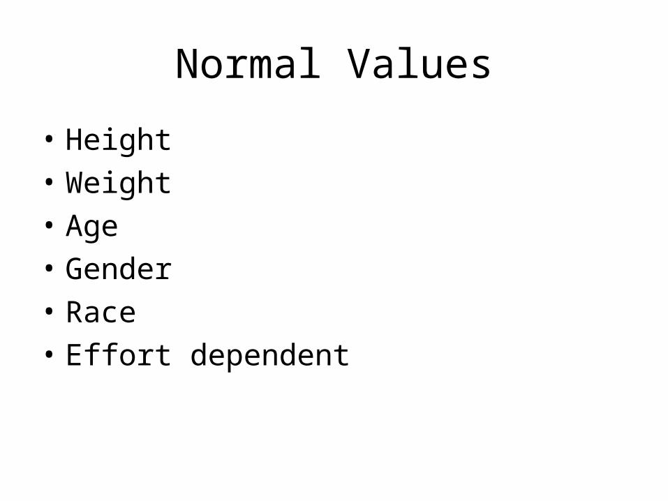
Normal Values
• Height • Weight• Age• Gender• Race• Effort dependent
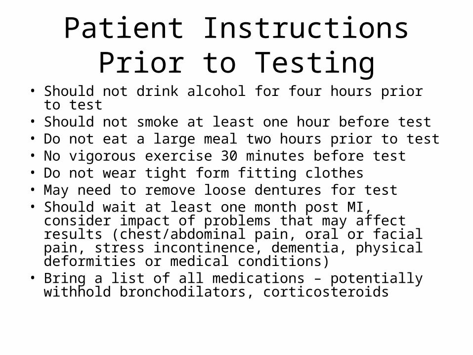
Patient Instructions Prior to Testing
• Should not drink alcohol for four hours prior to test• Should not smoke at least one hour before test• Do not eat a large meal two hours prior to test• No vigorous exercise 30 minutes before test• Do not wear tight form fitting clothes• May need to remove loose dentures for test• Should wait at least one month post MI, consider impact of
problems that may affect results (chest/abdominal pain, oral or facial pain, stress incontinence, dementia, physical deformities or medical conditions)
• Bring a list of all medications – potentially withhold bronchodilators, corticosteroids
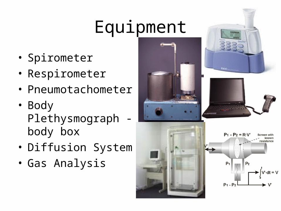
Equipment
• Spirometer• Respirometer• Pneumotachometer• Body Plethysmograph -
body box• Diffusion System• Gas Analysis
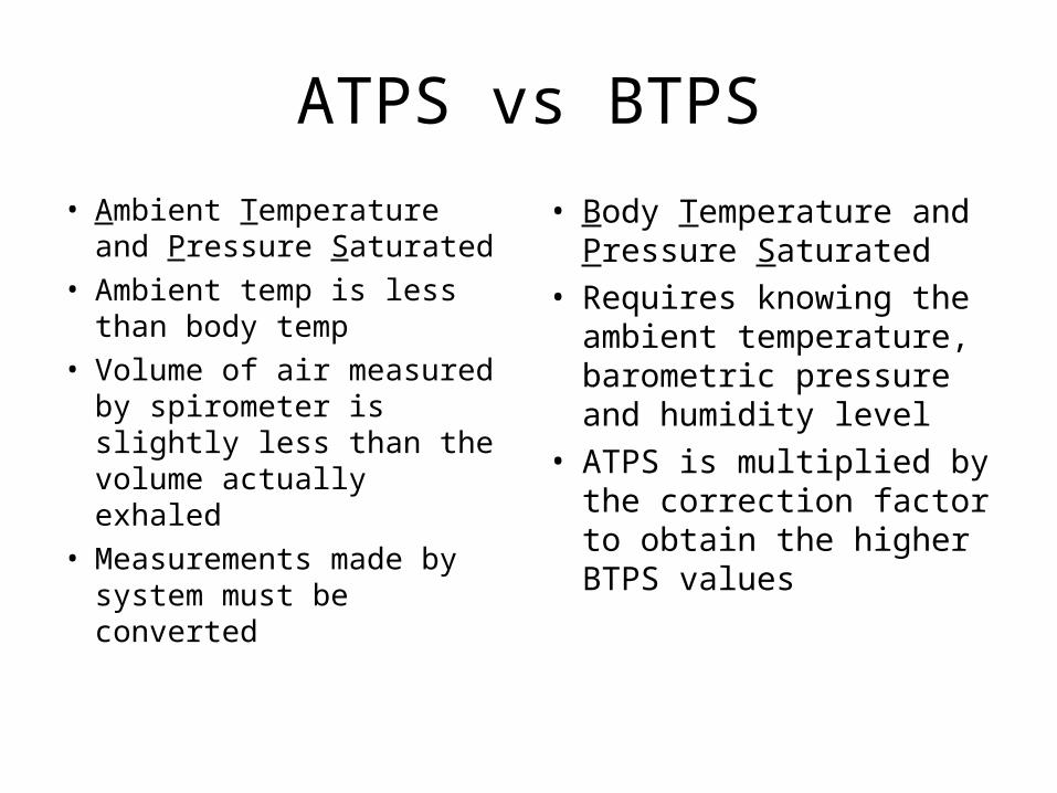
ATPS vs BTPS
• Ambient Temperature and Pressure Saturated
• Ambient temp is less than body temp
• Volume of air measured by spirometer is slightly less than the volume actually exhaled
• Measurements made by system must be converted
• Body Temperature and Pressure Saturated
• Requires knowing the ambient temperature, barometric pressure and humidity level
• ATPS is multiplied by the correction factor to obtain the higher BTPS values

Temp.ºC Corr.factor16 1.123 21 1.097 26 1.069 31 1.03917 1.118 22 1.091 27 1.063 32 1.03318 1.113 23 1.086 28 1.057 33 1.02619 1.107 24 1.080 29 1.051 34 1.02020 1.102 25 1.074 30 1.045 35 1.013
Conversion from ATPS to BTPS conditions

Classification of Lung Defects
OBSTRUCTIVE• Expiratory flow is below
normal• Anatomic site can be
identified• Diseases:
– Cystic fibrosis– Bronchitis– Asthma– Bronchiectasis– Emphysema
RESTRICTIVE• Lung volumes are reduced• Diseases:
– Neuromuscular – Cardiovascular – Pulmonary– Trauma/chest wall
dysfunction– Obesity

NORMAL SPIROGRAM

Spirogram
Volumes• Tidal Volume• Minute Volume• Residual Volume• Inspiratory Reserve Volume• Expiratory Reserve Volume
Capacities• Vital Capacity• Total Lung Capacity• Function Residual Capacity• Inspiratory Capacity

Vital Capacity
• Forced (FVC)– Requires proper
coaching– Three distinct phases– Decreased in both
obstructive and restrictive diseases
• Slow (SVC)– Helps avoid air trapping

Slow Vital Capacity

Total Lung Capacity
• Increased with obstructive disease• Decreased with restrictive disorders• Sum of the vital capacity and residual volume• Obtain RV by:
– Body plethysmography– Nitrogen washout– Helium dilution

Body Plethysmography• Uses the “body box”• Boyles Law
Unknown lung gas vol = Gas pressure of the boxKnown box gas vol Gas pressure of the lungs

In body plethysmography, the patient sits inside an airtight box, inhales or exhales to a particular volume (usually FRC), and then ashutter drops across their breathing valve. Thesubject makes respiratory efforts against the closed shutter causing their chest volume to expand and decompressing the air in their lungs. The increase in their chest volume slightly reduces the box volume and thus increases the pressure in the box. This method of measuring FRC actually measures all the conducting pathways including abdominal gas; the actual measurement made is VTG (Volume of Thoracic gas).

Nitrogen Washout
• Open circuit method• Patient breathes
100% oxygen while the nitrogen washed out of the lungs ismeasured
• Assumes 79% of lung volume is nitrogen
• Several “problems” with this test
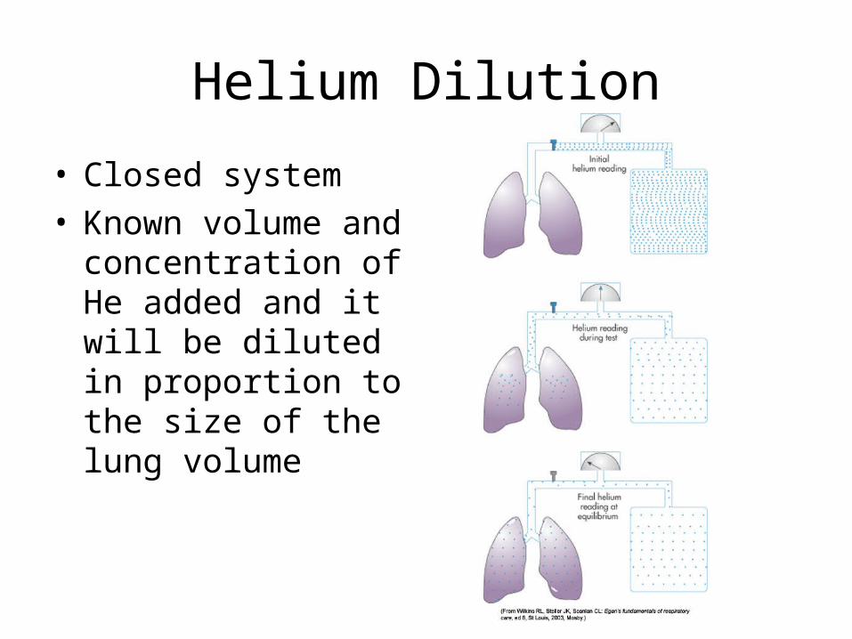
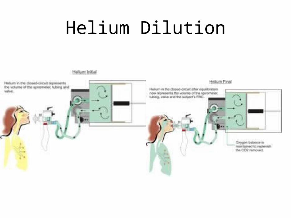
Helium Dilution
• Closed system• Known volume and
concentration of He added and it will be diluted in proportion to the size of the lung volume
Helium Dilution
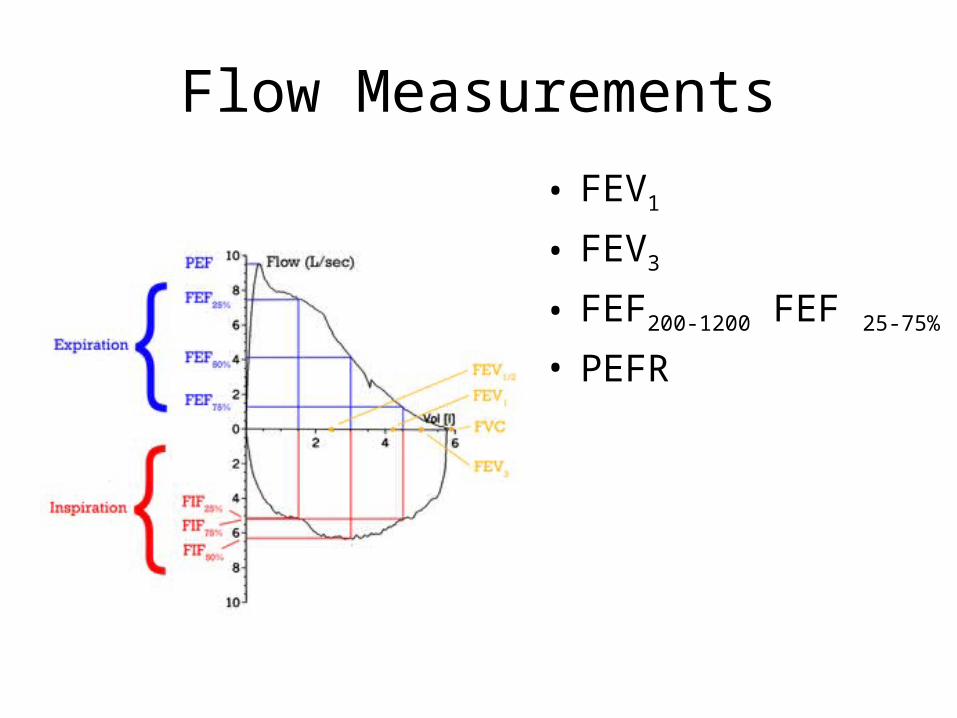
Flow Measurements
• FEV1
• FEV3
• FEF200-1200 FEF 25-75%
• PEFR

FEV1
• Maximal volume exhaled during the first second of expiration
• Best indicator of obstructive lung disease• Flow characteristics of the larger airways• Best expressed as a percentage of the FVC
(FEV1/FVC)– Should be able to exhale 70% of the vital capacity in
the first second– Decreased in obstructive disorders

FEV3
• Evaluates flow 3 seconds into expiration• Indicates flow in the smaller airways

Forced Expiratory Flow
• Examines the middle 50% of the exhaled curve
• Reflects degree of airway patency/condition of the medium to small airways
• Early indicator of obstructive dysfunction
• Normal value is 4-5 L/sec
FEF 25-75%
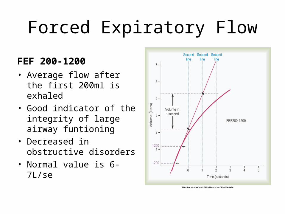
Forced Expiratory Flow
FEF 200-1200• Average flow after the first
200ml is exhaled• Good indicator of the
integrity of large airway funtioning
• Decreased in obstructive disorders
• Normal value is 6-7L/se
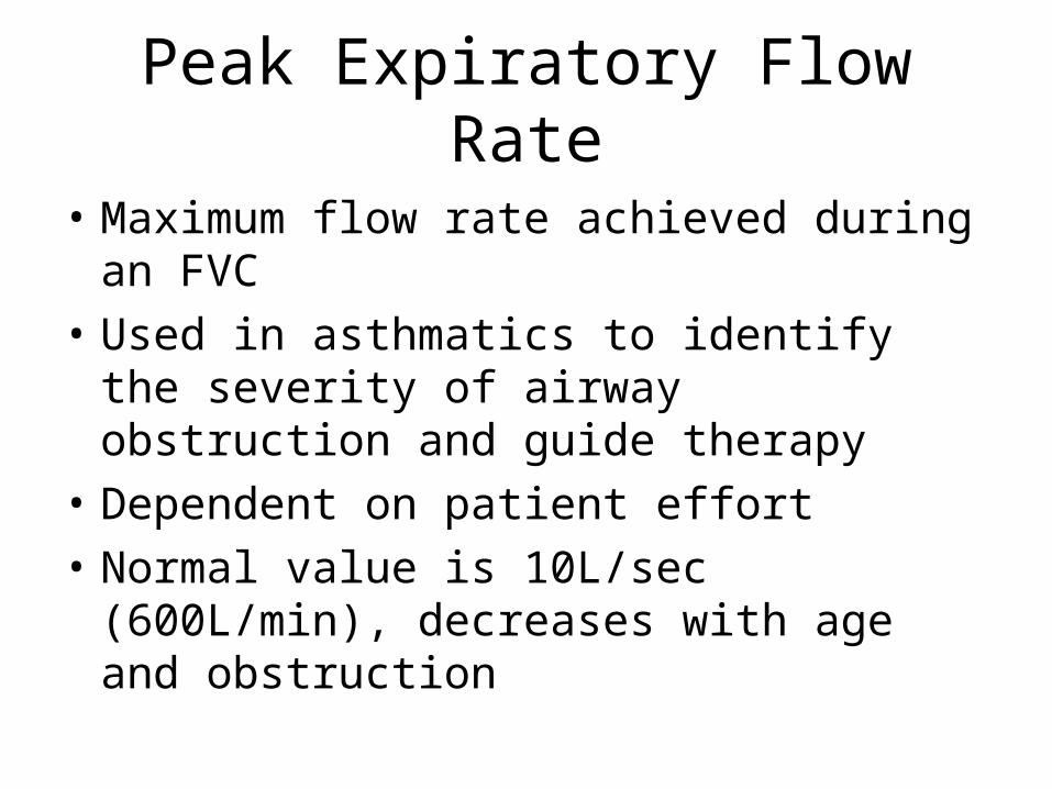
Peak Expiratory Flow Rate
• Maximum flow rate achieved during an FVC• Used in asthmatics to identify the severity of
airway obstruction and guide therapy• Dependent on patient effort• Normal value is 10L/sec (600L/min), decreases
with age and obstruction

Maximum Voluntary Ventilation• MVV – patient breathes as
fast and deep as possible for 12-15 seconds
• Tests for overall lung function, ventilatory reserve capacity and air trapping
• Normal = 170L/min• Decreased in obstructive
disorders
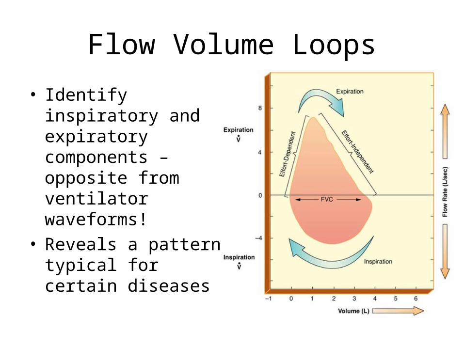
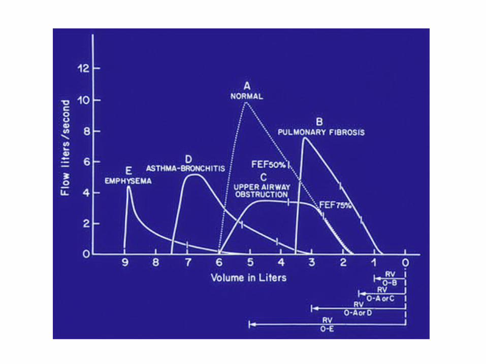
Flow Volume Loops
• Identify inspiratory and expiratory components – opposite from ventilator waveforms!
• Reveals a pattern typical for certain diseases

Graphic Representation of Values
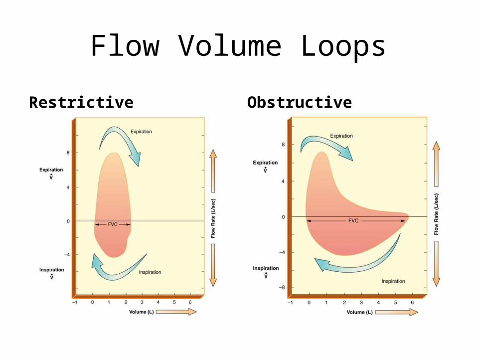
Flow Volume Loops
Restrictive Obstructive

Figure 08-07. Flow volume loop. These flow volume loops are typical patterns seen with (A) normal, (B) restrictive lung diseases, (C) upper airway
obstruction, and (D) severe chronic obstructive lung disease


Flow-volume loops of (a) fixed upper airway obstruction, (b) variable extrathoracic upper airway obstruction, and (c) variable intrathoracic upper airway obstruction.
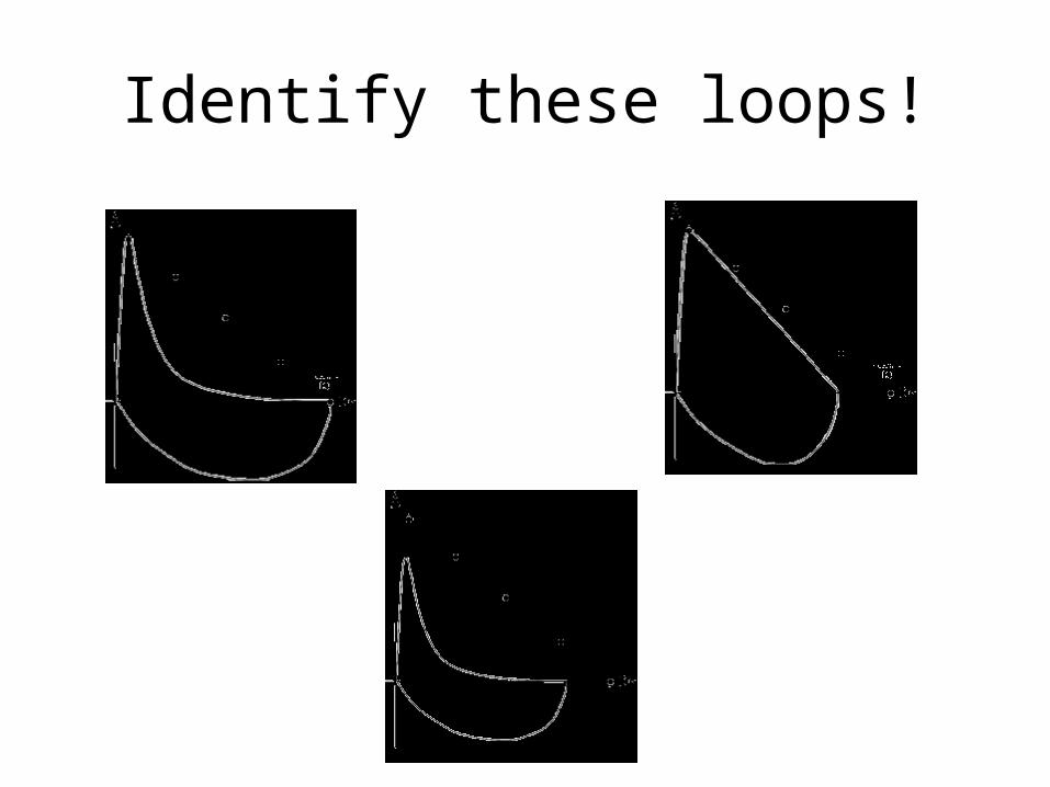
Identify these loops!


Bronchodilators
• Test before and after to assess the degree of reversibility of the airway obstruction
• Medication is not standardized• A positive response is demonstrated by:
– FVC increase >10%– FEV1 increase of 200ml or 15% over baseline
– FEF25-75% 20%-30% increase
• Often given a trial even if no response is seen
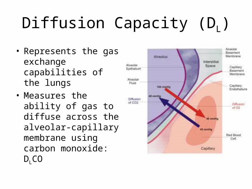
Diffusion Capacity (DL)
• Represents the gas exchange capabilities of the lungs
• Measures the ability of gas to diffuse across the alveolar-capillary membrane using carbon monoxide: DLCO

DLCO
• Diseases that reduce surface area – DL
– emphysema
• Interstitial altering of the membrane integrity - DL
– Pulmonary fibrosis, Asbestosis, Sarcoidosis
Other Studies
• Airway Resistance– Quantifying allows
understanding of the severity of the disease
– Measured using plethysomograph
• Compliance Studies– Identifies the relative
stiffness of the lung– Esophageal balloon
catheter
• Nitrogen Washout– Determines if there is
gross maldistribution of ventilation
• Closing Volume– Used for diagnosis of
small airway obstruction
• Respiratory Quotient– Determines the amount of
carbon dioxide produced and oxygen consumed

Exercise Testing
• 6 minute walk test• Anaerobic threshold• Exercise challenge• Ventilatory Capacity

Bronchoprovocation Testing
• Used to diagnose “occult” asthma• Challenge the patient with an inhaled
bronchoconstrictor – Methacholine (also can use cold air or exercise)
• Object is to determine the minimum level that elicits a 20% decrease in FEV1
• Requires bronchodilator ready for use as well as resuscitation equipment!
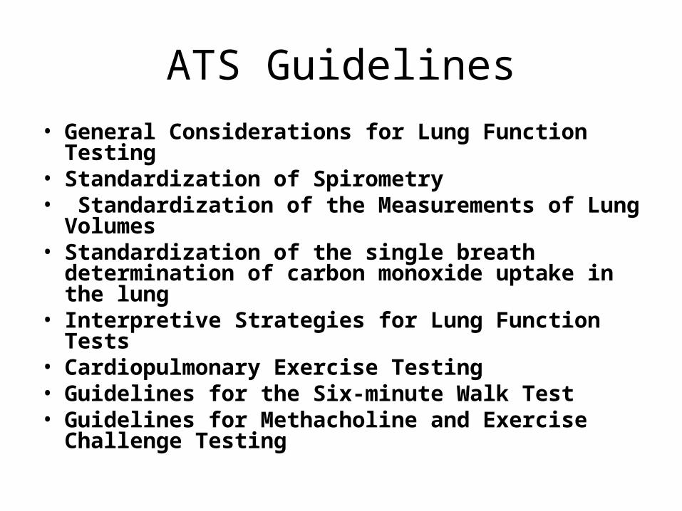
ATS Guidelines• General Considerations for Lung Function Testing• Standardization of Spirometry• Standardization of the Measurements of Lung Volumes• Standardization of the single breath determination of
carbon monoxide uptake in the lung• Interpretive Strategies for Lung Function Tests• Cardiopulmonary Exercise Testing• Guidelines for the Six-minute Walk Test• Guidelines for Methacholine and Exercise Challenge
Testing
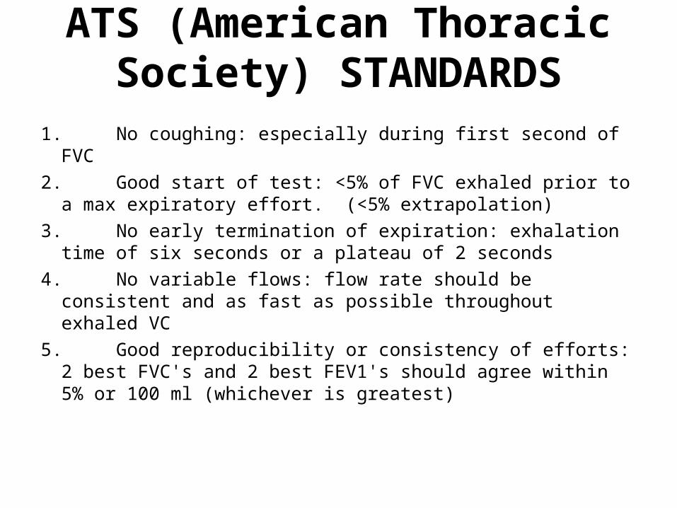
ATS (American Thoracic Society) STANDARDS
1. No coughing: especially during first second of FVC2. Good start of test: <5% of FVC exhaled prior to a max
expiratory effort. (<5% extrapolation)3. No early termination of expiration: exhalation time of six
seconds or a plateau of 2 seconds4. No variable flows: flow rate should be consistent and as fast
as possible throughout exhaled VC5. Good reproducibility or consistency of efforts: 2 best FVC's
and 2 best FEV1's should agree within 5% or 100 ml (whichever is greatest)

Evaluation/Interpretation of PFT’sINTERPRETATION CRITERIATEST NORMAL MILD MODERATE SEVEREFVC >80% 61-80% 50-60% <50% RestrictionFEV1 >80% 61-80% 50-60% <50% ObstructionPEFR >80% 61-80% 50-60% <50%FEF25-75 >80% 61-80% 50-60% <50% Small Airway DiseaseFEV1/FVC 70-75% 60-69% 50-59% <50% Obstruction
POSITIVE RESPONSE TO BRONCHODILATOR1. FVC: increase greater than 10%2. FEV1: increase of 200cc or 15% over baseline3. FEF25-75: 20% increase4. 2 out of 3 should improve to indicate a positive response
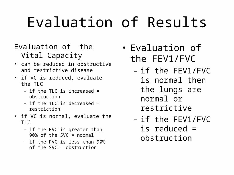
Evaluation of Results
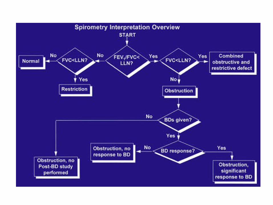
Evaluation of the Vital Capacity
• can be reduced in obstructive and restrictive disease
• if VC is reduced, evaluate the TLC– if the TLC is increased =
obstruction– if the TLC is decreased =
restriction
• if VC is normal, evaluate the TLC– if the FVC is greater than 90% of
the SVC = normal– if the FVC is less than 90% of the
SVC = obstruction
• Evaluation of the FEV1/FVC– if the FEV1/FVC is
normal then the lungs are normal or restrictive
– if the FEV1/FVC is reduced = obstruction
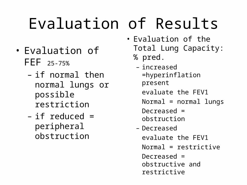
Evaluation of Results
• Evaluation of FEF 25-75%
– if normal then normal lungs or possible restriction
– if reduced = peripheral obstruction
• Evaluation of the Total Lung Capacity: % pred.– increased
=hyperinflation presentevaluate the FEV1Normal = normal lungsDecreased = obstruction
– Decreased evaluate the FEV1Normal = restrictiveDecreased = obstructive and restrictive
Evaluation of Results
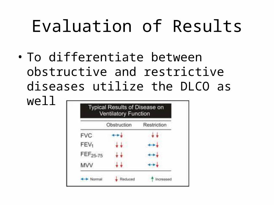
• To differentiate between obstructive and restrictive diseases utilize the DLCO as well
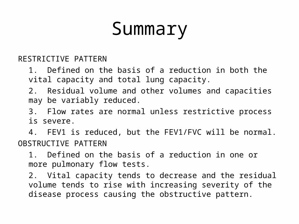
SummaryRESTRICTIVE PATTERN
1. Defined on the basis of a reduction in both the vital capacity and total lung capacity.2. Residual volume and other volumes and capacities may be variably reduced.3. Flow rates are normal unless restrictive process is severe.4. FEV1 is reduced, but the FEV1/FVC will be normal.
OBSTRUCTIVE PATTERN1. Defined on the basis of a reduction in one or more pulmonary flow tests.2. Vital capacity tends to decrease and the residual volume tends to rise with increasing severity of the disease process causing the obstructive pattern.
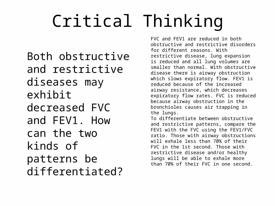
Critical Thinking
Both obstructive and restrictive diseases may exhibit decreased FVC and FEV1. How can the two kinds of patterns be differentiated?
FVC and FEV1 are reduced in both obstructive and restrictive disorders for different reasons. With restrictive disease, lung expansion is reduced and all lung volumes are smaller than normal. With obstructive disease there is airway obstruction which slows expiratory flow. FEV1 is reduced because of the increased airway resistance, which decreases expiratory flow rates. FVC is reduced because airway obstruction in the bronchioles causes air trapping in the lungs. To differentiate between obstructive and restrictive patterns, compare the FEV1 with the FVC using the FEV1/FVC ratio. Those with airway obstructions will exhale less than 70% of their FVC in the 1st second. Those with restrictive disease and/or healthy lungs will be able to exhale more than 70% of their FVC in one second.

Critical ThinkingA patient has spirometry and lung volumes typical of the obstructive pattern. The FEV1, FEV1/FVC and FEF's are significantly reduced and the FRC and TLC are increased. Two common obstructive diseases are chronic bronchitis and pulmonary emphysema. How can pulmonary function data differentiate between these two diseases?
THE DLCO! Chronic bronchitis involves mostly airways and is characterized by chronic inflammation of the mucosa, excessive mucus, and bronchospasm; all of which narrow the airways. Pulmonary emphysema primarily involves alveolar structures and is characterized by destruction of alveolar architecture, elastic fibers, and the alveolar capillary membrane. Emphysema decreases gas exchange surface area. Chronic bronchitis does not involve alveoli and therefore does not change surface area for gas exchange. A decreased diffusion capacity is associated with emphysema
Critical Thinking
In the advanced stages of pulmonary emphysema, the FRC and the RV are increased; in addition the VC is often decreased. Why do these changes occur?
Emphysema is characterized by a destruction of elastic tissue in the lung, which causes a lower lung recoil force. When lung recoil forces decrease, as in emphysema, chest wall expansion forces predominate, the chest wall expands outward pulling the lung with it. A new equilibrium occurs at increased lung volume so the FRC is increased. The RV is increased in emphysema because the VC is decreased because of small airway obstruction. When a person with emphysema tries to exhale completely, his or her bronchioles collapse, trapping air in the lungs. Increased FRC = hyperinflation; Increased RV = air trapping