Pulmonary Function Testing - Duke University
42
Pulmonary Function Testing Neil MacIntyre MD Duke University Medical Center Durham NC
Transcript of Pulmonary Function Testing - Duke University
Pulmonary Function
Testing
Neil MacIntyre MD
Duke University Medical Center
Durham NC
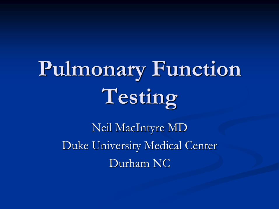
Normal structure/function
Branching airway system (23 divisions)
1st 5-6 generations cartilagenous, smooth muscle,
mucus glands
More distal airways membranous
300,000,000 alveoli with surface area that of a
tennis court
Pulmonary capillary blood volume 100-200 ml at
any given time
aao9
Callout
Deeper in the lungs we lose the cartilage and the mucus. Airways become collapsible. They actually collapse in disease states (atelectasis)
aao9
Text Box
The "choke point" is the part of the airway that most considerably resists airflow. At low volumes, the choke point (i.e. Point of Maximum Resistance - PMR) is in the small airways. They aren't full of air so they're collapsible. When the lung is full, the distal airways lose their collapsibility and the PMR is in the larger airwars.
aao9
Text Box
Blood thickness in the lung is only one cell thick because we spread the blood out over the large surface area. This "thinness" is important for gaseous exchange.
aao9
Text Box
Lungs start off as single tubes and branch off 23 times!
Normal structure/function
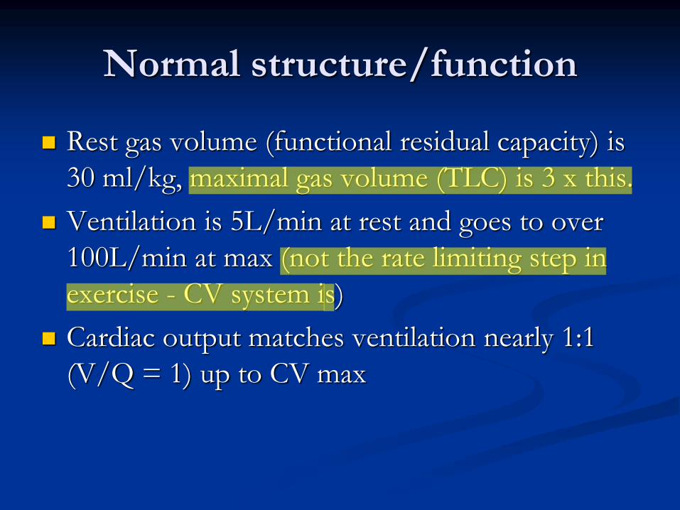
Rest gas volume (functional residual capacity) is
30 ml/kg, maximal gas volume (TLC) is 3 x this.
Ventilation is 5L/min at rest and goes to over
100L/min at max (not the rate limiting step in
exercise - CV system is)
Cardiac output matches ventilation nearly 1:1
(V/Q = 1) up to CV max
aao9
Callout
Rest point of the lung = Volume of gas in the lung when you take the lungs out of a person (i.e. eliminate the contribution from chest muscles)
aao9
Highlight
aao9
Highlight
aao9
Text Box
Lungs will not make you stop exercising. The heart will. Doesn't make much sense to "train your lungs"
aao9
Text Box
Altogether the lungs match ventilation (air flow) and perfusion (blood flow) very well. The top of the lung has more ventilation than perfusion and the bottom has more perfusion than ventilation.... but on average matching is almost perfect.
aao9
Highlight

Pulmonary Function Testing
Goals of PFTs
Normal values - interpretive principles
Spirometry
Lung volumes
Diffusing capacity
aao9
Rectangle
aao9
Text Box
Important for understanding the value of PFT
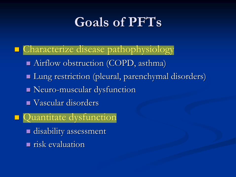
Goals of PFTs
Characterize disease pathophysiology
Airflow obstruction (COPD, asthma)
Lung restriction (pleural, parenchymal disorders)
Neuro-muscular dysfunction
Vascular disorders
Quantitate dysfunction
disability assessment
risk evaluation
aao9
Text Box
Restriction makes the lungs stiffer
aao9
Text Box
How bad is it? Describe impairment compared to normal values. Therapy is based on deviation from the norm. This is also important for analyzing risk. He gave surgery as an example. For any procedure that requires anesthesia, we have to analyze for risk since some drugs cause pulmonary toxicity. We can predict the patients who are susceptible and also follow them post-op to see how they're doing when we do start treatment
aao9
Text Box
Why PFT: 1. Put people in "physiological buckets" - Categorize according to disease states 2. Determine how ill our patients are and/or how likely they are to become ill (risk)
aao9
Text Box
Categorize patients
aao9
Highlight
aao9
Highlight
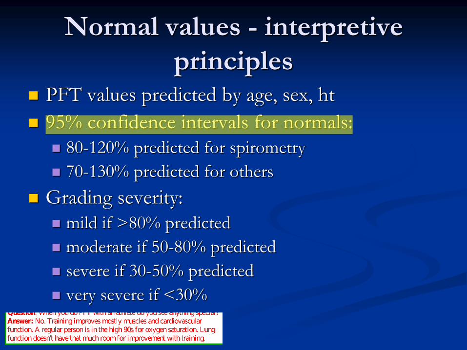
Normal values - interpretive
principles PFT values predicted by age, sex, ht
95% confidence intervals for normals:
80-120% predicted for spirometry
70-130% predicted for others
Grading severity:
mild if >80% predicted
moderate if 50-80% predicted
severe if 30-50% predicted
very severe if <30%
aao9
Rectangle
aao9
Text Box
What affects PFT: Age: PFT values peak during the late teens Sex: Different b/w men and women Height: "Just because of physics..." -------------- NB. Race and ethnicity probably play a role but we don't know yet. Most of the info we have now is derived from caucasians.
aao9
Callout
I.e Normal is where 95% of the population exists. Note that 5% of normal people will be "abnormal." Just the way it works.
aao9
Callout
Usually, 95% confidence intervals are sufficiently represented in this rage of 80% to 120% for spirometry
aao9
Text Box
These grading rules are arbitrary (convenient mathematically)
aao9
Text Box
Question: When you do PFT with an athlete do you see anything special? Answer: No. Training improves mostly muscles and cardiovascular function. A regular person is in the high 90s for oxygen saturation. Lung function doesn't have that much room for improvement with training.
aao9
Highlight

Pulmonary Function Testing
Goals of PFTs
Normal values - interpretive principles
Spirometry
Lung volumes
Diffusing capacity
aao9
Rectangle
aao9
Text Box
These 3 tests are 99% of work you do in PFT.

Spirometry
Oldest clinical test still in use today - John
Hutchinson in 1848 still has largest collection of
normal values
Patient inhales to total lung capacity and then
completely exhales as rapidly as possible
American Thoracic Society has comprehensive
performance standards
aao9
Callout
This guy put a pair of buckets in each other, had people blow into tubes (after taking a deep breath) and decided on vital capacity
aao9
Rectangle
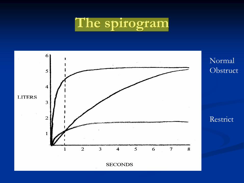
The spirogram
Normal
Obstruct
Restrict
aao9
Text Box
... don't use buckets anymore. Now use little flow censors.
aao9
Highlight
aao9
Callout
Start here with deep breath (full lung)
aao9
Text Box
Breath as rapidly as possible. Should go for 6 seconds. If the patient doesn't go for at least 6 seconds, we might need to repeat.
aao9
Callout
Volume of curve at this line is the volume exhaled after 1s (FEV1)
aao9
Text Box
The ratio FEV1/FVC is always an important value to have. ---- Most mammals can get 70% of vital capacity out in the first second. Irrespective of height, weight, etc
aao9
Callout
In obstructive disease, FEV1 is reduced but FVC is usually normal. The FEV1/FVC ratio is low. FEV1 is low because obstruction is worse during expiration. This is the case because of the external pressure from our chest wall (During inspiration, we're expanding the chest and reducing the pressure). This is why we take spirometry readings during expiration. This curve is representative. Patients eventually get air out (parenchyma is normal) but it takes forever.
aao9
Callout
In restrictive disease, FVC is lower but the ratio of FEV1/FVC remains intact at >70%. It often increases.

Patterns of pathophysiology
CATEGORY FEV1/FVC FVC RV DLCO
Obstructed
Asthma nl/dec* nl
Bronchitis dec nl
Emphysema dec nl
Restricted nl dec
Neuromuscular dec dec
Vascular nl nl
_________
* during exacerbations/methacholine
aao9
Callout
FEV1/FVC changes are episodic in asthma
aao9
Callout
For vascular disease, neither FEV1 nor FVC changes
aao9
Rectangle

The spirogram
After medications
bronchodilators
methacholine
Plotted as a flow-volume curve (“loop”)
aao9
Callout
Used in patients with normal FEV1/FVC ratio in whom we suspect asthma. This drug attempts to initiate an asthma attack.
aao9
Text Box
Information from the spirogram can be manipulated by taking medications and/or plotting a flow/volume curve.
aao9
Rectangle

The spirogram - post medications
Post bronchodilator (4 puffs beta agonist)
increase by 15% considered significant
?does it change clinical decisions
Post methacholine (up to 8mg/ml)
diagnostic of asthma when FEV1 falls >20%
Dose response defines severity
aao9
Callout
With albuterol for example
aao9
Text Box
Patient comes in with history that fits asthma... but their spirogram is normal. You can provoke an asthma attack using methacoline!
aao9
Text Box
Question: Why don't normal people feel the responses? Answer: If you could answer it I'd give you a fellowship in pulmonology (I.e. We don't know)
aao9
Text Box
Controversy about how much this should affect decision making. Should you not give someone albuterol for emergency relief because they aren't improving by 15%?
aao9
Highlight
aao9
Text Box
Interestingly, methacholine only works in people with asthma. It has no effect in people who don't have asthma

Methacholine challenge
aao9
Callout
This happens for most of us (Hardly any effect)
aao9
Callout
This is what happens for people who have asthma that doesn't manifest itself with tests ("Closet asthmatics"). FEV1 reduces with increasing doses of methcoline

The spirogram
After medications
bronchodilators
methacholine
Plotted as a flow-volume curve (“loop”)
aao9
Rectangle
aao9
Text Box
Plot graph as flow rate against volume (compare volume against time curve from normal spirometry)
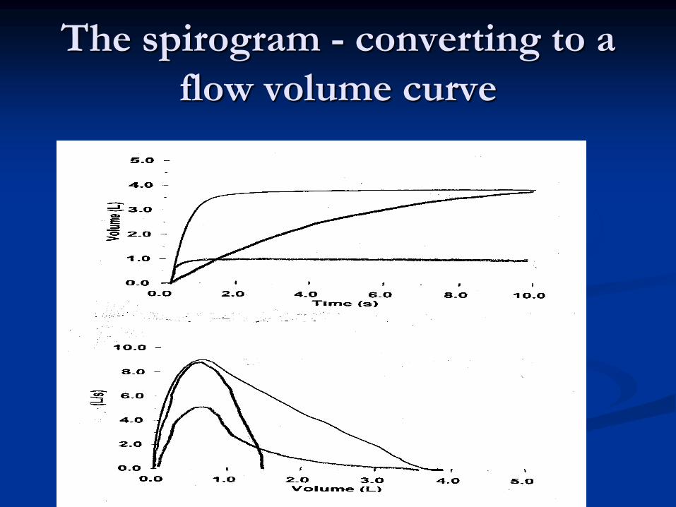
The spirogram - converting to a
flow volume curve
aao9
Text Box
Manipulate the information from spirometry measurements to get flow rate data
aao9
Text Box
The Flow rate vs. Volume graph helps us distinguish between small and large airway abnormalities. It's particularly useful at low lung volumes where small airways are PMR - Small airway disease is often a precursor to large airway disease and loss of lung volume. Finding this early allows us catch the process before total compromise of lung function.
aao9
Text Box
Normal Curve: Flow Volume against Time
aao9
Text Box
Converted Curve: Flow Rate against Volume
aao9
Text Box
A
aao9
Text Box
A
aao9
Text Box
B
aao9
Text Box
B
aao9
Text Box
C
aao9
Text Box
C
aao9
Text Box
Key: A: Normal B: Obstructive Lung Disease C: Restrictive Lung Disease

Flow-volume curve
Allows better assessment of airway
characteristics at low lung volumes
as lung empties, “choke point” for flow moves
distally from large cartilagenous airways to small
membranous airways
these small airways may be earliest site for airway
diseases
aao9
Rectangle
aao9
Highlight
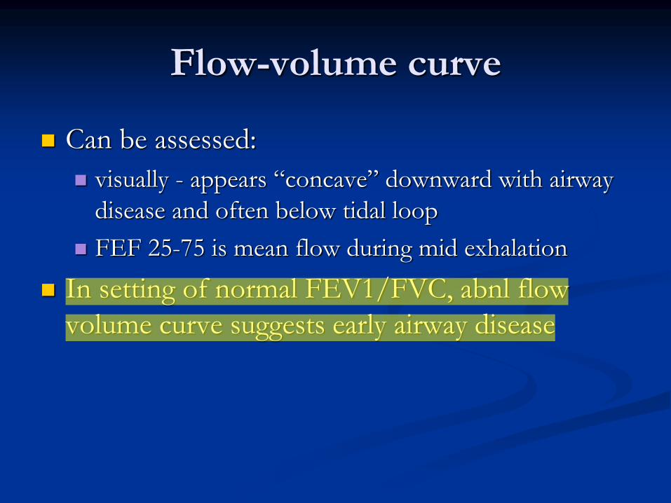
Flow-volume curve
Can be assessed:
visually - appears “concave” downward with airway
disease and often below tidal loop
FEF 25-75 is mean flow during mid exhalation
In setting of normal FEV1/FVC, abnl flow
volume curve suggests early airway disease
aao9
Text Box
The flow rate vs volume curves give very "noisy" numbers. The range of normal is very wide and it's difficult to interpret. He doesn't use it.
aao9
Highlight
aao9
Callout
I.e. in early stages of disease, FEV1/FVC might be normal. We can catch the small airway disease early using the flow rate vs volume curves.

aao9
Callout
Breathing at Rest (Tidally)
aao9
Callout
Graph for sequence of deep breath followed by rapid exhalation This person has terrible lung function with a low initial flow and very dramatic disappearance of flow
aao9
Text Box
When the patient is breathing tidally, there's a certain flow rate. When the patient takes the deep breath and breathes forcefully, flow actually ends up being slower than what it was during tidal breathing. This is very abnormal. Characteristic of collapsing airways. You push on them and they slam shut.

Upper Airway Evaluation
aao9
Text Box
For each of these flow volume loops, the part above the x axis represents exhalation . The part below represents inhalation.
aao9
Line
aao9
Line
aao9
Text Box
Volume
aao9
Text Box
Flow rate
aao9
Text Box
Exhalation is near normal but inhalation is impaired.
aao9
Text Box
Variable Obstruction: Exhalation is impaired but inhalation is normal (during exhalation, pressure collapses airways)
aao9
Text Box
Fixed obstruction: Both exhalation and inhalation are impaired (E.g. Upper airway narrowing at the anastomotic site of lung transplant)
aao9
Text Box
Easy point on your pulmonary boards: Upper airway obstruction (laryngeal obstruction, goiter) constricts the trachea and flow volume loop becomes almost a square. The third one is most common. Upper airway narrowing is now very common because of lung transplants

Pulmonary Function Testing
Goals of PFTs
Normal values - interpretive principles
Spirometry
Lung volumes
Diffusing capacity
aao9
Callout
Most common, cheap but requires patient cooperation
aao9
Text Box
Question: Where's the midpoint for switching the chokepoint from large to small airways? Answer: It's right around the midpoint of inspiration.
aao9
Highlight
aao9
Callout
Least important. Most expensive. Doesn't require too much patient cooperation

Lung volumes
The spirogram measures the maximal amount of
gas a subject can voluntarily move
Lung volume testing is primarily aimed at
measuring the remaining gas in the lung after
full exhalation (residual volume)
aao9
Text Box
Much more complicated than the spirogram, costs $40, 000! Not performed as often.
aao9
Rectangle
aao9
Highlight
Lung volumes
Lung gas volume at the “rest point” or
functional residual capacity is measured by one
of several techniques:
plethysmography
inert gas dilution
nitrogen washout
Residual volume is then calculated by having
patients fully exhale and subtracting this volume
aao9
Callout
Knowing that you started with 80% you can measure volume.
aao9
Text Box
"We don't have time to go through this"
aao9
Text Box
Put patient in a box. Patient exhales through a mouth piece. Mouth piece is closed to "seal" the box. Patient then inhales. Since it's a closed system, decrease in lung pressure with inhalation leads to changes in box pressure. Boyle's law is used to find volumes.
aao9
Text Box
Patient exhales entire vital capacity (leaving only residual volume) and then inhales nitrogen gas. This nitrogen mixes with nitrogen in residual volume. Since most of the air in residual volume is nitrogen, total volume of nitrogen gives TLC (make assumptions about CO2, O2, etc left in the residual gas)
aao9
Callout
Here, replace all the gas in the lung with inert gas, often helium (along with a known concentration of O2 so people keep breathing). This is done by diluting the lung gas with the inert gas over a period of time (4 - 7 minutes). To get TLC, measure the amount of inert gas that comes out when patient starts to breathe normal air (accounting for O2 part of "inert gas")
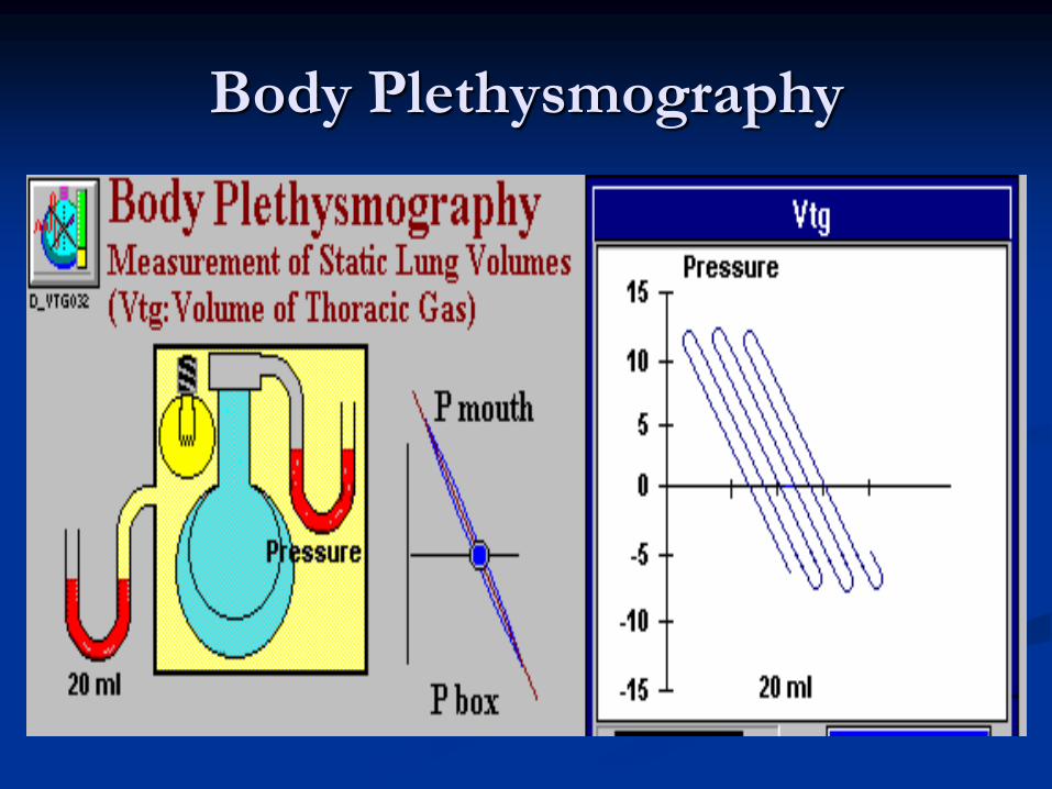
Body Plethysmography
aao9
Text Box
... skipped
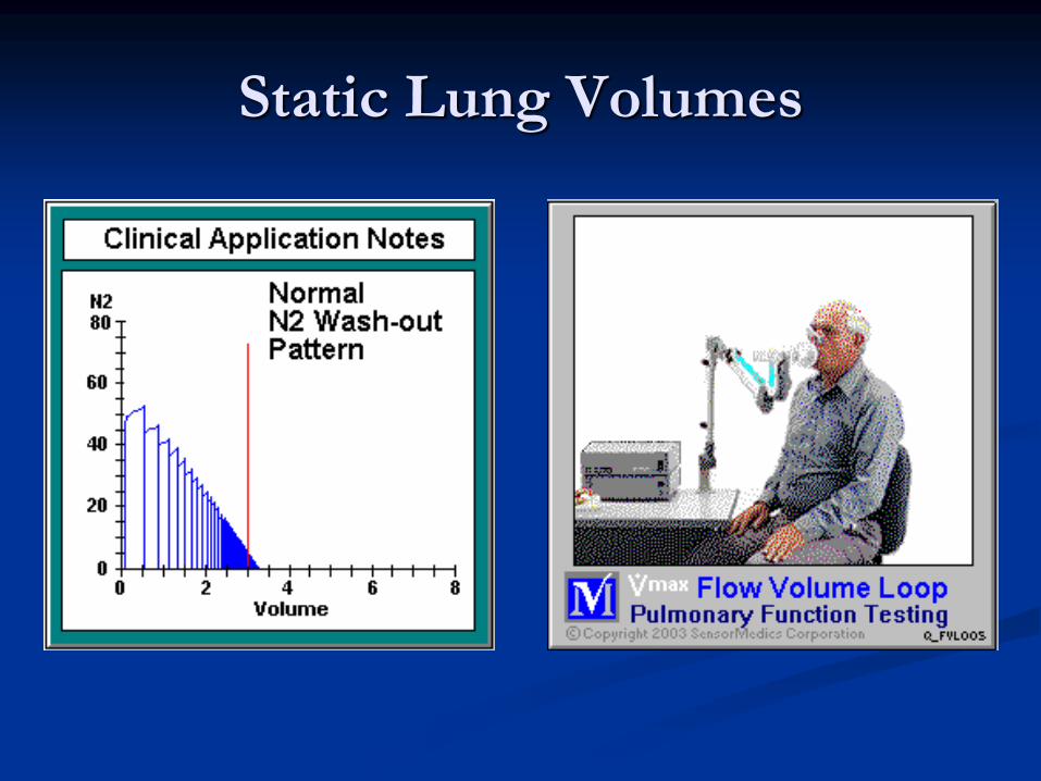
Static Lung Volumes
aao9
Text Box
... skipped

aao9
Text Box
... skipped

Patterns of pathophysiology
CATEGORY FEV1/FVC FVC RV DLCO
Obstructed
Asthma nl/dec* nl inc*
Bronchitis dec nl inc
Emphysema dec nl inc
Restricted nl dec dec
Neuromuscular dec dec inc
Vascular nl nl nl
_________
* during exacerbations/methacholine
aao9
Rectangle
aao9
Callout
With obstruction, air can't leave well so residual volume increases (remember expiration is the worse part of obstructive disease as the increased pressure collapses airways)
aao9
Callout
With restriction, you don't get enough air into the lungs. RV decreases
aao9
Callout
No change in RV with vascular pulmonopathy
aao9
Callout
With neuromuscular pulmonopathy, you just can't push out enough air. RV increases.
Pulmonary Function Testing
Goals of PFTs
Normal values - interpretive principles
Spirometry
Lung volumes
Diffusing capacity
aao9
Rectangle

Diffusing capacity
CO uptake dependent on:
Vc: capillary blood volume (incl Hb)
Dm: alv-cap membrane properties
CO uptake measured by
inhaling small concentrations CO
holding breath 10 sec
measuring exhaled CO
aao9
Text Box
Measure of the amount of blood reaching alveolar capillary membranes
aao9
Text Box
How much gas diffuses between the capillaries and the alveoli
aao9
Highlight
aao9
Highlight
aao9
Callout
Go through these steps to measure how well gas is diffusing into the capillaries (Compare with standards)

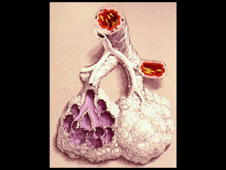
Pathologic changes that affect
Dm, Vc and DLCO
A. Normal
B. Emphysema
C. IPF
D. Lobectomy
E. Vasculitis
F. CHF
aao9
Callout
Diffusion actually goes up in acute situation with left ventricular failure because capillary beds are engorged. Long standing failure however, damages vessels.
aao9
Text Box
3 ways to increase diffusion without touching you: - Increase HR (exercise) - Lay you flat. Gravity affects much less - Molar manoeuver. Close glottis and inspire (opposite of valsalvar)
aao9
Text Box
Think of diffusion test as a measure the amount of blood that is there to pick up CO
aao9
Highlight
aao9
Text Box
Fibrosis
aao9
Text Box
Destroy vessels
aao9
Text Box
Loss of diffusion

The single breath DLCO
DLCO = ln (CO/i/Cot) x VA x 1/t
aao9
Text Box
... skipped
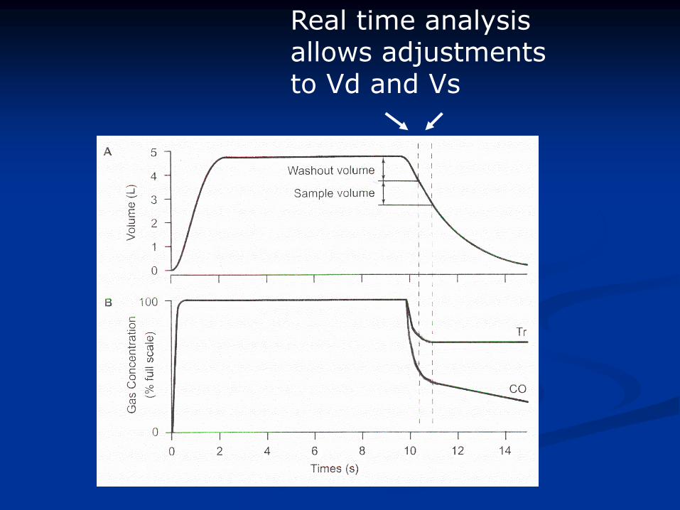
Real time analysis allows adjustments to Vd and Vs
aao9
Text Box
... skipped

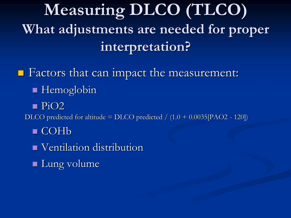
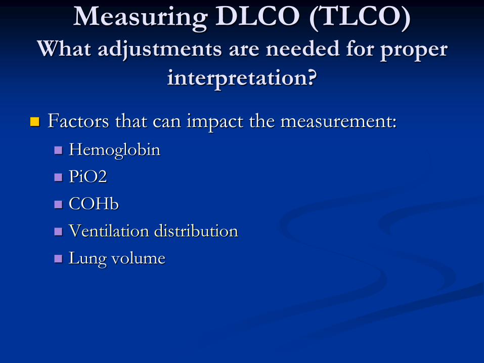
Measuring DLCO (TLCO) What adjustments are needed for proper
interpretation?
Factors that can impact the measurement:
Hemoglobin Men:
DLCOpredicted for Hb = DLCOpredicted 1.7 Hb / (10.22 + Hb)
Women:
DLCOpredicted for Hb = DLCOpredicted 1.7 Hb / (9.38 + Hb)
PiO2
COHb
Ventilation distribution
Lung volume
aao9
Callout
Most important
aao9
Callout
The factors on this page can skew your measurements for DLCO.
aao9
Rectangle
aao9
Callout
Equations to appropriately adjust DLCO to match standards (Didn't emphasize these equations so I wouldn't cram them...)
Measuring DLCO (TLCO) What adjustments are needed for proper
interpretation?
Factors that can impact the measurement:
Hemoglobin
PiO2 DLCO predicted for altitude = DLCO predicted / (1.0 + 0.0035[PAO2 - 120])
COHb
Ventilation distribution
Lung volume
aao9
Callout
Patient with a lot of O2 will outcompete CO
aao9
Text Box
More correction equations

Measuring DLCO (TLCO) What adjustments are needed for proper
interpretation?
Factors that can impact the measurement:
Hemoglobin
PiO2
COHb DLCOpredicted for COHb = DLCOpredicted (102% - COHb%)
Ventilation distribution
Lung volume
aao9
Callout
Cigarettes have CO which affects the measurement since it's CO based test
aao9
Text Box
CO correction equation
Measuring DLCO (TLCO) What adjustments are needed for proper
interpretation?
Factors that can impact the measurement:
Hemoglobin
PiO2
COHb
Ventilation distribution
Lung volume
aao9
Callout
Top of the lung has a bit of dead space (more ventilation than perfusion). Bottom of the lung is a bit of a shunt (more perfusion than diffusion). This distribution varies in different individuals. Can skew results in one direction or the other
aao9
Text Box
... skipped

Effects of poorly ventilated
regions
DLCO measures CO uptake from regions into
which it is inhaled
In severe OAD, many regions cannot get
measurable CO into them during a single breath
and thus global DLCO appears reduced
Suspect this when the tracer gas dilution Va is
very low - if the tracer gas cannot distribute,
neither can the CO
aao9
Text Box
... skipped. Read the slide.
aao9
Text Box
Tracer gas is put into the lungs and its distribution is observed. Provides a rough measure of lung ventilation.

Measuring DLCO (TLCO) What adjustments are needed for proper
interpretation?
Factors that can impact the measurement:
Hemoglobin
PiO2
COHb
Ventilation distribution
Lung volume
aao9
Callout
Lung volume will affect rate of gas diffusion

Lung volume effects - low Vi
- Less than maximal Vi
- lower Vc and Dm (dark)
- Lobectomy/pneumonectomy
- lower Dm, VC recruited
(light)
- Simple Dl/Va does NOT
“correct” (ie not 1:1)
aao9
Text Box
... skipped
aao9
Callout
With a lobectomy, you'd suspect that there's decreased diffusion because of the decreased volume. This is true but not to the extent that you would imagine. The perfusion in the remaining lobes increases to provide compensation for the lost diffusion capacity.
aao9
Text Box
Dm = Membrane resistance VC = Vital Capacity
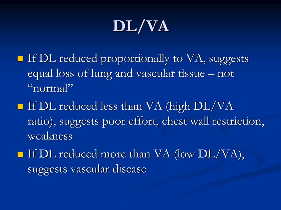
DL/VA
If DL reduced proportionally to VA, suggests
equal loss of lung and vascular tissue – not
“normal”
If DL reduced less than VA (high DL/VA
ratio), suggests poor effort, chest wall restriction,
weakness
If DL reduced more than VA (low DL/VA),
suggests vascular disease
aao9
Text Box
... skipped. Read the slide
aao9
Text Box
Ratio of Diffusing Capacity of Lung (DL) to Alveolar Volume (VA)
aao9
Text Box
Gas diffuses into the blood okay. To do this, we have to get the gas to the alveoli. Here we're just not doing that well enough. (I.e. we're not getting enough of gas in contact with blood vessels)
aao9
Text Box
Gas is getting into the alveoli but it's not diffusing into the blood.
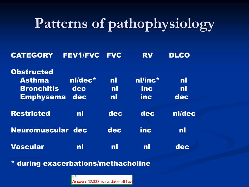
Patterns of pathophysiology
CATEGORY FEV1/FVC FVC RV DLCO
Obstructed
Asthma nl/dec* nl nl/inc* nl
Bronchitis dec nl inc nl
Emphysema dec nl inc dec
Restricted nl dec dec nl/dec
Neuromuscular dec dec inc nl
Vascular nl nl nl dec
_________
* during exacerbations/methacholine
aao9
Callout
Differentiating test for emphysema. If there's a problem with diffusion (in addition to the rest of the FEV1, FVC, FV profile), problem is emphysema since this problem reduces absorptive ability. If there's no effect on diffusion then the bronchi are affected.
aao9
Rectangle
aao9
Rectangle
aao9
Callout
You see this as an isolated finding (only DLCO is abnormal) and you know its a vascular issue
aao9
Callout
Measures gaseous exchange as opposed to mechanical behavior
aao9
Text Box
Question: Seeing as RV test is least important but most expensive, does anyone do it? Answer: 12,000 tests at duke - all have spiro, 50% have DLCO, 25% have RV

Pulmonary Function Testing
Goals of PFTs
Normal values - interpretive principles
Spirometry
Lung volumes
Diffusing capacity
aao9
Text Box
Recap of the things we talked about


![Shrinking Lung Syndrome: A Pulmonary Manifestation of ... · scan]) and pulmonary function tests (PFTs). Pulmonary function tests were carried out in our pulmonary function laboratory,](https://static.fdocuments.net/doc/165x107/5f03189c7e708231d40783f1/shrinking-lung-syndrome-a-pulmonary-manifestation-of-scan-and-pulmonary-function.jpg)