Pulmonary Function Indices Critical Care Patients978-3-642-73040...Josef X. Brunner Gunther Wolff...
16
Transcript of Pulmonary Function Indices Critical Care Patients978-3-642-73040...Josef X. Brunner Gunther Wolff...


Josef X. Brunner Gunther Wolff
Pulmonary Function Indices in Critical Care Patients
Springer-Verlag Berlin Heidelberg New York London Paris Tokyo
Josef X. Brunner, Dr. Sc. techno
Address during preparation of this book: Research Assistant of Clinical Physiology University of Basel Kantonsspital CH-4031 Basel Switzerland
Current address: Department of Anesthesiology Division of Biomedical Engineering 3 C 444 University of Utah Medical Center Salt Lake City, Utah 84132 USA
Gunther Wolff, M. D., FMH Surgery
Professor for Surgery' and Intensive Care Medicine Head of Clinical Physiology
Address: Clinic for Thoracic and Cardiac Surgery Department of Surgery University of Basel Kantonsspital CH-4031 Basel Switzerland
ISBN-13: 978-3-540-18432-4 001: 10.1007/978-3-642-73040-5
e-ISBN-13: 978-3-642-73040-5
Library of Congress Cataloging~in-Publication Data. Brunner, Josef X., 1956-. Pulmonary function indices in critical care patients. Bibliography: p. Includes index.!. Pulmonary function tests. 2. Critical care medicine. I. Wolff, Gunther, 1932-. iI. Title. RC 734.P84B78 1988 616.2'4'028 88-3161
This work is subject to copyright. All rights are reserved, whether the whole or part of the material is concerned, specifically the rights of translation, reprinting, reuse of illustrations, recitation, broadcasting, reproduction on microfilms orin other ways, and storage in data banks. Duplication of this publication or parts thereof is only permitted under the provisions of the German Copyright Law of September 9, 1965, in its version ofJune 24, 1985, and a copyright fee must always be paid. Violations fall under the prosecution act of the German Copyright Law.
© Springer-Verlag Berlin Heidelberg 1988
The use of general descriptive names, trade names, trade marks, etc. in this publication, even if the former are not especially identified, iSl),ot to be taken as a sign that such names, as understood by the Trade Marks and Merchandise Marks Act, may accordingly be used freely by anyone.
Product Liability: The publisher can give no guarantee for information about drug dosage and application thereof contained in this book. In every individual case the respective user must check its accuracy by consulting other pharmaceutical literature.

Foreword
Respiration is a unique topic among various subdisciplines of physiology. Physiologists and clinicians are now able to communicate quantitative functional properties of lung mechanics and gas exchange in the language of the engineer, physicist and mathematician. This is largely due to intensive and stimulating work during the last decades of brilliant minds in a handful of excellent schools in the international family of physiologists. Among these founders of respiratory physiology are a number of clinicians, and they have. taken significant ,part both in shaping the theoretical knowledge to clinical applicability and developing technical devices for diagnosis and therapy in pulmonology. However, the theory behind the evaluation of measurements, and their interpretation in terms of clinical function tests, is so confusingly complex that the ordinary physician, not specifically trained in respiratory physiology, finds himself unable to critically apply these techniques. We, therefore, need descriptions of respiratory physiology and of its clinical application presented in the language of the clinician.
And that is what this book is meant to be. Written by an expert in electrical and biomedical engineering, and by an expert in intensive care medicine, this text constitutes an "operational manual" of clinical respiratory physiology. It does not intend to be another textbook of basic respiratory physiology or pathophysiology. This book not only addresses practical clinicians, particularly those of intensive care medicine, by describing the essentials of clinically relevant respiratory knowledge. But it also addresses the engineer, by describing the essentials of biomedical engineering relevant to lung function analysis. The reliability and the elegance ofthe measuring techniques described in this book are to be commended. And what makes the authors eligible for such a task is that they have plunged deep into the understanding of the physiological basis and have, at the same time, kept their view as practical clinicians, since they maintain an everyday responsibility for the intensive care ward.
Hence, the book may be profitable read by clinicians, respiratory physiologists and engineers, particularly those with interests in intensive care medicine and pulmonology, who wish to learn details concerning both the theoretical background and the practical aspects of clinical function tests.
I trust that this book will find friendly acceptance by the colleagues in the field.
October 1987 Prof. Peter Scheid
Head of the Institute of Physiology Ruhr-Universitat Bochum West-Germany
Preface
This book started out 10 years ago with the question: How do I measure functional residual capacity in the intensive care patient? The following 5 years were filled with disappointment, mainly caused by the expensive measuring equipment. We were not able to perform measurements without running into serious limitations, and the algorithms were "buggy". The measurements turned out to be inaccurate and, worst of all, they could not be verified. The only solution to these problems seemed to be developing a custommade system. In the end, this decision proved to be the right one.
The task required an additional member in the team: an engineer. The first problem was communication. It turned out that even though they spoke the same language, the words did not mean the same for the engineer as they did for the physician. Almost completely opposite were the opinions as to what kinds of problems a computer can solve. Soon, another problem was discovered: the scatter of information. Although many of the techniques needed had already been developed, in physiology as well as in engineering, there was no common source of information. This book, which is the enhanced and translated version of a PhD thesis (Brunner 1985), attempts to fill the gap. We tried to cover most of the terminology used in both, medicine and engineering, with respect to lung function analysis. The prime goal is to enable efficient communication between physician and engineer in order to evaluate commercially available equipment or to design new equipment.
There is no claim for completeness regarding engineering or pulmonary physiology. We strongly recommend that the original literature be consulted. In particular, we encourage the reader to familiarize himself with the concepts developed by Fenn, Rahn, and Farhi in Rochester, which are not dealt with in detail in this book (V'/Q' diagram, see Chapter 2.4.3). As an excellent source of information we also recommend the "Handbook of Physiology" published by the American Physiological Society.
The success of the close (and pleasant) collaboration of physician and engineer extended the original project far beyond the initial plan to measure functional residual capacity. This book is one of the results. Its major purpose is to save interested researchers the "first five unpleasant years" .
In Chapter 1 the properties of a clinically important pulmonary function index are presented. The particular modes of application of the measuring method in the clinic and in seriously ill patients are discussed. In Chapter 2 the basic principles of model development for spontaneous respiration and mechanical ventilation are discussed. In Chapter 3 we describe our solutions for the measuring technique and analysis. We

VIII Preface
present three cases to document the efficiency of the entire measuring system. The next 3 chapters deal with applications. In Chapter 4 lung function indices of a standard situation are presented. In Chapter 5 the effects of isolated changes in individual variables of the breathing pattern are described through a comparison of various steady states (paired differences). Chapter 6 deals with the possibilities and limits of time resolution, i. e. the breath by breath analysis. Intermittent mandatory ventilation (IMV) is assessed by separate analysis of spontaneous breaths and mandatory breaths.
Appendices are included, some of which are designed to give the physicists/ technologists of the team an idea of the anatomy and physiology of the lungs, others should help the physician of the team to understand the physics and mathematics involved. A further appendix contains all of the formulas used in the program package. Finally, a last appendix presents in tables all of the results of the investigations found in Chapters 4, 5, and 6.
It is our hope that this book will help to make pulmonary function testing in the critically ill patient more reliable, precise, rational, critical, and less expensive. Our goal should be to treat our patients with better differentiation and to deepen our understanding of functional disturbances. For this purpose we have made our software with documentation available to the interested reader. In return, our only request (apart from copying costs) is feedback, in the hope that such a widespread exchange of experience and information will be a valuable contribution to the fascinating discussion of pulmonary function in the critically ill patient.
January 1988 Josef X. Brunner and Gunther Wolf!
Salt Lake City, Utah, USA and Basel, Switzerland
Contents
Foreword V
Preface . VII
Acknowledgements 1
List of Abbreviations 3
1. Evaluation of Pulmonary Function in the Intensive Care Patient 7
1.1. The Clinically Important Pulmonary Function Index .... 7
1.1.1. Evaluation and Monitoring of Circulatory Functions 1.1.2. Evaluation and Monitoring of Pulmonary Functions 1.1.3. "Simple" Measured Values ............ . 1.1.4. Physiological Models of Pulmonary Function Indices 1.1.5. The Clinically Important Pulmonary Function Indices
1.2. What Clinicians Expect of Transducers and Data Processing
1.2.1. The Chain ofInstruments: DataFlow .... 1.2.2. Measuring Head and Measuring Instruments 1.2.3. Data Processing 1.2.4. Result-output ......... .
1.3. A Measuring System for Clinical Research
1.3.1. Obtaining Primary Data from the Measurements at the Airway
7 7 8 9 9
10
10 10 11 11
12
Opening ............. 13 1.3.2. Transducers ................... 14 1.3.3. Preprocessing and Storage of Data . . . . . . . . 15 1. 3.4. The Selection of the Pulmonary Indices Presented 15
2. Derivation of the Pulmonary Function Indices 18
2.1. Introduction ................. . 18

X Contents
2.2. Breathing Mechanics 18
2.2.1. The Patient-ventilator Unit 18 2.2.2. Mathematical Model . . . 21 2.2.3. Determining Lung Compliance and Airway Resistance 23 2.2.3.a Pleural Pressure ................... 23 2.2.3. b Approximation ofthe Pressure-flow-volume Curve 24 2.2.4. Influence of the Lung Mechanics on the Distribution of Venti la-
tion . . . . . . . . . . . . . . . . . . 25
2.3. Lung Volume and Intrapulmonary Gas Mixing 26
2.3.1. Nitrogen Washout .......... 26 2.3.2. Measurement of the Functional Residual Capacity (FRC) 26 2.3.3. GasMixingintheAcinus:TheStationaryInterface.... 28 2.3.4. Gas Mixing in the Lungs ................. 29 2.3.5. Measurement of the Airway Dead Space (Series Dead Space) 32 2.3.6. Washout Efficiency from the Washout Curve (Decay Curve) 33
2.4. Transpulmonary Gas Transport: Exchange of O2 and CO2
2.4 .1. Diffusion from and into the Pulmonary Capillary Blood 2.4.2. Dissociation of O2 and CO2 in the Blood 2.4.3. The "Riley" 3-compartment Model . . . . . 2.4.4. Analysis of the COrdiagram . . . . . . . . 2.4.4.a Series Dead Space and Alveolar Ventilation 2.4.4. b Alveolar Efficiency of COrelimination 2.4.4.c Slope of the Alveolar Plateau ....... .
3. Assessment ofPnlmonary Function Indices
3.1. Sensors . . . . . . . . . .
3.1.1. Measuring Head . 3.1.2. Flow Measurement 3.1.2. a Principle of Measurement 3.1.2.b Linearity ........ . 3.1.2.c Calibration . . . . . .. . 3.1. 2. d Correction of Errors Caused by Viscosity Changes 3.1.2.e Bandwidth .... . 3.1.3. Gas Analysis ........ . 3.1.3.a MeasuringPrinciple .... . 3.1.3.b Automatic Sensitivity Control 3.1.3. c Linearity and "Cracking Pattern" 3.1.3.d Calibration ... 3.1.3.e TransportDelay 3.1.3.f ResponseTime .
36
36 36 37 39 40 40 42
44
44
44 44 44 46 47 48 50 50 50 51 51 51 52 52
Contents XI
3.1.4. Pressure Measurement 52 3.1.4.a MeasuringPrinciple 52 3.1.4.b Linearity . . . . . . . 53 3.1.4.c Bandwidth . . . . . . 53 3.1.5. STPD andBTPS Conditions 54
3.2. Data Processing 55
3.2.1. HardwareandDataFlow.. 55 3.2.2. Automatic Recognition of the Respiratory Phases 57 3.2.3. Software . . . . . . . 58
3.3. Testing the Measuring System 58
3.3.1. Absolute Accuracy . . 59 3.3.1.a PhysicalModeloftheLung 59 3.3.1. b Accuracy of Dead Space Determination 61 3.3.1.c Accuracyofthe APVMeasurements . . 61 3.3.1.d DecayCurveoftheModelLung 62 3.3.2. Precision of the Indices on the Stable Patient in "Steady State" 63 3.3.3. Reproducibility ofthe Nitrogen Washout . . . 63
3.4. Use ofthe Measuring System in the Intensive Care Unit 65
3.5. Sensitivity of the Indices in the Presence of Acute Pathological Changes in the Lungs: Case Studies ............ 66
3.5 .1. Hypovolemia Before and After Correction 66 3.5.1.a Results . . . . . . . . . . . . . . . . . . 67 3.5.1. b Comparison with Conventional Methods . 68 3.5.2. Acute Bronchial Constriction Before and After Therapy 68 3.5.2.a Results . . . . . . . . . . . . . . . . . . . . . . . . 68 3.5.2.b Comparison with ConventionalIndices . . . . . . . . 69 3.5.3. Transient Processes: Effects of Inflating the Balloon
of a Pulmonary Arterial Swan-Ganz Catheter 69 3.5.3.a AModelforPulmonaryEmbolism 69 3.5.3.b Measurements in the Patient 70 3.5.3.c Results . . . . . . . . . . . . . . 71
4. Application I: Standard Values 'During Mechanical Ventilation After Cardiac Surgery 73
4.1. Patients and Examination 73
4.2. The Ventilation ... 76
4.3. Breathing Mechanics 76
XII Contents
4.4. Accessible Pulmonary Volume 77
4.5. Washout Efficiency and Moment Analysis 77
4.6. CO2 Production and O2 Consumption . . 78
4.7. ConventionalIndices for CO2 Exchange 79
4.8. SpecificIndices for CO2 Exchange 79
4.9. Cardiac Output 80
4.10. Correlations 81
5. Application II: A Study on Optimizing Mechanical Ventilation 83
5.1. Problem
5.2. Studies by Other Authors
5.3. Hypotheses .....
5.4. Patients and Methods
5.4.1. Patients 5.4.2. Measurements 5.4.3. Presentation of the Results
5.5. Results and Discussion . . . . . .
5.5.1. Effects of Changing the Tidal Volume (VT)
5.5.2. Effects of the Positive End-expiratory Pressure (PEEP) 5.5.3. Effects of the Inspiratory Flow Rate (V'r) . 5.5.4. Effects of the End-inspiratory Pause (EIP)
5.6. Summarizing Discussion and Conclusions
83
83
86
86
86 87 88
88
88 91 94 96
98
5.6.1. Non-quantifiable Observations . 98 5.6.2. Optimizing Ventilation . . . . . 99 5.6.3. Recruitment of Compensatory Mechanisms 100
6. Application ill: A Study on Intermittent Mandatory Ventilation (IMV) ........ 102
6.1. Patients and Methods 103
6.1.1. Patients 103
6.1.2. Measurements .... 6.1.3. Presentation of Results
6.2. Results ..
6.3. Discussion
6.3.1. CPPVComparedwithIMV 6.3.2. IMVMB Compared with IMVsB
6.3.3. IMV Compared with CPAP
7. Appendices .......... .
7.1. On the Morphology of the Lungs
7.2. Technical Principles of Mechanical Ventilation
7.3. Measuring the Pleural Pressure with an Esophageal Balloon
7.4. Transport Equation for Convection and Diffusion
7.5. Relationship Between Dead Space and V' A/Q' Scatter
Contents XIII
103 104
104
105
106 107 109
110
110
115
118
122
124
7.6. Viscosity of Gas Mixture ............... 126
7.7. Determination and Dependencies of the Delay Time Between Flow Sensor and Mass Spectrometer 128
7.8. List of Formulas 133
7.9. Result-Tables . 142
7.9.1. Tables to Application I: Standard Values After Cardiac Surgery 142 7.9.2. Tables to Applications II: Effect of Ventilation Variables . .. 146 7.9.3. Tables to Application III: Intermittent Mandatory Ventilation . 158
References 163
Subject Index . . . . . . . . . . . . . . . . . . . . . . . . . . . . . . .. 169
Acknowledgements
This work was made possible by the exchange of ideas and extensive discussions with many of our colleagues in the scientific community. Most of these discussions took place not at large congresses, but rather in the laboratories. For their interest, support, and hospitality ,we are most grateful to Professor Johannes Piiper (G6ttingen, W. Germany), Professor Peter Scheid (Bochum, W. Germany), Professor Gordon Cumming (Midhurst, GB), Dr. G6ran Hedenstierna (Stockholm, Sweden), Professor Emilio Agostoni (Milano, Italy), and Professor Peter Suter (Geneva, Switzerland).
Interdisciplinary research is impossible without open communication between medical doctors, physiologists, and engineers. In this respect we gratefullyacknowledge the invaluable discussions with Dr. Manuel Paiva (Brussels, Belgium), Dr. Chris Mills (Midhurst, GB), Professor David Dennison (London, GB), and Professor Max Anliker (Zurich, Switzerland).
Our research was fully supported by the Director of the Clinic for Cardiac and Thoracic Surgery ofthe University of Basel, Switzerland, Professor Erich Gradel. We are most grateful for the research opportunities he provided us with, as well as his personal support. Our thanks also go to the Head of the Department of Surgery of the University of Basel, Professor Felix Harder, for his valuable support.
Each improvement of the software was tested in our cardiac surgery intensive care unit, which interfered immensely with the work on the ward. We want to thank our nurses and their staff for their patience and cooperation. Their critical interest as users of the measuring system proved to be invaluable. Many of the hardware improvements were carried out with the hospital technicians, in particular, Werner Nyffeler. We are indebted to them for their kind assistance and constructive imagination.
The original manuscript was written in German. The translation into English proved to be an unexpectedly difficult task. Virginia Sonntag-O'Brien deserves special commendation for the translation of most parts of the book. We are further indebted to Dr. Robert Loeb and Dr. Tom East for their scientific critique and help with the English. We wish to give special thanks to Dr. Toni Graf-Baumann from Springer-Verlag, who encouraged us to write this book and whose advice and suggestions on how to improve it didactically were a major asset.
We have not forgotten that our work was only possible thanks to the financial support of many institutions. We therefore wish to express our appreciation to those who showed their confidence in our project. We were supported by the Swiss National Science Foundation, the Roche Research Foundation, the Jubilee Foundation of

2 Acknowledgements
Ciby-Geigy, the Emilia Guggenheim-Schnurr Foundation, the Science Foundation of the Basel Chamber of Commerce, the Sandoz Research Foundation, and the Science Foundation of the Kantonsspital-Basel. Last but not least we would like to thank Dragerwerk and Dr. Eric Hecker (Liibeck, W. Germany). He provided the opportunities for international researchers to get together at productive workshops in our department. We sincerely appreciate his interest in the physiological aspects as well as his critical and stimulating contributions to the discussions.
List of Abbreviations
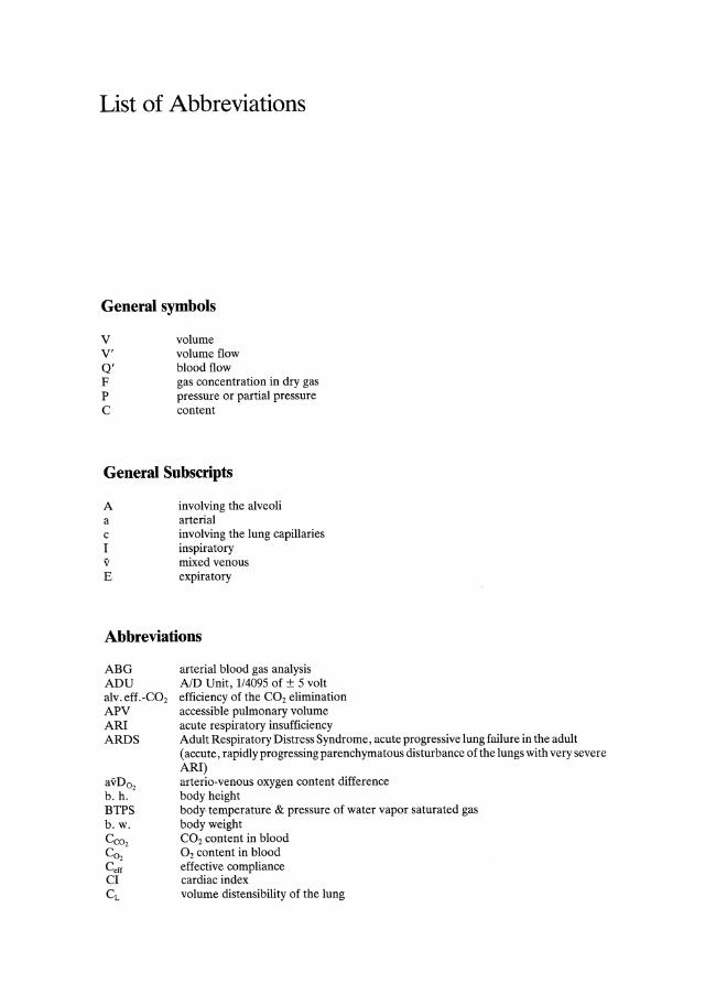
General symbols
v v' Q' F P C
volume volume flow blood flow gas concentration in dry gas pressure or partial pressure content
General Snbscripts
A a c I v E
involving the alveoli arterial involving the lung capillaries inspiratory mixed venous expiratory
Abbreviations
ABG ADU alv. eff. -C02
APV ARI ARDS
avD02
b. h. BTPS b.w. CC02
CO2
Ceff
CI CL
arterial blood gas analysis AID Unit, 114095 of ± 5 volt efficiency of the CO2 elimination accessible pulmonary volume acute respiratory insufficiency Adult Respiratory Distress Syndrome, acute progressive lung failure in the adult (accute, rapidly progressing parenchymatous disturbance of the lungs with very severe ARI) arterio-venous oxygen content difference body height body, temperature & pressure of water vapor saturated gas body weight CO2 content in blood O2 content in blood effective compliance cardiac index volume distensibility of the lung

4 List of Abbreviations
CO Ctot CV Cw E ECC EIP Fco,a-E Fco,-et FEco2
Fro, F~,o FRC I:E LAP mBP MS PA
Pco,
Po, PAP Pawo
PawoEI
Pawo1min Pawomax PECO,
PEa, PEEP PIEco, PIEN,
Pes PpJ
PT PN Q'S/Q'T R,RQ RAP RAW
RL RR RRN Rtj RV rF SAP SD SEM SI slopeco, So, STPD SV02
T T r, TE TLC V'A/Q' VC
cardiac output volume distensibility of the entire respiratory system variation coefficient volume distensibility of chest wall and diaphragm expiration extracorporeal circulation (heart-lung machine) end-inspiratory pause difference between arterial and expired (end-tidal) CO2
maximum CO2 concentration in the expirate mixed expiratory CO2 concentration inspiratory O2 concentration initial nitrogen concentration functional residual capacity ratio of inspiration time to expiration time left atrial (blood) pressure mean (arterial) blood pressure mass spectrometer pressure in the alveoli partial pressure of CO2
partial pressure of O2
pulmonary artery (blood) pressure pressure at the airway opening, i. e., at the mouth, at outer end of tube end-inspiratory P awo minimal airway pressure during inspiration maximum P awo mixed expiratory partial pressure of CO2
mixed expiratory partial pressure of O2
positive end-expiratory pressure ventilator setting approximative value for serial dead space from the CO2 diagram approximative value for serial dead space from the N2 diagram pressure in the esophagus pressure in the pleura pneumotachograph pressure volume (PN) diagram venous admixture (shunt fraction) respiratory quotient right atrial blood pressure airway resistance viscous resistance of lung tissue respiratory rate, breathing or ventilation frequency (i. e. both) relative remaining nitrogen viscous resistance of muscle or connective tissue residual volume relative humidity O2 saturation of a pulmonary artery blood sample = mixed venous O2 saturation standard deviation standard error of the mean stationary interface slope of the alveolar plateau in CO2 diagram O2 saturation standard temperature & pressure of dry gas mixed venous O2 saturation temperature inspiratory and expiratory time total lung capacity ventilation perfusion ratio vital capacity
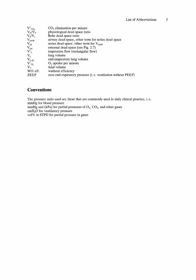
V'C02
VDIVT
VdlV, VdAW
V dS
Vex' V'I VL ·
V L-EI
V'02 VT
CO2 elimination per minute physiological dead space ratio Bohr dead space ratio airway dead space, other term for series dead space series dead space, other term for V clAW
external dead space (see Fig. 2.7) inspiration flow ( rectangular flow) lung volume end-inspiratory lung volume O2 uptake per minute tidal volume washout efficiency
List of Abbreviations 5
WOo eff. ZEEP zero end-expiratory pressure (i. e. ventilation without PEEP)
Conventions
The pressure units used are those that are commonly used in daily clinical practice, i. e. mmHg for blood pressure mmHg and (kPa) for partial pressures of 02, CO2, and other gases cmH20 for ventilatory pressure vol% in STPD for partial pressure in gases