PTPIP51 in adipose tissue
92
PTPIP51 in adipose t iss ue INAUGURAL-DISSERTATION (Cumulative Thesis) in partial fulfillment of the requirements for the Doctoral degree of the Faculty of Medicine of the Justus-Liebig-University Giessen by Manuel Anton Bobrich of Bamberg Giessen 2012
Transcript of PTPIP51 in adipose tissue

PTPIP51 in adipose tissue
INAUGURAL-DISSERTATION
(Cumulative Thesis)
in partial fulfillment of the requirements
for the Doctoral degree
of the Faculty of Medicine
of the Justus-Liebig-University Giessen
by
Manuel Anton Bobrich
of Bamberg
Giessen 2012

From the Institute of Anatomy and Cell Biology
Managing Director: Prof. Dr. med. W. Kummer
Faculty of Medicine, Justus-Liebig-University Giessen 1. Reviewer: Frau Prof. Dr. Wimmer
2. Reviewer: Prof. Dr. Bellusci
Day of doctoral defense: 18.10.2012

To my father, who will always be with me;
To my mother, who will always take care of me;
To my uncle, who will always be there for me;
To my siblings, who will always be part of me;
To my niece, who will always be sunshine for me;
To Janika, who will always mean everything to me.

DECLARATIONS
“I declare that I have completed this dissertation single-handedly without the
unauthorized help of second party and only with the assistance acknowledged
therein. I have appropriately acknowledged and referenced all text passages that
are derived literally from or based on the content of published or unpublished
work of others, and all information that relates to verbal communications. I have
abided by the principles of good scientific conduct laid down in the character of
the Justus-Liebig-University of Giessen in carrying out the investigations
described in the dissertation.“
Ich erkläre: Ich habe die vorgelegte Dissertation selbständig und ohne fremde
Hilfe und nur mit den Hilfen angefertigt, die ich in der Dissertation angegeben
habe. Alle Textstellen, die wörtlich oder sinngemäß aus veröffentlichten oder nicht
veröffentlichten Schriften entnommen sind, und alle Angaben, die auf mündlichen
Auskünften beruhen, sind als solche kenntlich gemacht. Bei den von mir
durchgeführten und in der Dissertation erwähnten Untersuchungen habe ich die
Grundsätze guter wissenschaftlicher Praxis, wie sie in der „Satzung der Justus-
Liebig-Universität Gießen zur Sicherung guter wissenschaftlicher Praxis“
niedergelegt sind, eingehalten.
Manuel Anton Bobrich

DECLARATIONS
This dissertation is submitted as a cumulative thesis according to the doctoral
degree regulations of the Faculty of Medicine of the Justus-Liebig-University of
Giessen. The thesis includes an interconnection of three original papers.
A general survey and summary of the state of the art of PTPIP51 implications in
adipose tissue will be given here as the introduction.

TABLE OF CONTENTS Table of contents
I. List of papers submitted for thesis 1
II. Introduction 2
1. Introduction 2
1.1.
1.2.
1.3.
2.
Scientific background of PTPIP51 2
PTPIP51 in different cellular processes 3
PTPIP51 in cancer 5 PTPIP51 – Gene, mRNA and protein 5
2.1. PTPIP51 gene 5
2.2. PTPIP51 mRNA 6
2.3. PTPIP51 protein 7
3. Interaction partners of PTPIP51 8
3.1. Interaction with phosphatases 9
3.2. Interaction with 14-3-3β 10
3.3. Interaction with kinases 10
4. Adipose tissue 11
4.1. Energy storage 11
4.2. Obesity 13
4.3. Adipocyte metabolism 13
4.4. Two important pathways in adipocytes 14
5. Experimental setup to determine the role of 15
PTPIP51 in adipose tissue
6. Role of PTPIP51 in adipose tissue 17

TABLE OF CONTENTS
7. Prospect 19
8. References 20
III. Summary 29
IV. Zusammenfassung 30
V. List of publications 31
VI. Acknowledgements 33
VII. Curriculum vitae 34
VIII. Annexes (Original papers) 36

LIST OF PAPERS SUBMITTED FOR THESIS
1
I. List of papers submitted for thesis Original papers
(1) Bobrich M, Brobeil A, Mooren FC, Krüger K, Steger K, Tag C, Wimmer M
(2011). PTPIP51 interaction with PTP1B and 14-3-3β in adipose tissue of
insulin-resistant mice. Int J Obes (Lond) 35(11): 1385-1394. (2) Brobeil A, Bobrich M, Wimmer M (2011a). Protein tyrosine phosphatase
interacting protein 51--a jack-of-all-trades protein. Cell Tissue Res 344(2): 189-
205. (3) Bobrich M, Schwabe SA, Brobeil A, Viard M, Kamm M, Mooren FC, Krüger K,
Tag C, Wimmer M (2012). PTPIP51 – a new interaction partner of the insulin
receptor and PKA in adipose tissue. Metabolism, under review

2
INTRODUCTION
II. INTRODUCTION
1. Introduction
1.1 Scientific background of PTPIP51
Protein tyrosine phosphatase interacting protein 51 (PTPIP51) was discovered about
ten years ago. Using the yeast two hybrid screening method, it was initially identified
as an interaction partner of T-cell protein tyrosine phosphatase (TcPTP) and later, to
a much higher extent, Protein tyrosine phosphatase 1B (PTP1B) (Stenzinger et al.,
2009). To reveal the function of PTPIP51 several studies dealt with its expression
pattern and its interaction partners in different tissues and cell culture systems.
PTPIP51 protein is expressed in epithelial cells of various organs. Highest evidence
of PTPIP51 in epithelia can be found in skin where the expression of PTPIP51 is
most likely associated with differentiation processes. The protein was also described
in ciliated cells of the trachea, the fallopian tube and ependyma of the brain
(Stenzinger et al., 2009). Recently, PTPIP51 was detected in non-keratinized
epithelia of the mouth. Furthermore it is expressed in neurons in the brain and
several brain tumours. In brain tissue, the expression is restricted to distinct
functional nuclei, for example nucleus subthalamicus and nucleus paraventricularis.
Additionally, PTPIP51 could be found in Purkinje cells of the cerebellum (Koch et al.,
2009b).
Stenzinger et al. (2005) described a fast-fibre-type specific expression pattern of
PTPIP51 in skeletal muscle, here being significantly associated to fast-contracting
type-2a-fibres (Stenzinger et al., 2005).
PTPIP51 is also expressed in male reproductive tissue, where it can be detected
throughout the complete differentiation process, from the primary spermatocytes up
to the spermatid stage (Stenzinger et al., 2009). In placenta, PTPIP51 is expressed
in syncytiotrophoblast and cytotrophoblast layer from the first, second, and third
trimesters (Stenzinger et al., 2009). Stenzinger et al. (2007) investigated the
expression of PTPIP51 in liver tissue. The tissue featured PTPIP51 protein in non-
parenchymal cells, namely Kupffer cells, stellate cells and natural killer cells. Despite
that specific association with non-parenchymal cells, a subset of hepatocyte nuclei

3
INTRODUCTION
and cholangiocytes of the bile tract also expressed PTPIP51 (Stenzinger et al.,
2007).
PTPIP51 also was seen in all investigated primary and secondary cell lines. Different
studies revealed that PTPIP51 is expressed in a multitude of cell lines, such as
glucagon-producing cells (INR1G9), human keratinocytes
(HaCat), human placental cells (BeWo), acetylcholine-
producing cells (NS20Y), Jurkat T-lymphoma cells
(Stenzinger et al. 2005) and human skeletal muscle cells
(Barop et al. 2009) as well as in a mouse fibroblast cell line
Fig. 1: PTPIP51 in proliferating 3T3 mouse fibroblast
(3T3) (Fig. 1) and a leukemia cell line (HL60) (Brobeil,
personal communication).
These observations gave basis for further functional studies.
1.2 PTPIP51 in different cellular processes
First studies indicated an involvement of PTPIP51 in differentiation processes, as the
protein was solely expressed in suprabasal cells of the epidermis (Koch et al., 2008).
Further experimental exploration was conducted by investigating human keratinocyte
cell culture (HaCat cells). Several experiments revealed that PTPIP51 expression
can be upregulated by stimulation with Epithelial Growth Factor (EGF) or 1,25OH2D3,
two factors stimulating differentiation and inhibiting proliferation (Hashimoto, 2000;
Stenzinger et al., 2009). Recently, a phosphorylation dependent activation of the
MAPK pathway was proven by Brobeil et al. (2012). The MAPK pathway is important
for proliferation and differentiation of cells. Activation of the MAPK pathway can be
induced by several receptors like epithelial growth factor receptor (EGFR) (Petri et
al., 2011) or downstream by the receptor kinases. One possibility of such a
downstream activation is the activation of raf-1. 14-3-3β is able to interact with raf-1
and thus activate the MAPK pathway. PTPIP51 seems to play an important role in
this interaction (Brobeil et al., 2012).
Barop et al. (2009) investigated the expression of PTPIP51 during muscle cell
differentiation. Proliferating myoblasts displayed low expression levels of both mRNA
and protein. Induction of differentiation to myotubes led to a sharp increase in
PTPIP51 expression. In differentiated multinuclear myotubes PTPIP51 expression
was highest. Re-induction of proliferation again decreased the level of PTPIP51

4
INTRODUCTION
protein in the myotubes, so a differentiation dependent expression profile seems
likely.
Further experiments by Brobeil et al. (2010) described PTPIP51 expression in
differentiated cells of the immune system. Bone marrow specimen, umbilical cord
blood and peripheral venous blood were all positively tested for PTPIP51 expressing
cells. Especially neutrophil granulocytes showed high PTPIP51 expression levels in
all investigated samples. In human bone marrow specimen PTPIP51 could also be
traced in myeloid precursor cells of neutrophils (Brobeil et al., 2010).
Based on the observation of a nucleic localization and an increased expression in
several types of cancer, experiments were conducted to investigate whether or not
PTPIP51 is also involved in proliferation processes. PTPIP51 exhibits partial
sequence homology with the microtubule-associated protein regulator of microtubule
dynamics 1 (RMD-1). Therefore, PTPIP51 has been internalized in a family of
proteins which are named regulator of microtubule dynamics (RMD), according to
their assumed function. PTPIP51 is also called regulator of microtubule dynamics 3
(RMD-3) (Oishi et al., 2007; Brobeil et al., 2011a).
In vivo experiments associated PTPIP51 with the cell cycle. Important regulators of
mitotic processes, CGI-99 and Nuf-2, were found to interact with PTPIP51 through a
two-hybrid screen that used PTPIP51 as bait (Stenzinger et al., 2009). The
interaction with CGI-99 hints towards an involvement of PTPIP51 in the regulation of
polymerization of microtubules, whereas the interaction with Nuf-2 indicates a
modulating function on the spindle apparatus (Brobeil et al., 2012). Therefore,
PTPIP51 seems to be an important protein in proliferating cells, which is also shown
by its expression in tumour tissue.
Additionally, PTPIP51 seems to be involved in apoptosis. Overexpression of
PTPIP51 in HEK293T and HeLa cells led to the induction of apoptosis (Lv et al.,
2006). Transfection of these cells with PTPIP51 led to a significant increase in the
active caspase 3 and its substrate poly ADP ribose polymerase (Lv et al., 2006), two
indicators of apoptosis. These findings were accompanied by a reduction of the
mitochondrial membrane potential and cytochrome c release.

5
INTRODUCTION
1.3 PTPIP51 in cancer
Studies on different kinds of cancer revealed an association of PTPIP51 with
proliferating cells.
In glioblastoma cells, PTPIP51 expression was significantly upregulated compared to
healthy glial cells. The expression of PTPIP51 was linked to 14-3-3β expression and
to the tumour grade (Petri et al., 2011).
In prostate cancer, PTPIP51 mRNA levels were slightly increased compared to
benign prostate hyperplasia (Koch et al., 2009a). Furthermore, the gene promoter
region of prostate cancer was unmethylated. In benign prostate hyperplasia, 70% of
CpG-rich islands were methylated.
Malignant melanoma cells displayed specific expression patterns of PTPIP51. The
protein was also detected in peritumoural tissue. Koch et al. (2008) assumed that
PTPIP51 might play a role in the development of keratinocyte tumours.
Current data concerning PTPIP51 in cancer reveals an increased expression in
several leukemia cells and in squamous cell carcinoma (SCC) of the oropharynx
(Brobeil et al., 2010; Planz, personal communication)
These observations of several different functions result in the question how this
protein is capable of influencing oppositely directed signal pathways.
2. PTPIP51 - Gene, mRNA and protein
2.1 PTPIP51 gene
The gene of PTPIP51 is located on chromosome 15 (15q15.1, position 41,028,086-
41,047,458), encompasses 13 exons and has a length of 19,373 base pairs (bp). Up
to now, information about the gene and its regulation are rare.
Three mechanisms for the regulation of PTPIP51 gene transcription have been
described so far.
Koch et al. (2009a) investigated the methylation status of the gene promoters and
disclosed methylation dependence in PTPIP51 gene expression. In comparison to
benign prostate hyperplasia, the PTPIP51 gene is hypomethylated in prostate
cancer, whereas PTPIP51 protein expression is upregulated. Methylation of CpG-rich

6
INTRODUCTION
islands associated with gene promoters leads to gene silencing through inhibition of
binding sites for relevant transcription factors. Demethylation leads to the opposite
effect: Cells display increased transcription and translation of demethylated genes
(Ellis et al., 2009). In benign prostate hyperplasia, eighteen investigated CpG-rich
regions are hypermethylated compared to their methylation status in prostate cancer
(Koch et al., 2009a).
Another regulatory mechanism for increasing PTPIP51 expression is the regulation
by the cAMP responsive element binding protein (CREB). A CREB binding site on
the PTPIP51 gene has been identified by recent database research (Brobeil et al.,
2011a). Several factors that are influenced by PTPIP51 seem to increase its
expression by interaction with CREB, namely PKA and p90 ribosomal S6 kinase
(p90RSK), leading to the regulation of the MAPK pathway (Zhang et al., 2005;
Shaywitz and Greenberg, 1999; McCubrey et al., 2000).
Roger et al. (2007) identified the ciliary neurotrophic factor (CNTF) to inhibit PTPIP51
expression during retinal differentiation. CNTF is a neuroprotective factor (De
Almeida et al., 2001) that possibly protects the differentiating retina cells from the
pro-apoptotic effects of PTPIP51 (Brobeil et al., 2011a)
2.2. PTPIP51 mRNA
PTPIP51 mRNA consists of 2251 basepairs (bp), distributed over 13 exons. Exon 1
forms the untranslated 5’ region and is a noncoding exon. Protein synthesis ends at a
triplet on exon 13 at position 1596. This position holds a stop-codon related
sequence (UAA) (Brobeil et al., 2011a). Initiation of translation starts at particular
sequences, namely Kozak-sequences (GCCRCCaugG, whereas R stands for purine
bases) (Kozak et al., 2005). AUG triplets mostly start Open Reading Frames (ORFs).
Five ORFs have been identified so far. One of them is located upstream of the
coding exons. This upORF is possibly able to regulate translation of PTPIP51, as
observed in several other proteins (Kozak et al., 2005). As PTPIP51 holds different
functions, an expression of different isoforms of the protein is assumed. Two different
reasons for isoforms could be important for PTPIP51: leaky scanning and alternative
splicing. Leaky scanning results in N-truncated isoforms (Kochetov 2008). Alternative
splicing is a second possibility to explain isoforms. It is estimated that about 95-100%
of pre-mRNAs from multi-exon-genes are alternatively spliced (Nilsen and Graveley
2010).

INTRODUCTION
7
2.3 PTPIP51 protein
The full length form of human PTPIP51 consists of 470 amino acids with a molecular
weight of 52,118 kDa. Calculations revealed several potential isoforms with different
molecular weights (45 kDa, 38 kDa and 30 kDa). They display partially different
expression patterns than the full length form. These isoforms are possibly generated
through alternative splicing or leaky scanning, two mechanisms accounting for
variations in protein length and weight.
The full length protein possesses several domains that are important for its functions
but lack in some of its isoforms (Fig. 2).
Fig. 2: Domains and phosphorylation sites of PTPIP51 S: Serine; Y: Tyrosine TMD: transmembrane domain; CR1 and CR2: Conserved region 1 and 2; CC: Coiled-coil domain; TPR: Tetratricopeptide repeat domain Numbers: amino acids
The N-terminal transmembrane domain is required for its association to
mitochondria. The association of PTPIP51 to mitochondria seems to induce
apoptosis as seen in HEK293T cells and HeLa cells (Lv et al. 2006).
Conserved region 1 (CR1) and 2 (CR2) are two crucial domains of the protein. CR1,
spanning amino acid (aa) 43 to aa 48 and CR2, spanning aa 146 to aa 154, are
binding sites for 14-3-3β. Yu et al. (2008) indicated that, after deletion of one of the
conserved regions, the other one alone is able to bind 14-3-3β and activate the
MAPK pathway. These findings indicate that there might be more functions for these
regions than only the MAPK pathway activation.
PTPIP51 also possesses a coiled-coil domain, spanning aa 92 to aa 124. Such a
coiled-coil domain indicates an involvement of the protein in vesicle trafficking in
general and in hormone release in particular, as seen in hypothalamic neurons
(Gillingham and Munro, 2003; Fotheringham et al. 1991; Koch et al. 2009b). Proteins

INTRODUCTION
8
with coiled-coil domains are also important during mitosis. Nuf-2, for example, is a
protein with a coiled-coil domain (Brobeil et al., 2011a). The interaction of Nuf-2 and
PTPIP51 seems to be important during mitosis (Brobeil et al., 2012).
A tetratricopeptide repeat (TPR) like domain at aa303 to aa447 also seems to be
associated with the cell cycle (Sikorski et al. 1990). Proteins containing several TPR-
like domains seem to be especially involved in mitosis (Goebl and Yanagida, 1991;
Oishi et al., 2007).
An important mechanism to regulate protein action in cells is phosphorylation and
dephosphorylation. As PTPIP51 seems to be involved in a multitude of signal
pathways, its activity needs to be controlled tightly. Therefore, several serine and
tyrosine phosphorylation sites can be found in the protein. Tyrosines are at positions
53, 158, 176 und 300, whereas serines are at positions 44, 46, 50, 212, 225 (Brobeil
et al., 2011a).
3. Interaction partners of PTPIP51
PTPIP51 is a multifunctional protein which can accomplish different functions by
participating in several signalling cascades. Up to now, several interaction partners of
PTPIP51 have been identified, either hypothetically or practically (Fig. 3).
Fig. 3: Interaction partners of PTPIP51, as determined by string software: FAM82A2: PTPIP51; C14orf166: UPF0568 protein C14orf166; DGKA: Diacylglycerolkinase α; NIN: Ninein; NUF: Nuf2; PTPN1-3: Protein Tyrosine Phosphatase non-receptor type 1-3; RAF1: v-raf-1 murine leukemia viral oncogene homolog 1;
YWHAB: tyrosine 3- monooxygenase/tryptophan 5-
monooxygenase activation protein, β polypeptide; YWHAG: tyrosine 3- monooxygenase/tryptophan 5-mono- oxygenase activation protein, gamma polypeptide;. Many these predicted interaction partners have already been proofed to interact with PTPIP51 in vivo.Calculated with string software, version 9.0

INTRODUCTION
9
3.1 Interaction with phosphatases
TcPTP was the first interaction partner of PTPIP51 to be identified (Stenzinger et al.,
2009). TcPTP is involved in apoptotic signal pathways, especially by increasing the
expression of p53, a strong pro-apoptotic factor (Gupta et al., 2002; Radha et al.,
1999). The interaction between TcPTP and PTPIP51 has not been investigated
further yet.
PTP1B is a protein tyrosine phosphatase that dephosphorylates several important
proteins, thus influencing intracellular signalling pathways. It is upregulated in
different tumours, so it seems to be involved in tumourigenic processes (Wang et al.,
2011; Lessard et al., 2012). Overexpression of PTP1B leads to hypophosphorylation
of several receptor tyrosine kinases, thus decreasing the activity of the associated
signal pathways (Stenzinger et al., 2009).
We could also find an association between PTPIP51 and PTP1B in adipose tissue
which could be related to the insulin sensitivity of the investigated animals. PTP1B is
already known to interact with the insulin receptor. Knockout of PTP1B in mice
prevented them from adiposity, even after high fat diet (Klamann et al., 2000). This
effect seems to take place mainly due to knockout of PTP1B in liver and skeletal
muscle. PTP1B in adipose tissue seems to play a minor role in this action. Recent
research in this area revealed that PTP1B has an influence on lipogenesis in adipose
tissue (Owen et al., 2012).
Our experiments concerning the role of PTP1B interaction with PTPIP51 revealed
that it is an essential factor in the interaction of PTPIP51 with 14-3-3β. Those
experiments showed that PTP1B mediated dephosphorylation on tyrosine 53 or
tyrosine 158 lead to an interaction of PTPIP51 with 14-3-3β and raf-1, whereas
phosphorylation of these two sites resulted in dissociation of the complex (Brobeil et
al., 2012).
Therefore, PTP1B seems to be one of the most important regulators of PTPIP51
function. Its impact on the action of PTPIP51 in adipose tissue has been investigated
recently (Bobrich et al., 2011). The interaction of both proteins seems to be
correlated to the status of insulin sensitivity, but the exact impact of this interaction is
still not well known.

INTRODUCTION
10
3.2 Interaction with 14-3-3β
14-3-3β is a crucial protein for MAPK pathway activation. Activation of this pathway
leads to several changes in the cells, for example an increase in proliferation and
differentiation (Yu et al., 2008). Thus, an increase in 14-3-3β can be found in different
tumours, for example hepatocellular carcinoma or glioblastoma multiforme (Liu et al.,
2012; Petri et al., 2011). In glioblastoma multiforme, 14-3-3β expression correlates to
the grade of malignancy (Petri et al., 2011). Recent investigation concerning the
relation between PTPIP51 and 14-3-3β in glioblastoma multiforme revealed an
association of both, hence PTPIP51 expression seems also to correlate with the
grade of malignancy. Controversially, the MAPK pathway seems to be involved in
both lipogenesis and lipolysis in adipocytes (Gehart et al., 2010). An association
between PTPIP51 and 14-3-3β was found in several tissues and cell culture systems.
In adipose tissue, the interaction between both depends on the grade of insulin
sensitivity. Several studies revealed that the interaction of PTPIP51 and 14-3-3β
influences the MAPK pathway (Yu et al., 2008; Stenzinger et al., 2009; Bobrich et al.,
2009; Brobeil et al., 2012). Interaction leads to an activation of raf-1. This activation
stimulates the MAPK pathway, which is an important regulator of proliferation and
differentiation (Brobeil et al., 2012).
3.3 Interaction with kinases
C-src is a kinase that plays an important role in mitosis (Brobeil et al., 2012). It is
overexpressed in several tumours and in healthy tissue. Recently, c-src was found to
influence the MAPK pathway through phosphorylation of tyrosine residue 176 in
PTPIP51. Thus raf-1 is activated to stimulate the MAPK pathway (Brobeil et al.,
2012)
Protein kinase A (PKA) fulfils a multitude of functions. It regulates calcium signalling
(Vang et al., 2001) as well as the MAPK pathway (Wu et al., 1993). It also plays a
role in apoptosis (Lizcano et al., 2000). In adipocytes, PKA mediates the β-
adrenergic signal of catecholamines which leads to lipolysis (Collins et al., 2004). Its
interaction with PTPIP51 seems to downregulate the inhibitory action of the IR on
adenylylcyclase and to increase the lipolytic activity of the MAPK pathway (Bobrich et
al., 2012).

INTRODUCTION
11
Several receptor tyrosine kinases have been found to interact with PTPIP51. The
epithelial growth factor receptor (EGFR) promotes proliferation. It is important in
malignant transformation of glioblastoma multiforme and HNSCC (head and neck
squamous cell carcinoma) of the oropharynx. In glioblastoma multiforme, a
correlation between PTPIP51 and EGFR could be found (Petri et al., 2011). In
HNSCC, a colocalization between PTPIP51 and the membranous EGF receptor
seems likely (Planz, personal communication). The Insulin receptor (IR) activates
several pathways following activation by insulin. Its functions in different organs such
as liver or skeletal muscle aim to decrease blood glucose levels. Thus, it induces the
translocation of specific glucose transporters to the plasma membrane. In adipose
tissue it also increases lipogenesis and suppresses lipolysis (Bobrich et al., 2012).
These effects can, at least partially, be mediated through PTPIP51 by direct
phosphorylation. The tyrosine 176 phosphorylation of PTPIP51 leads to decreased
interaction with 14-3-3β. Without being bound to PTPIP51, 14-3-3β does not
stimulate the MAPK signal pathway and is able to inhibit PKA through interaction with
phosphodiesterase 3B (PDE-3B) (Onuma et al., 2002; Bobrich et al., 2012).
The MAPK pathway is an important pathway in adipose tissue where it can either
promote lipolysis or lipogenesis (Gehart et al., 2010). 14-3-3β is an interesting protein
in adipocytes as it is able to interact with phosphodiesterase 3B (PDE-3B), thus
decreasing the intracellular cAMP levels (Onuma et al., 2002). This leads to
decreased lipolysis. As the interaction partners of PTPIP51 are important in
adipocytes, we investigated the expression and interaction profile of PTPIP51 in
adipose tissue in relation to the metabolism of the investigated individuals. The main
focus of these investigations lies on the balance between lipolysis and lipogenesis,
as imbalance between both can cause obesity.
4. Adipose tissue
4.1 Energy storage
Excessive energy in the body is stored in the form of fat, consisting of esters of
glycerin and fatty acids. Fatty acids are absorbed in the intestine and are further

INTRODUCTION
12
processed in the liver. They are converted to triglycerides and transported through
the hepatocyte cell membrane to the blood as very low density lipoproteins (VLDL).
VLDLs are transported via the blood stream to the adipose tissue, especially to the
visceral adipose tissue. Several enzymes convert triglycerides into free fatty acids
which can get incorporated into fat molecules and stored as fat droplets (Dallinga-
Thie et al., 2010).
Adipocytes are highly specialized cells that represent the most important energy
storage of the body. In case of energy demand fatty acids are transferred to the liver
where they are converted into energy in form of ATP. To ensure a constant high
energy level in the body, adipose tissue needs to be a dynamic storage which easily
and quickly can take up and release fatty acids. These functions need to be
controlled tightly, e.g. by hormones. Several hormones, such as insulin, epinephrine
or glucagon influence adipocytes by binding to specialized receptors on the cellular
surface.
Insulin, a multifunctional hormone, especially influences muscle, liver and adipose
tissue where it induces several intracellular changes. One important function of
insulin is its ability to increase glucose uptake by the translocation of the glucose
transporter 4 (GLUT-4) to the cell membrane (Choi et al., 2010). In adipocytes,
insulin acts lipogenic by tyrosine phosphorylation of several proteins, especially
insulin receptor substrate 1 (IRS-1) and antilipolytic by the inhibition of the lipolytic
cAMP-activated kinase A (PKA) (Gual et al., 2005; Choi et al., 2010). PKA
inactivation is mainly conducted by phosphodiesterase 3B (PDE-3B), a protein that is
activated via Akt signalling (Ahmad et al., 2007; Kitamura et al., 1999). PDE-3B is not
only activated via Akt but also through 14-3-3β (Onuma et al., 2007). The 14-3-3β
protein (Onuma et al., 2002) interacts with the MAPK pathway not only in adipose
tissue but also in several other tissues. As PTPIP51 interacts with 14-3-3β in adipose
tissue, we assume that it might play a role in the metabolism of adipocytes.
The opponents of insulin, epinephrine and glucagon, bind to different receptors,
namely β-adrenergic receptors resp. glucagon receptors. Both act lipolytic through
the activation of adenylylcyclase, thus increasing intracellular cAMP levels and PKA
activity (Duncan et al., 2007).
Lipolysis and lipogenesis can only be regulated by interplay of both regulation
mechanisms. Thus, the lipolytic and lipogenic pathways are connected via several
proteins which can act as switchers between both pathways. Protein Tyrosine

INTRODUCTION
13
Phosphatase Interacting Protein 51 (PTPIP51) is involved in a broad panel of
regulations. It could possibly play a role in the modulation of both pathways.
4.2 Obesity
The number of obese people in developed countries steadily increases. Obesity is
defined through a body-mass-index (BMI) of above 30 kg/m2. Metabolic changes in the obese individual abet several diseases such as diabetes type II, atherosclerosis
or impaired wound healing (Sibal et al., 2011; Tirosh et al., 2011; Xing et al., 2011).
Recent findings point out that adipocytes play an important role in energy balance, although adipocyte differentiation does not seem to cause obesity itself (Rosen et al.
2006). However, adipocytes seem to play an important role in the development of the
metabolic syndrome (Barth, 2011). Adipose tissue does not only possess the ability
to store fatty acids, it is also an important regulatory and hormone producing organ
(Wozniak et al., 2008). Adipose tissue produces several cytokines like adiponectin
and leptin. Both are mediators that influence the energy state of individuals (Li, 2011;
Ahima and Osei 2008; Hotta et al., 2008; Rosen and Spiegelman 2006).
Adipokines, produced by adipocytes, are suspected to play an outstanding role in the
development of the metabolic syndrome, especially when the balance of adipokines
is disturbed (Tersigni et al., 2011). The adipokine production is influenced by the
metabolism of the adipocyte and by the activation levels of several signalling
pathways, such as the PKA signalling pathway (Than et al., 2011). Insulin and leptin
seem to exhibit overlapping actions through activation of different pathways in the
body (Scherer et al., 2011).
4.3 Adipocyte metabolism
Fatty acids are stored as triacylglycerids (TAG). There are two ways to produce TAG
in adipocytes. One way is de novo lipogenesis. In human adipocytes, de novo
lipogenesis seems to play a minor role in TAG genesis, as it has been found in
several investigations (Sjostrom 1973; Lafontan, 2008; Roberts et al., 2009). Recent
findings by Collins et al. (2009) revealed that de novo lipogenesis might play a more
important role in the membrane lipid generation.
The more frequented way in the generation of TAGs is its formation from fatty acids
that accumulate within the bloodstream as chylomicrons or very low density
INTRODUCTION
14
lipoproteins (VLDLs). The lipolysis of these particles takes place in the lumen of the
blood vessels (Goldberg, 1996). The lipoprotein lipase (LPL) is secreted by
adipocytes and released into the luminal space where it is bound to
glycosaminoglycans on endothelial cells. The hydrolysis of both lipoproteins results in
non-esterified fatty acids (NEFA) which can be absorbed by adipocytes.
FATP1 is one of the membrane-bound transporters for fatty acids. It does not play an
important role in the basal fatty acid uptake, but it is important in the insulin
stimulated fatty acid uptake (Lobo et al., 2006). Its translocation is stimulated by
insulin, whereas its expression is increased by PPARγ and decreased by insulin
(Lobo et al., 2006). These in vivo findings have recently been corroborated by in vitro
experiments (Lenz et al., 2011).
Besides FATP1, another fatty acid transporter protein namely FATP4 has been
detected in adipocytes. The role of FATP4 has not been fully determined yet. Its
knockout does not influence the fatty acid mobilization in adipocytes, but lipolysis
seems to be downregulated (Lenz et al., 2011).
4.4 Two important pathways in adipocytes
Important regulatory hormones in adipose tissue metabolism are on the one hand
insulin and on the other hand epinephrine and glucagon. They fulfil opposing
functions. Insulin increases glucose uptake and promotes lipogenesis, whereas
epinephrine and glucagon induce lipolysis.
Insulin acts by activation of the insulin receptor. Extracellular binding of insulin leads
to autophosphorylation of the two intracellular β-subunits, thus activating these
subunits. The insulin receptor itself activates several molecules by tyrosine
phosphorylation in favour of the following signalling cascades. One of them is the
insulin receptor substrate 1 (IRS-1), a protein which induces most of the insulin
actions in adipocytes. For example, IRS-1 is able to induce trafficking of intracellular
glucose transporters to the plasma membrane (Peres et al., 2009). These GLUT-4
transporters increase glucose uptake into the cell. The insulin receptor also plays a
role in the MAP kinase pathway, the Cbl-associated protein (CAP)/Cbl pathway,
PI3K/Akt pathway or PTEN (Siddle, 2011). To sum up, the insulin receptor controls
the metabolism in adipocytes in many different ways.

INTRODUCTION
15
Adrenaline, as an antagonist of insulin, augments blood glucose levels and lipolysis.
Its function in adipose tissue is mediated by two different receptors, α- and β-
adrenergic receptors.
Adrenaline is able to either provoke lipolysis, mediated by β2-adrenergic receptors
or, to a minor percentage, to initiate antilipolytic pathways by the activation of α2-
adrenergic receptors. Β2-adrenergic receptors induce lipolysis through activation of
adenylyl-cyclase. Activated adenylyl-cyclase transforms ATP into cyclic AMP (cAMP)
which in turn activates the cAMP dependent protein kinase A (PKA) (Mantovani et
al., 2009; Lafontan, 2008). Protein kinase A thereupon initiates lipolysis (Mantovani
et al., 2009). Α2-adrenergic activation leads to opposite effects by inhibition of
adenylyl-cyclase and PKA. (Lafontan, 2008).
PKA activity is the first step of the following downstream pathway. Thus, PKA is a
constitutive kinase for the regulation of adipocyte energy status (Omar et al., 2009).
PKA activity is inhibited by the activated insulin receptor (Aitken, 2006). Amongst
other effects insulin signalling results in the activation of phosphodiesterase 3B,
which in turn hydrolyses cAMP, thus reducing the lipolytic activity of cAMP activated
PKA (Lafontan, 2008).
PTPIP51 is involved in several signalling cascades. It interacts with 14-3-3β,
influences the MAPK pathway and is controlled by receptor kinases such as the
insulin receptor. All those reasons make PTPIP51 a possibly interesting target in
diabetes and obesity, as the named proteins play important roles in the metabolism
of adipocytes. Therefore, we investigated PTPIP51 in adipose tissue.
5. Experimental setup to determine the role of PTPIP51 in adipose tissue
To enclose the role of PTPIP51 especially in the metabolism of adipocytes we
created mouse models with different states of insulin sensitivity. To accomplish these
models, several groups of 10 mice each were distributed to different types of feeding
and physical activity. The control group was fed a standard diet. Another group was
fed a standard diet and trained on a running wheel at 80% of individual oxygen
consumption to obtain a significant endurance training effect (Fig. 4). The third group
was fed a high fat diet, and the fourth group was fed a high fat diet and submitted to
the same training protocol as the second group.

INTRODUCTION
16
Fig.4: Experimental setup A: Running wheel B: Spirometry to determine the maximum oxygen consumption.
After a defined time interval, a glucose
tolerance test was performed. The test
revealed that the high fat diet mice
had high blood glucose levels, even 2
hours after application of glucose.
Training in these mice lowered the
values. Normal fed mice displayed
normal blood glucose levels,
independent of the training state. After
the glucose tolerance tests, the mice
were killed and the visceral adipose tissue was investigated. PTPIP51, PTP1B, 14-
3-3β, PKA and the IR in adipocytes were identified by immunofluorescence. To
confirm possible interactions, a Duolink proximity ligation assay was performed. To
review the principle of the Duolink proximity ligation assay, please refer to figure 5.
PCR experiments detected PTPIP51 mRNA in adipose tissue of all investigated
groups and could link its expression to the expression of PTP1B and 14-3-3β.
Fig. 5: Principle of the Duolink proximity ligation assay
The hybridization of the detection probes leads to a point-shaped signal, which can be quantified in special computer programmes. The amount of dots per cm³ tissue can be compared to other probes of the same tissue.
Our experiments revealed that PTPIP51 interacts with PTP1B, 14-3-3β, PKA and the
insulin receptor (IR), in correlation with the state of insulin sensitivity.
A partial colocalization in the immunofluorescent labelled probes could be detected
for PTPIP51 with all investigated proteins. Interaction profi les differed widely. Animals
fed a high fat diet displayed reduced interaction of PTPIP51 with PTP1B and 14-3-
3β, whereas interaction with the IR was significantly increased. Submitting high fat
diet animals to endurance training led to a slight increase in the interaction with
PTP1B and the IR, a strong increase in 14-3-3β interaction and no significant
difference in PKA interaction. Endurance training in normal fed animals led to a

INTRODUCTION
17
further decrease of interaction with the IR compared to all other groups. PKA
interaction showed a very strong decrease.
6. Role of PTPIP51 in adipose tissue
Basing on our results, we assume that PTPIP51 acts as a switcher between lipolytic
and antilipolytic pathways in adipocytes.
The insulin receptor interacts with PTPIP51, probably phosphorylating PTPIP51 at
Tyrosine 176. This phosphorylation site is known to influence the interaction profile of
PTPIP51, especially with 14-3-3β. Phosphorylation at Tyrosine 176 decreased the
interaction of PTPIP51 with 14-3-3β in HaCaT cells (Brobeil et al., 2012). In
consequence the activation of the MAPK pathway was reduced in those cells. This
mechanism is probably transferable to the regulation of the MAPK pathway in
adipocytes. Phosphorylation of PTPIP51 at Tyrosine 176 could lead to a decrease in
the interaction between PTPIP51, 14-3-3β and raf-1 thus reducing the MAPK
pathway activity. Instead, 14-3-3β could interact with other proteins, most likely
phosphodiesterase 3B (PDE-3B). Stimulated PDE-3B is able to influence PKA. The
interaction of 14-3-3β with PDE-3B is known to decrease the activity of PKA, thus
inhibiting lipolysis.
As has been known for long, PTP1B plays a crucial role in the development of
diabetes and obesity. PTP1B knockout mice are unable to develop adiposity, not
even when submitted to high fat diet (Ali et al., 2009). So far, PTP1B in adipocytes
seems to play a minor role in the development of adiposity. Previous knockout
experiments identified especially liver PTP1B and muscle PTP1B to be important.
Although the definite role of adipocyte PTP1B still remains unclear, it seems to
dephosphorylate proteins in the insulin signalling pathway (Venable et al., 2000).
Recent investigations concerning the role of PTP1B in adipocytes state that adipose
tissue specific deletion of PTP1B increases lipogenesis (Owen et al., 2012).
PKA interacts with PTPIP51 by phosphorylating it at Ser46. This phosphorylation site
is crucial for binding of PTPIP51 to 14-3-3β and its interaction with raf-1. These
interactions result in activation of the MAPK pathway (Bobrich et al., 2012). The
interaction pattern of PKA and PTPIP51 leads to the assumption that it increases the
lipolytic action of the MAPK pathway in adipocytes by phosphorylation of PTPIP51.

INTRODUCTION
18
Our experiments showed that, in states of physical training, interaction between PKA
and PTPIP51 is reduced. This seems controversial, especially if PTPIP51 is a
mediator of the lipolytic action of PKA. But there is another, very potent interaction
partner of PKA. The hormone sensitive lipase (HSL) is a strong activator of lipolysis
(Campbell et al., 2009; Collins et al., 2004). A switch of PKA from PTPIP51 to HSL to
force lipolysis seems most likely. We hypothesize that PTPIP51 acts as a switcher
between the lipolytic action of PKA and the antilipolytic action of the IR (Fig. 6).
Fig. 6: Possible function of PTPIP51 in adipose t issue The insulin receptor phosphorylates PTPIP51 at tyrosine 176. This leads to interaction of the PTPIP51- 14-3-3β-complex with PDE3B, thus leading to a decrease in the PKA- activating cAMP-levels. β-adrenergic receptor activation in contrast leads to an increase in cAMP- levels and PKA activation. PKA phosphorylates PTPIP51 at serine 46, thus activating the lipolytic MAPK pathway through activation of raf-1. The impact of PTP1B dephosphorylation of PTPIP51 in adipose tissue is not yet clear.

INTRODUCTION
19
7. Prospect
The impact of PTPIP51 on adipose tissue metabolism is still not fully understood. Our
investigations linked PTPIP51 to insulin and PKA signalling in adipocytes. Yet, there
are numerous other pathways, so the definite role and importance of PTPIP51 in
adipocytes is not completely understood. Functional assays in PTPIP51 knockout
models could enclose the role of the protein in adipocyte metabolism. Furthermore,
PTPIP51 expression in other insulin dependent organs like liver, pancreas or skeletal
muscle will enrol more details of PTPIP51 effects on fatty acid metabolism. Thus,
further investigations are needed to identi fy the role of PTPIP51 in the development
of metabolic syndrome and diabetes.

INTRODUCTION
20
8. References
Ahmad F, Lindh R, Tang Y, Weston M, Degerman E, Manganiello VC (2007). Insulin-
induced formation of macromolecular complexes involved in activation of cyclic
nucleotide phosphodiesterase 3B (PDE3B) and its interaction with PKB. Biochem J
404(2): 257-268.
Ahima RS, Osei SY (2008). Adipokines in obesity. Front Horm Res 36: 182-197.
Aitken A (2006). 14-3-3 proteins: a historic overview. Semin Cancer Biol 16(3): 162-
172.
Ali MI, Ketsawatsomkron P, Belin de Chantemele EJ, Mintz JD, Muta K, Salet C et al.
(2009). Deletion of protein tyrosine phosphatase 1b improves peripheral insulin
resistance and vascular function in obese, leptin-resistant mice via reduced oxidant
tone. Circ Res 105: 1013–1022.
Barop J, Sauer H, Steger K, Wimmer M (2009). Differentiation-dependent PTPIP51
expression in human skeletal muscle cell culture. J Histochem Cytochem 57(5): 425-
435.
Barth RJ (2011). Insulin resistance, obesity and the metabolic syndrome. S D Med
Spec No: 22-7.
Bobrich M, Brobeil A, Mooren FC, Krüger K, Steger K, Tag C, Wimmer M et al.
(2011). PTPIP51 interaction with PTP1B and 14-3-3β in adipose tissue of insulin-
resistant mice. Int J Obes (Lond) 35(11): 1385-1394.
Bobrich M, Schwabe SA, Brobeil A, Viard M, Kamm M, Mooren FC et al. (2012).
PTPIP51 – a new interaction partner of the insulin receptor and PKA in adipose
tissue. Metabolism, submitted manuscript

INTRODUCTION
21
Brobeil A, Graf M, Oeschger S, Steger K, Wimmer M (2010). PTPIP51-a myeloid
lineage specific protein interacts with PTP1B in neutrophi l granulocytes. Blood Cells
Mol Dis 45(2): 159-168.
Brobeil A, Bobrich M, Wimmer M (2011a). Protein tyrosine phosphatase interacting
protein 51--a jack-of-all-trades protein. Cell Tissue Res 344(2): 189-205.
Brobeil A, Bobrich M, Graf M, Kruchten A, Blau W, Rummel M, Oeschger S, Steger K,
Wimmer M (2011b). PTPIP51 is phosphorylated by Lyn and c-Src kinases lacking
dephosphorylation by PTP1B in acute myeloid leukemia. Leuk Res 35(10):1367-
1375.
Brobeil A, Bobrich M, Tag C, Wimmer M (2012). PTPIP51 in Protein Interactions:
Regulation and In Situ Interacting Partners. Cell Biochem Biophys; Epub ahead of
print.
Campbell JE, Fediuc S, Hawke TJ, Riddell MC (2009). Endurance exercise training
increases adipose tissue glucocorticoid exposure: adaptations that facilitate lipolysis.
Metabolism 58(5): 651-660.
Choi SM, Tucker DF, Gross DN, Easton RM, DiPilato LM, Dean AS, Monks BR,
Birnbaum MJ (2010). Insulin regulates adipocyte lipolysis via an Akt-independent
signalling pathway. Mol Cell Biol 30(21): 5009-5020.
Collins S, Cao W, Robidoux J (2004). Learning new tricks from old dogs: β-
adrenergic Receptors teach new lessons on firing up adipose tissue metabolism. Mol
Endocrinol 18(9): 2123-2131.
Collins JM, Neville MJ, Hoppa MB, Frayn KN (2010). De novo lipogenesis and
stearoyl-CoA desaturase are coordinately regulated in the human adipocyte and
protect against palmitate-induced cell injury. J Biol Chem 285(9): 6044-6052.
Dallinga-Thie GM, Franssen R, Mooij HL, Visser ME, Hassing HC, Peelman F,
Kastelein JJ, Péterfy M, Nieuwdorp M (2010). The metabolism of triglyceride-rich

INTRODUCTION
22
lipoproteins revisited: new players, new insight. Atherosclerosis 211(1): 1-8.
De Almeida LP, Zala D, Aebischer P, Déglon N (2001). Neuroprotective effect of a
CNTF-expressing lentiviral vector in the quinolinic acid rat model of Huntington’s
disease. Neurobiol Dis 8: 433–446
Duncan RE, Ahmadian M, Jaworski K, Sarkadi-Nagy E, Sul HS (2007). Regulation of
lipolysis in adipocytes. Annu Rev Nutr 27: 79-101.
Ellis L, Atadja PW, Johnstone RW (2009). Epigenetics in cancer: targeting chromatin
modifications. Mol Cancer Ther 8: 1409–1420
Fotheringham AP, Davidson YS, Davies I, Morris JA (1991). Ageassociated changes
in neuroaxonal transport in the hypothalamoneurohypophysial system of the mouse.
Mech Ageing Dev 60: 113–121
Gehart H, Kumpf S, Ittner A, Ricci R (2010). MAPK signalling in cellular metabolism:
stress or wellness? EMBO Rep 11(11): 834-840.
Gillingham AK, Munro S (2003). Long coiled-coil proteins and membrane traffic.
Biochim Biophys Acta 1641: 71–85
Goebl M, Yanagida M (1991). The TPR snap helix: a novel protein repeat motif from
mitosis to transcription. Trends Biochem Sci 16: 173–177
Goldberg IJ (1996). Lipoprotein lipase and lipolysis: central roles in lipoprotein
metabolism and atherogenesis. J Lipid Res 37(4): 693-707.
Gual P, Le Marchand-Brustel Y, Tanti JF (2005). Positive and negative regulation of
insulin signalling through IRS-1 phosphorylation. Biochimie 87(1): 99-109.
Gupta S, Radha V, Sudhakar CH, Swarup G (2002). A nuclear protein tyrosine
phosphatase activates p53 and induces caspase-1-dependent apoptosis. FEBS Lett
532, 61–66.

INTRODUCTION
23
Hashimoto K (2000). Regulation of keratinocyte function by growth factors. J
Dermatol Sci 24 Suppl 1: S46-50.
Hotta K, Funahashi T, Arita Y, Takahashi M, Matsuda M, Okamoto Y et al. (2000).
Plasma concentrations of a novel, adipose-specific protein, adiponectin, in type 2
diabetic patients. Arterioscler Thromb Vasc Biol 20: 1595–1599.
Kabir M, Stefanovski D, Hsu IR, Iyer M, Woolcott OO, Zheng D, Catalano et al.
(2011). Size Cells in the Visceral Adipose Depot Predict Insulin Resistance in the
Canine Model. Obesity (Silver Spring); Epub ahead of print
Kitamura, T, Kitamura Y, Kuroda S, Hino Y, Ando M, Kotani K et al. (1999). Insulin-
induced phosphorylation and activation of cyclic nucleotide phosphodiesterase 3B by
the serine-threonine kinase. Akt Mol Cell Biol 19: 6286–6296.
Klaman LD, Boss O, Peroni OD, Kim JK, Martino JL, Zabolotny JM et al. (2000)
Increased energy expenditure, decreased adiposity, and tissue-specific insulin
sensitivity in protein-tyrosine phosphatase 1B-deficient mice. Mol Cell Biol 20: 5479–
5489.
Koch P, Stenzinger A, Viard M, Märker D, Mayser P, Nilles M et al. (2008). The novel
protein PTPIP51 is expressed in human keratinocyte carcinomas and their
surrounding stroma. J Cell Mol Med 12(5B): 2083-2095.
Koch P, Petri M, Paradowska A, Stenzinger A, Sturm K, Steger K, Wimmer M
(2009a). PTPIP51 mRNA and protein expression in tissue microarrays and promoter
methylation of benign prostate hyperplasia and prostate carcinoma. Prostate 69(16):
1751-1762.
Koch P, Viard M, Stenzinger A, Brobeil A, Tag C, Steger K, Wimmer M (2009b).
Expression profile of PTPIP51 in mouse brain. J Comp Neurol 517: 892–905
Kochetov AV (2008). Alternative translation start sites and hidden coding potential of
eukaryotic mRNAs. Bioessays 30: 683–691.

INTRODUCTION
24
Kozak M (2005). Regulation of translation via mRNA structure in prokaryotes and
eukaryotes. Gene 361: 13–37
Lafontan M (2008). Advances in adipose tissue metabolism. Int J Obes (Lond) 32
Suppl 7: S39-51.
Lenz LS, Marx J, Chamulitrat W, Kaiser I, Gröne HJ, Liebisch G et al. (2011).
Adipocyte-specific inactivation of the acyl-CoA-synthetase fatty acid transport protein
4 (Fatp4) in mice causes adipose hypertrophy and alterations in metabolism of
complex lipids under high-fat diet. J Biol Chem; Epub ahead of print
Lessard L, Labbé DP, Deblois G, Bégin LR, Hardy S, Mes-Masson AM et al. (2012).
PTP1B is an androgen receptor-regulated phosphatase that promotes the
progression of prostate cancer. Cancer Res72(6): 1529-1537.
Li MD (2011). Leptin and beyond: an odyssey to the central control of body weight.
Yale J Biol Med 84(1): 1-7.
Liu TA, Jan YJ, Ko BS, Chen SC, Liang SM, Hung YL et al. (2012). Increased
expression of 14-3-3β promotes tumor progression and predicts extrahepatic
metastasis and worse survival in hepatocellular carcinoma. Am J Pathol 179(6):
2698-2708.
Lizcano JM, Morrice N, Cohen P (2000). Regulation of BAD by cAMP-dependent
protein kinase is mediated via phosphorylation of a novel site, Ser155. Biochem J
349, 547–557.
Lobo S, Wiczer BM, Smith AJ, Hall AM, Bernlohr DA (2007). Fatty acid metabolism in
adipocytes: functional analysis of fatty acid transport proteins 1 and 4. J Lipid
Res 48(3): 609-620.
Lv BF, Yu CF, Chen YY, Lu Y, Guo JH, Song QS et al. (2006). Protein tyrosine
phosphatase interacting protein 51 (PTPIP51) is a novel mitochondria protein with an

INTRODUCTION
25
N-terminal mitochondrial targeting sequence and induces apoptosis. Apoptosis 11(9):
1489-1501.
Mantovani G, Bondioni S, Alberti L, Gilardini L, Invitti C, Corbetta S et al. (2009).
Protein kinase A regulatory subunits in human adipose tissue: decreased R2B
expression and activity in adipocytes from obese subjects. Diabetes 58(3): 620-626.
McCubrey JA, May WS, Duronio V, Mufson A (2000). Serine/threonine
phosphorylation in cytokine signal transduction. Leukemia 14: 9–21
Nilsen TW, Graveley BR (2010). Expansion of the eukaryotic proteome by alternative
splicing. Nature 463: 457–463
Oishi K, Okano H, Sawa H (2007). RMD-1, a novel microtubule-associated protein,
functions in chromosome segregation in Caenorhabditis elegans. J Cell Biol 179:
1149-1162.
Omar B, Zmuda-Trzebiatowska E, Manganiello V, Göransson O, Degerman E
(2009). Regulation of AMP-activated protein kinase by cAMP in adipocytes: roles for
phosphodiesterases, protein kinase B, protein kinase A, Epac and lipolysis. Cell
Signal 21(5): 760-766.
Onuma H, Osawa H, Yamada K, Ogura T, Tanabe F, Granner DK, Makino H (2002).
Identification of the insulin-regulated interaction of phosphodiesterase 3B with
14-3-3 β protein. Diabetes 51(12): 3362-3367.
Owen C, Czopek A, Agouni A, Grant L, Judson R, Lees EK et al. (2012). Adipocyte-
specific protein tyrosine phosphatase 1B deletion increases lipogenesis, adipocyte
cell size and is a minor regulator of glucose homeostasis. PLoS One 7(2): e32700.
Peres SB, de Moraes SM, Costa CE, Brito LC, Takada J, Andreotti S et al. (2005).
Endurance exercise training increases Insulin responsiveness in isolated adipocytes
through IRS/PI3-kinase/Akt pathway. J Appl Physiol 98(3): 1037-1043.

INTRODUCTION
26
Petri MK, Koch P, Stenzinger A, Kuchelmeister K, Nestler U, Paradowska A, et al.
(2011). PTPIP51, a positive modulator of the MAPK/Erk pathway, is upregulated in
glioblastoma and interacts with 14-3-3β and PTP1B in situ. Histol Histopathol 26(12):
1531-1543.
Radha V, Sudhakar C, Swarup G (1999). Induction of p53 dependent apoptosis upon
overexpression of a nuclear protein tyrosine phosphatase. FEBS Lett 453, 308–312.
Roberts R, Hodson L, Dennis AL, Neville MJ, Humphreys SM, Harnden KE et al.
(2009). Markers of de novo lipogenesis in adipose tissue: associations with small
adipocytes and insulin sensitivity in humans. Diabetologia 52(5): 882-890.
Roger J, Goureau O, Sahel JA, Guillonneau X (2007) Use of suppression subtractive
hybridization to identify genes regulated by ciliary neurotrophic factor in postnatal
retinal explants. Mol Vis 13: 206–219
Rosen ED, Spiegelman BM (2006). Adipocytes as regulators of energy balance and
glucose homeostasis. Nature 14;444(7121): 847-853.
Scherer T, Buettner C (2011). Yin and Yang of hypothalamic insulin and leptin
signalling in regulating white adipose tissue metabolism. Rev Endocr Metab Disord
12(3): 235-243.
Shaywitz AJ, Greenberg ME (1999). CREB: a stimulus-induced transcription factor
activated by a diverse array of extracellular signals. Annu Rev Biochem 68: 821–861.
Sibal L, Agarwal SC, Home PD (2011). Carotid intima-media thickness as a
surrogate marker of cardiovascular disease in diabetes. Diabetes Metab Syndr Obes
4: 23-34.
Siddle K (2011). Signalling by insulin and IGF receptors: supporting acts and new
players. J Mol Endocrinol 17:47(1): R1-10.

INTRODUCTION
27
Sikorski RS, Boguski MS, Goebl M, Hieter P (1990). A repeating amino acid motif in
CDC23 defines a family of proteins and a new relationship among genes required for
mitosis and RNA synthesis. Cell 60: 307–317
Sjostrom L (1973). Fatty acid synthesis de novo in adipose tissue from obese
subjects on a hypercaloric high-carbohydrate diet. Scand J Clin Lab Invest 32: 339–
349.
Stenzinger A, Kajosch T, Tag C, Porsche A, Welte I, Hofer HW et al. (2005). The
novel protein PTPIP51 exhibits tissue- and cell-specific expression. Histochem Cell
Biol 123(1): 19-28.
Stenzinger A, Schreiner D, Tag C, Wimmer M et al. (2007). Expression of the novel
protein PTPIP51 in rat liver: an immunohistochemical study. Histochem Cell Biol
128(1): 77-84.
Stenzinger A, Schreiner D, Koch P, Hofer HW, Wimmer M (2009). Cell and molecular
biology of the novel protein tyrosine-phosphatase-interacting protein 51. Int Rev Cell
Mol Biol 275: 183-246.
Teff KL (2008). Visceral nerves: vagal and sympathetic innervation. JPEN J Parentel
Enteral Nutr 32(5): 569-571.
Tersigni C, Di Nicuolo F, D'Ippolito S, Veglia M, Castellucci M, Di Simone N (2011).
Adipokines: new emerging roles in fertility and reproduction. Obstet Gynecol Surv
66(1): 47-63.
Than A, Ye F, Xue R, Ong JW, Poh CL, Chen P (2011). The crosstalks between
adipokines and catecholamines. Mol Cell Endocrinol 332(1-2): 261-270.
Tirosh A, Shai I, Afek A, Dubnov-Raz G, Ayalon N, Gordon B (2011). Adolescent BMI
trajectory and risk of diabetes versus coronary disease. N Engl J Med 364(14): 1315-
1325.
INTRODUCTION
28
Vang T, Torgersen KM, Sundvold V, Saxena M, Levy FO, Ska°lhegg BS (2001).
Activation of the COOH-terminal Src kinase (Csk) by cAMP-dependent protein kinase
inhibits signalling through the T cell receptor. J Exp Med 193, 497–507.
Venable CL, Frevert EU, Kim YB, Fischer BM, Kamatkar S, Neel BG et al. (2000).
Overexpression of protein-tyrosine phosphatase-1B in adipocytes inhibits insulin-
stimulated phosphoinositide 3-kinase activi ty without altering glucose transport or
Akt/Protein kinase B activation. J Biol Chem 275: 18318–18326.
Wang J, Liu B, Chen X, Su L, Wu P, Wu J, Zhu Z (2011). PTP1B expression
contributes to gastric cancer progression. Med Oncol; Epub ahead of print
Wu J, Dent P, Jelinek T, Wolfman A, Weber MJ, Sturgill TW (1993). Inhibition of the
EGF-activated MAP kinase signalling pathway by adenosine 3’,5’-monophosphate.
Science 262: 1065–1069.
Wozniak SE, Gee LL, Wachtel MS, Frezza EE (2009). Adipose tissue: the new
endocrine organ? A review article. Dig Dis Sci 54(9): 1847-1856.
Xing L, Culbertson EJ, Wen Y, Robson MC, Franz MG (2011). Impaired laparotomy
wound healing in obese rats. Obes Surg 21(12): 1937-1946.
Yu C, Han W, Shi T, Lv B, He Q, Zhang Y et al. (2008). PTPIP51, a novel 14-3-3
binding protein, regulates cell morphology and motility via Raf-ERK pathway. Cell
Signal 20: 2208–2220
Zhang X, Odom DT, Koo SH, Conkright MD, Canettieri G, Best J et al. (2005).
Genome-wide analysis of cAMP-response element binding protein occupancy,
phosphorylation, and target gene activation in human tissues. Proc Natl Acad Sci
USA 102: 4459–4464

29
SUMMARY
III. Summary
The thesis aimed to investigate the expression and interaction profile of Protein
tyrosine phosphatase interacting protein 51 (PTPIP51) in adipose tissue. Several
groups of mice were trained and fed different diets to obtain different levels of insulin
resistance. The training was held at 80% of maximum oxygen consumption, as an
indicator for aerobe endurance training. The glucose tolerance of each mice was
determined at the end of the test period. The expression profile of PTPIP51 in
adipose tissue was investigated by immunofluorescence, PCR and Duolink proximity
ligation assay.
PTPIP51 was detected in all investigated samples. Its interaction partners PTP1B,
14-3-3β, Insulin receptor (IR) and PKA also were expressed throughout all
investigated groups.
The interaction profi le of PTPIP51 with its interaction partners differed widely
between the groups. PTPIP51 interacts with PTP1B in trained high fat diet animals,
normal diet animals and, to a lower extent, high fat diet animals. Interactions between
PTPIP51 and 14-3-3β was highest in trained high fat diet animals, whereas it was
lowest in high fat diet animals. IR-PTPIP51 interaction was highest in high fat diet
animals and lowest in trained standard diet animals. Normal diet and trained high fat
diet animals did not show significant differences. The interaction between PTPIP51
and PKA was low in trained standard diet animals, all other groups did not differ
significantly in their interaction profile.
In conclusion, this thesis demonstrates the involvement of PTPIP51 in metabolic
pathways. The protein seems to act as a switcher between the essential regulatory
pathways for lipogenesis and lipolysis in adipocytes.

30
ZUSAMMENFASSUNG
IV. Zusammenfassung
Die vorliegende Arbeit untersucht die Expression und das Interaktionsprofil des
Protein tyrosine phosphatase interacting Proteins 51 (PTPIP51) im Fettgewebe.
Hierzu wurden Mäuse in verschiedenen Gruppen trainiert und mit unterschiedlichen
Futterzusammenstellungen gefüttert um Unterschiede in der Insulinresistenz zu
provozieren. Das Training wurde bei 80% der maximalen Sauerstoffausschöpfung
abgehalten, um ein aerobes Ausdauertraining durchzuführen. Am Ende des Tests
wurde die Glukosetoleranz der Mäuse bestimmt. Das Fettgewebe wurde mit Hilfe
von Immunfluoreszenz, PCR und Duolink proximity ligation assay untersucht.
PTPIP51 wurde in allen untersuchten Tieren nachgewiesen. Auch die
Interaktionspartner PTP1B, 14-3-3β, der Insulin Rezeptor (IR) und PKA wurden in
allen untersuchten Gruppen exprimiert.
Das Interaktionsprofil zwischen den Gruppen zeigte ausgeprägte Unterschiede.
PTPIP51 interagiert mit PTP1B in trainierten fettgefütterten Tieren, in normal
gefütterten und, in geringerer Ausprägung, in fettgefütterten Tieren. Die Interaktion
zwischen PTPIP51 und 14-3-3β war am stärksten in trainierten fettgefütterten Tieren
zu sehen, fettgefütterte Tiere zeigten die wenigsten Interaktionen. IR-PTPIP51-
Interaktion war am höchsten in fettgefütterten Tieren und am niedrigsten in trainierten
normal gefütterten Tieren. Normal gefütterte und trainierte fettgefütterte Mäuse
zeigten keine signifikanten Unterschiede im Interaktionsprofil. Die Interaktion
zwischen PTPIP51 und PKA war niedrig in trainierten normal gefütterten Tieren, die
anderen Gruppen zeigten keine signifikanten Unterschiede.
Diese Arbeit zeigt die Beteiligung von PTPIP51 in metabolischen Signalwegen. Das
Protein scheint in Adipozyten als Vermittler zwischen den essentiellen
regulatorischen Signalwegen der Lipogenese und Lipolyse zu agieren.

31
LIST OF PUBLICATIONS
V. List of publications
A) Original Papers
Viard MJ, Kamm M, Bobrich M, Brobeil A, Petri M, Wimmer M.
PTPIP51 – a multifunctional protein in brain tissue
submitted
Bobrich M, Schwabe SA, Viard M, Kamm M, Brobeil A, Krüger K, Mooren FC, Tag C,
Wimmer M. PTPIP51 – Connecting lipolysis and lipogenesis
metabolism, under review
Brobeil A, Bobrich M, Tag C, Wimmer M (2012). PTPIP51 in Protein Interactions:
Regulation and In Situ Interacting Partners. Cell Biochem Biophys; Epub ahead of
print.
Brobeil A, Bobrich M, Graf M, Kruchten A, Blau W, Rummel M, Oeschger S, Steger
K, Wimmer M.
PTPIP51 is phosphorylated by Lyn and c-Src kinases lacking dephosphorylation by
PTP1B in acute myeloid leukemia.
Leuk Res 2011 Oct;35(10):1367-1375.
Brobeil A, Bobrich M, Wimmer M.
Protein tyrosine phosphatase interacting protein 51--a jack-of-all-trades protein.
Cell Tissue Res. 2011 May;344(2):189-205.
Bobrich M, Brobeil A, Mooren FC, Krüger K, Steger K, Tag C, Wimmer M.
PTPIP51 interaction with PTP1B and 14-3-3β in adipose tissue of insulin-resistant
mice.
Int J Obes (Lond). 2011 Jan 25.

32
LIST OF PUBLICATIONS
B) Poster presentations
Protein Tyrosine Phosphatase Interacting Protein 51 (PTPIP51) –
a possible switcher between insulin and adrenaline signalling
WORLD DIABETES CONGRESS, DUBAI
Protein Tyrosine Phosphatase Interacting Protein 51 in head and neck squamous cell
carcinoma
53rd SYMPOSIUM OF THE SOCIETY FOR HISTOCHEMISTRY, MUNICH
(Co-Author)
PTPIP51 – A possible target for novel Diabetes and obesity therapies
7th ANNUAL MEETING OF THE OLIGONUCLEOTIDE THERAPEUTICS SOCIETY,
COPENHAGEN
PTPIP51 – A new marker for ependymoma?
52nd SYMPOSIUM OF THE SOCIETY FOR HISTOCHEMISTRY, PRAGUE

33
ACKNOWLEDGEMENTS
VI. Acknowledgements
First of all, I would like to thank my supervisor and mentor Prof. Dr. Monika Wimmer for her unbelievable help and constant support not only in professional but also in personal matters. She was always there when I needed her and always had time for helpful discussions and reviews of my sometimes imperfect ideas. Prof. Dr. Wimmer also encouraged me to publish papers and to participate in international congresses. It is also her credit that I am where I am now, and I want to thank her for all she has done for me, even though we differed in some points. I hope we will not lose contact, because I really feel honoured that I have had the opportunity to work with such an outstanding person. Hopefully, this work will continue in the future.
Further I would like to thank Prof. Dr. Klaus Steger and his team (Department of Urology and Pediatric Urology, JLU Giessen) for the introductions of methods performed in their laboratory.
Special thanks to Karsten Krüger and Frank-Christoph Mooren for their fair and open cooperation with me and our working group. I wi ll hopefully be able to give them some of the love and support back that they gave me. I am sure we will meet again someday, and I am really looking forward to it.
I would also like to thank the whole team of the Institute of Anatomy and Cell Biology, especially Claudia Tag and Martin Bodenbenner for their very much appreciated technical support and Karola Michael for help with the photographical preparations.
I very much enjoyed to work together with several other doctoral candidates at the Institute of Anatomy and Cell Biology. Max Kamm transcended all my problems and manifold sorrows, I am glad to know him and to spend time with him. Thank you for revision. Jutta Planz was not only a great reviewer of my work but also a good friend. Alexander Brobeil, from the first day of our studies to the last, we kept friends with highs and lows. Thank you for the time together, hopefully we will see again from time to time.
Although they have left the institute, they are not forgotten. I want to thank Stefanie Schwabe for the interesting work together and the review of my thesis. I am glad I found another friend in there. Thanks to Dr. Maxime Viard for a good time and a good work flow. I’m glad to know him, and I’ll consult only him in case of severe headache.
Thanks to Tabea Siekmann for the support aside my work in the lab, especially through the hard times in South Africa and after return.
Special thanks to Janika Zinke, who supported me in rough times. Thank you for your very much appreciated help with the layout, and thank you for everything that will be.
Finally my thanks go to my parents, my brother and my sister for their enduring support and love. They were there for me even when I did not realize it. Special thanks to my mom who ensured I am not troubled with other problems.

Der Lebenslauf wurde aus der elektronischen Version der Arbeit entfernt.
The curriculum vitae was removed from the electronic version of the paper.

36
ANNEXES
VIII. Annexes
(1) Bobrich M, Brobeil A, Mooren FC, Krüger K, Steger K, Tag C, Wimmer M
(2011). PTPIP51 interaction with PTP1B and 14-3-3β in adipose tissue of
insulin-resistant mice. Int J Obes (Lond) 35(11): 1385-1394.
(2) Brobeil A, Bobrich M, Wimmer M (2011a). Protein tyrosine phosphatase
interacting protein 51--a jack-of-all-trades protein. Cell Tissue Res 344(2): 189-
205.
(3) Bobrich M, Schwabe SA, Brobeil A, Viard M, Kamm M, Mooren FC, Krüger K,
Tag C, Wimmer M (2012). PTPIP51 – a new interaction partner of the insulin
receptor and PKA in adipose tissue. Metabolism, under review

ORIGINAL ARTICLE
International Journal of Obesity (2011) 1–10 & 2011 Macmillan Publishers Limited All rights reserved 0307-0565/11 www.nature.com/ijo
PTPIP51 interaction with PTP1B and 14-3-3b in adipose tissue of insulin-resistant mice
M Bobrich1 , A Brobeil1 , FC Mooren2 , K Kru ger2, K Steger3 , C Tag1 and M Wimmer1
1Institute of Anatomy and Cell Biology, Justus-Liebig-University, Giessen, Germany; 2Department of Sports Medicine, Justus-Liebig-University, Giessen, Germany and 3Department of Urology and Paediatric Urology, Justus-Liebig-University Giessen, Giessen, Germany
Objective: We investigated the express ion of protein tyrosine phosphatase-interacting protein 51 (PTPI P51) and its interaction with protein tyrosine phosp hat ase 1B (PTP1B) and 14-3-3b in mice exhibiting insulin resistance and obesity. Design: A total of 20 mice were included in the study. Eight control animals were fed a normal standard diet, six animals were fed a high-fat diet and six animals were submitted to a treadmill training paral lel to the feeding of a high-fat diet. After 10 weeks, a glucose tolerance test was performed and abdominal adipose tissue samples of the animals were collected. Results: PTPIP51 protein was identified in the adipocytes of all samples. PTPIP51 interacted with PTP1B and with 14-3-3b protein. Compared with untrained mice fed a standard diet, the interaction of PTPIP51 with PTP1B was reduced in high-fat diet- fed animals. The highest interaction of PTPIP51 with 14-3-3b was seen in trained animals on high-fat diet, wherea s untrained animals on high-fat diet displayed lowest values. Conclusion: PTPIP51 is expressed in adipose tissue of humans, rats and mice. Obesity with enhanced insulin resist ance resulted in a reduction of PTPIP51 levels in adipocytes and influenced the interactions with PTP1B and 14-3-3b. The interaction of PTPIP51 with PTP1B suggests a regulatory function of PTPI P51 in insulin receptor signal transduction. The interaction of PTPIP51 with 14-3-3b, espec ial ly in trained individuals, hints to an involvement of PTPI P51 in the downstream regulation of insulin action. International Journal of Obesity advance online publication, 25 January 2011; doi:10.1038/ijo.2010.283
Keywords: PTPIP51; PTP1B; 14-3-3b; insulin resistance
Introduct ion
Adipose tissue is multifu n ctio n al. Its main function is its ability to store energy in the form of fatty acids.
The storage of fat is t ightly r egulat ed by diff erent signalin g pat h way s. Two of the mo st im p o rt ant sign al t r an sd uct io n ways are the in sulin an d the prot ein kin ase A (PKA) signalin g p ath way s, wh ich o p po se each oth er ’s fun ctio n. In sulin inhibits lipolysis, whereas PKA activates it .1
The role of prot ein tyro sin e ph o sph at ases (PTPs) has been descr ibed as crucial in in sulin signal tran sduct ion. PTP s gain im po rt an ce in in sulin- resist ant stat es, as th ey act as regulat or s of in sulin r ecepto r (IR) sign alin g. 2–4 In p articular , protein t yro sine p ho sp h atase 1B (PTP1B) was fo un d to depho sp ho rylat e the IR and the IR substr ate-1. 5,6 It has a
Correspondence: M Bobrich, Institute of Anatomy and Cell Biology , Justu s-Lie bi g -U ni versit y Giesse n, Aulweg 123, 35392 Giessen , Germany . E-mail: manuel.bobrich @ a n at o m i e . m e d . u ni - gi e sse n . d e Receiv ed 6 October 2010; rev ised 22 Nov ember 2010; accepted 24 Nov ember 2010
m ajor role in in sulin r esist an ce an d o besit y in wh ich its ex pression is up regulat ed.7 ,8 Mice lackin g a fun ct ion al PTP1B gene are resist ant to weight gain on a high- f at diet , an d sh o w in creased in sulin sen sitiv ity in liver an d sk eleta l m uscle. PTP1B-null mice do not exh ibit in creased in sulin sen sit ivit y in adipose tissue. 9
Accordin g to Asante-Ap piah and Kennedy,10 PTP1B regu- lates the resp on se to IR activ ation by dep ho sp h ory latio n of IR sub st rat e-1 an d oth er m olecules ph o sph o ry lated o n tyro sine in r esp on se to in sulin-m ediat ed sign alin g. Yet, up to no w, the ex act m ech an ism of PTP1B in the regulat ion of th e in sulin sign alin g p ath way has n ot be en fully establish ed, p art icular ly n ot in adipo se tissue. Nev erth eless, PTP1B an d its inter actio n p art n er s m ight beco m e im p o rtant new target s for ph arm aceut ical int erv entio n in in sulin -r esist ant states of diabet es.
Prot ein tyro sin e p h o sp h atase- interact in g protein 5 1 (PTPIP51), a protein with t issue- specif ic ex pressio n, tak es p art in the regulatio n of pro lif er atio n, diff er entiation, ap op - tosis an d cell m ot ility.1 1 PTPIP51 int er act s wit h PTP1B11 , 12
and with several molecules that are located upstream of the

Values in kcal per 100 g Standa rd diet High-f at diet Protein 24 20 Carbohy drates 65 35 Fat 11 45
PTPIP51 in adipose tissue M Bobrich et al
2 mitogen -activ ated protein kinase pathway, for example, 14-3-3 b, a prot ein t h at is able to act iv at e the m ito gen- activ ated p rotein k in ase p ath way thro ugh dir ect int er - action wit h raf-1. 13 The mit o gen-activ ated prot ein k in ase p ath way is crucial for gro wt h an d dif f er ent iatio n o f adipocyt es. 1 4, 1 5
We an aly zed the expressio n pro file of PTPIP51 in mice fed a st an dard diet an d in mice fed a high -fat diet that in d uces in sulin resist an ce .16 As in sulin r esistance is in fluen ced by ph y sical activ ity,17 a third gro up of anim als fed a high -f at diet was sub m itt ed to an en duran ce t r ain in g proto co l. The ex p ressio n p att ern of PTP1B an d 1 4-3-3 b, an d the analy sis of th eir int er actio n with PTPIP51, were in clu ded in the study.
Our exp erim ents r ev ealed a p art ia l association of PTPIP51 wit h prot ein s in vo lv ed in sign alin g processes in adip o- cytes fro m an im als un der ph y sio lo gical an d p ath o lo gical conditio n s.
Materials and methods
Study design The ex p er im ent s were p erform ed with Bl6 mice (n ¼ 20). The exp er im ent s were ap prov ed by the local Animal Care and Use Comm ittee (Gi 20/24 Nr 94 2010).
The anim als were kept un der st an dard con dit ion s (1 2-h ligh t/dark cycle) an d fed ad libitum wit h free access to water. The experim ents were run for 10 weeks.
Control group (n ¼ 8). The an im als were fed a st an dard diet (Altro m in stan dard-diet no. 1324 , Altrom in, Lage, German y). For nutrient compo s itio n, see Table 1.
High-fat diet group (n ¼ 6). The anim als were fed a specially assem bled high -f at diet co nt ain in g 45% fat. For n utr ien t compo sit io n of the high-fat diet, see Table 1.
High-fat diet and training group (n ¼ 6). The anim als were fed a high -fat diet an d su b m itt ed to en d uran ce tr a in in g on a treadm ill for 35 min five t im es a week. The animals were accusto m ed to the treadm ill 1 week befo re the begin nin g of the tra in in g. The perfo rm an ce of the anim als was contro lled by m easur in g VO2 max usin g a sp iro m eter (Arnfinn Sira , Trondheim, Norway). Runnin g velocity was adapt ed to 80% of VO2 max.
The insulin r esist an ce was est im at ed by a gluco se to leran ce test at the en d of the ex p er im ent al p er io d. The test was performed by an intraper ito n eal injection of 2g kg-1 body
Table 1 Energy content of the standard diet and the high-f at diet
wei ght of 20% D-Gluco se gluco se disso lv ed in ster ile 0 .9 % NaCl so lut io n. Fastin g bloo d gluco se levels were det erm in ed after 15, 45 and 90 min follo win g applicat ion . 18
After 10 weeks, the anim als were killed an d the abdo m in al adip o se t issue was fro zen in liq uid nitro gen p re-co o led isopent an and transfer r ed to -80 1C till further analysis.
Quantitative real-time PCR The am p lif icat io n of cDNA was car ried out in 25 ml react io n vo lum e on the iCycler iQ Real-T im e PCR Detectio n Sy st em (Bio-Rad, Mun ich, Germ any) . The final react io n t ubes co ntain ed 100 n M of PTPIP51, PTP1B, 14-3-3 b an d r eferen ce gene b- actin p rim er, 12.5 ml iQ SYBR Green Sup ermix (Bio-Rad) an d 2 ml of DNA tem p late . The PCR co n dit io n s were 94 1C for 3 min followed by 40 cycles for 30 s, 60 1C for 30 s an d 72 1C for 1 min. Melt in g curv es were generat ed fo r all genes after amp lification. Negativ e co ntro ls were in clude d in each run . Pairs of ch em ically synt h esized o ligon ucleotides were design ed usin g freely av ailable pr im er design so ft ware (Pr im er3, http ://fro do. wi.m it. edu/p rim er3/). Mo use-specif ic mRNA tem p lates were used as in p ut seq uen ce. All pr im er s used in this study are list ed in Table 2. mRNA expressio n levels of target genes were norm alized to the expression of the ho usekeepin g gene b-actin an d q uan t if ied usin g t h e comparat iv e CT metho d.1 9
PTPIP51 antibody production The cDNA sequen ce en co din g am ino acids 1 31– 470 was in sert ed into the BamHI an d Hin dIII sites of the plasm id pQE30 an d ex pressed as His6 -t ag ge d p rot ein in the protease-def icient E. coli exp ressio n stra in AD202 (araD139 DE(argF- lac)169 om pT1000: kan flhD5301 fruA25 relA1 rps150(strR) rbsR22 deoC1). The prot ein was p ur ified to electrop ho retic ho m o gen eity by ch ro m at o graph y on a nickel– agaro se colum n . Im m un izatio n of r abbit s was per- form ed wit h 0.5 mg of the pur if ied p rot ein in 0.5 ml RIBI adjuv ant fo llo wed by bo o st er inject io n s with 0.5 an d 0.3 m g on days 14 an d 21, r esp ectiv ely. The antiserum was co llect ed on day 28. Monospecif ic antibodies were prepared followin g the method descr ibed by Olmsted. 20 Briefly, 2 mg of purified antigen were blott ed on nitro cellulo se after SDS electro- ph oresis. The prot ein ban d was m ark ed usin g Po n ceau so lut io n an d h ad been cut o ut . After blo ck in g of th e m em bran e strip wit h 1% lo w-fat milk po wder in ph o s- ph ate-buff er ed salin e, the m em bran e was in cubat ed wit h the antiserum for 1 h follo we d by ext en siv e wash in g wit h Tris-EDTA-buffered saline. The antibo dies were eluted with 0.2 M gly cine (pH 2.0) for 2 min fo llo we d by im m e diat e neutralizat io n with 1 M triethan o lam in e.
Immunohistoch emistr y The primary polyclon al antibo dy to PTPIP51 was used in 1:10 0 0 dilut ion s for im m un o cyto ch em ist ry an d v isualized by Alexa 555 (Mo lecular P ro bes, Darm stadt, Germ an y, cat. no. A21428). Primary monoclo n al mouse and goat
International Journal of Obesity

PTPIP51 in adipose tissue M Bobrich et al
3 Table 2 List of the primers used f or quantitativ e reverse transcrip ta se PCR experiments
Target Sequence (50 –30 ) Product length, bp Exon Template PTPIP51 Forward (25 bp), GGTCCGCTCTCATATAGAAGAGAAC 70 3–4 510–579 Reverse (22 bp), CCTCTCTCTGGCAAAAGGGAAC PTP1B Forward (26 bp), CATCAAGAAAGTACTGCTGGAGATGC 144 ND 902–1045 Reverse (24 bp), TCCTTCCACTGATCCTGCACTGAC 14-3-3b Forward (24 bp), TGACAATCTTGTTAGTGCCTGTCA
Reverse (24 bp), AATGTCAAGGTAGAGGGGTGAGAC 175 ND 2132–2306
Abbrev iations: ND, not determined; PTP1B, protein ty rosine phosph a t a se 1B; PTPIP51, protein ty rosine phosphatase-interacting protein 51.
antibo dies were used for double stainin g experim ent s with 14-3-3 b an d PTP1B. The reactio n of the prim ary m on o clon al m o use ant ibo dy was visualized usin g f luo rescein isoth io - cy an at e-do n k ey -anti-m o use-F(ab)2 seco n dary antibo dy (Diano v a, Ham burg, Germ an y, cat. no. 715-09 6-15 0). The prim ary m o no clo n al goat ant ibo dy was visualized usin g f luorescein isot hiocyan ate-m o use-anti-go at-Ig G secon- dary antibo dy (Si gm a-Aldr ich Ch em ie Gm bH, St ein h eim , Germ an y, cat. no. G2 904 ). Nuclei were disp lay ed by DAPI (4 0 ,6-diamid in o-2-ph eny lin do le) stainin g.
Prim ary m o no clo n al m o use antibo dies used for do uble st ain in g exp erim ent s were again st PTP1B an d 14-3-3 b (Santa Cruz Biotech no lo gy, Sant a Cruz, CA, USA, cat. nos. sc-171 8 an d sc-25 2 76) . The reactio n of the prim ary m o n o clo n al m o use antibo dy was visualized usin g Alexa Fluor 48 8 secon dary antibo dies (Molecular Pro bes, Darm st adt, Ger - m an y, cat. no. A110 01). Nuclei were disp lay ed by DAPI stainin g.
Five samp les of rat adip o se tissue an d t wo samp les of h um an v isceral adip o se tissue were used for im m un o - cytochemist ry.
All sam ples of m o use, h um an an d rat were an alyzed and representat iv e samples were docum ent ed.
Duolink proximity ligation assay Int eract ion of PTPIP51 wit h e ith er PTP1B or 14-3-3b was det ect ed by the Duo Link prox im ity ligat ion assay k it (DPLA; Olin k bio scien ces, Up p sala, Swe den; PLA pro be ant i- rab bit m in us for the det ect io n of the rabbit PTPIP51 antibo dy, cat. no. 90602 ; PLA pro be anti-m o use plus for the det ectio n of the mo use ant i PTP1B or 14-3 -3 b ant i- bo dy, cat. no. 90 70 1; Detection Kit 563, cat. no. 901 34) . The DPLA seco n dary an tibo di es o n ly h y br i dize wh en t h e two diff er ent PLA pro bes (p ro be anti-r abbit m in us an d pro be anti-m o use plus) h av e bo un d to protein s in such pro xim ity th at t h ey are closer than 40 n m . After ligation, fo rm in g a c ir cular tem p lat e, an d am p lif icat ion step, the fluorop ho re- co up led t est in g pro be bin ds the am p lified o ligo n ucleotide str an ds. Ad dit io n of the fluo rescently labeled o ligo n ucleo- tides th at h y br idize to the rollin g circle am plif icatio n pro duct leads to a point- sh ap ed sign al t h at is visible in fluorescen ce microsco p y.
Meth an ol-f ix ed air -dr ied samp les were in cu bat ed wit h blockin g agent for 1 h. After washin g in phosphate-b uff ered
saline for 10 min, pr im ary antibo dies for PTPIP51 an d PTP1B, resp ectiv ely 1 4-3-3 b were ap plied to the samp les. The antibo dies were dilut ed in the blo ck in g agent in a con centr at ion of 1:500 an d 1 :10 0, r esp ectively. In cubat ion was car ried out o v ernight in a p r e-h eat ed h um i dity cham ber. Slides were wash ed th ree t im es in ph o sp h at e- buf fered salin e for 10 m in. Duo lin k PLA p ro bes detect in g rab bit or mo use antibo dies were dilut ed in the blocking agent in a co n centr at io n of 1:5 an d app lied to the slides fo llo win g in cubat io n for 2 h in a pr e-h eat ed h u m idity cham ber at 37 1C. Wash in g th ree t im es in pho sp h ate- buf fered salin e for 10 min r em o v ed un bo un d PLA pro bes. For hy bridizat ion of the two Duolink PLA p ro bes, Duo lin k hy br idizat ion stock was dilut ed in the ratio 1:5 in high - p ur ity wat er an d slides were in cubat ed in a pre-h eated h um idity ch am ber for 15 m in at 37 1C. The slides were wash ed in tris-buff er ed salin e-T ween 20 for 1 min un der gent le agit ation . The samp les were in cu bat ed in the ligat io n solutio n co n sistin g of Duo link L igation stock (1:5) an d Duo link Ligase (1:40) dilut ed in h igh-p urit y wat er for 90 m in at 37 1C. Detect io n of the am plif ied pro be was do n e wit h t h e Duo link Det ectio n kit . Duo lin k Detect io n stock was dilut ed in the ratio 1:5 in high-p urit y water and applied for 1 h at 37 1C. Final wash in g steps were carr ied o ut by salin e- so dium citrate buffer and 70% ethano l. 2 1, 2 2
Analysis of DPLA results The Blo bFin der soft ware f ro m the Centr e for Im age An aly sis Quan tif icat ion was used for quant if icatio n of the detect ed DPLA signals.2 3 Po sitiv e r eaction dots of the DPLA, in dicat - ing the int er act io n of the analy zed protein s (PTPIP51, PTP1B an d 14-3-3 b) were co unt ed. Sen sit iv it y an d blob thresh o ld were set identically for all probes. Fluorescence microscopy The Axiop lan 2 fluo rescen ce m icro sco p e eq uip p ed wit h Plan-Apo ch ro m at o bjectives (Carl Zeiss, Jena, Germ any) was used for phot o do c um en t at ion . For visualization of the seco n dary ant ibo dy, Alexa Fluor 555, an ex citat ion filter with a sp ectrum of 530– 560 n m an d an emission f ilt er wit h a sp ectrum 57 2–6 47 n m were used. Alexa Fluor 488 was visualized by an ex cit atio n filter in a ran ge of 46 0–5 00 n m and an emission filter in a range of 512–542 nm.
International Journal of Obesity

4 Results
PTPIP51 in adipose tissue M Bobrich et al
perinuclear region and was low in the cytoplasm . In human s and mice, PTPIP51 was distrib uted within the whole
In prelim in ary exp er im ent s, PTPIP51 was sho wn to be exp ressed in adipo se t issue of diff er ent sp ecies, nam ely, m o use, rat an d h um an s. Yet, subcellular lo calization of PTPIP51 diff er ed wit hin the inv estigat ed species (Figure 1). In rat adipocytes, PTPIP51 was concentr at ed in the
cytoplasm, with higher concentr atio n s in the nuclear region. Owin g to the fact of comparable expression values of
PTPIP51 in human and mice adipose t issue, mice were used in this study. Glucose tolerance test and weight gain After 10 weeks of bein g fed on high -fat diet or high- f at diet an d en d uran ce tr a in in g prot o co l, the gluco se to leran ce was det erm in ed. As sh o wn in Table 3, 15 m in after gluco se load, bloo d gluco se con centrat io n s in creased to 130% of basal values in control animals, which almost normalized after 45 min and reached basal values after 90 min.
The high- fat diet-f ed an im als exh ibited a dif f er ent respo n se to gluco se. Mean gluco se con centr atio n s were m o re th an do u bled at 15 m in after gluco se load an d st ay ed at higher levels even at 90 min post injection .
High -f at diet- fed an d tr a in ed anim als su bjected to the gluco se tolerance test displayed glucose values of 180% at 15 min, 158% at 45 min and 132% at 90 min.
Over the exp er im ent al p erio d, the weight gain of th e an im als diff ered wit hin the thr ee gro up s. No rm al-f ed anim als h ad a m ean weigh t gain of 46%, high-f at diet- fed an im als gained 132% an d the high- f at die t- f ed an d tr a in ed mice gain ed 128% (Table 4). The weight gain was r ef lected by in creased size of adipo cyt es basin g on the elev ated calor ic int ak e. Adip ocytes f ro m mice on high- f at diet displayed t h e high est vo lum es, wh ereas adip o cyt es fro m co ntro l anim als were smallest (Figure 2).
PTPIP51 protein and mRNA expression PTPIP51 protein in adipose tissue sections from mice fed on a control diet, a high-fat diet or a high-fat diet accomp an ied
Table 3 Av erage blood glucose concentration af ter intraperitoneal applica- tion of glucose solution
Bef ore
application
At 15 min af ter
application
At 45 min af ter
application
At 90 min af ter
application
Standard diet fed 148.0±21.4 205.25±47.8 163.6±42.5 138.6±24.4 High-f at diet fed 170.2±25.2 403.2±98.0 415.3±70.8 353.3±65.5 High-f at diet fed and trained
153.6±26.6 277.2±64.6 243.2±50.4 202.0±35.2
Table 4 Mean weight of the animals of all three inv estigated groups at the beginning (week 0) and the ending (week 10) of the test period
Figure 1 Immunostaining of PTPIP 5 1 in adipose tissue of mouse, rat and human. (a) Mouse adipose tissue; (b) rat adipose tissue; (c) human adipose ti ssu e. Nuclei are stained using D AP I. Arrows, high PTPI P 51 concentrations in the perinuclear area. Bar, 10 mm.
Week 0 Week 10
Standard diet f ed 20.1±1.1 29.4±1.7 High-f at diet f ed 19.5±1.3 45.3±2.3 High-f at diet f ed and trained 18.8±1.3 42.9±4.2
International Journal of Obesity

PTPIP51 in adipose tissue M Bobrich et al
5
Figure 2 Immunostaining of PTPI P5 1 in mouse adipose tissue. (a) Adipose tissue of standard-diet-f ed animal; (b) adipose tissue of high-f at diet-f ed animal; (c) adipose tissu e of animal f ed on high-f at diet and subjected to training. Nuclei are stained using D A PI . Ar rows, high P TPIP 5 1 concentrations in the perinuclear area. Bar, 20 mm.
Figure 3 Immunostaining of PTP1B protein (a–c) and PTPI P5 1 (d–f ). (a) PTP 1B in a di p o se ti ssu e of standard-diet-f ed animal; (b) PTP1 B in a di p o se ti ssu e of high-f at diet-f ed animal; (c) PTP 1B in adipose tissue of animal f ed on high-f at diet and subjected to training; (d) PTPI P5 1 in adipose tissue of standard-diet-f ed animal; (e) P TPI P 51 in a di p o se ti ssu e of high-f at diet-f ed animal; (f ) PTPI P5 1 in a di p o se ti ssu e of animal f ed on high-f at diet and subjected to training. Nuclei are stai n e d using DAPI. Arrows, co-localization of PTP1B and PTPIP51. Bar, 50 mm.
by an en durance tr eadm ill tr a in in g r egim en was ident if ied by a specif ic po lyclon al antibo dy. In dep en dent of the pre-tr eat m ent of the mice, PTPIP51 prot ein was traceable in adip o se tissue of all mice (Figure 2). Adip ocytes fro m high -f at an im als disp lay ed a m uch weak er PTPIP51 react io n wh en co m p ared with adip o cyt es fro m an im als fed a stan dard diet or tho se fed on a h igh -f at diet acco m p an ied by a treadm ill tra inin g (Figure 2). In stan dard diet an im als, PTPIP51 was con centr at ed aro un d the n ucleus. The cyto p lasm rem ain ed wi dely un st ain ed, except in the direct prox im ity of plasm a m em bran es. In adipo cytes f ro m h igh- fat diet-f ed anim als, PTPIP51 was lo cat ed aro un d the n ucle us an d the cytop lasm rem ain ed un st ain ed. The distr ibut ion of PTPIP51 in anim als fed on high- f at diet an d su bject ed to en d uran ce tr a inin g co rr espo n ded to the distr ib ut io n p att ern in adipo cytes fro m standard diet animals.
Highest v alues of PTPIP51 mRNA were observ ed in adipo se tissue of h igh-f at diet-f ed an im als, wh ereas h igh-f at diet-f ed an d tr ain ed anim als disp lay ed lo wer am o un t s of PTPIP51 mRNA. Norm al diet anim als display ed lo west PTPIP 51 mRNA levels (Figure 4a).
PTP1B protein and mRNA expression As seen by im m un o st ainin g ex p er im ent s, PTP1B prot ein was exp ressed in anim als of all three gro up s (Figures 3a– c). The prot ein was traceable in the wh o le cy to p las m an d co -lo calized with PTPIP51 mainly in the n uclear r egion (Figures 3d–f). No diff er en ce in the PTP1B prot ein am o un t could be assessed.
Highest v alues of PTP1B mRNA were seen in adip o se tissue of high -f at diet- f ed an im als, wh ereas st an dard-diet- f ed an im als disp lay ed lo wer am o un t s of PTP1B mRNA. High-f at diet-f ed an d tr ain ed an im als displayed lo west PTP1B mRNA levels (Figure 4b). Interaction of PTPIP51 and PTP1B To cor ro bo rate a p o ssible interact io n of PTPIP51 and PTP1 B, a DPLA was app lied to adipo se tissue sectio n s. The int er- actio n of the two protein s was eviden ced by the ligation of the PLA min us oligon ucleotide identifyin g PTPIP 51 an d PLA plus o ligo n ucleotide ident ifyin g PTP1B. Ligated rolling circles were amplified by polymerase activity, with
International Journal of Obesity

PTPIP51 in adipose tissue M Bobrich et al
6
Figure 4 Quantitativ e real-time PCR a nal ysi s of PTPIP 5 1, P TP1 B and 14-3-3b in adipose ti ssu e f rom mice fed a standard diet, high-f at diet or high-f at diet plus training. (a) Normalization a p p ro a ch e s on the lev el of PTP IP 51 gene; (b) normalization a p p ro a ch e s on the l e vel of PTP1 B; (c) normalization approaches on the lev el of 14-3-3b. Normalizations were made using b-actin as ref erence gene.
su bseq uent det ect ion by a fluo rop h ore-co up led test in g pro be. Each amp lif icat io n co rr espo n ds to a fluorescent dot. In adip o cyt es of all an im als, th ese hy br idized an d am p lif ied antibo dy -link ed n ucleot ide st r an ds were det ect ed, with each dot corr esp on din g to an inter act io n bet ween PTPIP51 an d PTP1B. As sho wn in Figure 5, PTPIP51 an d PTP1B were dir ect ly int er actin g in adip o cy t es in dep en den t f ro m th eir pr e-tr eat m ent. Yet, the inter actio n was p art ia l an d the ext ent dif f er ed stro n gly bet ween the three gro up s. The high est grade of inter act io n was o bserved in the adip o cytes deriv ed fro m anim als on st an dard diet, with values bein g 1.7- fo ld higher compared with the interact io n in adipocytes of
an im als fed wit h h igh - f at die t. An im als s u b m itt ed to treadm ill tr a in in g h ad an int erm ediat e int er actio n grade (Figure 5). 14-3-3b protein and mRNA expression, and interaction of 14-3-3b with PTPIP51 Im m un o st ain in g of 14-3-3 b protein in adip o cytes disp lay ed its expressio n in animals of all three experiment al group s. 14-3-3 b was lo cated wit h in the cyto plasm an d co- lo calized to PTPIP51, especially in the nuclear area (Figure 6).
Highest values of 14-3-3 b mRNA were seen in adipo se tissue of h igh- fat diet-f ed an d tr a in ed an im als, wh ereas an im als fed on a stan dard diet or a high- fat diet disp lay ed low amount s of 14-3-3b mRNA (Figure 4c). Interaction of PTPIP51 and 14-3-3b To pro be a po ssible interact io n of PTPIP51 an d 14 -3-3 b, t h e DPLA was ap plied. PTPIP51 an d 1 4-3-3 b were p art ia lly int er actin g in all inv estigat ed sam p les, as ev idenced by a po sitiv e DPLA sign al (f luorescen ce dot s). The extent of int er action diff er ed accordin g to the pre-t reat m ent. High est inter act io n grade was observ ed in adipo cytes der ived f ro m anim als su bm itt ed to a high -f at diet feedin g acco m p an ied by en duran ce tr a in in g. The lo west int er action grade was fo un d in adipocytes from high-fat diet-fed animals (Figure 7). Discussion PTPIP51 prot ein is exp ressed in adip o se tissue of h u m an , r at an d m o use. On the basis of our result s, we co n clu de that PTPIP51 expression in mice adipo se tissue is co rr elated to th e grade of in sulin r esist ance. Fast recov ery of bloo d gluco se after glucose load was asso ciat ed wit h h igh PTPIP51 prot ein levels, as seen in co ntrol an im als an d to a so m ewh at lo wer degree in high -fat diet-f ed, tr ain ed an im als. In h igh-fat diet- fed anim als, the recovery of blo o d gl uco se co n centr at io n s was extensively slowed down.
High- f at diet-f ed an im als disp lay ed h yp ertrop hic adip o- cytes, as seen in our experim ent s and reported before.24
As suggested by Kadowak i et al.,25 hypertrop hic adipocyt es are likely to develop insulin resistan ce.
In sulin r esist an ce is asso ciat ed with a r educed t yro sin e ph o sp h ory latio n of the IR.26 PTP1B is one of the crucial ph o sp hat ases r esp on sible for the deph o sp ho ry lat ion of th e IR. In our study, PTP1B mRNA was ex pressed in high levels in adipo cyt es fro m h igh -f at diet-f ed an im als, wh ereas h igh- fat diet- f ed an d t ra in ed mice display ed lo west levels. Dif fer in g fro m the observ ed RNA levels, PTP1B protein levels were th e same in the three ex p er im ental gro up s. Similar result s co uld be fo un d for PTPIP51 mRNA an d p rot ein. The mRNA was high est in h igh -f at diet- f ed an im als an d lo w in no rm al diet - fed anim als an d in an im als fed on high- f at diet an d subject ed to tra inin g. The p rot ein levels of PTPIP51 were h igh in normal diet-fed animals and animals fed on high-fat diet and
International Journal of Obesity

PTPIP51 in adipose tissue M Bobrich et al
7
Figure 5 Duolink proximity ligation assa y f or P TPI P 51 and P TP 1B in se cti o n s of mouse adi p o se ti ssu e . Roll ing circle amplif ication (R C A ) t a ke s place if both proteins are in cl o se proximity . The positiv e reaction is see n a s dots, indicating the position of both interacting proteins. (a) Standard-diet-f ed animal; (b) high-f at diet-f ed animal; (c) animal f ed on high-f at diet and subjected to training; (d) quantitativ e anal ysi s of the interaction. Ordinate, dots per complete section an al ysi s; ar rows, nuclei stained with DAPI; arro w h e a d s, interaction between PTPIP51 and PTP1B. Bars, 50 mm.
Figure 6 Immunostaining of 14-3-3b (a–c) and P TPIP 5 1 (d–f ). (a) 14-3-3b in adi p o se ti ssu e of standard-diet-f ed animal; (b) 14-3-3b in a di p o se ti ssu e of high-f at diet-f ed animal; (c) 14-3-3b in adipose ti ssu e of animal f ed on high-f at diet and subjected to training; (d) PTPI P5 1 in adipose ti ssu e of standard-diet-f ed animal; (e) P TPI P 51 in a di p o se ti ssu e of high-f at diet-f ed animal; (f ) PTPI P5 1 in a di p o se ti ssu e of animal f ed on high-f at diet and subjected to training. Nuclei are stai n e d using DAPI. Arrows, co-localization of 14-3-3b and PTPIP51. Bar, 50 mm.
subject ed to tra inin g, but low in high -fat diet-f ed an im als. These dif feren ces in ex pression levels an d prot ein co uld be due to diff er ent p o sttr an slatio n al m o dificatio n or stabiliza- t ion of the mRNA.
Our exp er im ent s r ev ealed a co -localizatio n of PTPIP51 wit h PTP1B. Prev io us ex perim ents co nf irm ed an int er act ion of PTPIP51 wit h PTP1B bot h in vitro an d in vivo.11 We were also able to verify this interactio n in mouse adipocytes by
International Journal of Obesity

PTPIP51 in adipose tissue M Bobrich et al
8
Figure 7 Duolink proximity ligation assa y f or P TP IP 51 and 14-3-3b in sections of mouse adipose tissue. Rolling circle amplif ication (R C A ) takes place if both proteins are in cl o se proximity . The positiv e reaction is se en a s dots, indicating the position of both interacting proteins. (a) Standard-diet-f ed animal; (b) high-fat diet-f ed animal; (c) animal f ed on high-f at diet and subjected to training; (d) quantitativ e anal ysi s of the interaction. Ordinate, dots per f ull section anal ysi s. Arrows, nuclei are marked by DAPI. Arrow h e ad s, interaction between PTPIP51 and 14-3-3b. Bars, 50 mm.
the DPLA. Int erestin gly, this inter act io n was co rr ela t ed t o the feedin g an d tr a in in g st at us of the mice. High int er act io n was seen in norm al-f ed an im als an d was stro n gly r educe d in high -fat diet-f ed an im als. Despit e th eir r ed uced con centr a- t io n of PTPIP51, interact io n v alues seen for high-f at diet- fed mice subm itt ed to en duran ce t r ain in g cor r espo n ded to th o se observed in control animals.
In vario us tissues, nam ely, m uscle an d liver, PTP1B affects in sulin sign alin g by dep h o sph o ry latio n of the IR.7,8,2 6 T he role of PTP1B in adipo se tissue is less well st udied. Ov er- ex pression of PTP1B in adip o cytes is associated with a decrease in IR- an d IR sub st rat e-1-ph o sph ory lat io n, a lth o u gh the effect of in sulin r em ain s ev ident. 27 Lack of PTP1B in adip o cyt es leads to increased ty ro sin e ph o sp ho ry lation of the IR and, thus, to an increase in IR activity.28 , 29
14-3-3b mediates insulin action in adipocytes30 ,31 by m o dify in g the Ras/Raf/MEK/ERK path way, wh ich is import ant in adipo cyte remodelin g and different iation . 15
Erk-1 activ atio n is o bserv ed in in sulin sign alin g in high -f at diet-f ed anim als.13 PTPIP51 can affect Raf1 act iv it y an d, in co n sequen ce, r egulat e the Ras/Raf/MEK/ERK p at h way by it s int er actio n wit h 14 -3 -3 b. 32 Two co n serv ed prot ein do m ain s are crucial for the int er act ion of PTPIP51 an d 1 4-3-3 b, namely, conserv ed region 1 (CR1)- sp an nin g amino acids 43–48 and CR2 spann in g amino acids 146–154,32 leadin g to the observ ed hypertroph y of the adipocyt es. 14-3-3b
m ediat es effects of the in sulin sign alin g cascade.13, 30,3 1 Th us, PTPIP51 is able to act as a do wn str eam r egulat or of the insulin signalin g pathway via 14-3-3b.
We observed a stron g int eract io n of PTPIP51 an d 14 -3-3 b in train ed high -f at diet-f ed an im als, sug gest in g the rev er sio n of insulin r esist an ce o bserv ed in an im als on high-f at diet by the en duran ce tr a in in g. In addit io n to that, 14 -3-3 b m RNA levels were very high in an im als fed on high -f at diet an d subject ed to train in g, wh ereas no rm al-diet- f ed an d h igh- fat diet-fed animals display ed low 14-3-3b mRNA expressio n.
The energy status of adipocyt es is regulat ed by PKA .3 3
Adren alin- in duced l ip oly sis works by activ atio n of the PKA signalin g cascade. 34 Gluco se uptak e leads to the activ atio n of the IR an d to the reductio n of PKA act iv it y, an d in con sequen ce to reduced PKA do wn st ream sign alin g.35 T h e GP S 2.1 dat abase ident if ied PTPIP51 as a potentia l inter- actio n p art n er of P K A ,3 6 an d PTPIP51 is p ho sp h ory lated at its ser in e 212 residue by PKA in vitro.11 This ph o sp h o ry latio n site is locat ed in pro xim it y of CR2 of PTPIP51. Phosp ho - ser in e/t hreon ine-co nt ain in g m otif s in int er act ion p art n er s of 14-3-3b can influen ce the bindin g of the partners. 3 5
Takin g t hese facts into acco unt , we hyp othesize that PTPIP51 is a shifter bet ween the lipo ly sis- in ducin g PKA sign alin g p ath way an d the l ip o ly sis- inh ibit in g in sulin sign alin g p ath way. P h o sp h orylat io n at the serin e 212 resid ue m i gh t trigger the bindin g of PTPIP51 to 14-3-3b.
International Journal of Obesity

Trainin g affects adipo se tissue by in creasin g lypo ly sis3 7, 3 8
an d up reg ulat io n of cAMP-activat ed kinase P K A .3 8 We o bserved in creased PTPIP51 expression in the adip o se tissue of train ed, high -f at diet- fed m ice co m p ared wit h un tr ain ed high -f at diet- fed mice an d an elev at ed int eraction of PTPIP51 wit h 1 4-3 -3 b, suggest in g a p o ssible sh ift s to ward h igh er insulin sensitivity.
Furth er st udies will follo w to disclo se the co m plete function of PTPIP51 in diabetes and obesity.
Conflict of interest
The authors declare no conflict of interest.
Acknowledgements
We are grateful to Ms A Erkel an d Ms B Fr o h lich (Dep art m ent of Uro lo gy an d P aediatr ic Urolo gy, Giessen, Germany ) for their excellent technical assist an ce.
References
1 Choi SM, Tucker DF, Gross DN, Easton RM, Dipil ato LM, Dean AS et al. Insulin regulat es adipocyte li polysis via an Akt -independent signalling pathway. Mol Cell Biol 2010; 30: 5009–5020.
2 Kenner KA, Anyanwu E, Olefsky JM, Kusari J. Prot ein-tyrosine phosphatase 1B is a negative regulator of i nsulin- and insulin-l ik e growth factor-I-stimulat ed signalling. J Biol Chem 1996; 271: 19810–19816.
3 Seely BL, Staubs PA, Rei chart DR, Berhanu P, Milarski KL, Saltiel AR et al. Prot ein t yrosine phosphatase 1B int eracts with the acti vat ed insulin receptor. Diabetes 1996; 45: 1379–1385.
4 Calera MR, Vallega G, Pilch PF. Dynamics of prot ein-tyrosine phosphatases in rat adipocytes. J Biol Chem 2000; 275: 6308–6312.
5 Salmeen A, Andersen JN, Myers MP, Tonks NK, Barford D. Molecular basis for t he dephosphoryl at i on of t he acti vat ion segment of the i nsulin receptor by prot ein tyrosine phosphatase 1B. Mol Cell 2000; 6: 1401–1412.
6 Goldst ein BJ, Bittner-Kowalczyk A, W hit e MF, Harbeck M. Tyrosine dephosphoryl ati on and deactivation of i nsulin receptor subst rat e-1 by prot ein-tyrosine phosphatase 1B: possibl e facili - t at i on by the for mat ion of a ternary complex with the Grb2 adaptor protein. J Biol Chem 2000; 275: 4283–4289.
7 McGui re MC, Fields RM, Nyomba BL, Raz I, Bogardus C, Tonks NK et al. Abnormal regulation of prot ein t yrosine phosphatase activities in skeletal muscle of insulin-res ist ant humans. Diabetes 1991; 40: 939–942.
8 Ahmad F, Goldst ein BJ. Increased abundance of speci fi c skel et al muscl e prot ein-tyrosine phosphatases in a geneti c model of insulin-resi st ant obesity and diabet es mellitus. Metabolism 1995; 44: 1175–1184.
9 Elchebly M, Payett e P, Michaliszyn E, Cromli sh W, Collins S, Loy AL et al. Increased insulin sensiti vit y and obesity resis t ance in mice l acking the prot ein t yrosine phosphatase-1B gene. Science 1999; 283: 1544–1548.
10 Asante-Appiah E, Kennedy BP. Prot ein t yrosine phosphatases : the ques t for negative regulators of i nsulin acti on. Am J Physiol Endocrinol Metab 2003; 284: 663–670.
PTPIP51 in adipose tissue M Bobrich et al
9 11 Stenzinger A, Schreiner D, Koch P, Hofer HW, Wimmer M. Cell and
molecular biology of the novel prot ein tyrosine-phosphatase- interact ing protein 51. Int Rev Cell Mol Biol 2009; 275: 183–246.
12 Brobeil A, Graf M, Oeschger S, Steger K, Wimmer M. PTPIP51-a myeloid lineage specifi c protein interacts with PTP1B in neutrophil granulocytes. Blood Cells Mol Dis 2010; 45: 159–168.
13 Bost F, Aouadi M, Caron L, Bine truy B. The role of MAPKs in adipocyte differenti ati on and obesity. Biochimie 2005; 87: 51–56.
14 Crowe S, Turpin SM, Ke F, Kemp BE, Watt MJ. Metabolic remodel ing in adipocytes promotes ciliary neurot rophic factor- mediat ed fat loss in obesity. Endocrinology 2008; 149: 2546–2556.
15 Capeau J. Insulin signalli ng: mechanisms altered in insulin resistance. Med Sci (Paris) 2003; 19: 834–839.
16 Storli en LH, Pan DA, Kriketos AD, Baur LA. High-fat -di et -i nduced insulin resi st ance. Lessons and impli cati ons from animal studi es. Ann NY Acad Sci 1993; 683: 82–90.
17 Marques CM, Motta VF, Torres TS, Aguila MB, Mandarim- de-Lacerda CA. Benefi ci al effects of exerci se t raining (t readmill ) on insulin res ist ance and nonalcoholi c fat t y l ive r disease in high-fat fed C57BL/6 mice. Braz J Med Biol Res 2010; 43: 467–475.
18 Bradley RL, Jeon JY, Liu FF, Maratos-Fli er E. Voluntary exercise improves insulin sensiti vi ty and adipose ti ssue i nfl am mat ion in diet-induced obese mice. Am J Physiol Endocrinol Metab 2008; 295: 586–594.
19 Livak KJ, Schmittgen TD. Analysis of rel ati ve gene express ion dat a us ing real -t ime quanti t ati ve PCR and the 2(-Delt a Delta C(T)) Method. Methods 2001; 25: 402–408.
20 Olmsted JB. Affinity purificat ion of antibodies from diazoti zed paper blots of heterogeneous protein samples. J Biol Chem 1981; 256: 11955–11957.
21 So derberg O, Gullberg M, Jarvius M, Ridders t ra le K, Leuchowius KJ, Jarvius J et al. Direct observation of individual endogenous protein complexes in situ by proximity ligation. Nat Methods 2006; 3: 995–1000.
22 Gullberg M, Andersson A-C. Vi suali zati on and quanti fi cati on of protein-prot ein interactions in cells and tissues. Nat Methods 2010; 6: 641–647.
23 Allalou A, Wahlby C. BlobFinder, a t ool for fl uorescence microscopy image cytomet ry. Comput Methods Programs Biomed 2009; 94: 58–65.
24 Berger JJ, Barnard RJ. Effect of diet on fat cell size and hormone- sensitive lipase activity. J Appl Physiol 1999; 87: 227–232.
25 Kadowaki T, Hara K, Yamauchi T, Terauchi Y, Tobe K, Nagai R. Molecular mechani sm of insulin resistance and obesity. Exp Biol Med (Maywood) 2003; 228: 1111–1117.
26 Klaman LD, Boss O, Peroni OD, Kim JK, Mart ino JL, Zabolotny JM et al. Increased energy expenditure, decreased adiposi ty, and tissue-speci fi c insulin sensitivity in protein-tyrosine phosphatase 1B-defi ci ent mice. Mol Cell Biol 2000; 20: 5479–5489.
27 Venable CL, Frevert EU, Kim YB, Fischer BM, Kamatkar S, Neel BG et al. Overexpress ion of prot ein-tyrosine phosphatase-1B in adipo- cytes inhibit s i nsul in-stimulat ed phosphoinositi de 3-kinase acti - vity without alt ering glucose t ransport or Akt/Prot ein kinase B activation. J Biol Chem 2000; 275: 18318–18326.
28 Chen C, Zhang Y, Huang C. Berberine inhibits PTP1B activity and mimics insulin action. Biochem Biophys Res Commun 2010; 397: 543–547.
29 Ali MI, Ketsawatsomkron P, Belin de Chantemele EJ, Mintz JD, Muta K, Salet C et al. Deleti on of prot ein tyros ine phosphatase 1b improves peripheral i nsul in resi st ance and vascular funct ion in obese, leptin-res ist ant mice via reduced oxidant tone. Circ Res 2009; 105: 1013–1022.
30 Xiang X, Yuan M, Song Y, Ruderman N, Wen R, Luo Z. 14-3-3 facili t at es i nsulin-s timulat ed i nt racellul ar t raffi cking of insuli n receptor substrat e 1. Mol Endocrinol 2002; 3: 552–562.
31 Onuma H, Osawa H, Yamada K, Ogura T, Tanabe F, Granner DK, Makino H. Identi fi cat ion of the insulin-regulat ed interaction of
International Journal of Obesity
PTPIP51 in adipose tissue M Bobrich et al
10 phosphodiest erase 3B with 14-3-3bet a protein. Diabetes 2002; 51:
3362–3367. 32 Yu C, Han W, Shi T, Lv B, He Q, Zhang Y et al. PTPIP51, a novel
14-3-3 binding protein, regulat es cell morphology and motility via Raf-ERK pathway. Cell Signal 2008; 20: 2208–2220.
33 Omar B, Zmuda-Trzebiatowska E, Manganiell o V, Go ransson O, Degerman E. Regulation of AMP -activat ed prot ein kinase by cAMP in adipocytes: roles for phosphodiest erases, prot ein kinase B, protein kinase A, Epac and lipolysis. Cell Signal 2009; 21: 760–766.
34 Mantovani G, B ondioni S, Alberti L, Gil ardini L, Invitti C, C orbett a S et al. Prot ein kinase A regulatory subunits in human adipose tis sue: decreased R2B expression and activi t y in adipo- cytes from obese subjects. Diabetes 2009; 58: 620–626.
35 Aitken A. 14-3-3 proteins: a historic overview. Semin Cancer Biol
2006; 16: 162–172. 36 Yu X, Jian R, Xinjiao G, Changji ang J, Longping W, Xuebiao Y.
GPS 2.0, a tool to predict kinase-spe ci fi c phosphoryl at ion sites in hierarchy. Moll Cell Proteomics 2008; 7: 1598–1609.
37 Campbel l JE, Fediuc S, Hawke TJ, Riddell MC. Endurance exercise t raining i ncreases adipose t issue glucocort i coid exposure: adaptati ons that facilit at e lipolysis. Metabolism 2009; 58: 651–660.
38 Nomura S, Kawanami H, Ueda H, Kizaki T, Ohno H, Izawa T. Possible mechani sms by which adipocyte lipolysis is enhance d in exercise-t rained rats. Biochem Biophys Res Commun 2002; 295: 236–242.
International Journal of Obesity
Cell T issue Res DOI 10.1007/s00441-011-1146-1
AT-A-GLANCE ARTICLE
Protein tyrosine phosphatase interacting protein 51—a jack-of-all-trades protein
Alexander Brobeil & Manuel Bobrich & Monika Wimmer
Received: 14 December 2010 / Accepted: 5 February 2011 # Springer-Verlag 2011
Abstract Protein tyrosine phosphatase interacting protein 51 (PTPIP51) interacts both in vit ro and in v ivo with PTP1B, a protein tyrosine phosphatase involved in cellular regulation. PTPIP51 is known to be expressed in many different types of tissues. It is involved in cellular processes such as proliferation, differentiat ion and apoptosis. Never- theless, the exact cellular function of PTPIP51 is still unknown. The present review summarizes our current knowledge of the PTPIP51 gene and its mRNA and protein structure.
Keywords PTPIP 51 . Gene regula tio n . mRNA structur e . Protein doma ins . Isoforms
Introduction
About 10 years ago, protein tyrosine phosphatase interact- ing protein 51 (PTPIP51) was discovered as an interacting partner o f protein ty rosine phosphatase 1B (PTP1B; Stenzinger et al. 2009b). Several publications have subse- quently dealt with its expression pattern in d iverse tissues from various animals. The init ial study revealed PTPIP51 to be expressed in epithelial cells of the epidermis, in ciliated cells of the trachea, in the fallopian tubes and in the ependyma of the brain ventricles. Reproductive tissue was also identified as expressing the PTPIP51 protein, PTPIP51 being present in primary spermatocytes with continuous
This paper is dedicated to Prof. Dr. Drs. mult. h.c. Andreas Oksche on the occasion of his 85th birthday.
A. Brob e il : M. Bobric h : M. Wimmer (*) Institute of Anatomy and Cell Biology, Justus-Liebig-University, 35392 Giessen, Germany e-mail: Monik a .W im m e r @ a n a tom ie .m e d .un i-g ie ss e n.de
expression throughout differentiation up to the spermatid stage. Nervous tissue and skeletal muscle also show PTPIP51 protein expression, in the latter in a fibre-type- specific manner. Additionally, PTPIP51 has been revealed to be expressed in every cell line investigated, e .g . glucagon-producing cells (INR1G9), human kerat inocytes (HaCat), human p lacental cells (BeWo), acetylcholine- producing cells (NS20Y) and Jurkat T-lymphoma cells (Stenzinger et al. 2005). Rat liver tissue exhib its the PTPIP51 antigen in a subset of hepatocyte nuclei, in cholangiocytes of the bile tract and in non-parenchymal cells, namely Kupffer cells, stellate cells and natural killer cells (Stenzinger et al. 2007).
The expression pattern of PTPIP51 suggests its associ- ation with cellular processes such as proliferation and differentiation. Subsequent studies have identified PTPIP51 protein as being involved in apoptosis and malignant transformation (Lv et al. 2006; Koch et al. 2008). This multifunctional character of PTPIP51 protein raises the question as to how such a single p rotein can fulfill opposing cellular functions. The present review combines the latest experimental knowledge with theoretical database informat ion and computational calculat ions regarding the PTPIP51 gene and its mRNA and protein structure and gives possible explanations for the mult itude of functions performed by PTPIP51 with in the processes of prolifera- tion, differentiation and apoptosis. Present experimental data concerning PTPIP51 Following the basic description of PTPIP51 expression by Stenzinger and co-workers (2005), subsequent studies aimed to create a more detailed p icture o f its expression correlated to cellular function. Investigations of HaCat cells

Cell T issue Res
treated with epidermal growth factor (EGF), transform- ing growth factor β, retino ic acid (RA) and 1,25- dihydroxyvitamin D3 (vitamin D3) p rovided the first data concerning the functional involvement of PTPIP51 in p rotein t ranslat ion (Stenzinger et al. 2006). PTPIP51 expression was found to be influenced by these growth factors. HaCat cells in it ially d isplayed a decrease in PTPIP51-positive cells treated with a low concentration of EGF (10-9 M) fo llowed by a step-wise up-regu lation with h igher concent rat ions o f EGF (10-7 M). The different iat ion-inducing vitamin D3 stimulated PTPIP51 protein expression and augmented the number of PTPIP51- positive HaCat cells. RA had no effect on the number of PTPIP51-positive cells when used at physiological concen- trations. Exposure of cells to a supr ap hysiologic al concentra- tion of RA (10-5 M) resulted in a dramatic increase of PTPIP51-positive cells. Based on these findings, Barop and co-workers (2009) probed cultured human muscle cells for PTPIP51 protein hoping to corroborate the hypothesis that PTPIP51 is involved in differentiation processes. When differentiating into multinucleated myotubes, cultured myo- blasts displayed an up-regulat ion of PTPIP51 mRNA expression. Exposure of the differentiated myotubes to proliferation-promoting conditions resulted in a sharp drop in PTPIP51 mRNA expression. Nev erthel ess, in both studies, the investigated cell lines always displayed a basic expres- sion of PTPIP51 during the proliferation state.
The haematopoietic system is characterized by a high turnover of cells, which subsequently undergo terminal differentiation. In healthy bone marrow trephine specimens, PTPIP51 is neither associated with proliferating cells nor with apoptotic cells. PTPIP51 protein is restricted to the myeloid lineage. Furthermore, mature neutrophil granulo- cytes of bone marrow t rephine specimens, umbilical cord blood and peripheral venous blood contain large amounts of PTPIP51 protein (Brobeil et al. 2010). Tyrosine kinases mediate the signals of various haematopoietic cytokines (Miranda and Johnson 2007). The signal transduction is controlled by distinct protein tyrosine phosphatases such as PTP1B (Heinonen and Tremblay 2006; Barreda et al. 2004). As PTPIP51 and PTP1B interact in neutrophil granulocytes of umbilical cord blood and peripheral venous blood, a signal transducing function of PTPIP51 can be assumed (Brobeil et al. 2010). Moreover, Bobrich and co- workers have confirmed the interaction of PTPIP51 and PTP1B in mice adipose tissue (Bobrich et al. 2011).
Apart from this, the PTPIP51 protein, if over e xp r ess ed in HEK293T and in HeLa cells, is connected to the apoptotic mach inery (Lv et al. 2006). Hallmarks of apoptosis are divided into morphological and biochemical alterations. Cell shrinkage, chromat in condensation and cell fragmen- tation into small vesicles are typical morphological charac- teristics for apoptotic cells. In biochemical terms, apoptosis
is characterized by DNA degradation through endogenous DNases, the activation of caspases and the depolarizat ion of the mitochondrial membrane with the subsequent release of pro-apoptotic factors (Saraste and Pulkki 2000; Ly et al. 2003). These processes have also been observed when PTPIP51 is overexpressed in HEK293T cells. After transfection, a significant increase in the active caspase 3 and its substrate poly ADP ribose polymerase is detected (Lv et al. 2006). Moreover, the membrane potential of the mitochondria is reduced and cytochrome c is released. Thus, PTPIP51-t ransfected cells with subsequent over- expression display all the features of apoptosis. Interesting- ly, PTPIP51 protein contains a transmembrane domain (TMD) that is essential for its translocation to mitochondria and the induction of apoptosis. This association of PTPIP51 with apoptotic processes has also been observed in vivo; however, a restriction solely to apoptotic functions has been excluded (Stenzinger et al. 2009a; Maerker et al. 2008).
Apoptotic cell death is crucial fo r the normal develop- ment of various organs such as the retina (Linden et al. 2005). The screening of embryonic tissue samples at various stages of murine eye development has stressed the importance of PTPIP51 expression during the maturation of this organ (Maerker et al. 2008). Apoptotic cells labelled by the TdT-mediated dUTP-b iotin n ick-end labelling assay display no exclusive association with the PTPIP51 protein. On the contrary, not all PTPIP51-positive cells undergo apoptosis.
During retinal differentiat ion, PTPIP51 expression is down-regulated by the ciliary neurotophic factor (CNTF; Roger et al. 2007). CNTF has neuroprotective effects and can prevent neurons from degenerating in diseases such as H u nt ingt on’s (de Almeida et al. 2001). Hence, PTPIP51 down-regulation mediated through CNTF is probably a regulatory mechanis m to adjust the pro-apoptotic effect of PTPIP51 in a cell-specific manner contributing to the differentiation process.
Summarizing these find ings, we postulate that the PTPIP51 protein is regulated through growth factors and cytokines that thereby adapt the expression of PTPIP51 to cellu lar requ irements such as proliferat ion, d ifferentiation or apoptosis.
In a systemat ic morphological study, the PTPIP51 protein was found to be restricted to specific areas and nuclei o f the mouse brain . Neurons were labeled with antibodies against the PTPIP51 antigen, whereas glial cells remained unstained (Koch et al. 2009b). In malignant transformation such as glioblastoma, however, glial cells started to express PTPIP51 (M. Pet ri, P. Koch , A. Stenzinger, K. Kuchelmeister, U. Nestler, K. Steger, M. Viard, M. Wimmer, in preparation).
Such a correlation of PTPIP51 protein expression and malignant transformation was also confirmed for various

Cell T issue Res
skin carcinomas such as basal cell carcinomas (BCC), squamous cell carcinomas (SCC), Bo w e n’s disease and keratoacanthomas (Koch et al. 2008). Malignant cells of BCC and SCC showed a co-lo cal iz at ion o f PTPIP51 and β- catenin. Dysregu lated β-caten in signalling was found with in various cancer types such as colon ic cancer, melanoma and prostate cancer (Morin 1999). PTPIP51 expression in colon ic cancer and melanoma is being confirmed by ongoing studies in our laboratory. In addition, breast cancer samples of ductal and lobular orig in display a variable expression pattern of PTPIP51. A recent study has shed light on PTPIP51 expression in prostate cancer (Koch et al. 2009a; see also below). In addit ion to occurring with in solid tumours, the PTPIP51 protein has also been traced in a malignant transformed myeloid cell in acute myelo id leukemia (AML). PTPIP51 expression is not correlated with the subtype of AML as classified by the French-American-British system (A. Brobeil, M. Bobrich , M. Graf, A. Kruchten, W. Blau, M. Rummel, S. Oeschger, K. Steger, M. Wimmer, in preparation).
However, the exact cellular function of PTPIP51 is still unknown. Recent studies on the impact of PTPIP51 on signalling pathways have identified PTPIP51 as being involved in the mitogen-activated protein kinase (MAPK) pathway (Yu et al. 2008; Bobrich et al. 2011). Yu and co- workers (2008) have shown that PTPIP51 is capable of increas ing ERK act ivat ion by interact ing with Raf-1 through 14-3-3 proteins. Consequently, the cells display an elongated cell morphology and increased cell migration, adhesion and spreading. The down-regulation of PTPIP51 abrogates these effects. Apart from its effect on cellular morphology and cell migration, PTPIP51 interacts with 14- 3-3beta in ad ipocytes of specifically fed and trained mice, as verified in vivo by duolink proximity ligation assay (DPLA). The punctate pattern of the hybridized and amplified antibody-linked nucleotide strands for PTPIP51 and the investigated interaction partner is only apparent when both proteins are in closest proximity. Strikingly, the high-fat diet trained mice d isplay the highest grade of interaction compared with high-fat diet mice or control mice. Recent studies (to be published) have revealed an up- regulation of the insulin receptor mRNA in h igh-fat diet trained mice. 14-3-3 proteins are known to mediate the insulin signal (Xiang et al. 2002; Onuma et al. 2002). Under these cond it ions, PTPIP51 expression is up- regulated because of the increased demand for the linker partner. Furthermore, PTPIP51 acts as a cross-linking molecu le with in the c-Kit signalling pathway in cells of AML bone marrow trephine biopsy specimens (A. Brobeil, M. Bobrich, M. Graf, A. Kruchten, W. Blau, M. Rummel, S. Oeschger, K. Steger, M. Wimmer, in prepara- tion). Two Src kinases, namely c-Src and Lyn, phosphor- ylate PTPIP51 at its tyrosine residue 176. In AML blasts,
the entire PTPIP51 protein is nearly completely phosphor- ylated at tyrosine 176. Probing AML samples by DPLA has shown that PTPIP51 has no effect on MAPK pathway regulation. The phosphorylation of crit ical tyrosine residues might be a regulatory mechanis m controlling the effect of PTPIP51 on the MAPK pathway (see below).
These experimental data stress the functional involve- ment of PTPIP51 in proliferation, differentiat ion, apoptosis and malignant t ra nsf or m at io n. In the following sections, the experimental findings are discussed in relation to theoret- ical considerat ions gained by database searches and computational calculations, thus extending the knowledge of the function of PTPIP51. PTPIP51 gene: structure and mechanisms of regulation The PTPIP51 gene is located on the minus strand of human chromosome 15 (15q15.1, position 41,028,086-41,047,458) encompassing 13 exons and having a length of 19,373 base pairs (bp). Up to now, the nomenclature for the PTPIP51 gene has been inconsistent. The gene belongs to the family with sequence similarity 82 (FAM82). The gene family is subdivided into three groups, named alphabetically with A, B and C. PTPIP51 is a member of subclass C, wherefore the PTPIP51 gene is named FAM82C. Alternatively, the FAM82 members are categorized solely into groups A and B. In this case, the PTPIP51 gene can be identified as FAM82A2. Th is nomenclature of the PTPIP51 gene was established in the 17th revision of the human genome in March 2006. Prior to that, the PTPIP51 gene and its protein were known as FLJ10579.
Apart from this basic information, little is known about the PTPIP51 gene and its regulation. A recent study has provided crit ical new insights into the regulation of the PTPIP51 gene by investigating the methylation status of the gene promoters (Koch et al 2009a). The investigated CpG- rich promoter region is located on intron 2 at position 793 to 1112 (Fig. 1b). Another predicted CpG-rich island is located ahead of exon 1 (Fig. 1a). Th is island is probably the main in itiat ion site for transcription attributable to the full transcription of the PTPIP51 gene. Figure 1c, d g ives an overv iew of the promoter locat ion and pred icted transcription factors (Tr ans c ript ion Element Search System, http://www.cbil.upenn.edu/cgi-bin/tess/tess).
DNA methylation stabilizes the chromatin structure and is crucial to epigenet ic gene regu lat ion in mammals (Shames et al. 2007). Gene silencing is achieved by the methylat ion of CpG-rich islands associated with gene promoters thereby inhib it ing the b ind ing of relevant transcription factors. Demethylation leads to the opposite effect: cells display increased transcription and translation of demethylated genes and their products (Ellis et al. 2009).

Cell T issue Res
Fig. 1 The protein tyrosine phosphatase interacting protein 51 (PTPIP51) gene has two Cp G-rich islands, which resemble possible promoter regions. These CpG-rich islands can be methylated and silenced, preventing transcription. a Potential PTPIP51 promoter region located in front of exon 1. b PTPIP51 promoter sequence includin g the investigated CpG-rich island. c Location of the two promoters (PROM1, PROM2) of the PTPIP51 gene. d Calculated transcription factors binding to the promoters (MYOG myogenin,
CTCF CCCTC-bin ding factor [zinc finger protein], NF-1 nuclear factor 1, GCF GC-bindin g factor, ER α oestrogen receptor alpha, AP-1 activator protein 1, TCF T-cell factor). e Quantitative reverse transcription with the polymerase chain reaction of samples of benign prostate hyperplasia (BPH ) an d prostate carcinoma (PCA ). f Promoter methylation of the promoter region 2 of the PTPIP51 gene in prostate cancer (PC a ). g Promoter methylation of the promoter region 2 of the PTPIP51 gene in benign prostate hyperplasia (BPH)
Koch and co-workers (2009a) have linked the overexpres- sion of the PTPIP51 protein to the methylation status of the PTPIP51 gene in prostate cancer. In benign p rostate hyperplasia, eighteen investigated CpG-rich regions are hypermethylated compared with their methylat ion status in prostate cancer (Fig. 1f, g).
Glial cells from mouse brain samples show no transcrip- tion of the PTPIP51 protein (Koch et al. 2009b). Interest- ingly, in g lioblastoma, malignant transformed glial cells exhibit PTPIP51 expression. Two changes are known to occur with regard to DNA methylat ion status during carcinogenesis: genome-wide hypomethylation and dysre-

Cell T issue Res
gulation of the methylation status at distinct CpG-rich promoters (Shames et al. 2007). Thus, promoter methyla- tion might be one mechanism of PTPIP51 gene regulat ion. Subsequent studies are needed to correlate the promoter methylation status to diseased and healthy tissue.
As genes are regulated multifactorially, we performed database searches for other regulatory mechanisms. The cAMP-response element binding protein (CREB) target gene database (http://natural.salk.edu/CREB/) added evi- dence that the PTPIP51 gene has a cAMP-response element (CRE) with the full site sequence (TGACGTCA). Prelim- inary experiments revealing the presence of a CRE were pe rfo r m ed. Us ing a chromatin immunoprecip itation-on-chip (ChIP-ch ip), CREB and phosphorylated CREB were shown to bind to the promoter region of the PTPIP51 gene. Two values were provided by the study: the confidence level of the binding (P-value) and the ratio of ChIP signal over control genomic DNA (binding ratio). The recommended cut-off values for ChIP-positive gene were P<0.001 and a binding ratio of >2. The obtained values were highly significant except for islet cells, according to the recom- mended cut-off values. Furthermore, CREB and phosphor- ylated CREB b inding was shown to increase with exposing time to the protein kinase A activator forskolin in the HEK293T cell line (Zhang et al. 2005). Various signalling pathways were shown to activate CREB by phosphoryla- tion at serine 133. In addition to protein kinase A and the Ca2+/calmodulin-dependent protein kinases, the MAPK pathway is capable of activating CREB by phosphorylation mediated through p90 ribosomal S6 kinase (p90RSK; Shaywitz and Greenberg 1999; McCubrey et al. 2000). The archetype of the MAPK pathway activating receptor is the EGF receptor (EFGR). Activation of the EGFR by its main ligand, viz. EGF, leads to cell pro liferat ion paralleled by the activation of the MAPK pathway and CREB. Immortalized human kerat inocyte cancer cells (HaCat) exposed to EGF show a concentration-dependent up- regulation of PTPIP51 expression (Stenzinger et al. 2006). Subsequent in vivo studies are crucial to ascertain PTPIP51 gene regulation by a CRE but the theoretical information related to experiments hints at the presence of a CRE near its gene locus.
Moreover, glioblastoma cells express a mutant of the EGFR. Th is EGFR variant III enhances the tumourigenic potential by act ivating and sustaining mitogenic, anti- apoptotic and pro-invasive signalling pathways (Gan et al. 2009). The constitut ive signal might enhance CREB phosphorylation with the subsequent up-regulat ion o f PTPIP51. This suggests the existence of an additional mechanis m for PTPIP51 expression in transformed glioma cells.
Roger et al. (2007) have identified CNTF as down- regulating PTPIP51 gene expression in retinal explants.
CREB activation is reduced in retinal explants of wild-type mice subjected to CNTF and brain-derived neurotrophic factor (Azad i et al. 2007). Thus, decreased PTPIP51 expression correlates with lower amounts of active CREB.
In summary, the PTPIP51 gene is assumed to be regulated by at least two mechanis ms: on the one hand, the methylation and demethylat ion of critical CpG-rich residues in the promoter reg ions of the PTPIP51 gene and, on the other hand, the CRE-controlled transcription. PTPIP51 mRNA: structure, post-transcriptional processing and protein translation The mRNA sequence (National Center for Biotechnology Informat ion [NCBI] Reference Sequence database acces- sion code NM_018145) consists in 2251 bp distributed over 13 exons, whereby exon 1 is a non-coding exon. Exon 1 forms the 5′ untranslated region (5′-UTR) o f PTPIP51 mRNA.
Exon 13 encompasses 707 bp but only the first 51 bp en cod e for amin o a cids 454 to 470 of the PTPIP51 protein. The triplet on exon 13 at position 1596 resembles the stop-codon- related sequence UAA and terminate s protein synthesis.
For the init iation of t ranscription, an AUG trip let is required with a specific surrounding sequence. The optimal consensus sequence has been iden t ified as fo llows : GCCRCCaugG (R stands for purine bases). Positions -3 and +4 play key roles for the strength of initiation (Kozak 2005). Strikingly, the first AUG t rip let is located on exon 1, which does not code for the PTPIP51 protein. The surrounding bases are in accordance with the optimal consensus sequence for the initiation of translation. Anal- ysis of the full mRNA sequence of PTPIP51 by an open reading frame (ORF) finder (http://www.bot.uni-heidelberg. de/sms/javascript/orf_find.html) identifies five ORFs by using AUG as the exclusive start codon, reading frame 3 on the direct strand and a min imum ORF length of four codons. The first ORF is located at position 45 to 47 of the PTPIP51 mRNA. The coding mRNA sequence ends after nine codons with a stop codon at position 72 to 74. The following ORF from base 186 to base 1598 encompasses the coding sequence for the PTPIP51 fu ll-length protein (Fig. 2a). Hence, the upstream ORF (upORF) might serve as a regu lator for the translation of the PTPIP51 protein. This mechanis m has been observed for several mRNAs (Kozak 2005). The PTPIP51 mRNA structure including upORF can also be traced in other mammals and thus evolut ionary conservat ion can be assumed. PTPIP51 mRNAs of mouse and rat exh ib it an upORF that differs in its localization compared with the human PTPIP51 mRNA. The upORF overlaps the protein coding ORF resulting in a low level of PTPIP51 protein translation

Cell T issue Res
Fig. 2 PTPIP51 mRNA exhibits a regulatory upstream open reading frame (upORF). The upORF is conserved bet ween the three investigated species. a Location of the upORF in human PTPIP51 mRNA. b The upORF of rats shows an overlap with the main open reading frame (ORF). c PTPIP51 mRNA of mice exhibit an overlapping upORF with the main ORF
(Fig. 2b, c). Such strict regulation of mRNA translation is mainly observed fo r g rowth facto rs and other proto - oncogenes (Kotechov 2008). The express ion of the PTPIP51 protein and mRNA in human skeletal muscle cell lines is differentiation-dependent. Undifferentiated myo- blasts show both low mRNA and protein levels of PTPIP51. Differentiation into multinucleated myotubes results in a linear increase of PTPIP51 protein levels proceeded by an intense increase of PTPIP51 mRNA expression (Barop et al. 2009). UpORF repression of translation is unlikely. However, in cases of reaching a critical protein level that could induce apoptosis, upORF might sustain the mRNA/protein ratio by limit ing the translation of PTPIP51 mRNA. Interestingly, the transcript starting at the alternative promoter of the PTPIP51 gene lacks exon 1 and thus the upORF. This leads to the translation of the full-length protein without any control of the protein expression by an upORF. The observed over- expression of the PTPIP51 protein in prostate cancer corroborates this hypothesis. In benign prostate hyperplasia and in prostate cancer, PTPIP51 mRNA has been observed to be expressed in the same amounts by quantitative re ve rse -t ra ns cript ase po lymerase chain reaction (qRT-PCR) experiments (Fig . 1e). Compared with benign prostate hyperp lasia , the alternat ive p romoter is found to be hypomethylated in prostate cancer (Fig. 1f, g; Koch et al. 2009a). DNA and promoter methylation is one reason for switching from the core-promoter to an alternative promoter (Davuluri et al. 2008). The new transcript lacks the upORF resulting in an overexpression of PTPIP51. Thus, we
hypothesize that promoter switching and the multiple start codons are a cancer-dependent mechanism for the dysregu- lation of both PTPIP51 mRNA transcription and translation leading to independent growth and proliferation.
The full translation of PTPIP51 mRNA results in a protein product of 52 kDa. This protein corresponds to the full-length form of PTPIP51. As listed in Table 1, immu- noblotting of various tissues displays different PTPIP51 levels with isoforms of variable molecular weights. Hither- to, no study has analysed the underlying mechanism of PTPIP51 isoform synthesis and no experimental data have verified the existence of isoforms at the mRNA level. Experimental findings are limited to Western blot analyses. Nevertheless, two theoretical mechanis ms exp lain ing the expression of PTPIP51 protein isoforms are discussed here. The molecular weights obtained by Western blot analyses have been correlated to hypothetical database information and computational calculat ions: one probable mechanis m is the use of different initiat ion-sites on PTPIP51 mRNA for translation. The scanning of the coding ORF for internal start codons has led to six addit ional AUG triplets being found at positions 375 to 377, 573 to 575, 804 to 806, 1044 to 1046, 1275 to 1277 and 1506 to 1508 corresponding to exon 3, 4, 5, 6, 9 and 12, respectively. These alternative initiation codons result in expected molecular weights of 45 kDa, 38 kDa and 30 kDa (Prote inC a lcu lato r v3.3, http:// www .sc rip p s.e d u/∼c dp utn a m /p rotc a lc .h tm l; Stenzinger et al. 2005). The underlying mechanis m is called leaky scanning. Optimal sequences border the AUG triplet and are recognized by ribosomes as translational initiat ion site (TIS). AUG t rip lets lacking this consensus sequence are reduced in their ability to start translation. Thus, a subset of ribosomes misses the start codon and continues scanning downstream until it recognizes an alternative AUG trip let. This mechanis m allows the ribosomes to detect different TIS within the ORF lead ing to distinct isoforms of the protein. The resu lt ing products are t runcated at the N-terminal end (Kochetov 2008).
The first and second alternat ive AUG trip lets marking the start of PTPIP51 protein translat ion are not in accordance with the optimal consensus. In contrast, the third alternative start codon is bordered by an adenosine at position -3 and a guanine at position +4. This is an optimal context for t ranslation in itiation. Thus, PTPIP51 isoforms with molecular weights lower than 30 kDa are unlikely to be t rans lated exclusively by the leaky scanning mechanism.
Immunoblot analyses corroborate the expression of PTPIP51 isoforms with molecular weights of 30, 45, 52 kDa, e.g. in isolated neutrophil granulocytes of peripheral venous blood (Table 1). The 38-kDa isoform is known and assigned under Q96TC7-2 in the Uniprot database. More- over, the 45-kDa and 30-kDa isoform are listed in the NCBI

Cell T issue Res
Table 1 Molecular weights of PTPIP51 isoforms revealed by immunoblotting
T issue Origin PTPIP51 isforms [kDa]
Acute myeloid leukmia Human 52, 38.25,13 Embryonic tissue 12day: 75,60,30
13day: 100,75,60,48,35,30 14day: 100,75,60,48,35,30
Adult eye Human 28 Brain Mouse Hippocampus: 45,38,37,35
Cortex: 150,65,52,48,33,30,27 Cerebellum: 150,120,100,75,52,48,35,33,30,27
Skeletal muscle Rat 40,25 Skeletal muscle cell line Human 0day: 60,48
13day: 80,60,48 13da y + 10: 60-48
Trachea Rat 65,36 Jurkat Human 75,52,48,38 HaCat Human 120,100,68,52,48,38,35,33,25,17, Blood Human 65,52,45,38 Testis Rat 48 Sperm Human 37 Placenta Human 52,45,38,35,28 Spinal cord Human 40
Liver Tat 75,52,33,27
protein database under accession numbers BAG53446 and BAB15298, respectively.
Nevertheless, some of the observed isoforms cannot be explained by leaky scanning suggesting a second mecha- nism: alternative splicing is another possibility for aug- menting the number o f PTPIP51 isoforms. Proteomic diversity in multicellular eukaryotes is based mainly on alternative splicing (Nilsen and Gravely 2010). Recent studies report that almost 95%–100% of p re-mRNAs transcribed from multi-exon genes are alternatively spliced (Nilsen and Graveley 2010). PTPIP51 mRNA exhibits 13 exons and is therefore highly likely to be alternatively spliced. Alternative splicing occurs in a developmentally, different iat ion and even s ignal-transduction-dependent manner (Sánchez 2008; Calarco et al. 2009; Lynch 2007). Up to now, experimental data corroborating the presence of alternatively spliced transcripts are missing. However, the theoretical alternative splicing prediction (ASPic, http:// t.caspur.it/ASPIC/index.php, Bonizzoni et al. 2005) web- tool calculates 24 alternative spliced mRNAs for PTPIP51 (Fig. 3a). As has been shown recently, the prediction is reliable, e.g. the predicted alternat ively spliced transcripts of the mitochondrial transcription factor A have all been corroborated by RT-PCR (De Virg ilio et al. 2011). In the alternative splicing and transcript diversity (ASTD) data- base (ht tp:/ /w w w. e bi .a c. uk/ astd/m ain. ht ml) o f the European Bioinfor mat ics Inst itute (EBI) a splice variant of PTP IP5 1 has been assigne d the identi fic atio n numbe r TRAN00000049919.
The splice variant codes for 107 amino acids (aas) of the C-terminus of PTPIP51 (PEP T 00 000 03 90 18). This variant is also a predict ed spliced product listed by the ASPic web-tool. Hence, the observed alternatively spliced variant expression and the predicted spliced variants support the hypothesis that PTP IP5 1 mR N A is al ternat ively spl iced . In a ddi t ion, PTPIP51 isoforms are also expressed depending on the differentiation status and origin o f the cel l . In huma n ne ut rophi l granulocytes, PTPIP51 is expressed with molecular weights of 30, 38, 45 and 52 kDa. As shown by qRT-PCR with exon-specific primers for PTPIP51 mRNA detection, PTPIP51 exons are expressed in similar amounts making alternative splicing unl ikely (F ig. 3b). Thus, in neutrophil gra nulo cytes, the leaky scanning mechanism is probably responsible for PTPIP51 isoform production. To confirm this, qRT-PCR experiments covering all exons are needed. Furthermore, the assembled cDNA has to be sequenced. Other t issues exh ib it a heterogeneous mix of PTPIP51 isoforms with variab le molecular weights (Table 1). Here, alternative splicing might play an important role in the expression of specific PTPIP51 isoforms for the mainten an c e o f its various cellular functions. Further qRT-PCR experiments covering all exons of PTPIP51 might shed light on alternative splicing processes concerning PTPIP51 mRNA.
These findings indicate that PTPIP51 protein translation is strict ly controlled, by the inhibit ion of overexpression, in order to prevent subsequent cell dysfunction. Alternative AUG triplets serving as start codons enable the expression

Cell T issue Res
Fig. 3 PTPIP51 mRNA con- sists in 13 exons, which are likely to be processed by alter- native splicing. a The alternative splicing prediction server (ASPic, http://t .caspur.it/ASPIC/ index.php, Bonizzoni et al. 2005) calculated 24 possible splice variants (UTR untranslated region, CDS coding sequence, PTC premature termination codon, chr chromo- some). The overview illustrates their structures (image source: http://t .caspur.it/ASPIC/tmp/ trans_list874_job1221.png) b qRT-PCR of isolated neutrophil granulocytes by using exon- specific primer. 1 = sample 1; 2 = sample 2

Cell T issue Res
of various isoforms. Alternative splicing is a second mechanis m used to enhance the cellular adapted expression of PTPIP51 isoforms. We hypothesize that both mecha- nisms can occur singularly or in combination, depending on the investigated tissue or cells.
PTPIP51 protein: isoforms exhibit functional adapted domains
As shown by various studies, the PTPIP51 protein is associated with proliferation, differentiat ion and apoptosis (Barop et al. 2009; Koch et al. 2008; Lv et al. 2006). To date, the protein structure of PTPIP51 is unclear. Therefore, a predicted model of PTPIP51 protein structure has been generated by the I-TASSER server (http://zhanglab.ccmb. m ed. u mic h. ed u/ I- TAS S ER/; Roy et al. 2010). The predicted protein structure and surface are presented in Fig. 4a, b.
T h e P T P I P 5 1 p r o t ein b e l o n gs to a f a m i l y o f microtubule-associated proteins called regulators of micro - tubule dynamics (RMD). The human family consists in three members, which are consecutively numbered. The alternative name for PTPIP51 is human RMD-3 (hRMD-3; Uniprot accession no. Q96TC7). Oishi and co-workers (2007) have cosedimented PTPIP51 associated to taxol- stabilized microtubu les. In interphase cells, PTPIP51 protein is found within defined spots throughout the cytoplasm, whereas in mitotic cells, PTPIP51 is trans- located to the spindle apparatus (own observations; Oishi et al. 2007 supplement). As indicated in Fig. 5a, the sequences of RMD-1 and PTPIP51 both show conserved regions. The tetratricopeptide repeat (TPR) domains of RMD-1 marked by a line spanning the relevant protein sequence (Fig. 5a) are also present within the PTPIP51 protein. Furthermore,
Fig. 4 The tertiary protein structure of PTPIP51 is unknown. The I-Tasser server (http :// zh a ngl a b .c c m b. m e d.u m ic h. ed u/I - TA S S E R /; Roy et al. 2010) predicts a possible structure of the PTPIP51 protein. a Ribbon structure of PTPIP51 (ye llow transmembrane domain, purple coiled-coil, blue conserved domains 1 and 2, cyan tetratricopeptide- like domain, red known serine phosphorylation residues, green known tyrosine phosphorylation residues). b Calculated surface of the PTPIP51 protein model
PTPIP51 is known to encompass a TPR-like domain spanning aa303 to aa447. Leaky scanning has found all PTPIP51 isoforms containing the TPR-like domain. An example of this is shown for the full-length protein and the 38-kDa isoform in Fig. 5b-e. A subset of alternatively spliced isofoms of PTPIP51 lacks this domain. First, the TPR domain is associated with the cell cycle (Sikorsky et al. 1990). The two mic ro tubu le-as sociated p ro teins PTPIP51 and RMD-1 are recruited to the spindle apparatus during mitosis and RMD-1 is known to be crucial for normal kinetochore binding of spindle microtubules (Oishi et al. 2007). Interestingly, the proteins most important for mitosis contain several TPR domains (Goebl and Yanagida 1991). Moreover, PTPIP51 protein has been found to interact with the kinetochore protein Nuf2 in vivo by DPLA (A. Brobeil, M. Bobrich, M. Wimmer, in prepara- tion). The human Nuf2 (hNuf2) protein has been shown to be associated with the outer kinetochore p late. Addit ional short interfering RNA experiments have corroborated the stab ilizing funct ion in microtubu le attachment to the kinetochore. hNuf2 is a co iled-co il protein (DeLuca et al. 2002) and acts as a linker between the plus end of the microtubules and the kinetochore. Various co iled-co il p roteins are associated with the kinetochore, such as mitosin, cent romere protein F o r centromere/kinetochore protein ZW10 (Rose and Meier 2004). Thus, PTPIP51 is high ly likely to fo rm a complex with hNuf2 during mitosis. We can therefore assume that PTPIP51 is involved in mitotic processes such as the regulat ion of the spind le apparatus and accurate chromo- some segregation.
The centrosome is an essential part for the formation of the mitotic spindle apparatus. In addition, the centrosome is the microtubule-organizat ion centre of the cell and nucle- ates the microtubules (Rieder et al. 2001). The centrosome is formed by two centrioles, which are sourrounded by the pericentrio lar matrix (PCM). Human n inein (hNinein) is one com pon e nt o f the PCM and possibly stabi lizes the linkage between the microtubules and the centrioles (Delgehyr et al. 2005). In a yeast two-hybrid screen, CGI-99 (alternatively C14orf166) has been identified as an interaction partner of hNinein, which is associated with the centrosome. Interest- ingly, CGI-99 is able to inhibit the phosphorylation of
Fig. 5 PTPIP51 is also known as a regulator of microtubule dynam ic s„ 3 (RMD-3) and belongs to a new family of microtubule-associated proteins. PTPIP51 and RMD-1 exhibit sequence homologies. Both proteins contain a tetratricopeptide-like domain and PTPIP51 pos- sesses an additional coiled-coil domain. a Sequence alignment of PTPIP51 and RMD-1. b PTPIP51 full-length protein (52 kDa) with its coiled-coiled and tetratricopeptide-like domains. c The PTPIP51 38-kDa isoform exclusively exhibits the tetr atri c op ep tid e -lik e domain. d Tertiary structure of PTPIP51 coiled-coil domain. e Tertiary structure of PTPIP51 tetratr ic op ep tid e- lik e domain

FRRGA ?G
. .. .
a
------------------------- -MA LAAR)RLP A
Cell T issue Res :!0 RD I -li\.1!'1AN
KSRLGAIGGARAGLG LGTAAG GFIC YSQRXKRTQRHGRSOS)PNStOYTO-SDPG
SR ?AGTSGSRGHCGPCRFRGFEV MGPG-!K -LSAl SV .GFr.TYQVI S------- RHII LLRAVPGG;AGDASVLPS!..PRECIQEKVLORLDFVl.ITS VA .RR 1vEELRS • RGLI'\C
6C R'')13 -11\.1:-17\N
'J? R'lO1 Hi.IMI\t\ 1?0 R'ID3 Hi.I:-IAN
: . : : : 72 R:-101 Hi.IMAN
E!VGEVRCHMEENQRIIARRRRFPFVRERSDSTGSSSVYFTASSGATFTDAESEGGYTTAN
-----------------------------------------------0AA VVH A1AK V e AESD ERDSDKESEOG OEVSCETVKKGRKDSLD E EAASGASSALE A GSSG EDV LP
IEOADYYESG ---EKLYOLTOYKF.SED-- AEL RLARASR VAOSRTSE K
180 R!-103 HU!-1AN 85 R!'l.) 1-Hv!-1AN 240 R!-103-H \Jl•1AN
14 1 R!-101-ili.IMM lOOA:»RGOBOG KREGFOLNN"VYGSRODFL RIARA YS TCE!!VSl- KS ?.99 R l')3-II i.J:-111 N
2C1 R Ol HU:'-I AN 359 R IOJ HU:O.IAN
261 R Ol-HU:-1AN 19 R:-!::>3-HUMAN . .. •. • ••. j, ' . . ..
" ... .. ...... . N LLGk1Y LKHNKKA AF MKAKDY rAHEOKQ 1QTEAA0LLTSFSEKN 314 GRVYlSkY RE K SEA RWKKLA LE L PDV KO wA lOKD-ELEVlwRO 70
R!'lOl HU:OlAN R!'I03=HUMAN
b Coiled-coil
l92 12
f4
c
opeptide-repeat-likeFi
309 447 470
atricopeptide-repeat-like do
180 318 341

Cell T issue Res
hNinein mediated by glycogen synthase kinase 3 beta and therefore influences the nucleation of the microtubules (Howng et al. 2004). The direct interaction of PTPIP51 and CGI-99 has been revealed by co-immunoprecipitat ion experiments and in vivo by DPLA (A. Brobeil, M. Bobrich, M. Wimmer, in preparation). Furthermore, during mitosis, PTPIP51 is also located at the spindle body suggesting the format ion of a complex with CGI-99 that regulates hNinein phosphorylation. CGI-99 is strikingly overexpressed in various brain tumours such as astrocytomas (Hwong et al. 2004). Data obtained in our laboratory confirm that PTPIP51 is expressed in malignantly transformed glial cells (M. Petri, P. Koch, A. Stenzinger, K. Kuchelmeister, U. Nestler, K. Steger, M. Viard, M. Wimmer, in preparation). Ongoing studies are examining the interaction of PTPIP51 with CGI-99 in astrocytoma and the impact resulting from this interaction for malignant growth.
In addit ion to this mitotic function , TPR-domain-containing proteins are involved in processes such as protein transport,
neurotransmitter release, phosphate turnover and splicing (Blatch and Lässle 1999). Vesicle trafficking is functionally associated with coiled-coil proteins (Gillingham and Munro 2003). PTPIP51 possesses such a coiled-coil domain.
The transport and release of the neurohormones vaso- pressin and oxytocin are hallmarks of the function of the posterior p ituitary. Both hormones are produced in hypo- thalamic magnocellular neurons (Russel and Leng 2000). Expression studies in the mouse brain have shown PTPIP51 to be expressed in hypothalamic magnocellular neurons with their fibres extending to the neurohypophysis. Furthermore, PTP IP5 1 is co-loc al ized with the hormone vasopr essin and its carrier protein neurophysin II (Fotheringham et al. 1991; Koch et al. 2009b). Here, PTPIP51 probably mediates vesicle transport and terminal release of the hormones by association with the microtubule transport system.
PTPIP51 can also act as a signalling molecu le in a distinct signalling pathway. Interacting with Raf-1 mediated by proteins of the 14-3-3 family, PTPIP51 influences the
Fig. 6 PTPIP51 has two bind- ing sites for 14-3-3 proteins, named conserved regions 1 and 2 (CR1, CR2), each of which is capable of binding to 14-3-3. A transmembrane domain (TMD) is located in close proximity to the conserved regions of PTPIP51. These domains are surrounded by serine and tyro- sine phosphorylation sites. The serine residues aa46 and aa149 are critical for 14-3-3 binding to PTPIP51. The tyrosine residues aa53 and aa158 are probable regulation sites inhibiting the interaction of PTPIP51 and 14-3-3. a Localization of the TMD and the two conserved regions of PTPIP51. b Serine phosphorylation sites (red) and tyrosine phosphorylation sites (green) and their relationship to the conserved regions (PTPIP51 full-length protein). c Serine phosphorylation sites (red) and tyrosine phosphorylation sites (green) and their relationship to the conserved regions (PTPIP51 38-kDa isoform). d Tertiary structure of CR1 and its serine aa46 and tyrosine 53 residues. e Tertiary structure of CR2 and its serine aa149 and tyrosine aa158 residues. f Tertiary structure of serine aa212 and tyrosine aa176 residues

Position Kinase Peptide Experimental data
53 Abl SLPNSLDYTQTSDPG No 158 Abl STGSSSVYFTASSGA No 176 Abl DAESEGGYTTANAES No 53 Abl2 SLPNSLDYTQTSDPG No 53 EGFR SLPNSLDYTQTSDPG No 158 EGFR STGSSSVYFTASSGA No 176 EGFR DAESEGGYTTANAES No 53 FGFR3 SLPNSLDYTQTSDPG No 53 IGF1R SLPNSLDYTQTSDPG No 158 IGF1R STGSSSVYFTASSGA No 176 InsR DAESEGGYTTANAES No 53 JAK1 SLPNSLDYTQTSDPG No 176 JAK1 DAESEGGYTTANAES No 53 JAK2 SLPNSLDYTQTSDPG No 176 JAK2 DAESEGGYTTANAES No 53 JAK3 SLPNSLDYTQTSDPG No 158 Fyn STGSSSVYFTASSGA No 176 Fyn DAESEGGYTTANAES No 53 Yes SLPNSLDYTQTSDPG No 300 Yes EVSEKKSYALDGKEE No 158 HCK STGSSSVYFTASSGA No 158 Lyn STGSSSVYFTASSGA Yes 176 Lyn DAESEGGYTTANAES Yes 158 Lck STGSSSVYFTASSGA No 176 Lck DAESEGGYTTANAES No 158 Src STGSSSVYFTASSGA Yes 176 Src DAESEGGYTTANAES Yes 158 BLK STGSSSVYFTASSGA No 176 BLK DAESEGGYTTANAES No 176 Brk DAESEGGYTTANAES No 53 Syk SLPNSLDYTQTSDPG No 176 Syk DAESEGGYTTANAES No 53 ZAP70 SLPNSLDYTQTSDPG No 158 ZAP70 STGSSSVYFTASSGA No 176 ZAP70 DAESEGGYTTANAES No 158 PDGFRa STGSSSVYFTASSGA No 176 PDGFRa DAESEGGYTTANAES No 158 KIT STGSSSVYFTASSGA Yes
Cell T issue Res
activity of the MAPK pathway. The interactions with the 14-3-3 proteins take place at two defined sites of PTPPI51. These sites, named conserved regions (CR) 1 and 2, span aas43 to 48 and aas 146 to 154. Notably, the p resence of only one conserved region is sufficient for binding to 14-3- 3 with subsequent Raf-1 modulat ion (Jin et al. 2004; Yu et al. 2008). Figure 6a, d, e illustrates the localization of the two conserved regions of the PTPIP51 protein. Two recent studies have added evidence that PTPIP51 is involved in the MAPK pathway (Bobrich et al. 2011; M. Petri, P. Koch,
A. Stenzinger, K. Kuchelmeister, U. Nestler, K. Steger, M. Viard, M. Wimmer, in preparation). In contrast, AML blasts display no interaction of PTPIP51 and Raf-1 (A. Brobeil, M. Bobrich, M. Graf, A. Kruchten, W. Blau, M. Rummel, S. Oeschger, K. Steger, M. Wimmer, in preparat ion). This may be attributable to the observed high phosphorylation status of PTPIP51 in AML blasts. PTP1B interacts with PTPIP51 in normal neutrophil granulocytes (Brobeil et al. 2010). The interaction is abolished in AML b lasts leading to the high phosphorylation status of PTPIP51. PTP1B
Table 2 Tyrosine kinases phos- phorylating PTPIP51 (Abl V-abl Abelson murine leukemia viral oncogene homolog 1, Abl2 Abelson-related gene protein, EGFR epidermal growth factor receptor, FGFR fibroblast growth factor receptor, IGF insulin-like growth factor 1 receptor, InsR insulin receptor, JAK Janus kinase, Fyn proto- oncogene tyrosine protein, HCK haematopoietic cell kinase, Lyn Src-family tyrosine kinase, Lck lymphocyte-specific protein tyrosine kinase, Scr Rous sarco- ma nonreceptor tyrosine kinase, BLK B lymphocyte kinase, brk breast tumor kinase, Syk spleen tyrosine kinase, ZAP70 zeta- chain-associated protein kinase 70, PDGFR platelet-derived growth factor receptor, KIT CD117)

Cell T issue Res
function is double-edged in cancer. It can act both as a tumour suppressor and as a tumour promoter dependent on the cancer type (Lessard et al. 2010). As shown in Fig. 6b, d-f, PTPIP51 possesses serine and tyrosine phosphorylation sites. The majority of kinases involved in their phosphor- ylation are still unknown. Serine residues 46 and 212 have been demonstrated to be phosphorylated during cell cycle progression and mitosis (Cantin et al. 2008; Daub et al. 2008; Dephoure et al. 2008). Furthermore, PTPIP51 is phosphorylated by c-Src at the tyrosine 176 residue (A. Brobeil, M. Bobrich, M. Wimmer, in preparat ion). In addition, A. Brobeil, M. Bobrich, M. Graf, A. Kruchten, W. Blau, M. Rummel, S. Oeschger, K. Steger, M. Wimmer (in preparation) have identified Lyn, another member of the Src family kinases (SFK) and revealed that it interacts with PTPIP51 in human AML b lasts by phosphorylat ing tyrosine 176. Minor phosphorylations by SFK or other kinases have been observed for tyrosine residues 53 and 158. The phosphorylation is reversed by the T-cell protein tyrosine phosphatase (Stenzinger et al. 2009b). Computa- tional predictions of PTPIP51-interacting kinases for the phosphorylation at tyrosine residues 53, 158 and 176 and at serine residues 46, 149 and 212 are listed in Tables 2, 3. Strikingly, except for serine residue 212, the serine residues are located in the two conserved regions (Fig. 6b, S46 and S149). The serine residues aa46 and aa149 have to be phosphorylated and are critical for 14-3-3 binding (Yu et al. 2008). Using the group-based prediction system 2.1 (GPS 2.1; http://gps.biocuckoo.org/; Xue et al. 2008), protein kinase C (PKC) has been ident ified to mediate the phosphorylation at the serine residues of CR1 and CR2. The two serine residues of the conserved regions are surrounded by two tyrosine residues, viz. aa53 and aa158. An additional protein domain is located in close proximity to the CR1, namely a TMD (Fig. 6b, d), which is part of the
mitochondrial apoptotic system. Apoptosis is initiated by the localization of the PTPIP51 fu ll-length protein to the mitochondria (Lv et al. 2006). The 38-kDa isoform lacks this TMD and is therefore incapable o f inducing apoptosis (Lv et al. 2006). Nevertheless, the CR2 is located at the truncated N-terminus of the 38-kDa isoform promoting 14- 3-3 b inding with subsequent Raf-1 activ ity modulat ion (Yu et al. 2008; for a representation, see Fig. 6c). Bound 14-3-3 protein caps this TMD structure (Fig. 6d). This capping could be a mechanism to prevent uncontrolled entry into apoptosis during the cell cycle. According to the phosphor- ylation atlas of mitotic proteins, PTPIP51 is phosphorylated at serine residues aa49 and aa212 (Dephoure et al. 2008). A similar regulatory mechanis m is well established for BAD, a pro-apoptotic protein of the Bcl-2 family. 14-3-3 proteins separate BAD from the mitochondrial membrane. More- over, BAD and PTPIP51 are both controlled by the same kinase p90RSK phosphorylating the critical serine residue 46 in PTPIP51 (Tan et al. 2000; see Table 3). Figure 7 illustrates the hypothetical involvement of PTPIP51 in signalling pathways maintaining cell survival.
The c-Abl proto-oncoprotein is a member of the Src family of non-recep tor ty rosine kinases. c-Abl is a ubiquitously expressed protein that localizes both to the nucleus and the cytoplasm where it plays distinctly different roles (Shaul and Ben-Yehoyada 2005). Expression of c-Abl in the cytoplasm results in cell proliferat ion and survival. Cytoplasmic c-Abl is involved in the response to growth factor stimulation by promoting DNA synthesis, F-act in assembly and receptor trafficking (Sirvent et al. 2008). In contrast, activated nuclear c-Abl induces apoptosis after genotoxic stress. The molecular mechanis ms that transport c-Abl to the nucleus have been studied by Yoshida (2007a). 14-3-3 proteins sequester c-Abl in the cytosol of normal cells. Genotoxic stress leads to the phosphorylation of 14-3-
Table 3 Serine/threonine kinases phosphorylating PTPIP51 (PK protein kinase, RSK ribosomal s6 kinase family, IPL1 Aurora protein kinase)
Position Kinase Peptide Experimntal data 46 PKA QRHGRSQSLPNSLDY No 149 PKA FPFVRERSDSTGSSS No 212 PKA VKMGRKDSLDLEEEA Yes 46 PKC QRHGRSQSLPNSLDY No 149 PKC FPFVRERSDSTGSSS No 46 p70RSK QRHGRSQSLPNSLDY No 149 p70RSK FPFVRERSDSTGSSS No 212 p70RSK VKMGRKDSLDLEEEA No 46 IPL1-yeast QRHGRSQSLPNSLDY No 212 IPL1-yeast VKMGRKDSLDLEEEA No 46 RSK1 QRHGRSQSLPNSLDY No 149 RSK1 FPFVRERSDSTGSSS No 212 RSK1 VKMGRKDSLDLEEEA No 46 RSK2 QRHGRSQSLPNSLDY No

Cell T issue Res
Fig. 7 Cell survival is mediated through distinct signalling path- ways, namely the mitogen- activated protein kinase (MAPK) pathway and protein kinase C (PKC) signalling. In both pathways, PTPIP51 can interact with 14-3-3 and thus modify the two pathways. A necessary prerequisite is the phosphorylation of PTPIP51 by PKC. This results in the inhibi- t ion of the translocation of PTPIP51 to the mitochondrial membrane and thus in the inhi- bition of apoptosis (pathway left). The star indicates the computational calculation of the interaction (CREB cAMP- response element binding pro- tein, ERK1/2, p90RSK, SOS, Grb2, Ras, Raf-1 members of the MAPK pathway). The dia- gram was created by using the Pathwaybuilder online tool at www.proteinlounge.com
3 by c-jun N-terminal kinase. Thus, c-Abl is able to enter the nucleus and induce apoptosis (Yoshida 2007a).
A possible downstream substrate of the c-Abl tyrosine kinase is PKCdelta. DNA damage is followed by the activation of c-Abl, which then phosphorylates PKCdelta, thereby inducing apoptosis (Yoshida 2007b). Not only
DNA damage but also endoplasmic reticu lum (ER) stress can be followed by apoptosis. ER stress leads to the interaction of PKCdelta and c-Abl. The PKCdelta-Abl complex conveys the apoptotic signal to the mitochondria (Qi and Mochly-Rosen 2008). Earlier studies by Lv et al. (2006) and Stenzinger et al. (2009b) have revealed that
Fig. 8 Localization of PTPIP51 protein containing the N-terminus, sequence aas114-129 or the C-terminus in placenta. a The N-terminal- containing isoform of PTPIP51 is restricted to the syncytial knots (arrow ) and endothelial cells lining the blood vessels (double
arrow head). b The isoforms of PTPIP51 including the sequence aas114-129 are exclusively expressed in the cytot ro ph obl as t (ar row ). c The syncytiotrophoblast reacted positively for the C-terminal se- quence of PTPIP51 (double arrow ). Bars 20 μm

Cell T issue Res
PTPIP51 plays a pivotal role in apoptosis. GPS 2.1 (http:// gps.biocuckoo.org/) has shown PTPIP51 to be a substrate of c-Abl phoshorylating tyrosine residues 53 and 176. The close proximity of serine res idue 46 to the tyrosine residue 53 suggests that both have a regulatory function. Recent cell culture experiments have revealed that tyrosine phosphorylation is critical for the regulat ion of 14-3-3 binding to PTPIP51. In HaCat cells, phosphatases have been blocked by exposing cells to pervanadate. As a consequence, tyrosine residues of PTPIP51 are hyper- phosphorylated. Subsequent interaction quantification has revealed a significant decrease of PTPIP51 and 14-3-3beta interaction. Therefore, we conclude that tyrosine phosphor- ylation is an important regulatory mechanism for 14-3-3 binding to PTPIP51 (A. Brobeil, M. Bobrich, M. Wimmer, in preparation). The interact ion of Bcr with 14-3-3 proteins is comparably regulated. The adaptor proteins 14-3-3 bind to specific phosphoserine motifs of Bcr. Phosphorylation of tyrosine residues on Bcr inhibit the binding of 14-3-3 proteins. In consequence, their ability to form a complex with Raf-1 is inhibited with respect to modulating Raf-1 activity (Peters and Smithgall 1999). PTPIP51 also modu- lates Raf-1 act ivity through 14-3-3 adaptor molecu les (Yu et al. 2008). This mechanis m is probably responsible for the c-Abl-induced dissociation of the 14-3-3 proteins from the CR1, rendering the TMD accessible and leading to protein translocation to the mitochondria and the induction of apoptosis. In addition, serine/threonine protein phospha- tases (PPs) such as PP1, PP2A and PP2B enable BAD to interact with Bcl-XL and in itiate cell death (Klumpp and Krieglstein 2002). These protein phosphatases might also desphosphorylate PTPIP51 at its serine 46 residue leading to the dissociation of the 14-3-3 proteins with subsequent apoptosis. By regulating the serine phosphorylation by PKC, the PTPIP51/14-3-3 complex p robably inhibits PKC activity, which is reflected in the alternative name of 14-3-3 proteins, namely kinase C inh ib itor p roteins. 14-3-3 proteins have a mot if exhib iting similarity to the pseudo- substrate site on PKC and might inhibit kinase activity by blocking the active site (Robinson et al. 1994).
The important functions of PTPIP51 are reflected by its expression in many different tissues with diverse subcellu- lar localizations, probably determined by its local function. Brobeil and co-workers (2010) have used peptide-specific antibodies to detect sequences of PTPIP51 comprising the C-terminus, N-terminus and an amino acid sequence spanning aa114 to 129. The subcellular distributions of PTPIP51 isoforms are given for the human placenta in Fig. 8. The human placenta exhibits an isoform-specific localizat ion of PTPIP51 (Fig . 8a-c). The N-terminal containing PTPIP51 is restricted to a s mall subset of syncytioth rophob last cells lin ing the syncyt ial knots (Fig. 8a, arrow). Endothelial cells also express PTPIP51
containing this peptide sequence (Fig. 8a, double arrow- head). All other cells remain negative for the N-terminal peptide sequence (Fig. 8a). Only the cytotrophoblast stains positively for the PTPIP51 including aas114-129 (Fig. 8b, arrowhead). A ll cells of the syncytiotrophoblast express PTPIP51 isoforms that include the C-terminal sequence (Fig. 8c, double arrow).
As PTPIP51 takes part in many cellular p rocesses, it is present in almost every cell compartment. The leaky scanning mechanis m prov ides an effective tool to generate mult iple isoforms delivered to d ifferent compartments (Kochetov 2008). For example, use of the second AUG triplet within the main ORF results in the lack o f the N- terminal-located mitochondrial target sequence and, there- fore, the protein cannot be translocated to the mitochondria. Concluding remarks PTPIP51 is a multifunctional protein with an important impact on major cellular functions such as proliferat ion, di ffe re nt iat ion and apoptosis, thereby being a real function- al jack–of-all-trades protein. The strict regulation of the PTPIP51 gene and its mRNA indicates that PTPIP51 is probably a potent oncogene, the dysregulation of which might result in the onset of cancer. Further studies are needed to reveal the detailed functions of PTPIP51 in the integration of distinct signalling networks. Acknowledgements We thank the native speaker Mr. Phillip Grant (Department of Differential Psychology and Personality Research, Justus Liebig University, Gießen) for excellent linguistic revision. References Azadi S, Johnson LE, Paquet-Durand F, Perez MT, Zhang Y, Ekström
PA, Van Veen T (2007) CNTF + BDNF treatment and neuro- protective pathways in the rd1 mouse retina. Brain Res 1129:116–129
Barop J, Sauer H, Steger K, Wimmer M (2009) Differentiation- dependent PTPIP51 expression in human skeletal muscle cell culture. J Histochem Cytochem 57:425–435
Barreda DR, Hanington PC, Belosevic M (2004) Regulation of myeloid development and function by colony stimulating factors. Dev Comp Immunol 28:509–554
Blatch GL, Lässle M (1999) The tetratricopeptide repeat: a structural motif mediating protein-protein interactions. Bioessays 21:932– 939
Bobrich M, Brobeil A, Mooren FC, Krüger K, Steger K, Wimmer M (2011) PTPIP51 interaction with PTP1B and 14-3-3beta in adipose tissue of insulin resistant mice. Int J Obes (Lond) [Epub ahead of print]
Bonizzoni P, Rizzi R, Pesole G (2005) ASPIC: a novel method to predict the exon-intron structure of a gene that is optimally compatible to a set of transcript sequences. BMC Bioinform 6:244

Cell T issue Res
Brobeil A, Graf M, Oeschger S, Steger K, Wimmer M (2010) PTPIP51-a myeloid lineage specific protein interacts with PTP1B in neutrophil granulocytes. Blood Cells Mol Dis 45:159–168
Calarco JA, Superina S, O’ H a nl on D, Gabut M, Raj B, Pan Q, Skalska U, Clarke L, Gelinas D, Van der Kooy D, Zhen M, Ciruna B, Blencowe BJ (2009) Regulation of vertebrate nervous system alternative splicing and development by an SR-related protein. Cell 138:898–910
Cantin GT, Yi W, Lu B, Park SK, Xu T, Lee JD, Yates JR 3rd (2008) Combining protein- bas ed IMAC, peptid e-b ase d IMAC, and MudPIT for efficien t phosph oprote om ic analysis . J Prote om e Res 7:1346–1351
Daub H, Olsen JV, Bairlein M, Gnad F, Oppermann FS, Körner R, Greff Z, Kéri G, Stemmann O, Mann M (2008) Kinase-selective enrichment enables quantitative phosphoproteomics of the kinome across the cell cycle. Mol Cell 31:438–448
Davuluri RV, Suzuki Y, Sugano S, Plass C, Huan g TH (2008) The functional consequences of alternative promoter use in mamma- lian genomes. Trends Genet 24:167–177
De Almeida LP, Zala D, Aebischer P, Déglon N (2001) Neuro- protective effect of a CNTF-expressing lentiviral vector in the quinolinic acid rat model of Hun tin gto n’s disease. Neurobiol Dis 8:433–446
Delgehyr N, Sillibourne J, Bornens M (2005) Microtubule nucleation and anchoring at the centrosome are independent processes linked by ninein function. J Cell Sci 118:1565–1575
DeLuca JG, Moree B, Hickey JM, Kilmartin JV, Salmon ED (2002) hNuf2 inhibition blocks stable kinetochore-microtubule attach- ment and induces mitotic cell death in HeLa cells. J Cell Biol 159:549–555
Dephoure N, Zhou C, Villén J, Beausoleil SA, Bakalarski CE, Elledge SJ, Gygi SP (2008) A quantitative atlas of mitotic phosphoryla- t ion. Proc Natl Acad Sci USA 105:10762–10767
De Virgilio C, Pousis C, Bruno S, Gadaleta G (2011) New isoforms of human mitochondrial transcription factor A detected in normal and tumoral cells. Mitochondrion 11:287–295
Ellis L, Atadja PW, Johnston e RW (2009 ) Epige ne tics in cance r : targeting chromatin modific a tions. Mol Cancer Ther 8:1409–1420
Fotheringham AP, Davidson YS, Davies I, Morris JA (1991) Age- associated changes in neuroaxonal transport in the hypothalamo- neurohypophysial system of the mouse. Mech Ageing Dev 60:113–121
Gan HK, Kaye AH, Luwor RB (2009) The EGFRvIII variant in glioblastoma multiforme. J Clin Neurosci 16:748–754
Gillingham AK, Munro S (2003) Long coiled-coil proteins and membrane traffic. Biochim Biophys Acta 1641:71–85
Goebl M, Yanagida M (1991) The TPR snap helix: a novel protein repeat motif from mitosis to transcription. Trends Biochem Sci 16:173–177
Heinonen KM, Tremblay ML (2006) Protein tyrosine phosphatase 1B in hematopoiesis. Cell Cycle 5:1053–1056
Howng SL, Hsu HC, Cheng TS, Lee YL, Chang LK, Lu PJ, Hong YR (2004) A novel ninein-interaction protein, CGI-99, blocks ninein phosphorylation by GSK3beta and is highly expressed in brain tumors. FEBS Lett 566:162–168
Jin J, Smith FD, Stark C, Wells CD, Fawcett JP, Kulkarni S, Metalnikov P, O’ D on n ell P, Taylor P, Taylor L, Zougman A, Woodgett JR, Langeberg LK, Scott JD, Pawson T (2004) Proteomic, functional, and domain-based analysis of in vivo 14-3-3 binding proteins involved in cytoskeletal regulation and cellular organization. Curr Biol 14:1436–1450
Klumpp S, Krieglstein J (2002) Serine/threonine protein phosphatases in apoptosis. Curr Opin Pharmacol 2:458–462
Koch P, Stenzinger A, Viard M, Märker D, Mayser P, Nilles M, Schreiner D, Steger K, Wimmer M (2008) The novel protein PTPIP51 is expressed in human keratinocyte carcinomas and their surrounding stroma. J Cell Mol Med 12:2083–2095
Koch P, Petri M, Paradowska A, Stenzinger A, Sturm K, Steger K, Wimmer M (2009a) PTPIP51 mRNA and protein expression in tissue microarrays and promoter methylation of benign prostate hyperplasia and prostate carcinoma. Prostate 69:1751–1762
Koch P, Viard M, Stenzinger A, Brobeil A, Tag C, Steger K, Wimmer M (2009b) Expression profile of PTPIP51 in mouse brain. J Comp Neurol 517:892–905
Kochetov AV (2008) Alternative translation start sites and hidden coding potential of eukaryotic mRNAs. Bioessays 30:683–691
Kozak M (2005) Regulation of translation via mRNA structure in prokaryotes and eukaryotes. Gene 361:13–37
Lessard L, Stuible M, Tremblay ML (2010) The two faces of PTP1B in cancer. Biochim Biophys Acta 1804:613–619
Linden R, Martins RA, Silveira MS (2005) Control of programmed cell death by neurotran smitters an d neuropeptides in the developing mammalian retina. Prog Retin Eye Res 24:457–491
Lv BF, Yu CF, Chen YY, Lu Y, Guo JH, Song QS, Ma DL, Shi TP, Wang L (2006) Protein tyrosine phosphatase interacting protein 51 (PTPIP51) is a novel mitochondria protein with an N-terminal mitochondrial targeting sequence and induces apoptosis. Apo- ptosis 11:1489–1501
Ly JD, Grubb DR, Lawen A (2003) The mitochondrial membrane potential (deltapsi(m)) in apoptosis; an update. Apoptosis 8:115– 128
Lynch KW (2007) Regulation of alternative splicing by signal transduction pathways. Adv Exp Med Biol 623:161–174
Maerker D, Stenzinger A, Schreiner D, Tag C, Wimmer M (2008) Expression of PTPIP51 during mouse eye development. Histo- chem Cell Biol 129:345–356
McCubrey JA, May WS, Duronio V, Mufson A (2000) Serine/ threonine phosphorylation in cytokine signal transduction. Leukemia 14:9–21
Miranda MB, Johnson DE (2007) Signal transduction pathways that contribute to myeloid differentiation. Leukemia 21:1363–1377
Morin PJ (1999) Beta-catenin signaling and cancer. Bioessays 21:1021–1030
Nilsen TW, Graveley BR (2010) Expansion of the eukaryotic proteome by alternative splicing. Nature 463:457–463
Oishi K, Okano H, Sawa H (2007) RMD-1, a novel microtubule- associated protein, functions in chromosome segregation in Caenorhabditis elegans . J Cell Biol 179:1149–1162 [Erratum in: J Cell Biol (2009) 186:629]
Onuma H, Osawa H, Yamada K, Ogura T, Tanabe F, Granner DK, Makino H (2002) Identification of the insulin-regulated interac- tion of phosphodiesterase 3B with 14-3-3 beta protein. Diabetes 51:3362–3367
Peters KL, Smithgall TE (1999) Tyrosine phosphorylation enhances the SH2 domain-bin ding activity of Bcr and inhibits Bcr interaction with 14-3-3 proteins. Cell Signal 11:507–514
Qi X, Mochly-Rosen D (2008) The PKCdelta-Abl complex commu- nicates ER stress to the mitochondria—an essential step in subsequent apoptosis. J Cell Sci 121:804–813
Rieder CL, Faruki S, Khodjakov A (2001) The centrosome in vertebrates: more than a microtubule-organizin g center. Trends Cell Biol 11:413–419
Robinson K, Jones D, Patel Y, Ma rtin H, Madrazo J, Martin S, Howell S, El mo r e M, Finnen MJ, Aitken A (1994) Me ch a nis m of inhibiti on of protein kinase C by 14-3-3 isoforms. 14-3-3 isoforms do not have phospholipase A2 activity. Biochem J 299:853–861
Roger J, Go ureau O, Sahel JA, Guillonneau X (2007) Use of suppression subtractive hybridization to identify genes regulated by ciliary neurotrophic factor in postnatal retinal explants. Mol Vis 13:206–219
Rose A, Meier I (2004) Scaffolds, levers, rods and springs: diverse cellular functions of long coiled-coil proteins. Cell Mol Life Sci 61:1996–2009

Cell T issue Res
Roy A, Kucukural A, Zhang Y (2010) I-TASSER: a unified platform for automated protein structure and function prediction. Nat Protoc 5:725–738
Russell JA, Leng G (2000) Veni, vidi, vici: the neurohypophysis in the twentieth century. Exp Physiol 85:1–6
Sánchez L (2008) Sex-determining mechanisms in insects. Int J Dev Biol 52:837–856
Saraste A, Pulkki K (2000) Morphologic and biochemical hallmarks of apoptosis. Cardiovasc Res 45:528–537
Shames DS, Minna JD, Gazdar AF (2007) DNA methylation in health, disease, and cancer. Curr Mol Med 7:85–102
Shaul Y, Ben-Yehoyada M (2005) Role of c-Abl in the DNA damage stress response. Cell Res 15:33–35
Shay witz AJ, Greenberg ME (1999) CREB: a stimulus-induced transcription factor activated by a diverse array of extracellular signals. Annu Rev Biochem 68:821–861
Sikorski RS, Boguski MS, Goebl M, Hieter P (1990) A repeating amino acid motif in CDC23 defines a family of proteins and a new relationship among genes required for mitosis and RNA synthesis. Cell 60:307–317
Sirvent A, Benistant C, Roche S (2008) Cytoplasmic signalling by the c-Abl tyrosine kinase in normal and cancer cells. Biol Cell 100:617–631
Stenzinger A, Kajosch T, Tag C, Porsche A, Welte I, Hofer HW, Steger K, Wimmer M (2005) The novel protein PTPIP51 exhibits t issue- and cell- sp e cif ic expression. Histochem Cell Biol 123:19– 28
Stenzinger A, Schreiner D, Pfeiffer T, Tag C, Hofer HW, Wimmer M (2006) Epidermal growth factor-, transforming growth factor- beta-, retinoic acid- and 1, 25-dihydroxyvitamin D3-regulated expression of the novel protein PTPIP51 in keratinocytes. Cells T issues Organs 184:76–87
Stenzinger A, Schreiner D, Tag C, Wimmer M (2007) Expression of the novel protein PTPIP51 in rat liver: an immunohistochemical study. Histochem Cell Biol 128:77–84
Stenzinger A, Märker D, Koch P, Hoffmann J, Baal N, Steger K, Wimmer M (2009a) Protein tyrosine phosphatase interacting protein 51 (PTPIP51) mRNA expression and localization and its in vitro interacting partner protein tyrosine phosphatase 1B (PTP1B) in human placenta of the first, second, and third trimester. J Histochem Cytochem 57:143–153
Stenzinger A, Schreiner D, Koch P, Hofer HW, Wimmer M (2009b) Cell and molecular biology of the novel protein tyrosine- phosphatase-interacting protein 51. Int Rev Cell Mol Biol 275:183–246
Tan Y, Demeter MR, Ruan H, Comb MJ (2000) BAD Ser-155 phosphorylation regulates BAD/Bcl-XL interaction and cell survival. J Biol Chem 275:25865–25869
Xiang X, Yuan M, Song Y, Ruderman N, Wen R, Luo Z (2002) 14-3-3 facilitates insulin-stimulated intracellular trafficking of insulin receptor substrate 1. Mol Endocrinol 3:552–562
Xue Y, Ren J, Gao X, Jin C, Wen L, Yao X (2008) GPS 2.0, a tool to predict kinase-specific phosphorylation sites in hierarchy. Mol Cell Proteomics 7:1598–1608
Yoshida K (2007a) Regulation for nuclear targeting of the Abl tyrosine kinase in response to DNA damage. Adv Exp Med Biol 604:155–165
Yoshida K (2007b) PKCdelta signaling: mechanisms of DNA damage response and apoptosis. Cell Signal 19:892–901
Yu C, Han W, Shi T, Lv B, He Q, Zhang Y, Li T, Zhang Y, Song Q, Wang L, Ma D (2008) PTPIP51, a novel 14-3-3 binding protein, regulates cell morphology and motility via Raf-ERK pathway. Cell Signal 20:2208–2220
Zhang X, Odom DT, Koo SH, Conkright MD, Canettieri G, Best J, Chen H, Jenner R, Herbolsheimer E, Jacobsen E, Kadam S, Ecker JR, Emerson B, Hogenesch JB, Unterman T, Young RA, Montminy M (2005) Genome-wide analysis of cAMP-response element binding protein occupancy, phosphorylation, and target gene activation in human tissues. Proc Natl Acad Sci USA 102:4459–4464
*Manuscript
1 PTPIP51 – a new interaction partner of the insulin receptor and PKA in adipose
2 tissue
3 Bobrich M1, Schwabe SA1, Brobeil A1, Viard M2, Kamm M1, Mooren FC3, Krüger K3,
4 Tag C1, Wimmer M1
5
6 1Institute of Anatomy and Cell Biology, Justus Liebig University, Giessen, Germany
7 2Department of Neurology, Justus Liebig University Giessen, Germany
8 3Department of Sports Medicine, Justus Liebig University, Giessen, Germany
9
10 11 12 Corresponding author:
13 Manuel Bobrich
14 Institute of Anatomy and Cell Biology
15 Justus-Liebig-University Giessen
16 Aulweg 123
17 35392 Giessen
18 Germany
19 Tel.: +49.(0)641.99.47012
20 Fax: +49.(0)641.99.47009
21 22 23 24 25 26 Page heading: PTPIP51 interaction in adipose tissue 27 28 Abstract: 225 29 Manuscript: 2326 30 References: 25 31 Tables: 2 32 Figures: 5 33 34 There is no conflict of interest to declare.

2
35 Abstract
36 Aims: Our previous experiments revealed an association of PTPIP51 (Protein
37 Tyrosine Phosphatase interacting protein 51) with the insulin signalling pathway
38 through PTP1B and 14-3-3beta. We aimed to clarify the role of PTPIP51 in adipocyte 39 metabolism.
40 Methods: Four groups of ten black six mice each were used. Two groups were fed a
41 standard diet; two groups were fed a high fat diet. After four weeks of adaptation, two 42 groups (one high fat diet and one standard diet) were submitted to endurance
43 training, while the remaining two groups served as untrained control groups. After ten
44 weeks, we measured the insulin resistance of every mouse using a glucose tolerance
45 test. Adipose tissue samples were analyzed by immunofluorescence and Duolink
46 proximity ligation assay to verify and quantify interactions of PTPIP51 with either
47 insulin receptor (IR) or cAMP activated protein kinase A (PKA).
48 Results: PTPIP51 and the IR respectively PKA were colocalized in all groups.
49 Standard diet animals that were submitted to endurance training showed low
50 PTPIP51-IR- and PTPIP51-PKA-interaction. The interaction levels of both the IR and 51 PKA were associated to the grade of insulin sensitivity, feeding and training status of
52 the animals.
53 Conclusion: PTPIP51 might serve as a linking protein in adipocyte metabolism by
54 connecting the IR-triggered lipogenesis with the PKA-dependent lipolysis. PTPIP51 55 interacts with both proteins, therefore being a potential gateway for the cooperation
56 of both pathways.
57
58

3
59
60
61
62
63
64
65
66
67
68
69
70
71
72
73
74 Keywords
75 Diabetes, Obesity, insulin resistance, adipocyte
76
77
78
79 Abbreviations
80 cAMP activated Protein Kinase A (PKA); Duolink Proximity Ligation Assay (DPLA);
81 High fat diet (HFD); High fat diet and training (HFDT); Hormone sensitive lipase
82 (HSL); Insulin receptor (IR); Mitogen Activated Protein Kinase (MAPK);
83 Phosphodiesterase-3B (PDE-3B); Protein Tyrosine Phosphatase Interacting Protein
84 51 (PTPIP51); Standard diet (SD); Standard diet and training (SDT).

4
Introduction 85
86
87 Obesity abets several life style diseases such as diabetes type II, atherosclerosis or
88 impaired wound healing. These adverse effects can be avoided or, if already present,
89 reduced by physical activity. Physical activity increases sympathetic activity and
90 blood catecholamine levels. Adaption to periodic physical exercise is well-studied.
91 Exercise training and increased catecholamine levels affect adipocyte metabolism,
92 on the one hand by influencing insulin secretion [1] and on the other hand by
93 additionally channelling the adipocyte metabolism towards lipolysis [2]. This effect is
94 mediated by beta2-adrenergic receptors which activate the intracellular adenylyl-
95 cyclase. Adenylyl-cyclase transforms ATP into cyclic AMP (cAMP), which in turn
96 increases the activity level of cAMP dependent protein kinase A (PKA) [3, 4]. Protein
97 kinase A thereupon initiates lipolysis [4].
98 The most potent antagonist of catecholamines on adipose tissue is insulin. Activation
99 of the insulin receptor results in decreased lipolysis, increased lipogenesis and
100 uptake of fatty acids.
101 Lipolysis is decreased by insulin receptor induced phosphodiesterase-3B (PDE-3B)
102 and 14-3-3beta dependent reduction of PKA activity [3, 5]. Furthermore, the insulin
103 receptor activates several lipogenic enzymes by tyrosine phosphorylation and
104 promotes GLUT-4 recruitment to the cell membrane [6].
105 Protein Tyrosine Phosphatase Interacting Protein 51 (PTPIP51) is expressed in
106 different organs with a multitude of functions [7, 8]. As previously shown for mouse
107 adipose tissue, PTPIP51 acts as an effector in insulin signalling. PTPIP51 expression
108 was correlated to the insulin resistance of the animals [9], according to the
109 expression of its interaction partner Protein Tyrosine Phosphatase 1B (PTP1B). In
110 addition to that, PTPIP51 is connected to the lipolytic MAPK pathway by its

interaction with raf-1 mediated by 14-3-3beta [7]. 14-3-3beta also interacts with 111
5
112 phosphodiesterase 3B, resulting in reduced intracellular cAMP levels and decreased
113 PKA activity [9]. As 14-3-3beta and PTP1B are important for adipocyte metabolism
114 control, the role of its interaction partner PTPIP51 needs to be disclosed.
115 The interaction of PTPIP51 with the insulin receptor and PKA was investigated in
116 adipose tissue from animals kept under different defined experimental conditions to
117
118
119
120
121
122
123
124
125
126
127
128
129
130
131
132
133
134
135
136
clarify the role of PTPIP51 in adipocyte metabolism.
6
137
138
Material and Methods
139 Study design
140 The experiments were performed with male Bl6 mice (n=40) and approved by the
141 local Animal Care and Use Committee (Gi 20/24 Nr.94/2010). The animals were kept
142 under standard conditions (12 hr light / dark cycle) with no access to running wheels.
143 They were fed ad libitum with free access to water. The animals were aged ten
144 weeks at the beginning of the training. The experiments were run for 14 weeks, with
145 a four week period of fat feeding followed by a ten week training period.
146 Control group (SD): The animals were fed a standard diet (Altromin standard-diet
147 #1324, Altromin, Lage, Germany).
148 Endurance training control group (SDT): The animals were fed a standard diet
149 (Altromin standard-diet #1324, Altromin, Lage, Germany) and trained for a period of
150 ten weeks. The endurance training was performed on a motorized treadmill for 35
151 min/day at 12 m/min, at 12 % grade, 5 times per week, for ten weeks. Running speed
152 was 0.27 m/s ± 0.05 m/s corresponding to 80 % of maximal oxygen consumption.
153 High-fat-diet group (HFD): The animals were fed a high-fat-diet containing 45% fat for
154 14 weeks of the experimental period.
155 High-fat-diet, training group (HFDT): The animals were fed a high-fat-diet during the
156 complete experimental time period. The endurance training period started four weeks
157 after the beginning of high fat diet feeding. The endurance training was performed on
158 a motorized treadmill for 35 min/day at 12 m/min, at 12 % grade, five times per week,
159 for ten weeks. Running speed was 0.27 m/s ± 0.05 m/s corresponding to 80 % of
160 maximal oxygen consumption.
161 For diet composition see table 1, for mean body weights see table 2.

7
Values in kcal/100g Standard diet High fat diet
Protein 24 20
Carbohydrates 65 35
Fat 11 45
162
163
Table 1: Energy content of standard diet and high fat diet.
Basic value (10
weeks of age)
Beginning of training (14 weeks
of age)
End of test period (24 weeks of age)
Normal diet (ND) 23.22+- 2.41 23.16 ± 1.03 27.74 ± 0.96
Normal diet and training (NDT)
22.81+- 3.11 22.79 ± 0.77 26.54 ± 1.15
High fat diet (HFD) 23.67+- 3.02 40.37 ± 2.97 50.91 ± 2.56
High fat diet and training (HFDT)
23.44+-2.88 42.22 ± 3.18 49.23 ± 3.43
164
165
Table 2: Mean body weight in grams with standard deviation.
166 After 14 weeks the animals were sacrificed in the morning between 10 and 12 o’clock
167 and the abdominal adipose tissue was frozen in liquid nitrogen precooled isopentan
168 and transferred to -80°C till further analysis. See also Bobrich et al. [9]. A glucose
169 tolerance test was performed to determine the Insulin sensitivity of the mice [9, 10,
170 11]. For testing the glucose tolerance of the animals they were injected
171 intraperitoneally with 2g of 20% D-Glucose glucose dissolved in sterile 0.9% NaCl
172 solution per kg body weight. The blood was collected and glucose concentration was

8
173 determined. The test was performed in the morning; last feeding of the animals took
174
175
place the previous day.
176 PTPIP51 Antibody production
177 An antibody against a defined peptide sequence at the C-terminus of PTPIP51
178 (sequence: IQKDLEELEVILRD, exon 13) was produced (BioLux, Stuttgart,
179 Germany). Identity and purity of the synthesized peptide were approved by ESI-MS
180 and UV-analysis. Rabbits were immunized with the KLH-conjugated peptide. The
181 specificity of the antibody was tested by ELISA and Western blot. Preabsorption
182
183
experiments were performed [9, 12].
184 Immunofluorescence
185 Immunofluorescence staining was performed according to a standard protocol [9].
186 The primary PTPIP51 antibody to the C-Terminus was used in 1:800 dilutions and
187 visualized by Alexa 555 (Molecular Probes, Darmstadt, Germany, Cat.-# A21428).
188 Primary monoclonal mouse antibodies were used for double staining experiments
189 with PKA (ab58187, Abcam plc, 330 Cambridge Science Park, Cambridge, CB4 0FL)
190 and the beta-subunit of the IR (clone CT-3, Millipore, 28820 Single Oak Drive,
191 Temecula, CA 92590). To avoid unspecific binding of the primary mouse antibodies,
192 samples were preincubated with biotin-coupled anti-mouse-antibodies for one hour.
193 The reaction of the primary monoclonal mouse antibody was visualized using Alexa
194 fluor 488 secondary antibodies (Molecular Probes, Darmstadt, Germany, Cat.-#
195
196
A11001).
197 Confocal laser scanning microscopy

9
198 Confocal images of cells were obtained with a Leica confocal laser scanning
199 microscope (CLSM, 5 TCS SP2, Leica, Bensheim, Germany). Confocal images of
200 Alexa Fluor 555 fluorescence were acquired using 6 Plan-Apochromat ×40/1.4 oil
201 objective, 548 nm excitation wavelengths (helium-neon laser) and a 560–585 nm
202 band-pass filter. The pinhole diameter was set to yield optical sections of 1 Airy unit.
203 For the detection of Alexa Fluor 488, we used a Plan-Apochromat ×40/1.4 oil
204 objective, the 488 nm excitation wavelength of an argon laser, and a 505–530 nm
205 band-pass filter. The pinhole diameter was set to yield optical sections of 1 Airy unit.
206 Confocal images of To-Pro-3 fluorescence (Molecular probes, Cat.# T3605) (nuclear
207 staining) were acquired using Plan-Apochromat ×40/1.4 oil objective, 633 nm
208 excitation wavelengths (helium-neon laser), the 650–670 nm bandpass filter. The
209 pinhole diameter was set to yield optical sections of 1 Airy unit. Acquired DIC and
210 confocal images were analyzed and combined using the LCS software (Leica
211 Confocal Software).
212 Acquired images were subsequently processed by ImageJ (v1.43m; Rasband, W.S.,
213 ImageJ, U. S. National Institutes of Health, Bethesda, Maryland, USA,
214 http://imagej.nih.gov/ij/, 1997-2011) using an iterative deconvolution plug-in by Bob
215 Dougherty (http://www.optinav.com/imagej.html, Iterative Deconvolution). Options
216 were set for all confocal acquired images as follows: 8 numbers of iteration and 2.0
217 pixels of LP filter diameter. Point spread function was calculated for each channel
218 separately by the ImageJ plug-in created by Bob Dougherty
219
220
(http://www.optinav.com/imagej.html, Diffraction Limit PSF).
221 Intensity correlation analysis

10
222 Intensity correlation analysis (ICA) was carried out using ImageJ (v1.43m; Rasband,
223 W.S., ImageJ, U. S. National Institutes of Health, Bethesda, Maryland, USA,
224 http://imagej.nih.gov/ij/, 1997-2011) and an appropriate plug-in for ICA included in the
225 plug-in package of the Wright cell imaging facility [13]
226
227
(http://www.uhnres.utoronto.ca/faci lities/wcif/fdownload.html).
228 Duolink proximity ligation assay (DPLA)
229 Interaction of PTPIP51 with either PKA or beta-subunit of the IR was detected by the
230 proximity ligation assay kit DuoLink (Olink biosciences, Uppsala, Sweden, PLA probe
231 anti-rabbit minus for the detection of the rabbit PTPIP51 antibody, Cat.# 90602; PLA
232 probe anti-mouse plus for the detection of the mouse anti PKA or IR antibody, Cat.#
233 90701; Detection Kit 563, Cat.# 90134).
234 The DPLA was performed according to the manufacturer’s protocol; negative controls
235
236
237
were included [9, 14, 15]. For principle of the DPLA see figure 1.
Figure 1
238 Fluorescence microscopy
239 The Axioplan 2 fluorescence microscope equipped with Plan-Apochromat objectives
240 (Carl Zeiss Jena, Germany) was used for photo documentation. For visualization of

11
241 the DPLA an excitation filter with a spectrum of 530-560nm and an emission filter
242
243
with a spectrum of 572-647nm were used.
244 Analysis of Duolink proximity ligation assay results
245 The Duolink Image Tool (Olink biosciences, Uppsala, Sweden) was used for
246 quantification of the detected DPLA signals. All analyzed sections were investigated
247 with equal light intensity and shutter speed. Sensitivity of the software scan and blob
248 threshold were set identically for all probes. The positive reaction dots were counted
249
250
automatically.
251 Database research
252 We accomplished a database research using the GPS 2.1 database [16] to identify
253 kinases within adipocyte metabolism that possess the ability to phosphorylate
254
255
256
257
258
259
260
261
262
PTPIP51.

12
263
264
Results
265 Glucose tolerance test
266 The glucose tolerance was determined by a standard glucose tolerance test at the
267 end of the experimental period. SD and SDT animals displayed a slight increase of
268 blood glucose levels 30 minutes after application of glucose which was almost
269 normalized after 60 minutes.
270 In HFD animals, blood glucose levels strongly increased from the basal level of
271 1mg/dl to 4 mg/dl 60 minutes after application. Blood glucose levels did not return to
272 normal values within 120 minutes.
273 In HFDT animals, blood glucose levels increased to 2.7 mg/dl 30 minutes after
274
275
276
application and almost returned to normal values after 120 minutes (Fig. 2).
Figure 2
277 PTPIP51 protein is colocalized with the insulin receptor (IR) and protein kinase A
278 (PKA)
279 PTPIP51 localization in adipose tissue did not differ between the four experimental
280 set ups. PTPIP51 immunoreactivity was detected within the cytoplasm and at the
281 plasma membrane (Fig. 3).

13
282 The IR revealed a similar expression pattern when compared to PTPIP51. In the
283 cytoplasm, the immune reaction of the IR antibody was lower compared to PTPIP51.
284 The computed data of the intensity correlation analysis is displayed in figure 3B. Co-
285 localization is displayed in yellow to orange and non-co-localized parts are shown in
286 blue. Most colocalization was detected at the plasma membrane.
287 PKA is also co-localized with PTPIP51, especially concerning its expression in the
288 cytoplasm. The computed data of the intensity correlation analysis is displayed in
289 figure 3D. Co-localization is displayed in yellow to orange and non-co-localized parts
290
291
292
are shown in blue. Most colocalization was detected within the cytoplasm.
Figure 3
293 PTPIP51 and IR interact and the interaction depends on the feeding status
294 The Duolink proximity ligation assay (DPLA) was performed to detect interactions
295 between PTPIP51 and the beta-subunit of the IR. The interaction of PTPIP51 and IR
296 was verified as seen in figure 4, where each dot indicates the interaction of both
297 proteins. This interaction of PTPIP51 and the IR was found in adipocytes of animals
298 from all experimental groups. Specifity of the DPLA was tested (Fig. 4).

14
299 Figure 4
300 The grade of interaction in adipose samples from SD animals corresponds to the
301 level in HFDT animals. A decrease in the interaction was detected in samples from
302
303
SDT animals. HFD animals displayed the highest interaction level (Fig. 5A).
304 Interaction between PTPIP51 and PKA displayed a dependency on training status
305 The Duolink proximity ligation assay was performed to detect interactions between
306 PTPIP51 and PKA. The assumption of an interaction of PTPIP51 and PKA based on
307 the database research was verified (Fig. 4). PTPIP51 and PKA were interacting in
308 adipocytes of animals from all experimental groups.
309 The Interaction levels of SD, HFD and HFDT mice did not show significant
310
311
312
313
differences. The interaction levels seen in SDT mice were decreased (Fig. 5B).
Figure 5

15
314 Discussion
315 To sum up the results, adipocytes from mice subjected to different diets and activity
316 levels displayed a connection between PTPIP51 and the insulin receptor respectively
317 PKA. Animals of the four experimental groups displayed differences in their glucose
318 resistance. The glucose tolerance test revealed a glucose intolerant status in HFD
319 and HFDT animals, whereas the glucose tolerance of HFDT animals improved
320 compared to HFD animals.
321 Immunofluorescence data displayed colocalization of PTPIP51 with IR and PKA
322 suggesting an interaction between PTPIP51 and IR respectively PKA. Basing on
323 these facts, we performed a GPS 2.1 database research. Search results displayed
324 PTPIP51 to be phosphorylated at tyrosine 176 by the insulin receptor and at serine
325 46 by PKA [16]. These two phosphorylation sites are possible regulatory sites for the
326 interaction of PTPIP51 with 14-3-3beta, thus being able to influence the MAPK
327 pathway by interaction with raf-1 [17]. Phosphorylation of Serine 46 in PTPIP51
328 results in augmented interaction with raf-1 via 14-3-3 and thus in increased ERK1/2
329 phosphorylation, promoting the MAPK pathway [12, 17]. The MAPK pathway is
330 suspected to possess two opposite functions in adipocytes. Depending on the
331 activator, ERK1/2 conveys either adipocyte growth or lipolysis [18]. PKA is known to
332 induce lipolysis through the MAPK pathway [18, 19].
333 The insulin receptor induces tyrosine phosphorylation of PTPIP51 at tyrosine 176,
334 which leads to reduced interaction of PTPIP51 with 14-3-3beta and raf-1 [20],
335 thereby decreasing the MAPK pathway activity.

16
336 Analysis of the PTPIP51 interaction profile of adipocytes displayed high interaction
337 levels with the insulin receptor in HFD animals. Interaction levels were reduced by
338 30% in SD and HFDT and by 50% in SDT animals, compared to HFD animals.
339 High fat feeding increases the lipogenic activity in adipose tissue [21, 22]. This is
340 consistent with our observation of high interaction levels in HFD animals. Training
341 leads to reduced adipocyte size [23]. The reduced PTPIP51-IR interaction in trained
342 groups compared to the corresponding untrained groups suggests that PTPIP51 is
343 especially involved in processes of insulin induced lipogenesis or in its antilipolytic
344 action.
345 Interaction levels between PTPIP51 and PKA did not show significant differences in
346 SD, HFD and HFDT animals. In NDT animals, interaction was reduced.
347 The low interaction in NDT animals seems to be controversial. But, as PKA is known
348 to also interact with hormone-sensitive lipase (HSL), a change of the interaction
349 partner of PKA away from PTPIP51 under these preconditions is assumable. The
350 interaction between PKA and HSL is a potent promoter for lipolysis in adipocytes.
351 The activity of both PKA and HSL are increased in endurance trained animals [24,
352 25].
353 Our experiments show that PTPIP51 could mediate between lipogenesis and
354 lipolysis by switching between the lipogenic insulin pathway and the lipolytic PKA
355 pathway. Further experiments need to be conducted in order to determine the
356
357
358
magnitude in which PTPIP51 engages in adipocyte metabolism.

17
359 Acknowledgements
360 We are grateful to Ms Silvia Tietz (Department of Neurology, Justus-Liebig-University
361
362
363
364
Giessen, Germany) for her excellent technical assistance.
365 Disclosure statement
366
367
368
369
There is no conflict of interest.
370 Author contribution
371 Stefanie Schwabe, Alexander Brobeil, Maxime Viard, Max Kamm and Claudia Tag
372 performed experiments and revised the manuscript.
373 Prof. Mooren, Dr. Krüger and Prof. Wimmer revised the manuscript and designed the
374 study.
375 Manuel Bobrich designed the study, performed experiments, wrote and revised the
376
377
378
379
380
manuscript.
381 Dedication
382 This publication is dedicated to Mr. Hans Werner Hofer who promoted the research
383 on PTPIP51 with all his energy. He died much too early († 18.05.2011).

18
References 384
385 386 387
1. Teff KL. Visceral nerves: vagal and sympathetic innervation. JPEN J Parentel
388
389
Enteral Nutr 2008;32(5):569-71.
390 2. Leskinen T, Rinnankoski-Tuikka R, Rintala M et al. Differences in muscle and
391 adipose tissue gene expression and cardio-metabolic risk factors in the members of
392
393
physical activity discordant twin pairs. PLoS One 2010;5(9):e12609.
394 3. Lafontan M. Advances in adipose tissue metabolism. Int J Obes (Lond)
395
396
2008;32:S39-51.
397 4. Mantovani G, Bondioni S, Alberti L et al. Protein kinase A regulatory subunits in
398 human adipose tissue: decreased R2B expression and activity in adipocytes from
399
400
obese subjects. Diabetes 2009;58(3):620-6.
401 5. Aitken A. 14-3-3 proteins: a historic overview. Semin Cancer Biol 2006;16(3):162-
402
403
72.
404 6. Kersten S. Mechanisms of nutritional and hormonal regulation of lipogenesis.
405
406
EMBO Rep 2001;2(4):282-6.
407 7. Koch P, Viard M, Stenzinger A et al. Expression profile of PTPIP51 in mouse
408
409
brain. J Comp Neurol 2009;517(6):892–905.
19
8. Stenzinger A, Schreiner D, Koch P et al. Cell and molecular biology of the novel 410
411 protein tyrosine-phosphatase-interacting protein 51. Int Rev Cell Mol Biol
412
413
2009;275:183-246.
414 9. Bobrich M, Brobeil A, Mooren FC et al. PTPIP51 interaction with PTP1B and 14-3-
415 3beta in adipose tissue of Insulin-resistant mice. Int J Obes (Lond).
416
417
2011;doi:10.1038/ijo.2010.283
418 10. Fukuda S, Ohta T, Sakata S et al. Pharmacological profiles of a novel protein
419 tyrosine phosphatase 1B inhibitor, JTT-551. Diabetes Obes Metab 2010;12(4):299-
420
421
306.
422 11. Matsuda M, DeFronzo RA. Insulin sensitivity indices obtained from oral glucose
423 tolerance testing: comparison with the euglycemic Insulin clamp. Diabetes Care
424
425
1999;22(9):1462-70.
426 12. Brobeil A, Graf M, Oeschger S, et al. PTPIP51-a myeloid lineage specific protein
427 interacts with PTP1B in neutrophil granulocytes. Blood Cells Mol Dis 2010;45(2):159-
428
429
68.
430 13. Li Q, Lau A, Morris TJ et al. A syntaxin 1, Galpha(o), and N-4 type calcium
431 channel complex at a presynaptic nerve terminal: analysis by quantitative 5
432
433
immunocolocalization. J Neurosci 2004;24:4070-81.
434 14. Gullberg M, Andersson AC. Visualization and quantification of protein-protein
435 interactions in cells and tissues. Nat Methods 2010;6:641–7.

20
436
15. Söderberg O, Gullberg M, Jarvius M. Direct observation of individual endogenous 437
438
439
protein complexes in situ by proximity ligation. Nat Methods 2006;3:995–1000.
440 16. Yu X, Jian R, Xinjiao G et al. GPS 2.0, a tool to predict kinase-specific
441
442
phosphorylation sites in hierarchy. Moll Cell Proteomics 2008;7:1598–609.
443 17. Yu C, Han W, Shi T et al. PTPIP51, a novel 14-3-3 binding protein, regulates cell
444
445
morphology and motility via Raf-ERK pathway. Cell Signal 2008;20(12):2208-20
446 18. Gehart H, Kumpf S, Ittner A, et al. MAPK signalling in cellular metabolism: stress
447
448
or wellness? EMBO Rep 2010;11(11):834-40.
449 19. Carmen GY, Víctor SM. Signalling mechanisms regulating lipolysis. Cell Signal
450
451
2006;18(4):401-8.
452 20. Brobeil A, Bobrich M, Wimmer M. Protein tyrosine phosphatase interacting
453
454
protein 51--a jack-of-all-trades protein. Cell Tissue Res. 2011;344(2):189-205.
455 21. Berger JJ, Barnard RJ. Effect of diet on fat cell size and hormone-sensitive lipase
456
457
activity. J Appl Physiol 1999;87(1):227-32
458 22. Kadowaki T, Hara K, Yamauchi T et al. Molecular mechanism of Insulin
459
460
resistance and obesity. Exp Biol Med (Maywood) 2003;228(10):1111-7.

21
23. Peres SB, de Moraes SM, Costa CE et al. Endurance exercise training increases 461
Insulin responsiveness in isolated adipocytes through IRS/PI3-kinase/Akt pathway. J 462
463
464
Appl Physiol 2005;98(3):1037-43
465 24. Campbell JE, Fediuc S, Hawke TJ et al. Endurance exercise training increases
466 adipose tissue glucocorticoid exposure: adaptations that facilitate lipolysis.
467
468
Metabolism 2009;58(5):651-60.
469 25. Collins S, Cao W, Robidoux J. Learning new tricks from old dogs: beta-
470 adrenergic Receptors teach new lessons on firing up adipose tissue metabolism. Mol
471
472
473
474
475
476
477
478
479
480
481
482
483
484
485
486
Endocrinol 2004;18(9):2123-31.
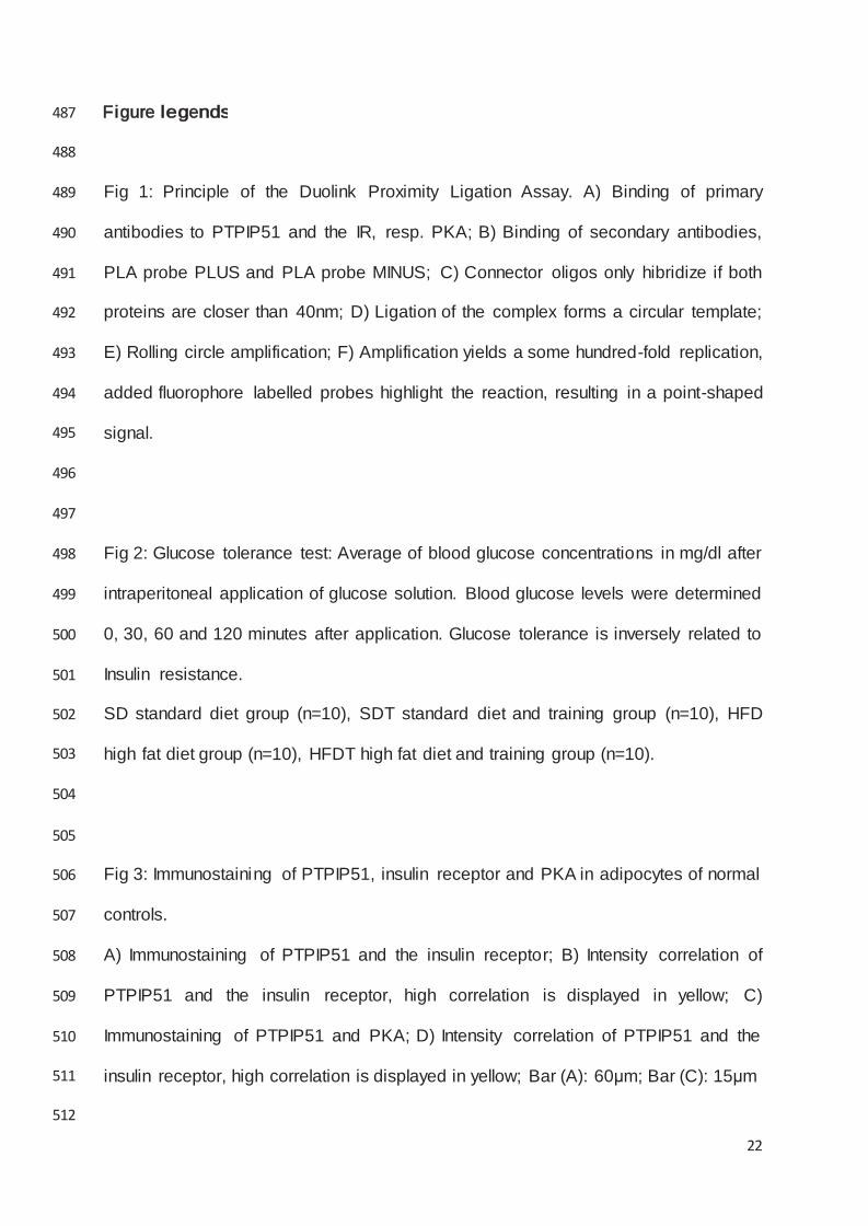
Figure legends 487
22
488
489 Fig 1: Principle of the Duolink Proximity Ligation Assay. A) Binding of primary
490 antibodies to PTPIP51 and the IR, resp. PKA; B) Binding of secondary antibodies,
491 PLA probe PLUS and PLA probe MINUS; C) Connector oligos only hibridize if both
492 proteins are closer than 40nm; D) Ligation of the complex forms a circular template;
493 E) Rolling circle amplification; F) Amplification yields a some hundred-fold replication,
494 added fluorophore labelled probes highlight the reaction, resulting in a point-shaped
495
496
497
signal.
498 Fig 2: Glucose tolerance test: Average of blood glucose concentrations in mg/dl after
499 intraperitoneal application of glucose solution. Blood glucose levels were determined
500 0, 30, 60 and 120 minutes after application. Glucose tolerance is inversely related to
501 Insulin resistance.
502 SD standard diet group (n=10), SDT standard diet and training group (n=10), HFD
503
504
505
high fat diet group (n=10), HFDT high fat diet and training group (n=10).
506 Fig 3: Immunostaining of PTPIP51, insulin receptor and PKA in adipocytes of normal
507 controls.
508 A) Immunostaining of PTPIP51 and the insulin receptor; B) Intensity correlation of
509 PTPIP51 and the insulin receptor, high correlation is displayed in yellow; C)
510 Immunostaining of PTPIP51 and PKA; D) Intensity correlation of PTPIP51 and the
511
512
insulin receptor, high correlation is displayed in yellow; Bar (A): 60μm; Bar (C): 15μm

23
513
514 Fig 4: Duolink proximity ligation assay to detect interactions between PTPIP51 and
515 the insulin receptor (IR) resp. PKA
516 A) Interaction between PTPIP51 and the IR; B) Interaction between PTPIP51 and
517 PKA; C) Negative control, PTPIP51 and the IR in testis, no interaction known; D)
518 Negative control, DPLA without primary antibodies in adipose tissue; Nuclei are
519 marked by DAPI staining; Arrows: positive reactions; Bars (A, B): 50μm; Bars (C, D):
520
521
522
20μm
523 Fig 5: Semiquantitative analysis of the interaction between PTPIP51 and its
524 interaction with IR and with PKA.
525 Every section was subdivided into rectangles of the same size and dots were
526 counted. The results were averaged for each section.
527 A) Semiquantitative evaluation of PTPIP51 interaction with IR;
528 B) Semiquantitative evaluation of PTPIP51 interaction with PKA.
529 SD: standard diet group; SDT: standard diet and training group; HFD: high fat diet
530
531
532
group; HFDT: high fat diet and training group.