Pseudocyst of pancreas and benign cystic neoplasms
35
PSEUDOCYST OF PANCREAS AND BENIGN CYSTIC NEOPLASMS Dr. E.Kaushik Kumar Stanley Medical College Hospital,Chennai
-
Upload
kaushik-kumar-eswaran -
Category
Health & Medicine
-
view
39 -
download
2
Transcript of Pseudocyst of pancreas and benign cystic neoplasms

PSEUDOCYST OF PANCREAS AND BENIGN CYSTIC NEOPLASMS
Dr. E.Kaushik KumarStanley Medical College Hospital,Chennai

குறள் 84
அகனமர்ந்துசெய்யா�ள்உறை�யும்முகனமர்ந்து
நல்வி�ருந் தோ��ம்புவி�ன்இல்
வி�ளக்கம் மனமக�ழ்ச்!றையாமுகமலர்ச்!யா�ல் க�ட்டி
வி�ருந்��னறை%வி%தோவிற்பவிர்வீட்டில்அமர்ந்து செல்விம் எனும்��ருமகள்வி�ழ்வி�ள்
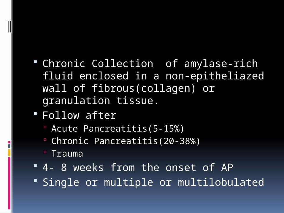
Chronic Collection of amylase-rich fluid enclosed in a non-epitheliazed wall of fibrous(collagen) or granulation tissue.
Follow after Acute Pancreatitis(5-15%) Chronic Pancreatitis(20-38%) Trauma
4- 8 weeks from the onset of AP Single or multiple or multilobulated
Duct leak Intrapancreatic or extend beyond into
other cavities or compartments Regress spontaneously 50% Chronic pseudocysts may persist
Thick-walled or large (over 6 cm in diameter)
Lasted for a long time (over 12 weeks) Arisen in the context of chronic pancreatitis

Symptoms and Complications Pain Fullness Satiety Loss of weight

Complications include Abscess /systemic sepsis SMV/PV thrombosis Intracystic
hemorrhage/pseudoaneurysms Peritonitis/intraperitoneal bleeding Pressure efects Pancreatico pleural fistula
INVESTIGATIONS
Ultasonogram CT EUS ERCP MRCP FNA

US,CT- Identification of pseudocyst and differentiates from abscess
EUS- Guided FNA Differentiates between chronic pseudocyst and cystic neoplasms
ERCP/MRCP- ductal communication,ductal anomalies, chronic pancreatitis,plan treatment

CEA -High level in mucinous tumours >400ng/ml
Amylase- Level usually high in pseudocysts, but occasionally in tumours
Cytology- Inflammatory cells in pseudocyst

INTERVENTION
Symptoms
If complications develop
Distinction has to be made between a pseudocyst and a tumour

Internal drainage vs external drainage Endoscopic
Percutaneous
Surgical –Open/Laparoscopic
Percutaneous
Usually avoided
Recurrence
Pancreatico-cutaneous fistula
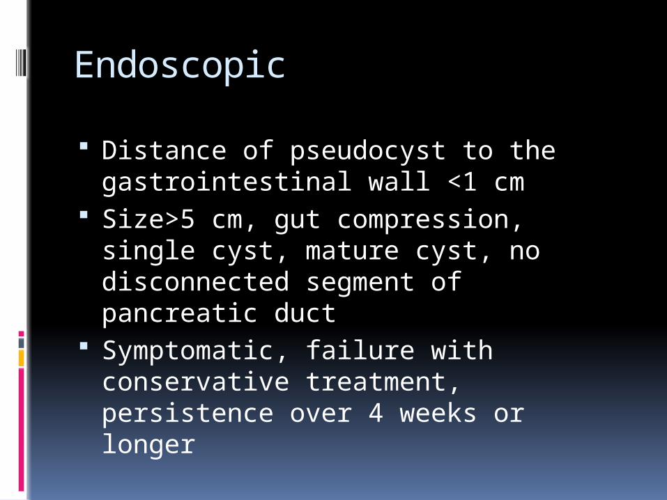
Endoscopic
Distance of pseudocyst to the gastrointestinal wall <1 cm
Size>5 cm, gut compression, single cyst, mature cyst, no disconnected segment of pancreatic duct
Symptomatic, failure with conservative treatment, persistence over 4 weeks or longer

Location of transmural approach based on maximal bulge of the pseudocyst to the adjacent wall
Mature cyst, perform pancreatography first, prefer transpapillary approach, if feasible
Check for debris within pseudocyst Neoplasm and pseudoaneurysm
have to be ruled out

Surgical Complications Drainage internally
Stomach Duodenum Jejunum
Recurrence <5%


Newer techiques
Forward-viewing echoendoscope PFC puncture devices Combination devices Devices for maintenance of
cystenterostomy Multiple transluminal gateway
technique (MTGT)


CYSTIC NEOPLASMS
Second most common neoplasm of exocrine pancreas Mucinous cystic neoplasm Serous cystic neoplasm Intraductal papillary neoplasm

Mucinous cystic neoplasm
Most common Histologic spectrum from benign to
invasive carcinomas. MCNs contain mucin-producing
epithelium Mucin-rich cells and ovarian-like
stroma Estrogen and progesterone staining
are positive in most cases.

Frequently seen in young women, the mean age at presentation is in the fifth decade.
Men are rarely affected Body and tail of the pancreas Up to 50% of patients present with
vague abdominal pain. A history of pancreatitis may be found
in up to 20% of patients-common misdiagnosis of pseudocyst

CT Appearance Solitary cyst Fine septations Rim of calcification
Malignant Large tumour size Egg-shell calcification Mural nodule


Potential to turn to malignancy
Resection Curative No further surveilance needed

Serous cystic neoplasm
Higher median age Head of pancreas Vague abdominal pain,weight
loss,obstructive jaundice Large well circumscribed mass

CT appearance Central calcification with radiating septa “Sun-burst” appearance
Microscopic appearance Multiloculated,glycogen rich small cysts
Resection >4cm Rapidly growing Diagnosis of malignancy is uncertain

Intraductal papillary mucinous neoplasm
6th-7th decade Wide spectrum of epithelial
changes,including benign adenoma, borderline, carcinoma in situ, and invasive adenocarcinoma
Types Side branch Main duct Mixed

Side branch IPMNInvolves dilation of the pancreatic duct side branches that communicate with but do not involve the main pancreatic duct.
Focal/multifocal Malignant transformation directly
proportional to cystic dilatation 10-15% risk


Main duct IPMN
Abnormal cystic dilation of the main pancreatic duct with columnar metaplasia and thick mucinous secretions
Focal or diffuse 30%-50% risk of invasive cancer Surgical resection is the corner stone
of treatment –Partial Pancreatectomy

50% present with abdominal pain 25% present with AP Diagnostic confusion with – Chronic
Pancreatitis Risk of malignancy
Jaundice Elevated serum alkaline phosphatase level Mural nodules Diabetes Main pancreatic duct diameter of 7 mm

Mixed type
Side branch IPMN that has extended to involve the main pancreatic duct to a varying degree
Individuals with side branch cysts who exhibit upstream dilation of the pancreatic duct
Characteristics and management- similar to Main duct IPMN


Conclusion
Observation for patients with asymptomatic small (<3 cm) branch duct IPMNs that have no associated nodularity.
A plan for watchful surveillance with delayed intervention in these patients is reasonable because Risks for malignancy with small,
asymptomatic branch duct tumors is low Most Patients are older Time required to develop invasive malignancy
>patient’s life expectancy.