Debility Unspecified and Adult Failure to Thrive: Determining Appropriate Diagnosis

University of North DakotaUND Scholarly Commons
Physical Therapy Scholarly Projects Department of Physical Therapy
2016
Proximal Muscle Weakness and DebilitySecondary to Polymyositis: A Case ReportAdam MeidingerUniversity of North Dakota
Follow this and additional works at: https://commons.und.edu/pt-grad
Part of the Physical Therapy Commons
This Scholarly Project is brought to you for free and open access by the Department of Physical Therapy at UND Scholarly Commons. It has beenaccepted for inclusion in Physical Therapy Scholarly Projects by an authorized administrator of UND Scholarly Commons. For more information,please contact [email protected].
Recommended CitationMeidinger, Adam, "Proximal Muscle Weakness and Debility Secondary to Polymyositis: A Case Report" (2016). Physical TherapyScholarly Projects. 554.https://commons.und.edu/pt-grad/554

PROXIMAL MUSCLE WEAKNESS AND DEBILITY SECONDARY TO POLYMYOSITIS: A CASE REPORT
by
ADAM MEIDINGER Bachelor of Science Marketing, Psychology; Minor Philosophy
North Dakota State University, 2011
A Scholarly Project Submitted to the Graduate Faculty of the
Department of Physical Therapy
School of Medicine
University of North Dakota
in partial fulfillment of the requirements for the degree of
Doctor of Physical Therapy
Grand Forks, North Dakota May, 2016

This Scholarly Project, submitted by Adam Meidinger in partial fulfillment of the requirements for the Degree of Doctor of Physical Therapy from the University of North Dakota, has been read by the Advisor and Chairperson of Physical Therapy under whom the work has been done and is hereby approved.
r
(Chairperson, P sical Therapy)
ii

Title
Department
Degree
PERMISSION
PROXIMAL MUSCLE WEAKNESS AND DEBILITY SECONDARY TO POLYMYOSITIS: A CASE REPORT
Physical Therapy
Doctor of Physical Therapy
In presenting this Scholarly Proj ect in partial fulfillment of the requirements for a graduate degree from the University of North Dakota, I agree that the Department of Physical Therapy shall make it freely available for inspection. T further agree that permission for extensive copying for scholarly purposes may be granted by the professor who supervised my work or, in her absence, by the Chairperson of the department. It is understood that any copying or publication or other use of this Scholarly Project or part thereof for financial gain shall not be allowed without my written permission. It is also understood that due recognition shall be given to me and the University of North Dakota in any scholarly use which may be made of any material in this Scholarly Project.
Signature
Date 11/9/15. • •
iii

TABLE OF CONTENTS
LIST OF TABLES............................................................................... v
ACKNOWLEDGEMENTS..................... ................................................ VI
ABSTRACT ......................................................................... ".... ........ vu
CHAPTER I BACKGROUND AND PURPOSE.......................................... 1
II CASE DESCRIPTION........................................................ 5
Examination, Evaluation, and Diagnosis.................................... 6
Progress and Plan of Care..................................................... 9
III INTERVENTION ............................................................. . 10
IV OUTCOME ................................................................... .. 14
V DISCUSSION ................................................................. . 16
Reflective Practice............................................................. 17
Addendum.. ...................... ............... ....... .............. ....... ... 18
REFERENCES.................................... .................................. ........ ...... 19
iv

LIST OF TABLES
1. Table 1: DE and LE Range of Motion................................................... 7
2. Table 2: DE and LE Strength.................. ............................ ...... ......... 7
3. Table 3: Strengthening Exercises...................... ................................... 11
4. Table 4: Therapeutic Activity................... ..... ............................... ...... 12
v

ACKNOWLEDGEMENTS
I'd like to thank the faculty of the UND PT department, my first clinical instructors, and
my friends and families for giving me the foundation and support necessary to become a physical
therapist. You have made this a great journey. Also, all my patients - past and future - you're the
reason we do this.
vi

ABSTRACT
BACKGROUND AND PURPOSE: Polymyositis (PM) is the most common inflammatory myopathy in persons over age 50. 1 It predominantly causes weakness at proximal musculature including the trunk, hips, thighs, shoulders, upper arms, and neck leading to challenges with ambulation and mobility, community activities, and difficulty swallowing, eating, and talking.2
Studies have shown physical therapy to be a staple in the plan of care for treatment of PM to aid in the restoration of muscle function and to improve or prevent further loss of strength.3,4 The purpose of this case report is to outline physical therapy's role in the treatment of a patient who showed both improvement and digression in tenns of strength and mobility secondary to polymyositis.
CASE DESCRIPTION: The patient was a 69-year-old female presenting to swing bed physical therapy for weakness and debility secondary to PM. The patient had decreased balance and was unable to independently rise from a chair. She demonstrated 2/5 MMT bilaterally at the hip and shoulder preventing her from being able to complete most ALDs, transferring, and bed mobility. She could ambulate up to 200 feet with SBA for safety but fatigued quickly.
INTERVENTION: The patient was seen for 11 sessions during her IS-day swing bed stay and two to three times per week for three weeks as an outpatient. Physical therapy interventions included task specific full body strengthening and endurance interventions, gait training, balance activities, and as needed manual therapy techniques for pain management. Strengthening included upper extremity assisted ROM at the shoulder, weight resisted UE exercises, assisted hip and knee exercises in supine and/or sitting, weight resisted LE exercises, sit to stands, ambulation for distance, and core-focused rolling and assisted sitting exercises.
OUTCOMES: The patient gained the ability to rise from a lift chair and roll in bed independently. Endurance increased, and she was able to ambulate up to 550 feet with SBA for safety. Upper extremity strength remained unchanged while in swing bed, however as an outpatient strength increased slightly. The patient reported better-perceived tolerance to transfers and demonstrated improved balance. Tluee weeks into outpatient therapy, she had a reaction to her Prednisone treatments and developed situational diabetes mellitus causing muscle weakness and debility. She was hospitalized for management.
DISCUSSION: The patient had a long and difficult stay at different medical facilities to manage her PM. Even with her unexpected medical difficulties, the patient demonstrated improvement of muscle function, greater in her LEs than UEs. This led to improved functional mobility, improved balance, and improved tolerance to ADLs. This case report outlines the need for further research and suggests that a detailed physical therapy plan of care should be created and driven by functional and ADL specific goals.
vii

CHAPTER I
BACKGROUND AND PURPOSE
Idiopathic inflammatory myopathies are a classification ofrare, systemic, acquired
diseases. There are four types: dermatomyositis, polymyositis, immune-mediated necrotizing
myopathies, and sporadic inclusion body myositis. 5 Myopathy is a term that means "muscle
disease." Inflammatory myopathies are a group of diseases that involve muscle inflarmnation
with accompanying weakness. Although exact causes are unlmown, evidence suggests this can
be caused by an allergic reaction, exposure to certain substances or medicines, or exposure to a
virus or other infectious agent6 The chronic myopathic diseases also have no specific
identifiable cause and are thought to be autoimmune disorders with a rheumatic component. 6
Idiopathic inflammatory myopathies most commonly affect adult patients of Caucasian ethnicity.
Females are affected at a 5: 1 ratio to males for people ages 18-44 years of age. Incidence rates
are estimated between 5-10 cases per million in adults and 1-5 cases per million in children. The
condition may affect those under the age of 18 and is referred to as juvenile dermaJpolymyositis 5
Polymyositis (PM) is defined as an uncommon acquired idiopathic inflammatory
myopathy. As with all inflammatory myopathies, PM affects women more than men and causes
can be varied to unknown. It is the most common inflarmnatory myopathy in persons over age
50. 1 PM also resembles an autoimmune disorder where the body attacks its own tissues. It is
believed the condition is triggered by environmental factors in genetically susceptible
individuals. Associated conditions include Raynaud's, connective tissue disorders such as lupus,
rheumatoid arthritis, scleroderma, and Sjogren's, cardiovascular diseases such as myocarditis,
1

congestive heart failure, and arrhythmias, and lung diseases such as fibrosis. Possible triggers
include drugs such as statins, viruses, cancer, or exacerbation of connective tissue disease.
Typical presentation of PM in both juvenile and adult populations includes predominant
weakness at proximal musculature including the trunk, hips, thighs, shoulders, upper arms, and
the neck. 2 Proximal muscle weakness can progress to the respiratory and/or the accessory
respiratory muscles as well as muscles involved in speech and swallowing. Affected muscles
digress in strength at a rate unique to each patient's condition. A similar condition known as
dermatomyositis (DM) is a connective tissue disease related to PM but involves inflammation of
the skin as well as inflammatory myopathy.7 Managing inflanunatory myopathies is driven by
the goals of reducing inflammation, restoring muscle performance, and preventing chronic
muscle disease and extra-muscular organ damage to restore quality of life. 5 Common
medications used to treat PM include corticosteroids such as prednisone or corticosteroid-sparing
agents such as azathioprine and methotrexate, or rituximab which are commonly used for
arthritis. 7
The medical management team, under the direction of a rheumatologist, often includes
physical therapists, dieticians, and consultations to cardiologists, pulmonary specialists, and
speech therapists. Together this tearn will work to restore quality of life with intervention
strategies determined on a case-by-case basis. l
Physical therapy intervention is considered a staple in the plan of care for a person
affected by PM or any inflammatory myopathy3,4 The goal of therapy is to restore muscle
function and improve or prevent further loss of strength during acute episodes. 5 Because lower
extremity, hip, core, and shoulder musculatures are commonly affected, persons face challenges
with ambulation and mobility, ADLs including grooming, washing, and eating, community
involvement due to difficulties with balance, transferring, and mobility, and difficulty
2

swallowing, eating, and talking.] Due to the physical nature of the condition, there have been
studies that examine the efficacious use of physical therapy interventions and exercise as a
treatment for PM8
Physical rehabilitation interventions present unique difficulties in patients with PM
because of the degree of physical limitations that present as well as multi-muscle and system
involvement, often times including respiratory musculature. It is recommended that exercise
prescription focus on task-specific performance, such as eating and grooming as well as mobility
and ambulation, and that pulmonary physical therapy intervention be included to improve
respiratory function and improve tolerance to exercise9 It is also recommended that diet be
considered a vital intervention strategy for PM to help combat any extraneous sources of
inflammation, which often warrants a referral to a registered dietician.!
During an acute onset of PM, patients often receive medical management and forgo
therapy services until medical stabilization has occurred. This is because it is assumed
inflammation will inhibit proper exercise and strengthening interventions. A study examined
patients 2-3 weeks post-acute phase PM. Active PM patients received proximal, distal, and
respiratory muscle strengthening interventions. No decrease in muscle function was noted and
respiratory muscle strength increased during disease progression. This study supports early
physical therapy intervention for PM/DM patients to prevent muscle atrophy.lO A similar early
intervention study by Alexanderson et al showed that long-term improvements in strength were
found in exercise groups not otherwise present in non-exercise groups. However, general
functional mobility was not shown to be sustained without continued exercise for patients with a
history ofPMll
Resisted exercise prescription programs for adults with PM were shown to reduce pain,
improve function, and improve physical conditioning as measured by intramuscular Type-I fiber
3

concentration, intramuscular gene expression for protein synthesis, and SF-36 scores. 12 Resisted
exercise programs were also shown to decrease levels of intramuscular inflammation as
measured by medical biomarkers believed to be the result of increased intramuscular processes.
While it is understood that resisted exercise may temporarily increase intramuscular
inflammation, this was not shown to exacerbate inflammation caused by PM/DM. 12 Volume of
oxygen maximal uptake (V02 max), overall health and disability, as well as overall disease
activity was shown to improve with a combination of aerobic and resisted exercise programs in
adults with chronic PMIDM.4 Physiologic changes in aerobic milieu in the muscle as well as
aerobic capacity were improved in juveniles and adults with both acute and chronic conditions
leading to reduced disability3
Based on literature review and accepted medical practice, physical therapy plays an
important role in the treatment and management of functional strength, mobility, and quality of
life of patients diagnosed with polymyositis.
The purpose of this case report is to outline a patient who showed both improvement and
digression in terms of strength and mobility secondary to polymyositis. She also faced
complications secondary to medical treatment. This case study outlines the role of physical
therapy in her treatment and recovery.
4

HISTORY
CHAPTER II
CASE DESCRIPTION
A 69-year-old female of Caucasian descent presented to physical therapy following
admission to swing bed services for weakness and debility secondary to polymyositis (PM). Her
condition started approximately six months prior to her swing bed admission. She began with
outpatient medical management for treatment but was demonstrating proximal muscle weakness
with weakness progressing into her upper extremities. This prevented her from being fully able
to care for herself but she did participate in self-care. Her husband, her main caregiver, recently
underwent a cholecystectomy surgery and was unable to assist her with ADLs.
As the disease progressed she noticed a slow loss ofUE strength and increased balance
impairments with a decrease in ambulation endurance. Prior to the disease progression she was
independent with ADLs. Prior to her diagnosis with PM the patient was in good health with a
medical history of hypertension, hysterectomy, and bilateral cataract extraction. The patient was
currently taking Prednisone for treatment of PM. She was also on a medication for management
of hypertension.
At admission, the patient exhibited signs of hemochromatosis and diabetes mellitus
believed to a complication of Prednisone. Physically, she was unable to independently rise from
a chair but did ambulate up to 200 feet without an assistive device and with SBA for safety. She
had not been able to participate in normal social interactions such as visiting friends and family
5

and attending church due to weakness and difficulty with mobility. She reported no smoking, use
of drugs, or regular use of alcohol. Her family medical history was unavailable.
The patient's primary language was identified as English but she did speak some
German. The patient worked as a school lunch provider at a rural school and retired about one
year priol'. Before being affected by PM, she also helped her husband with farm and ranch work.
Their closest child lived approximately 90 miles away. Her social activities included spending
time with friends and family, attending church, and helping her husband with yard work. She
lived in the country approximately 15 miles from the nearest town with medical services. All
services she needed to access were on the main floor of her home but there were four steps to
enter. She used assistive devices for safety including a walker and railings to navigate within her
home. Her goal for discharge was to return back to her home following her husband's recovery.
Her therapy needs consisted of strengthening and endurance training so that she would be able to
complete ADLs independently. This was her first time receiving physical therapy.
EXAMINATION AND EVALUATION
The patient was admitted to swing bed with HR and BP WNL, no signs of edema
peripherally, and clear lung sounds. She was 65" tall and 175lbs with a BMI of29.1 classifying
her as overweight. The patient reported being right handed. She was unrestricted with sleep,
cognitively orientated x 4, and demonstrated no apparent psychological impairments such as
delusions or hallucinations. She did demonstrate signs of lethargy and possible situation-induced
anxiety as identified by therapy services and her admitting physician. The patient reported no
pain. Skin integrity was intact and the patient demonstrated no anatomical abnormalities. Lower
and upper extremity muscle symmetry was normal and posture appropriate; she did exhibit
slightly stooped posture in sitting due to weakness. Static standing posture appeared appropriate.
The patient did have some ROM and strength limitations as noted in Tables 2-1 and 2-2.
6

Table 2-1: UE and LE Range of Motion Bilateral AROM wrist, elbow WFL shoulder flex 70°
abduction 65° remaining WFL
UE PROM grossly WFL
Bilateral LE
AROM knee, ankle WFL hip was unable to be tested in appropriate position due to weakness
PROM grossly WFL
Table 2-2: UE and LE Strength Bilateral Gross handlwrist MMT UE 4/5 Bilateral LE
Gross ankle MMT 4+15
Gross elbow MMT 4-/5 Gross knee MMT 4/5
Gross shoulder MMT 2/5 Gross hip MMT 2/5
Mobility: Patient's standing balance was diminished due to LE and core weakness. She was able
to ambulate 200 feet independently on level surfaces with no AD before needing to sit due to
muscle fatigue. Stand by assist of therapist and gait belt were used for safety. Static sitting
balance was good, however dynamic sitting balance, although not specifically tested, appeared to
be diminished. Patient required Min/Mod-A x 1 for sit to stand transfers and bed mobility,
including supine-to-sit and rolling.
Functional Assessment: The Elderly Mobility Scale (EMS) was completed as part of the initial
evaluation as mobility was a large part of this patient's impairment. The patient's EMS score was
12/20, which placed the patient borderline in terms of safe mobility and independence in ADLs
as she required some help with mobility. Even with this score, the patient needed assistance with
basic ADLs including transfers and toileting. Strength loss due to PM was the largest underlying
factor for patient's functional level.
The EMS was shown to have high intra and inter-rater reliability as well as high validity
for classification of elderly mobility indicated in acute care settings. 13 The EMS was designed to
assess mobility in £i'ail, elderly people on seven items considered essential for performing ADLs.
7

These include transfer, gait, and balance tasks. A score of 14-20 indicates the patient maneuvers
alone and is independent in basic ADLs. Individuals are considered generally safe to go home
but may require some help. A score of 10-13 indicates the patient is borderline in terms of safe
mobility and independence in ADLs. A score <10 indicates the patient is dependent in mobility
maneuvers and requires help with basic ADLs. They may require home care or long-term care. 13
PT DIAGNOSIS
Patient demonstrated severe strength and mobility impairments correlating with common
symptoms of PM. Deconditioning was also apparent in her ambulation abilities and distance.
Proximal muscles of the UE, LE, and trunk demonstrated the greatest amount of weakness with
weakness also progressing to her more distal UE as apparent in loss of strength at her elbows.
Because of these impairments, the patient was unable to perform self-care, sit-to-stand transfers,
and was limited in bed mobility. She could not reach to the back of her head so dressing,
grooming, and eating activities had also become affected. She was unable to participate in
community and leisure activities due to her lack of strength and endurance. She was at a high
risk for falling and had standing and dynamic sitting balance impairments secondary to
weakness. She also faced potential respiratory difficulties if accessory muscle strength continued
to decline.
SHORT TERM GOALS (to be met in I week)
I. Patient will improve bilateral shoulder and hip strength to 3/5 to increase her
independence with activities of daily living.
2. Patient will demonstrate sit-to-stand transfer with Min-A of I to increase
independence in mobility.
3. Patient will improve Elderly Mobility Scale score by 2-3 points to demonstrate
improved safety in transfers, gait, and balance.
8

LONG TERM GOALS (to be met prior to discharge from swing bed)
1. Patient will improve bilateral shoulder and hip strength to 4/5 to increase her
independence with activities of daily living.
2. Patient will demonstrate sit-to-stand transfers independently using a lift chair to
increase independence in mobility.
3. Patient will improve Elderly Mobility Scale score by 4-6 points to demonstrate
improved safety in transfers, gait, and balance.
PROGNOSIS
With proper medical management of PM, the prognosis was good for the patient to return
home and be more independent with ADLs. It was also anticipated that she would return to
community and leisure activities with the potential of using an assistive device for added safety.
DISCHARGE CRITERIA
Patient would be discharged from physical therapy services when anticipated goals were
achieved, when she was no longer receiving benefit from physical therapy services, or when
requested by the patient and/or referring physician.
PLAN OF CARE
The patient was appropriate for PT services for treatment of strength and endurance loss
secondary to PM. The patient's treatment plan of care included modalities for pain management,
manual therapy to improve soft tissue and joint mobility, therapeutic exercise to improve
strength and mobility, gait and balance training, and endurance exercises. The initial plan of care
consisted of seeing the patient 4 to 5 times per week during her swing bed stay.
9

CHAPTER III
INTERVENTION
The patient was seen once daily, five times per week, for 45-60 minute sessions for a
total of 11 visits. Three different therapists saw her, as each therapist would travel to different
community clinics on different days of the week, rendering them unavailable to see inpatients on
those specific days. The patient's primary symptoms from polymyositis (PM) were severe
strength and mobility impairments. Because ofthe decrease in her mobility, the patient also
exhibited signs of deconditioning apparent in her decreased tolerance to exercise time and
ambulation distances. Exercise was started immediately as strengthening has been shown to slow
and combat the strength lost due to PM. 4.5.14 PT interventions included general full body
strengthening and endurance interventions, as strength and endurance were the patient's main
functional limitations.
Intervention strategies were based on patient's symptoms and progression varied on a
day-to-day basis. Interventions were chosen based on the patient's tolerance during therapy
sessions with numerous strengthening exercises completed (see Table 3-1). Therapeutic activities
were also varied and based on the patient's response and are described in Table 3-2. The tables
are not comprehensive of every exercise and activity as the patient was seen by three therapists
and all intervention data was not available at the time of this case report.
10
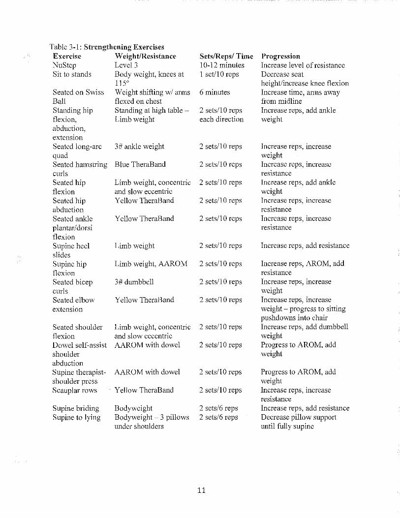
Table 3-1: Strengthening Exercises Exercise Weight!Resistance SetslReps! Time Progression NuStep Level 3 10-12 minutes Increase level of resistance Sit to stands Body weight, knees at 1 setilO reps Decrease seat
1150 heightlincrease knee flexion Seated on Swiss Weight shifting w! arms 6 minutes Increase time, arms away Ball flexed on chest from midline Standing hip Standing at high table - 2 setsll 0 reps Increase reps, add ankle flexion, Limb weight each direction weight abduction, extension Seated long -arc 3# ankle weight 2 setsll 0 reps Increase reps, increase quad weight Seated hamstring Blue TheraBand 2 sets! 1 0 reps Increase reps, increase curls resistance Seated hip Limb weight, concentric 2 setsll 0 reps Increase reps, add ankle flexion and slow eccentric weight Seated hip Yellow TheraBand 2 setsll 0 reps Increase reps, increase abduction resistance Seated ankle YeHow TheraBand 2 setsll 0 reps Increase reps, increase plantar!dorsi resistance flexion Supine heel Limb weight 2 setsll 0 reps Increase reps, add resistance slides Supine hip Limb weight, AAROM 2 setsll 0 reps Increase reps, AROM, add flexion resistance Seated bicep 3# dumbbell 2 sets!IO reps Increase reps, increase curls weight Seated elbow Yellow TheraBand 2 sets!IO reps Increase reps, increase extension weight - progress to sitting
pushdowns into chair Seated shoulder Limb weight, concentric 2 setsll 0 reps Increase reps, add dumbbell flexion and slow eccentric weight Dowel self-assist AAROM with dowel 2 setsll 0 reps Progress to AROM, add shoulder weight abduction Supine therapist- AAROM with dowel 2 setsll 0 reps Progress to AROM, add shoulder press weight Scauplar rows YeHow TheraBand 2 sets!IO reps Increase reps, increase
resistance Supine briding Bodyweight 2 sets!6 reps Increase reps, add resistance Supine to lying Bodyweight - 3 pillows 2 sets!6 reps Decrease pillow support
under shoulders until fully supine
11

Table 3-2: Therapeutic Activity% Activity Degree Gait training 400 feet x 2
(needed rest break) Stair mobility On to and off of 6"
or 8"box
Bed mobility, rolling Bed mobility, scooting Transfer training Mini squats Marching while holding handrail
Supine to side lying Seated, left to right and right to left Sit to stand from hospital bed Body weight Body weight
Condition SBA for safety
Hand rail and quad cane. Max-A on ascending, Mod-A on descending Independent - needed cuemg Independent - moderate difficulty Mod-A, verbal cues required Increase endurance Increase dynamic balance
Progression Increase distance, increase speed Decrease assistive devices, increase step number, increase step height Supine to prone, Supine or side lying to seated Increase number of times
Increase number Decrease stability, eliminate hand support, increase instability of surface
Stair training 5 steps ascend and Mod-A with handrail Increase step number, descend and quad cane decrease stability, decrease
assistance * A gait belt was used with all seated or standing exercises and activities for safety.
Gait training was performed to ensure patient's safety during ambulation. Gait training
also doubled as a flllctional endurance exercise. Gait training was performed with the use of a
front-wheeled-walker and gait belt with assist/contact guard for patient safety. She ambulated in
the hallways of the hospital and therapy department. Initial distance patient tolerated during gait
training was 800 feet with a rest break after 400 feet. The patient demonstrated increased lateral
weight shift, wide base of support, and decreased step length. Distance was increased each day as
tolerated.
Therapeutic activity, focusing on balance training, was included as the patient exhibited
decreased balance. Some alternative therapeutic activities included standing on 1 to 3" foam pads
while incorporating reaching, standing on a trampoline incorporating reaching, and stepping
exercises to the sides and backwards. The patient was assisted or guarded by two therapists
during these activities.
12

The patient would experience some pain primarily due to new exercises and activities.
Pain was mostly experienced in UE musculature such as upper trapezius and deltoids as well as
low back postural musculature. Modalities for pain management were performed PRN and
included interferential current therapy: TENS, with parameters set to 80-120Hz, intensity as
tolerated, for 10 minutes. Manual therapy techniques consisting of soft tissue mobilization,
myofascial therapy, and trigger point release techniques were also used for pain management and
to improve tissue mobility prior to exercise. She reported a decrease in pain with these
interventions.
The patient was discharged to her home after 15 days in swing bed care. She continued
therapy as an outpatient and was seen two to three times per week for continued strengthening
and balance activities. She had been given a home exercise program at outpatient therapy to be
completed two to three times per day including the strengthening and balance activities the
patient was perfo=ing during therapy sessions. Her husband was coached on proper assistive
transfer techniques to ensure both his and the patient's safety during transfers. More advanced
balance and strengthening activities were also performed during therapy under the supervision of
physical therapists for the patient's safety.
13

CHAPTER IV
OUTCOMES
Throughout the course of the patient's IS-day stay in swing bed, she did demonstrate
gains in terms of functional strength and endurance. Although MMT grades did not change in her
UE or LEs, she was able to rise from a lift chair independently. She also demonstrated
improvements in bed mobility and was able to roll independently. Her supine-to-sit transfers still
required MIA. She continued to exhibit balance deficits and it was decided that, for patient
safety, she ambulate at all times using a FWW. Her endurance progressed and she was able to
ambulate 550 feet before needing to rest. Shc still required assistance for many ADLs as her UE
strength was not improving as quickly as her LE strength and her UE AROM remained limited.
Due to her continued improvement and the recovery of her husband, the plan to discharge the
patient home with continued physical therapy as an outpatient was discussed with the patient, her
spouse, and her children.
She was discharged home and as an outpatient, her UE strength increased slightly, up to
y, MMT grade, but continued to be impaired. Her balance and endurance improved which
lowered her fall probability. It was recommended that the patient continue to use a FWW for
safety during anlbulation. She reported better tolerance to transfers within her house, needing
less assistance from her husband. Socially, she was able to travel with assistance from her home
to therapy and also to church activities. The patient was satisfied with her progress in terms of
physical therapy and was motivated to work hard and continue outpatient physical therapy. An
Elderly Mobility Scale score was reassessed during outpatient therapy and was found to be
14

14/20, an increase of two points. This increase indicates that a patient is generally safe to go
home but may still require some help with ADLs.
Three weeks after beginning outpatient therapy, the patient had a reaction to her
Prednisone treatment and developed uncontrolled diabetes mellitus. It was unclear what
specifically prompted the reaction. She also demonstrated rapid-onset muscle weakness and
debility related to the Prednisone reaction. She was hospitalized for management of both the
diabetes mellitus and debility and began inpatient physical therapy. She demonstrated a severe
loss of strength with this re-admission and was unable to independently rise from a lift chair,
position herself in bed, or walk independently with the use of an assistive device. Eight days
later, following medical stabilization of her condition she was discharged to a local skilled
nursing facility for continued medical care and therapy. Within a week of admittance to the
nursing facility, she contracted Methicillin-resistant Staphylococcus Aureus (MRSA) from a
medical port. She was readmitted to the hospital under quarantine. Once the MRSA was
controlled, she was transferred to a larger metropolitan city with a specialized SNF where she
was able to receive physical therapy and assisted exercise throughout the day and was more
closely medically monitored. From there, a referral was made to the Mayo Clinic where the
patient was medically managed for PM. After Mayo, she was discharged back to the original
SNF where she remained at the time of this case report. Her goal was to return home by the end
of the year.
15

CHAPTER V
DISCUSSION
Once medically managed for Polymyositis, the patient began to show success utilizing
physical therapy services to improve her strength, mobility, and tolerance to ADLs. As a swing
bed patient, her LE strength and endurance improved as she was able to independently rise from
a lift chair and she increased ambulation distance between rest breaks. She also demonstrated
improved bed mobility by being able to independently reposition herself with less overall cueing.
During outpatient physical therapy, the patient demonstrated up to Yz MMT grade improvement
in her UE strength bilaterally allowing her to participate in more ADLs such as grooming and
brushing her teeth. She also reported improved tolerance to community mobility with the
assistance of her husband.
During both her swing bed and outpatient stays, she began to demonstrate restoration and
improvement of muscle function, especially in her LEs. These outcomes are consistent with
physical therapy's role in the treatment of PM, as physical therapy is cited as an essential
treatment to restore muscle strength and function. 5•9.lO.l5 The patient's treatment protocol was
dependent on the patient's tolerance and was based on task-specific exercises as suggested by
Aboussouan's research.9 Task-specific interventions included ambulation, stepping, and reaching
activities, as mobility impairments and self-care were the patient's main functional limitations.
This supports the research of Pappu and Seetharaman! stating the most common challenges of a
PM patient include ambulation and mobility, ADLs including grooming, washing, and eating,
and community involvement due to difficulties with balance, transferring, and mobility. This is
16

due to LE, hip, core, and shoulder musculature being most commonly affected. l Additional
treatment could have included respiratory physical therapy interventions with the goal of
strengthening the patient's respiratory musculature as these muscles are also commonly affected
by PM9 Dietary interventions or a referral to a dietician could have been provided to help
combat any extraneous sources of inflammation. 1
REFLECTIVE PRACTICE
There are limitations to this case report, mainly the difficulty the patient experienced with
medical management of her PM. The patient's transfers to different medical facilities were
appropriate considering the medical challenges she experienced. Her acute exacerbations
regressed her muscle strength and endurance gains, as well as decreased her selt:reported
motivation and tolerance to therapeutic interventions. Because the patient was transferred to
numerous care facilities during the course of her care, detailed information on the patient's
current status was unable to be obtained at the time of this case report.
Being able to complete a full physical therapy re-evaluation of the patient's current status
and compare changes in her medical treatment with therapy progressions would give a more
complete picture of physical therapy's influence on her outcomes. It is difficult to draw complete
conclusions about therapy's influence because of the length of therapy in each specific setting
(i.e. swing bed, outpatient, acute care, etc.), in addition to the medical difficulties the patient
encountered.
The physical therapy plan of care established for the patient focused on task-specific
interventions including exercises to improve reaching abilities, ambulation, transfers, and bed
mobility. The severe PM involvement and other medical issues the patient experienced required
physical therapy to challenge the patient and mimic the activities she may experience day-to-day
while working to prevent learning substitutions if possible. As the therapy team knew the time
17

with the patient was potentially limited (dependent on her swing bed stay length), and it was not
known if she would seek outpatient services, the goal was to regain muscle strength and function
as quickly as possible.
Areas offurther research should include case studies and reports outlining cases similar
in nature where medical management was difficult or delayed. Intervention outcomes based off
of therapy services should be examined. Also, a more detailed understanding of how medications
such as Prednisone can intramuscularly cause decreases in strength and muscle function could
have led to an improved plan of care.
One major benefit of this case report is that it provides an example ofthe extraneous
difficulties a patient can experience during the course of physical therapy sessions. This case
report outlines the need for further research and suggests that a detailed physical therapy plan of
care should be created and driven by functional and ADL specific goals.
ADDENDUM
The author of this case report has connections to the patient outside of physical therapy
services. The patient's husband informed the author that the patient returned home 10 months
after her initial swing bed stay. The patient remained moderately dependent on him for help with
transfers and ADLs. She continued outpatient physical therapy and twelve months after her
initial swing bed stay the patient was able to independently perform sit-to-stand transfers from a
regular dining room chair as well as from her bed. Her strength, function, and motivation
continues to improve.
18

REFERENCES
1. Pappu R, Seetharaman M. Polymyositis treatment and management. Medscape Web site. http://emedicine.medscape.comlarticle/3 3 5925-treatment. Updated 2014. Accessed March 14,2015.
2. Mayo Clinic Staff. Polymyositis. Mayo Clinic Web site. http://www.mayoclinic.org/diseases-conditions/polymyositis/basics/symptoms/con-20020710. Updated 2014. Accessed March 19, 2015.
3. Lundberg I, E., Vencovsky J, Alexanderson H. Therapy of myositis: Biological and physical. Curr Opin Rheumatol. 2014;26(6):704-711.
4. Alemo Munters L, Dastmalchi M, Andgren V, et a. Improvement in health and possible reduction in disease activity using endurance exercise in patients with established polymyositis and dermatomyositis: A multicenter randomized controlled trial with a 1-year open extension followup. Arthritis Care Res (Hoboken). 2013;65(12): 1959-68.
5. Idiopathic inflammatory myopathy. DynaMed. http://Search.e bscohost. com.ezproxy. undmedlibrary. orgllogin.aspx? direct=true&si te= Dy naMed&id=113862. Updated December 2014. Accessed March 2015.
6. National Institute of Neurological Disorders and Stroke. Inflammatory myopathies. 2007. 7. Femia A. Dermatomyositis. Medscape Web site.
http://emedicine.medscape.comlarticle/332783-overview. Updated 2014. Accessed March 19, 2015.
8. B6schung P., de Bruin ED., Tobler-Ammann BC., Maurer B., Knols RH. The relevance of applying exercise training principles when designing therapeutic interventions for patients with inflammatory myopathies: A systematic review. Rheumatology International. 2015;35(10): 1641-1654.
9. Aboussouan L. Mechanisms of exercise limitation and pulmonary rehabilitation for patients with neuromuscular disease. Chronic Respiratory Disease. 2009;6(4):231-249.
10. Varju C, Peth6 E, Kutas R, Czirjak L. The effect of physical exercise following acute disease exacerbation in patients with dermato/polymyositis. Clin Rehabil. 2003;17(1):83-87.
11. Alexanderson H, Munters L, Alemo, Dastmalchi M, et al. Resistive home exercise in patients with recent-onset polymyositis and dermatomyositis - A randomized controlled single-blinded study with a 2-year followup. J Rheumatol. 2014;41(6):1124-1132.
12. Alexanderson H. Exercise effects in patients with adult idiopathic inflammatory myopathies. Curr Opin Rheumatol. 2009;21(2):158-163.
13. Nolan JS, Remilton LE, Green MM. The reliability and validity of the elderly mobility scale in the acute hospital setting. J Allied Health Sci Pract. 2008;6(4):1-7.
14. de Sousa D, Junior IL. Evaluation of capacity aerobic and resistance exercises in patients with juvenile dermatomyositis and polymyositis: Literature review [Portuguese]. FISTOTER MOVIMENTO. 2009;22(4):489-496.
15. Wiesinger GF. Quittan M. Aringer M. Seeber A. Vole-Platzer B. Smolen J. Graninger W. Improvement of physical fitness and muscle strength in polymyositis/dermatomyositis patients by a training programme. Journal of Rheumatology. 1998;37(2):196-200.
19