DIFFERENTIAL DIAGNOSIS OF NEUROGENIC DISORDERS & MYOPATHIES · 1 DIFFERENTIAL DIAGNOSIS OF...
46
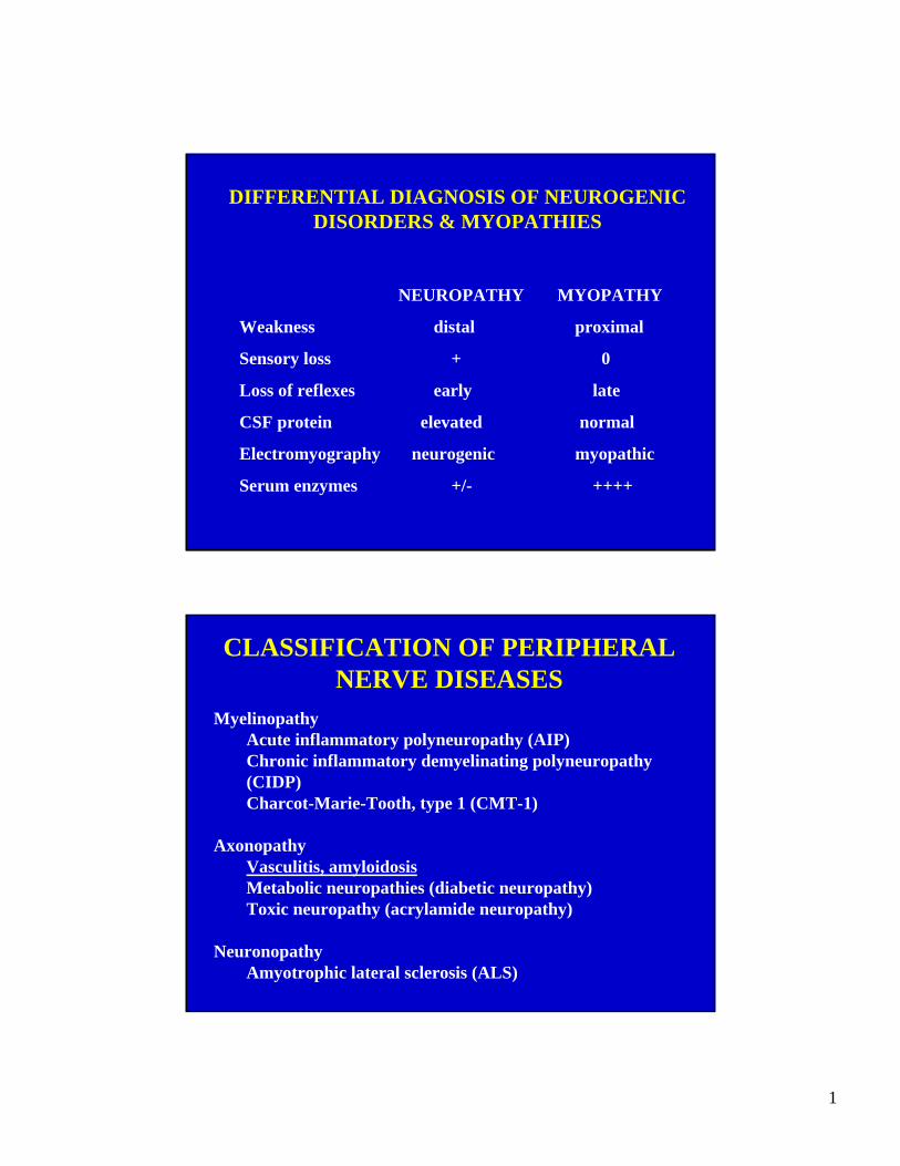
1 DIFFERENTIAL DIAGNOSIS OF NEUROGENIC DISORDERS & MYOPATHIES NEUROPATHY MYOPATHY Weakness distal proximal Sensory loss + 0 Loss of reflexes early late CSF protein elevated normal Electromyography neurogenic myopathic Serum enzymes +/- ++++ CLASSIFICATION OF PERIPHERAL NERVE DISEASES Myelinopathy Acute inflammatory polyneuropathy (AIP) Chronic inflammatory demyelinating polyneuropathy (CIDP) Charcot-Marie-Tooth, type 1 (CMT-1) Axonopathy Vasculitis, amyloidosis Metabolic neuropathies (diabetic neuropathy) Toxic neuropathy (acrylamide neuropathy) Neuronopathy Amyotrophic lateral sclerosis (ALS)
Transcript of DIFFERENTIAL DIAGNOSIS OF NEUROGENIC DISORDERS & MYOPATHIES · 1 DIFFERENTIAL DIAGNOSIS OF...
1
DIFFERENTIAL DIAGNOSIS OF NEUROGENIC DISORDERS & MYOPATHIES
NEUROPATHY MYOPATHY
Weakness distal proximal
Sensory loss + 0
Loss of reflexes early late
CSF protein elevated normal
Electromyography neurogenic myopathic
Serum enzymes +/- ++++
CLASSIFICATION OF PERIPHERALNERVE DISEASES
MyelinopathyAcute inflammatory polyneuropathy (AIP)Chronic inflammatory demyelinating polyneuropathy (CIDP)Charcot-Marie-Tooth, type 1 (CMT-1)
AxonopathyVasculitis, amyloidosisMetabolic neuropathies (diabetic neuropathy)Toxic neuropathy (acrylamide neuropathy)
NeuronopathyAmyotrophic lateral sclerosis (ALS)
2
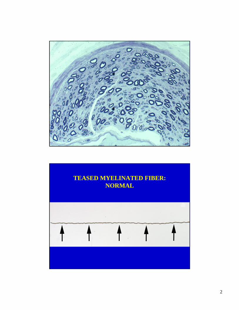
TEASED MYELINATED FIBER:NORMAL
3
4
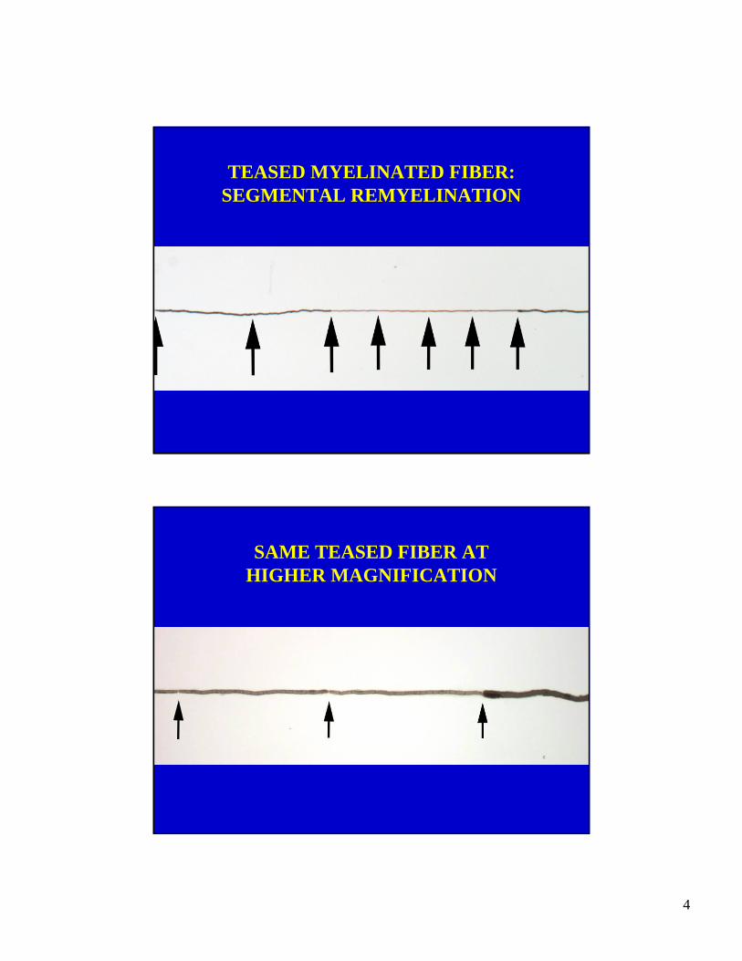
TEASED MYELINATED FIBER:SEGMENTAL REMYELINATION
SAME TEASED FIBER AT HIGHER MAGNIFICATION
5
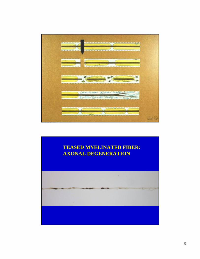
TEASED MYELINATED FIBER:AXONAL DEGENERATION
6
CLASSIFICATION OF PERIPHERALNERVE DISEASES
MyelinopathyAcute inflammatory polyneuropathy (AIP)Chronic inflammatory demyelinating polyneuropathy(CIDP)Charcot-Marie-Tooth, type 1 (CMT-1)
AxonopathyVasculitis, amyloidosisMetabolic neuropathies (diabetic neuropathy)Toxic neuropathy (acrylamide neuropathy)
NeuronopathyAmyotrophic lateral sclerosis (ALS)
ACUTE INFLAMMATORY POLYNEUROPATHY(GUILLAIN-BARRE SYNDROME [GBS])
• Rapidly progressive neuropathy, chiefly motor, reachingmaximum weakness usually within 1 to 2 weeks.
• An acute infectious illness precedes weakness in two thirds.
• Electrophysiology: slow conduction velocity & conduction blockbut also show axonal degeneration, usually of mild degree.
• Recovery takes weeks or months. Permanent handicap in 5%.
• Plasmapheresis or intravenous gamma globulin speeds recovery.
7
PATHOLOGY OF ACUTE INFLAMMATORY POLYNEUROPATHY
(GBS)
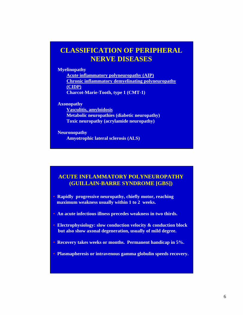
• Immune complexes (C3, IgG, IgM) are detectable on thesurface of myelin sheaths in the early stage.
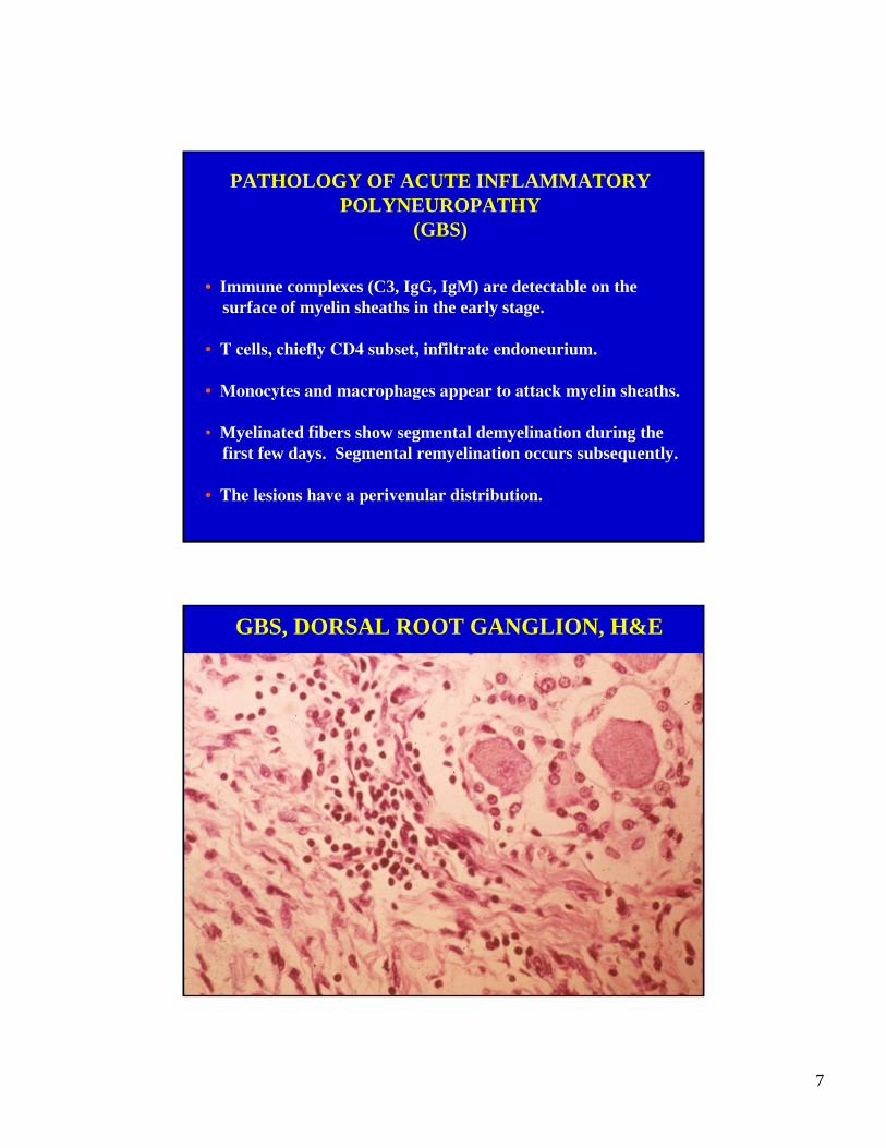
• T cells, chiefly CD4 subset, infiltrate endoneurium.
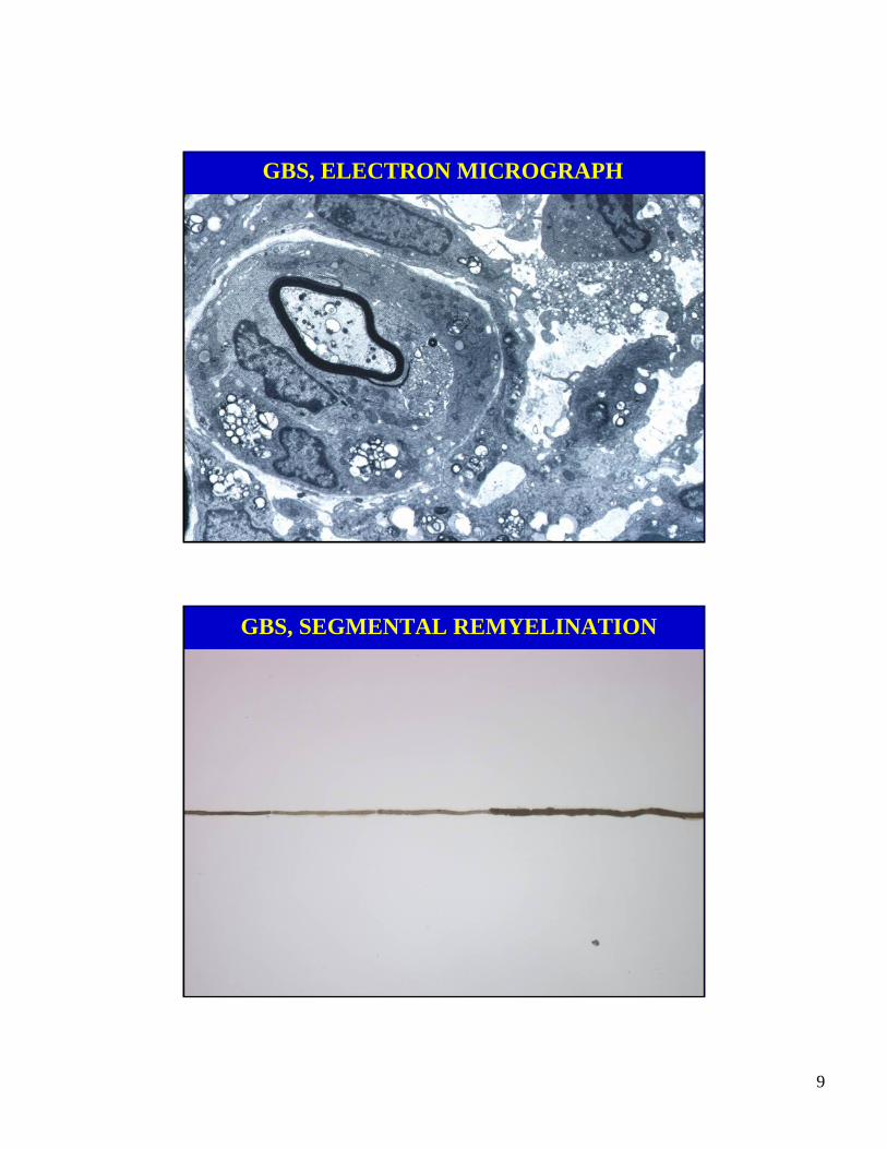
• Monocytes and macrophages appear to attack myelin sheaths.
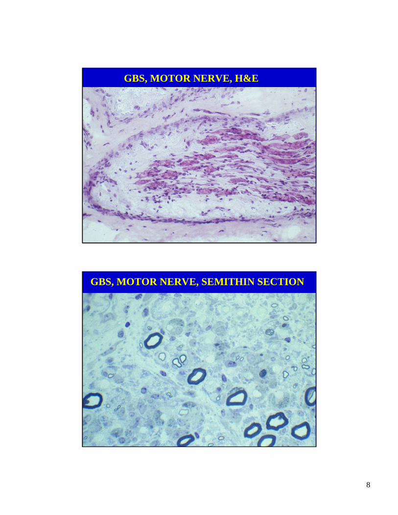
• Myelinated fibers show segmental demyelination during thefirst few days. Segmental remyelination occurs subsequently.
• The lesions have a perivenular distribution.
GBS, DORSAL ROOT GANGLION, H&E
8
GBS, MOTOR NERVE, H&E
GBS, MOTOR NERVE, SEMITHIN SECTION
9
GBS, ELECTRON MICROGRAPH
GBS, SEGMENTAL REMYELINATION
10
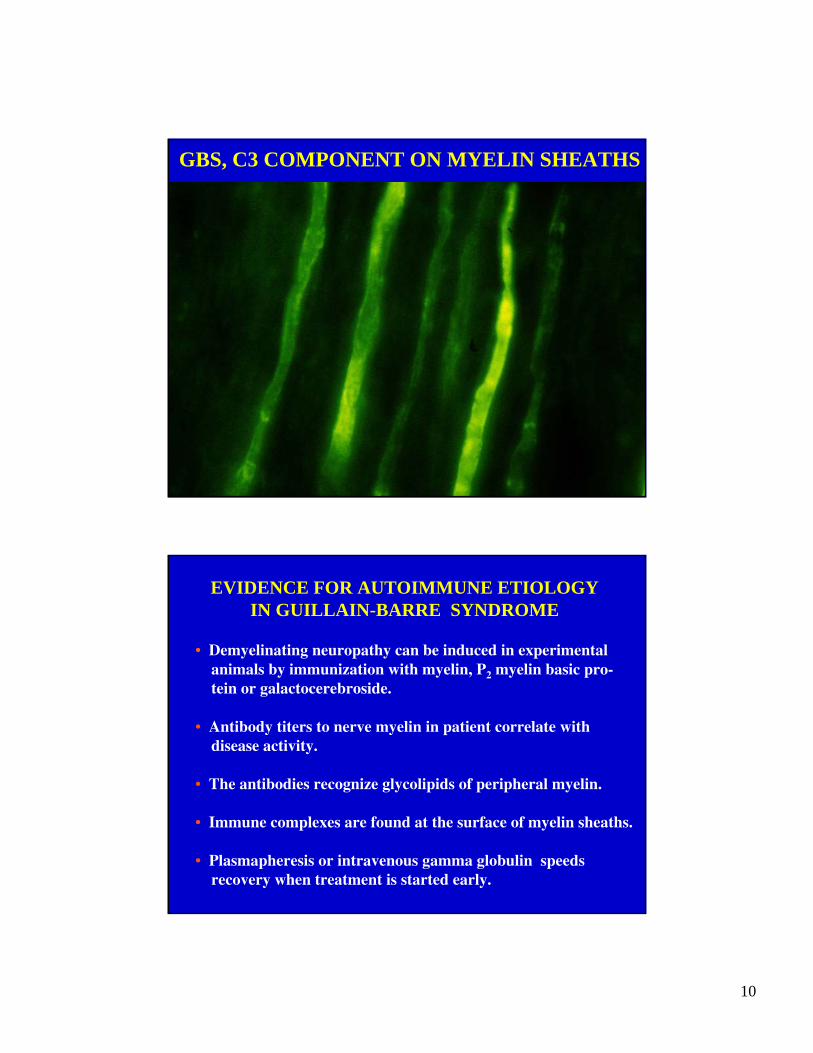
GBS, C3 COMPONENT ON MYELIN SHEATHS
EVIDENCE FOR AUTOIMMUNE ETIOLOGY IN GUILLAIN-BARRE SYNDROME
• Demyelinating neuropathy can be induced in experimental animals by immunization with myelin, P2 myelin basic pro-tein or galactocerebroside.
• Antibody titers to nerve myelin in patient correlate with disease activity.
• The antibodies recognize glycolipids of peripheral myelin.
• Immune complexes are found at the surface of myelin sheaths.
• Plasmapheresis or intravenous gamma globulin speedsrecovery when treatment is started early.
11
AXONAL VARIANT OF GUILLAIN-BARRE SYNDROME
• Clinical syndrome resembles Guillain-Barre syndrome, but is often purely motor.
• It was first described in children in China by investigatorsfrom John Griffin and others from Johns Hopkins.
• Electrodiagnostic studies suggested a purely axonal dis-order with little slowing of conduction velocity or block.
• Some of the patients died within a few days of the onset ofweakness allowing autopsy study of the nerves.
AXONAL VARIANT OF GUILLAIN-BARRE SYNDROME
• Autopsy showed axonal degeneration with little or no demy-elination or lymphocytic infiltration.
• Immune complexes were found at the nodes of Ranvier.
• The disorder was often preceded by a gastrointestinal illness caused by the bacterium, Campylobacter jejuni.
• Elevated serum antibodies to GD1 & GM1 ganglioside;these antibodies recognized the terminal oligosaccharidechain, which is a component of both gangliosides.
12
AXONAL VARIANT OF GUILLAIN-BARRE SYNDROME
• It turned out that the chemical structure of thelipopolysaccharide of the bacterium includes thesame oligosaccharide chain present in GD1a and GM1.
• This suggests that the immune response to the infectioncould react to both Campylobacter jejuni and gangliosidesexpressed on axons.
• This data provides support for the idea that molecular mimicry can be the basis for this autoimmune neurop-athy.
CHRONIC INFLAMMATORY DEMYE-LINATING POLYNEUROPATHY
• Chronic progressive or relapsing neuropathy, motor > sensory.
• An antecedent infectious illness is uncommon.
• Electrophysiology: slow conduction velocity & conduction block.
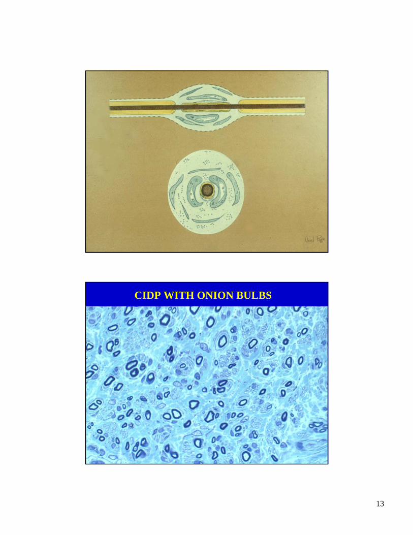
• Pathology: segmental demyelination and remyelination, onion bulbs, fibrosis and little or no lymphocytic infiltration of tissue.
• Autoimmune disorder of myelin, probably antibody-mediated.
• Patients respond to plasmapheresis, intravenous gamma globulinor corticosteroid treatment.
13
CIDP WITH ONION BULBS
14
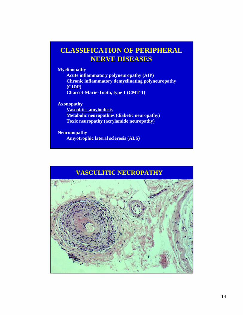
CLASSIFICATION OF PERIPHERALNERVE DISEASES
MyelinopathyAcute inflammatory polyneuropathy (AIP)Chronic inflammatory demyelinating polyneuropathy (CIDP)Charcot-Marie-Tooth, type 1 (CMT-1)
AxonopathyVasculitis, amyloidosisMetabolic neuropathies (diabetic neuropathy)Toxic neuropathy (acrylamide neuropathy)
NeuronopathyAmyotrophic lateral sclerosis (ALS)
VASCULITIC NEUROPATHY
15
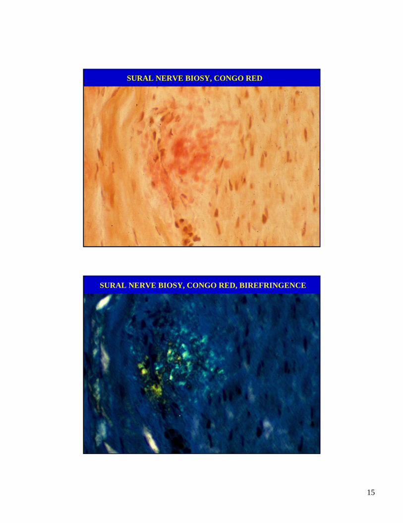
SURAL NERVE BIOSY, CONGO RED
SURAL NERVE BIOSY, CONGO RED, BIREFRINGENCE
16
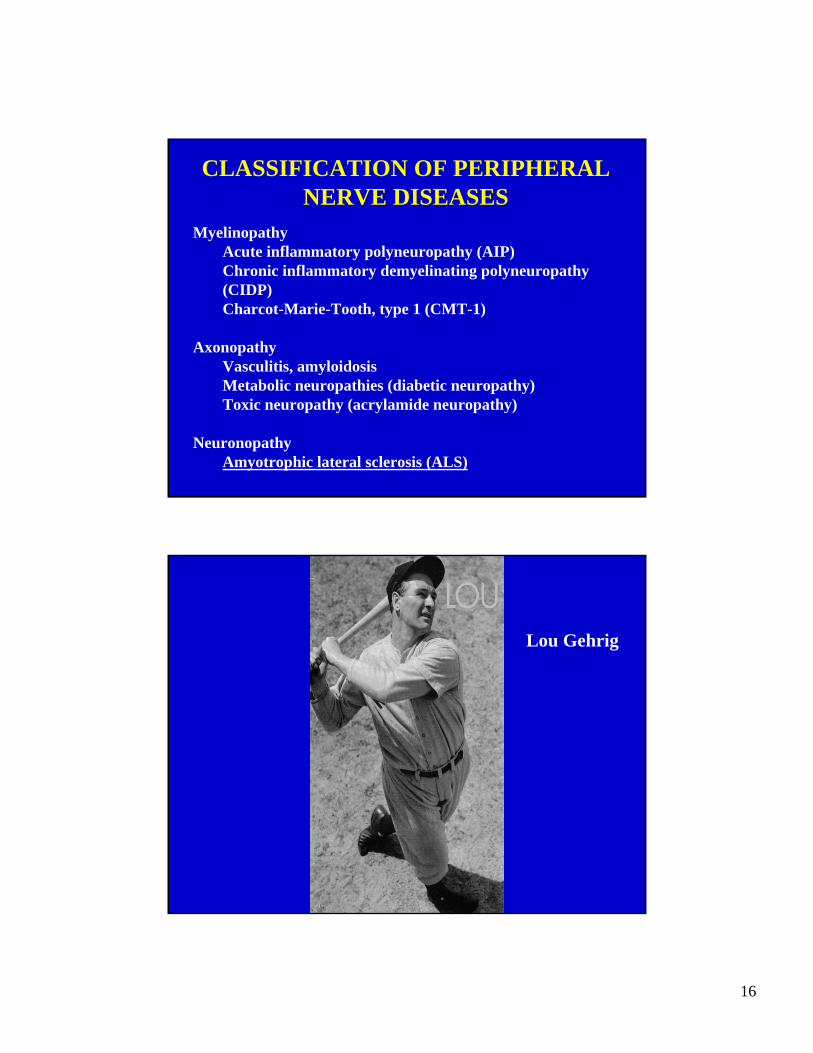
CLASSIFICATION OF PERIPHERALNERVE DISEASES
MyelinopathyAcute inflammatory polyneuropathy (AIP)Chronic inflammatory demyelinating polyneuropathy (CIDP)Charcot-Marie-Tooth, type 1 (CMT-1)
AxonopathyVasculitis, amyloidosisMetabolic neuropathies (diabetic neuropathy)Toxic neuropathy (acrylamide neuropathy)
NeuronopathyAmyotrophic lateral sclerosis (ALS)
Lou Gehrig
17
AMYOTROPHIC LATERAL SCLEROSIS(LOU GEHRIG’S DISEASE)
• Progressive weakness and wasting with fasciculations, oftenasymmetrical in the beginning.
• Hyperactive tendon reflexes, clonus and Babinski signs.
• Symptoms usually begin after the age of 40.
• Electrodiagnostic: denervation & normal nerve conductions.
• Most are sporadic; 5-10% are familial.
• Death usually within 3 to 5 years from onset.
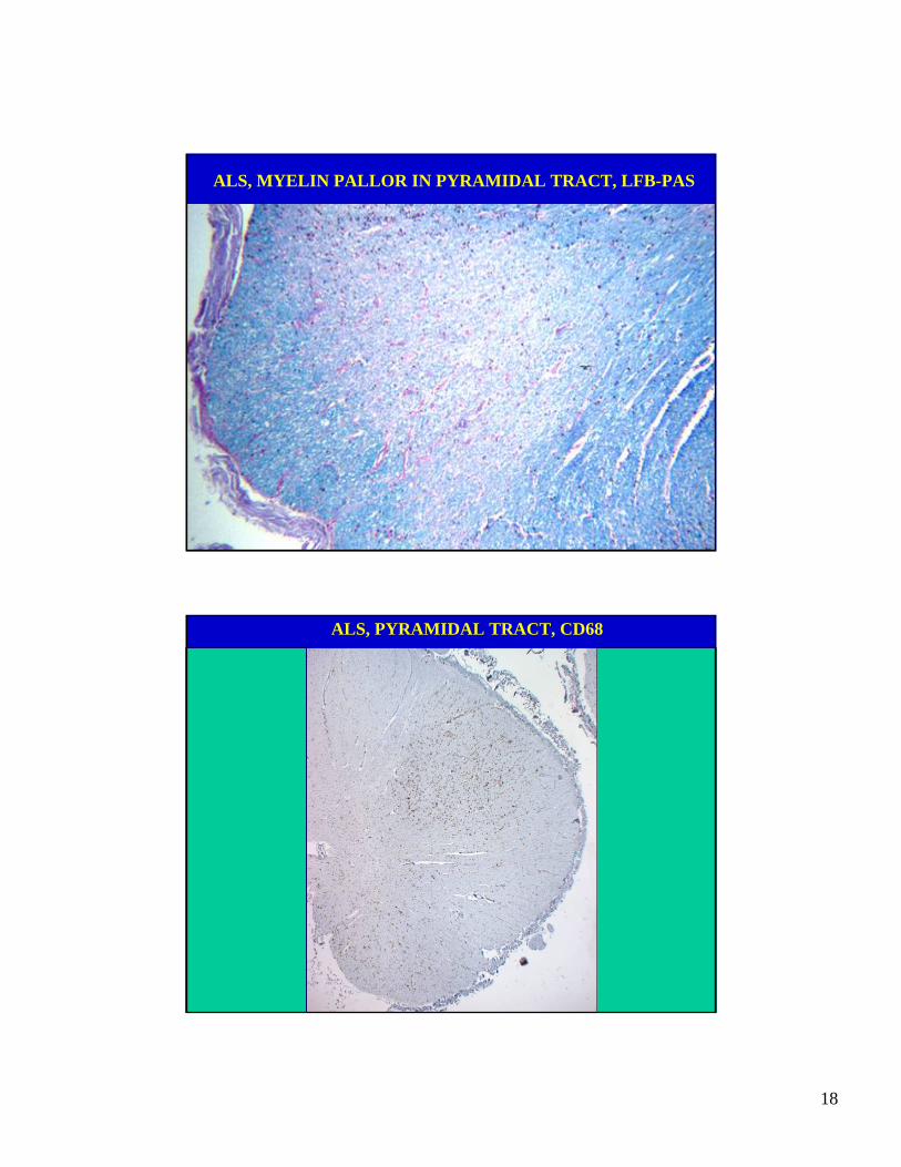
ALS: UPPER MOTOR NEURON PATHOLOGY
• Loss of Betz cells in precentral gyrus.
• Pyramidal degeneration with gradually increasingmyelin pallor in caudal direction due to loss ofaxons.
• The tract degeneration is marked by macrophages(removing myelin debris) and numerous activatedmicroglia.
18
ALS, MYELIN PALLOR IN PYRAMIDAL TRACT, LFB-PAS
ALS, PYRAMIDAL TRACT, CD68
19
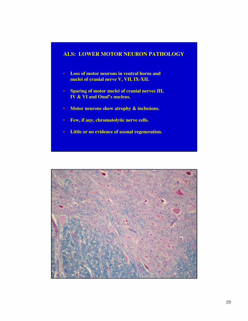
ALS: LOWER MOTOR NEURON PATHOLOGY
• Loss of motor neurons in ventral horns andnuclei of cranial nerve V, VII, IX-XII.
• Sparing of motor nuclei of cranial nerves III,IV & VI and Onuf’s nucleus.
• Motor neurons show atrophy & inclusions.
• Few, if any, chromatolytic nerve cells.
• Little or no evidence of axonal regeneration.
20
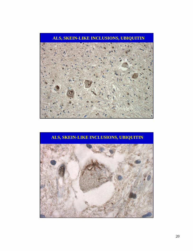
ALS, SKEIN-LIKE INCLUSIONS, UBIQUITIN
ALS, SKEIN-LIKE INCLUSIONS, UBIQUITIN
21
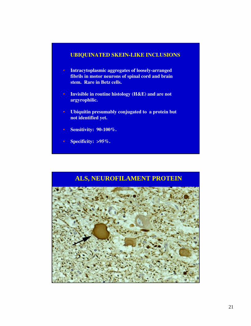
UBIQUINATED SKEIN-LIKE INCLUSIONS
• Intracytoplasmic aggregates of loosely-arrangedfibrils in motor neurons of spinal cord and brainstem. Rare in Betz cells.
• Invisible in routine histology (H&E) and are notargyrophilic.
• Ubiquitin presumably conjugated to a protein butnot identified yet.
• Sensitivity: 90-100%.
• Specificity: >95%.
ALS, NEUROFILAMENT PROTEIN
22
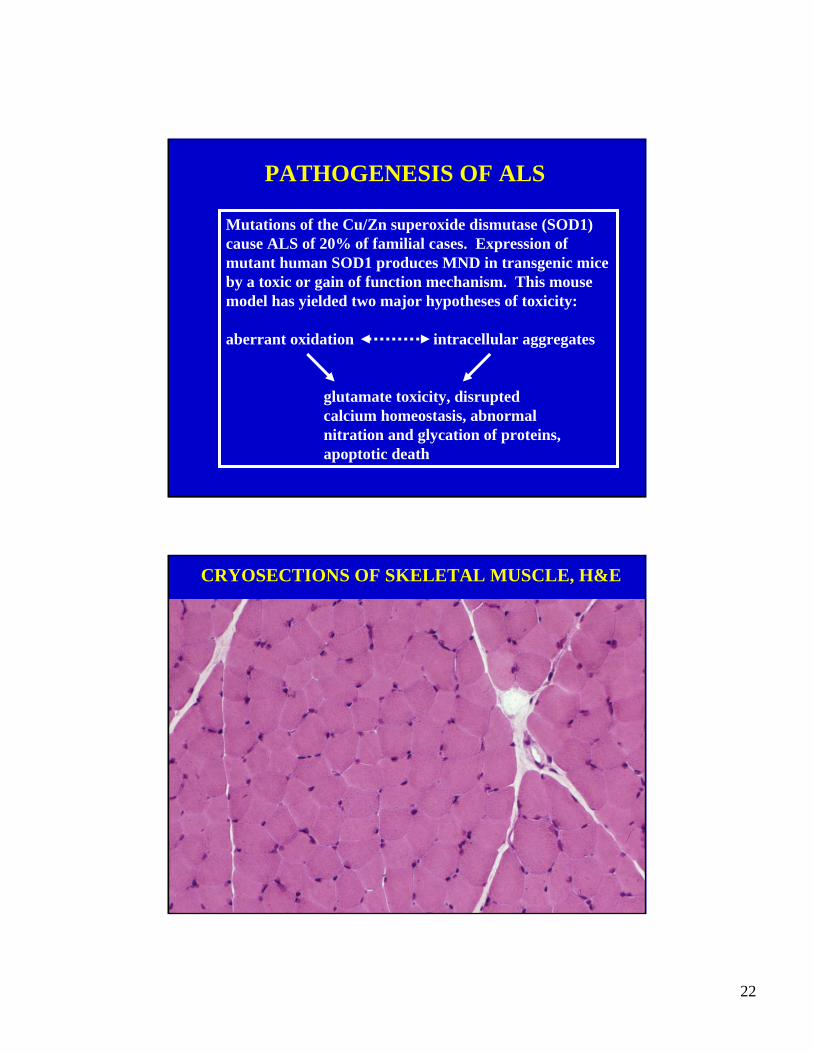
PATHOGENESIS OF ALS
Mutations of the Cu/Zn superoxide dismutase (SOD1)cause ALS of 20% of familial cases. Expression of mutant human SOD1 produces MND in transgenic miceby a toxic or gain of function mechanism. This mouse model has yielded two major hypotheses of toxicity:
aberrant oxidation intracellular aggregates
glutamate toxicity, disruptedcalcium homeostasis, abnormalnitration and glycation of proteins,apoptotic death
CRYOSECTIONS OF SKELETAL MUSCLE, H&E
23
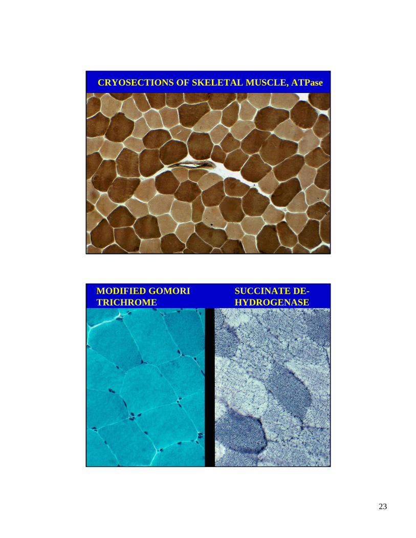
CRYOSECTIONS OF SKELETAL MUSCLE, ATPase
MODIFIED GOMORI TRICHROME
SUCCINATE DE-HYDROGENASE
24
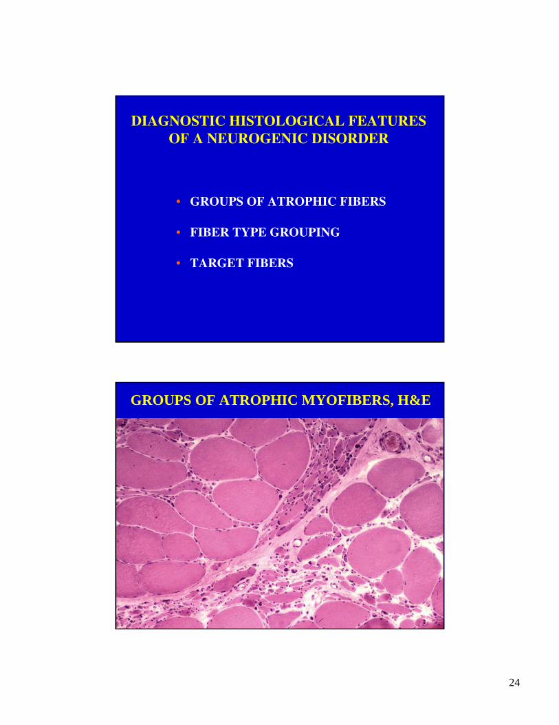
DIAGNOSTIC HISTOLOGICAL FEATURESOF A NEUROGENIC DISORDER
• GROUPS OF ATROPHIC FIBERS
• FIBER TYPE GROUPING
• TARGET FIBERS
GROUPS OF ATROPHIC MYOFIBERS, H&E
25
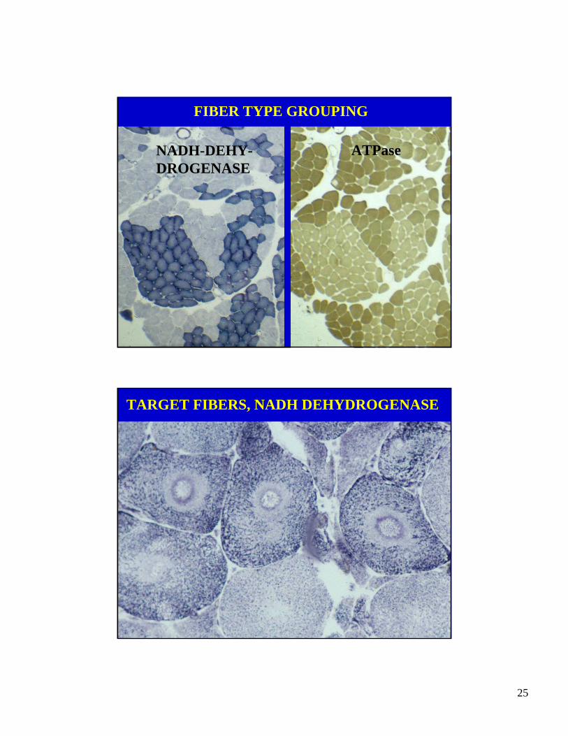
FIBER TYPE GROUPING
NADH-DEHY-DROGENASE
ATPase
TARGET FIBERS, NADH DEHYDROGENASE

26
DIAGNOSTIC HISTOLOGICAL FEATURES OF MYOPATHIES
• ABSENCE OF NEUROGENIC ABNORMALITIES
• NECROTIC MUSCLE FIBERS
• BASOPHILIC (REGENERATING) MYOFIBERS
• FIBROSIS OF THE ENDOMYSIUM
• SPECIAL PATHOLOGICAL FEATURES (INFLAMMATORYCELLS, RAGGED RED FIBERS ETC.)
NECROTIC FIBER, H&E
27
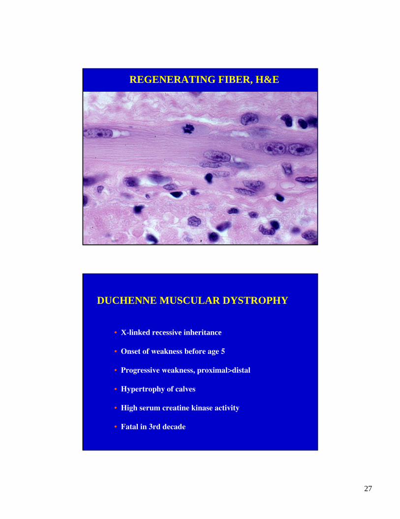
REGENERATING FIBER, H&E
DUCHENNE MUSCULAR DYSTROPHY
• X-linked recessive inheritance
• Onset of weakness before age 5
• Progressive weakness, proximal>distal
• Hypertrophy of calves
• High serum creatine kinase activity
• Fatal in 3rd decade
28
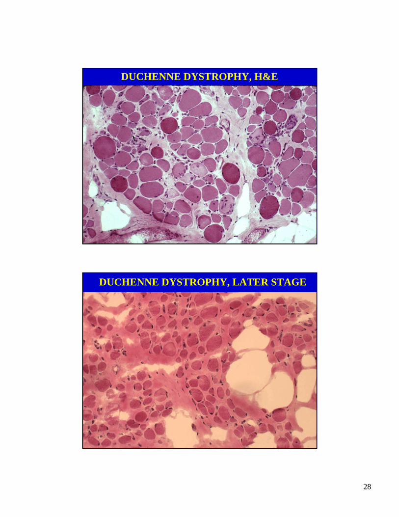
DUCHENNE DYSTROPHY, H&E
DUCHENNE DYSTROPHY, LATER STAGE
29
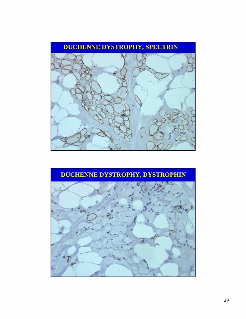
DUCHENNE DYSTROPHY, SPECTRIN
DUCHENNE DYSTROPHY, DYSTROPHIN
30
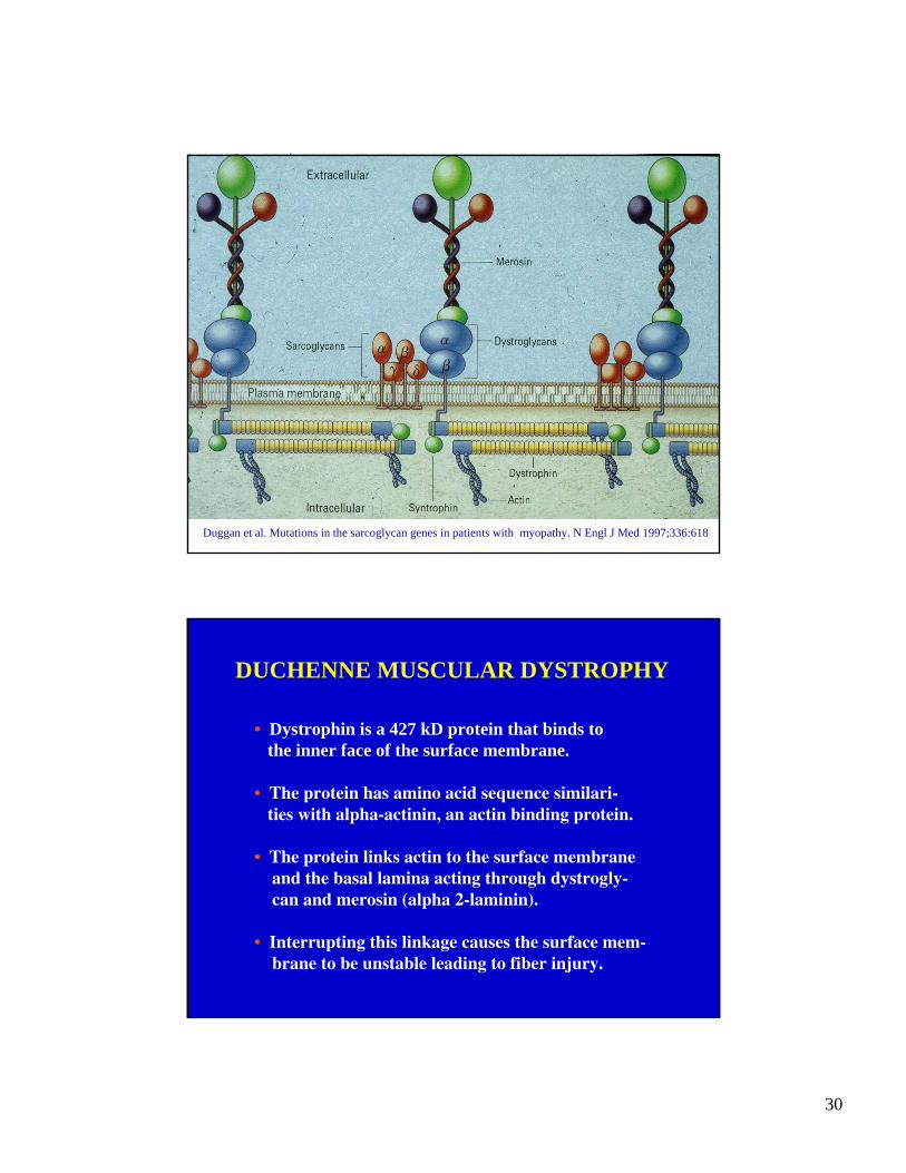
Duggan et al. Mutations in the sarcoglycan genes in patients with myopathy. N Engl J Med 1997;336:618
DUCHENNE MUSCULAR DYSTROPHY
• Dystrophin is a 427 kD protein that binds tothe inner face of the surface membrane.
• The protein has amino acid sequence similari-ties with alpha-actinin, an actin binding protein.
• The protein links actin to the surface membraneand the basal lamina acting through dystrogly-can and merosin (alpha 2-laminin).
• Interrupting this linkage causes the surface mem-brane to be unstable leading to fiber injury.
31
INFLAMMATORY MYOPATHIES
• Polymyositis
• Inclusion body myositis
• Dermatomyositis
POLYMYOSITIS
32
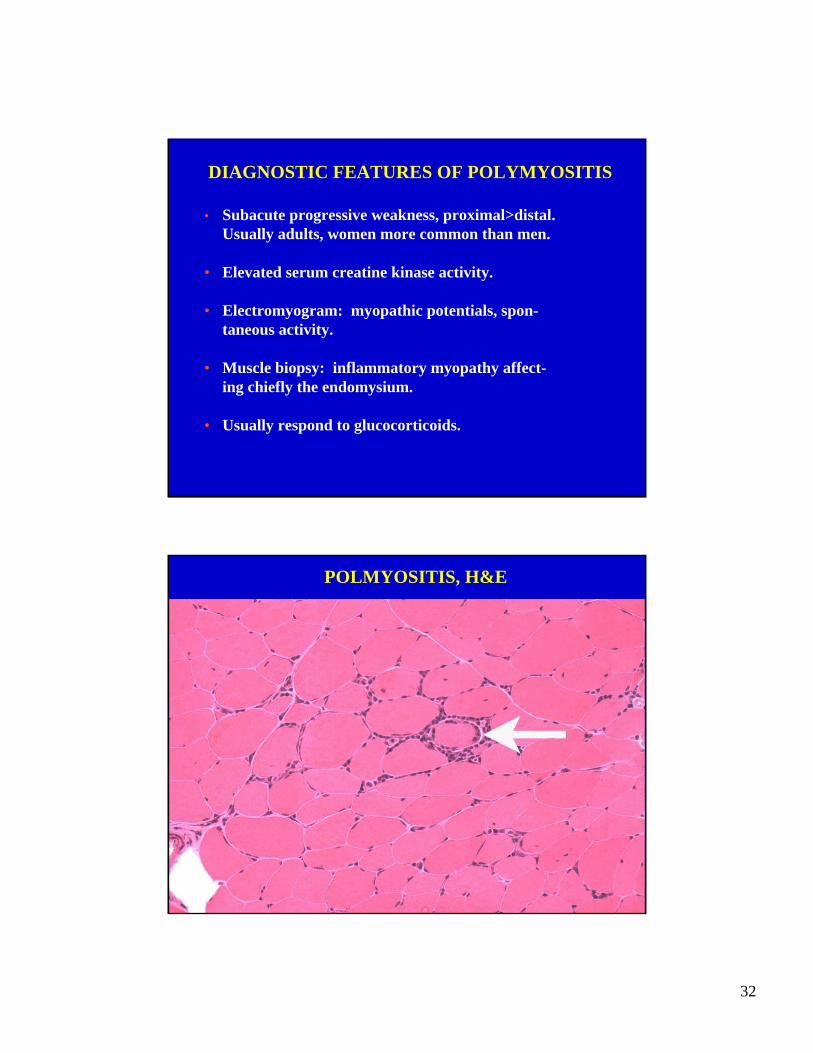
DIAGNOSTIC FEATURES OF POLYMYOSITIS
• Subacute progressive weakness, proximal>distal.Usually adults, women more common than men.
• Elevated serum creatine kinase activity.
• Electromyogram: myopathic potentials, spon-taneous activity.
• Muscle biopsy: inflammatory myopathy affect-ing chiefly the endomysium.
• Usually respond to glucocorticoids.
POLMYOSITIS, H&E
33
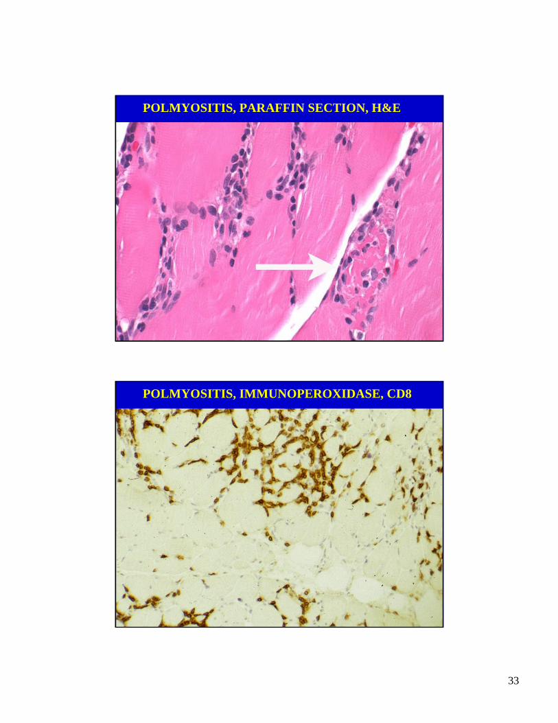
POLMYOSITIS, PARAFFIN SECTION, H&E
POLMYOSITIS, IMMUNOPEROXIDASE, CD8
34
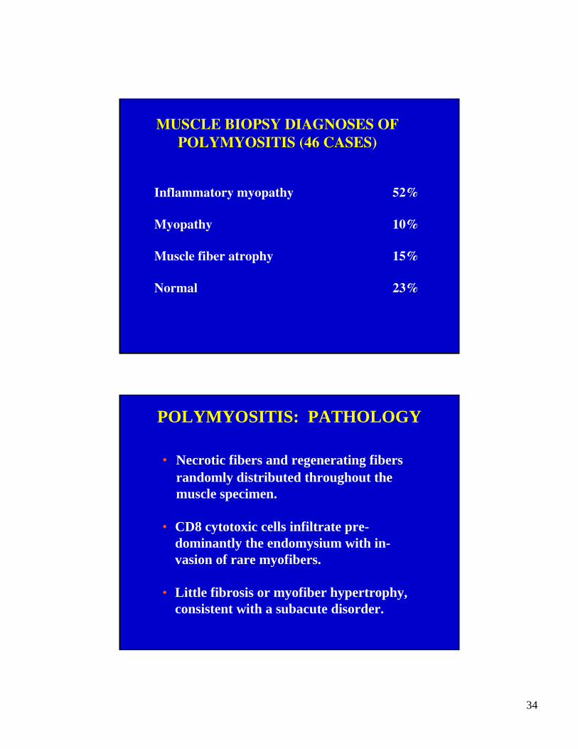
MUSCLE BIOPSY DIAGNOSES OFPOLYMYOSITIS (46 CASES)
Inflammatory myopathy 52%
Myopathy 10%
Muscle fiber atrophy 15%
Normal 23%
POLYMYOSITIS: PATHOLOGY
• Necrotic fibers and regenerating fibersrandomly distributed throughout the muscle specimen.
• CD8 cytotoxic cells infiltrate pre-dominantly the endomysium with in-vasion of rare myofibers.
• Little fibrosis or myofiber hypertrophy, consistent with a subacute disorder.
35
INCLUSION BODYMYOSITIS
DIAGNOSTIC FEATURES OF IBM
• Slowly progressive weakness, proximal and distal.Usually in adults, mostly men.
• Mildly elevated serum creatine kinase or normal.
• Electromyogram: myopathic potentials, spon-taneous activity.
• Muscle biopsy: inflammatory myopathy affect-ing chiefly the endomysium, but chronic and hasrimmed vacuoles and amyloid inclusions.
• Usually does not respond to glucocorticoids.
36
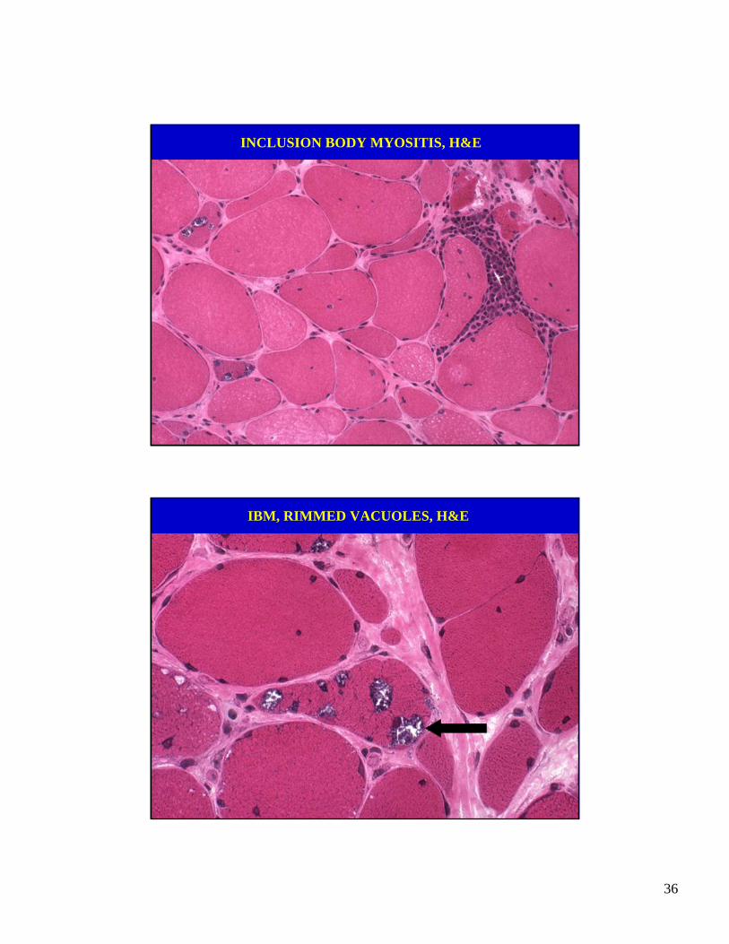
INCLUSION BODY MYOSITIS, H&E
IBM, RIMMED VACUOLES, H&E
37
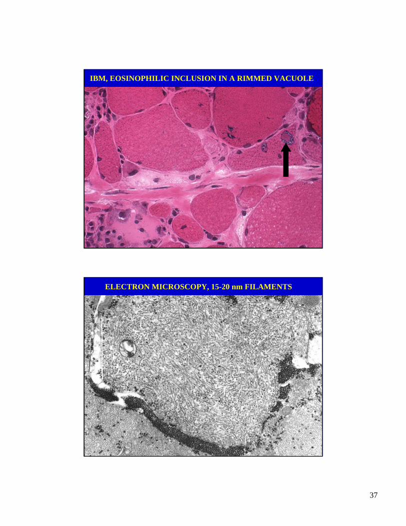
IBM, EOSINOPHILIC INCLUSION IN A RIMMED VACUOLE
ELECTRON MICROSCOPY, 15-20 nm FILAMENTS
38
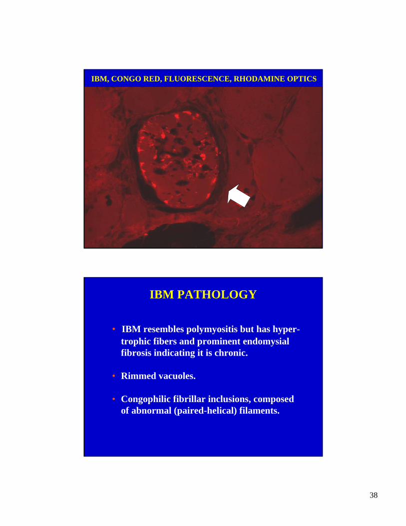
IBM, CONGO RED, FLUORESCENCE, RHODAMINE OPTICS
IBM PATHOLOGY
• IBM resembles polymyositis but has hyper-trophic fibers and prominent endomysial fibrosis indicating it is chronic.
• Rimmed vacuoles.
• Congophilic fibrillar inclusions, composedof abnormal (paired-helical) filaments.
39
DERMATOMYOSITIS
DIAGNOSTIC FEATURES OF DERMATOMYOSITIS
• Subacute progressive weakness, proximal>distal.Children and adults, women more common than men.
• Characteristic rash and periorbital heliotrope.
• Elevated serum creatine kinase activity.
• Electromyogram: myopathic potentials, spon-taneous activity.
• Muscle biopsy: inflammatory myopathy affectingchiefly the perimysium with perifascicular atrophy.
• Usually respond to glucocorticoids or IVGG.
40
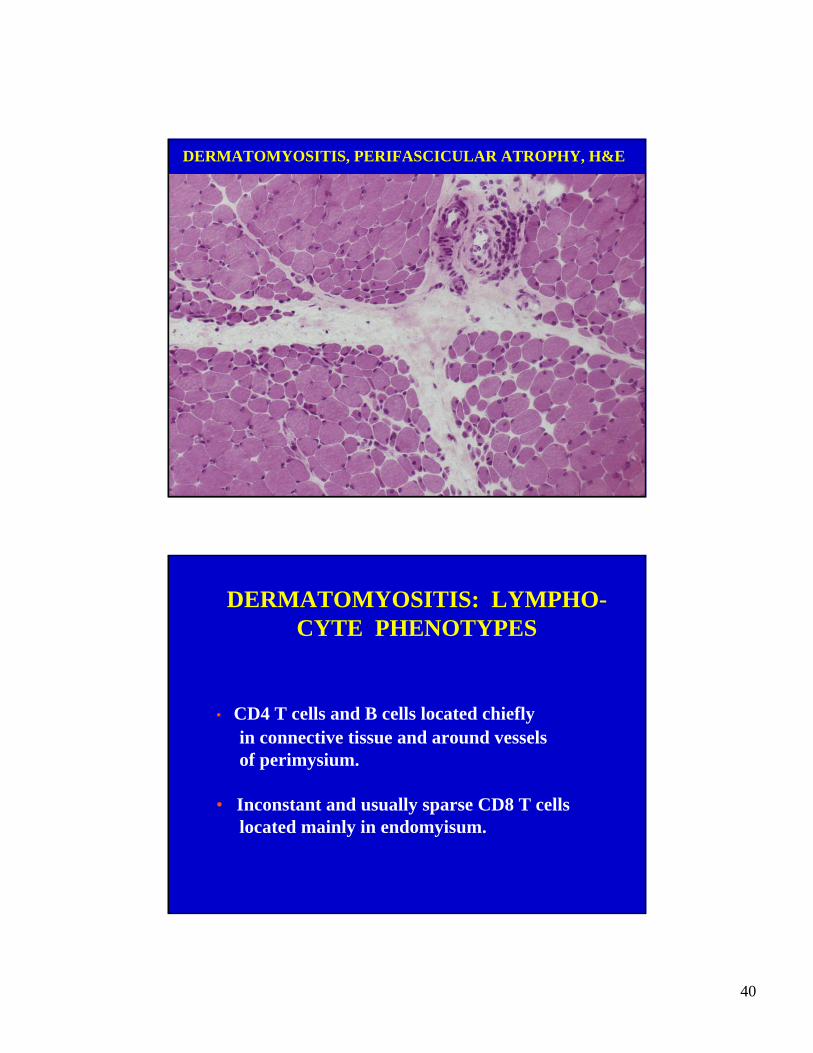
DERMATOMYOSITIS, PERIFASCICULAR ATROPHY, H&E
DERMATOMYOSITIS: LYMPHO-CYTE PHENOTYPES
• CD4 T cells and B cells located chieflyin connective tissue and around vesselsof perimysium.
• Inconstant and usually sparse CD8 T cellslocated mainly in endomyisum.
41
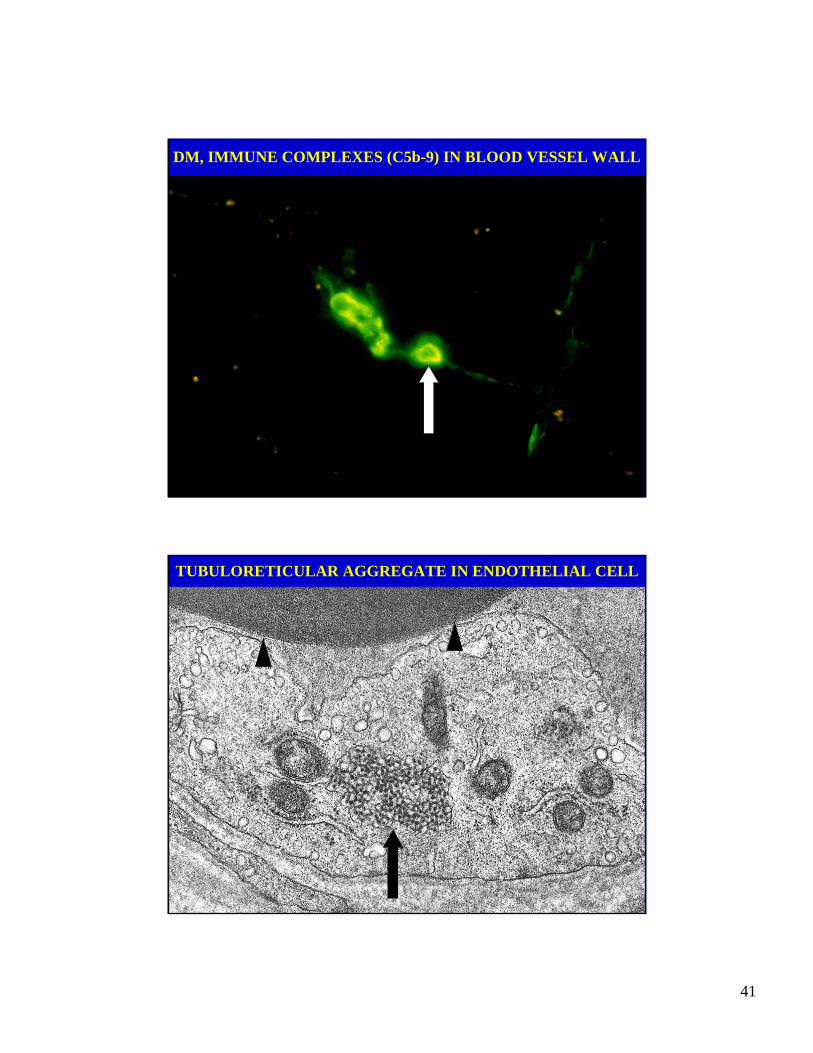
DM, IMMUNE COMPLEXES (C5b-9) IN BLOOD VESSEL WALL
TUBULORETICULAR AGGREGATE IN ENDOTHELIAL CELL
42
DERMATOMYOSITIS: PATHOLOGY
• Perifascicular atrophy of muscle fibers, with or with-out necrotic fibers or regenerating fibers.
• Immune complexes of immunoglobulins and comple-ment components in the walls of blood vessels.
• Tubuloreticular aggregates (undulating tubules).
• Reduced number of capillaries at periphery of fascicle.
• Lymphocytes are often sparse and located in chiefly perimysium.
INFLAMMATORY MYOPATHIES:PATHOPHYSIOLOGY
• Polymyositis and inclusion body myositis (IBM) haveautoaggressive CD8 lymphocytes that appear to attack myofibers and suggest an autoimmune role. However, a major question exists about the etiology of IBM.
• Dermatomyositis is thought to be caused by auto-antibodies, possibly targeting an antigen of theendothelium. Fiber injury may be caused by ischemia.
43
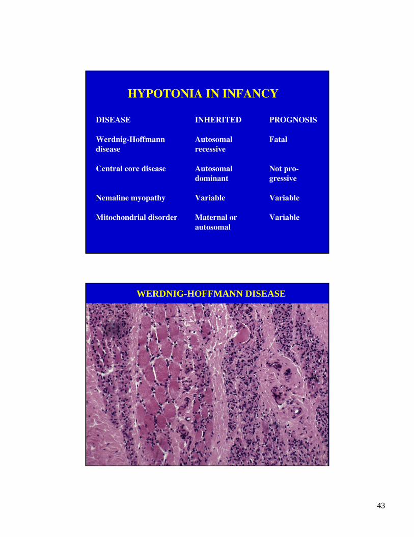
HYPOTONIA IN INFANCY
DISEASE INHERITED PROGNOSIS
Werdnig-Hoffmann Autosomal Fataldisease recessive
Central core disease Autosomal Not pro-dominant gressive
Nemaline myopathy Variable Variable
Mitochondrial disorder Maternal or Variableautosomal
WERDNIG-HOFFMANN DISEASE
44
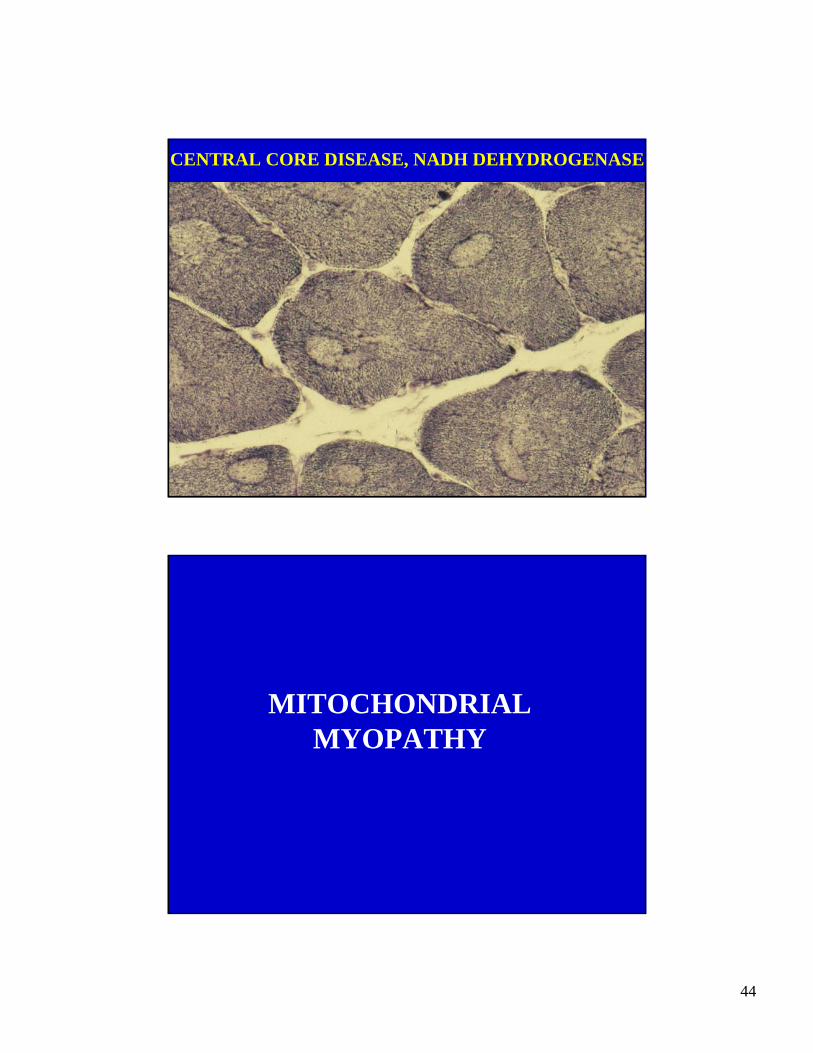
CENTRAL CORE DISEASE, NADH DEHYDROGENASE
MITOCHONDRIALMYOPATHY
45
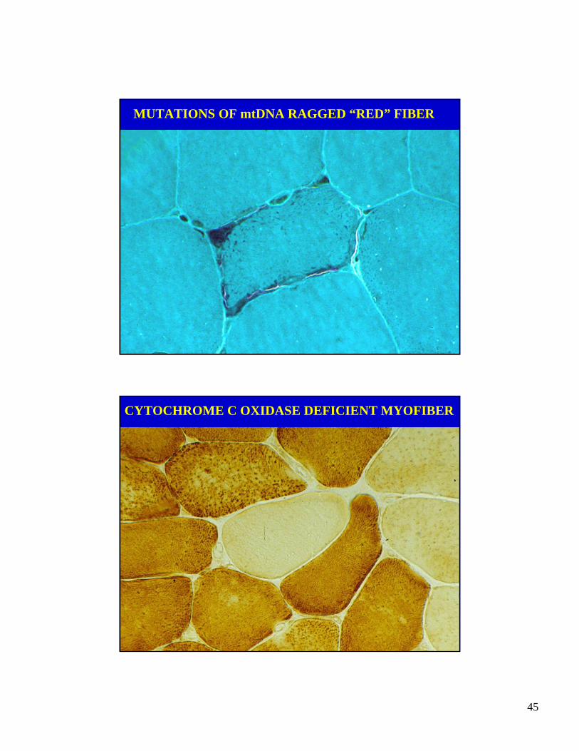
MUTATIONS OF mtDNA RAGGED “RED” FIBER
CYTOCHROME C OXIDASE DEFICIENT MYOFIBER
46
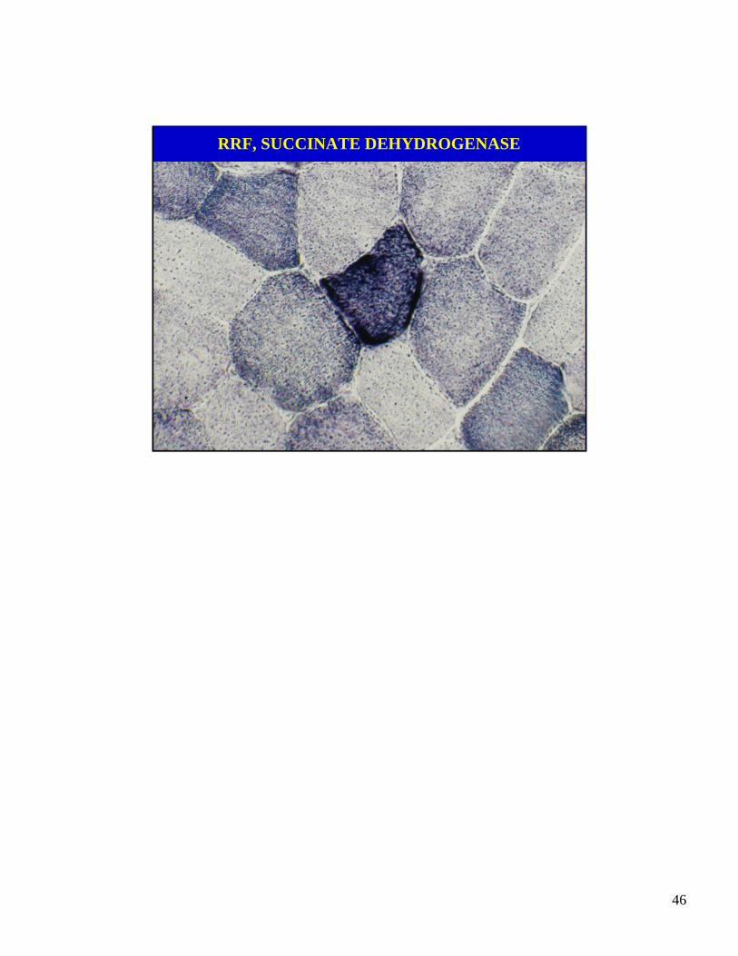
RRF, SUCCINATE DEHYDROGENASE