Proteomics for biodeterioration of wood (Pinus taeda L.): Challenging analysis by 2-D PAGE and...
9
Proteomics for biodeterioration of wood (Pinus taeda L.): Challenging analysis by 2-D PAGE and MALDI-TOF/TOF/MS Young-Min Kang, M. Lynn Prewitt, Susan V. Diehl * Department of Forest Products, Forest and Wildlife Research Center, Mississippi State University, PO Box 9820, Mississippi State, MS 39762, USA article info Article history: Received 11 November 2008 Received in revised form 15 July 2009 Accepted 16 July 2009 Available online 4 September 2009 Keywords: Biodeterioration Gloeophyllum trabeum Proteomics MALDI-TOF/TOF/MS Two-dimensional polyacrylamide gel electrophoresis abstract Proteins expressed by the brown-rot fungus Gloeophyllum trabeum were characterized from inoculated southern yellow pine sapwood undergoing decay, from pure cultures of the fungus and from uninocu- lated pinewood. Analysis was carried out by two-dimensional polyacrylamide gel electrophoresis and MALDI-TOF/TOF/MS. No proteins were detected from the clean uncontaminated wood. The inoculated wood undergoing active brown-rot decay produced 76 proteins, including the Fenton-chemistry related enzymes, alcohol oxidase, lipoxygenase, and catalase. One hundred and eleven proteins were detected from the pure culture and most were common metabolic proteins. A majority of proteins in both samples were identified as hypothetical proteins. A surprising result is that there was very little overlap between proteins found in both sets of samples, indicating a very different mechanism in action when the fungus is growing on a cellulose-based nutrient source (wood) versus glucose media. This study also highlights a current limitation of this approach, which is the limited protein and genomic sequence information annotated on the public databases. Of the 187 proteins characterized, only 36 were identified with confidence. To our knowledge, this is the first reported proteomic analysis of pinewood decayed by a brown-rot fungus and provides the initial characterization of proteins involved in this type of wood biodeterioration. Although significant limitations still exist in identifying the proteins, this limitation will diminish as functional proteins are identified and added to the databases. Ó 2009 Elsevier Ltd. All rights reserved. 1. Introduction Wood is an important renewable and natural resource with a multitude of uses (Goodell, 2003). Wood decay results in the loss of billions of U.S. dollars annually (Preston, 2000). The traditional method for protecting wood from decay is treatment with broad- spectrum chemical preservatives, usually without knowing the specific identity of the fungi causing decay in many different envi- ronments. Identification of the microorganisms involved in decay is necessary in order to develop a complete understanding of the roles the different microorganisms play in the decay process. However, the presence of a microorganism does not mean it is metabolically active in decay. Wood decay is caused by enzymes and low-molec- ular-weight mediators secreted by fungi in their efforts to obtain a carbon source for their survival (Cease et al., 1989; Flournoy et al., 1991; Pointing et al., 2003). The resulting wood decay is classified into three types: soft rot, brown rot, and white rot. Basidiomycota members typically cause the more active and destructive brown- or white-rot decay (Nilsson et al., 1989; Bruce et al., 1991). Brown-rot fungi and brown-rot decay predominate on pines and pine products such as lumber in the USA (Goodell, 2003). During brown-rot decay the carbohydrate fraction of the holocellulose is actively metabolized, leaving behind a chemically modified lignin residue, which is typically brown and crumbly. The depolymer- ization of the cellulosic fraction of the S2 cell wall dramatically and rapidly reduces the strength of the wood. The S2 layer is degraded in advance of the actual hyphae, and since most enzymes are too large to penetrate intact cell wall layers, it is hypothesized that non- enzymatic low-molecular mediators or chealators diffuse into the cell wall, producing localized hydroxyl free radicals via modified Fenton reactions (Green and Highley, 1997; Goodell, 2003; Daniel et al., 2007). However, the exact mechanism of brown-rot decay is still hypothetical and controversial. Recently, the genome of the brown-rot fungus Postia placenta was sequenced and annotated (Martinez et al., 2009). In relation to the decay of wood, these authors detected none of the common lignin decay enzyme genes, such as lignin peroxidase, manganese peroxidase, or versatile peroxidase; however, there were two possible laccase genes present. There were high transcript levels of numerous hemi- cellulases and a single putative b 1-4 endoglucanase from cultures grown on cellulose media compared to those grown on glucose media. No genes coding for cellobiohydrolases or cellulose-binding * Corresponding author. Tel.: þ1 662 325 3101; fax: þ1 662 325 8126. E-mail address: [email protected] (S.V. Diehl). Contents lists available at ScienceDirect International Biodeterioration & Biodegradation journal homepage: www.elsevier.com/locate/ibiod 0964-8305/$ – see front matter Ó 2009 Elsevier Ltd. All rights reserved. doi:10.1016/j.ibiod.2009.07.008 International Biodeterioration & Biodegradation 63 (2009) 1036–1044
-
Upload
young-min-kang -
Category
Documents
-
view
215 -
download
2
Transcript of Proteomics for biodeterioration of wood (Pinus taeda L.): Challenging analysis by 2-D PAGE and...

lable at ScienceDirect
International Biodeterioration & Biodegradation 63 (2009) 1036–1044
Contents lists avai
International Biodeterioration & Biodegradation
journal homepage: www.elsevier .com/locate/ ib iod
Proteomics for biodeterioration of wood (Pinus taeda L.): Challenging analysisby 2-D PAGE and MALDI-TOF/TOF/MS
Young-Min Kang, M. Lynn Prewitt, Susan V. Diehl*
Department of Forest Products, Forest and Wildlife Research Center, Mississippi State University, PO Box 9820, Mississippi State, MS 39762, USA
a r t i c l e i n f o
Article history:Received 11 November 2008Received in revised form15 July 2009Accepted 16 July 2009Available online 4 September 2009
Keywords:BiodeteriorationGloeophyllum trabeumProteomicsMALDI-TOF/TOF/MSTwo-dimensional polyacrylamide gelelectrophoresis
* Corresponding author. Tel.: þ1 662 325 3101; faxE-mail address: [email protected] (S.V. Diehl
0964-8305/$ – see front matter � 2009 Elsevier Ltd.doi:10.1016/j.ibiod.2009.07.008
a b s t r a c t
Proteins expressed by the brown-rot fungus Gloeophyllum trabeum were characterized from inoculatedsouthern yellow pine sapwood undergoing decay, from pure cultures of the fungus and from uninocu-lated pinewood. Analysis was carried out by two-dimensional polyacrylamide gel electrophoresis andMALDI-TOF/TOF/MS. No proteins were detected from the clean uncontaminated wood. The inoculatedwood undergoing active brown-rot decay produced 76 proteins, including the Fenton-chemistry relatedenzymes, alcohol oxidase, lipoxygenase, and catalase. One hundred and eleven proteins were detectedfrom the pure culture and most were common metabolic proteins. A majority of proteins in both sampleswere identified as hypothetical proteins. A surprising result is that there was very little overlap betweenproteins found in both sets of samples, indicating a very different mechanism in action when the fungusis growing on a cellulose-based nutrient source (wood) versus glucose media. This study also highlightsa current limitation of this approach, which is the limited protein and genomic sequence informationannotated on the public databases. Of the 187 proteins characterized, only 36 were identified withconfidence. To our knowledge, this is the first reported proteomic analysis of pinewood decayed bya brown-rot fungus and provides the initial characterization of proteins involved in this type of woodbiodeterioration. Although significant limitations still exist in identifying the proteins, this limitation willdiminish as functional proteins are identified and added to the databases.
� 2009 Elsevier Ltd. All rights reserved.
1. Introduction
Wood is an important renewable and natural resource witha multitude of uses (Goodell, 2003). Wood decay results in the loss ofbillions of U.S. dollars annually (Preston, 2000). The traditionalmethod for protecting wood from decay is treatment with broad-spectrum chemical preservatives, usually without knowing thespecific identity of the fungi causing decay in many different envi-ronments. Identification of the microorganisms involved in decay isnecessary in order to develop a complete understanding of the rolesthe different microorganisms play in the decay process. However,the presence of a microorganism does not mean it is metabolicallyactive in decay. Wood decay is caused by enzymes and low-molec-ular-weight mediators secreted by fungi in their efforts to obtaina carbon source for their survival (Cease et al., 1989; Flournoy et al.,1991; Pointing et al., 2003). The resulting wood decay is classifiedinto three types: soft rot, brown rot, and white rot. Basidiomycotamembers typically cause the more active and destructive brown- orwhite-rot decay (Nilsson et al., 1989; Bruce et al., 1991).
: þ1 662 325 8126.).
All rights reserved.
Brown-rot fungi and brown-rot decay predominate on pines andpine products such as lumber in the USA (Goodell, 2003). Duringbrown-rot decay the carbohydrate fraction of the holocellulose isactively metabolized, leaving behind a chemically modified ligninresidue, which is typically brown and crumbly. The depolymer-ization of the cellulosic fraction of the S2 cell wall dramatically andrapidly reduces the strength of the wood. The S2 layer is degradedin advance of the actual hyphae, and since most enzymes are toolarge to penetrate intact cell wall layers, it is hypothesized that non-enzymatic low-molecular mediators or chealators diffuse into thecell wall, producing localized hydroxyl free radicals via modifiedFenton reactions (Green and Highley, 1997; Goodell, 2003; Danielet al., 2007). However, the exact mechanism of brown-rot decay isstill hypothetical and controversial. Recently, the genome of thebrown-rot fungus Postia placenta was sequenced and annotated(Martinez et al., 2009). In relation to the decay of wood, theseauthors detected none of the common lignin decay enzyme genes,such as lignin peroxidase, manganese peroxidase, or versatileperoxidase; however, there were two possible laccase genespresent. There were high transcript levels of numerous hemi-cellulases and a single putative b 1-4 endoglucanase from culturesgrown on cellulose media compared to those grown on glucosemedia. No genes coding for cellobiohydrolases or cellulose-binding

Y.-M. Kang et al. / International Biodeterioration & Biodegradation 63 (2009) 1036–1044 1037
domains were found. On cellulose media there were also hightranscript levels of a methanol oxidase similar to one in Gloeo-phyllum trabeum, as well as genes tentatively identified as glucose-1-oxidases, polyphenol oxidases, iron reductases, and quinonereductases, all supporting Fenton chemistry and hydroxyl radicalsas the brown-rot mechanism of decay. The annotated genome isavailable at http://genome.jgi-psf.org/Postia.
During the process of wood decay, fungi express differentproteins involved in both the decay of wood and the metabolism ofthe fungus. Proteomic profiles or protein fingerprints can be used tocompare decaying wood to non-decayed wood or cultured fungi todetermine which proteins are being uniquely expressed during thedecay process. The proteome itself is defined as the complete set ofproteins generated by the genome (Gromov and Celis, 2000;Phillips and Bogyo, 2005). The genome is the complete set of anorganism’s DNA. The proteome of a given organism is constantlychanging in response to internal and external conditions includingits environment and carbon sources. Proteomics is much morecomplex than either genomics or transcriptomics because eachprotein can be chemically modified in different ways post synthesis(Keller and Hettich, 2009). Strong gene expression results inabundant mRNA and includes all transcripts in the cell. The tran-scriptome reflects the genes that are being actively expressed at anygiven time, but it does not necessarily mean that the correspondingprotein is also abundant or active in the cell (Prabakaran et al.,2001; Zhou et al., 2004). Expressional proteomics can provide botha qualitative and quantitative snapshot of all proteins expressed byan organism at the time of extraction (Keller and Hettich, 2009).
In the wood-decay field, a proteomics approach has been usedby numerous authors toward understanding the white-rot decayprocess using Phanerochaete chrysosporium as the model (Abbaset al., 2005; Vanden Wymelenberg et al., 2005; Sato et al., 2007;Matsuzaki et al., 2008; Shary et al., 2008). The genome of thiswhite-rot fungus has been sequenced (Martinez et al., 2004). To ourknowledge, this is the first reported proteomic analysis of pine-wood deteriorated by a brown-rot fungus and provides a useful toolfor characterization of proteins involved in wood biodeterioration.Our objective was to identify those proteins expressed during thedecay of pine. This research will lay the foundation to begin tounderstand the suites of decay genes and their proteins which areexpressed during brown-rot biodeterioration. A related currentstudy is looking at the hypothesis that gene expression of wood-decay enzymes is influenced by the decay resistance of the woodspecies being degraded.
2. Material and methods
2.1. Wood samples and fungal culture
The wood wafer samples for these studies were from loblollypine (Pinus taeda L.) sapwood lumber. This wood had been kiln-dried at high pressure and temperature to 15% moisture contentbefore being cut into wafers (18� 18� 5 mm). Samples consisted ofuncontaminated wood (a: undecayed pine stake), inoculateddecayed wood (b: G. trabeum inoculated pine stake) and pureculture of G. trabeum. The uncontaminated wood (UW) was cleanand no microorganisms were detected; its moisture content wasless than 15%. The inoculated wood (IW) was incubated with thebrown-rot fungus, G. trabeum, for approximately 2 months at roomtemperature at a moisture content w60%. The uncontaminatedwood (Fig. 1a) and inoculated wood after incubation (Fig. 1b) wereground in a Wiley mill. The proteins were extracted from each ofthe ground samples (listed as c and d in Fig. 1). Furthermore, purecultures of G. trabeum (GT) were grown on a malt extract medium,and the proteins were extracted by the same procedure.
2.2. Extraction and quantification of proteins from wood and fungi
The proteins were extracted and quantified from all threesamples by published methods (Bradford, 1976; Shimizu andWariishi, 2005). In general, 1.0 g of each sample (ground samplesand fungal tissue) was homogenized using a clean pestle withprotein extraction buffer (5.0 ml for 1.0 g wet weight of biomass).The protein extraction buffer was composed of 0.7 M sucrose, 0.5 MTris–HCl (pH 8.5), 0.05 M Na2EDTA, 0.1 M KCl, and 2% (v/v) 2-mercaptoethanol. Water-saturated phenol was then used to extracttotal proteins from the buffer. Proteins in the phenol phase wereprecipitated with five volumes of ammonium acetate in methanol(0.1 M NH4OAc and 1% 2-mercaptoethanol in methanol) at �70 �Cfor 2 h. The resultant pellets were sequentially washed withammonium acetate in methanol and 80% acetone. The proteinsamples were then air-dried and stored at�70 �C until needed. Theconcentration of the fungal proteins extracted from the foursamples was quantified by the 2-D-Quant Kit (Bio-Rad Hercules,CA) using bovine serum albumin (BSA) as the standard.
2.3. Analysis of the protein patterns using two-dimensionalPAGE (2-D PAGE)
Immobilized pH gradient (IPG) strips (Dry Strips, 7 cm, pH 3–10non-linear, Bio-Rad, Hercules, CA) were passively rehydratedovernight with the protein sample buffer (9.5 M urea, 4% CHAPS, 1%DTT, 0.2% ampholites). The isoelectric focusing (IEF) of sampleproteins (300 mg) was performed using the following step gradient:500 V for 1 h, 1000 V for 1 h, and 8000 V until a total of 67,500 V hwas reached. After IEF, strips were equilibrated in a buffer con-taining 7 M urea, 2% SDS, 375 mM Tris (pH 8.8), and 10% glycerolplus either 50 mM DTT for reduction or 100 mM iodoacetamide foralkylation. Equilibrated IPG strips were loaded onto a 12% acryl-amide gradient sodium dodecyl sulfate (SDS) – PAGE gel and sealedwith 1% agarose.
Two-dimensional electrophoresis was carried out usinga Protean IIxi system (Bio-Rad, Hercules, CA) until the dye frontreached the bottom (Fryksdale et al., 2002). Protein molecularmarkers were purchased from Amersham Biosciences Corp. (Pis-cataway, NJ, USA). Isoelectric focusing and 2-D PAGE were con-ducted three times on each of the three protein extractions. Theproteins in the gels were stained with Commassie Brilliant Blue R-250, and images were acquired with a digital camera. Protein spotswere detected and numbered with PD Quest software (Bio-Rad,Hercules, CA). The proteins on each gel were cut using a roboticdigester and spot cutter (Investigator Proprep 4 block system,Genomics Solutions, Ann Arbor, MI; Robotic Bio-Rad proteomework station) in the Life Sciences and Biotechnology Institute (LSBI)at Mississippi State University following the procedure of Pecha-nova et al. (2008). All proteins were digested by trypsin followingthe in-gel digestion method of Pechanova et al. (2008) using theProPrep Robotic Digester (Genomics Solution). The digestedpeptides were desalted with C18 ZipTips and subjected to MALDI-TOF/TOF/MS (matrix-assisted laser desorption ionization – time offlight/mass spectrometry) analysis (ABI 4700, Applied Biosystems,Foster City, CA).
2.4. Identification of proteins and functional grouping
From the mass spectral data, protein identification was auto-matically performed with ABI GPS Explorer software using theResult Dependent Analysis Mode. This system ran a search for eachprotein against the NCBInr (National Center for Biotechnology non-redundant database) using the MASCOT (version 1.8.0, MatrixScience Ltd., London, UK) search engine (Pappin et al., 1993).

Fig. 1. Wood samples used for protein extractions (a) Uncontaminated wood (UW) (b) Gloephyllum trabeum inoculated wood (IW). (c) Ground samples from ‘‘a’’. (d) Ground samplesfrom ‘‘b’’.
Y.-M. Kang et al. / International Biodeterioration & Biodegradation 63 (2009) 1036–10441038
Identifications via MASCOT were performed by searching mono-isotopic peptide masses against the basidiomycete and fungaldatabases. In these searches the peptide mass tolerance was set at150 ppm, mass tolerance for fragmented ions was set to 0.2 Da, onemissed cleavage by trypsin was allowed, and protein modificationsincluded oxidation of methionine and carbamidomethylation ofcysteine, when appropriate. The data were finally compiled asa fungal database. Proteins with a MASCOT high cross confidenceinterval (C.I.% > 95) score were considered identified. Proteins thatwere matched with a lower confidence score were consideredtentative. Proteins with unknown or unconfirmed function werelisted as hypothetical proteins. In addition, hypothetical proteinswere placed into predicted functional groups if possible bysearching the NCBI Conserved Domain Database (CDD) and PDBJ(Protein Data Bank Japan) using the BLAST search engine. Func-tional categories were based on those listed at PDBJ.
2.5. Image systems for biodeterioration of wood
In order to visually determine the extent of decay and whetheror not fungal tissues were present, selected samples were observedwith both scanning electron microscopy (JSM-6500F, JEOL, Japan)and 3-D digital microscopy (KH-7700, Hirox Co., Japan). For SEMobservation, small specimens of ground samples from each waferwere prepared, cut, and fixed with 3% glutaraldehyde in 0.1 Mphosphate cacodylate buffer overnight (Fromm et al., 2003). Thespecimens were washed three times for 10 min each (total 30 min)with distilled water. Post-fixation was carried out with 1% osmiumtetraoxide in 0.1 M phosphate cacodylate buffer for 4 h. Fixedspecimens with osmium tetraoxide were washed with distilledwater three times for 10 min each. Dehydration with the ethanolseries (10, 30, 50, 70, 80, 90, and three times 100%) was carried outfor 30 min (each step) followed by critical point drying with CO2 at42 �C. The specimens were coated in a Polaron E5100 (Quorum
Technologies, Newhaven, UK) with Au/Pd at 0.4 torr and 20 mA for3 min (45 nm thickness) and examined under a JEOL JSM-6500 SEMat 5–10 kV in the Electron Microscope Center at Mississippi StateUniversity. For observation with 3-D digital microscopy, previouspublished protocol was used (Kobayashi, 2006). The uncontami-nated wood and decayed wood were scanned for each image(600� 1200 pixels) by moving (15 frames/second) the lens to X–Y–Z axis and a 3-D image synthesizing function from a 1-D image byinputting information.
3. Results
3.1. 2-D PAGE and protein composition
Three 2-D gels were run for each of the three samples, uncon-taminated wood, inoculated wood, and fungal culture. No proteinswere detected from the clean, uncontaminated wood samples (datanot shown) in any of the three gel replicates. This is not unexpectedsince kiln-dried wood cells are non-living, and thus no proteins areextracted from the wood itself; the wood had not been exposed tosoil or water; and the wood moisture content was well below thatneeded to support microbial growth. An additional wood samplewas run as a second negative control. This wood sample had beenactively decayed, but held in the laboratory for an extended periodin a dried state. One fungal protein, actin, was detected from thissample (data not shown). When wood that contained active fungalmycelia is allowed to desiccate, the fungal mycelia dehydrate. Nofungi could be cultured from this sample, yet enough myceliaremained to produce one protein spot.
From the inoculated wood samples, the three gels contained 76,74, and 72 protein spots and there were no obvious differences inthe protein patterns among the gels. The gel containing 76 proteinspots was selected for extraction and identification (Fig. 2a). Fromthe pure culture of G. trabeum, the three gels contained 106, 108,
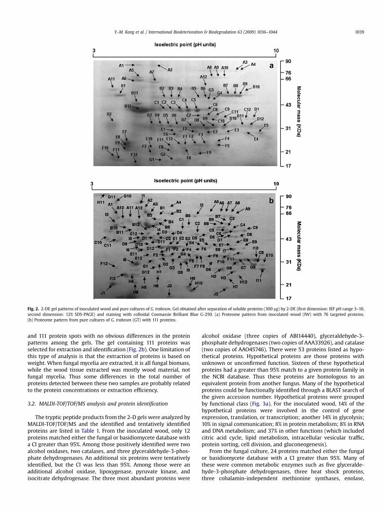
Fig. 2. 2-DE gel patterns of inoculated wood and pure cultures of G. trabeum. Gel obtained after separation of soluble proteins (300 mg) by 2-DE (first dimension: IEF pH range 3–10,second dimension: 12% SDS-PAGE) and staining with colloidal Coomassie Brilliant Blue G-250. (a) Proteome pattern from inoculated wood (IW) with 76 targeted proteins.(b) Proteome pattern from pure cultures of G. trabeum (GT) with 111 proteins.
Y.-M. Kang et al. / International Biodeterioration & Biodegradation 63 (2009) 1036–1044 1039
and 111 protein spots with no obvious differences in the proteinpatterns among the gels. The gel containing 111 proteins wasselected for extraction and identification (Fig. 2b). One limitation ofthis type of analysis is that the extraction of proteins is based onweight. When fungal mycelia are extracted, it is all fungal biomass,while the wood tissue extracted was mostly wood material, notfungal mycelia. Thus some differences in the total number ofproteins detected between these two samples are probably relatedto the protein concentrations or extraction efficiency.
3.2. MALDI-TOF/TOF/MS analysis and protein identification
The tryptic peptide products from the 2-D gels were analyzed byMALDI-TOF/TOF/MS and the identified and tentatively identifiedproteins are listed in Table 1. From the inoculated wood, only 12proteins matched either the fungal or basidiomycete database witha CI greater than 95%. Among those positively identified were twoalcohol oxidases, two catalases, and three glyceraldehyde-3-phos-phate dehydrogenases. An additional six proteins were tentativelyidentified, but the CI was less than 95%. Among those were anadditional alcohol oxidase, lipoxygenase, pyruvate kinase, andisocitrate dehydrogenase. The three most abundant proteins were
alcohol oxidase (three copies of ABI14440), glyceraldehyde-3-phosphate dehydrogenases (two copies of AAA33926), and catalase(two copies of AAO45746). There were 53 proteins listed as hypo-thetical proteins. Hypothetical proteins are those proteins withunknown or unconfirmed function. Sixteen of these hypotheticalproteins had a greater than 95% match to a given protein family inthe NCBI database. Thus these proteins are homologous to anequivalent protein from another fungus. Many of the hypotheticalproteins could be functionally identified through a BLAST search ofthe given accession number. Hypothetical proteins were groupedby functional class (Fig. 3a). For the inoculated wood, 14% of thehypothetical proteins were involved in the control of geneexpression, translation, or transcription; another 14% in glycolysis;10% in signal communication; 8% in protein metabolism; 8% in RNAand DNA metabolism; and 37% in other functions (which includedcitric acid cycle, lipid metabolism, intracellular vesicular traffic,protein sorting, cell division, and gluconeogenesis).
From the fungal culture, 24 proteins matched either the fungalor basidiomycete database with a CI greater than 95%. Many ofthese were common metabolic enzymes such as five glyceralde-hyde-3-phosphate dehydrogenases, three heat shock proteins,three cobalamin-independent methionine synthases, enolase,

Table 1Results of BLAST homology search of proteins identified using the GPS Explorer� protein analysis software (Applied Biosystems). Each protein spot corresponds to a number inthe gel shown in Fig. 2. The identified 18 proteins from the inoculated wood (IW) and identified 45 proteins from the pure culture of) Gloephyllum trabeum (GT) are listed. Thecalculated molecular weight (MW) and pI are also shown.
Sample name Protein score (%) Protein name Accession n. MW pI
A6 (IW) 100 Heat shock 70 protein (Aspergillus terreus) XP_001209480 69,550.50 5.08A8 (IW) 100 Alcohol oxidase (Gloeophyllum trabeum) ABI14440 72,468.20 6.25A9 (IW) 99.998 Alcohol oxidase (Gloeophyllum trabeum) ABI14440 72,468.20 6.25A10 (IW) 62.263 Alcohol oxidase (Gloeophyllum trabeum) ABI14440 72,468.20 6.25B1 (IW) 61.249 Lipoxygenase (Aspergillus ochraceus) AAZ13594 23,687 8.28B2 (IW) 59.422 Isocitrate dehydrogenase (Aspergillus fumigatus) XP_754749 55,739.60 8.85B7 (IW) 99.536 Catalase (Pleurotus sajor-caju) AAO45746 43,597.50 6.19B8 (IW) 100 Catalase (Pleurotus sajor-caju) AAO45746 43,597.50 6.19B9 (IW) 73.191 Pyruvate kinase (Aspergillus clavatus) XP_001267897 57,924.70 7.12B11 (IW) 100 ATP synthase beta chain (Coprinopsis cinerea) EAU88709 57,891.20 5.58D8 (IW) 97.127 Cystein synthase, putative (Aspergillus fumigatus) XP_747323 43,187.4 8.85C10 (IW) 97.38 Pyruvate dehydrogenase E1 component alpha subunit, putative (Aspergillus clavatus) XP_001269463 45,246.60 7.67E3 (IW) 100 Glyceraldehyde-3-phosphate dehydrogenase (Schizophyllum commune) AAA33926 36,032.70 7.01E5 (IW) 99.773 Glyceraldehyde-3-phosphate dehydrogenase (Schizophyllum commune) AAA33926 36,032.70 7.01E6 (IW) 100 Glyceraldehyde-3-phosphate dehydrogenase (Schizophyllum commune) AAA33926 36,032.70 7.01E7 (IW) 86.564 Vesicle-mediated transport protein (Imh1), putative (Aspergillus fumigatus) XP_746485 113,456.60 5.26E9 (IW) 41.552 DNA-directed DNA polymerase (Crinipellwas perniciosa) AAQ74286 106,154 8.84E10 (IW) 100 DNA damage checkpoint protein (Coprinopsis cinerea) EAU84422 28,941.30 4.76A1 (GT) 100 Heat shock protein (Aspergillus terreus) XP_001210539 80582.2 5.14A2 (GT) 100 Cell division cycle protein (Aspergillus terreus) XP_001209335 90410 4.98A3 (GT) 100 Heat shock protein (Aspergillus terreus) XP_001210539 80582.2 5.14A5 (GT) 88.564 Cobalamin-independent methionine synthase MetH/D (Aspergillus fumigatus) XP_752090 86840.5 6.33A7 (GT) 97.87 Cobalamin-independent methionine synthase MetH/D (Aspergillus fumigatus) XP_752090 86840.5 6.33A8 (GT) 99.974 Cobalamin-independent methionine synthase MetH/D (Aspergillus fumigatus) XP_752090 86840.5 6.33A9 (GT) 99.87 Cobalamin-independent methionine synthase MetH/D (Aspergillus fumigatus) XP_752090 8684.05 6.33A10 (GT) 93.419 Heat shock protein (Penicillium marneffei) AAX63812 69453.6 5.03A11 (GT) 51.215 Toxin biosynthesis proten (Fum3) (Aspergillus fumigatus) XP_754154 40826.9 6.65A12 (GT) 0 ER Hsp70 chaperone BiP, putative (Aspergillus clavatus) XP_001272734 73343.6 4.87B4 (GT) 0 Beta-glucosidase (Aspergillus fumigatus) XP_753915 92149.7 5.3B7 (GT) 100 Transketolase, putative (Aspergillus fumigatus) CAF32073 75352 6.13B12 (GT) 99.767 Heat shock protein (Aspergillus nidulans) XP_663693 61808.6 5.53C4 (GT) 0 Acetyl-CoA hydrolase (Aspergillus terreus) XP_001216368 58035.5 6.36C5 (GT) 0 Acetyl-CoA hydrolase (Aspergillus terreus) XP_001216368 58035.5 6.36C6 (GT) 0 Acetyl-CoA hydrolase (Aspergillus terreus) XP_001216368 58035.5 6.36C12 (GT) 99.994 Tubulin alpha-1 subunit (Aspergillus clavatus) XP_001269877 50048.2 4.99D3 (GT) 0 tRNA ligase (Aspergillus fumigatus) XP_752336 94155.1 8.42D4 (GT) 34.191 Phosphoglyceromutase (Aspergillus oryzae) BAB12237 57252.9 5.3D7 (GT) 91.715 UTP-glucose-1-phosphate uridylyltransferase Ugp1 (Aspergillus fumigatus) XP_746749 56889.5 6.41D9 (GT) 100 ATP synthase NADP-dependent XP_753589 55586 5.3D12 (GT) 100 Enolase (Aspergillus terreus) XP_001212082 47318.2 5.3E4 (GT) 80.126 Glutamate dehydrogenase (Penicillium chrysogenum Q9URS1 49801.1 6E6 (GT) 0 Mitochondrial ribosomal protein (Aspergillus clavatus) XP_001274476 16416 10.33E9 (GT) 100 Citrate synthase (Aspergillus clavatus) XP_001276108 52073.2 8.84E11 (GT) 100 Eukaryotic translation initiation factor 4 (Aspergillus clavatus) XP_001270835 44903 4.98F2 (GT) 100 Actin (Aspergillus terreus) XP_001209659 40112.2 5.84F10 (GT) 100 NAD-dependent formate dehydrogenase AciA/Fdh (Aspergillus fumigatus) XP_747586 45717.7 8.42G1 (GT) 96.625 DUF636 domain protein (Aspergillus clavatus) XP_001275720 15634.5 6.88G3 (GT) 0 Conidiophore development protein HymA (Aspergillus clavatus) XP_001271242 43774.8 8.55G4 (GT) 0 Alkaline serine protease (Aspergillus fumigatus) XP_749473 13429 9.72G6 (GT) 0 Carbonic anhydrase family protein (Aspergillus clavatus) XP_001271988 25482.2 5.86G9 (GT) 100 Glyceraldehyde-3-phosphate dehydrogenase gpdA-Aspergillus niger XP_001397496 36164.4 6.6H1 (GT) 0 Quinone oxidoreductase (Aspergillus clavatus) XP_001269933 36667.9 6.2H2 (GT) 0 6-phosphofructo-2-kinase (Aspergillus clavatus) XP_001270037 61336.7 7.76H3 (GT) 0 Alkaline serine protease (Aspergillus fumigatus) XP_749473 13429 9.72H7 (GT) 0 Stress responsive A/B barrel domain protein (Aspergillus clavatus) XP_001273319 12160.1 5.73H12 (GT) 0 Annexin (Aspergillus clavatus) XP_001275766 49004.5 8.33I2 (GT) 100 Mitochondrial aconitate hydratase, putative (Aspergillus clavatus) XP_001268345 84,730.80 5.87I4 (GT) 100 Hsp70 chaperone (HscA), putative (Aspergillus clavatus) XP_001276496 66,901.60 5.19I5 (GT) 100 Beta-tubulin (Aspergillus fumigatus) AAL01593 44,722.00 6.01I10 (GT) 100 Glyceraldehyde-3-phosphate dehydrogenase (Aspergillus terreus) XP_001209501 35,687.30 7.68I11 (GT) 100 Glyceraldehyde-3-phosphate dehydrogenase (Aspergillus niger) XP_001397496 36,164.40 6.6I12 (GT) 100 Glyceraldehyde-3-phosphate dehydrogenase, putative (Aspergillus fumigatus) XP_748238 36,117.50 6.17J1 (GT) 100 Citrate synthase (Aspergillus nidulans) AAC49728 52,128.30 8.75
Y.-M. Kang et al. / International Biodeterioration & Biodegradation 63 (2009) 1036–10441040
citrate synthase, actin, and formate dehydrogenase (Table 1). Anadditional 21 proteins were tentatively identified, but the CI wasless than 95%. Among those were an additional heat shock protein,cobalamin-independent methionine synthase, beta-glucosidase,glutamate dehydrogenase, acetyl-CoA hydrolase, and quinoneoxidoreductase, although many of these had a very low CI (Table 1).
The most abundant proteins were cobalamin-independent methi-onine synthases (three copies of XP_752090), heat shock protein(two copies of XP_001210539), acetyl-CoA hydrolase (two copies ofXP_001216368), citrate synthase (two copies of XP_001276108,actin (2 copies of XP_001209659), and alkaline serine protease (twocopies of XP_749473). There were 58 proteins listed as hypothetical

Fig. 3. Protein functional grouping for comparisons between inoculated wood (a) and pure cultures of G. trabeum (b). Hypothetical proteins were placed into predicted functionalgroups if possible by searching the NCBI Conserved Domain Database (CDD) and PDBJ (Protein Data Bank Japan) using the BLAST search engine. Functional groups were based onPDBJ nomenclature.
Y.-M. Kang et al. / International Biodeterioration & Biodegradation 63 (2009) 1036–1044 1041
proteins and of these only eight showed greater than a 95% matchto a given protein family. The hypothetical proteins were groupedby functional class (Fig. 3b). There were also eight unidentifiedproteins. For the fungal culture, 6% of the hypothetical proteinswere involved in control of gene expression, translation, or tran-scription, another 9% in glycolysis, 4% in signal communication, 19%in protein metabolism, 4% in RNA and DNA metabolism, and 48% inother functions.
3.3. Comparison of hypothetical proteins between inoculated woodand the pure cultures of G. trabeum
In order to determine how many of the hypothetical proteinswere common in both the inoculated wood and the pure culture ofG. trabeum, a comparison between these two using the NCBI proteindatabase and its search engine was performed. None of the 53hypothetical proteins from the inoculated wood were identical tothe 58 hypothetical proteins from the pure culture of G. trabeum.
3.4. Image systems for biodeterioration of wood
The analysis of uncontaminated wood (UW) by both 2-D and 3-D imaging showed the external surface of the UW was intact and nodamage from microorganisms was found (Fig. 4a and 4c). Scanningelectron microscopic observations of the surfaces of the uncon-taminated wood indicate that this wood was structurally intact andno fungi were observed in the pits of the wood (Fig. 5a and c). Bycontrast, the external surfaces of the field-decayed wood shown by2-D and 3-D imaging indicated that the samples were deteriorated(Fig. 4b and d). The external surfaces were highly irregular andcontained many troughs where decay had occurred. The surfaces ofthe wood, when viewed by SEM (Fig. 5b and d), showed similarroughness in addition to fungal spores and mycelia that covered thepits. When brown-rot fungi attack wood, they first enter throughthe ray parenchyma cells and spread to other cells by pits or bore
holes (Green and Highley, 1997; Goodell, 2003; Daniel et al., 2007).As decay progresses the dissolution of the cell wall structure as wellas the presence of hyphae should be visible by microscopy. Thusmicroscopy confirmed that uncontaminated wood was structurallysound and no proteins were detected because no fungi werepresent.
4. Discussion
As stated previously, the exact mechanism of brown-rot decay isbeing debated; however, all of the proposed models involve H2O2,hydroxyl radicals, and modified Fenton chemistry. This study foundthree enzymes involved in Fenton chemistry in the brown-rotfungus growing on wood as a substrate, but not in the fungusgrowing in liquid media.
Alcohol oxidase (AOX, EC 1.1.3.13), which oxidizes short-chainaliphatic alcohols into aldehydes producing H2O2, has been isolatedand characterized from G. trabeum while growing on wood (Danielet al., 2007). The AOX from G. trabeum was localized outside of thefungus on hyphal walls and tripartite membranes, and within theslime layer. This was the first report of an extracellular AOX which iscritical for AOX to play a role in brown-rot decay. Additionally, theAOX characterized by Daniel et al. (2007) exhibited very highactivities in liquid culture with methanol a preferred substrate.Brown-rot fungi can modify lignin through demethylation yieldingmethanol, providing the necessary substrate for AOX activity.Additionally, Shary et al. (2008) found a likely AOX associated withthe outer membranes of the P. chrysosporium secretome. Thetranscripts from this AOX were 361-fold higher in cellulose-growncultures compared to glucose-grown cultures. Additionally, theShury AOX was 88% identical to the alcohol oxidase from G. trabeum(accession no. ABI14440), which is the same alcohol oxidaseidentified in this study. The three AOX identified in this currentstudy were 100%, 99.99%, and 62.26% similar to G. trabeum AOX(accession no. ABI14440) and were found only in the inoculated

Fig. 4. Image comparisons between uncontaminated wood (UW) and decayed wood (DW) using the KH-7700 digital microscope. (a) UW (bar: 1.0 mm). (b) DW (bar: 1.0 mm).(c) 3-D Scan of UW (bar: 0.16 mm). (d) 3-D Scan of DW (bar: 0.16 mm). The external surface of the decayed wood indicates a highly irregular surface and extensive decay while thesurface of the uncontaminated wood is relatively even and not decayed.
Y.-M. Kang et al. / International Biodeterioration & Biodegradation 63 (2009) 1036–10441042
wood samples. Both white-rot and brown-rot systems employhydroxyl radicals, and all of these findings support the possible roleof AOX as a source of H2O2 for the Fenton reaction.
Lipoxygenases (LOX, EC 1.13.11) are a family of iron-containingdioxygenases whose preferred substrates are polyunsaturated fattyacids containing a series of cis double bonds. Very few fungal LOXhave been identified or characterized. One unique fungal LOX thathas been characterized from Gaeumannomyces graminis is the onlymanganese, not iron, containing LOX (Oliw, 2002). One role of LOXin plants and animals is the production of hydroperoxy end prod-ucts that induce structural changes in lipid membranes andprovoke a series of secondary lipid peroxidations (Brash, 1999).Lipid peroxides are unstable in the presence of reduced metals,such as Feþ2 involved in the Fenton reaction, and form alkoxylradicals which are as damaging as hydroxyl radicals (Arora et al.,2002). Additionally, some products of lipoxygenase activity arefurther modified and used as signaling compounds. Ten percent ofthe proteins produced by G. trabeum in this study were for signalcommunication.
Catalase (EC 1.11.1.6) is a ubiquitous enzyme involved in thebreakdown of H2O2 into molecular oxygen. This enzyme helps toprotect cells from the toxic effects of H2O2 without the productionof free radicals. Catalase has been found to be an effective inhibitorof lipid peroxidation caused by hydroquinone-derived hydroxyl
radicals formed during wood degradation by G. trabeum (Varelaand Tien, 2003). In P. chrysosporium, catalase transcripts were35-fold higher in cellulose-grown ligninolytic cultures compared toglucose-grown cultures. This catalase appears to be associated withthe outer membrane of the fungus and is proposed to play a role indetection of reactive oxygen species. The P. chrysosporium genomeencodes for four different catalases, but only one appeared to be themajor isozyme expressed under ligninolytic conditions (Shary et al.,2008). Two catalases were detected in this study from the inocu-lated wood sample.
None of the primary fungal decay enzymes such as hemi-cellulases or cellulases were detected in this study. In the fungalculture samples, these enzymes are not needed because they werenot grown in a cellulose medium. Green and Highley (1997)reported that these primary decay enzymes are produced by thedecay fungi during the beginning of decay (less than 4 weeks afterthe wood was inoculated). The inoculated wood samples used inthis study were held past 4 weeks before extracted; thus it is likelysome of these enzymes were ‘‘missed’’ due to the timing of theextractions. In P. placenta Martinez et al. (2009) found numeroushemicellulases, but only one endoglucanase from cultures grownon cellulose media compared to glucose media, and no genescoding for cellobiohydrolases or cellulose-binding domains werefound in this genome. In the future, a decay time series study

Fig. 5. Visual comparisons of the state of decay between uncontaminated wood (UW) and decayed wood (DW) using a JOEL JSM-6500F SEM. (a) UW wood is structurally intactand no fungi were observed in the pits of the wood; the bar indicates 10 mm. (b) The surfaces of DW contain fungal spores and mycelia covering the pits; the bar indicates 10 mm.(c) Magnification of ‘‘a’’, the bar indicates 1 mm. (d) Magnification of ‘‘b’’, the bar indicates 10 mm. The circle on images a and b indicates that location of the enlargements seen inc and d.
Y.-M. Kang et al. / International Biodeterioration & Biodegradation 63 (2009) 1036–1044 1043
beginning with inoculation is planned in order to characterize all ofthe proteins involved in the breakdown of cellulose, hemicellulose,and lignin at different stages of decay.
A comparison of the functional grouping of proteins in this studyhighlights some interesting differences. The inoculated woodsample expressed approximately twice as many different proteinsinvolved in transcription, translation, control of gene expression,electron transport, and DNA/RNA metabolism as did the fungalculture sample. The inoculated sample also produced more thantwice as many different signal communication proteins as thefungal culture and 1.5 times more proteins involved in glycolysis. Incontrast, the inoculated sample produced fewer proteins involvedin protein metabolism (8% versus 19%). When the fungus is growingon wood, such as the inoculated sample, there is a requirement formany different enzymes involved in the complex breakdown ofthe wood components. So the fungus invests more resources toproduce these enzymes, which ultimately feeds glycolysis as thecellulose breaks down into glucose. When the fungus is grown inliquid media containing high glucose levels, these types of enzymesare not required, nor does the fungus need to invest its resources inobtaining a source of carbon that is readily available in the media.A surprising result from this study is that none of the hypotheticalproteins that were a majority of the proteins found in this study andonly a few of the identified proteins were found in both sets ofsamples. This implies that there is a very different mechanism inaction when the fungus is growing on a cellulose-based nutrientsource (wood) versus glucose media.
5. Conclusions
Proteomic profiles or ‘‘pictures in time’’ will ultimately lead toa better understanding of the complex metabolic processesinvolved in the decay of wood cell wall components. They may alsolead to the discovery of new enzymes critical to the decay process.By comparing protein expression levels under different growthconditions, environments, or stages of decay, this approach couldhelp determine how different wood preservatives or differentwood species with natural durability or microbial interactions alterthe expression of decay enzymes and ultimately decay. Althoughmany authors consider 2-D PAGE with MALDI-TOF/TOF/MS themost accepted approach for expression proteomics (Keller andHettich, 2009), there are still considerable limitations, which arehighlighted by this study.
Of the 76 protein spots excised from the inoculated woodsample, only 12 were identified with confidence; six were identi-fied with low confidence, 53 were hypothetical, although manycould be grouped into a functional category, and five wereunidentified. Similar results occurred for the fungus grown inculture. Of 111 protein spots, 24 were identified with confidence, 21were identified with low confidence, 58 were hypothetical, andeight were unidentified. It is not surprising that more proteins wereidentified from this sample since many of these proteins wereubiquitous metabolic proteins. This approach is hampered by thelack of well-annotated genomic sequence data and protein data onthe public databases. Identification of unknown proteins is only as

Y.-M. Kang et al. / International Biodeterioration & Biodegradation 63 (2009) 1036–10441044
good as the existing searchable databases. This problem parallelsthat which occurred in years past with the lack of sequence infor-mation on GenBank. As more and more researchers sequence thegenome of wood-decay fungi or use proteomics as a tool to answerquestions in science, more proteins will be identified and thedatabase will continue to grow. Ultimately the identity of theproteins in this study may be resolved.
Another limitation of this type of analysis is that the extractionof proteins is based on weight. When fungal mycelia are extracted,it is all fungal biomass, while the wood tissue extracted was mostlywood material, not fungal mycelia. Thus differences in the totalnumber of proteins detected between these two samples areprobably related to the protein concentrations or extraction effi-ciency. Also it is likely that proteins were missed by this analysisbecause the concentration was not sufficient for detection andidentification.
The brown-rot mechanism of decay is unique. Brown-rot fungiare the only known organisms that can breakdown celluloseleaving behind an intact but modified lignin. From the study pre-sented here, it is evident that the brown-rot decay mechanism isvery complex and very different from ‘‘normal’’ metabolism. Therewas very little overlap between the proteins found from G. trabeumwhile growing on wood (cellulose) compared to the proteins foundfrom the same fungus growing in a glucose medium. Althoughsignificant limitations still exist in identifying the proteinsexpressed, this limitation will diminish as functional proteins areidentified and added to the databases. We are currently sequencingthe genome of the copper-tolerant brown-rot fungus, Antrodiaradiculosa, and as this sequence is annotated we will add to thepublic databases. Future studies will look at expression of decayproteins over the course of decay as well as gene expression ofwood-decay enzymes during decay of different wood species. Toour knowledge, this is the first reported proteomic analysis ofpinewood decayed by a brown-rot fungus, and it provides the initialcharacterization of proteins involved in this type of woodbiodeterioration.
Acknowledgements
The authors wish to thank Dr. Tibor Pechan in the LSBI(Life Sciences and Biotechnology Institute) and Dr. OlgaPechanova at Mississippi State University, who provided the MSanalysis and protein database analysis; and Mr. Min Lee, forassisting with this experiment. This work was supported bya LSBI grant, the Lucas Biodeterioration Laboratory, and theState of Mississippi. This article was approved for publication asFP505 of the Forest and Wildlife Research Center at MississippiState University.
References
Abbas, A., Koc, H., Liu, F., Tien, M., 2005. Fungal degradation of wood: initial pro-teomic analyses of extracellular proteins of Phanerochaete chrysosporium grownon oak substrate. Current Genetics 47, 49–56.
Arora, A., Sairam, R.K., Srivastava, G.C., 2002. Oxidative stress and antioxidativesystem in plants. Current Science 82, 1227–1238.
Bradford, M., 1976. A rapid and sensitive method for the quantitation of microgramquantities of protein utilizing the principle of protein–dye binding. AnalyticalBiochemistry 72, 248–254.
Brash, A., 1999. Lipoxygenases: occurrence, functions, catalysis, and acquisition ofsubstrate. Journal of Biological Chemistry 274, 23679–23682.
Bruce, A., King, B., Highley, T.L., 1991. Decay resistance of wood removed from polesbiologically treated with Trichoderma. Holzforschung 45, 307–311.
Cease, K.R., Blanchette, R.A., Highley, T.L., 1989. Interactions between Scytalidiumspecies and brown- or white-rot basidiomycetes in birch wood decayed in thelaboratory. Wood Science and Technology 23, 151–161.
Daniel, G., Volc, J., Filonova, L., Plihal, O., Kubatova, E., Halada, P., 2007. Character-istics of Gloeophyllum trabeum alcohol oxidase, an extracellular source of H2O2
in brown rot decay of wood. Applied and Environmental Microbiology 73,6241–6253.
Flournoy, D., Kirk, T., Highley, T., 1991. Wood decay by brown-rot fungi: changes inpore structure and cell wall volume. Holzforschung 45, 383–388.
Fromm, J., Rockel, B., Lautner, S., Windeisen, E., Wanner, G., 2003. Lignin distributionin wood cell walls determined by TEM and backscattered SEM techniques.Nature Structural Biology 143, 77–84.
Fryksdale, B.G., Jedrzejewski, P.T., Wong, D.L., Gaertner, A.L., Miller, B.S., 2002.Impact of deglycosylation methods on two-dimensional gel electrophoresis andmatrix assisted laser desorption/ionization–time of flight-mass spectrometryfor proteomic analysis. Electrophoresis 23, 2184–2193.
Green, F., Highley, T.L., 1997. Brown-rot wood decay-insights gained from a low-decay isolate of Postia placenta. Trends in Plant Pathology 1, 1–17.
Gromov, P.S., Celis, J.E., 2000. From genomics to proteomics. Molecular Biology 34,508–520.
Goodell, B., 2003. Brown-rot fungal degradation of wood: our evolving view. Wooddeterioration and preservation: advances in our changing world. In: Goodell, B.,Nicholas, D., Schultz, T. (Eds.), ACS Symposium Series, 845. American ChemicalSociety, Washington, DC, pp. 97–118.
Keller, M., Hettich, R., 2009. Environmental proteomics: a paradigm shift in char-acterizing microbial activities at the molecular level. Microbiology and Molec-ular Biology Reviews 73 (1), 62–70.
Kobayashi, Y., 2006. HIROX developed next generation digital microscope. Semi-conductor FPD World 25, 100–101.
Martinez, D., Larrondo, L.F., Putnam, N., Gelpke, M.D.S., Huang, K., Chapman, J.,Helfenbein, K.G., Ramaiya, P., Detter, J.C., Larimer, F., Coutinho, P.M.,Henrissat, B., Berka, R., Cullen, D., Rokhsar, D., 2004. Genome sequence of thelignocellulose degrading fungus Phanerochaete chrysosporium strain RP78.Nature Biotechnology 22, 695–700.
Martinez, D., Challacombe, J., Morgenstern, I., Hibbett, D., Schmoll, M., Kubicek, C.P.,Ferreira, P., Ruiz-Duenas, F.J., Martinez, A.T., Kersten, P., Hammel, K.E., VandenWymelenberg, A., Gaskell, J., Lindquist, E., Sabat, G., Splinter BonDurant, S.,Larrondo, L.F., Canessa, P., Vicuna, R., Yadav, J., Doddapaneni, H., Subramanian, V.,Pisabarro, A.G., Lavin, J.L., Oguiza, J.A., Master, E., Henrissat, B., Coutinho, P.M.,Harris, P., Magnuson, J.K., Baker, S.E., Bruno, K., Kenealy, W., Hoegger, P.J., Kues, U.,Ramaiya, P., Lucas, S., Salamov, A., Shapiro, H., Tu, H., Chee, C.L., Misra, M., Xie, G.,Teter, S., Yaver, D., James, T., Mokrejs, M., Pospisek, M., Grigoriev, I.V., Brettin, T.,Rokhsar, D., Berka, R., Cullen, D., 2009. Genome, transcriptome, and secretomeanalysis of wood decay fungus Postia placenta supports unique mechanisms oflignocellulose conversion. Proceedings of the National Academy of Sciences 106(6), 1954–1959.
Matsuzaki, F., Shimizu, M., Wariishi, H., 2008. Proteomic and metabolomic analysesof the white-rot fungus Phanerochaete chrysosporium exposed to exogenousbenzoic acid. Journal of Proteome Research 7, 2342–2350.
Nilsson, T., Daniel, G., Kirk, T.K., Obst, J.R., 1989. Chemistry and microscopy of wooddecay by some higher ascomycetes. Holzforschung 43, 11–18.
Oliw, E.H., 2002. Plant and fungal lipoxygenases. Prostaglandins and Other LipidMediators 68-69, 313–323.
Pappin, D.J.C., Hojrup, P., Bleasby, A.J., 1993. Rapid identification of proteins bypeptide mass fingerprinting. Current Biology 3, 327–332.
Pechanova, O., Stone, W.D., Monroe, W., Nebeker, T.E., Klepzig, K.D., Yuceer, K.D.C.,2008. Global and comparative protein profiles of the pronotum of the southernpine beetle, Dendroctonus frontalis Zimmermann (Coleoptera: Scolytidea).Insect Molecular Biology 17 (3), 261–277.
Phillips, C., Bogyo, M., 2005. Proteomics meets microbiology: technical advances inthe global mapping of protein expression and function. Cellular Microbiology 7,1061–1076.
Pointing, S., Parungao, M.M., Hyde, K.D., 2003. Production of wood-decay enzymes,mass loss and lignin solubilization in wood by tropical Xylariaceae. MycologicalResearch 107, 231–235.
Preston, F., 2000. Wood preservation: trends of today will influence the industrytomorrow. Forest Products Journal 50, 12–19.
Prabakaran, P., An, J., Gromiha, M., Selvaraj, S., Uedaira, H., Kono, H., Sarai, A., 2001.Thermodynamic database for protein-nucleic acid interactions (ProNIT). Bio-informatics 17, 1027–1034.
Sato, S., Liu, F., Koc, H., Tien, M., 2007. Expression analysis of extracellular proteinsfrom Phanerochaete chrysosporium grown on different liquid and solidsubstrates. Microbiology 153, 3023–3033.
Shary, S., Kapich, A.N., Panisko, E.A., Magnuson, J.K., Cullen, D., Hammel, K.E., 2008.Differential expression in Phanerochaete chrysosporium of membrane-associ-ated proteins relevant to lignin degradation. Applied and EnvironmentalMicrobiology 74 (23), 7252–7257.
Shimizu, M., Wariishi, H., 2005. Development of a sample preparation method forfungal proteomics. FEMS Microbiology Letters 247, 17–22.
Vanden Wymelenberg, A., Sabat, G., Martinez, D., Rajangam, S., Teeri, T.T., Gaskell, P.,Kersten, P.J., Cullen, D., 2005. The Phanerochaete chrysosporium secretome:database predictions and initial mass spectrometry peptide identification incellulose-grown medium. Journal of Biotechnology 118, 17–34.
Varela, E., Tien, M., 2003. Effect of pH and oxalate on hydroquinone-derivedhydroxyl radical formation during brown rot wood degradation. Applied andEnvironmental Microbiology 69, 6025–6031.
Zhou, X.W., Blackman, M.J., Howell, S.A., Carruthers, V.B., 2004. Proteomic analysisof cleavage events reveals a dynamic two-step mechanism for proteolysis ofa key parasite adhesive complex. Molecular and Cellular Proteomics 3,565–576.