Protein profiles of dengue-infected Aedes aegypti (L) -...
9
Dengue Bulletin – Volume 33, 2009 115 Protein profiles of dengue-infected Aedes aegypti (L) H.L. Lee # , Y.C. Wong and A. Rohani Medical Entomology Unit/Infectious Disease Research Centre and WHO Collaborating Center for Vectors, Institute for Medical Research, Jalan Pahang, 50588 Kuala Lumpur, Malaysia Abstract The protein profiles of DENV-2 and DENV-4 viruses-infected adult Aedes aegypti were analysed using Coomassie-stained SDS-PAGE samples. For this study, adult female mosquitoes were infected orally with either DENV-2 or DENV-4 virus using spiked human blood samples. The infections in the individual mosquitoes were confirmed using the reverse transcription-polymerase chain reaction (RT-PCR). From the SDS-PAGE analyses, a total of 11 proteins with molecular weights of 22 kDa to greater than 181.8 kDa were identified in DENV-2 and DENV-4-infected mosquito homogenates. Three proteins (Mr 49, 64 and 200 kDa) from DENV-2-infected mosquitoes and five proteins (Mr 49, 50, 60, 135 and 200 kDa) from DENV-4-infected mosquitoes were identified at higher concentrations than in uninfected controls. Further work is now needed to specifically identify these proteins and study their biological roles in DENV infections. Keywords: Dengue; Aedes aegypti; infection proteins; protein profiles. # E-mail: [email protected] Introduction In Malaysia, classical dengue fever was first documented in 1901-1902, [1] while the first reported outbreak of dengue haemorrhagic fever (DHF) occurred in 1962. [2] Since then dengue has remained endemic, with one of the four dengue serotypes circulating in the country and outbreaks of DHF being reported periodically. At present, neither an effective vaccine nor a specific drug treatment for DF/DHF is available, making the laboratory- based surveillance system an important tool to provide an early warning of an impending outbreak of the disease. Vector surveillance allows timely implementation of emergency mosquito control measures such as space application of chemical insecticides against adult mosquitoes and destruction of their breeding places to contain an outbreak from spreading. However, when the adult mosquito density is low, direct entomological monitoring is not a sensitive enough indicator to serve as an early warning surveillance system for outbreak prevention. It is in this particular situation that detection of dengue viruses in vector population becomes an important element as part of an early alert system. [3,4,5]
Transcript of Protein profiles of dengue-infected Aedes aegypti (L) -...
Dengue Bulletin – Volume 33, 2009 115
Protein profiles of dengue-infected Aedes aegypti (L)H.L. Lee#, Y.C. Wong and A. Rohani
Medical Entomology Unit/Infectious Disease Research Centre and WHO Collaborating Center for Vectors, Institute for Medical Research,
Jalan Pahang, 50588 Kuala Lumpur, Malaysia
Abstract
The protein profiles of DENV-2 and DENV-4 viruses-infected adult Aedes aegypti were analysed using Coomassie-stained SDS-PAGE samples. For this study, adult female mosquitoes were infected orally with either DENV-2 or DENV-4 virus using spiked human blood samples. The infections in the individual mosquitoes were confirmed using the reverse transcription-polymerase chain reaction (RT-PCR). From the SDS-PAGE analyses, a total of 11 proteins with molecular weights of 22 kDa to greater than 181.8 kDa were identified in DENV-2 and DENV-4-infected mosquito homogenates. Three proteins (Mr 49, 64 and 200 kDa) from DENV-2-infected mosquitoes and five proteins (Mr 49, 50, 60, 135 and 200 kDa) from DENV-4-infected mosquitoes were identified at higher concentrations than in uninfected controls. Further work is now needed to specifically identify these proteins and study their biological roles in DENV infections.
Keywords: Dengue; Aedes aegypti; infection proteins; protein profiles.
# E-mail: [email protected]
IntroductionIn Malaysia, classical dengue fever was first documented in 1901-1902,[1] while the first reported outbreak of dengue haemorrhagic fever (DHF) occurred in 1962.[2] Since then dengue has remained endemic, with one of the four dengue serotypes circulating in the country and outbreaks of DHF being reported periodically. At present, neither an effective vaccine nor a specific drug treatment for DF/DHF is available, making the laboratory-based surveillance system an important tool to provide an early warning of an impending outbreak of the disease.
Vector survei l lance al lows t imely implementation of emergency mosquito control measures such as space application of chemical insecticides against adult mosquitoes and destruction of their breeding places to contain an outbreak from spreading. However, when the adult mosquito density is low, direct entomological monitoring is not a sensitive enough indicator to serve as an early warning surveillance system for outbreak prevention. It is in this particular situation that detection of dengue viruses in vector population becomes an important element as part of an early alert system.[3,4,5]
116 Dengue Bulletin – Volume 33, 2009
Protein profiles of dengue-infected Ae. aegypti (L)
The current “gold s tandard” for dengue detection in mosquitoes involves isolation of the virus followed by indirect immunofluorescence.[5] However, this requires cell culture facilities which are difficult to maintain and the technique is not sensitive. The reverse transcriptase-polymerase reaction (RT-PCR) technique is sensitive and specific but very costly, especially during an outbreak where large-scale screening needs to be performed. Hence, alternative methods of detection are needed.
It is observed that dengue is less likely to cause mortality to the Aedes vector, leading to the belief that the mosquitoes may synthesize certain proteins (known as “infection proteins”) to work against the dengue viruses during invasion. Detection of these proteins, therefore, not only provides further understanding of the interaction between both the dengue virus and its vector but also could be the potential antigen used for detecting the presence of the virus in the mosquito.
Materials and methods
Preparation of dengue virus source for membrane feeding
The Aedes albopictus cell line C6/36, maintained with 10% fetal bovine serum in the minimum essential medium (MEM), was used to propagate dengue virus (DENV). DENV-2 and DENV-4-infected, as well as non-infected, C6/36 Ae. albopictus cell-culture supernatants (DENV-2/-4 ICS and NICS, respectively) were used to spike normal human blood for membrane feeding. The DENV-2/-4 ICS and NICS were clarified by centrifugation, and confirmed to contain either DENV-2/-4 ICS or to be uninfected (NICS) using the DENV serotype-specific RT-PCR (reference) and stored at –70 °C.
Preparation of blood meal for artificial membrane feeding
Initially, equal volumes of blood and phosphate buffered saline (PBS) were mixed in a 10 ml culture tube and centrifuged at 500 x g for 10 minutes at room temperature. The supernatant was then removed and the large blood cells (erythrocytes and leukocytes) were further washed five times with PBS using this method until the supernatant became clear. The blood cells were then transferred to fresh culture tubes.
Artificial membrane feeding
An artificial membrane feeding technique employed was modified from Graves (1980). Two hundred four- to seven-day-old adult female Ae. aegypti mosquitoes were collected into paper cups and starved overnight prior to blood feeding. A total of 30 female mosquitoes were placed into each paper cup covered with netting. A glass feeder fitted with a water jacket at 37 °C was covered at the bottom by wrapping a small piece of membrane made from chicken skin moistened with normal saline.
In the preparation of the artificial infective blood meal, 6 ml of washed red blood cells was mixed with 3 ml of D2/4-ICF. One ml of adenosine 5′-triphosphate (ATP) was added to the final concentration of 0.02 M to stimulate mosquitoes to engorge.[6,7] For negative control, the same ratio of washed red blood cells, normal CF and ATP was used. The blood meals were heated to 37 °C before being presented to the mosquitoes. Blood was presented to the mosquitoes by placing the cup containing the mosquitoes below the feeder, with the surface of the nylon netting of the cup in contact with the membrane of the feeder. The mosquitoes were then allowed to feed for approximately 10 to 30 minutes.
Dengue Bulletin – Volume 33, 2009 117
Protein profiles of dengue-infected Ae. aegypti (L)
Rearing of infected mosquitoes
Fully-engorged mosquitoes were selected and transferred to a small cage. The dengue-infected mosquitoes were reared at 28 °C and 70%–80% relative humidity in ACL-2 insectarium for 15 days before being killed. A single mosquito was transferred into a nuclease-free 1.5 ml microcentrifuge tube and kept at –70 °C in a freezer for further analysis.
Detection of dengue virus using RT-PCR
The single mosquito was homogenized on ice using a homogenizer attached to a Pellet Pestle Motor (Kontes, Japan) in a nuclease-free 1.5 ml microcentrifuge tube containing 210 µl of nuclease-free, double-distilled water. The homogenized samples were then centrifuged at 5000 x g for 10 minutes at 4 °C. A similar procedure was performed for non-infected mosquito homogenates. The rest of the samples were thawed and put on ice. High pure viral nucleic acid kit (Roche Diagnostic Corp., USA) was used to extract the viral RNA from the mosquito homogenates following the manufacturer’s guidelines. Extracted RNA was kept at –20 °C until used.
RT-PCR
The RT-PCR method of Lanciotti et al. (1992) was employed. DENV-2-specific primers (GTTCCTCTGCAAACACTCCA and GTGTTATTTTGATTTCCTTG) or DENV-4-specific primers (CCATTATGGCTGTGTTGTTT and CTTCATCCTGCTTCACTTCT) were used. The Access RT-PCR System (Promega, USA) was used to trans cribe the DENV RNA to cDNA. Each reaction contained 10.25 µl of double-distilled water, 2 µl of dNTP mixture, 1.25 µl of dithiotreitol, 0.5 µl of RNAse inhibitor, 5.0 µl of RT-PCR buffer, 0.5µl of enzyme mixture, 0.5 µl of each dengue primer and 0.5µl of RNA.
For dengue virus detection, the reaction was carried out at 51 °C for 30 minutes to produce cDNA, which was then amplified by the following PCR steps: initial denaturation at 92 °C for three minutes, 41 cycles of 92 °C for 30 seconds, 51 °C for 45 seconds and 72 °C for one minute; followed by 72 °C for five minutes. For every RT-PCR, a positive control and a negative control were included.
PCR products were analysed by performing electrophoresis in 2.0% Nusieve PCR gel (FC Bio, USA) at 100 volts and staining with ethidium bromide. The gel was viewed under ultraviolet illuminator (Ultra Lum Inc, California, USA) and the resulting bands were photographed with a polaroid camera.
Protein analysis of DENV-2- and DENV-4-infected mosquitoes
SDS-PAGE gel was prepared using Dual Slab Gel system. Both RT-PCR-detected infective mosquito homogenates and non-infected mosquito homogenates (negative control) were homogenized in double-distilled water and the extract was separated on SDS-polyacrylamide slab gel using the discontinuous system consisting of 4% acrylamide stacking gel and 12% acrylamide separating gel. Approximately 10 ul of the mosquito samples were boiled at 100 °C for five minutes before loading onto the gel. The separated protein bands were visualized by staining with Coomassie brilliant blue.
Results
The results obtained in a previous study[8] showed that the extrinsic incubation period for Aedes mosquitoes (DENV-2-infected) decreased from Day 11 to Day 17 and (DENV-4-infected) from Day 7 to Day 19 as the temperature increased from 26 °C to

118 Dengue Bulletin – Volume 33, 2009
Protein profiles of dengue-infected Ae. aegypti (L)
30 °C. The mortality rate of mosquitoes was found to be high at 30 °C compared with that at 28 °C or 26 °C. Based on these results, the extrinsic incubation temperature of 28 °C and incubation period of 15 days was, therefore, selected in this experiment in order to obtain a high number of live infected mosquitoes and a shorter viral replication time.
Protein synthesis in DENV-2-infected Ae. aegypti
Figure 1 shows the agarose gel electrophoresis of individual DENV-2-infected Ae. aegypti after RT-PCR assay. Lane 1, 2, 4, 5, 6, 7 and 9 represent the dengue-infected Aedes mosquito.
The protein profiles of individual DENV-2-infected Ae. aegypti are shown in Figures 2 and 3. There are at least 11 protein bands, which fall in the range of approximately 200 kDa to less than 24 kDa for both experimental and control mosquito. The predominant
Figure 1: Result of agarose gel electrophoresis of individual DENV-2-infected Ae. aegypti collected on 15th day after dengue viral amplification through RT-PCR assay
Figure 2: Protein profiles of DENV-2-infected Ae. aegypti sacrificed on 15th day
Dengue Bulletin – Volume 33, 2009 119
Protein profiles of dengue-infected Ae. aegypti (L)
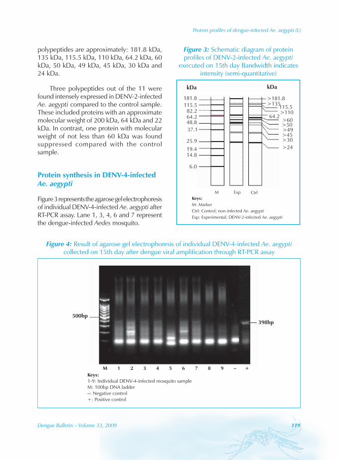
polypeptides are approximately: 181.8 kDa, 135 kDa, 115.5 kDa, 110 kDa, 64.2 kDa, 60 kDa, 50 kDa, 49 kDa, 45 kDa, 30 kDa and 24 kDa.
Three polypeptides out of the 11 were found intensely expressed in DENV-2-infected Ae. aegypti compared to the control sample. These included proteins with an approximate molecular weight of 200 kDa, 64 kDa and 22 kDa. In contrast, one protein with molecular weight of not less than 60 kDa was found suppressed compared with the control sample.
Protein synthesis in DENV-4-infected Ae. aegypti
Figure 3 represents the agarose gel electrophoresis of individual DENV-4-infected Ae. aegypti after RT-PCR assay. Lane 1, 3, 4, 6 and 7 represent the dengue-infected Aedes mosquito.
Figure 3: Schematic diagram of protein profiles of DENV-2-infected Ae. aegypti
executed on 15th day Bandwidth indicates intensity (semi-quantitative)
Figure 4: Result of agarose gel electrophoresis of individual DENV-4-infected Ae. aegypti collected on 15th day after dengue viral amplification through RT-PCR assay

120 Dengue Bulletin – Volume 33, 2009
Protein profiles of dengue-infected Ae. aegypti (L)
The protein profiles of individual DENV-4-infected Ae. aegypti are shown in Figure 5. The predominant protein bands observed were in the range of approximately 200 kDa to less than 24 kDa in both experimental and control mosquito (Figure 6). These proteins are having molecular weight of approximately 181.8 kDa, 135 kDa, 115.5 kDa, 110 kDa, 64.2 kDa, 60 kDa, 50 kDa, 49 kDa, 45 kDa, 30 kDa and 24 kDa.
Out of the 11 proteins observed, there were five proteins with approximate molecular weight of 181.8 kDa, 135 kDa, 60 kDa, 50 kDa and 49 kDa that were highly expressed in DENV-4-infected Aedes mosquito.
The Table summarizes the protein profiles obtained in both DENV-2- and DENV-4-infected Ae. aegypti. It is obvious that uninfected mosquitoes also produced similar protein profiles, although in both
Figure 5: Protein profiles of DENV-4-infected Ae. aegypti sacrificed on 15th day
Figure 6: Schematic diagram of protein profiles of DENV-4-infected Ae. aegypti
executed on 15th day
Table: Molecular weight of proteins synthesized in both experimental and
control mosquito
No.Molecular weight, kDa
Control DENV-2 DENV-4
1 >181.8 >181.8* >181.8*
2 >135 >135 >135*
3 115.5 115.5 115.5
4 >110 <110 >110
5 64.2 64.2* 64.2
6 >60 >60** >60*
7 >50 >50 >50*
8 >49 >49* >49*
9 >45 >45 >45
10 >30 >30 >30
11 >24 >24 >24
* highly expressed protein ** suppressed protein

Dengue Bulletin – Volume 33, 2009 121
Protein profiles of dengue-infected Ae. aegypti (L)
DENV-2- and DENV-4-infected mosquitoes some of the similar proteins were over-expressed.
DiscussionAs mentioned earlier, RT-PCR is a powerful tool which can be used in the detection of infection due to virus such as dengue. This technique, however, needs to be used in the laboratory as it is difficult to carry out in the field. Therefore, development of an ELISA-based antigen detection system may be necessary if anti-dengue viral “proteins” (or “infection proteins”) are to be obtained from the dengue virus-infected mosquitoes, and to be used as an ELISA-based detection kit to detect dengue infection in mosquitoes.
The “infection proteins” produced in a vector may act as a kind of defence mechanism to protect itself from a detrimental condition. For example, innate immune response is activated against the infection of plasmodium in Anopheles gambiae[9] as defensive and putative Gram-negative bacteria-binding proteins are synthesized mainly to block the parasite from entering into the mosquito. Chee and AbuBakar[10] had identified a tubulin or tubulin-like C6/36 mosquito cell protein which was able to bind to DENV-2 virus. It is believed that Aedes mosquito may also elicit the defense component (or “infection proteins”) to protect itself from the invaded dengue viruses. Yunus[11] found the presence of “infection proteins” in dengue-infected Ae. albopictus with molecular weight of 18 kDa, 27 kDa, 28 kDa and 70 kDa, while Rohani et al.[12] found that there are four such proteins having molecular weight of 24 kDa, 25 kDa, 31 kDa and 76 kDa from the DENV-2-infected Ae. aegypti.
There are 11 predominant polypeptides observed in the control with molecular weight of not less than 181.8 kDa, 135 kDa, 115.5 kDa, 110 kDa, 64.2 kDa, 60 kDa, 50 kDa, 49 kDa, 45 kDa, 30 kDa and 24 kDa. The result reported herein, however, is not identical to the findings reported by Rohani et al.[12] In their study, there were seven conspicuous proteins in the range of 72 kDa to 17 kDa detected in the normal blood-fed Ae. aegypti. The difference could be due to the concentration of the SDS-PAGE gel used since 10% of separating gel was used to separate the proteins present in the control mosquito in this study, as compared with 12% separating gel employed in Rohani et al.[12] study. Therefore, the major proteins determined in this study are relatively higher in molecular weight compared with the study done by Rohani et al.[12] where lower molecular weight of polypeptides was observed. On the other hand, Lee et al.[13] reported that 29 protein bands were observed in the sugar-fed Ae. aegypti. This shows that the protein synthesized in Aedes mosquito could also be closely related to the food consumed by the mosquitoes.
The protein profiles of the dengue-infected mosquito were found similar to that of the control, with an overall of 11 conspicuous polypeptides found. These proteins, however, exhibited dif ferent expression levels represented by the bandwidth. Proteins with molecular weight of not less than 181.8 kDa, 64.2 kDa and not less than 49 kDa were highly expressed in DENV-2-infected mosquito compared with the control sample, while proteins with molecular weight of not less than 181.8 kDa, 135 kDa, 60 kDa, 50 kDa and 49 kDa showed a broader bandwidth in the protein profiles of DENV-4-infected mosquito compared with the control. Such patterns may be due to the high expression

122 Dengue Bulletin – Volume 33, 2009
Protein profiles of dengue-infected Ae. aegypti (L)
of the ordinary proteins in dengue-infected Aedes mosquito during dengue virus invasion in Aedes mosquito. Another possibility could be the different type of protein(s), or “infection protein(s)”, having similar molecular weight(s) to the ordinary protein(s), being synthesized that hence fall in the same distance of the SDS-PAGE gel.
Suppression of protein with molecular weight of not less than 60 kDa was assumed to occur in the DENV-2-infected Aedes mosquito when compared with the control. The molecular weight of 28kDa also seemed to be suppressed in DENV-2-infected Ae. aegypti and such finding was also reported before by Rohani et al.[12] Since no protein suppression was observed in DENV-4-infected mosquito, there is a possibility that the protein synthesis could be serotype-specific.
Alcon et al.[14] reported that NS1 protein, which is not part of the virion and having molecular weight of 48 kDa, was found on the surface of the infected cells or secreted extracellularly into the blood circulation in dengue patients. The function of this protein remains unknown, but it is believed to correlate with the development of dengue haemorrhagic fever.[15,16] Tubulin or tubulin-like mosquito cell protein reported by Chee and AbuBakar[10] is having similar molecular weight of about 48 kDa. Wang et al.[17] reported that there are abundant brush borders found in the midgut of Ae. aegypti, and this element could be the initial interaction site between dengue virus and the mosquito. Hence, it is possible that the
“infection protein” synthesized may appear in the brush border to act as a defence barrier to block the entry of the dengue viruses.
Huang et al.[18] had successfully proved that this viral NS1 protein could be used as an ELISA antigen to detect dengue infection in patients. The protein with a molecular weight of about 49 kDa found in this study is unlikely to be similar to 48 kDa protein reported in cell culture and dengue patient, as this protein was also secreted in DENV-2- and DENV-4-infected and control adult mosquitoes. However, although similar proteins were found in both infected and control Aedes mosquitoes, several over-expressed proteins observed only in infected mosquitoes could be considered potential diagnostic antigens to be used for detecting dengue infection in mosquitoes, based on the quantitative differences in protein concentrations. It is pertinent, therefore, to quantify the various over-expressed proteins in any future study.
AcknowledgementsThe authors thank the Director-General of Health, Malaysia, for his permission to publish this research. Works mentioned in this paper were a part of the Master of Science thesis of the second author at the University of Malaya, Kuala Lumpur, and were partially supported by a research grant No. JPP-05-006 from the National Institute of Health, Ministry of Health, Malaysia.
ReferencesSkae FM. Dengue fever in Penang. [1] Br Med J 1902;2:1581-2.
Lam SK. Two decades of dengue in Malaysia. [2] Trop Med 1993;35(4):195-200.
Liotta DJ, Cabanne G, Campos R , Tonon [3] SA. Molecular detection of dengue viruses in field caught Aedes aegypti mosquitoes from northeastern Argentina. Rev Latinoam Microbiol 2005;47(3-4):82-7.

Dengue Bulletin – Volume 33, 2009 123
Protein profiles of dengue-infected Ae. aegypti (L)
Chow VTK, Chan YC, Rita Yong, Kim LK, [4] Lim LK, Chung Y, Lam-Phua SG. Monitoring of dengue viruses in field-caught Aedes aegypti and Aedes albopictus mosquitoes by a type-specific polymerase chain reaction and cycle sequencing. Am J Trop Med Hyg 1998;58(5):578-86.
Harris E, Roberts TG, Smith L, Selle J, Kramer [5] LD, Valle S, Sandoval E, Balmaseda A. Typing of dengue viruses in clinical specimens and mosquitoes by single-tube multiplex reverse transcriptase PCR. J Clin Microbiol 1998;36(9):2634-9.
Rutledge LC, Ward RA, Gould DJ. Studies on [6] the feeding response of mosquitoes to nutritive solutions in a new membrane feeder. Mosq News 1964;24(4):407-19.
Wirtz RA, Rutledge LC. Reconstituted collagen [7] sausage casings for the blood feeding of mosquitoes. Mosq News 1980;40(2):287-8.
Rohani A, Wong YC, Zamree I, Lee HL, [8] Zurainee MN. The effect of extrinsic incubation temperature on development of dengue serotype 2 and 4 viruses in Aedes aegypti (L.). Southeast Asian J Trop Med Pub Hlth 2009; 40(5):942-50.
Richman AM, Dimopoulos G, Seeley D, [9] Kafatos FC. Plasmodium activates the innate immune response of Anopheles gambiae mosquitoes. EMBO J 1997;16(20):6114-9.
Chee HY, AbuBakar S. Identification of a [10] 48kDa tubulin or tubulin-like C6/36 mosquito cells protein that binds dengue virus 2 using mass spectrometry. Biochem Biophys Res Commun 2004;320(1):11-7.
Yunus W. [11] Protein synthesized by mosquito in response to dengue virus infection. Thesis in Applied Parasitology and Entomology. Kuala Lumpur: Institute for Medical Research, 2000.
Rohani A, Yunus W, Zamree I, Lee HL. Protein [12] synthesized by Aedes aegypti and Aedes albopictus. Trop Biomed 2005;22(2):233-42.
Lee HL, Murahwa FC, Gan SC, Nasuruddin HA. [13] Protein profiles of Malaysian Aedes aegypti and Anopheles maculates and their characterization. Trop Biomed 1994;11:155-60.
Alcon S, Talarmin A, Debruyne M, Falconar [14] A, Deubel V, Flamand M. Enzyme-linked immunosorbent assay specific to dengue virus type 1 nonstructural protein NS1 reveals circulation of the antigen in the blood during the acute phase of disease in patients experiencing primary or secondary infections. J Clin Microbiol 2002;40(2):376-81.
Falconar AK. The dengue virus nonstructural-1 [15] protein (NS1) generates antibodies to common epitopes on human blood clotting, integrin/adhesion proteins and binds to human endothelial cells: potential implication in haemorrhagic fever pathogenesis. Arch Virol 1997;142(5):897-916.
Libraty DH, Young PR, Pickering D, Endy TP, [16] Kalayanarooj S, Green S, Vaughn DW, Nisalak A, Ennis FA, Rothman AL. High circulating levels of the dengue virus non-structural protein NS1 early in dengue illness correlate with the development of dengue hemorrhagic fever. J Infect Dis 2002;186:1165-8.
Wang P, Conrad JT, Shahabuddin M. [17] Localization of midgut-specific protein antigens from Aedes aegypti (Diptera: Culicidae) using monoclonal antibodies. J Med Entomol 2001;38(2):223-30.
Huang JL, Huang JH, Shyu RH, Teng CW, Lin [18] YL, Kuo MD, Y`ao CW, Shaio MF. High level expression of recombinant dengue viral NS1 protein and its potential use as a diagnostic antigen. J Med Virol 2001;65:553-60.
![RESEARCHARTICLE The wMelStrainof Wolbachia Aedes aegypti · strain ofAe.aegypti essentially asdescribedin[27].ThewMel-infected laboratorypopulation ofAe.aegyptioriginatedfrom eggsprovided](https://static.fdocuments.net/doc/165x107/5f0e91e47e708231d43fe076/researcharticle-the-wmelstrainof-wolbachia-aedes-strain-ofaeaegypti-essentially.jpg)