Proposed Changes to the WHO Classification of Acute ...sdhematopathology.org/ppt/2015/Arber.pdf ·...
66
Proposed Changes to the WHO Classification of Acute Leukemias and Myeloid Disorders Daniel A. Arber, MD Ronald F. Dorfman, MBBch, FRCPath Professor of Hematopathology Stanford, California
Transcript of Proposed Changes to the WHO Classification of Acute ...sdhematopathology.org/ppt/2015/Arber.pdf ·...

Proposed Changes to the WHO Classification of Acute Leukemias
and Myeloid Disorders
Daniel A. Arber, MD Ronald F. Dorfman, MBBch, FRCPath
Professor of Hematopathology Stanford, California
Revision of the 4th edition
• Current “blue book” is part of the 4th edition series, starting in 2008
• Needs updating, but WHO has not completed all 4th edition books and will not allow 5th edition to begin until entire 4th edition series is complete
• Will allow an on-line and printed revision of the 4th edition

Clinical Advisory Committee
• March 31 and April 1, 2014, Chicago, IL – Organized by Jim Vardiman and Michelle LeBeau – 50 invited participants (pathologists, cytogeneticists,
hematologists) and for acute leukemia and myeloid neoplasms topics
– 50 invited participants for lymphoid neoplasms
• Acute Leukemia and Myeloid Neoplasms CAC Co-Chairs Clara Bloomfield and Mario Cazzola
• A series of questions were proposed by the co-chairs and involved pathologists for discussion and vote by the CAC

Lymphoid CAC

Myeloid CAC
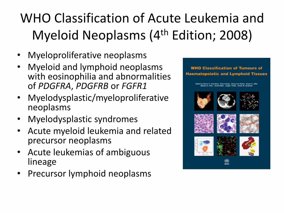
WHO Classification of Acute Leukemia and Myeloid Neoplasms (4th Edition; 2008)
• Myeloproliferative neoplasms • Myeloid and lymphoid neoplasms
with eosinophilia and abnormalities of PDGFRA, PDGFRB or FGFR1
• Myelodysplastic/myeloproliferative neoplasms
• Myelodysplastic syndromes • Acute myeloid leukemia and related
precursor neoplasms • Acute leukemias of ambiguous
lineage • Precursor lymphoid neoplasms

Myeloproliferative Neoplasms
• Chronic myelogenous leukemia, BCR-ABL1 positive
• Chronic neutrophilic leukemia
• Polycythemia vera
• Primary myelofibrosis
• Essential thrombocythemia
• Chronic eosinophilic leukemia
• Mastocytosis
• Myeloproliferative neoplasm, unclassifiable

Chronic Myeloid Leukemia, BCR-ABL1 positive
• Mostly unchanged • Definition of accelerated
phase – Will include development of
TKI/therapy resistance
• Definition of lymphoid blast crisis – Any lymphoblast population
in the blood should raise concern for blast crisis
– >5% (aberrant) lymphoblasts in the marrow should be considered blast crisis

Chronic Neutrophilic Leukemia
• Rare disorder of sustained neutrophilia without reactive cause or evidence of other MPN
• Mutations – CSF3R T6181
• Mutations disrupt the JAK-STAT pathway and are considered disease defining for CNL
– SETBP1, ASXL1 • Frequent, but not disease
specific
From Gotlib J. The Hematologist Sept 2013

Polycythemia Vera criteria (2008)
• Increased red cell production – Hemoglobin >18.5/16.5 g/dL in men/women – Hemoglobin >17/15 g/dL in men/women, with
sustained increase of 2 g/dL over baseline – Increased red cell mass (>25% above normal) – Hemoglobin or hematocrit >99th percentile
• JAK2 mutation
1. Bone marrow showing typical PV histology 2. Decreased serum EPO levels 3. Endogenous erythroid colony formation M
ino
r

Proposed revision to PV criteria
• Increased red cell production – Hemoglobin >16.5/16.0 g/dL in
men/women or hematocrit >49/48% in men/women
– Hemoglobin >17/15 g/dL in men/women, with sustained increase of 2 g/dL over baseline
– Increased red cell mass (>25% above normal)
– Hemoglobin or hematocrit >99%le
• Bone marrow showing typical PV histology
• JAK2 mutation 1. Decreased serum EPO levels 2. Endogenous erythroid colony
formation Min
or

Primary Myelofibrosis and Essential Thrombocythemia
• Addition of CALR mutations to diagnostic criteria for both and MPL for ET
• Lower fibrosis requirement for prefibrotic/early PMF – Provide more
information on the ddx of prePMF vs ET

Myeloproliferative Neoplasms
• Chronic myeloid leukemia, BCR-ABL1 positive
• Chronic neutrophilic leukemia
• Polycythemia vera
• Primary myelofibrosis
• Essential thrombocythemia
• Chronic eosinophilic leukemia
• Mastocytosis
• Myeloproliferative neoplasm, unclassifiable

Myelodysplastic Syndromes (2008)
• Refractory cytopenia with unilineage dysplasia – Refractory anemia – Refractory neutropenia – Refractory thrombocytopenia
• Refractory anemia with ring sideroblasts • Refractory cytopenia with multilineage
dysplasia • Refractory anemia with excess blasts
– RAEB-1 – RAEB-2
• Myelodysplastic syndrome with isolated del(5q)
• Myelodysplastic syndrome, unclassifiable • Childhood myelodysplastic syndrome
– Refractory cytopenia of childhood

Myelodysplastic Syndromes – Revised Terminology
• Refractory cytopenia with unilineage dysplasia – Refractory anemia – Refractory neutropenia – Refractory thrombocytopenia
• Refractory anemia with ring sideroblasts
• Refractory cytopenia with multilineage dysplasia
• Refractory anemia with excess blasts – RAEB-1 – RAEB-2
• Myelodysplastic syndrome with isolated del(5q)
• Myelodysplastic syndrome, unclassifiable
• Childhood myelodysplastic syndrome – Refractory cytopenia of childhood
• MDS with single lineage dysplasia
• MDS with ring sideroblasts with
unilineage dysplasia • MDS with ring sideroblasts with
multilineage dysplasia • MDS with multilineage dysplasia • MDS with excess blasts
– With excess blasts-1 – With excess blasts-2
• MDS with isolated del(5q)
• MDS, unclassifiable
• Childhood myelodysplastic syndrome – Refractory cytopenia of childhood

MDS with ring sideroblasts
• Frequent association with mutations of SF3B1 and a favorable prognosis with low risk of transformation to acute leukemia
• >15% ring sideroblasts (among erythroid precursors), or
• >5% in the presence of an SF3B1 mutation
• Blast cell increases exclude this diagnosis – If multilineage dysplasia without
a blast cell increase is present, case is classified as MDS with multilineage dysplasia with ring sideroblasts
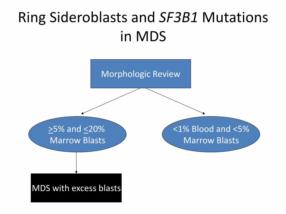
Morphologic Review
>5% and <20% Marrow Blasts
<1% Blood and <5% Marrow Blasts
Ring Sideroblasts and SF3B1 Mutations in MDS
MDS with excess blasts

Single lineage
<1% Blood and <5% Marrow Blasts
Ring Sideroblasts and SF3B1 Mutations in MDS
MDS with ring sideroblasts (and uni-
lineage dysplasia)
Dysplasia Multilineage
Iron Stain
>15% MDS with ring
sideroblasts and multilineage dysplasia
5-14% SF3B1
Yes
<5% Single lineage MDS with single lineage dysplasia
Multilineage MDS with multilineage
dysplasia
Dysplasia
No

MDS with Isolated del(5q) (5q-minus Syndrome)
• Currently restricted to del(5q) as the only abnormality
• Will now allow a second (non-high risk; i.e. -7) cytogenetic abnormality
• Cases with >2 abnormalities, multilineage dysplasia or increase blasts will not qualify for this category
• Recommend TP53 mutation assessment or p53 staining
Germing Leukemia 26:1286, 2012; Mallo Leukemia24:110, 2011; Jadersten JCO 29:1971; 2011

Erythroid/myeloid leukemia is now considered as MDS with excess blasts • Prior definition of
erythroleukemia (erythroid/myeloid type) in AML, NOS required >50% marrow erythroid precursors and >20% myeloblasts among non-erythroid cells
• These cases will now be classified as MDS based on the total blast cell count
Grossman et al. Leukemia 27:1940, 2013 Hasserjian et al. Blood 115:1985, 2010

Evaluation of Dysplastic Marrows with over 50% Erythroids
>50% Marrow Erythroids
Determine Absolute Blast Cell Count
<5%
Iron Stain Dysplasia
MDS with unilineage dysplasia
(+/- ring sideroblasts)
MDS with multilineage dysplasia
(+/- ring sideroblasts)
5-19%
MDS with excess blasts
>20% AML

WHO Classification of Acute Leukemia and Myeloid Neoplasms (4th Edition; 2008)
• Myeloproliferative neoplasms • Myeloid and lymphoid neoplasms
with eosinophilia and abnormalities of PDGFRA, PDGFRB or FGFR1
• Myelodysplastic/myeloproliferative neoplasms
• Myelodysplastic syndromes • Acute myeloid leukemia and related
precursor neoplasms • Acute leukemias of ambiguous
lineage • Precursor lymphoid neoplasms

Familial Myeloid Neoplasms
• Familial MDS/AML is likely more prevalent than realized • Familial MDS/AML associated with germline mutations
– CEBPA (AML) – SRP72 (AML) – DDX41 (MDS/AML)
• Familial hematologic malignancies associated with platelet disorders and gene mutations – RUNX1 (AML) – ANKRD26 (AML) – ETV6 (AL and solid tumors)
• Familial MDS/AML associated with other organ dysfunction – GATA2 (MDS/AML) – TERC/TERT – DNA repair gene syndromes – Tumor suppressor gene syndromes
West AH, Godley LA & Churpek JE. Ann NY Acad Sci 1310:111, 2014
Zhang MY, et al. Nature Genetics Epub Jan 12, 2015

WHO Classification of Precursor Myeloid and Lymphoid Neoplasms (4th Edition)
Acute myeloid leukemia (AML) and related precursor neoplasms
• AML with recurrent genetic abnormalities
– AML with t(8;21) (q22;q22) (RUNX1-RUNX1T1)
– AML with inv(16)(p13.1q22) or t(16,16) (p13.1;q22) (CBFB-MYH11)
– Acute promyelocytic leukemia with t(15;17)(q24.1;q21.1) (PML-RARA)
– AML with t(9;11)(p22;q23) (MLLT3-MLL)
– AML with t(6;9)(p23;q34) (DEK-NUP214)
– AML with inv(3)(q21q26.2) or t(3;3)(q21;q26.2) (RPN1-EVI1)
– AML (megakaryoblastic) with t(1;22)(p13;q13) (RBM15-MKL1)
– Provisional entity: AML with mutated NPM1
– Provisional entity: AML with mutated CEBPA
• AML with myelodysplasia-related changes
• Therapy-related myeloid neoplasms
• AML not otherwise specified
– AML minimally differentiated
– AML without maturation
– AML with maturation
– Acute myelomonocytic leukemia
– Acute monoblastic and monocytic leukemia
– Acute erythroid leukemia
– Acute megakaryocytic leukemia
– Acute basophilic leukemia
– Acute panmyelosis with myelofibrosis
• Myeloid sarcoma
• Myeloid proliferations related to Down syndrome
• Blastic plasmacytoid dendritic cell neoplasm
Acute leukemias of ambiguous lineage
• Acute undifferentiated leukemia
• Mixed phenotype acute leukemia with t(9;22)(q34;q11.2); BCR-ABL1
• Mixed phenotype acute leukemia with t(v;11q23); MLL rearranged
• Mixed phenotype acute leukemia, B/myeloid, NOS
• Mixed phenotype acute leukemia, T/myeloid, NOS
• Mixed phenotype acute leukemia, NOS, rare types
• Other ambiguous lineage leukemias
Precursor lymphoid neoplasms
• B-lymphoblastic leukemia/lymphoma, not otherwise specified
• B-lymphoblastic leukemia/lymphoma with recurrent genetic abnormalities
– B-lymphoblastic leukemia/lymphoma with t(v;11q23)(MLL)
– B-lymphoblastic leukemia/lymphoma with t(12;21)(p13;q22) (ETV6-RUNX1)
– B-lymphoblastic leukemia/lymphoma with t(5;14)(q31;q32) (IL3-IGH@)
– B-lymphoblastic leukemia/lymphoma with t(1;19)(q23;p13.3) (TCF3-PBX1)
– B-lymphoblastic leukemia/lymphoma with hyperdiploidy
– B-lymphoblastic leukemia/lymphoma with hypodiploidy
• T-lymphoblastic leukemia/lymphoma

• AML with recurrent genetic abnormalities
– AML with t(8;21) (q22;q22) (RUNX1-RUNX1T1)
– AML with inv(16)(p13.1q22) or t(16,16) (p13.1;q22) (CBFB-MYH11)
– Acute promyelocytic leukemia with t(15;17)(q24.1;q21.1) (PML-RARA)
– AML with t(9;11)(p22;q23) (MLLT3-MLL)
– AML with t(6;9)(p23;q34) (DEK-NUP214)
– AML with inv(3)(q21q26.2) or t(3;3)(q21;q26.2) (RPN1-EVI1)
– AML (megakaryoblastic) with t(1;22)(p13;q13) (RBM15-MKL1)
– Provisional entity: AML with mutated NPM1
– Provisional entity: AML with mutated CEBPA
• AML with myelodysplasia-related changes
• Therapy-related myeloid neoplasms
• AML, not otherwise specified
– AML minimally differentiated
– AML without maturation
– AML with maturation
– Acute myelomonocytic leukemia
– Acute monoblastic and monocytic leukemia
– Acute erythroid leukemia
– Acute megakaryocytic leukemia
– Acute basophilic leukemia
– Acute panmyelosis with myelofibrosis
• Myeloid sarcoma
• Myeloid proliferations related to Down syndrome
• Blastic plasmacytoid dendritic cell neoplasm
2008 WHO Classification of AML

Precursor Lymphoid Neoplasms (2008)
• B-lymphoblastic leukemia/lymphoma, not otherwise specified
• B-lymphoblastic leukemia/lymphoma with recurrent genetic abnormalities – B-lymphoblastic leukemia/lymphoma
with t(v;11q23)(MLL) – B-lymphoblastic leukemia/lymphoma
with t(12;21)(p13;q22) (ETV6-RUNX1) – B-lymphoblastic leukemia/lymphoma
with t(5;14)(q31;q32) (IL3-IGH@) – B-lymphoblastic leukemia/lymphoma
with t(1;19)(q23;p13.3) (TCF3-PBX1) – B-lymphoblastic leukemia/lymphoma
with hyperdiploidy – B-lymphoblastic leukemia/lymphoma
with hypodiploidy
• T-lymphoblastic leukemia/lymphoma

Since 2008
• Classification systems move slowly, but science does not
• Mutations
• Protein expression
• Methylation

Since 2008
• Classification systems move slowly, but science does not
• Next generation sequencing (NGS) has resulted in an explosion of new information

Advances in ALL
• IKZF1 deletions – at 7p12 encodes the zinc
finger transcription factor IKAROS
– Associated with gene expression signature similar to Ph+ ALL
– Very poor prognosis independent of age, WBC count and genetic subtype
• JAK mutations • CRLF2 translocations
Mullighan et al. Nature 453;110, 2008 Mullighan et al. N Engl J Med 360;470, 2009

Advances in ALL
• IKZF1 deletions • JAK mutations
– JAK1, JAK2 and JAK3 mutations found in 10.7% of Ph-negative B-ALL (80% JAK2)
– Mutations associated with deletion of IKZF1 and CDKN2A/B and a Ph+ ALL gene expression profile
– Very poor prognosis of IKZF1 deleted/JAK mutated cases
• CRLF2 translocations
Mullighan et al. PNAS 106;9414, 2009

Advances in ALL
• IKZF1 deletions
• JAK mutations
• CRLF2 translocations – Found in 7-14% of B-ALLs
and in 53% of Down-syndrome associated ALL
– Located at Xp22.3/Yp11.3 – 62% are translocations
with IGH – Associated with
• JAK1 and JAK2 mutations • IKZF1 deletions • Hispanic ethnicity • Very poor prognosis
Russell L J et al. Blood 114:2688, 2009 Mullighan et al. Nat Genet 41:1243, 2009 Harvey et al. Blood 115:5312, 2010

• BCR-ABL1-like B-ALL is a high risk ALL with a
gene expression profile similar to that of BCR-
ABL1+ ALL
• Accounts for 10% of pediatric and 25% of adult
ALL; poor clinical outcomes; may be amenable to
targeted therapy
• Need to establish clear diagnostic criteria
• CRLF2 translocations • Usually show increased expression of CRLF2
by flow cytometry analysis
• Some have activating mutations or
rearrangements of genes, such as ABL1,
ABL2, JAK2, PDGFRB, NTRK3, TYK2,
CSF1R, and/or EPOR • Diagnostic significance of deletions/mutations
of IKZF1, CDKN2A/B, JAK1 less clear
• The full spectrum of genetic changes is still
being investigated
BCR-ABL1-like B-ALL (B-ALL with Translocations Involving Tyrosine Kinases or Cytokine
Receptors)
van der Veer et al. Blood 122:2622, 2013 Izraeli. Curr Opin Hematol 21:289, 2014

• Intrachromosomal amplification of
chromosome 21 (iAMP21) accounts for
about 2% of B-ALL
• Generally in older children (median age 9
years) with lower WBC count
• Adverse outcomes when treated with
standard risk therapy; but improved when
treated as high risk ALL
• Presence of 5 or more copies of RUNX1 on
a single cell or 3 or more extra copies on a
single abnormal chromosome 21
• Reliably detected by FISH for RUNX1 and
confirmed by cytogenetics Image provided by Dana Bangs
ALL with iAMP21
Harrison et al. Leukemia 28:1015, 2014
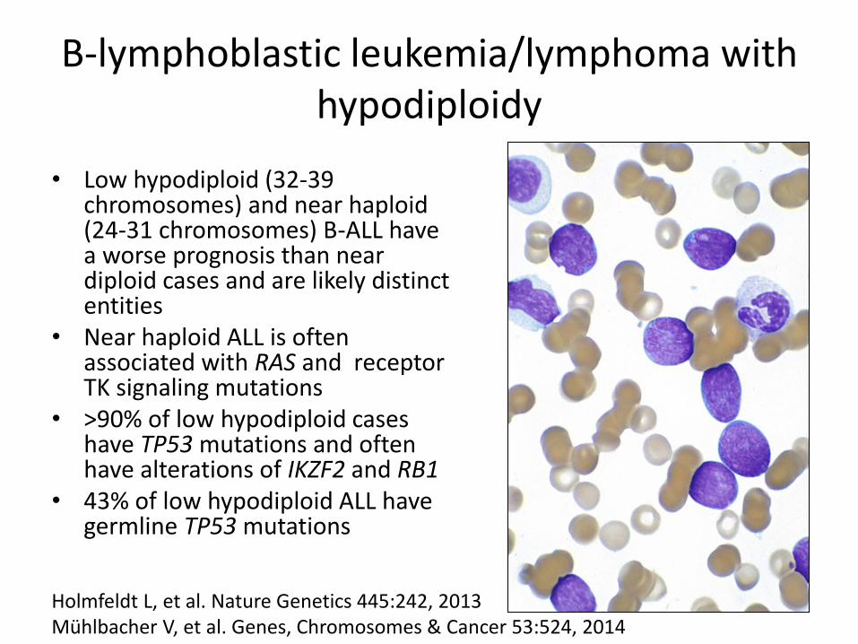
B-lymphoblastic leukemia/lymphoma with hypodiploidy
• Low hypodiploid (32-39
chromosomes) and near haploid (24-31 chromosomes) B-ALL have a worse prognosis than near diploid cases and are likely distinct entities
• Near haploid ALL is often associated with RAS and receptor TK signaling mutations
• >90% of low hypodiploid cases have TP53 mutations and often have alterations of IKZF2 and RB1
• 43% of low hypodiploid ALL have germline TP53 mutations
Holmfeldt L, et al. Nature Genetics 445:242, 2013 Mühlbacher V, et al. Genes, Chromosomes & Cancer 53:524, 2014

• Early T-Precursor (ETP) ALL comprises 10-15% of T-ALL • Defined immunophenotypically by expression of cCD3,
CD7, low CD5, but no CD1a, CD4 or CD8 • Expresses CD34 and myeloid-related antigens
(CD117, CD33, or CD13) but not MPO • Thought to arise from an early progenitor cell with
lineage plasticity that may be more closely related to human stem cells than to early T-cell precursors
• Molecular genetics • Increase in AML-associated mutations • Rare NOTCH pathway (T-ALL-associated) mutations
• Initially considered high risk due to higher rate of induction failure
• Recent COG study showed no outcome difference with current T-ALL therapy
Early T-Precursor Acute Lymphoblastic Leukemia (ETP-ALL)
• Coustan-Smith E, et al. Lancet Oncol 10:147, 2009
• Haydu JE and Ferrando AA. Curr Opin Hematol 20:369, 2013
• Wood B, et al. ASH Abstract #1, 2014

Proposed WHO Revisions for ALL
• B-ALL – BCR-ABL1-like B-ALL
– B-ALL with iAMP21
– Hypodiploid ALL will be subdivided • Near haploid
• Low hypodiploid
• Near diploid
• T-ALL – Early T-Precursor ALL

2001
2008
AML with Multilineage Dysplasia
AML, Not Otherwise Categorized
AML with inv(3) or t(3;3)
AML with t(6;9) AML with Myelodysplasia-
Related Changes AML, Not Otherwise
Specified
History and/or cytogenetics
AML with mutated NPM1 AML with mutated CEBPA
NPM1 and CEBPA
AML

Weinberg et al. Blood 113:1906, 2009

What about new translocations in AML?

AML with BCR-ABL1
• Difficult to distinguish from myeloid blast crisis of chronic myelogenous leukemia – Few basophils – Less splenomegaly
• Deletion of antigen receptors, particularly IGH, recently shown to be specific for de novo disease – Detection of t(9;22) in only blasts supports diagnosis
• Subset of cases have mutated NPM1 • Important to recognize due to presence of
targeted (TKI) therapy
Soupir CP, et al. Am J Clin Pathol 127:642, 2007 Konoplev S, et al. Leuk Lymphoma 54:138, 2013 Nacheva EP, et al. Br J Haematol 161:541, 2013

Mutations in AML
• Only four mentioned in 2008 WHO
– Provisional entities
• NPM1
• CEPBA
– Prognostic markers
• FLT3
• KIT
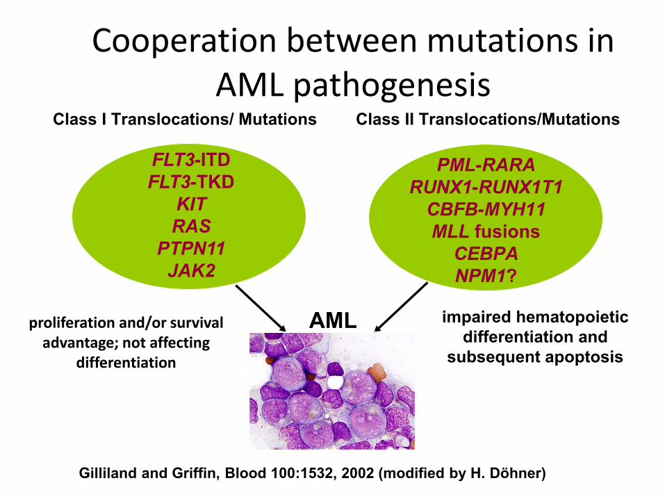
Class I Translocations/ Mutations Class II Translocations/Mutations
FLT3-ITD
FLT3-TKD
KIT
RAS
PTPN11
JAK2
PML-RARA
RUNX1-RUNX1T1
CBFB-MYH11
MLL fusions
CEBPA
NPM1?
AML impaired hematopoietic
differentiation and
subsequent apoptosis
Gilliland and Griffin, Blood 100:1532, 2002 (modified by H. Döhner)
proliferation and/or survival advantage; not affecting
differentiation
Cooperation between mutations in AML pathogenesis

AML with mutated CEBPA
• 7-20% of AMLs have mutations of CEBPA – More frequent with normal
or intermediate karyotype
• 12.5-47% are single/monoallelic
• Double mutant/biallelic cases (CEBPAdm) predict a favorable prognosis – Low frequency of other
mutations or other cytogenetic abnormalities
Green CL, et al. J Clin Oncol 28:2739, 2010

NPM1 and CEBPA Mutations in AML-MRC and Secondary AML
• Significance of multilineage dysplasia in the presence of NPM1 mutation, a normal karyotype and no history of MDS – MLD found in 74/318 (23%) de novo
NPM1 mutated AML – No prognostic significance for MLD
(Falini et al. Blood 115:3776, 2010) • NPM1 mutations in secondary AML
– Approximately 16% of AMLs arising from MDS, post therapy or following an MPN or CMML have mutations
– NPM1 mutation usually not present in original disease
– Such cases lack the favorable prognosis of de novo AML with mutated NPM1
• CEBPA mutations – MLD found in 28/108 (25.9%) CEPBA
mutated AML patients – No significant survival difference in
MLD+ and MLD- groups
Courville EL, et al. Modern Pathol 26:751, 2013 Döhner et al. Blood 106:3740, 2005 Gale RE et al. Blood 111;2776, 2008 Schnittger et al Leukemia 25:615, 2011 Bacher U et al. Blood 119:4719, 2012

Survival curves of patients up to 60 years with intermediate-risk cytogenetics AML depending on NPM1 status and presence of
multilineage dysplastic features (MLD)
Díaz-Beyá M et al. Blood 2010;116:6147-6148
©2010 by American Society of Hematology

AML with mutated NPM1 or CEPBA and an abnormal karyotype
• Abnormal karyotype identified in 14.7% of NPM1 and 26% of CEBPA mutated AML cases
• +8, +4, -Y, del(9q) and +21 most frequent with NPM1 mutation
• del(9q), del(11q), -Y, +10, +21 most frequent with biallelic CEBPA mutation
• del(9q) is currently considered an MDS-related cytogenetic abnormality, but it appears to be unusually common in NPM1 and CEBPA mutated cases
• In this setting, del(9q) does not appear to have prognostic significance
Haferlach C, et al. Blood 114:3024, 2009 Schlenk RF, et al. Blood 122:1576, 2013

Mutations in AML Gene Frequency in AML Reported prognosis NPM1 30-35% Favorable FLT3 ITD 25% Unfavorable DNMT3A 15-25% Unfavorable NRAS/KRAS 15-20% Neutral WT1 10-15% Neutral to unfavorable RUNX1 10-15% Unfavorable IDH2 R132 7-16% Variable IDH2 R140 and R172 8-15% Variable TET2 8-12% Unfavorable MLL 5-10% Unfavorable ASXL1 3-19% Unfavorable FLT3 TKD 7% Neutral CEBPA 6% Favorable PTPN11 3% Unknown PHF6 2-4% Unfavorable TP53 2-5% Unfavorable KIT 2-3% Unfavorable CBL 1-3% Unknown EZH2 1-3% Unknown JAK2 1% Unfavorable

AML with mutated RUNX1
• Gene located at 21q22 • Encodes the alpha subunit of the
core binding factor • Mutation in 4-16% of AML • More frequent in older male patients • Frequent prior history of MDS, or
prior exposure to radiation • Immature morphology and
phenotype common • Frequently associated ASXL1, KMT2A-
PTD, (FLT3-ITD), IDH1R132, and IDH2R140 and R172 mutations
• Rare CEBPA or NPM1 mutations • Poor response to therapy with
shortened survival Tang et al. Blood 114:5352, 2009
Mendler et al. J Clin Oncol 30:3109, 2012

Proposed WHO Revisions for AML
• AML, NOS – Mostly unchanged
– Move erythroid/myeloid type of acute erythroid leukemia to the MDS section
• New cytogenetic subgroups – Rare ones will be mentioned, but
not added to the classification
– AML with BCR-ABL1 • Antigen receptor deletion
• BCR-ABL1 absent in background cells
– Refine APL with PML-RARA fusion

Proposed WHO Revisions for AML
• New and revised mutation subgroups – AML with mutated
RUNX1 • Category will only
include de novo cases
• Cases arising from MDS will still be called AML-MRC
• Cases with prior therapy will still be therapy-related AML

Proposed WHO Revisions for AML
• New and revised mutation subgroups – AML with RUNX1 mutation
– AML with CEBPA mutation will have to be heterozygous/double mutation
– NPM1 and CEBPAdm mutations will trump multilineage dysplasia in de novo disease without MDS-related cytogenetic abnormalities other than del(9q)

Proposed WHO Revisions for AML
• Revise criteria for AML with myelodysplasia-related changes – Remove de novo cases
with no MDS-related cytogenetic abnormalities if NPM1 or CEBPAdm mutated
– Revise MDS-related cytogenetic abnormalities • Allow del(9q) only in the
absence of NPM1 and CEBPA mutation

MDS-related cytogenetic abnormalities
• Complex karyotype*
• Unbalanced abnormalities – -7/del(7q)
– -5/del(5q)/t(5q)
– i(17q)/t(17p)
– -13/del(13q)
– del(11q)
– del(12p)/t(12p)
– del(9q)**
– idic(X)(q13)
• Balanced abnormalities – t(11;16)(q23.3;p13.3)
– t(3;21)(q26.2;q22.1)
– t(1;3)(p36.3;q21.1.2)
– t(2;11)(p21;q23.3)
– t(5;12)(q32;p13.2)
– t(5;7)(q32;q11.2)
– t(5;17)(q32;p13.2)
– t(5;10)(q32;q21)
– t(3;5)(q25.3;q35.1)
*>3 abnormalities ** mutation of NPM1 or CEBPA trumps this abnormality

Proposed WHO Revisions for AML
• Revise criteria for AML with myelodysplasia-related changes – Remove de novo cases
with no MDS-related cytogenetic abnormalities if NPM1 or CEBPAdm mutated
– Revise MDS-related cytogenetic abnormalities
• Add section on familial myeloid neoplasms

Algorithmic Approach
Morphologic Review
>20% Blood or
Marrow Blasts
<20% Blood or
Marrow Blasts

Algorithmic Approach
<20% Blood or
Marrow Blasts
Cytogenetics
AML with recurrent
genetic abnormality ALL with t(5;14) Not acute leukemia
t(8;21), inv(16),
t(16;16) or PML-RARA t(5;14)
Normal or other
abnormalities
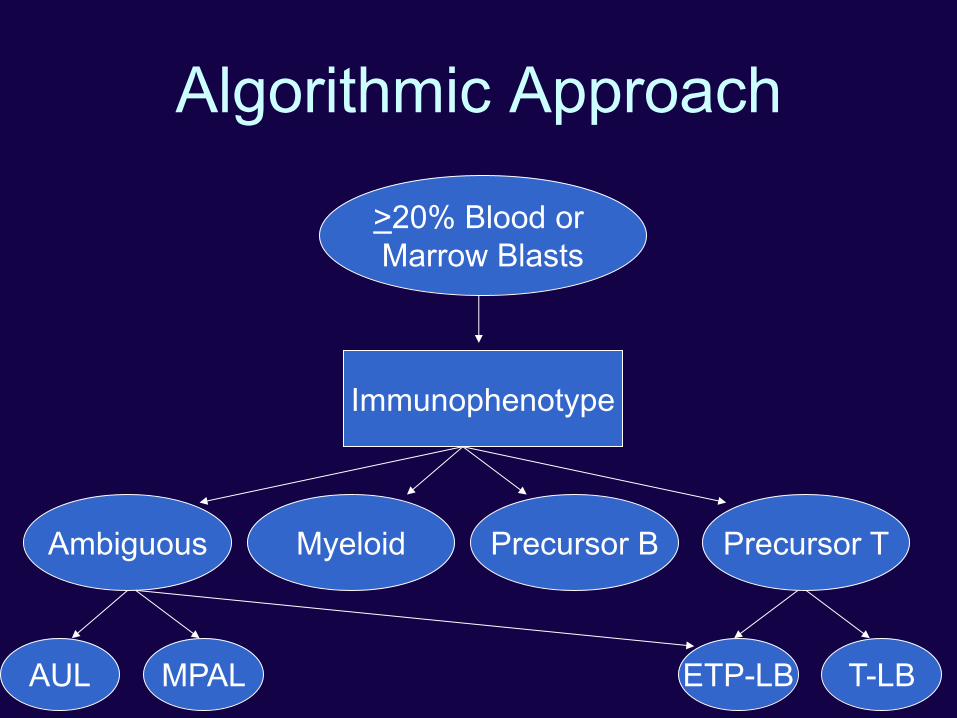
Algorithmic Approach
>20% Blood or
Marrow Blasts
Immunophenotype
Ambiguous Myeloid Precursor B Precursor T
T-LB ETP-LB AUL MPAL
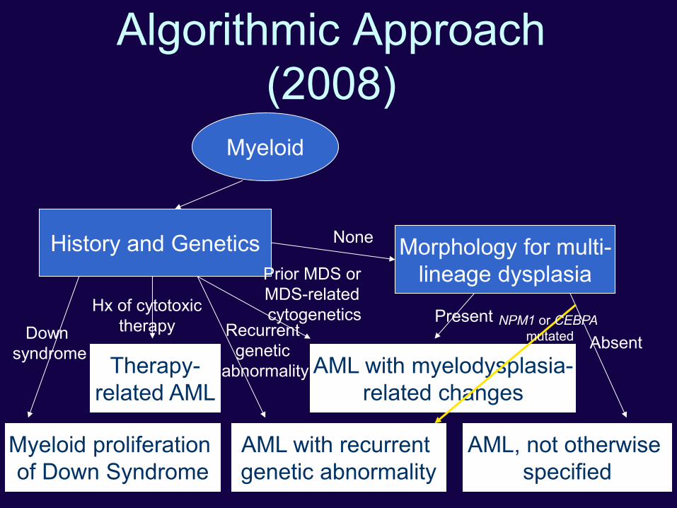
Algorithmic Approach
(2008)
Myeloid
History and Genetics
Therapy-
related AML
Myeloid proliferation
of Down Syndrome
AML with recurrent
genetic abnormality
Morphology for multi-
lineage dysplasia
AML with myelodysplasia-
related changes
AML, not otherwise
specified
Down
syndrome
Hx of cytotoxic
therapy Recurrent
genetic
abnormality
Prior MDS or
MDS-related
cytogenetics Present
Absent
None
NPM1 or CEBPA
mutated

Algorithmic Approach
(Proposed)
Myeloid
History and Genetics
Therapy-
related AML
Myeloid proliferation
of Down Syndrome
AML with recurrent
genetic abnormality
Morphology for multi-
lineage dysplasia
AML with myelodysplasia-
related changes
AML, not otherwise
specified
Present
Absent
-
NPM1, CEBPAdm
mutated
Mutation
Studies
RUNX1
mutated

AML Mutation Studies (FLT3, NPM1, CEPBA, KIT, RUNX1, DNMT3A, TET2, IDH1/2, ASXL1, WT1 ….)
Mutated NPM1
or CEBPAdm
History of Prior
Therapy
Therapy-
related AML
History of MDS
or MDS/MPN AML with MDS-
related changes MDS-related
CG abnormality
other than del(9q) AML with
mutated NPM1
or AML with
biallelic
CEPBA mutation
None Other recurring
CG abnormality
AML with re-
current genetic
abnormality

AML Mutation Studies (FLT3, NPM1, CEPBA, KIT, RUNX1, DNMT3A, TET2, IDH1/2, ASXL1, WT1 ….)
Mutated RUNX1
History of Prior
Therapy
Therapy-
related AML
History of MDS
or MDS/MPN AML with MDS-
related changes MDS-related
CG abnormality
AML with
mutated RUNX1
None Other recurring
CG abnormality
AML with re-
current genetic
abnormality
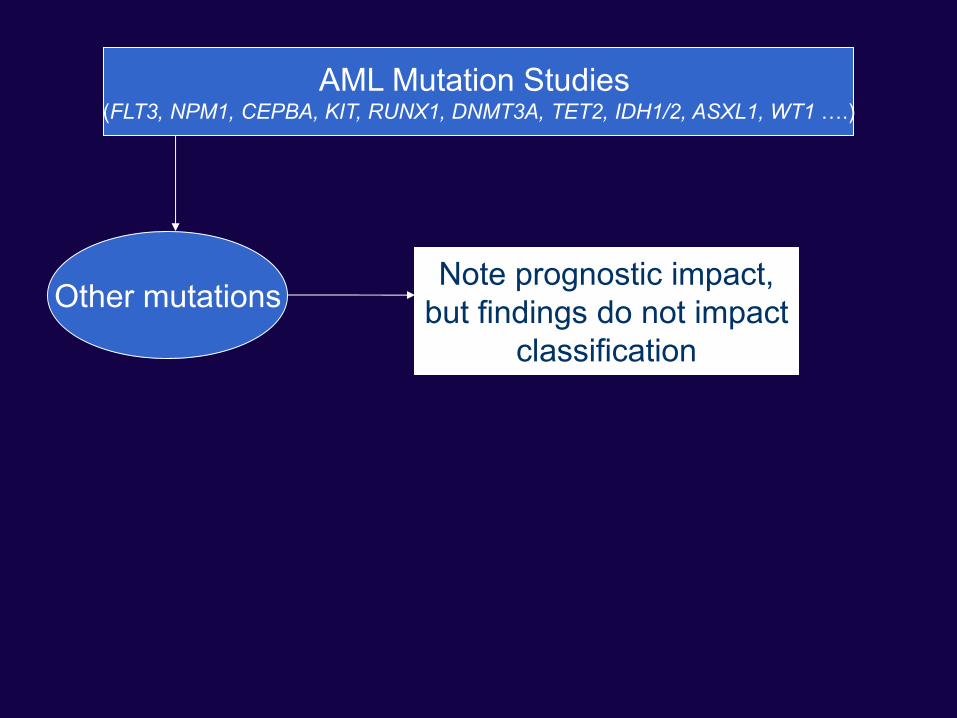
AML Mutation Studies (FLT3, NPM1, CEPBA, KIT, RUNX1, DNMT3A, TET2, IDH1/2, ASXL1, WT1 ….)
Other mutations Note prognostic impact,
but findings do not impact
classification
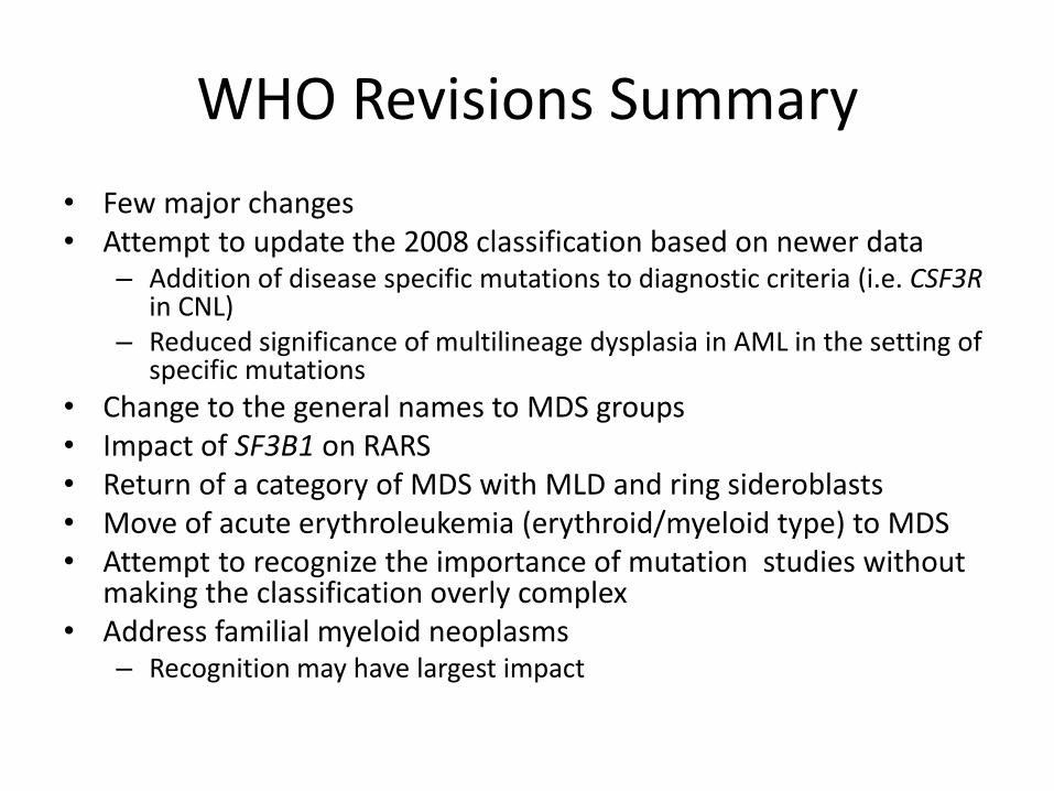
WHO Revisions Summary
• Few major changes • Attempt to update the 2008 classification based on newer data
– Addition of disease specific mutations to diagnostic criteria (i.e. CSF3R in CNL)
– Reduced significance of multilineage dysplasia in AML in the setting of specific mutations
• Change to the general names to MDS groups • Impact of SF3B1 on RARS • Return of a category of MDS with MLD and ring sideroblasts • Move of acute erythroleukemia (erythroid/myeloid type) to MDS • Attempt to recognize the importance of mutation studies without
making the classification overly complex • Address familial myeloid neoplasms
– Recognition may have largest impact

Acknowledgements
• Jim Vardiman • Jürgen Thiele • Attilio Orazi • Rob Hasserjian • Kathy Foucar • LoAnn Peterson • Dick Brunning • Michelle LeBeau • Mike Borowitz • Myeloid CAC
– Clara Bloomfield – Mario Cazzola