Propagation and restoration of mussel species of concern.
75
1 Endangered Species Grant Interim Report Grant No. E-1-42 Propagation and restoration of mussel species of concern. Grant Period: 9/1/03 – 8/31/04 Propagation and restoration of mussel species of concern. Prepared by: Chris Barnhart, Ph.D. Department of Biology Southwest Missouri State University 901 S. National Avenue, Springfield, MO 65804 [email protected] Date Prepared: November 14, 2004 Project Leader: _____________________________ Stephen E. McMurray Missouri Department of Conservation
Transcript of Propagation and restoration of mussel species of concern.

1
Endangered Species Grant Interim Report
Grant No. E-1-42 Propagation and restoration of mussel species of concern.
Grant Period: 9/1/03 – 8/31/04
Propagation and restoration of mussel species of concern.
Prepared by: Chris Barnhart, Ph.D.
Department of Biology Southwest Missouri State University 901 S. National Avenue, Springfield, MO 65804 [email protected]
Date Prepared: November 14, 2004 Project Leader: _____________________________ Stephen E. McMurray Missouri Department of Conservation
2
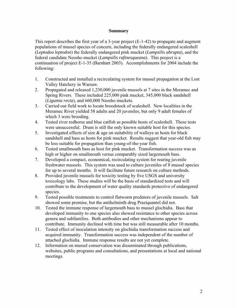
Summary
This report describes the first year of a 3-year project (E-1-42) to propagate and augment populations of mussel species of concern, including the federally endangered scaleshell (Leptodea leptodon) the federally endangered pink mucket (Lampsilis abrupta), and the federal candidate Neosho mucket (Lampsilis rafinesqueana). This project is a continuation of project E-1-35 (Barnhart 2003). Accomplishments for 2004 include the following: 1. Constructed and installed a recirculating system for mussel propagation at the Lost
Valley Hatchery in Warsaw. 2. Propagated and released 1,230,000 juvenile mussels at 7 sites in the Meramec and
Spring Rivers. These included 225,000 pink mucket, 345,000 black sandshell (Ligumia recta), and 660,000 Neosho muckets.
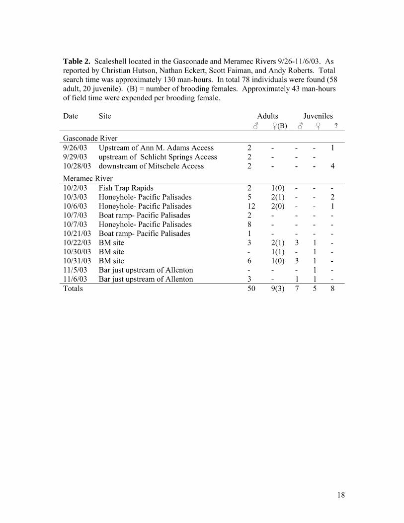
3. Carried out field work to locate broodstock of scaleshell. New localities in the Meramec River yielded 58 adults and 20 juveniles, but only 9 adult females of which 3 were brooding.
4. Tested river redhorse and blue catfish as possible hosts of scaleshell. These tests were unsuccessful. Drum is still the only known suitable host for this species.
5. Investigated effects of size & age on suitability of walleye as hosts for black sandshell and bass as hosts for pink mucket. Results suggest that year-old fish may be less suitable for propagation than young-of-the-year fish.
6. Tested smallmouth bass as host for pink mucket. Transformation success was as high or higher on smallmouth versus comparably sized largemouth bass.
7. Developed a compact, economical, recirculating system for rearing juvenile freshwater mussels. This system was used to culture juveniles of 8 mussel species for up to several months. It will facilitate future research on culture methods.
8. Provided juvenile mussels for toxicity testing by five USGS and university toxicology labs. These studies will be the basis of standardized tests and will contribute to the development of water quality standards protective of endangered species.
9. Tested possible treatments to control flatworm predators of juvenile mussels. Salt showed some promise, but the antihelminth drug Praziquantel did not.
10. Tested the immune response of largemouth bass to mussel glochidia. Bass that developed immunity to one species also showed resistance to other species across genera and subfamilies. Both antibodies and other mechanisms appear to contribute. Immunity declined with time but was still measurable after 10 months.
11. Tested effect of inoculation intensity on glochidia transformation success and acquired immunity. Transformation success was independent of the number of attached glochidia. Immune response results are not yet complete.
12. Information on mussel conservation was disseminated through publications, websites, public programs and consultations, and presentations at local and national meetings.

3
TABLE OF CONTENTS
SUMMARY.............................................................................................................................2 CONTENTS .............................................................................................................................3 LIST OF TABLES AND FIGURES...............................................................................................4 ACKNOWLEDGEMENTS..........................................................................................................5 PROJECT OBJECTIVES.............................................................................................................5 ACCOMPLISHMENTS IN 2004 .................................................................................................5
Installation of RPS at Lost Valley .................................................................................5 Propagation and Release ................................................................................................6
Scaleshell fieldwork and broodstock .......................................................................7 Scaleshell propagation and host tests......................................................................7 Pink mucket propagation .........................................................................................8 Comparison of older and younger bass as hosts for pink mucket...........................8 Smallmouth bass as hosts for pink mucket...............................................................9 Black sandshell propagation....................................................................................9 Comparison of older and younger walleye as hosts for black sandshell.................9 Neosho mucket propagation ....................................................................................9 Fatmucket propagation ..........................................................................................10
A compact recirculating system for rearing juveniles .................................................10 Juvenile mussels for toxicity tests ...............................................................................11 Treatments for controlling flatworms ..........................................................................11 Acquired resistance and cross-resistance of largemouth bass to glochidia .................12 Effects of inoculation intensity on transformation and immunity ...............................13 Duration of acquired immunity to glochidia................................................................13
DISSEMINATION OF RESULTS...............................................................................................14 LITERATURE CITED.............................................................................................................16 APPENDIX 1: A COMPACT RECIRCULATING SYSTEM FOR REARING JUVENILE FRESHWATER MUSSELS ......38 APPENDIX 2: CROSS-RESISTANCE OF LARGEMOUTH BASS TO GLOCHIDIA OF UNIONID MUSSELS ..............55
4
LIST OF TABLES AND FIGURES Tables
1. Releases of mussels propagated in 2004 ........................................................................17 2. Scaleshell located in the Gasconade and Meramec Rivers. ...........................................18 3. Brood condition of Meramec pink muckets propagated 6/24 and 6/25/04 ....................19 4. Size and fecundity of Meramec pink muckets ...............................................................19 5. Propagation of Meramec pink mucket on 6/24/04 .........................................................20 6. Propagation of Meramec pink muckets on 6/25/04 .......................................................21 7. Brood condition of Meramec black sandshell propagated 6/18/04. .............................22 8. Size and fecundity of Meramec black sandshell propagated 6/18/04 ............................22 9. Propagation of Meramec black sandshell on 1-year old walleye 6/18/04......................23 10. Brood condition of Spring River Neosho muckets propagated 7/14/04 .......................24 11. Size and fecundity of Spring River Neosho muckets propagated
7/14 and 8/2/04. ............................................................................................................24 12. Propagation of Neosho muckets on largemouth bass 7/14/04 .......................................25 13. Propagation of Neosho muckets on largemouth bass 8/2/04. .......................................26 14. Propagation of fatmucket on largemouth bass 7/14/04..................................................27 15. Effect of infection intensity on transformation success of
Lampsilis reeveiana glochidia on largemouth bass. ......................................................27 Figures
1. MDC personnel transporting RPS tanks from Springfield to Warsaw .........................28 2. Cod-ends of recovery filters for RPS. ............................................................................28 3. Lost Valley RPS nearing completion. ............................................................................29 4. Completed RPS in use for propagation of black sandshell on walleye..........................29 5. Hyphae of water mold that infested female scaleshell. ..................................................30 6. Recovery of 6/24/04 pink muckets in RPS. ...................................................................31 7. Recovery of 6/25/04 pink muckets in RPS ....................................................................31 8. Recovery of 6/24/04 pink muckets from smallmouth bass
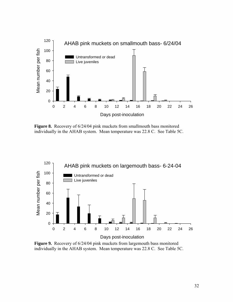
monitored individually in the AHAB system.................................................................32 9. Recovery of 6/24/04 pink muckets from largemouth bass
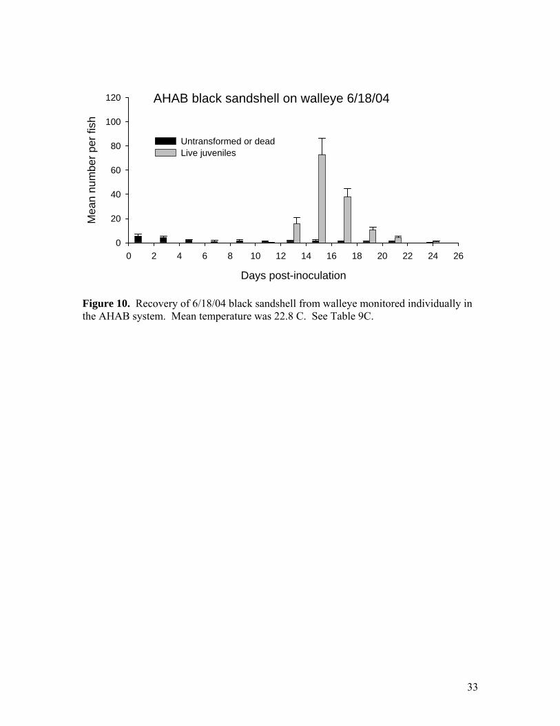
monitored individually in the AHAB system.................................................................32 10. Recovery of 6/18/04 black sandshell from walleye monitored
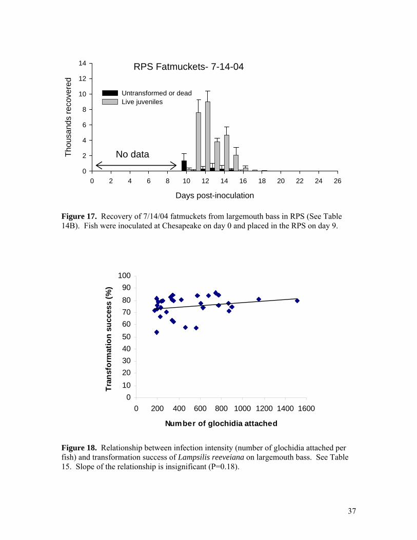
individually in the AHAB system. .................................................................................33 11. Time course of Neosho mucket attachment to bass during
inoculation on 7/14/04....................................................................................................34 12. Time course of Neosho mucket attachment to bass during
inoculation on 8/2/04......................................................................................................34 13. Recovery of 7/14/04 Neosho muckets from largemouth bass in the RPS......................35 14. Recovery of 8/2/04 Neosho muckets from largemouth bass in RPS. ............................35 15. Recovery of 7/14/04 Neosho muckets from largemouth bass monitored
individually in the AHAB system. .................................................................................36 16. Recovery of 8/2/04 Neosho muckets from largemouth bass monitored
individually in the AHAB system. ................................................................................36 17. Recovery of 7/14/04 fatmuckets from largemouth bass in RPS ....................................37 18. Relationship between infection intensity and transformation success
of Lampsilis reeveiana on largemouth bass. .................................................................37
5
ACKNOWLEDGEMENTS I would like to thank SMSU students Bob Brown, Zach Beussink, Ben Dodd, Todd Fobian and Brianna Kaiser, and staff members Christian Hutson, Nathan Eckert and Bob Holmes for their hard work and creativity, which made this work possible. Scott Faiman and Steve McMurray (MDC) and Andy Roberts (USFWS) played essential roles in planning and carrying out this project and also spent many hours in the field locating rare mussels. Andy Cornforth, Dennis Whelan, and John Clayton (MDC Chesapeake Hatchery) provided fish, raceway space for inoculation, fish transport, and expert advice concerning fish husbandry. Ken Neubrand, Richard Cook, Lesly Conaway and J.R. Booth (MDC Lost Valley Hatchery) carried out mussel propagation, developed methods and provided facilities, fish and logistic support. Dr. Conrad Kleinholtz (Langston University) provided freshwater drum for propagation of scaleshell. Bill Anderson (MDC) assisted with collection of river redhorse for scaleshell host tests. Dave Hendrix and his colleagues at Neosho Hatchery transported drum for scaleshell propagation. Ed Miller (KDWP) and Brian Obermeyer (TNC) made observations on propagated Neosho muckets in Kansas and provided information and advice regarding collection and release sites. I am grateful to Charlie Scott (USFWS) and Tommie Crawford (MDC) for supporting this work. This project is a cooperative effort supported by the Missouri Department of Conservation (MDC), the U.S. Fish and Wildlife Service (USFWS) and Southwest Missouri State University (SMSU).
PROJECT OBJECTIVES This report describes the first year of a 3-year project (E-1-42). The overall objectives of the project are 1) to augment populations of three target mussel species of conservation concern (Neosho mucket, pink mucket, and scaleshell) through propagation and release of wild-caught glochidia larvae, 2) to test factors affecting the suitability of host fish for the larval stages of native mussels, including the significance of acquired immunity, and 3) to investigate the susceptibility of juvenile mussels and mussel reproduction to low dissolved oxygen.
ACCOMPLISHMENTS IN 2004 Installation of RPS at Lost Valley
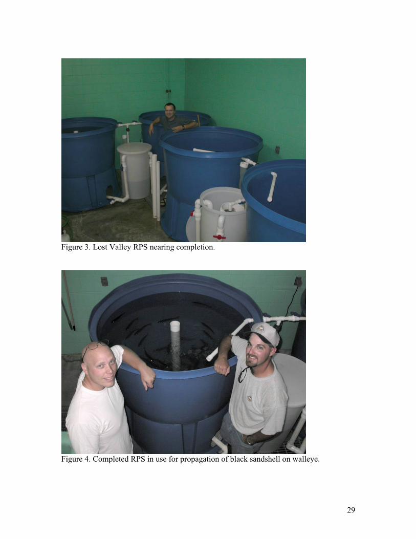
The recirculating propagation system (RPS) is an aquaculture system for recovering juvenile mussels from large numbers of host fish. The prototype RPS was designed and constructed at SMSU last year (Barnhart 2003). In the spring of 2004 we installed a similar system at the MDC Lost Valley Hatchery in Warsaw (Figures 1-4). The installation at Lost Valley consists of 4 conical-bottom 250-gallon tanks with sumps for mechanical and biological filtration, and recovery filters to recover juveniles from each tank. Host fish are inoculated with glochidia in raceways at the hatchery and later moved into the RPS during the 1-2 week period of excystment of the juvenile mussels. The RPS
6
systems were used at both facilities this year and allowed us to expand the production of juvenile mussels of several species. Propagation and release In 2004 we released a total of 1,230,000 propagated juveniles of 3 species at 7 sites in the Meramec and Spring Rivers (Table 1). Pink mucket glochidia were obtained from 6 Meramec River females and were placed on bass at Lost Valley Hatchery and at Chesapeake Hatchery. The fish were later transported to SMSU for recovery of juveniles. Black sandshell glochidia were obtained from 4 Meramec River females and were placed on walleye at Lost Valley Hatchery. Recovery of juveniles took place in the new RPS at Lost Valley. Releases of pink mucket and black sandshell were made at 4 sites in the Meramec River by MDC and SMSU personnel on 7/16/04. Neosho mucket glochidia from 6 Spring River females were placed on largemouth bass at Chesapeake Hatchery. The fish were later transported to SMSU for recovery of juveniles. Releases were carried out by SMSU personnel at 4 sites in the Spring River on 8/3/04 and 8/20/04. No scaleshell were released this year. Snuffbox mussel propagation at Lost Valley is not presented in this report. We continued to refine methods for propagation (Barnhart 2003). We generally use 300-500 3-4 inch host fish (largemouth bass, walleye, or drum) as hosts for glochidia from each female mussel and may inoculate up to 2,000 fish at a time. The fish are concentrated in a measured segment of a hatchery raceway with blocking screens. The length and depth of the segment are adjusted to a volume of approximately 200 ml per fish. The flow in the raceway is interrupted and frames covered with plastic sheet are inserted to isolate the raceway segment. Glochidia are added at (ideally) 3,000-5,000 per liter. The water is aerated vigorously at four points to keep the glochidia in suspension. During exposure the unattached glochidia tend to close and little further attachment occurs after 15-20 minutes. Attachment of 40-50% of the glochidia after 15 minutes is typical for Lampsilis. Smaller bath volume improves attachment success, probably because the fish ventilate a larger proportion of the bath in a short time. After inoculation the fish are usually kept in the hatchery raceway until a day or two before juvenile drop-off is expected. Feed is interrupted after 5-7 days to allow the fish to purge before drop-off begins. Depending on temperature, drop-off of juveniles begins at about 10 days post-inoculation and continues for 8-12 days. The fish are moved into the RPS to collect the juveniles. We usually also monitor a subset of fish individually in the AHAB system at SMSU, which allows quantification of transformation success and timing (Barnhart 2002). These fish are moved to the AHAB immediately after inoculation so that sloughed glochidia are also counted. Adequate data gathering and analysis is essential if we are to get maximum benefit from propagation efforts. The propagation of rare mussels is an excellent opportunity to gather life cycle data. Detailed records of results such as fecundity, brood condition, timing of transformation, transformation success on different host species and age classes, and

7
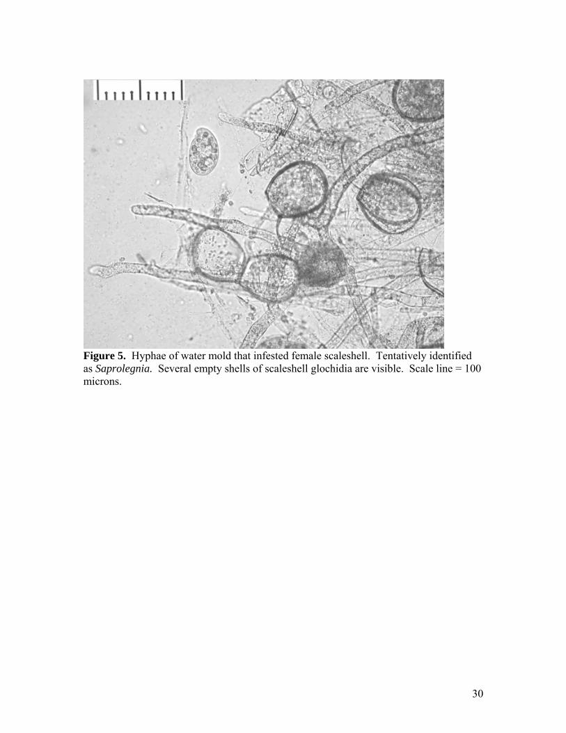
other data are needed to improve methods and ensure efficiency. Of course, gathering these data is time consuming and must be balanced against other demands. We hope to streamline the process and have made considerable progress toward that goal. In 2004 we refined data spreadsheets for brood harvest and condition, inoculation, RPS recovery, and AHAB recovery. A protocol will be written this winter so that Lost Valley and SMSU can follow similar procedures in the spring. Scaleshell fieldwork and broodstock Scaleshell recovery continues to be a very difficult challenge. An intensive effort was made in fall 2003 to locate brooding females for propagation. Table 2 summarizes results by locality and date (as reported by Nathan Eckert and Christian Hutson). In total 58 adults and 20 juveniles were located in 130 man-hours of search time. Of the 58 adult individuals, only 9 were female (15.5%), and only 3 of the 9 adult females were brooding. The highly skewed sex ratio in this species has been documented previously (Barnhart 2001). Approximately 43 man-hours of field time were expended per brooding female found. From 10/2/03 to 11/6/03, 7 adult males, 4 adult females, and 9 juvenile scaleshell were moved from sites near Pacific Palisades to the Opechee Beach site. These mussels are intended to serve as brood stock that can be accessed for future propagation efforts. The mussels were placed in a “mussel corral” to restrict their movement and facilitate future recovery for propagation. The corral is a square open-top tray 90 x 90 x 18 cm (35 x 35 x 7 inches) , made of 1.3 cm (1/2 inch) expanded 1/16” stainless steel (expanded steel is a mesh with diamond-shaped openings). The corral was buried in the substrate to a depth of 15 cm, leaving the sides 3 cm high above the substrate as a “fence”. Scaleshell propagation and host tests Two of the three brooding females found in fall 2003 were collected, while the third was caged at the Opechee Beach site (see above). One brooding female was brought to Chesapeake Hatchery on 10/24/03 to overwinter. This female was placed in a raceway in a tray of substrate from the Meramec which was 10 cm deep (4 inches). The tray was equipped with an undergravel filter and airlift to maintain water flow through the substrate. The raceway was supplied with flowing pond water at ambient temperature. Condition of the mussel was checked at 1-2 week intervals. Unfortunately, when the mussel was examined on January 23, 2004, the mantle and siphons were found to be infested with a water mold, possibly Saprolegnia (Figure 5) and the mussel was moribund. We removed approximately 2,950,000 total glochidia and estimated from salt test that only 15% of these were viable. The female mussel was preserved in ethanol, and is presently stored at SMSU. The second brooding female was brought to SMSU on 10/4/03 to over-winter. The mussel was held in an incubator in reconstituted moderately hard freshwater. Temperature was adjusted downward over the next month to 7 C. Water was changed at 2-week intervals. This mussel survived the winter in good condition and was eventually
8
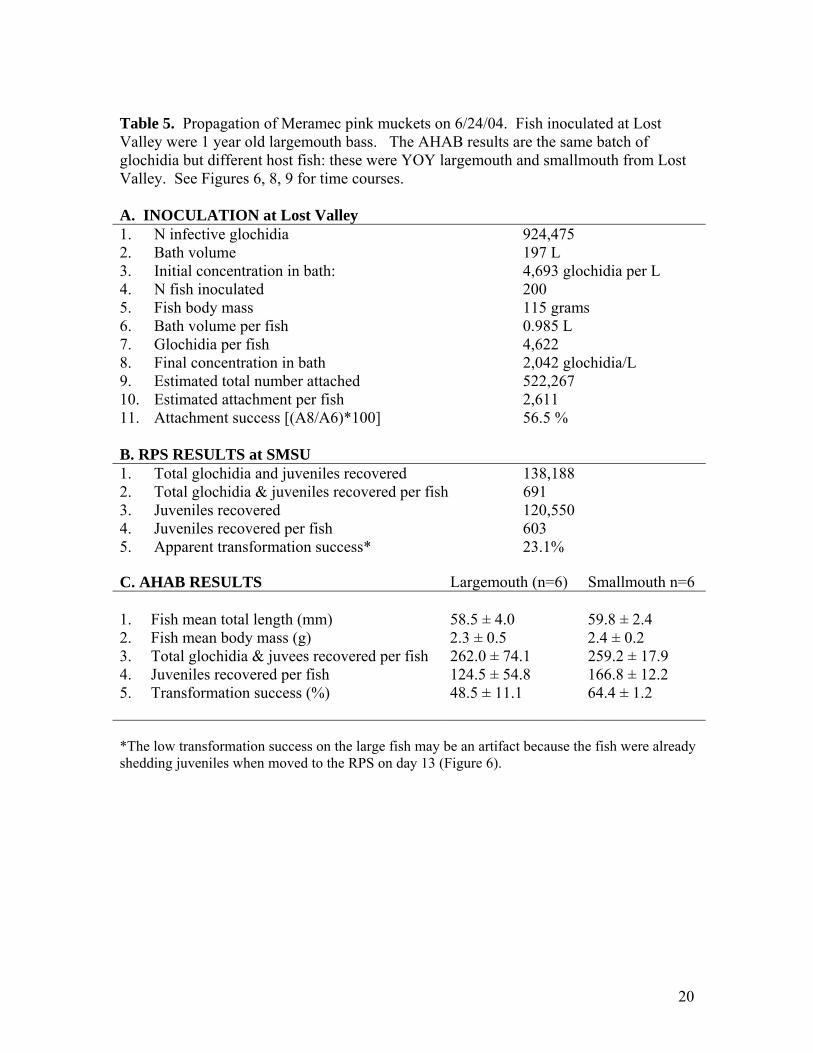
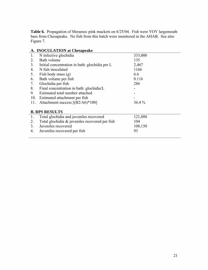
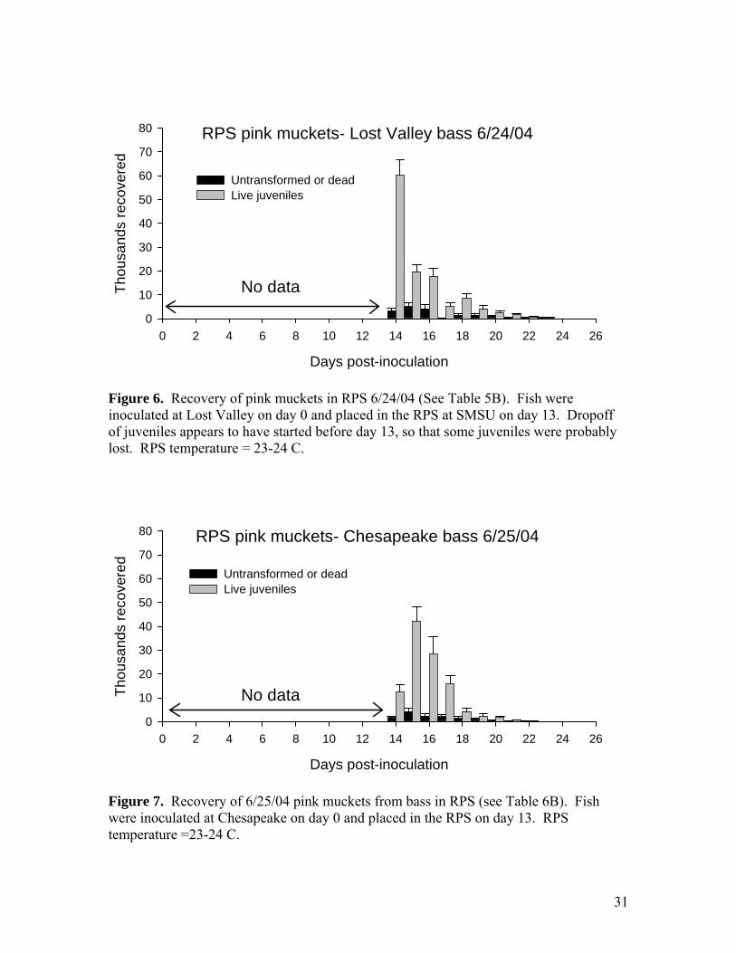
returned to the Meramec River at the Opechee Beach site on June 27, 2004. A portion of the glochidia were used to inoculate drum at Chesapeake Hatchery on June 1, 2004. Unfortunately, only 200 fish were available because of unexpected winter losses in the culture ponds at Langston University, and these fish proved to be unusually poor hosts. Transformation success was less than 35%. Approximately 800 juveniles were recovered from the RPS and were kept for grow-out experiments at SMSU (see below). A portion of the glochidia from the SMSU female was sent to Dr. Greg Cope at North Carolina State University for toxicity testing experiments. The rest of the glochidia were used at SMSU for host tests of river redhorse and blue catfish. Both hosts failed to produce any juveniles. River redhorse were adult fish collected by electroshock from the lower James River. Six redhorse were inoculated with scaleshell glochidia on June 8. One redhorse died on June 10 and was examined for encysted glochidia. None were found. The other redhorse later succumbed to Ich infestation and no glochidia cysts or juveniles were recovered. Blue catfish were hatchery juveniles obtained from a state hatchery in Tennessee. A group of 12 fish was inoculated on June 8. Several of the blue cats were sacrificed on June 10 and no encysted glochidia were found. Pink mucket propagation From 5/11/04 to 6/9/04 eight female pink muckets were brought to SMSU to obtain glochidia for propagation (Tables 3-4). Four specimens were from the Pacific Palisades area of the Meramec River, 2 from the “Show” site, and 2 from Opechee Beach. These sites are all within a reach of 3 miles. Of these eight females 6 were brooding and were marked 04-1 (=PP7), 04-2 (=BM1), 04-5, 04-6, 04-7(=PP5), and 04-8. The 2 non-brooding mussels were marked 04-3 and 04-4. These 8 mussels were returned to the Opechee Beach site on 6/27/04. Glochidia from 4 of the pink muckets (04-1, 04-2, 04-5, 04-6) were placed on hosts on 6/24/04 at Lost Valley Hatchery (Table 5). Glochidia from two of the females (04-7 and 04-8) were placed on hosts on 6/25/04 at Chesapeake Hatchery (Table 6). Both batches of fish were later moved to SMSU for recovery of juveniles. On July 15, a total of 225,000 juveniles derived from the 6 females were released at 4 sites in the Meramec (Table 1). A subset of approximately 4,500 juveniles was held at SMSU for grow-out experiments and for use in toxicity testing by USGS. Comparison of older and younger bass as hosts for pink mucket The bass inoculated with pink muckets on 6/24/04 at Lost Valley were about 1 year old, and it is interesting to compare the propagation results on older and younger fish (Table 5). The large bass had mean mass of 115 g and attachment of glochidia was 2,611 per fish or 22.7 per gram. A group of YOY (young of the year) largemouth with mean 2.3 gram mass carried 262 glochidia per fish, or 114 per gram. Thus, it appears that the small fish carried about 5 times more glochidia per gram body mass. Transformation success also appeared to be higher on the smaller fish. The yield from the large bass in the RPS was only 603 juveniles per fish. The apparent transformation success was only 23%, less
9
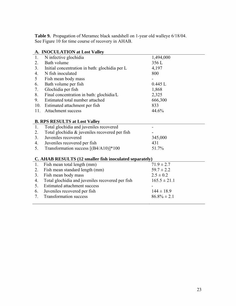
than half that observed on the smaller largemouth bass. The low yield may be at least partly artifact. It appears that a proportion of the juveniles were lost because the fish were already shedding juveniles when moved to the RPS on day 13 (Figure 6). However, it is also possible that transformation success was lower on the larger fish, as it appeared to be for black sandshell on older walleye (see below). Smallmouth bass as hosts for pink mucket Two groups of YOY largemouth bass and smallmouth bass from Lost Valley were inoculated with pink mucket on 6/24/04 and monitored in the AHAB. We have used largemouth and walleye for propagating pink mucket but have not previously compared transformation on smallmouth. MDC stocking of smallmouth in SE Missouri rivers raises the possibility of placing pink mucket glochidia on these fish before release. The AHAB results show that smallmouth is a suitable host for pink mucket (Table 5). Transformation success of these glochidia was 64% and was higher than that on comparable size largemouth (48%). The difference was nearly significant (p=0.07 by T-test). Black sandshell propagation Black sandshell were placed on 1-year-old (approximately 50 grams body mass) walleye at Lost Valley on 6/18/04 (Tables 7-9). This was the first time that 1-year-old walleye were used as hosts. Attachment success appears to have been good at about 45%. The number attached per fish was 833/fish, or about 17/gram. Recovery of juveniles was carried out using the new RPS system at Lost Valley. The RPS catch was 345,000 juveniles, or 431 per fish. Comparison of attachment with the RPS catch indicates a transformation success of about 52%. Most of these black sandshell juveniles were released at 4 sites in the Meramec River along with the pink muckets (Table 1). A few thousand juveniles were used in grow-out tests (see Appendix A) and were sent to NCSU for toxicology testing. Comparison of older and younger walleye as hosts for black sandshell A group of 12 YOY walleye was also inoculated with the same black sandshell glochidia described above and monitored in the AHAB system. Attachment was 165 glochidia/fish and 66.4 glochidia/gram, about 4 times more than the larger fish. Transformation success on the smaller fish was also higher (average 87%) and they produced an average of 144 juveniles per fish (Table 9C). These results seem to indicate that the younger fish may be better hosts, similar to the comparison with pink muckets YOY and 1-year-old largemouth bass (see above.). Neosho mucket propagation Two rounds of Neosho mucket propagation were carried out, each with glochidia from 3 females (6 females propagated this year total). The source population was the Spring
10
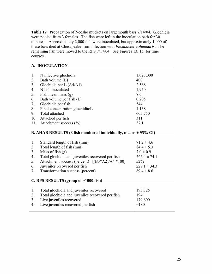
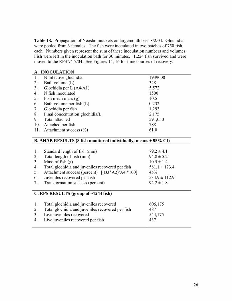
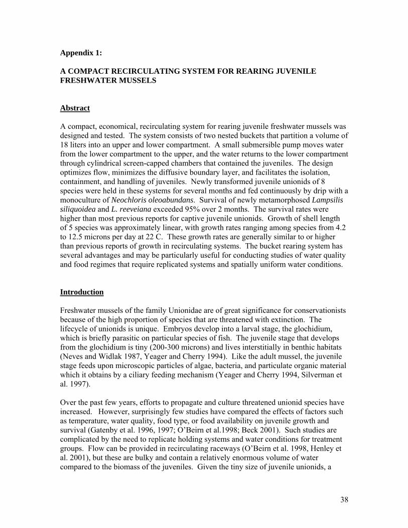
River at Carthage, Missouri. Four female mussels were collected 7/7/04. Three of the 4 females had glochidia (Table 10). The 4th female bore only unfertilized eggs. Glochidia were used to inoculate approximately 2,000 bass at Chesapeake on 7/14/04 (Table 12). Of these fish, about half died over the weekend due to infection with Flexibacter columnaris, a common bacterial pathogen that is sometimes problematic in the summer at hatcheries. The remaining bass were moved to SMSU on July 17 and treated with Kanamycin and salt (0.1%) in the RPS. Approximately 200 juveniles per fish were recovered (Table 12) and a total of 180,000 juveniles were released on August 3 in the Spring River at Hoberg, Stott City, and Carthage (Table 1). Another second group of 4 brooding females was collected July 27 at Carthage. Three of the 4 mussels yielded glochidia (Table 10). The fourth was brooding, but over 90% of the eggs were unfertilized, similar to one of the 4 females collected on July 7. Glochidia were used to inoculate approximately 1500 bass at Chesapeake on August 2, 2004 (Table 13). A subset of 12 fish was brought to SMSU to monitor in the AHAB system (Table 13, Figure 16). All fish at Chesapeake were treated prophylactically for 3 days with Tetracyclin after inoculation. Average temperature in the raceway over a 24-h period was 25.2 degrees Celsius. Flexibacter continued to be a problem. When the bass were delivered to SMSU on August 6 they were treated with Kanamycin. Mortality was moderate and 1,244 fish survived. Approximately 400 juveniles were recovered per fish (Table 13). A total of 480,000 of these were released on August 20 at 4 sites in the Spring River (Table 1). Another 5,000 were sent to CERC for use in toxicity tests and others were kept for grow out experiments (see below). All of these sites have received propagated juveniles previously. All adult mussels collected were marked, tissue sampled, and returned to the site of collection. Fatmucket propagation A group of 500 largemouth bass was inoculated with fatmucket glochidia at Chesapeake on 7/14/04 to provide juveniles for toxicity testing. The inoculation success was only ~24% which is relatively low compared to results with other Lampsilis under these conditions (typically ~50%) (Table 14). Approximately half of the fish died of Flexibacter. The recovery of juveniles from 200 survivors in the RPS was fair with approximately 112 juveniles per fish (about 86% transformation success). These juveniles did very well in grow-out and were the basis for a “round-robin” study involving 5 toxicology labs, developing standardized methods for tests (see below). A compact recirculating system for rearing juveniles A compact, economical, recirculating system for rearing juvenile freshwater mussels was designed and tested (see draft manuscript: Appendix 1). The system consists of two nested buckets that partition a volume of 18 liters into an upper and lower compartment. A small submersible pump moves water from the lower compartment to the upper, and the water returns to the lower compartment through cylindrical screen-capped chambers (downwellers) that contained the juveniles. The design minimizes space requirement and
11
facilitates the isolation, containment, and handling of juveniles. Newly transformed juvenile unionids of 8 species were held in these systems for several months and fed continuously by drip with a monoculture of Neochloris oleoabundans. Survival rates were higher than most previous reports for captive juvenile unionids. Survival of newly metamorphosed Lampsilis siliquoidea and L. reeveiana exceeded 95% over 2 months. Growth of shell length of 5 species was approximately linear, with growth rates ranging among species from 4.2 to 12.5 microns per day at 22 C. These growth rates are generally similar to or higher than previous reports of growth in recirculating systems. The bucket rearing system has several advantages and may be particularly useful for conducting studies of water quality and food regimes that require replicated systems and spatially uniform water conditions. Juvenile mussels for toxicity tests For the past 3 years SMSU has collaborated with researchers at the USGS Columbia Environmental Research Center (CERC) by providing technical consultation, glochidia, and juvenile mussels for toxicology studies. In 2004, SMSU participated in a USGS-led project titled “Acute copper round-robin toxicity tests with glochidia and newly-released juveniles of freshwater mussels”. The objective of the study is to examine the inter-laboratory variability in results of acute copper water-only toxicity tests conducted with glochidia and newly-released juvenile mussels. The study was carried out in July and August 2004. Five toxicology laboratories participated (North Carolina State University, Oklahoma State University, University of Wisconsin, USGS LaCrosse, and USGS Columbia). SMSU supplied glochidia and transformed juveniles. The study was completed successfully and reports are in preparation. SMSU also supplied 2-month-old juvenile pink mucket, black sandshell, and fat mucket to North Carolina State for chlorine and atrazine tests, and provided pink mucket and fatmucket to CERC for 28-d toxicity tests with copper and chlorine. Cultured alga was supplied to both labs weekly during the 28 day tests. Development of the bucket rearing system has made it possible for us to efficiently supply large numbers of older juveniles. We plan to continue collaborations with CERC, NCSU and other toxicology labs that are investigating unionid mussel sensitivity to toxicants. SMSU is a collaborator on a grant proposal submitted to USFWS 7/04 titled “Determining the sensitivity of Ozark mussels to zinc and lead in water or sediment”. Treatments for controlling flatworms Rhabdocoel flatworms are a significant problem in mussel propagation (Delp 2003, Zimmerman 2003). We investigated whether flatworms could be killed by NaCl concentrations tolerated by juvenile pink muckets (Lampsilis abrupta). We tested 9 different concentrations of NaCl: 0 g/L (control), 0.1, 0.25, 0.5, 1.0, 1.5, 2.0, 3.0, 4.0, and 6.0. The exposures were carried out in 5-ml aliquots in 12-well cell culture plates.
12
Flatworms were exposed in groups of 3 with three replicate groups (9 worms total) per concentration. Juvenile mussels were exposed in groups of 10 with 3 replicates per concentration. Observations were made once a day for seven days. Juvenile mussels appeared unaffected by NaCl at or below 0.5 g/l and no mortality was noted. At 1-2 g/L motility was reduced and foot extension became less common, but juveniles responded to tapping by closing the shell. At 3-4 g/l, responsiveness to tapping declined over time and a few mortalities occurred, but not clearly in excess of the control group. At 6 g/L most were dead by day 3 and all were dead by day 6. Flatworms were unaffected by NaCl up to 1.5 g/L. Responsiveness was reduced at 2 g/L but no mortality was observed. At 3-4 g/L most worms were dead by day 2 and all were dead by day 6. At 6 g/L all were dead by day 2. In conclusion, it appears that there may be some use for 2-day exposure to NaCl at 3 g/L to selectively kill Macrostomum. However, the long term affect of this treatment on juvenile survival should be tested further. Praziquantel (acylated quinoline-pyrazine) is a drug used for treatment of trematode and cestode flatworm parasites of mammals and fish. No information was found regarding effects on rhabdocoel flatworms. An experiment was conducted to test whether this anti-helminth drug might be useful in controlling Macrostomum tuba, the most prevalent flatworm predator. We exposed Macrostomum and juvenile broken-ray mussels (Lampsilis reeveiana), to 4 concentrations of Praziquantel: 0 mg/L (control), 1.0 mg/L, 5.0 mg/L, 10.0 mg/L, and 20.0 mg/L. A concentration of 2-3 mg/L is recommended for killing Monogenea and tapeworms of fish. The exposures were carried out in 5-ml aliquots in 12-well cell culture plates. Flatworms were exposed in groups of 2 with three replicate groups (6 worms total) per concentration. Juvenile mussels were exposed in groups of 10 with 3 replicates per concentration. Observations were made once a day for seven days. At the end of seven days, all worms present were still alive with 3 missing (perhaps cannibalized). Three juvenile mussels were dead in the control group. All juveniles exposed to Praziquantel survived. We conclude that Praziquantel is not an effective treatment for Macrostomum. Acquired resistance and cross-resistance of largemouth bass to glochidia We tested whether host fish that acquired resistance to glochidia of one mussel species were cross-resistant to glochidia of other species. Largemouth bass (Micropterus salmoides) were primed with 4-5 successive infections of glochidia of Lampsilis reeveiana. The percentage of attached glochidia that survived and transformed to the juvenile stage (transformation success) was compared between primed fish and naïve controls. Transformation success of L. reeveiana, Lampsilis abrupta, Villosa iris, and Utterbackia imbecillis was significantly lower on primed fish (37.8%, 43.5%, 67.0%, and 13.2% respectively) than on control fish (89.0%, 89.7%, 90.0%, and 22.2% respectively).
13
Immunoblotting was used to analyze the binding of serum antibodies from primed fish with glochidia proteins. Antibodies bound to glochidia proteins of similar molecular weight from L. reeveiana and L. abrupta. Bound proteins of V. iris differed in molecular weight from those of the Lampsilis species. There was no binding to specific glochidia proteins of U. imbecillis or Strophitus undulatus. Our results indicate that host acquired resistance can extend across mussel genera and subfamilies, and might involve both specific and nonspecific mechanisms. Understanding the specificity of acquired resistance of hosts to glochidia could enhance understanding of the evolutionary and ecological relationships between mussels and their host fishes. A manuscript describing these experiments has been prepared and submitted to Journal of Parasitology (Appendix 2).
Effects of inoculation intensity on transformation and acquired immunity This study was prompted by concern that the high infections intensities that we routinely use for propagation might negatively affect transformation success. We examined whether the number of attached glochidia of broken-rays mussel (Lampsilis reeveiana) affected transformation success on largemouth bass (Micropterus salmoides). Transformation success was quantified as the percent of attached glochidia that transformed to the juvenile stage and were recovered alive. Largemouth bass received either a low, medium or high intensity of infection (Table 15). Transformation success among the three groups did not differ significantly (One-way ANOVA, p>0.05). The data from the three groups were combined and a regression analysis was used to determine if intensity of infection affected transformation success. The relationship was insignificant (p= 0.18) (Figure 18). Therefore, it appears that a five-fold range of infection intensity not affect transformation success on naïve host fish. These intensities cover the range that we routinely obtain during propagation. These fish were also used to determine whether the intensity of the primary infection (described above) would affect the development of acquired immunity in host fish. The fish received a challenge infection 25 days after the primary infection. The fish were infected as a single group in order to ensure a uniform intensity of infection. Each fish was marked with a PIT tag prior to the challenge infection so that they could afterward be associated with their treatment groups. The experiment is still in progress at the time of this report. The duration of acquired immunity to glochidia Host fish acquire immunity to glochidia of freshwater mussels; however, the persistence of the immunity is not known. We investigated acquired immunity of largemouth bass (Micropterus salmoides) to glochidia of the broken rays mussel (Lampsilis reeveiana). Largemouth bass received 3 successive priming infections with glochidia of L. reeveiana to induce an immune response. Subsets of the primed fish were later challenged (re-
14
infected) at 2, 6, and 10 months post-priming. Transformation success was quantified as the percent of attached glochidia that transformed to the juvenile stage and were recovered alive. Significantly reduced transformation was observed on primed fish 2 months and 6 months post priming. The 10 month challenge infection is in progress and it appears that acquired immunity is diminished but still measurable. Serum antibody levels correlate with resistance of host fishes to a variety of other parasites. A second group of largemouth bass were used to determine whether serum antibody levels of host fish correlate with transformation success. Largemouth bass received 3 priming infections and blood was taken and pooled from one-fourth of the fish post-priming, 2 months post-priming, 6 months post-priming, and 10 months post priming. Immunoblotting was then used to detect antibodies specific to L. reeveiana glochidia in the serum. Antibody levels did not correlate with the persistence of acquired immunity. Antibodies were detected immediately post-priming, but diminished 2 months post-priming. Despite the fact that the fish retained their acquired immunity 6 months post-priming, serum antibodies were almost undetectable. Serum antibodies from 10 months post priming will be analyzed.
DISSEMINATION OF RESULTS Publications and reports • Dodd, B. J., M. C. Barnhart, C. L. Rogers-Lowery, T. B. Fobian, and R. V. Dimock Jr.
Cross-resistance of largemouth bass to glochidia of unionid mussels. Submitted to Journal of Parasitology, 11/04.
• Barnhart, M. C., J. Wigger and M. Duzan. 2004. Freshwater mussel survey of the Big Piney River and Roubidoux Creek. Final Report to the Missouri Department of Conservation. 24 pg. 11/6/04
• Hutson, C. and M. C. Barnhart. 2004. Survey of endangered and special concern mussel species in the Sac, Pomme de Terre, St. Francis, and Black River systems of Missouri, 2001-2003. Final Report to Missouri Department of Conservation & U.S. Fish and Wildlife Service. 369 pg. 10/14/04
Presentations (chronological order) • Wang N, Ingersoll CG, Greer IE, Whites DW, Dwyer FJ, Roberts AD, Augspurger T,
Kane C, Tibbott C, Neves RJ, Barnhart MC. 2003. Developing standardized guidance for conducting toxicity tests with glochidia of freshwater mussels. Presented at the 24th meeting of SETAC, Austin, TX, November 9-13,.
• Barnhart, M. C. 2004. Why fisheries professionals should care about native freshwater mussels. Platform presentation at American Fisheries Society Kansas Division Annual Meeting, Emporia KS. 2/21/04
• Eckert, N. E. and M. C. Barnhart. 2004. Diversity among Western fanshell mussel populations. Platform presentation at American Fisheries Society Annual Meeting Kansas Division, Emporia KS. 2/21/04. Best student paper award.
15
• Benjamin J. Dodd and M. C. Barnhart. 2004. The development, persistence and mechanism of acquired immunity of largemouth bass to mussel glochidia. Poster presentation at American Fisheries Society Kansas Division Annual Meeting, Emporia KS. 2/21/04 Best poster award.
• Barnhart, M. C. 2004. Conservation biology of native freshwater mussels. Invited seminar. University of Nebraska, Kearney, NE 2/27/04.
• Wang N, Ingersoll CG, Greer IE, Whites DW, Dwyer FJ, Roberts AD, Augspurger T, Kane C, Tibbott C, Neves RJ, Barnhart MC. 2004. Developing standardized guidance for conducting toxicity tests with glochidia of freshwater mussels. Presented at the joint meeting of the Midwestern chapter and Ozark-Prairie Region chapter of SETAC, La Crosse, WI, March 5, 2004.
• Barnhart, M. C. 2004. Artificial propagation as a management tool. U.S. Army Corps of Engineers, Memphis District, Mussel Workshop. 4/2/04
• John Harris and M. C. Barnhart. 2004 Work on Cyprogenia at SMSU. Arkansas Freshwater Mollusk Council meeting, Conway USFWS Field Office, 4-8-04
• Barnhart, M. C. 2004. Why we should care about native freshwater mussels. Mississippi Museum of Natural Science, Jackson, MS (invited). 6/3/04..
• Wang N, Ingersoll CG, Greer IE, Whites DW, Roberts A, Dwyer FJ, Augspurger T, Kane C, Tibbott T, Neves RJ, Barnhart MC. 2004. Developing standard guidance for conducting toxicity tests with glochidia of freshwater mussels. Seminar presented at Peking University in Beijing China (June 16, 2004), at the Research Center for Eco-environmental Sciences in Beijing China (June 18, 2004) and at the Institute of Hydrobiology in Wuhan China (June 21, 2004).
• Barnhart, M. C. 2004. Progress in the propagation of unionid mussels. Kansas Mussel Meeting, SW University, Winfield, KS. 7/28/04
• Dodd, B and M C Barnhart. 2004. Mechanisms and persistence of host fish immunity to glochidia of unionid mussels. Kansas Mussel Meeting, SW University, Winfield, KS. 7/28/04
• Kaiser, B and M C Barnhart. 2004. The effects of glochidia attachment on host fish respiration. Kansas Mussel Meeting, SW University, Winfield, KS. 7/28/04
• Serb, Jeanne M., John L. Harris, and M. Chris Barnhart. The Utility of Molecular Phylogenetics for Unionid Conservation: Identifying New Populations of the Endangered Winged Mapleleaf Quadrula fragosa (Bivalvia: Unionidae). Annual Meeting of the American Malacological Society, Sanibel, FLA. 8/2/04
• Barnhart, M. C. 2004. The intertwined interests of native mussels, native fish, and those who care for them. Invited, National Meeting of the American Fisheries Society, Madison, WI 8/24/04
Other programs & consultations (chronological order) • September 19, 2003. Presentation and display on freshwater mussels at Castlewood
State Park, with Scott Faiman, MDC, for “Meramec Expedition” program hosted by Richard Love, Missouri Department of Natural Resources.
• September 9, 2003. Interviewed by KOLR 10 television for the Morning Show, regarding work propagating endangered species. Aired September 10, 2003.
16
• September 15, 2003. Critical review of Draft Environmental Assessment and other documents related to the relicensing of Bagnell Dam by the Federal Energy Regulatory Commission. Review requested by U.S. Fish and Wildlife Service. Reviewed over 200 pages of documents, wrote 7 page commentary.
• January 16, 2004. Met with Donn L. Waage, Director, Central Region National Fish and Wildlife Foundation, and Martin-Williams Advertising in Minneapolis to discuss production of a video for use in fund-raising by the Fish and Wildlife Foundation.
• April 29, 2004. Hosted visitors from USFWS Mammoth Spring National Hatchery and Arkansas State University to tour our research facilities.
• May 1, 2004. Developed a workshop exercise, titled “Using baby clams to detect water pollution” for the Opening the Horizon program for middle-school girls, Drury University. Three of my students and I conducted 4 sessions for approximately 100 participants on May 1.
• May 7, 2004. Hosted Jim Carpowicz, Missouri Department of Conservation, to work on a video program for the “Missouri Outdoors” series. My lab group was interviewed, filmed, and I provided mussel video footage for the program, which is scheduled to run in January.
• July 10, 2004. Provided video footage to Kent Mayo, U.S. Department of Justice, for use in courtroom presentation on behalf of USFWS.
• July 12, 2004. Hosted Dr. Chris Ingersoll, Eugene Greer, and David White from Columbia Environmental Research Center of the US Geological Survey, who came to tour our facilities and learn methods for mussel propagation.
• July 14, 2004. Hosted two graduate students from the University of Oklahoma, Norman, to instruct them in research methods.
• July 21, 2004. Consultation meeting with city and county officials in Poplar Bluff and USFWS regarding proposed modifications on the Black River and potential impact on mussels.
• August 9, 2004. Hosted Dr. Paul Johnson and 3 other visitors from Tennessee Aquarium and Tennessee Aquatic Research Institute, who came to tour our facilities and learn methods for mussel propagation.
LITERATURE CITED Barnhart, M. C. 2003. Culture and restoration of mussel species of concern. Report to U.S. Fish and Wildlife Service and Missouri Department of Conservation. 56 pages. Delp, Angela 2002. Rhabdocoel flatworms as predators of juvenile freshwater mussels. Thesis, Master of Science, Southwest Missouri State University. Zimmerman, L.L., R.J. Neves and D. Smith. 2003. Control of predacious flatworms (Macrostomum sp.) in culturing freshwater mussels (Bivalvia: Unionidae). North American Journal of Aquaculture 65:28-32.
17
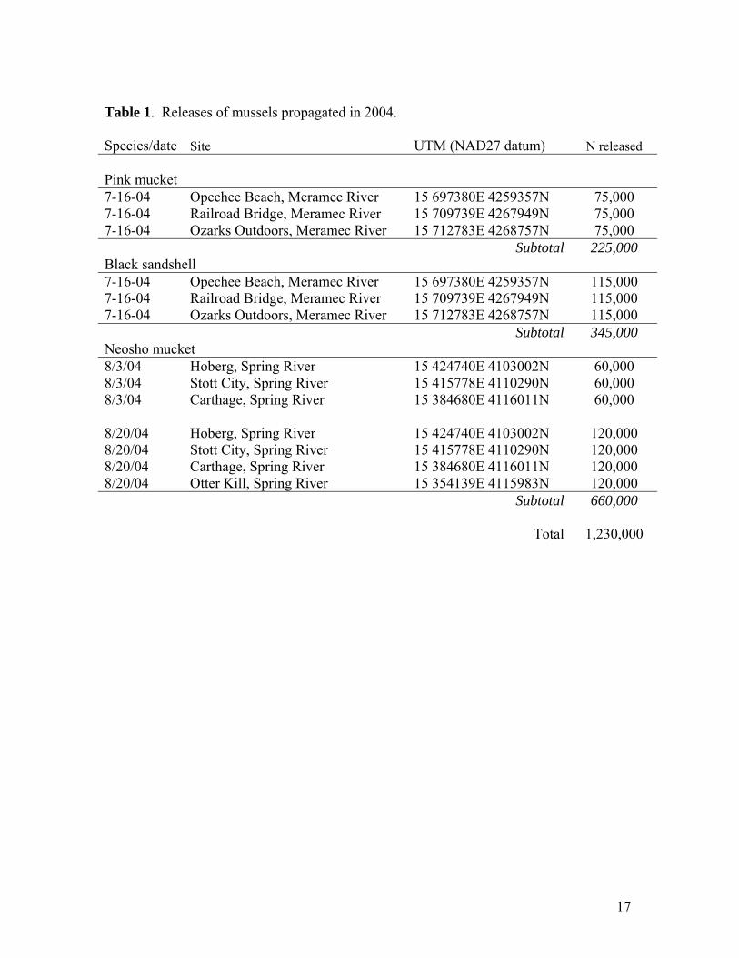
Table 1. Releases of mussels propagated in 2004. Species/date Site UTM (NAD27 datum) N released Pink mucket 7-16-04 Opechee Beach, Meramec River 15 697380E 4259357N 75,000 7-16-04 Railroad Bridge, Meramec River 15 709739E 4267949N 75,000 7-16-04 Ozarks Outdoors, Meramec River 15 712783E 4268757N 75,000 Subtotal 225,000 Black sandshell 7-16-04 Opechee Beach, Meramec River 15 697380E 4259357N 115,000 7-16-04 Railroad Bridge, Meramec River 15 709739E 4267949N 115,000 7-16-04 Ozarks Outdoors, Meramec River 15 712783E 4268757N 115,000 Subtotal 345,000 Neosho mucket 8/3/04 Hoberg, Spring River 15 424740E 4103002N 60,000 8/3/04 Stott City, Spring River 15 415778E 4110290N 60,000 8/3/04 Carthage, Spring River 15 384680E 4116011N 60,000 8/20/04 Hoberg, Spring River 15 424740E 4103002N 120,000 8/20/04 Stott City, Spring River 15 415778E 4110290N 120,000 8/20/04 Carthage, Spring River 15 384680E 4116011N 120,000 8/20/04 Otter Kill, Spring River 15 354139E 4115983N 120,000 Subtotal 660,000 Total 1,230,000
18
Table 2. Scaleshell located in the Gasconade and Meramec Rivers 9/26-11/6/03. As reported by Christian Hutson, Nathan Eckert, Scott Faiman, and Andy Roberts. Total search time was approximately 130 man-hours. In total 78 individuals were found (58 adult, 20 juvenile). (B) = number of brooding females. Approximately 43 man-hours of field time were expended per brooding female. Date Site Adults Juveniles ♂ ♀(B) ♂ ♀ ?
Gasconade River 9/26/03 Upstream of Ann M. Adams Access 2 - - - 1 9/29/03 upstream of Schlicht Springs Access 2 - - - 10/28/03 downstream of Mitschele Access 2 - - - 4 Meramec River 10/2/03 Fish Trap Rapids 2 1(0) - - - 10/3/03 Honeyhole- Pacific Palisades 5 2(1) - - 2 10/6/03 Honeyhole- Pacific Palisades 12 2(0) - - 1 10/7/03 Boat ramp- Pacific Palisades 2 - - - - 10/7/03 Honeyhole- Pacific Palisades 8 - - - - 10/21/03 Boat ramp- Pacific Palisades 1 - - - - 10/22/03 BM site 3 2(1) 3 1 - 10/30/03 BM site - 1(1) - 1 - 10/31/03 BM site 6 1(0) 3 1 - 11/5/03 Bar just upstream of Allenton - - - 1 - 11/6/03 Bar just upstream of Allenton 3 - 1 1 - Totals 50 9(3) 7 5 8

19
Table 3. Brood condition of Meramec River pink muckets propagated 6/24 ( #04-1, 2, 5, 6) and 6/25/04 (#04-7, 8). Numbers are in thousands. Undeveloped eggs were not counted but were uncommon. Dead glochidia were noted only when significant numbers were present. Glochidia from mussel 04-8 had been held 24 hours and were stale. Each figure is derived from a mean ± 95% CI of counts of 10 volumetric subsamples from the total suspension. Female ID#
A. Numbers (thousands) 04-1 04-2 04-5 04-6 04-7 04-81. Total brood 381 ±7.1 464 ± 5.7 141 ± 2.5 6.1 ±0.4 290±4.6 271±2.72. Undeveloped eggs - - - - - -3. Glochidia 381 ± 7.1 464 ± 5.7 141 ± 2.5 6.1 ±0.4 290±4.6 271±2.74. Live, open glochidia 354 ± 6.6 434 ± 5.3 131 ± 2.2 5.5 ±0.4 267±4.2 66±1.35. Live, closed glochidia 27 ± 1.3 30 ± 1.0 11 ± 0.4 0.6 ± 0. 21±0.7 62±1.26. Dead glochidia - - - - 2±0.4 143±1.8 B. Proportions 1. % brood fertile 100.0 100.0 100.0 100.0 100.0 100.02. % brood infertile - - - - - -3. % glochidia live 100.0 100.0 100.0 100.0 100.0 100.04. % glochidia dead - - - - - -5. % live glochidia open 92.9 93.5 92.6 90.1 92.7 51.66. % live glochidia closed 7.1 6.5 7.4 9.9 7.3 48.4
Table 4. Size and fecundity of Meramec River pink muckets. The mussels were marked as noted and returned to the site of collection. Mantle tissue samples were preserved. These numbers underestimate fecundity because the gills were not completely emptied. Female Length (mm) Width (mm) Height (mm) Whole mass (g) Fecundity
04-1 112.8 91.5 381,000 04-2 103.9 87.1 464,000 04-5 - - 141,000 04-6 115.4 98.2 6,100 04-7 133.6 104.7 290,000 04-8 - - 271,000
20
Table 5. Propagation of Meramec pink muckets on 6/24/04. Fish inoculated at Lost Valley were 1 year old largemouth bass. The AHAB results are the same batch of glochidia but different host fish: these were YOY largemouth and smallmouth from Lost Valley. See Figures 6, 8, 9 for time courses. A. INOCULATION at Lost Valley 1. N infective glochidia 924,475 2. Bath volume 197 L 3. Initial concentration in bath: 4,693 glochidia per L 4. N fish inoculated 200 5. Fish body mass 115 grams 6. Bath volume per fish 0.985 L 7. Glochidia per fish 4,622 8. Final concentration in bath 2,042 glochidia/L 9. Estimated total number attached 522,267 10. Estimated attachment per fish 2,611 11. Attachment success [(A8/A6)*100] 56.5 % B. RPS RESULTS at SMSU 1. Total glochidia and juveniles recovered 138,188 2. Total glochidia & juveniles recovered per fish 691 3. Juveniles recovered 120,550 4. Juveniles recovered per fish 603 5. Apparent transformation success* 23.1% C. AHAB RESULTS Largemouth (n=6) Smallmouth n=6 1. Fish mean total length (mm) 58.5 ± 4.0 59.8 ± 2.4 2. Fish mean body mass (g) 2.3 ± 0.5 2.4 ± 0.2 3. Total glochidia & juvees recovered per fish 262.0 ± 74.1 259.2 ± 17.9 4. Juveniles recovered per fish 124.5 ± 54.8 166.8 ± 12.2 5. Transformation success (%) 48.5 ± 11.1 64.4 ± 1.2
*The low transformation success on the large fish may be an artifact because the fish were already shedding juveniles when moved to the RPS on day 13 (Figure 6).
21
Table 6. Propagation of Meramec pink muckets on 6/25/04. Fish were YOY largemouth bass from Chesapeake. No fish from this batch were monitored in the AHAB. See also Figure 7. A. INOCULATION at Chesapeake 1. N infective glochidia 333,000 2. Bath volume 135 3. Initial concentration in bath: glochidia per L 2,467 4. N fish inoculated 1166 5. Fish body mass (g) 6.6 6. Bath volume per fish 0.116 7. Glochidia per fish 286 8. Final concentration in bath: glochidia/L - 9. Estimated total number attached - 10. Estimated attachment per fish - 11. Attachment success [(B2/A6)*100] 36.4 % B. RPS RESULTS 1. Total glochidia and juveniles recovered 121,888 2. Total glochidia & juveniles recovered per fish 104 3. Juveniles recovered 108,150 4. Juveniles recovered per fish 93

22
Table 8. Size and fecundity of Meramec River black sandshell propagated 6/18/04. The mussels were marked as noted and returned to the Meramec Opechee Beach site. Mantle tissue samples were preserved. These numbers underestimate fecundity because the gills were not completely emptied. Female Length (mm) Width (mm) Height (mm) Whole mass (g) Fecundity
04-1 154 - - 412 136,000 04-2 147 - - 326 842,000 04-3 152 - - 368 342,000 04-4 147 - - 376 274,000
Table 7. Brood condition of Meramec River black sandshell propagated 6/18/04. Numbers are in thousands. Each figure is derived from a mean ± 95% CI of counts of 10 volumetric subsamples from the total suspension. Table format is similar to previous reports. Undeveloped eggs and dead glochidia were not recorded for these mussels, but the proportion of both was very low. Female ID#
A. Numbers (thousands) 04-1 04-2 04-3 04-41. Total brood 136 ± 2.7 842 ± 9.0 342 ± 4.4 274 ± 5.22. Undeveloped eggs - - - -3. Glochidia 136 ± 2.7 842 ± 9.0 342 ± 4.4 274 ± 5.24. Live, open glochidia 110 ± 2..4 806 ± 9.1 326 ± 4.6 252 ± 5.05. Live, closed glochidia 26 ± 8.4 36 ± 1.7 16 ± 0.5 22 ± 0.46. Dead glochidia - - - - B. Proportions 1. % brood fertile 100 100 100 1002. % brood infertile - - - -3. % glochidia live 100 100 100 1004. % glochidia dead - - - -5. % live glochidia open 80.9 95.7 95.3 92.06. % live glochidia closed 19.1 4.3 4.7 8.0
23
Table 9. Propagation of Meramec black sandshell on 1-year old walleye 6/18/04. See Figure 10 for time course of recovery in AHAB. A. INOCULATION at Lost Valley 1. N infective glochidia 1,494,000 2. Bath volume 356 L 3. Initial concentration in bath: glochidia per L 4,197 4. N fish inoculated 800 5 Fish mean body mass - 6. Bath volume per fish 0.445 L 7. Glochidia per fish 1,868 8. Final concentration in bath: glochidia/L 2,325 9. Estimated total number attached 666,300 10. Estimated attachment per fish 833 11. Attachment success 44.6% B. RPS RESULTS at Lost Valley 1. Total glochidia and juveniles recovered - 2. Total glochidia & juveniles recovered per fish - 3. Juveniles recovered 345,000 4. Juveniles recovered per fish 431 5. Transformation success [(B4/A10)]*100 51.7% C. AHAB RESULTS (12 smaller fish inoculated separately) 1. Fish mean total length (mm) 71.9 ± 2.7 2. Fish mean standard length (mm) 59.7 ± 2.2 3. Fish mean body mass 2.5 ± 0.2 4. Total glochidia and juveniles recovered per fish 165.5 ± 21.1 5. Estimated attachment success - 6. Juveniles recovered per fish 144 ± 18.9 7. Transformation success 86.8% ± 2.1
24
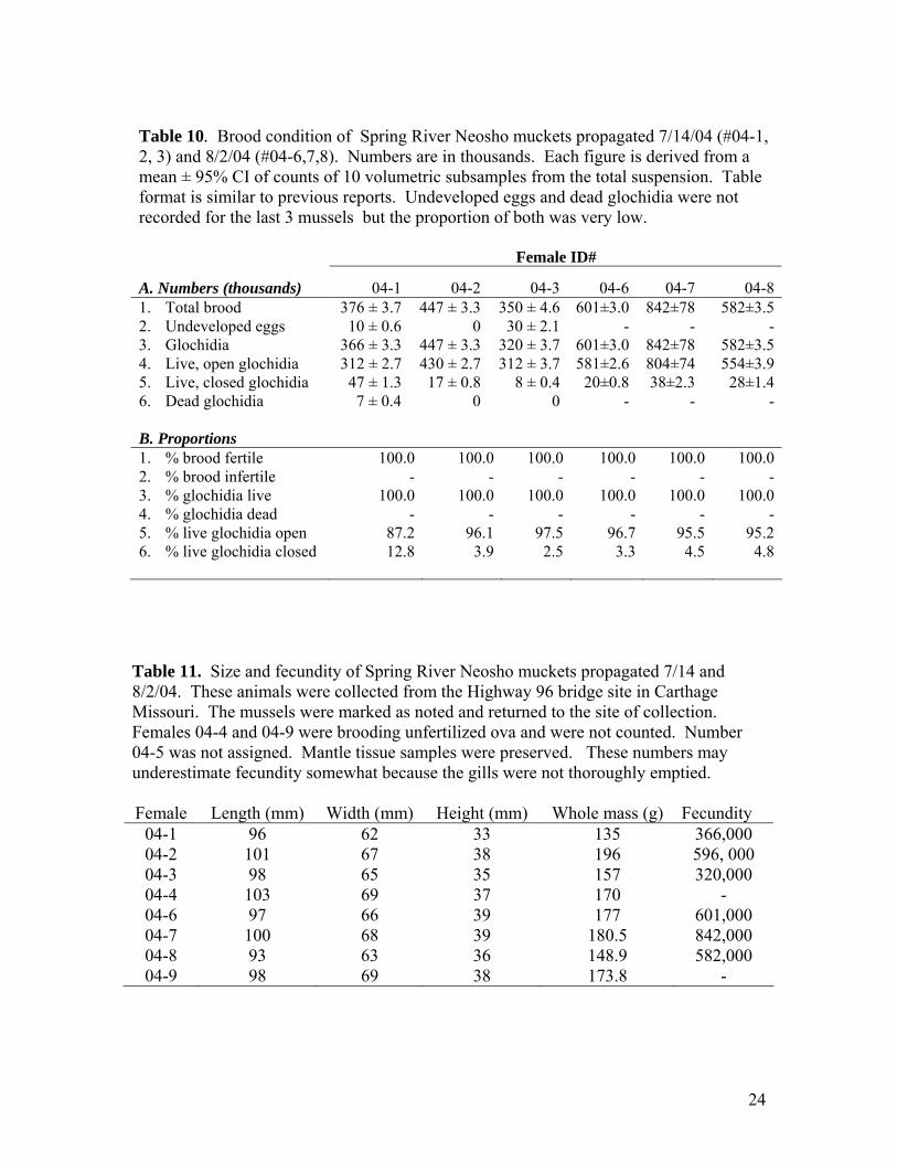
Table 10. Brood condition of Spring River Neosho muckets propagated 7/14/04 (#04-1, 2, 3) and 8/2/04 (#04-6,7,8). Numbers are in thousands. Each figure is derived from a mean ± 95% CI of counts of 10 volumetric subsamples from the total suspension. Table format is similar to previous reports. Undeveloped eggs and dead glochidia were not recorded for the last 3 mussels but the proportion of both was very low. Female ID#
A. Numbers (thousands) 04-1 04-2 04-3 04-6 04-7 04-81. Total brood 376 ± 3.7 447 ± 3.3 350 ± 4.6 601±3.0 842±78 582±3.52. Undeveloped eggs 10 ± 0.6 0 30 ± 2.1 - - -3. Glochidia 366 ± 3.3 447 ± 3.3 320 ± 3.7 601±3.0 842±78 582±3.54. Live, open glochidia 312 ± 2.7 430 ± 2.7 312 ± 3.7 581±2.6 804±74 554±3.95. Live, closed glochidia 47 ± 1.3 17 ± 0.8 8 ± 0.4 20±0.8 38±2.3 28±1.46. Dead glochidia 7 ± 0.4 0 0 - - - B. Proportions 1. % brood fertile 100.0 100.0 100.0 100.0 100.0 100.02. % brood infertile - - - - - -3. % glochidia live 100.0 100.0 100.0 100.0 100.0 100.04. % glochidia dead - - - - - -5. % live glochidia open 87.2 96.1 97.5 96.7 95.5 95.26. % live glochidia closed 12.8 3.9 2.5 3.3 4.5 4.8
Table 11. Size and fecundity of Spring River Neosho muckets propagated 7/14 and 8/2/04. These animals were collected from the Highway 96 bridge site in Carthage Missouri. The mussels were marked as noted and returned to the site of collection. Females 04-4 and 04-9 were brooding unfertilized ova and were not counted. Number 04-5 was not assigned. Mantle tissue samples were preserved. These numbers may underestimate fecundity somewhat because the gills were not thoroughly emptied. Female Length (mm) Width (mm) Height (mm) Whole mass (g) Fecundity
04-1 96 62 33 135 366,000 04-2 101 67 38 196 596, 000 04-3 98 65 35 157 320,000 04-4 103 69 37 170 - 04-6 97 66 39 177 601,000 04-7 100 68 39 180.5 842,000 04-8 93 63 36 148.9 582,000 04-9 98 69 38 173.8 -
25
Table 12. Propagation of Neosho muckets on largemouth bass 7/14/04. Glochidia were pooled from 3 females. The fish were left in the inoculation bath for 30 minutes. Approximately 2,000 fish were inoculated, but approximately 1,000 of these bass died at Chesapeake from infection with Flexibacter columnaris. The remaining fish were moved to the RPS 7/17/04. See Figures 13, 15 for time courses. A. INOCULATION 1. N infective glochidia 1,027,000 2. Bath volume (L) 400 3. Glochidia per L (A4/A1) 2,568 4. N fish inoculated 1,950 5. Fish mean mass (g) 8.6 6. Bath volume per fish (L) 0.205 7. Glochidia per fish 544 8. Final concentration glochidia/L 1,138 9. Total attached 605,750 10. Attached per fish 311 11. Attachment success (%) 57.1 B. AHAB RESULTS (8 fish monitored individually, means ± 95% CI) 1. Standard length of fish (mm) 71.2 ± 4.6 2. Total length of fish (mm) 84.4 ± 5.3 3. Mass of fish (g) 7.0 ± 0.9 4. Total glochidia and juveniles recovered per fish 265.4 ± 74.1 5. Attachment success (percent) [(B3*A2)/A4 *100] 52% 6. Juveniles recovered per fish 227.1 ± 34.3 7. Transformation success (percent) 89.4 ± 8.6 C. RPS RESULTS (group of ~1000 fish) 1. Total glochidia and juveniles recovered 193,725 2. Total glochidia and juveniles recovered per fish 194 3. Live juveniles recovered 179,600 4. Live juveniles recovered per fish ~180
26
Table 13. Propagation of Neosho muckets on largemouth bass 8/2/04. Glochidia were pooled from 3 females. The fish were inoculated in two batches of 750 fish each. Numbers given represent the sum of these inoculation numbers and volumes. Fish were left in the inoculation bath for 30 minutes. 1,224 fish survived and were moved to the RPS 7/17/04. See Figures 14, 16 for time courses of recovery. A. INOCULATION 1. N infective glochidia 1939000 2. Bath volume (L) 348 3. Glochidia per L (A4/A1) 5,572 4. N fish inoculated 1500 5. Fish mean mass (g) 10.5 6. Bath volume per fish (L) 0.232 7. Glochidia per fish 1,293 8. Final concentration glochidia/L 2,175 9. Total attached 591,050 10. Attached per fish 788 11. Attachment success (%) 61.0 B. AHAB RESULTS (8 fish monitored individually, means ± 95% CI) 1. Standard length of fish (mm) 79.2 ± 4.1 2. Total length of fish (mm) 94.8 ± 5.2 3. Mass of fish (g) 10.5 ± 1.4 4. Total glochidia and juveniles recovered per fish 581.1 ± 123.4 5. Attachment success (percent) [(B3*A2)/A4 *100] 45% 6. Juveniles recovered per fish 534.9 ± 112.9 7. Transformation success (percent) 92.2 ± 1.8 C. RPS RESULTS (group of ~1244 fish) 1. Total glochidia and juveniles recovered 606,175 2. Total glochidia and juveniles recovered per fish 487 3. Live juveniles recovered 544,175 4. Live juveniles recovered per fish 437
27
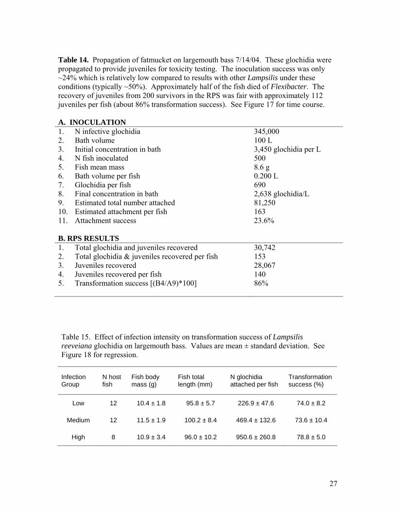
Table 14. Propagation of fatmucket on largemouth bass 7/14/04. These glochidia were propagated to provide juveniles for toxicity testing. The inoculation success was only ~24% which is relatively low compared to results with other Lampsilis under these conditions (typically ~50%). Approximately half of the fish died of Flexibacter. The recovery of juveniles from 200 survivors in the RPS was fair with approximately 112 juveniles per fish (about 86% transformation success). See Figure 17 for time course. A. INOCULATION 1. N infective glochidia 345,000 2. Bath volume 100 L 3. Initial concentration in bath 3,450 glochidia per L 4. N fish inoculated 500 5. Fish mean mass 8.6 g 6. Bath volume per fish 0.200 L 7. Glochidia per fish 690 8. Final concentration in bath 2,638 glochidia/L 9. Estimated total number attached 81,250 10. Estimated attachment per fish 163 11. Attachment success 23.6% B. RPS RESULTS 1. Total glochidia and juveniles recovered 30,742 2. Total glochidia & juveniles recovered per fish 153 3. Juveniles recovered 28,067 4. Juveniles recovered per fish 140 5. Transformation success [(B4/A9)*100] 86% Table 15. Effect of infection intensity on transformation success of Lampsilis reeveiana glochidia on largemouth bass. Values are mean ± standard deviation. See Figure 18 for regression.
Infection Group
N host fish
Fish body mass (g)
Fish total length (mm)
N glochidia attached per fish
Transformation success (%)
Low 12 10.4 ± 1.8 95.8 ± 5.7 226.9 ± 47.6 74.0 ± 8.2
Medium 12 11.5 ± 1.9 100.2 ± 8.4 469.4 ± 132.6 73.6 ± 10.4
High 8 10.9 ± 3.4 96.0 ± 10.2 950.6 ± 260.8 78.8 ± 5.0

28
Figure 1. MDC personnel transporting RPS tanks from Springfield to Warsaw
Figure 2. Cod-ends of recovery filters for RPS. Dr. Pepper bottles were donated by Coca-Cola Bottlers of Springfield.
29
Figure 3. Lost Valley RPS nearing completion.
Figure 4. Completed RPS in use for propagation of black sandshell on walleye.
30
Figure 5. Hyphae of water mold that infested female scaleshell. Tentatively identified as Saprolegnia. Several empty shells of scaleshell glochidia are visible. Scale line = 100 microns.
31
RPS pink muckets- Lost Valley bass 6/24/04
Days post-inoculation
0 2 4 6 8 10 12 14 16 18 20 22 24 26
Thou
sand
s re
cove
red
0
10
20
30
40
50
60
70
80
Untransformed or deadLive juveniles
No data
Figure 6. Recovery of pink muckets in RPS 6/24/04 (See Table 5B). Fish were inoculated at Lost Valley on day 0 and placed in the RPS at SMSU on day 13. Dropoff of juveniles appears to have started before day 13, so that some juveniles were probably lost. RPS temperature = 23-24 C.
RPS pink muckets- Chesapeake bass 6/25/04
Days post-inoculation
0 2 4 6 8 10 12 14 16 18 20 22 24 26
Thou
sand
s re
cove
red
0
10
20
30
40
50
60
70
80
Untransformed or deadLive juveniles
No data
Figure 7. Recovery of 6/25/04 pink muckets from bass in RPS (see Table 6B). Fish were inoculated at Chesapeake on day 0 and placed in the RPS on day 13. RPS temperature =23-24 C.
32
AHAB pink muckets on smallmouth bass- 6/24/04
Days post-inoculation
0 2 4 6 8 10 12 14 16 18 20 22 24 26
Mea
n nu
mbe
r per
fish
0
20
40
60
80
100
120
Untransformed or deadLive juveniles
Figure 8. Recovery of 6/24/04 pink muckets from smallmouth bass monitored individually in the AHAB system. Mean temperature was 22.8 C. See Table 5C.
AHAB pink muckets on largemouth bass- 6-24-04
Days post-inoculation
0 2 4 6 8 10 12 14 16 18 20 22 24 26
Mea
n nu
mbe
r per
fish
0
20
40
60
80
100
120
Untransformed or deadLive juveniles
Figure 9. Recovery of 6/24/04 pink muckets from largemouth bass monitored individually in the AHAB system. Mean temperature was 22.8 C. See Table 5C.
33
AHAB black sandshell on walleye 6/18/04
Days post-inoculation
0 2 4 6 8 10 12 14 16 18 20 22 24 26
Mea
n nu
mbe
r per
fish
0
20
40
60
80
100
120
Untransformed or deadLive juveniles
Figure 10. Recovery of 6/18/04 black sandshell from walleye monitored individually in the AHAB system. Mean temperature was 22.8 C. See Table 9C.

34
0
500
1000
1500
2000
2500
3000
0 5 10 15 20 25 30
Minutes elapsed
Glo
chid
ia p
er L
iter
Measured concentration
Predicted concentration
Figure 11. Time course of Neosho mucket attachment to bass during inoculation on 7/14/04. The initial concentration of glochidia in the bath was predicted from the number of glochidia and the bath volume. Concentration was measured at intervals by collecting 2-L samples of the bath and counting the glochidia remaining.
0
1000
2000
3000
4000
5000
6000
7000
0 5 10 15 20 25 30
Minutes elapsed
Glo
chid
ia p
er L
iter
Measured concentrationPredicted concentration
Figure 12. Time course of Neosho mucket attachment to bass during inoculation on 8/2/04. The initial concentration of glochidia in the bath was predicted from the number of glochidia and the bath volume. Concentration was also measured at intervals by collecting 2-L samples of the bath and counting the glochidia remaining.

35
RPS Neosho muckets- 7-14-04
Days post-inoculation
0 2 4 6 8 10 12 14 16 18 20 22 24 26
Thou
sand
s re
cove
red
0
10
20
30
40
50
60
70
80
Untransformed or deadLive juveniles
No data
Figure 13. Recovery of 7/14/04 Neosho muckets from largemouth bass in RPS (See Table 12C). Fish were inoculated at Chesapeake on day 0 and placed in the RPS on day 9.
RPS Neosho muckets- 8/2/04
Days post-inoculation
0 2 4 6 8 10 12 14 16 18 20 22 24 26
Thou
sand
s re
cove
red
0
20
40
60
80
100
120
140
160
Untransformed or deadLive juveniles
No data
Figure 14. Recovery of 8/2/04 Neosho muckets from largemouth bass in RPS (See Table 13C). Fish were inoculated at Chesapeake on day 0 and placed in the RPS on day 8. Temp = 24.0 in RPS- but 25.2 C (24 h) or warmer at Chesapeake.

36
AHAB Neosho muckets 7/14/04
Days post-inoculation
0 2 4 6 8 10 12 14 16 18 20 22 24 26 28 30 32 34 36
Mea
n nu
mbe
r per
fish
0
20
40
60
80
100
120
140
Untransformed or deadLive juveniles
Figure 15. Recovery of 7/14/04 Neosho muckets from largemouth bass monitored individually in the AHAB system. Mean temperature was 22.9 C. See Table 12C.
AHAB Neosho mucket 8/2/04
Days post-inoculation
0 2 4 6 8 10 12 14 16 18 20 22 24 26 28 30 32 34 36
Mea
n nu
mbe
r per
fish
020406080
100120140160180200220240260
Untransformed or deadLive juveniles
Figure 16. Recovery of 8/2/04 Neosho muckets from largemouth bass monitored individually in the AHAB system. Mean temperature was 23.0 C. See Table 13C. It is a puzzle as to why these juveniles took so long to leave the fish. The peak of drop-off was about 10 days later than that of Neosho muckets propagated on July 14. Temperature in the AHAB sump was essentially identical for the two tests.
37
RPS Fatmuckets- 7-14-04
Days post-inoculation
0 2 4 6 8 10 12 14 16 18 20 22 24 26
Thou
sand
s re
cove
red
0
2
4
6
8
10
12
14
Untransformed or deadLive juveniles
No data
Figure 17. Recovery of 7/14/04 fatmuckets from largemouth bass in RPS (See Table 14B). Fish were inoculated at Chesapeake on day 0 and placed in the RPS on day 9.
0102030405060708090
100
0 200 400 600 800 1000 1200 1400 1600
Number of glochidia attached
Tran
sfor
mat
ion
succ
ess
(%)
Figure 18. Relationship between infection intensity (number of glochidia attached per fish) and transformation success of Lampsilis reeveiana on largemouth bass. See Table 15. Slope of the relationship is insignificant (P=0.18).
38
Appendix 1: A COMPACT RECIRCULATING SYSTEM FOR REARING JUVENILE FRESHWATER MUSSELS Abstract A compact, economical, recirculating system for rearing juvenile freshwater mussels was designed and tested. The system consists of two nested buckets that partition a volume of 18 liters into an upper and lower compartment. A small submersible pump moves water from the lower compartment to the upper, and the water returns to the lower compartment through cylindrical screen-capped chambers that contained the juveniles. The design optimizes flow, minimizes the diffusive boundary layer, and facilitates the isolation, containment, and handling of juveniles. Newly transformed juvenile unionids of 8 species were held in these systems for several months and fed continuously by drip with a monoculture of Neochloris oleoabundans. Survival of newly metamorphosed Lampsilis siliquoidea and L. reeveiana exceeded 95% over 2 months. The survival rates were higher than most previous reports for captive juvenile unionids. Growth of shell length of 5 species was approximately linear, with growth rates ranging among species from 4.2 to 12.5 microns per day at 22 C. These growth rates are generally similar to or higher than previous reports of growth in recirculating systems. The bucket rearing system has several advantages and may be particularly useful for conducting studies of water quality and food regimes that require replicated systems and spatially uniform water conditions. Introduction Freshwater mussels of the family Unionidae are of great significance for conservationists because of the high proportion of species that are threatened with extinction. The lifecycle of unionids is unique. Embryos develop into a larval stage, the glochidium, which is briefly parasitic on particular species of fish. The juvenile stage that develops from the glochidium is tiny (200-300 microns) and lives interstitially in benthic habitats (Neves and Widlak 1987, Yeager and Cherry 1994). Like the adult mussel, the juvenile stage feeds upon microscopic particles of algae, bacteria, and particulate organic material which it obtains by a ciliary feeding mechanism (Yeager and Cherry 1994, Silverman et al. 1997). Over the past few years, efforts to propagate and culture threatened unionid species have increased. However, surprisingly few studies have compared the effects of factors such as temperature, water quality, food type, or food availability on juvenile growth and survival (Gatenby et al. 1996, 1997; O’Beirn et al.1998; Beck 2001). Such studies are complicated by the need to replicate holding systems and water conditions for treatment groups. Flow can be provided in recirculating raceways (O’Beirn et al. 1998, Henley et al. 2001), but these are bulky and contain a relatively enormous volume of water compared to the biomass of the juveniles. Given the tiny size of juvenile unionids, a
39
suitably designed recirculating system can maintain thousands of individuals in only a few gallons of water. Such a system can be replicated economically and provide statistical power for comparisons among treatment groups. The small size of juvenile mussels presents difficulties in handling and confining them in flowing water. Shell length of newly metamorphosed juveniles generally ranges between about 200 and 300 microns, depending on species. These tiny bivalves can be suspended by even small water currents, so that they are easily lost from open containers in flowing systems. In addition to drift, juvenile mussels are quite mobile and can crawl up the sides of containers. Losses in grow-out studies are sometimes attributed to emigration as well as death (Zimmerman 2003). Several studies have reported that providing a substrate of silt, in which juveniles can burrow, improves growth and survival. Silt is thought to serve as a source of food as well as a substrate (e.g. Hudson and Isom 1984, Gatenby et al. 1996, Rogers 1999, Mummert 2001, Zimmerman 2003, Kovitvadhi in press). However, the presence of silt further complicates maintenance, observation and handling, and may encourage the growth of other organisms in the culture system. Maintaining adequate flow in culture systems is essential, because juvenile unionids are small enough to occupy the diffusive boundary layer. The diffusive boundary layer is a benthic zone closely adjacent to surfaces, where friction reduces water movement to the point that diffusion, rather than convection, becomes the dominant mode of transport. Factors such as dissolved oxygen, ammonia, and food concentration in the boundary layer can differ substantially from those in adjacent flowing water (Boudreau 2001). Investigation of these factors should therefore be carried out in a system designed to minimize stagnant zones and maintain uniform flow and water quality. The system described in this report partitions a volume of 18 liters into an upper compartment and a lower compartment. A small submersible pump moves water from the lower compartment to the upper. The water then returns to the lower compartment through a set of cylindrical flow-through chambers (downwellers) that contain the juveniles. The design was tested by rearing juveniles of 8 unionid species for periods up to 12 weeks and quantifying growth and survival. Materials and Methods Chambers The flow-through chambers for containing juveniles were constructed from 2-inch diameter PVC plumbing pipe and couplings (Figure 1, 2). Nitex screen was placed over a 1.75-inch length of pipe and press-fit into a coupling, forming a unit called a filter cup. Pairs of filter cups were nested together to make chambers bounded by screen on both ends. The two halves of each chamber are press-fit together and can easily be opened and closed to allow access to the juveniles. The juveniles rest on the lower screen. Each
40
chamber is positioned vertically in the recirculating system with water flowing downward through the chamber. Bucket recirculating system The recirculating system consists of two nested plastic (HDPE) buckets, one of 3.5 gallon capacity and the other of 5 gallon capacity (Figures 1, 2) (Encore Plastics, Sandusky, Ohio). The smaller bucket is nested into the larger one, and its base forms a platform 6 inches above base of the lower bucket. Seven holes are drilled in the base of the upper bucket with a 2⅜ inch hole saw. The bases of the flow-through chambers are inserted into these holes. It is important that the chambers fit the openings closely, so that the only path for water to return to the lower compartment is through the chambers. A ¾-inch bulkhead fitting is mounted in the center of the base of the upper bucket. The fitting is attached to the outlet of a small “power-head” submersible aquarium pump (Aquarium Systems Mini-jet model MN-404). The pump circulates water from the lower to the upper compartment. Nominal flow rate is 106 gallons per hour. Food and feeding The unicellular green alga Neochloris oleoabundans Chantanachat and Bold was cultured and provided as food. This species has been identified previously as a suitable food source for juvenile unionids (Gatenby et al. 1997, O’Beirn et al.1998). Stock cultures were obtained from the University of Texas (UTEX Culture Collection of Algae, accession number 1185). The growth medium was autoclaved tap water fertilized with a commercial nutrient mix (Kent ProCulture® F2, Aquatic Ecosystems, Apopka FL). The alga was grown in 100-ml flasks and in 1.5-L glass jugs (Figure 3a). The 100 ml flasks were inoculated from a stock culture on agar or serially from other 100 ml cultures. Each 1.5-L culture was inoculated with a 100-ml culture. Each jug was aerated via a glass pipette inserted through a rubber stopper. The air was filtered (0.5-micron) to remove contaminant spores of other microorganisms. Four 1.5 liter cultures were prepared weekly and harvested after 4-5 days of growth. The algae were separated from the culture medium by centrifuging at about 1500 RCF for 10 minutes. The cells were then resuspended in water to achieve a concentration of about 20*106 cells per ml. Algae were refrigerated after resuspension and generally used within 1 week of harvest. Each recirculating system was fed from 500 ml drip bags, similar to those that are used to deliver intravenous solutions (Figure 3b). Each bag was filled daily with 400 ml of water from the system and 100 ml of algae suspension (total 20 * 109 cells). Flow through the drip line was controlled with a length of fine polyethylene tubing and was adjusted to approximately 500 ml/24 h. Cell concentration in the recirculating systems was checked with a hemocytometer weekly and remained at about 10-15,000 cells/ml. The bags were washed weekly with hot water and the drip lines rinsed with bleach to prevent them from becoming blocked.
41
Water quality and cleaning The chambers were moved into clean buckets and new water weekly. Natural river water (James River, Greene County MO) was used in order to provide a natural community of microorganisms, which may aid in digestion. The water was collected one day before use and was filtered to remove particles larger than 30 microns. The screens in each chamber were cleaned at least weekly by spraying with a modified garden sprayer. When replacing the chambers, care was taken to avoid trapping air beneath the screens because trapped air would block water flow. Air was removed from each chamber after submergence by drawing it out through the upper screen with a large rubber-bulb syringe (cooking baster). Temperature was 22-23 C. Juveniles Juvenile mussels were propagated on host fish for population augmentation of threatened species or during the course of research on host relationships and toxicology studies. From several hundred to several thousand juveniles of each species were collected and placed in the rearing systems within 2 days of excystment from the host fish. Four bucket systems (28 chambers) were used and all were treated similarly throughout the course of the tests. Growth rates of 5 species were checked at 10-20 day intervals. Each chamber was opened and the juveniles were rinsed into a 3-inch plastic Petri dish. The group of juveniles was then photographed several times under a compound microscope with a digital camera. A stage micrometer was photographed to provide scale. The photographs were later displayed in software and the maximum shell length of each suitably oriented individual was measured using ImageJ (NIH 2004) image analysis software. An average sample of 72 individuals was measured for each group. Survivorship in each of the 28 chambers was determined by complete counts of each group of juveniles using Bolgorov plankton counting cells and a dissecting microscope. Dead individuals were easily recognizable as empty shells. Results and Discussion Survivorship A total of 27,850 juveniles of 8 species were included in the study. The juveniles were counted and survivorship was determined only once during the study, at which time the age of the cohorts varied from 2-12 weeks (Table 1). Survivorship varied among species and among chambers but exceeded 75% in 25 of 28 groups. Lampsilis siliquoidea and L. reeveiana exceeded 95% survival at 2 months.

42
The low mortality observed in this study contrasts strongly with previous reports. Survival of captive juvenile unionids after 2 months is usually below 50% and often below 20% (summarized by Gatenby et al 1996, Rogers 1999, Beck 2001, Kovitvadhi et al. 2004). One problem frequently cited is predation by rhabdocoel flatworms (Delp 2003, Zimmerman 2003, 2004). Emigration (loss from culture containers) is also a common problem because early juveniles are highly mobile and small enough to easily be suspended by currents (Rogers 1999, Zimmerman 2003). The present culture system minimizes both of these problems by confining the juveniles. Growth rates in culture Growth was determined for 5 of the 8 species. In each species, the increase of shell length over time was remarkably linear after about 2 weeks of age (Figure 5, Table 2). Rates of growth ranged among species from 4.2 to 12.5 microns per day (Table 3). The fastest growth was observed in Lampsilis reeveiana, and the slowest in Lampsilis abrupta. Growth varied greatly among individuals within groups, so that some individuals were twice as large as others after 84 days (Figure 4, 6). Coefficients of variation of shell length increased with size and age (Figure 7) indicating sustained differences in growth rates among individuals. The growth rates observed in this study are generally similar to those reported in other studies of lampsiline juveniles in recirculating systems with cultured algae as food. For example, Villosa iris held in aerated dishes and fed an algal mix plus silt grew linearly from day 45-200 at about 4.5 microns per day (Gatenby et al.1996). Juveniles of Lampsilis fasciola held in a recirculating system and fed cultured algae grew to lengths of 1.1-2.1 mm (mean 1.7mm) after 105-112 days (Steg 1998). Extrapolating the present results for Lampsilis reeveiana (Figure 5) to those ages give lengths of 1.5-1.6 mm. Somewhat higher growth rates have been obtained using natural water and food supply from ponds or river water passing through ponds. Growth of Lampsilis fasciola and Villosa iris in flow-through systems with natural water and food is generally 13-15 microns per day (calculated from Mummert 2001 and references therein, Zimmerman 2003). Natural growth rates The growth rates observed in recirculating systems with artificial diets appear to be much slower than natural growth rates. Known-age juveniles of Lampsilis rafinesqueana reached shell lengths of about 10 mm within their first growing season after release in August in the Verdigris River (Barnhart 2002). Likewise, Lampsilis higginisi, a close relative of pink mucket, reached 8-22 mm within 90 days after excystment from the host fish in the St. Croix River and in Lake Pepin (USFWS 2002). The latter growth rates are 88-242 microns per day, or roughly 20-50 times higher than pink mucket growth in the present study.
43
Limitations to growth The vast disparity between growth in natural and artificial habitats indicates that one or more important limitations to growth in culture have not been addressed. These limitations probably include inadequate diet. As noted above, provision of natural water and food enhances growth in culture, although not to the extent seen in nature. Another possible factor is energy expenditure. Unionids are sessile filter feeders and are probably normally stationary. It is not uncommon to find tiny juveniles (2-3 mm) anchored by byssal threads to rocks or shells in rivers (personal observations). However, I am unaware of any reports of byssal thread formation in laboratory cultured juveniles. Possibly cultured juveniles simply do not “settle down” because of disturbance due to overcrowding with conspecifics, disturbances in handling, inadequate flow, or lack of suitable support in the substrate. Numerous studies have investigated the significance of a layer of silt (i.e. fine sediment) in the culture of juvenile mussels and these studies have generally shown measurable benefit (e.g. Hudson and Isom 1984, Gatenby et al. 1996, Rogers 1999, Mummert 2001, Zimmerman 2003, Kovitvadhi in press). It is therefore notable that the present study provided no silt substrate yet obtained superior survivorship and comparable or superior growth to many other studies in which silt was provided. Both nutritional and physical roles of silt have been suggested. That is, silt may be both a source of food and a medium in which juveniles can burrow and orient for effective feeding. The bucket system could be used to separate these roles and investigate their significance. The design of the confinement chambers largely prohibits the provision of a layer of fine sediment, which would either pass through or occlude the mesh. However, suspended silt could be provided as a nutritional source, and a layer of coarser particles, such as glass beads or sand, could be provided as habitat without blocking the screens. Summary A rearing system was developed for use in the culture of juvenile freshwater mussels. The system is compact and can be replicated economically for use in experimental investigations of for temporary holding of cultured juveniles. Over 10,000 juveniles in 7 groups can be supported in a volume of 18 L and the space requirement of a 5-gallon bucket. The system was tested with eight species of lampsiline mussels. Survivorship was considerably higher than has been reported in previous studies, and exceeding 95% at 2 months in some species. Growth rates were similar to previous results in recirculating systems.
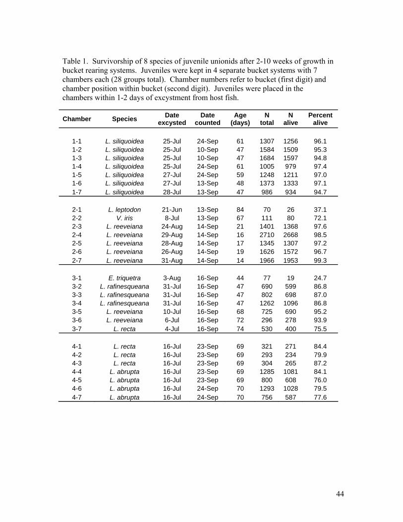
44
Table 1. Survivorship of 8 species of juvenile unionids after 2-10 weeks of growth in bucket rearing systems. Juveniles were kept in 4 separate bucket systems with 7 chambers each (28 groups total). Chamber numbers refer to bucket (first digit) and chamber position within bucket (second digit). Juveniles were placed in the chambers within 1-2 days of excystment from host fish.
Chamber Species Date excysted
Date counted
Age (days)
N total
N alive
Percent alive
1-1 L. siliquoidea 25-Jul 24-Sep 61 1307 1256 96.1 1-2 L. siliquoidea 25-Jul 10-Sep 47 1584 1509 95.3 1-3 L. siliquoidea 25-Jul 10-Sep 47 1684 1597 94.8 1-4 L. siliquoidea 25-Jul 24-Sep 61 1005 979 97.4 1-5 L. siliquoidea 27-Jul 24-Sep 59 1248 1211 97.0 1-6 L. siliquoidea 27-Jul 13-Sep 48 1373 1333 97.1 1-7 L. siliquoidea 28-Jul 13-Sep 47 986 934 94.7
2-1 L. leptodon 21-Jun 13-Sep 84 70 26 37.1 2-2 V. iris 8-Jul 13-Sep 67 111 80 72.1 2-3 L. reeveiana 24-Aug 14-Sep 21 1401 1368 97.6 2-4 L. reeveiana 29-Aug 14-Sep 16 2710 2668 98.5 2-5 L. reeveiana 28-Aug 14-Sep 17 1345 1307 97.2 2-6 L. reeveiana 26-Aug 14-Sep 19 1626 1572 96.7 2-7 L. reeveiana 31-Aug 14-Sep 14 1966 1953 99.3
3-1 E. triquetra 3-Aug 16-Sep 44 77 19 24.7 3-2 L. rafinesqueana 31-Jul 16-Sep 47 690 599 86.8 3-3 L. rafinesqueana 31-Jul 16-Sep 47 802 698 87.0 3-4 L. rafinesqueana 31-Jul 16-Sep 47 1262 1096 86.8 3-5 L. reeveiana 10-Jul 16-Sep 68 725 690 95.2 3-6 L. reeveiana 6-Jul 16-Sep 72 296 278 93.9 3-7 L. recta 4-Jul 16-Sep 74 530 400 75.5
4-1 L. recta 16-Jul 23-Sep 69 321 271 84.4 4-2 L. recta 16-Jul 23-Sep 69 293 234 79.9 4-3 L. recta 16-Jul 23-Sep 69 304 265 87.2 4-4 L. abrupta 16-Jul 23-Sep 69 1285 1081 84.1 4-5 L. abrupta 16-Jul 23-Sep 69 800 608 76.0 4-6 L. abrupta 16-Jul 24-Sep 70 1293 1028 79.5 4-7 L. abrupta 16-Jul 24-Sep 70 756 587 77.6

45
Table 2. Growth of five species of juvenile unionids in bucket rearing systems. These data are plotted in Figure 5.
Days post-excystment Mean Standard deviation
Coefficient of variation 95% C.I. N measured
Lampsilis abrupta
21 467.5 39.7 8.5 13.1 35 42 561.9 58.9 10.5 20.7 31 57 629.8 75.3 12.0 17.4 72 83 758.8 121.6 16.0 18.8 160
Lampsilis rafinesqueana
13 439.3 125.8 28.6 39.0 40 34 559.2 80.0 14.3 16.3 92 45 631.4 99.7 15.8 23.2 71 68 781.2 118.5 15.2 19.0 150
Lampsilis reeveiana
14 389.9 32.1 8.2 15.3 17 34 683.2 102.2 15.0 27.8 52 48 807.9 116.6 14.4 63.4 13 63 974.7 195.8 20.1 56.0 49 84 1184.0 209.0 17.7 31.1 193
Lampsilis siliquoidea
13 451.5 44.2 9.8 14.2 37 18 504.1 50.2 10.0 13.6 52 39 682.3 81.7 12.0 18.7 73 57 826.8 106.1 12.8 17.6 139 69 911.3 110.7 12.1 15.7 190
Ligumia recta (7-4 cohort)
14 440.2 46.3 10.5 19.3 22 20 493.2 44.0 8.9 17.2 25 33 547.1 53.0 9.7 19.3 29 45 687.8 77.8 11.3 22.2 47 61 816.0 111.7 13.7 32.3 46 78 909.6 168.9 18.6 40.2 68
Ligumia recta (7-16 cohort)
21 525.4 68.7 13.1 23.8 32 42 645.3 74.4 11.5 20.2 52 57 733.7 105.7 14.4 24.6 71 83 915.9 158.7 17.3 23.8 171

46
Table 3. Regressions of mean shell length on age (days post-excystment). Lines are shown in Figure 5. The slope of the regression is the growth rate in micrometers per day. Species Regression equation R2 Lampsilis reeveiana y = 12.24x + 200 0.989 Lampsilis siliquoidea y = 8.21x + 370 0.998 Ligumia recta (7-4 cohort) y = 7.58x + 332 0.987 Ligumia recta (7-16 cohort) y = 6.29x + 386 0.997 Lampsilis rafinesqueana y = 6.24x + 353 0.999 Lampsilis abrupta y = 4.70x + 366 0.999
Figure 1. Recirculating rearing system
Upper bucket
Lower bucket Pump
chambers
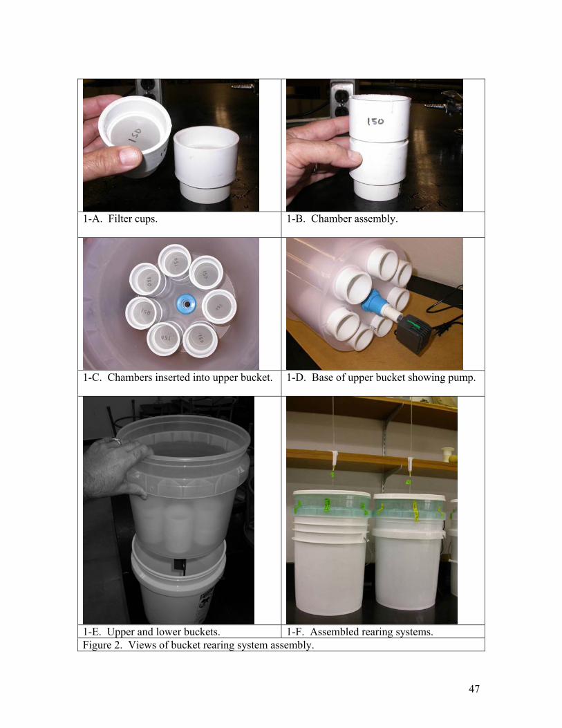
47
1-A. Filter cups.
1-B. Chamber assembly.
1-C. Chambers inserted into upper bucket.
1-D. Base of upper bucket showing pump.
1-E. Upper and lower buckets. 1-F. Assembled rearing systems. Figure 2. Views of bucket rearing system assembly.
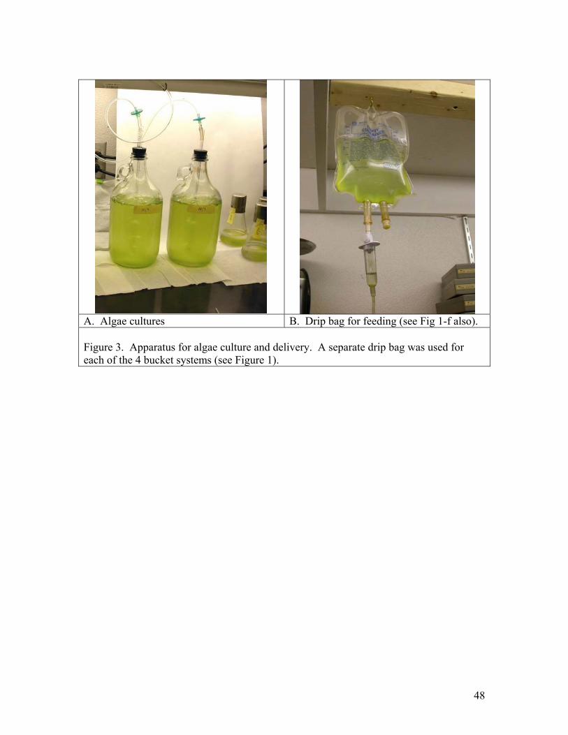
48
A. Algae cultures B. Drip bag for feeding (see Fig 1-f also). Figure 3. Apparatus for algae culture and delivery. A separate drip bag was used for each of the 4 bucket systems (see Figure 1).
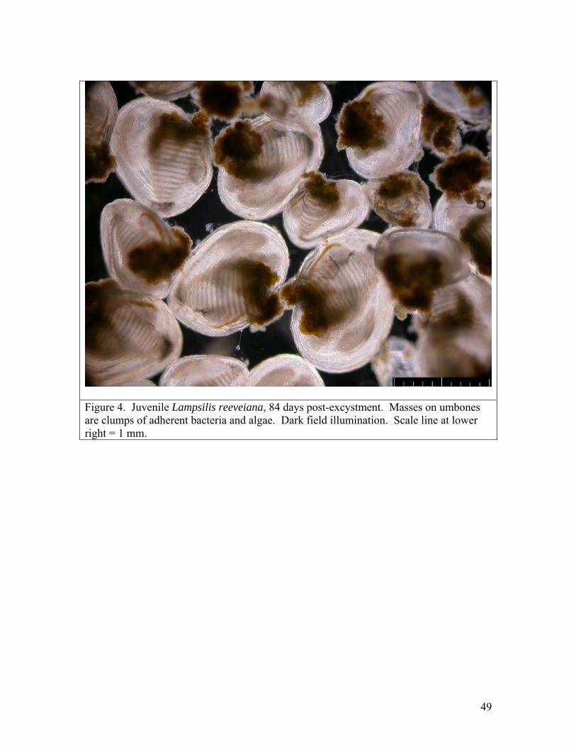
49
Figure 4. Juvenile Lampsilis reeveiana, 84 days post-excystment. Masses on umbones are clumps of adherent bacteria and algae. Dark field illumination. Scale line at lower right = 1 mm.
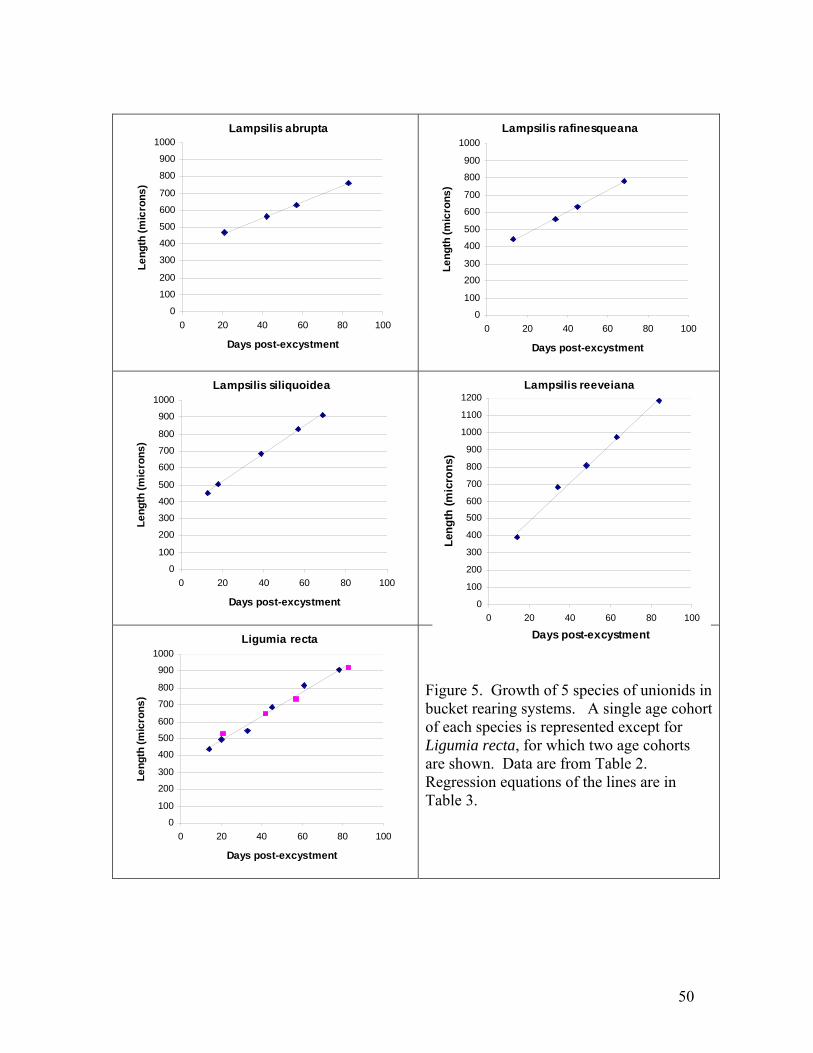
50
Lampsilis abrupta
0
100
200
300
400
500
600
700
800
900
1000
0 20 40 60 80 100
Days post-excystment
Leng
th (m
icro
ns)
Lampsilis rafinesqueana
0
100
200
300
400
500
600
700
800
900
1000
0 20 40 60 80 100
Days post-excystment
Leng
th (m
icro
ns)
Lampsilis siliquoidea
0
100
200
300
400
500
600
700
800
900
1000
0 20 40 60 80 100
Days post-excystment
Leng
th (m
icro
ns)
Ligumia recta
0
100
200
300
400
500
600
700
800
900
1000
0 20 40 60 80 100
Days post-excystment
Leng
th (m
icro
ns)
Figure 5. Growth of 5 species of unionids in bucket rearing systems. A single age cohort of each species is represented except for Ligumia recta, for which two age cohorts are shown. Data are from Table 2. Regression equations of the lines are in Table 3.
Lampsilis reeveiana
0
100
200
300
400
500
600
700
800
900
1000
1100
1200
0 20 40 60 80 100
Days post-excystment
Leng
th (m
icro
ns)
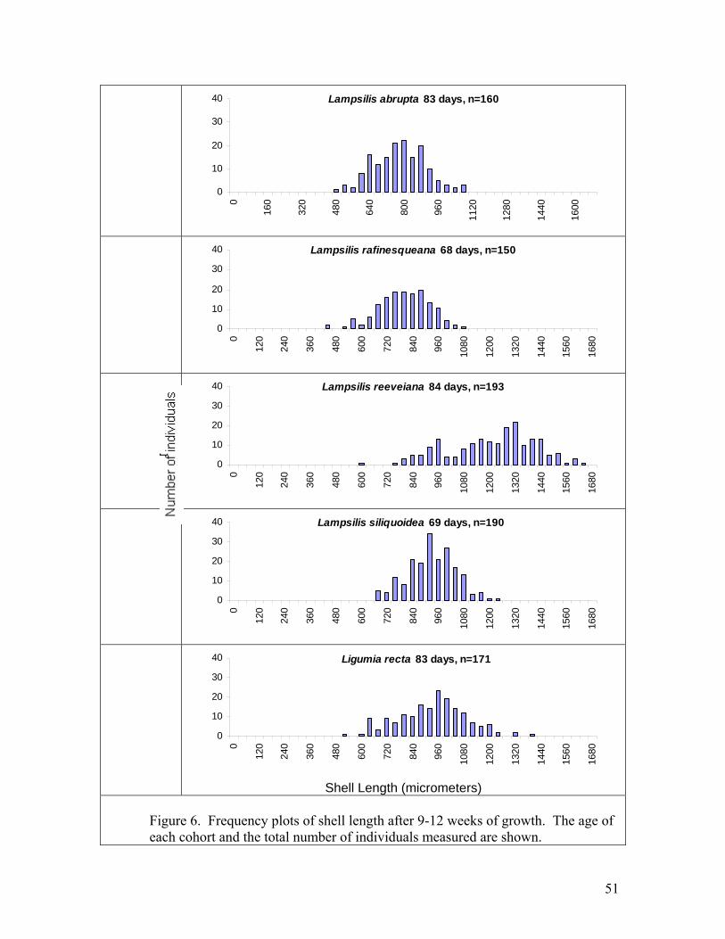
51
Lampsilis abrupta 83 days, n=160
0
10
20
30
40
0
160
320
480
640
800
960
1120
1280
1440
1600
Lampsilis rafinesqueana 68 days, n=150
0
10
20
30
400
120
240
360
480
600
720
840
960
1080
1200
1320
1440
1560
1680
Lampsilis reeveiana 84 days, n=193
0
10
20
30
40
0
120
240
360
480
600
720
840
960
1080
1200
1320
1440
1560
1680
Lampsilis siliquoidea 69 days, n=190
0
10
20
30
40
0
120
240
360
480
600
720
840
960
1080
1200
1320
1440
1560
1680
Ligumia recta 83 days, n=171
0
10
20
30
40
0
120
240
360
480
600
720
840
960
1080
1200
1320
1440
1560
1680
Shell Length (micrometers)
Figure 6. Frequency plots of shell length after 9-12 weeks of growth. The age of each cohort and the total number of individuals measured are shown.
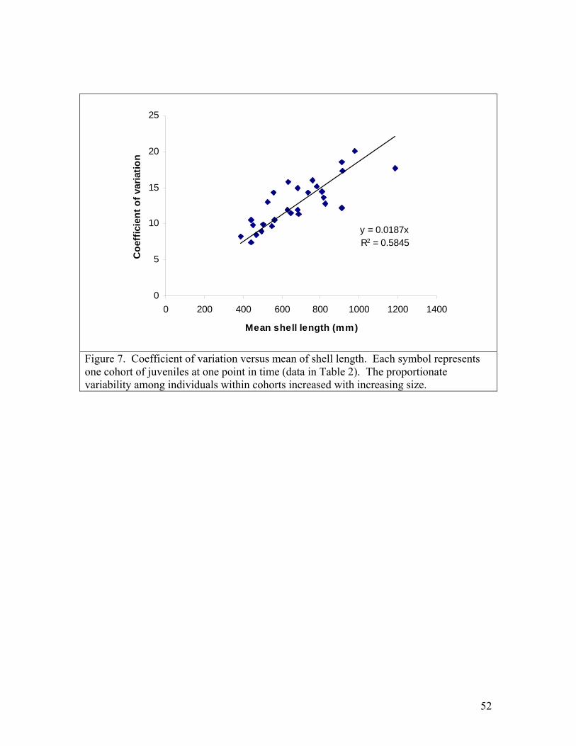
52
y = 0.0187xR2 = 0.5845
0
5
10
15
20
25
0 200 400 600 800 1000 1200 1400
Mean shell length (mm)
Coe
ffic
ient
of v
aria
tion
Figure 7. Coefficient of variation versus mean of shell length. Each symbol represents one cohort of juveniles at one point in time (data in Table 2). The proportionate variability among individuals within cohorts increased with increasing size.

53
Literature Cited Barnhart, M. C. 2002. Culture and restoration of special concern mussel species. Report
to Missouri Department of Conservation, Endangered Species Grant E-1-35. Beck, Kevin M. 2001. Development of an Algal Diet for Rearing Juvenile Freshwater
Mussels (Unionidae). Master’s Thesis, Virginia Polytechnic Institute and State University, Blacksburg, Virginia.
Boudreau, Bernard P. 2001. Life in the diffusive boundary layer. in: Boudreau, Bernard
P. and Bo Barker Jørgensen, eds. The benthic boundary layer: Transport processes and biogeochemistry. Oxford University Press.
Delp, Angela 2002. Rhabdocoel flatworms as predators of juvenile freshwater mussels.
Master of Science, Southwest Missouri State University. Gatenby, C., R. J. Neves, and B. C. Parker. 1996. Influence of sediment and algal food
on cultured juvenile freshwater mussels. Journal of the North American Benthological Society 15(4):597-609.
Gatenby C.M., Parker B.C., Neves R.J. 1997. Growth and survival of juvenile rainbow
mussels, Villosa iris (Bivalvia: Unionidae) reared on algal diets and sediment. Am. Malacol. Bull. 14(1): 57-66.
Hanlon, S. D. 2000. Release of juvenile mussels into a fish hatchery raceway: a
comparison of techniques. Master’s Thesis, Virginia Polytechnic Institute and State University, Blacksburg, Virginia.
Henley, W.F., Zimmerman, L.L. Neves, R.J., Kidd M.R. 2001. Design and evaluation of
recirculating water systems for maintenance and propagation of freshwater mussels. North American Journal of Aquaculture 63, 144-155.
Jørgensen, B. B., and N. P. Revsbech (1985) Diffusive boundary layers and the oxygen
uptake of sediments and detritus. Limnol.Oceanogr., 30, 111-122. Oxford University Press.
Kovitvadhi S., U. Kovitvadhi, P. Sawangwong, A. Thongpan, and J. Machado. In press.
Culturing glochidia and rearing juveniles to adult stage of freshwater pearl mussel, Hyriopsis (Limnoscapha) myersiana. Aquaculture, in press.
Mummert, Andrea K. 2001. Evaluating the Feasibility of Rearing Juvenile Freshwater
Mussels in a Flow-Through Pond System at White Sulphur Springs National Fish Hatchery. Master’s Thesis, Virginia Polytechnic Institute and State University, Blacksburg, Virginia

54
Neves, R.J. and J.C. Widlak. 1987. Habitat ecology of juvenile freshwater mussels (Bivalvia: Unionidae) in a headwater stream in Virginia. American Malacological Bulletin 5(1): 1-7.
NIH. 2004. ImageJ: Image Processing and Analysis in Java. Edition: 1.29. URL:
http://rsb.info.nih.gov/ij/ O’Beirn, F. X., R. J. Neves, and M. B. Steg. 1998. Survival and growth of juvenile
freshwater mussels (Unionidae) in a recirculating aquaculture system. American Malacological Bulletin 14: 165-171.
Rogers, Susan O. 1999. Population biology of the tan riffleshell (Epioblasma florentina
walkeri) and the effects of substratum and light on juvenile mussel propagation. Master’s Thesis, Virginia Polytechnic Institute and State University, Blacksburg, Virginia.
Silverman, H., S. J. Nichols, J. S. Cherry, E. Achberger, J. W. Lynn, and T. H. Dietz.
1997. Clearance of laboratory–cultured bacteria by freshwater bivalves: differences between lentic and lotic unionids. Can. J. Zool. 75: 1857–1866
Steg, Michelle B. 1998. Identification of Host Fish and Experimental Culture of
Juveniles for Selected Freshwater Mussel Species in Virginia. Master’s Thesis, Virginia Polytechnic Institute and State University, Blacksburg, Virginia.
Surber, T. 1912. Identification of the glochidia of freshwater mussels. Report and Special
Papers of the U.S. Bureau of Fisheries. [Issued separately as U.S. Bureau of Fisheries Document 771]. 1912:1-10 + 3 plates.
USFWS 2002. Propagation and Restoration of Higgins' Eye Pearlymussels in the Upper
Mississippi River Basin: Partnership Efforts and Achievements in 2000-2001. U.S. Fish and Wildlife Service, Fishery Resource Office, Onalaska, Wisconsin. URL: http://midwest.fws.gov/mussel/projects.html
Yeager, M. M. & D. S. Cherry, 1994. Feeding and burrowing behaviors of juvenile
rainbow mussels, Villosa iris (Bivalvia: Unionidae). Journal of the North American Benthological Society 13: 217-222.
Zimmerman Lora L. 2003. Propagation of Juvenile Freshwater Mussels (Bivalvia:
Unionidae) and Assessment of Habitat Suitability for Restoration of Mussels in the Clinch River, Virginia. Master’s Thesis, Virginia Polytechnic Institute and State University, Blacksburg, Virginia.
55
Appendix 2- Draft manuscript
Cross-resistance of largemouth bass to glochidia of unionid mussels Benjamin J. Dodd1, M. Christopher Barnhart, Constance L. Rogers-Lowery2, Todd B. Fobian, and Ronald V. Dimock Jr. 2
Department of Biology, Southwest Missouri State University, Springfield, Missouri 65804 Please address correspondence to: M. C. Barnhart Department of Biology, Southwest Missouri State University, Springfield, MO 65804. Email: [email protected] Telephone: 417-836-5166 1Present address: Iowa Department of Natural Resources, Fisheries Research Station, Chariton, Iowa 50049
2Department of Biology, Wake Forest University, Winston-Salem, North Carolina 27109
56
Abstract: We tested whether host fish that acquired resistance to glochidia of one
mussel species were cross-resistant to glochidia of other species. Largemouth bass (Micropterus salmoides) were primed with 4-5 successive infections of glochidia of Lampsilis reeveiana. The percentage of attached glochidia that survived and transformed to the juvenile stage (transformation success) was compared between primed fish and naïve controls. Transformation success of L. reeveiana, Lampsilis abrupta, Villosa iris, and Utterbackia imbecillis was significantly lower on primed fish (37.8%, 43.5%, 67.0%, and 13.2% respectively) than on control fish (89.0%, 89.7%, 90.0%, and 22.2% respectively). Immunoblotting was used to analyze the binding of serum antibodies from primed fish with glochidia proteins. Antibodies bound to glochidia proteins of similar molecular weight from L. reeveiana and L. abrupta. Bound proteins of V. iris differed in molecular weight from those of the Lampsilis species. There was no binding to specific glochidia proteins of U. imbecillis or Strophitus undulatus. Our results indicate that host acquired resistance can extend across mussel genera and subfamilies, and might involve both specific and nonspecific mechanisms. Understanding the specificity of acquired resistance of hosts to glochidia could enhance understanding of the evolutionary and ecological relationships between mussels and their host fishes.
57
INTRODUCTION Freshwater mussels of the family Unionidae have an obligate, parasitic larval stage, the glochidium, which typically attaches to the gills or fins of a host fish. Glochidia that attach to a compatible host species are encysted by migration of host cells. The larvae remain encysted for days to months depending on species and temperature, and transform to the juvenile stage. When development is complete the juveniles leave the host and become benthic suspension-feeders (Arey, 1921; 1932a; Fustish and Millemann, 1978; Waller and Mitchell, 1989).
Mussels are host-specific and are generally compatible with only a limited number of host species (Watters, 1994). Glochidia that attach to incompatible (non-host) species are lost from the host within a few days after attachment because they either fail to be encysted, or are subsequently sloughed from the host before transformation is complete. Incompatibility is thought to be innate, but the mechanisms involved are unknown (Reuling, 1919; Arey, 1932a; Meyers and Millemann, 1977; Meyers et al., 1980; Young and Williams, 1984b; O’Connell and Neves, 1999).
In addition to innate resistance, several studies have shown that compatible hosts acquire resistance to glochidia after one or more infections (Reuling, 1919; Arey, 1924; 1932a; Bauer and Vogel, 1987; Rogers and Dimock, 2003). Compared to naïve hosts, resistant host fish kill and slough a larger number of the attached glochidia, thus reducing the proportion that transform into juveniles (Bauer and Vogel, 1987; Rogers and Dimock, 2003). The underlying mechanisms of acquired resistance of host fish to glochidia are not fully understood. Fish infected with glochidia produce anti-glochidia factors in their serum, presumably antibodies (Meyers et al., 1980; Bauer and Vogel, 1987; O’Connell and Neves, 1999). However, the relationship between serum antibody levels and resistance has not been investigated.
Acquired resistance of fish to one species of parasite can result in resistance to other species (cross-resistance) (Buchmann et al., 1999, Larsen et al., 2002). Cross-resistance to glochidia of different mussel species has been documented but little information is available (Reuling, 1919). Further understanding of acquired resistance and cross-resistance could have practical application in efforts to understand mussel host relationships and to propagate endangered species. Captive propagation of mussels on host fish is increasingly used in efforts to conserve rare species of mussels and is an objective in many federal recovery plans (NNMCC, 1998). Propagating multiple species on the same host fish could be used to reduce labor and costs associated with collecting and maintaining hosts.
The main goals of this study were to determine 1) whether host fish that have acquired resistance to one mussel species are cross-resistant to other mussel species, and 2) whether serum antibodies from fish primed with glochidia from one species of mussel would cross-react with glochidia proteins of different species.
58
MATERIALS & METHODS Fish and mussels Six-month-old largemouth bass were obtained from Chesapeake State Fish Hatchery, Chesapeake, MO. Fish were held in a recirculating aquarium system at 23-24 C in moderately hard synthetic freshwater (SFW) (USEPA 2002). We fed fish 1-2% of their body weight daily (AquaMax pellet feed, Purina Mills), except during infections, when they were fed every other day to reduce feces production. The body mass (g) of each fish was measured following each infection. Gravid mussels were collected from Missouri and North Carolina, USA during 2003 and 2004. We collected Ozark broken-rays mussels (Lampsilis reeveiana brevicula, hereafter referred to as L. reeveiana), rainbow mussels (Villosa iris), and creeper (Strophitus undulatus) from Beaver Creek, Taney County, Missouri (UTM 15, 503804E, 4066693N). Pink muckets (Lampsilis abrupta) were collected from the Meramec River, Jefferson County, Missouri (UTM 15, 699328E, 4260349N). Paper pondshell (Utterbackia imbecillis) were collected from Lake Rockingham, Rockingham County, North Carolina (UTM 17, 625142E, 4026086N). Lampsilis reeveiana and V. iris were maintained at 19-21°C. Utterbackia imbecillis were kept at 10°C and S. undulatus were kept at 6.5°C to slow the release of glochidia. Lampsilis reeveiana, V. iris, and S. undulatus were maintained unfed in SFW. Lampsilis abrupta were kept in a flow-through raceway that received water from a pond at Chesapeake State Fish Hatchery, Missouri. Utterbackia imbecillis were fed once or twice per week with a mixture of algae, and maintained in SFW. Mussels and fish were kept on a 12:12 hour light dark photoperiod, except for L. abrupta and U. imbecillis, which were subject to natural photoperiod. Infection procedure We used glochidia from one female mussel per infection, and obtained glochidia from a different female mussel for each infection. We used a needle and syringe to perforate the marsupial gill and flush the glochidia into a beaker. The glochidia of S. undulatus were freed from the conglutinates (Ortmann, 1911) by spraying them with water through 400 µm mesh nylon fabric. Glochidia were suspended in a known volume of water which was sub sampled for counting. The water was stirred with a large, rubber-bulb syringe while ten 200-µL samples were removed using a volumetric pipette. Each 200 µL sample was placed as a drop on a plastic Petri dish. The glochidia in each drop were counted and classified as open or closed before and after adding NaCl. Open glochidia that closed after NaCl were classified as “viable”. The sample counts were averaged and used to estimate the concentration and the total number of viable glochidia. Fish were infected with glochidia by placing them as a group in a bath containing 2,000 viable glochidia L-1 of SFW. The volume of the suspension was 0.5 L fish-1. Aeration and stirring with a baster were used to keep the glochidia in suspension. After 15 minutes the fish were immediately transferred by dip net into individual 2.75 L tanks.
59
Transformation success
We monitored transformation success of mussel glochidia on individual fish in a recirculating system (AHAB® Aquatic Habitats, Inc. Apopka, FLA) modified for that purpose. Each 2.75 L tank received water continuously from a manifold, and the overflow entered a filter cup with a 125µm nylon screen (Nitex®). Flow rate through each tank was 0.5 L min-1. Before each count (see below) the tanks were “flushed” at 2 L min-1 for approximately 10 min. Filter cups rested upon gutters that returned the water to a sump. The water was conditioned by mechanical, biological, and carbon filtration and received ultraviolet sterilization before returning to the tanks. Temperature was recorded hourly (Optic Stowaway, Onset Computer Corporation) and remained at 23-24°C during the test infections. We counted the glochidia and juveniles present in the filter cups to monitor the timing of drop-off and the number recovered from each fish. We counted at one day after infection and every 2 days thereafter until no more glochidia or juveniles were recovered from any fish for at least 4 d. The contents of each filter cup were rinsed into a finger bowl and transferred to a Bogorov plankton counting tray with a pipette. We used a stereomicroscope at 10.5-40X to count the number of glochidia and juveniles. An individual was classified as a live juvenile if foot activity was observed. Priming and test infections We infected largemouth bass 4-5 times in succession with L. reeveiana glochidia to induce resistance (“priming”). Primed fish and naïve control fish (never exposed to glochidia) were then infected with each batch of test glochidia. The controls allowed us to distinguish differences due to priming from differences in the viability of glochidia from individual mussels. For each fish, we determined infection intensity (the total number of glochidia and juveniles recovered from the fish), transformation success (the percent of recovered individuals that were live juveniles), and mean duration of successful parasitism (i.e. days from infection to excystment of live juveniles). Two-tailed T-tests were used to compare fish body mass and intensity of infection between primed and control fish in each experiment. One-tailed t-tests were used to compare the number of recovered juveniles, transformation success, and the mean duration of successful parasitism between primed and control fish. The results are expressed as mean ± 1 SD unless otherwise noted, and differences are considered significant if p < 0.05. Antibody tests We then used immunoblotting procedures to test whether anti-glochidia factors (presumably antibodies) in fish blood serum would recognize glochidia proteins of L. reeveiana and the other test species. Serum was obtained from a separate group of largemouth bass from the same source and of similar size (~ 13.5 g) that were primed with 3 successive infections of L. reeveiana glochidia. Naïve bass that had never been exposed to mussel glochidia were also used for comparison.
60
Extraction and preparation of sera from fish: Fish were anesthetized with Finquel (MS-222). The caudal peduncle was severed with scissors and blood collected from the caudal vein with a pipette. Blood from different fish of the same treatment was pooled in a centrifuge tube and refrigerated (4°C) for 24 hours. Serum was separated from the blood by centrifugation (Labnet Spectrafuge 16M) at 3000 rpm, for 5 min. The serum was decanted from the blood cells and stored in aliquots at -80°C. The samples were later thawed for immunoblotting and 0.05% sodium azide was added to allow temporary storage at 2-4°C. Detection of bass antibody production: Bass antibodies were isolated using Protein A affinity column chromatography. Briefly, an ImmunoPure ® Immobilized Protein A column (Pierce, Rockford, IL, USA) was equilibrated with binding buffer (10 mM Tris, pH 7.5). Pooled sera from 5 naïve largemouth bass from a different source (Foster’s Lake and Pond Management, Garner, NC) was diluted in binding buffer and applied to the column for 3 h. The Protein A column was washed with binding buffer and the bound largemouth bass antibodies were eluted with elution buffer (0.1M glycine, pH 2.0). Eluted protein fractions were immediately neutralized with 1 M Tris, pH 7.5. The first two 1-mL fractions contained 90% of eluted antibodies and were pooled for subsequent use. Protein concentrations were determined using Bradford’s assay (Bio-Rad). SDS-PAGE was utilized to determine the purity of the eluted largemouth bass antibodies. Samples of the elutant, containing purified antibodies, and whole largemouth bass serum were mixed with Laemmli sample buffer (62.5 mM Tris-HCl, pH 6.8, 25% glycerol, 2% SDS, 0.01% bromophenol blue, 5% B-mercaptoethanol; Bio-Rad) and boiled for 4 minutes. The samples (4 ug total protein for purified antibodies and 10ug total protein for whole serum) were applied to a 4% stacking gel over a 12% resolving gel. Broad range SDS-PAGE molecular weight standards (Bio-Rad) were included. After electrophoresis, the gels were fixed and stained using Coomassie Brilliant Blue R250. Polyclonal mouse antibodies were then used to detect the production of antibodies in primed bass. The polyclonal antibodies were produced in BALB-c mice exposed to purified antibodies from bluegill sunfish (Rogers-Lowery et al., unpublished). To determine whether anti-bluegill antibodies would recognize largemouth bass antibodies, samples of purified largemouth bass antibodies and whole serum were first electrophoresed as described and then electrotransferred to 0.45 um nitrocellulose membrane using a Mini Trans-Blot ® Electrophoretic Transfer Cell (Bio-Rad). Prestained SDS-PAGE molecular weight standards (Bio-Rad) were included on the gels. After blotting, the gels were stained with Coomassie Brilliant Blue to confirm transfer of proteins to membrane. Membranes were blocked overnight with PBS containing 5% non-fat dry milk (PBS-NFDM) and then washed with PBS containing 0.5% Tween-20 (PBS-Tween). The membranes were initially probed with mouse anti-bluegill antibodies diluted 1:1000 in PBS containing 3% bovine serum albumin (PBS-BSA) for 1 h. After thoroughly rinsing in PBS-Tween, membranes were incubated in goat anti-mouse antibodies conjugated to horseradish peroxidase diluted 1:1000 in PBS-BSA. Antibody binding was visualized using 4-chloro-1-napthol and hydrogen peroxide to produce a colored precipitate.
61
Preparation, electrophoresis, and immunoblotting of glochidia extract: Glochidia were removed from gravid mussels of each species as described above and washed several times in SFW. The glochidia were frozen at -4°C until further use. Glochidia proteins were extracted by thawing and refreezing the samples several times and then homogenizing in 0.1 M Tris buffer containing a protease inhibitor cocktail (Sigma) using a Dounce homogenizer. Approximately 500 uL packed volume of glochidia was homogenized in 1500 uL total volume. Bradford’s assay (Bio-Rad) was utilized to determine protein concentrations. Samples of extracted proteins (each 10 ug total protein) were boiled in Laemmli sample buffer (Bio-Rad) for 4 minutes, and separated by SDS-PAGE on a 4% stacking gel over a 12% resolving gel with broad range molecular weight standards (Bio-Rad) included.. Gels were stained with Coomassie Brilliant Blue R250. Immunoblotting techniques were used to determine which glochidia proteins were recognized by antibodies from primed largemouth bass. Glochidia proteins were separated by SDS-PAGE and electrotransferred to 0.45 um nitrocellulose membrane. Prestained SDS-PAGE molecular weight standards (Bio-Rad) were included on the gels. Membranes were blocked overnight with PBS-NFDM. After washing with PBS-Tween, the membranes were initially probed with pooled sera collected from naïve (n=9) or primed (n=14) largemouth bass diluted 1:50 in PBS-BSA for 1 h. After thoroughly rinsing in PBS-Tween, membranes were incubated in mouse anti-bluegill antibodies diluted 1:1000 in PBS-BSA and subsequently incubated in goat anti-mouse antibodies conjugated to horseradish peroxidase. Antibody binding was visualized using 4-chloro-1-napthol and hydrogen peroxide as the substrate. RESULTS Transformation success During the course of the investigation, three different groups of host fish were primed with 4-5 infections of L. reeveiana (Figure 1). The mean intensity of infection (number of glochidia that attached) for each priming infections was 495 ± 149 glochidia per fish. All three groups exhibited similar resistance (1-way ANOVA p=0.5; mean transformation 32% ± 25) in the last priming infection. Primed fish were tested with glochidia of L. reeveiana and 4 other species. The mean body mass of the host fish was 34.6 ± 7.2 g. The mean intensity of the test infections was 655 ± 108 glochidia per fish and did not differ significantly between primed and control fish in any test (2-tailed T-tests). The control transformation success of the lampsiline species (L. reeveiana, L. abrupta, and V. iris) was similar at about 90%, while control transformation of the anondontine species was much lower (U. imbecillis 22%, S. undulatus 1%) (Table 1, Figure 3). Transformation success of S. undulatus on primed fish was similarly low to that of controls (Table 1-Figure 3). Transformation success of all the other species was significantly reduced on primed hosts and averaged about 56 % of control values (Table
62
1-Figure 3). The transformation success of L. reeveiana in the last two priming infections and the test infection were statistically similar (i.e. the priming appeared to have reached a plateau). The majority of glochidia sloughed from control fish were lost during the first day after attachment for all mussel species except S. undulatus (Figure 2). In contrast, primed fish continued to slough glochidia until juveniles were recovered (Figure 2). Both primed and control fish with S. undulatus continued to slough glochidia up until the appearance of transformed juveniles (Figure 2). The mean duration of successful parasitism was significantly reduced for L. reeveiana on primed fish, relative to controls (Table 1-Figure 2). The mean duration of successful parasitism was similar on primed and control fish for the rest of the test species (Table 1-Figure 2). Antibodies SDS-PAGE of largemouth bass antibodies purified on a Protein A column revealed two heavy chain bands with molecular weights of 78-85 kDa and a single light chain band with molecular weight of ~29 kDa. No other bands were present in the gels of purified antibodies. Both heavy chains and light chain were recognized by mouse anti-bluegill IgM polyclonal antiserum. Immunoblot of whole serum from largemouth bass probed with anti-bluegill IgM antiserum revealed a heavy chain, light chain, and a third band with a molecular weight of ~110 kDa, which may represent associated heavy and light chains. Antibodies produced in primed largemouth bass bound antigens in extracts of glochidia from the L. reeveiana and the other test species (Figure 4); however, antibodies from naïve largemouth bass did not (data not shown). Control blots probed with largemouth bass serum and goat anti-mouse antibodies (no mouse anti-bluegill antibodies), mouse anti-bluegill and goat anti-mouse antibodies (no largemouth bass serum), goat anti-mouse antibodies only, and substrate only all produced negative results (data not shown). Antibodies bound several high molecular weight proteins for L. reeveiana, an intensely stained band with molecular weight of 132.5 kDa and several less intense bands (120.1, 85.0, and 78.5 kDa). Only the 132.5 kDa band was recognized for L. abrupta. Additionally, 3 low molecular weight bands with molecular weights of 44.5, 41.2 and 38.1 kDa were recognized for both L. reeveiana and L. abrupta. The antibodies bound a 81.7 kDa protein band of V. iris, which is lighter than the major heavy molecular weight band (132.5 kDa) of the Lampsilis species. There was no evidence in V. iris of the 132.5 kDa protein of the Lampsilis species. However, very faint bands corresponding to the 81.7 kDa protein of V. iris were present for the Lampsilis species. Additionally, antibodies bound 5 low molecular weight bands ranging from 46.0 kDa to 22.0 kDa of V. iris.

63
No distinct bands were produced by serum from primed fish and extract of S. undulatus or U. imbecillis glochidia. However, diffuse staining was observed in the high molecular weight range (~183-109 kDa) for both species. DISCUSSION Glochidia initially attach to the host by clamping to host tissue, mainly the gills and fin margins. Attached glochidia are encysted within hours by migrating cells of the host epithelial and connective tissues. Glochidia on a compatible host species remain encysted for days or weeks and transform into juveniles before excystment occurs. On non-compatible hosts (nonhosts), or on hosts that have acquired immunity, cysts may fail to form, may regress, or the cyst may grow and detach from the underlying epithelium, so that glochidia are “sloughed” before transformation is complete. Glochidia may be sloughed live or may be killed within the cysts before sloughing occurs (Arey, 1921; 1932a; 1932b; Fustish and Millemann, 1978; Waller and Mitchell, 1989). Several studies have reported unusual cyst formation by resistant host fish. Largemouth bass resistant to fat mucket (Lampsilis siliquoidea) produced bulky and irregular shaped cysts around glochidia attached to their gills (Reuling, 1919; Arey, 1932a). Bluegills resistant to U. imbecillis produced cysts on fins more slowly than naïve fish, and the cysts were often thinner or incomplete (Rogers and Dimock, 2003). In the present study, we observed intact cysts containing glochidia that had been shed from resistant fish, as well as unencysted glochidia. Sloughing of cysts appears to result from weakening of the attachment to the underlying tissue (Arey, 1932a). Both live and dead glochidia were recovered from primed and control hosts in our study. We have also observed dead, open glochidia within cysts still attached to the host. Live and dead glochidia have both been recovered in other studies as well (Reuling, 1919; Arey, 1932a; Fustish and Millemann, 1978; Meyers et al., 1980; Bauer, 1987; Bauer and Vogel, 1987; Waller and Mitchell, 1989; Roberts and Barnhart, 1997; O’Connell and Neves, 1999; Rogers and Dimock, 2003). Presumably, elements of the immune system are responsible for death within the cysts (see below). The normal process of excystment of transformed juveniles is not fully understood. The cyst wall can become thinner late in the parasitism (Arey, 1932a, Waller and Mitchell, 1989). However, it is not known whether movements of the juvenile rupture the cyst or whether the cyst tissue simply regresses or disintegrates. Sloughing might involve an acceleration of processes that cause normal excystment. In the present study, duration of successful parasitism of L. reeveiana juveniles was reduced on primed hosts. This change was not evident for the other test species (Table 1). Another study also found shorter duration of successful parasitism on primed host fish (Rogers and Dimock, 2003). In contrast, Bauer and Vogel (1987) reported prolonged encystment of Margaritifera margaritifera on re-infected brown trout (Salmo trutta) when compared to naïve fish. Shortened duration of encystment could limit nutritional exchange, which occurs between the host fish and glochidia (Arey, 1932c; Fisher and Dimock, 2002), and might therefore affect nutritional status and perhaps survivorship of juveniles.
64
Both non-specific and specific (antibody-mediated) mechanisms are involved in acquired resistance and cross-resistance of teleost fish to parasites. Priming with interleukin (IL-1), bacterial polysaccharide (LPS), concanavalin A (Con A), and mannan provide rainbow trout (O. mykiss) partial protection against the parasitic ciliate Ichthyopthirius multifiliis (Buchmann et al., 1999). Complement binds and kills the ectoparasitic platyhelminth, Gyrodactylus derjavini (Buchmann, 1998). Non-specific cytotoxic cells (NCC) in teleosts are capable of killing certain protists (Evans et al., 1998). Cell-mediated mechanisms are involved in acquired immunity of rainbow trout (Oncorhynchus mykiss) to haemoflagellates, Cryptobia salmositica (Mehta and Woo, 2002). Acquired immunity to parasites involving antibodies is well documented in fish (Hines and Spira, 1974; Clark et al., 1987; Cross and Matthews, 1992; Xu et al., 2002). Antibodies to shared antigens of several different protist parasites are involved in cross-resistance to these parasites (Ling et al., 1993; Sin et al., 1992; Goven et al., 1980; 1981; Wolf and Markiw, 1982; Dickerson et al., 1984). Our results indicate that cross-resistance of host fish to different mussel species may be at least partly mediated by antibodies. Antibodies bound to glochidia proteins of 2 of the 3 test species that showed cross-resistance. These proteins were similar to those of the priming glochidia. Antibody-mediated cross-resistance is likely to be correlated with phylogenetic relatedness, because distantly related species may have proteins sufficiently different that they are not recognized by antibodies of primed fish. In this study, similar antigens were evidently present among the lampsiline species (members of the subfamily Lampsilinae; Ortmann, 1919; Parmalee and Bogan, 1998), but not in the less closely related anondontine species U. imbecillis or S. undulatus (members of subfamily Anodontinae) (Figure 4). Control largemouth bass were poor hosts for U. imbecillis and essentially incompatible with S. undulatus, yet no antibody binding with specific proteins of either species was observed (Figure 4). This observation indicates that innate resistance did not involve these antigenic proteins. In spite of the fact that no antibody binding could be shown to U. imbecillis, significant cross-resistance was observed (Table 1). This result indicates that non-specific mechanisms as well as antibodies may be involved in cross-resistance of fish to glochidia. Eosinophilic granulocytes (nonspecific immune cells) may be involved in the cross-resistance to U. imbecillis because these cells congregate around glochidial cysts on immune hosts (Arey, 1932a). There are few previous studies regarding cross-resistance of host fish to unionid mussel glochidia. Reuling (1919) found that largemouth bass that acquired resistance to L. siliquoidea glochidia were cross-resistant to glochidia of a congener, L. cardium and to glochidia of A. ligamentina, also a member of the Lampsilinae. Our results agree with Reuling’s findings. The possibility of cross-resistance of fish to glochidia and unrelated parasites has not been investigated since the early 1900’s. Wilson (1916) found that black sandshell
65
(Ligumia recta) glochidia had a lower attachment success on white crappie (Pomoxis annularis) infected with parasitic copepods (Ergasilus caeruleus) than on uninfected fish. Conversely, copepodid larvae had lower attachment to gills of P. annularis that had L. recta glochidia attached to them. Similar results were found using short-nosed gar (Lepisosteus platostomus), Lernaea copepods, and unspecified mussel glochidia (Wilson, 1917). The mechanism of interference is not known and deserves further attention. In eastern North America, mussel habitats generally support large numbers of species living in close proximity (Vaughn, 1997). In many cases different mussel species may utilize the same species of host fish (Watters, 1994; Haag and Warren, 1997). Given that fish can develop cross-resistance to glochidia, interspecific as well as intraspecific competition for naïve hosts might occur. There is evidence that fish acquire resistance to glochidia in nature (Young and Williams, 1984a; Bauer, 1987; Watters and O’Dee, 1996; Hastie and Young, 2001). Competition for hosts would be favored by prolonged retention by the host of acquired resistance. We have observed that largemouth bass retain measurable acquired resistance for at least 11 months (unpublished data). Competition for immunologically naïve host fish could be a factor in niche partitioning and perhaps in the evolutionary diversification of Unionidae. Many mussels in the subfamily Lampsilinae display mantle lures that attract host fish. In the Mobile Basin, the Alabama rainbow (Villosa nebulosa) displays a white lure primarily at night, while the sympatric southern rainbow (Villosa vibex) has a black lure and displays mostly during the day. Such differences in lures and in luring behavior might permit coexistence of species because they minimize immunological competition for hosts (Haag and Warren, 2000). Graf (1997) presented a model by which shifts in host utilization could promote sympatric speciation of unionids. In Graf’s model, individuals compatible with a new host might be distributed into different habitat because of habitat preferences of the new host. Non-random mating resulting from host-linked habitat use might lead to sympatric speciation. If acquired immunity of a host population to mussels were extensive, mussel variants that were compatible with a different host species, one less likely to encounter glochidia and acquire immunity, might be favored by natural selection. A new host with different habitat preferences from the parental mussel species might also be less likely to have acquired immunity to that species. Cross resistance of fish to mussel glochidia may have practical implications for efforts to propagate endangered mussel species. It appears that propagating either the same or different mussel species consecutively on the same host fish would reduce transformation success. Another question, which has apparently not been investigated, is whether the immune response of the host might affect the viability of those juveniles that do successfully transform. The shortened duration of successful parasitism observed in primed fish could affect the nutritional status of the juveniles. Study is also needed to establish whether infection intensity affects transformation success, duration of parasitism, or juvenile viability. Hypothetically, higher infection intensity could result in a stronger immune response, perhaps affecting the success of glochidia even during the
66
first infection of a host. Establishing the optimum intensity of infection might improve the efficiency of captive propagation. ACKNOWLEDGEMENTS Funding was provided by the United States Fish & Wildlife Service, Missouri Department of Conservation, Southwest Missouri State University, and Wake Forest University. We thank Dan Beckman, Elizabeth Bowen, Raymond Kuhn, John Heywood, Janice Horton, Janice Moll, Richard Myers, Joe Newton, and Clifford Starliper for their advice and assistance throughout this study. We greatly appreciate the assistance of Robert Brown, Christian Hutson, and Bri Kaiser in the lab and field. We would also like to thank Andy Cornforth and Dennis Whelan at Chesapeake State Fish Hatchery for providing fish for this study. LITERATURE CITED Arey, L. B. 1921. An experimental study on glochidia and the factors underlying
encystment. Journal of Experimental Zoology 33:463-499. --------. 1924. Observations on an acquired immunity to a metazoan parasite. Journal of
Experimental Zoology 38:377-381. --------. 1932a. A microscopical study of glochidial immunity. Journal of Morphology
53:367-379. --------. 1932b. The formation and structure of the glochidial cyst. Biological Bulletin
62:213-221. --------. 1932c. The nutrition of glochidia during metamorphosis. Journal of Morphology
53:201-221. Barnhart, M. C., and A. D. Roberts. 1997. Reproduction and fish hosts of unionids from
the Ozark Uplifts. In: Conservation and Management of Freshwater Mussels II. Proc. UMRCC Symp. 16-18 October 1995, St. Louis, Missouri. Cummings, K. S., Buchanan, A. C., Mayer, C. A., and Naimo, T. J., eds., pg 16-20. Upper Mississippi River Conservation Committee, Rock Island, Illinois.
Bauer, G., and C. Vogel. 1987. The parasitic stage of the freshwater pearl mussel
(Margaritifera margaritifera L.) I. Host response to glochidiosis. Archiv fuer Hydrobiologie Supplement 76:393-402.
Buchmann, K. 1998. Binding and lethal effect of complement from Oncorhynchus
mykiss on Gyrodactylus derjavini (Platyhelminthes, Monogenea). Diseases of Aquatic Organisms 32:195-200.
67
Buchmann, K., T. Lindenstrom, and J. Sigh. 1999. Partial cross protection against Ichthyophthirius multifiliis in Gyrodactylus derjavini immunized rainbow trout. Journal of Helminthology 73:189-195.
Clark, T. G., H. W. Dickerson, J. G. Gratzek, and R. C. Findly. 1987. In vitro response
of Ichthyophthirius multifiliis to sera from immune channel catfish. Journal of Fish Biology 31:203-208.
Cross, M. L. and R. A. Matthews. 1992. Ichthyophthiriasis in carp Cyprinus carpio L.:
fate of parasites in immunized fish. Journal of Fish Diseases 15:497-505. Dickerson, H. W., J. Brown, D. L. Dawe, and J. B. Gratzek. 1984. Tetrahymena
pyriformis as a protective antigen against Ichthyophthirius multifiliis infection: comparison between isolates and ciliary preparations. Journal of Fish Biology 24:523-528.
Evans, D. L., J. H. Leary, Jr., P. Nadella, and L. Jaso-Freidmann. 1998. Evidence for
antigen recognition by nonspecific cytotoxic cells: initiation of 3H-thymidine uptake following stimulation by a protozoan parasite and homologous cognate synthetic peptide. Developmental and Comparative Immunology 22:161-172.
Fisher, G. R., and R. V., Dimock, Jr. 2002. Ultrastructure of the mushroom body:
digestion during metamorphosis of Utterbackia imbecillis (Bivalvia: Unionidae). Invertebrate Biology 121(2):126-135.
Fustish, C. A., and R. E. Millemann. 1978. Glochidiosis of salmonid fishes. II.
Comparison of tissue response of coho and chinook salmon to experimental infection with Margaritifera margaritifera (L.) (Pelecypoda: Margaritanidae). Journal of Parasitology 64(1):155-157.
Goven B. A., D. L. Dawe, and J. B. Gratzek. 1980. Protection of channel catfish,
Ictalurus punctatus Rafinesque, against Ichthyophthirius multifiliis Fouquet by immunization. Journal of Fish Biology 17:311-316.
Goven B. A., D. L. Dawe, and J. B. Gratzek. 1981. In vitro demonstration of serological
cross-reactivity between Ichthyophthirius multifiliis Fouquet and Tetrahymena pyriformis (Lwoff). Developmental and Comparative Immunology 5:283-289.
Graf, D. L. 1997. Sympatric speciation of freshwater mussels (Bivalvia: Unionoidea): a
model. American Malacological Bulletin 14(1):35-40. Haag, W. R. and M. L. Warren, Jr. 1997. Host fishes and reproductive biology of 6
freshwater mussel species from the Mobile Basin, USA. Journal of the North American Benthological Society. 16:576-585.

68
Haag, W. R. and M. L. Warren, Jr. 2000. Effects of light and presence of fish on lure display and larval release behaviors in two species of freshwater mussels. Animal Behaviour 60:879-886.
Hastie, L. C., and M. R. Young. 2001. Freshwater pearl mussel (Margaritifera
margaritifera) glochidiosis in wild and farmed salmonid stocks in Scotland. Hydrobiologia 445:109-119.
Hines, R. S. and D. T. Spira. 1974. Ichthyophthiriasis in the mirror carp Cyprinus carpio
(L.) V. Acquired immunity. Journal of Fish Biology 6:373-378. Larsen, A. H., J. Bresciani, and K. Buchmann. 2002. Interactions between ecto- and
endo-parasites in trout Salmo trutta. Veterinary Parasitology 103:167-173. Ling, K. H., Y. M. Sin, and T. J. Lam. 1993. Protection of goldfish against some common
ectoparasite protozoans using Ichthyophthirius multifiliis and Tetrahymena for vaccination. Aquaculture 116:303-314.
Mehta, M., and P. T. K. Woo. 2002. Acquired cell-mediated protection in rainbow trout,
Oncorhynchus mykiss, against the haemoflagellate, Cryptobia salmositica. Parasitology Research 88: 956-962.
Meyers, T. R., and R. E. Millemann. 1977. Glochidiosis of salmonid fishes. I.
Comparative susceptibility to experimental infection with Margaritifera margaritifera (L.) (Pelecypoda: Margaritanidae). Journal of Parasitology 63(4): 728-733.
Meyers T. R., R. E. Millemann, and C. A. Fustish. 1980. Glochidiosis of salmonid
fishes. IV. Humoral and tissue responses of coho and Chinook salmon to experimental infection with Margaritifera margaritifera (L.) (Pelecypoda: Margaritanidae). Journal of Parasitology 66(2):274-281.
NNMCC (National Native Mussel Conservation Committee). 1998. National strategy for
the conservation of native freshwater mussels. Journal of Shellfish Research 17(5):1419-1428.
O’Connell, M. T., and R. J. Neves. 1999. Evidence of immunological responses by a
host fish (Ambioplites rupestris) and two non-host fishes (Cyprinus carpio and (Carassius auratus) to glochidia of a freshwater mussel (Villosa iris). Journal of Freshwater Ecology 14(1):71-78.
Ortmann, A. E. 1911. A monograph of the naiades of Pennsylvania. Parts I and II.
Memoirs of the Carnegie Museum 4(6):279-347 + 4 plates.
69
Ortmann, A. E. 1919. A mongraph of the naiades of Pennsylvania. Part III: Systematic account of the genera and species. Memoirs of the Carnegie Museum 8(1):xvi 384p + 21 plates.
Parmalee, P. W. and A. E. Bogan. 1998. The freshwater mussels of Tennessee.
University of Tennessee Press. 328p. Reuling, F. H. 1919. Acquired immunity to an animal parasite. Journal of Infectious
Diseases 24:337-346. Rogers, C. L., and R. V. Dimock, Jr. 2003. Acquired resistance of bluegill sunfish
Lepomis macrochirus to glochidia larvae of the freshwater mussel Utterbackia imbecillis (Bivalvia: Unionidae) following multiple infections. Journal of Parasitology 89(1):51-56.
Sin, Y. M., K. H. Ling, and T. J. Lam. 1992. Protection against velvet disease in goldfish
recovered from ichthyophthiriasis. Aquaculture 102:187-191. USEPA. 2002. Short-term Methods for Estimating the Chronic Toxicity of Effluents and
Receiving Waters to Freshwater Organisms, 4th ed. van Snik Gray, E., W. A. Lellis, J. C. Cole, and C. S. Johnson. 2002. Host identification
for Strophitus undulatus (Bivalvia: Unionidae), the Creeper, in the Upper Susquehanna River Basin, Pennsylvania. American Midland Naturalist 147:153-161.
Vaughn, C. C. 1997. Regional patterns of mussel species distributions in North American
rivers. Ecography 20:107-115. Waller, D. L., and L. G. Mitchell. 1989. Gill tissue reactions in walleye Stizostedion
vitreum vitreum and common carp Cyprinus carpio to glochidia of the freshwater mussel Lampsilis radiata siliquoidea. Diseases of Aquatic Organisms 6:81-87.
Watters, G. T. 1994. An annotated bibliography of the reproduction and propagation of
the Unionoidea (Primarily of North America). Ohio Biological Survey Miscellaneous Contributions No.1. 158 pp.
Watters, G. T. and S.H. O'Dee. 1996. Shedding of untransformed glochidia by fishes
parasitized by Lampsilis fasciola Rafinesque, 1820 (Mollusca: Bivalvia: Unionidae): evidence of acquired immunity in the field? Journal of Freshwater Ecology 11:383-389.
Williams, J. D., M. L. Warren, K. S. Cummings, J. L. Harris, and R. J. Neves. 1993.
Conservation status of freshwater mussels of the United States and Canada. Fisheries 18(9):6-22.

70
Wilson, C. B. 1917. The economic relations, anatomy, and life history of the genus Lernaea. Bulletin of the United State Bureau of Fisheries 35:165-198 + 10 plates.
Wilson, C. B. 1916. Copepod parasites of fresh-water fishes and their economic relations
to mussel glochidia. Bulletin of the United States Bureau of Fisheries (1914) 34:331-374 + 15 plates.
Wolf, K., and M. E. Markiw. 1982. Ichthyophthiriasis: Immersion immunization of
rainbow trout (Salmo gairdneri) using Tetrahymena thermophila as a protective immunogen. Canadian Journal of Fisheries and Aquatic Science 39:1722-1725.
Xu, D.H., P.H. Klesius, and R.A. Shelby. 2002. Cutaneous antibodies in the excised skin
from channel catfish, Ictalurus punctatus Rafinesque, immune to Ichthyophthirius multifiliis. Journal of Fish Diseases 25:45-52.
Young, M., and J. Williams. 1984a. The reproductive biology of the freshwater pearl
mussel Margaritifera margaritifera (LINN.) in Scotland I. Field Studies. Archiv fuer Hydrobiologie 99(4):405-422.
Young, M., and J. Williams. 1984b. The reproductive biology of the freshwater pearl
mussel Margaritifera margaritifera (LINN.) in Scotland II. Laboratory Studies. Archiv fuer Hydrobiologie 100(1):29-43.
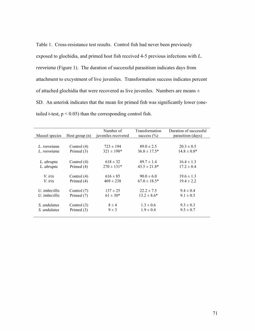
71
Table 1. Cross-resistance test results. Control fish had never been previously
exposed to glochidia, and primed host fish received 4-5 previous infections with L.
reeveiana (Figure 1). The duration of successful parasitism indicates days from
attachment to excystment of live juveniles. Transformation success indicates percent
of attached glochidia that were recovered as live juveniles. Numbers are means ±
SD. An asterisk indicates that the mean for primed fish was significantly lower (one-
tailed t-test, p < 0.05) than the corresponding control fish.
Mussel species Host group (n) Number of
juveniles recovered Transformation
success (%) Duration of successful
parasitism (days)
L. reeveiana Control (4) 723 ± 194 89.0 ± 2.5 20.3 ± 0.5 L. reeveiana Primed (3) 321 ± 198* 36.8 ± 17.5* 14.8 ± 0.8*
L. abrupta Control (4) 618 ± 32 89.7 ± 1.4 16.4 ± 1.3 L. abrupta Primed (4) 270 ± 131* 43.5 ± 21.8* 17.2 ± 0.4
V. iris Control (4) 616 ± 85 90.0 ± 6.0 19.6 ± 1.3 V. iris Primed (4) 469 ± 238 67.0 ± 18.5* 19.4 ± 2.2
U. imbecillis Control (7) 137 ± 25 22.2 ± 7.5 9.4 ± 0.4 U. imbecillis Primed (7) 61 ± 30* 13.2 ± 8.6* 9.1 ± 0.5
S. undulatus Control (3) 8 ± 4 1.3 ± 0.6 9.3 ± 0.3 S. undulatus Primed (3) 9 ± 3 1.9 ± 0.4 9.5 ± 0.7
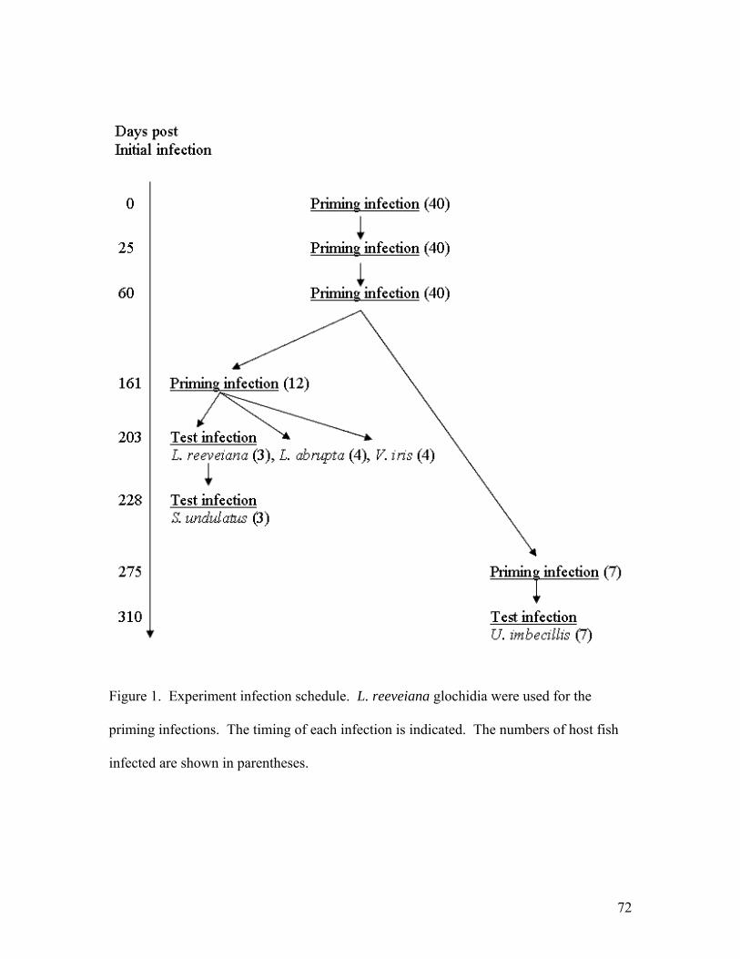
72
Figure 1. Experiment infection schedule. L. reeveiana glochidia were used for the
priming infections. The timing of each infection is indicated. The numbers of host fish
infected are shown in parentheses.
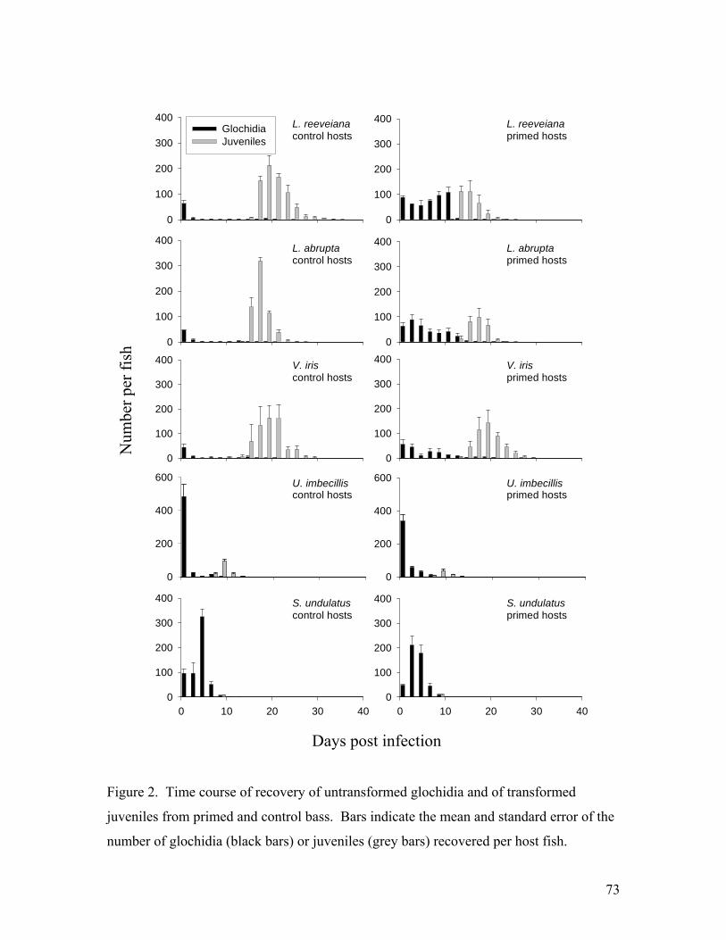
73
0 10 20 30 40
0
100
200
300
400
0
100
200
300
400
0
100
200
300
400
0
100
200
300
400
0
100
200
300
400Glochidia Juveniles
0
100
200
300
400
0 10 20 30 400
100
200
300
400
0
100
200
300
400
L. reeveianacontrol hosts
L. reeveianaprimed hosts
L. abruptacontrol hosts
V. iriscontrol hosts
L. abruptaprimed hosts
V. irisprimed hosts
S. undulatuscontrol hosts
S. undulatusprimed hosts
0
200
400
600
0
200
400
600 U. imbecilliscontrol hosts
U. imbecillisprimed hosts
Figure 2. Time course of recovery of untransformed glochidia and of transformed
juveniles from primed and control bass. Bars indicate the mean and standard error of the
number of glochidia (black bars) or juveniles (grey bars) recovered per host fish.
Days post infection
Num
ber p
er fi
sh
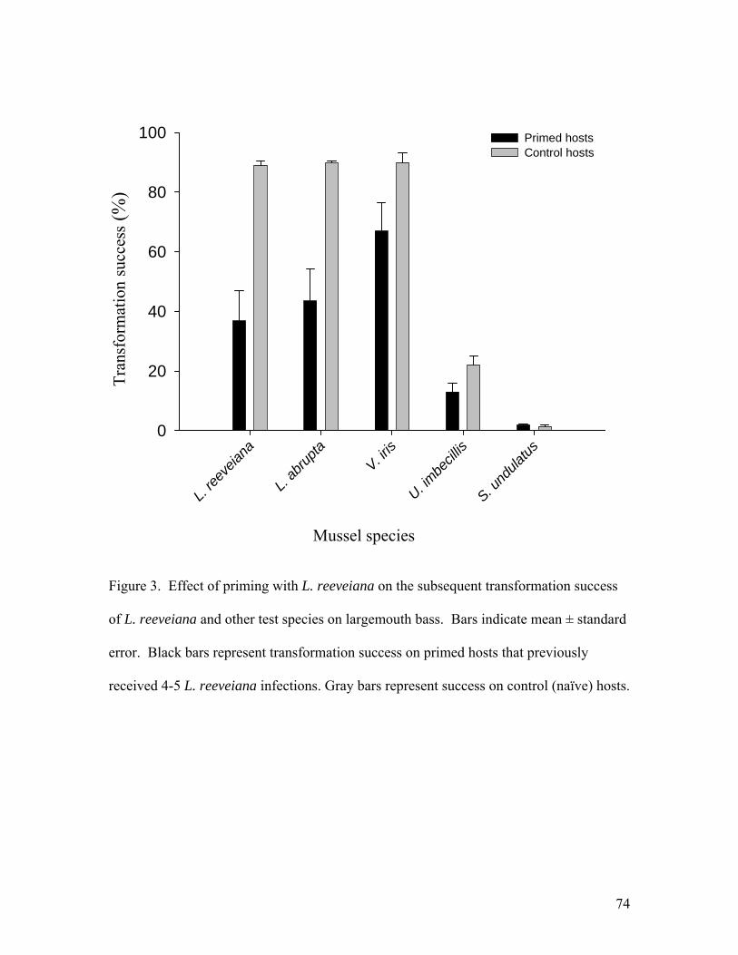
74
L. ree
veian
a
L. ab
rupta
V. iris
U. imbe
cillis
S. und
ulatus
0
20
40
60
80
100 Primed hostsControl hosts
Figure 3. Effect of priming with L. reeveiana on the subsequent transformation success
of L. reeveiana and other test species on largemouth bass. Bars indicate mean ± standard
error. Black bars represent transformation success on primed hosts that previously
received 4-5 L. reeveiana infections. Gray bars represent success on control (naïve) hosts.
Mussel species
Tra
nsfo
rmat
ion
succ
ess (
%)

75
Figure 4. Western Blot of glochidia antigens recognized by serum antibodies of
largemouth bass primed with L. reeveiana glochidia. The lanes are Molecular Weight
standards (MW), L. reeveiana proteins (1), recognized L. reeveiana proteins (2), L.
abrupta proteins (3), recognized L. abrupta proteins (4), V. iris proteins (5), recognized
V. iris proteins (6), S. undulatus proteins (7), recognized S. undulatus proteins (8), U.
imbecillis proteins (9), and recognized U. imbecillis proteins (10).