
progression and prognosis of lung adenocarcinoma Increased ...
22
Page 1/22 Increased STIP1 and Hsp90 correlate with progression and prognosis of lung adenocarcinoma Youwen Zhang Aliated Hospital of Jining Medical University Junye Wang Aliated Hospital of Jining Medical University Shucheng Ye Aliated Hospital of Jining Medical University Maoqing Guo Aliated Hospital of Jining Medical University Shenghua Jiang Aliated Hospital of Jining Medical University Wei Li ( [email protected] ) Aliated Hospital of Jining Medical University Research article Keywords: Lung adenocarcinoma, LAC, stress-inducible phosphoprotein 1, STIP1, heat shock protein 90, Hsp90 Posted Date: August 30th, 2019 DOI: https://doi.org/10.21203/rs.2.13680/v1 License: This work is licensed under a Creative Commons Attribution 4.0 International License. Read Full License
Transcript of progression and prognosis of lung adenocarcinoma Increased ...

Page 1/22
Increased STIP1 and Hsp90 correlate withprogression and prognosis of lung adenocarcinomaYouwen Zhang
A�liated Hospital of Jining Medical UniversityJunye Wang
A�liated Hospital of Jining Medical UniversityShucheng Ye
A�liated Hospital of Jining Medical UniversityMaoqing Guo
A�liated Hospital of Jining Medical UniversityShenghua Jiang
A�liated Hospital of Jining Medical UniversityWei Li ( [email protected] )
A�liated Hospital of Jining Medical University
Research article
Keywords: Lung adenocarcinoma, LAC, stress-inducible phosphoprotein 1, STIP1, heat shock protein 90,Hsp90
Posted Date: August 30th, 2019
DOI: https://doi.org/10.21203/rs.2.13680/v1
License: This work is licensed under a Creative Commons Attribution 4.0 International License. Read Full License

Page 2/22
AbstractBackground: Stress-inducible phosphoprotein 1 (STIP1) and heat shock protein 90 (Hsp90) have beenfound to be correlated with malignant tumors. The aim of this investigation was to study the relationshipbetween their expressions and lung adenocarcinoma (LAC). Methods: The expressions of STIP1 andHsp90 in LAC cells and tissues were tested by immunohistochemistry and western blot; the correlationbetween their expressions and clinicopathological parameters of LAC was analyzed by survival analysisand multiple regression analysis. Results: Expressions of STIP1 and Hsp90 were higher in A549 cells andLAC tissues than that in 16 human bronchial epithelial cells (16HBE cells) (P < 0.05) and adjacent normallung tissues (P < 0.05). The expression of STIP1 and Hsp90 in LAC showed a strong positive correlation(P < 0.05) and signi�cantly correlated with lymph node metastasis (P < 0.05), advanced clinical stage (P <0.05) and shorter survival (P < 0.05) of LAC. Conclusions: Increased expressions of STIP1 and Hsp90were closely related to malignant biological behavior of LAC, suggesting that they could be used aspotential biomarkers and prognostic indicators for LAC.
IntroductionLung cancer is one of the most malignant tumors with the fastest growth in morbidity and mortality andthe greatest threat to population health and life [1]. In the past 20 years, the incidence of lung cancer hasbeen reported to increase signi�cantly in China [2] and the incidence of lung adenocarcinoma (LAC) hasexceeded that of lung squamous cell carcinoma (LSCC) [2]. Clinical studies show that LAC is closelyrelated to speci�c carcinogenic driver genes such as epidermal growth factor receptor (EGFR),anaplasticlymphoma kinase (ALK), mesenchymal to epithelial transition factor (MET), rearranged duringtransfection (RET), and proto-oncogene tyrosine-protein kinase ROS (ROS1). Structural changes(mutation, overexpression or rearrangement) of these speci�c oncogenes can trigger and promote thedevelopment of LAC cells [3–5]. So far, it has been known that lung cancer patients associated withdriving genetic mutations have reached 62% of the total number of lung cancers [6, 7]. In any case, it isbelieved that there are more unknown driving genes associated with the development of LAC that havenot been discovered [8].
Although the human genome map has been drawn, the function of the protein encoded by the gene isextremely complicated, so the study of protein function has become the focus of the post-genome era [9].We previously screened a differential protein stress-inducible phosphoprotein 1 (STIP1) based on isobarictags for relative and absolute quanti�cation (iTRAQ) in combination with two-dimensional liquidchromatography/tandem mass spectrometry (2D-LC-MS/MS) and found that its expression wassigni�cantly higher in LAC cells than in 16 human bronchial epithelial cells (16HBE cells). The STIP1 geneis located in the human chromosome 11q13 coding region and consists of 14 exons with a proteinmolecular weight of 63 kDa, which is located near many cancer-related genes [10]. It is reported that up-regulated STIP1 promotes the growth, colony formation and migration of liver cancer cells [11] and highSTIP1 expression is correlated with advanced T stage of colorectal cancer [12]. Heat shock protein 90(Hsp90) plays an important role in protein folding, subunit assembly, and regulation of cell growth and
Page 3/22
survival [13, 14] and has been found to be highly expressed in some solid tumors, which is closely relatedto tumor progression and prognosis [15, 16]. The purpose of this study was to quantify the difference inexpressions of STIP1 and Hsp90 in LAC cells and tissues, and to verify their associations with LACclinical parameters.
Material And Methods
Cell cultureHuman LAC cell line A549 purchased from Life Sciences Cell Bank of Chinese Academy of Sciences.16HBE cells were purchased from the cell bank of the state key laboratory of environment and genetics,Xi’an Jiaotong University. The 16HBE cells were cultured in RPMI 1640 medium with 10% FBS, 100 g/mLpenicillin and 100 g/mL streptomycin. The A549 cells were cultured in culture �asks (culture mediumpreparation: 10% fetal bovine serum + 1% double antibody + 89% DMEM high glucose medium). Theincubator environment was set to 37 °C and with a 5% of CO2 content. When the cell density at thebottom of the bottle reached 80%, the cells were passaged. The cells in logarithmic growth wereharvested for total protein extraction.
Patients selectionFrom January 2013 to December 2015, LAC samples (LAC tissue and adjacent normal tissue which is atleast 3 cm away from the cancer tissue) from a total of 72 patients undergoing surgery were collected(A�liated Hospital of Jining Medical University, Jining, China and Gansu Provincial Hospital, Lanzhou,China). Important clinical data for patients were extracted (gender, age, smoking status, celldifferentiation, lymph node metastasis and clinical staging) (Table 1).. Survival data were de�ned as thetime from the time of surgery until the time of death or loss of follow-up. The study was approved by theInstitute’s Ethics Committee of A�liated Hospital of Jining Medical University, Jining, China. The patient’sconsent and informed consent were obtained before the collection of tissue samples from the patient.
Western blotThe cultured A549 cells were lysed with RIPA lysate to extract total cellular protein. The BCA method wasused to determine the protein concentration of each sample. After the boiling water bath denaturationtreatment, an equal amount of total protein was subjected to polyacrylamide gel electrophoresis. Afterelectrophoresis, the total protein was transferred to a PVDF membrane. After the PVDF membrane wasblocked with a 10% skim milk powder solution, the corresponding primary antibody was added andincubated at 4 °C overnight. The PVDF membrane was rinsed and then incubated with a secondaryantibody at 37 °C for 45 min. The PVDF membrane was illuminated by ECL method and was exposed in adark room with X-ray �lm.
Page 4/22
Tissue microarray (TMA) constructionThe tissue sections were observed and labeled under light microscope. Tissue array instrument was usedto locate and drill a hole with a diameter of 1mm on the recipient wax block. A tissue column with adiameter of 1 mm was drilled from the tumor tissue and inserted into the hole of the recipient wax block.The prepared tissue chip wax block was incubated in a 37 °C incubator for 1 h. After cooling at roomtemperature, it was cut into 4 μm thick tissue sections and placed in an oven at 60 °C overnight. Thesections were subjected to HE staining to see if the samples were accurate, and the quali�ed sectionswere kept at –4 °C for use.
Immunohistochemistry (IHC)The expressions of STIP1 (dilution concentration of mouse anti-human STIP1 polyclonal antibody, 1:500;Abcam) and Hsp90 (dilution concentration of mouse anti-human Hsp90 polyclonal antibody, 1:300;Boster) in tissues were detected by immunohistochemical staining. A positive expression of STIP1 inthyroid cancer was used as a positive control for STIP1 and a positive expression of Hsp90 in breastcancer was used as a positive control for Hsp90. PBS buffer instead of primary antibody was used as anegative control. The expressions of STIP1 and Hsp90 were stained as tan and brownish yellow, whichwere distributed in the cytoplasm of LAC cells. The semi-quanti�cation analysis of IHC were performed bythe following criteria: 0 for tumor cells without staining; medium staining was 1 point; more than 75% oftumor cells were stained for 2 points; more than 1 point is considered a positive expression [17].
Statistical analysisSPSS 20.0 software was used for statistical analysis. The chi-square test and Fisher’s exact test wereused for comparison of counting data between groups. The one-way ANOVA was used to compare themeasurement data. Correlation analysis between the enumeration data was performed by the cruskal-wallis rank sum test. Kaplan-meier method was used to analyze the survival data. Bilateral test was takenand P < 0.05 was considered statistically signi�cant.
Results
Expression levels of STIP1 and Hsp90 in A549 cells are higher thanthat in 16HBE cellsA Western blot method was used to determine the expression levels of STIP1 and Hsp90 in A549 and16HBE cells, we found that the expressions of STIP1 and Hsp90 in A549 cells were higher than that in16HBE cells (Figure 1A). Quantitative analysis suggested that the expression levels of STIP1 (0.88 ± 0.17)and Hsp90 (0.79 ± 0.27) in the A549 cells were higher than that in 16HBE (0.46 ± 0.13 for STIP1; 0.52 ±0.19 for Hsp90, P< 0.001) (Figure 1B).

Page 5/22
Expression levels of STIP1 and Hsp90 in LAC tissues are higher thanthat in adjacent normal lung tissuesThe expression rate of STIP1 in cancer adjacent normal tissues was 21.9% (16/73) and 57.7% (42/73) inLAC tissues, showing that the expression of STIP1 in LAC was higher than that in non-cancerous lungtissues (P < 0.001) (Table 2; Figure 1C; Figures 2A - D).. The expression of Hsp90 also showed asigni�cant increase in LAC tissues (43/73; 68.9%), compared with cancer adjacent normal tissues (24/73;32.9%) (P = 0.002) (Table 2; Figure 1D; Figures 2E - H)..
Expression of STIP1 is associated with poor differentiation, lymphnode metastasis and advanced stage of LACCompared with well differentiation (6/19; 31.6%), no-lymph node metastasis (3/28; 9.6%) and early stage(16/45; 33.3%), STIP1 was highly expressed in LAC tissues with poor differentiation (24/32; 75%) (P =0.019), lymph node metastasis (39/42; 92.9%) (P < 0.001), and advanced stage (26/28; 92.8%) (P =0.002), suggesting that elevation of STIP1 was associated with malignant biological behavior of LAC(Table 3; Figures 3A - C)..
Expression of Hsp90 is associated with lymph node metastasis andadvanced stage of LACA higher expression of Hsp90 was observed in lymph node metastasis (37/42; 88.1%) (P < 0.001) andadvanced LAC tissues (24/28; 85.7%) (P < 0.001) compared to no-lymph node metastasis (6/31; 19.4%)and early stage of LAC (9/45; 42.2%), suggesting that elevation of Hsp90 was associated withmetastasis and progression of LAC (Table 4; Figures 3D - F)..
Expressions of STIP1 and H sp90 in LAC showed a strong positivecorrelationSTIP1 and Hsp90 had a positive co-expression rate of 38.4% (28/73) and a co-negative rate of 54.8%(40/73). The Pearson correlation coe�cient was 0.86 (P < 0.001) and the Spearman correlationcoe�cient was 0.86 (P < 0.001), which indicated that there was a signi�cant positive correlation betweenthe expressions of STIP1 and Hsp90. The Kappa value was 0.859 (P < 0.001), indicating that there was aconsistent trend between their expressions.
Expressions of STIP1 and Hsp90 are negatively correlated with thesurvival of LAC patients

Page 6/22
As shown in Table 5, compared with patients with negative STIP1 (49.83 ± 1.23 months; 95% CI = 47.43 -52.24), LAC patients with STIP1 expression (29.59 ± 1.07 months; 95% CI = 27.49 - 31.69) had shortersurvival (Log Rank, P < 0.001; Breslow, P < 0.001) (Figures 4A and B).. In addition, survival of LAC patientswith expression of Hsp90 (30.46 ± 1.58 months; 95% CI = 28.03 - 32.85) was shorter than those withoutexpression of Hsp90 (49.34 ± 1.39 months; 95% CI = 46.74 - 51.95) (Log Rank, P < 0.001; Breslow, P <0.001) (Figures 4C and D)..
Multiple regression analysis of clinical parameters and overallsurvival of patients with LACAs shown in Table 6, a total of 8 parameters were included in the analysis, including gender, age, smokingor not, degree of tissue differentiation, lymph node metastasis, TNM staging, and expression of STIP1and Hsp90. Finally, three parameters, TNM staging (P value = 0.004; OR value = 2.991; 95% CI = 1.405—6.366), STIP1 (P value = 0.001; OR value = 9.614; 95% CI = 2.463—37.52) and Hsp90 expression (P value= 0.015; OR value = 3.585; 95% CI = 1.279—10.028), were included in the regression equation. The resultssuggested that TNM staging, STIP1 and Hsp90 expression were risk factors for shortening the survival ofpatients with LAC and the regression equation based on COX regression was H(t) = [h0(t)]e(1.612 X6 + 2.223
X7 + 1.277 X8).
DiscussionLung cancer is one of the major diseases that cause cancer-related death. Among them, LAC is the mostcommon pathological type, and it has gradually increased in recent years [1]. Studies have shown thatsome gene mutations and protein expression abnormalities are closely related to the occurrence anddevelopment of LAC, and some molecular targeted drugs have been used in clinical practice [3, 18, 19].STIP1 is a is an adaptor protein that combines heat shock protein 70 and heat shock protein 90 tomodulate biological effects such as transcription, translation, and protein folding, and promotes thegrowth of tumor cells [12, 20, 21]. According to a recent meta-analysis, the high expression of STIP1 wasrelated to lymph node metastasis, advanced TNM stage and shorter survival of patients compared withthat of tumor tissues with low STIP1 expression, which was expected to be a molecular marker ofmalignancy or provide new ideas for the development of targeted drugs [22]. However, the malignanttumors included in the meta-analysis have only ovarian cancer, liver cancer, gastric cancer, thyroid cancer,and colon cancer. This also re�ects the current lack of research on the expression pattern of STIP1 inLAC. In our study, we investigated the association and prognostic signi�cance of STIP1 and Hsp90 withLAC clinical features by methods of molecular biology and biostatistics.
We �rst used the Western blot method to determine the expression patterns of STIP1 and Hsp90 in A549and 16HBE cells and found that the expressions of STIP1 and Hsp90 in A549 cells were higher than thatin 16HBE cells. This �nding further validated our previous �ndings in proteomics, namely STIP1expression was signi�cantly higher in LAC cells than in 16HBE cells. And some studies have observed
Page 7/22
that the expression of STIP1 is increased in other tumor cells and tissues, and suggests that STIP1expression plays a role in the occurrence, invasion and metastasis of malignant tumors [23, 24]. Next, weused the method of IHC to detect expression patterns in STIP1 and Hsp90 in LAC tissues and adjacentnormal lung tissues. Similarly, we found that the expressions of STIP1 and Hsp90 in LAC tissues werehigher than that in adjacent normal lung tissues. These results from our study suggest that STIP1 andHsp90 may play an important role in the occurrence and development of LAC, and the synergisticincrease of the two may be risk factor for LAC. Hsp90 has been found to be highly expressed in the serumand tissues of lung cancer patients and up-regulation of Hsp90 is related to the occurrence, developmentand outcome of lung cancer [25]. We further analyzed the association between the expression of STIP1and Hsp90 and various clinical parameters of LAC and found that the expression of STIP1 wasassociated with poor differentiation, lymph node metastasis and advanced stage of LAC and that theexpression of Hsp90 was related to lymph node metastasis and advanced stage of LAC. Lymph nodemetastasis and poor differentiation are the malignant biological characteristics of tumors and are oftenused to determine the treatment of tumors and to predict the prognosis of tumors [26–28]. Previousstudies suggest that STIP1 is elevated in patients with lymph node metastasis and advanced clinicalstages, and indicate that STIP1 expression may serve as a potentially valuable biomarker for cancer [11,12, 20–22, 24]. As a chaperone, Hsp90 can activate signals of some cancer-related proteins, kinases andtranscriptional regulatory proteins, which lead to the proliferation of tumor cells and promote thedevelopment, migration and metastasis of tumors [25]. However, inhibition of Hsp90 expression hasshown inhibition of lung cancer cell proliferation, migration and metastasis, which involves complexsignaling pathways including induction of apoptosis, inhibition of vascular endothelial growth factor(VEGF) and EGFR pathways [15, 16, 29, 30]. Combining our �ndings with previous studies, wehypothesized that the high expression of STIP1 and Hsp90 may promote the progression and invasion ofLAC cells, thus speeding the development of LAC.
Based on the phenomenon that STIP1 and Hsp90 have increased synchronously in LAC, we furtheranalyzed the correlation between the two in LAC tissues and found that they had a signi�cant positivecorrelation, indicating that there was an intrinsic link between their expressions in LAC. STIP1 has beenshown to act as an adaptor which can guide Hsp90 to Hsp70 in cytoplasm to form a customer proteincomplex and ultimately regulate its molecular chaperone activity, which play complex roles in RNAsplicing, signal transcription, protein folding, signal transduction and cell cycle regulation [10, 31].Overexpression of STIP1 in cancer cells is associated with regulation of the JAK2-STAT3 signalingpathway. The interaction of STIP1 and HSP90 promotes maturation of the JAK2 protein and forms ascaffold complex that transduces JAK2-STAT3 signaling. However, inhibiting the expression of STIP1 canblock the interaction between STIP1-Hsp90 and showed an antitumor effect [23]. From our study, wededuce that STIP1 and Hsp90 have synergistic effects in the occurrence and development of LAC, andthe common increase in both may play a role in the progress of LAC. During our follow-up, 64 out of 73patients were followed up with complete survival data. We found that patients with expressions of STIP1and Hsp90 had a shorter survival than those without expressions, which means that STIP1 and Hsp90are prognostic risk factors for LAC. A meta-analysis points out that cancer patients with high STIP1

Page 8/22
expression often have shorter overall survival (hazard ratio = 2.15) compared with those with lower STIP1expression [22]. A clinical pathology analysis suggests that STIP1 expression is signi�cantly associatedwith tumor size, lymph node metastasis, and TNM stage, and patients with higher STIP1 expression havea shorter overall survival [32]. Our multiple regression analysis suggested three parameters, TNM staging(P = 0.004), STIP1 (P = 0.001) and Hsp90 expression (P = 0.015) were included in the regressionequation, indicating that advanced TNM staging, STIP1 and Hsp90 are risk factors for shortening thesurvival of patients with LAC. A study of ovarian cancer suggests that patients with STIP1 expressionhave worse survival (high STIP1 = 76 months; low STIP1 = 112 months) [21]. And, high expression ofSTIP1 is associated with the poor prognosis of breast cancer patients and HER–2 positive expression[20]. The expression of Hsp90 in lung cancer tissue has been found to be increased and the survival isshorter in patients with high expression than in those with low expression [33]. And, the high expressionof Hsp90 in lung cancer promotes the metastasis and proliferation of lung cancer [34–36]. However,inhibition of Hsp90 expression has been found to block proliferation, migration and metastasis of lungcancer cells [36].
ConclusionElevated expressions of STIP1 and Hsp90 in LAC cells and tissues closely related to lymph nodemetastasis, advanced clinical stage and shorter survival of LAC, suggesting that they could be used aspotential biomarkers and prognostic indicators for LAC.
Declarations
AcknowledgementsWe appreciate the great help of Mr.Li Yandong and Miss Wang Li as interviewers.
FundingThis work was supported by science and technology development Science and Technology PlanningProject of Jining, Shandong province, china, No [2016] 56 - 086. The funding body did not play a role inthe design of the study and collection, analysis, and interpretation of data and in writing the manuscript.
Availability of data and materialsThe datasets supporting the conclusions of this article are included within the article.
Authors’ contributions
Page 9/22
YW Z, J W, SC Y, MQ G, SH J and W L participated in the design and coordination of the study, carried outthe critical appraisal of studies, statistical analysis of studies and wrote the manuscript. All authors haveread and approved the manuscript, and ensure that this is the case.
Competing interestsNone.
Consent for publicationNot applicable.
Ethics approval and consent to participateThe study was approved by the Institute’s Ethics Committee of A�liated Hospital of Jining MedicalUniversity, Jining, China.
Author details1Department of Respiration, A�liated Hospital of Jining Medical University, Jining, China. 2OncologyDepartment, A�liated Hospital of Jining Medical University, Jining, China.
Abbreviations2D-LC-MS/MS, two-dimensional liquid chromatography/tandem mass spectrometry; 16HBE, 16 humanbronchial epithelial cells; ALK, anaplasticlymphoma kinase; EGFR, epidermal growth factor receptor;Hsp90, heat shock protein 90; iTRAQ, isobaric tags for relative and absolute quanti�cation; IHC,immunohistochemistry; LAC, lung adenocarcinoma; LSCC, lung squamous cell carcinoma; MET,mesenchymal to epithelial transition factor; RET, rearranged during transfection; ROS1, proto-oncogenetyrosine-protein kinase ROS; STIP1, stress-inducible phosphoprotein 1; TMA, tissue microarray; VEGF,vascular endothelial growth factor.
References1.Siegel RL, Miller KD, Jemal A: Cancer statistics, 2016. CA Cancer J Clin 2016, 66(1):7–30.
2.Chen W, Zheng R, Baade PD, Zhang S, Zeng H, Bray F, Jemal A, Yu XQ, He J: Cancer statistics in China,2015. CA Cancer J Clin 2016, 66(2):115–132.
3.Villalobos P, Wistuba, II: Lung Cancer Biomarkers. Hematol Oncol Clin North Am 2017, 31(1):13–29.

Page 10/22
4.Toyooka S, Mitsudomi T, Soh J, Aokage K, Yamane M, Oto T, Kiura K, Miyoshi S: Molecular oncology oflung cancer. Gen Thorac Cardiovasc Surg 2011, 59(8):527–537.
5.Tsao AS, Scagliotti GV, Bunn PA, Jr., Carbone DP, Warren GW, Bai C, de Koning HJ, Yousaf-Khan AU,McWilliams A, Tsao MS et al: Scienti�c Advances in Lung Cancer 2015. Journal of thoracic oncology:o�cial publication of the International Association for the Study of Lung Cancer 2016, 11(5):613–638.
6.Zhu QG, Zhang SM, Ding XX, He B, Zhang HQ: Driver genes in non-small cell lung cancer:Characteristics, detection methods, and targeted therapies. Oncotarget 2017, 8(34):57680–57692.
7.Nagano T, Tachihara M, Nishimura Y: Molecular Mechanisms and Targeted Therapies IncludingImmunotherapy for Non-Small Cell Lung Cancer. Current cancer drug targets 2018.
8.Black RC, Khurshid H: NSCLC: An Update of Driver Mutations, Their Role in Pathogenesis and ClinicalSigni�cance. Rhode Island medical journal (2013) 2015, 98(10):25–28.
9.Seijo LM, Peled N, Ajona D, Boeri M, Field JK, Sozzi G, Pio R, Zulueta JJ, Spira A, Massion PP et al:Biomarkers in lung cancer screening: achievements, promises and challenges. Journal of thoraciconcology: o�cial publication of the International Association for the Study of Lung Cancer 2018.
10.Odunuga OO, Longshaw VM, Blatch GL: Hop: more than an Hsp70/Hsp90 adaptor protein. BioEssays:news and reviews in molecular, cellular and developmental biology 2004, 26(10):1058–1068.
11.Luo X, Liu Y, Ma S, Liu L, Xie R, Li M, Shen P, Wang S: STIP1 is over-expressed in hepatocellularcarcinoma and promotes the growth and migration of cancer cells. Gene 2018, 662:110–117.
12.Zhang Z, Ren H, Yang L, Zhang X, Liang W, Wu H, Huang L, Kang J, Xu J, Zhai E et al: Aberrantexpression of stress-induced phosphoprotein 1 in colorectal cancer and its clinicopathologic signi�cance.Human pathology 2018, 79:135–143.
13.Pennisi R, Ascenzi P, di Masi A: Hsp90: a new player in DNA repair? Biomolecules 2015, 5(4):2589–2618.
14.Verma S, Goyal S, Jamal S, Singh A, Grover A: Hsp90: Friends, clients and natural foes. Biochimie2016, 127:227–240.
15.Whitesell L, Lindquist SL: HSP90 and the chaperoning of cancer. Nat Rev Cancer 2005, 5(10):761–772.
16.Lianos GD, Alexiou GA, Mangano A, Rausei S, Boni L, Dionigi G, Roukos DH: The role of heat shockproteins in cancer. Cancer Lett 2015, 360(2):114–118.
17.Liu B, Wan Z, Sheng B, Lin Y, Fu T, Zeng Q, Qi C: Overexpression of EMMPRIN is associated with lymphnode metastasis and advanced stage of non-small cell lung cancer: a retrospective study. BMC
Page 11/22
pulmonary medicine 2017, 17(1):214.
18.Lemjabbar-Alaoui H, Hassan OU, Yang YW, Buchanan P: Lung cancer: Biology and treatment options.Biochim Biophys Acta 2015, 1856(2):189–210.
19.Hirsch FR, Scagliotti GV, Mulshine JL, Kwon R, Curran WJ, Jr., Wu YL, Paz-Ares L: Lung cancer: currenttherapies and new targeted treatments. Lancet 2016.
20.Wu R, Liu F, Peng P, Qiu H, Xiong H, Yu S, Huang X, Zhang H, Zhuang L: Tumor stress-inducedphosphoprotein 1 as a prognostic biomarker for breast cancer. Annals of translational medicine 2018,6(15):302.
21.Chao A, Lai CH, Tsai CL, Hsueh S, Hsueh C, Lin CY, Chou HH, Lin YJ, Chen HW, Chang TC et al: Tumorstress-induced phosphoprotein1 (STIP1) as a prognostic biomarker in ovarian cancer. PloS one 2013,8(2):e57084.
22.Zhang S, Shao J, Su F: Prognostic signi�cance of STIP1 expression in human cancer: A meta-analysis. Clinica chimica acta; international journal of clinical chemistry 2018, 486:168–176.
23.Tsai CL, Chao A, Jung SM, Tsai CN, Lin CY, Chen SH, Sue SC, Wang TH, Wang HS, Lai CH: Stress-induced phosphoprotein–1 maintains the stability of JAK2 in cancer cells. Oncotarget 2016,7(31):50548–50563.
24.Chao A, Lee LY, Hsueh C, Lin CY, Tsai CL, Chao AS, Lin CT, Chou HH, Chang TC, Wang TH:Immunohistological analysis of stress-induced phosphoprotein 1 in ovarian cancer patients with lowserum cancer antigen 125 levels. Taiwanese journal of obstetrics & gynecology 2013, 52(2):185–191.
25.Biaoxue R, Shuangying Y: Molecular mechanism and targeted therapy of Hsp90 involved in lungcancer: New discoveries and developments (Review). Int J Oncol 2018, 52(2):321–336.
26.Blandin Knight S, Crosbie PA, Balata H, Chudziak J, Hussell T, Dive C: Progress and prospects of earlydetection in lung cancer. Open Biol 2017, 7(9).
27.Langevin SM, Kratzke RA, Kelsey KT: Epigenetics of Lung Cancer. Transl Res 2015, 165(1):74–90.
28.Larsen JE, Minna JD: Molecular biology of lung cancer: clinical implications. Clin Chest Med 2011,32(4):703–740.
29.Barrott JJ, Haystead TA: Hsp90, an unlikely ally in the war on cancer. FEBS J 2013, 280(6):1381–1396.
30.Chehab M, Caza T, Skotnicki K, Landas S, Bratslavsky G, Mollapour M, Bourboulia D: Targeting Hsp90in urothelial carcinoma. Oncotarget 2015, 6(11):8454–8473.

Page 12/22
31.Longshaw VM, Chapple JP, Balda MS, Cheetham ME, Blatch GL: Nuclear translocation of theHsp70/Hsp90 organizing protein mSTI1 is regulated by cell cycle kinases. Journal of cell science 2004,117(Pt 5):701–710.
32.Yuan MH, Zhou RS, She B, Xu HF, Wang JY, Wei LX: Expression and clinical signi�cance of STIP1 inpapillary thyroid carcinoma. Tumour biology: the journal of the International Society forOncodevelopmental Biology and Medicine 2014, 35(3):2391–2395.
33.Wang M, Feng L, Li P, Han N, Gao Y, Xiao T: Hsp90AB1 protein is overexpressed in non-small cell lungcancer tissues and associated with poor prognosis in lung adenocarcinoma patients. Zhongguo Fei Ai ZaZhi 2016, 19(2):64–69.
34.Biaoxue R, Xiling J, Shuanying Y, Wei Z, Xiguang C, Jinsui W, Min Z: Upregulation of Hsp90-beta andannexin A1 correlates with poor survival and lymphatic metastasis in lung cancer patients. J Exp ClinCancer Res 2012, 31:70.
35.Kim SH, Ji JH, Park KT, Lee JH, Kang KW, Park JH, Hwang SW, Lee EH, Cho YJ, Jeong YY et al: High-level expression of Hsp90beta is associated with poor survival in resectable non-small-cell lung cancerpatients. Histopathology 2015, 67(4):509–519.
36.Esfahani K, Cohen V: HSP90 as a novel molecular target in non-small-cell lung cancer. Lung Cancer(Auckland, NZ) 2016, 7:11–17.
Tables
Page 13/22
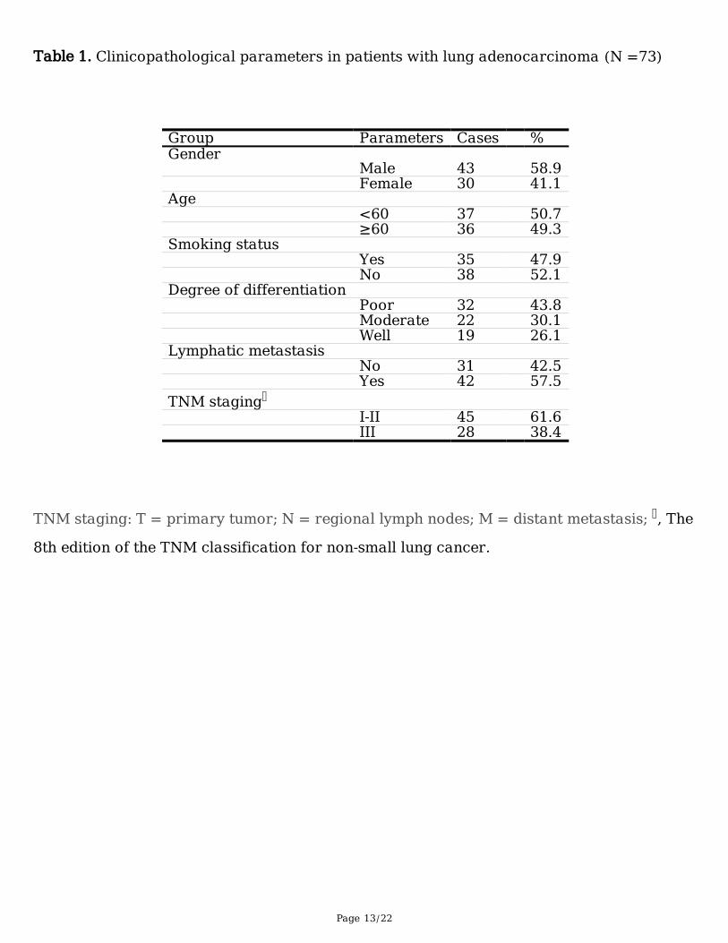
Table 1. Clinicopathological parameters in patients with lung adenocarcinoma (N =73)
Group Parameters Cases %Gender Male 43 58.9 Female 30 41.1Age <60 37 50.7 ≥60 36 49.3Smoking status Yes 35 47.9 No 38 52.1Degree of differentiation Poor 32 43.8 Moderate 22 30.1 Well 19 26.1Lymphatic metastasis No 31 42.5 Yes 42 57.5TNM staging I‐II 45 61.6 III 28 38.4
TNM staging: T = primary tumor; N = regional lymph nodes; M = distant metastasis; , The
8th edition of the TNM classification for non-small lung cancer.

Page 14/22
Table 2. Comparison of STIP1 and HSP90 expression in LAC and adjacent normal tissues
(73)
Items Groups N Comparison of expression rates
Negative (%) Positive (%) χ2 value P valueSTIP1 Normal 73 57(78.1) 16(21.9) 19.337 <0.001 LAC 73 31(42.5) 42(57.7)Hsp90
9.958 0.002 Normal 73 49(67.1) 24(32.9) LAC 73 30(41.1) 43(68.9)
LAC, lung adenocarcinoma; HSP90, heat shock protein 90; STIP1, stress-inducible
phosphoprotein 1.

Page 15/22
Table 3. Correlation between STIP1 expression and clinical features of LAC (n=73)
Items Groups N Expression of STIP1 in lung tissuesNegative (%) Positive (%) χ2 value P value
Gender Male 43 19(44.2) 24(55.8) 0.127 0.722 Female 30 12(40) 18(60)Ages <60 37 16(43.2) 21(56.8) 0.019 0.892 ≥60 36 15(41.7) 21(58.3)Smoking Yes 35 18(51.4) 17(48.6) 2.211 0.137 No 38 13(34.2) 25(65.8)Differentiation Poor 32 8(25) 24(75) 7.896 0.019 Moderate 22 10(40.9) 12(59.1) Well 19 13(68.4) 6(31.6)Lymph node metastasis No 31 28(90.4) 3(9.6) 50.51 <0.001 Yes 42 3(7.1) 39(92.9)TNM staing I‐II 45 29(66.7) 16(33.3) 10.037 0.002 III 28 2(7.2) 26(92.8)
LAC, lung adenocarcinoma; TNM, stage of lung cancer; STIP1, stress-inducible
phosphoprotein 1.

Page 16/22
Table 4. Correlation between HSP90 expression and clinical features of LAC (n=73)
Items Groups N Expression of HSP90 in lung tissuesNegative (%) Positive (%) χ2 value P value
Gender Male 43 17(39.5) 26(60.5) 0.105 0.746 Female 30 13(43.3) 17(56.7)Ages <60 37 16(43.2) 21(56.8) 0.143 0.705 ≥60 36 14(38.9) 22(61.1)Smoking Yes 35 15(42.9) 20(57.1) 0.086 0.769 No 38 15(39.5) 23(60.5)Differentiation Poor 32 9(34.4) 23(65.6) 3.016 0.221 Moderate 22 10(45.5) 12(54.5) Well 19 11(57.9) 8(42.1)Lymph node metastasis No 31 25(80.6) 6(19.4) 34.815 <0.001 Yes 42 5(11.9) 37(88.1)
TNM staging I‐II 45 26(57.8) 19(42.2) 13.487 <0.001 III 28 4(14.3) 24(85.7)
LAC, lung adenocarcinoma; TNM, stage of lung cancer; HSP90, heat shock protein 90.
Table 5. Correlation analysis between the expressions of STIP1 and Hsp90 and the survival
of patients with LAC
Items Groups Mean StandardDeviation
95% ConfidenceInterval
Log Rank Breslow
Lowerbound
Upperbound
Chi-square
P-value
Chi-square
P-valve
STIP1 Negative 49.83 1.23 47.43 52.24 65.11 <0.001
52.35 <0.001Positive 29.59 1.07 27.49 31.69
Hsp90 Negative 49.34 1.39 46.74 51.95 50.06 <0.001
43.74 <0.001Positive 30.46 1.58 28.03 32.85

Page 17/22
STIP1, stress-inducible phosphoprotein 1; HSP90, heat shock protein 90.

Page 18/22
Table 6. Multiple regression analysis of correlation between clinical parameters
and overall survival of patients with LAC
Variables (X) Items (single parametergrouping)
Pvalue
Odds ratiovalue
95% CI forodds ratio
lower upperGender (X1) Male (X1-0) vs. female (X1-1) 0.918 - - -
Age (X2) <60 (X2-0) vs. ≥60 (X2-1) 0.297 - - -
Smoking (X3) Yes (X3-0) vs. no (X3-1) 0.264 - - -
Differentiation(X4)
Poor (X4-0) vs. moderate (X4-1)vs. well (X4-2)
0.127 - - -
Lymphaticinvasion (X5)
Positive (X5-0) vs. negative (X5-1) 0.657 - - -
TNM (X6) I-II (X6-0) vs. III (X6-1) 0.004 2.991 1.405 6.366
STIP1 (X7) Negative (X7-0) vs. positive (X7-1)
0.001 9.614 2.463 37.52
Hsp90 (X8) Negative (X8-0) vs. positive (X8-1)
0.015 3.585 1.279 10.028
Risk function: H(t)=[h0(t)]e(1.612 X6 + 2.223 X7 +
1.277 X8)
LAC, lung adenocarcinoma; CI, confidence interval; TNM, stage of lung cancer; STIP1,
stress-inducible phosphoprotein 1; HSP90, heat shock protein 90.
Figures

Page 19/22
Figure 1
Protein expressions of STIP1 and Hsp90 in A549 and 16HBE cells. (A) Western blot showed that theexpressions of STIP1 and Hsp90 in A549 cells were higher than that in 16HBE cells. (B) Quantitativeanalysis suggested that the expression levels of STIP1 and Hsp90 in the A549 cells was higher than thatin 16HBE cells. (C) Expression of STIP1 in LAC tissues was higher than that in adjacent normal lungtissues. (D) Expression of STIP1 in LAC tissues was higher than that in adjacent normal lung tissues.LAC, lung adenocarcinoma; STIP1, stress-inducible phosphoprotein 1; Hsp90, heat shock protein 90;16HBE, 16 human bronchial epithelial cells.

Page 20/22
Figure 2
Protein expressions of STIP1 and Hsp90 in LAC tissues (×400). (A) Low expression of STIP1 in adjacentnormal lung tissues. (B) Low expression of STIP1 in well differentiated LAC tissues. (C) Moderateexpression of STIP1 in moderate differentiated LAC. (D) High expression of STIP1 in poor differentiatedLAC. (E) Low expression of Hsp90 in adjacent normal lung tissues. (B) Low expression of Hsp90 in welldifferentiated LAC tissues. (C) Moderate expression of Hsp90 in moderate differentiated LAC. (D) Highexpression of Hsp90 in poor differentiated LAC. LAC, lung adenocarcinoma; STIP1, stress-induciblephosphoprotein 1; Hsp90, heat shock protein 90; 16HBE, 16 human bronchial epithelial cells.

Page 21/22
Figure 3
Correlation between expressions of STIP1 and Hsp90 with clinicopathologic factors of LAC patients. (A)Increased STIP1 was observed in poorly differentiated LAC compared with the moderately and welldifferentiated LAC (p<0.05). (B) LAC tissues with lymph node metastasis revealed an increased STIP1expression compared with those without lymph node metastasis (p<0.05). (C) Tissues at stage III had ahigher STIP1 expression than those at stages I-II (p<0.05). (D) Increased Hsp90 was observed in poorlydifferentiated LAC compared with the moderately and well differentiated LAC (p<0.05). (E) LAC tissueswith lymph node metastasis had the same expression level compared with those without lymph nodemetastasis (p>0.05). (F) Tissues at stage III showed a higher Hsp90 expression than those at stages I-II(p<0.05). pTNM (pathologic TNM classi�cation); NSCLC, non-small cell lung cancer; LAC, lungadenocarcinoma; STIP1, stress-inducible phosphoprotein 1; Hsp90, heat shock protein 90; 16HBE, 16human bronchial epithelial cells.

Page 22/22
Figure 4
Relationships between the expressions of STIP1 and Hsp90 and prognosis of LAC patients. (A and B)Survival analysis showed that higher expression of STIP1 was associated with shorter survival of LACpatients (p<0.05). (C and D) Survival curve showed that higher expression of Hsp90 was associated withshorter survival of LAC patients (p<0.05). LAC, lung adenocarcinoma; STIP1, stress-induciblephosphoprotein 1; Hsp90, heat shock protein 90; 16HBE, 16 human bronchial epithelial cells.


![Tumor-Specific Chromosome Mis-Segregation Controls Cancer … · supported by prediction of tumor progression with genetic clonal diversity in esophageal adenocarcinoma [3], and now](https://static.fdocuments.net/doc/165x107/5faa35bda88b342e6e09c934/tumor-specific-chromosome-mis-segregation-controls-cancer-supported-by-prediction.jpg)
