Principles of drug therapy in dentistry
163
-
Upload
yousif-laftah -
Category
Health & Medicine
-
view
2.000 -
download
5
Transcript of Principles of drug therapy in dentistry


Principles of Drug Therapyin Dentistry


JAYPEE BROTHERS MEDICAL PUBLISHERS (P) LTD
New Delhi • Panama City • London
®
Principles of Drug Therapyin Dentistry
Hussain Ali BDS
Professor of PharmacognosyFaculty of Pharmacy
Misr University for Science and Technology (MUST)6th of October City, Giza, Egypt
Foreword
Sanna Shafshak

Headquarter
Jaypee Brothers Medical Publishers (P) Ltd4838/24, Ansari Road, DaryaganjNew Delhi 110 002, IndiaPhone: +91-11-43574357Fax: +91-11-43574314
Email: [email protected]
Overseas Offices
J.P. Medical Ltd.,83 Victoria Street LondonSW1H 0HW (UK)Phone: +44-2031708910Fax: +02-03-0086180
Email: [email protected]
Jaypee-Highlights Medical Publishers Inc.City of Knowledge, Bld. 237, ClaytonPanama City, PanamaPhone: 507-317-0160Fax: +50-73-010499
Email: [email protected]
Jaypee Brothers Medical Publishers (P) Ltd.
Website: www.jaypeebrothers.comWebsite: www.jaypeedigital.com
© 2012, Jaypee Brothers Medical Publishers
All rights reserved. No part of this book may be reproduced in any form or by anymeans without the prior permission of the publisher.
Inquiries for bulk sales may be solicited at: [email protected]
This book has been published in good faith that the contents provided by the authorcontained herein are original, and is intended for educational purposes only. Whileevery effort is made to ensure accuracy of information, the publisher and the authorspecifically disclaim any damage, liability, or loss incurred, directly or indirectly, fromthe use or application of any of the contents of this work. If not specifically stated,all figures and tables are courtesy of the author. Where appropriate, the readersshould consult with a specialist or contact the manufacturer of the drug or device.
Principles of Drug Therapy in Dentistry
First Edition: 2012
ISBN: 978-93-5025-380-9
Printed at
®

Dedicated to
My FatherMy MotherMy Wife
and My sons Sajjad and Ali


Foreword
This book is a model for merging two intimately integrated sciences—oral diagnosis and pharmacology. It is a very helpful guide to dentalpractitioners since it relates diagnostic criteria of oral diseases todetailed medical treatment.
The author is also an excellent model for graduates of MisrUniversity for Science and Technology (MUST), 6th of OctoberCity, Giza, Egypt.
Dr Hussain proved the accuracy of the vision of the universityas it was first stated by Prof (Dr) Soad Kafafy, the founder of theuniversity.
“The vision that aims at graduating dentists with highercapabilities than just practice dentistry.”
Hussain has to be appreciated for the great efforts he madeand for his will and courage to make this book.
Sanna ShafshakProfessor of Oral Diagnosis
Misr University for Science and Technology6th of October City, Giza, Egypt


Preface
It is not an easy task to cover all drugs used in dentistry; but in thisbook, the author summarized the most fundamental items.
The mouth and oral cavity is considered as the body gates foreither good health or diseases.
Oral hygiene and good health are the first line of prophylactictreatment in dentistry.
The curative medicines and drugs used in management of oraldiseases are mentioned in chapters, to facilitate understanding andhelp both undergraduate and postgraduate people who areconcerned with dentistry.• Gives an introduction to general pharmacology and general
forms and routes of administration of the drugs routinelyused. It also outlines the basics of drug interactions.
• The first chapter discusses all types of dental pain and theircharacters, pathogenesis of inflammation and its body responseand influence.
• The second chapter is concerned with all antimicrobial agentstreating bacterial diseases either prophylactic or curative, andspecific or nonspecific.
• The third and fourth chapters deal with antifungal and antiviralagents in common use, putting in consideration—theindications, contraindications, side effects and drug interactionsfor each drug.
• The fifth chapter is the most important one, because anemergency is a life-threatening condition. Dentists should knowvery well how to deal, manage and take prompt action in sucha way to save patient’s life. All physicians, pharmacists, andbefore all dentists should keep that chapter as a booklet foremergency cases.

Principles of Drug Therapy in Dentistry
The author has done his best to present this book in suchinformative and comprehensive manner.
Members of Pharmacology Department, Faculty of Pharmacy,MUST, have shared in revising and summarizing the main topics tobe easily digested by the readers.
We were keen to minimize and maximize the items accordingto its medicinal and surgical significance for dentists.
Hussain Ali
x

Acknowledgments
I wish to express my thanks and appreciation to everybody interestedin supporting and helping me. In the first place, all thanks go to“Allah” who provided us with knowledge and the ability to gain it.
I am more than grateful to my wife “Maryam” for standingwith me in the hard times.
I am indebted to Dr Sanna Shafshak, Professor of OralDiagnosis, for her efforts, without which, the completion of thisbook would not be possible. She was the source of support andinspiration.
A special recognition goes to Dr Husseiny Elgendy, Professorof Pharmacognosy, as he was interested in revising the book. Hisnotes were more than important as well as his suggestions. He didmotivate and encourage me.
Dr Salah Lofty, Assistant Lecturer and the right hand ofDr Elgendy, was more than a valuable source of help and support.He actively revised the book and provided me with valuablereferences.
My colleagues and friends, who deserve special recognitionsand thanks include, Ahmad Al Maskeen and the “Two Alis”—Ali AlHassar and Ali Sulais, for their support and provision of computerassistance. Thanks also go to Thani Al Sharrari and to the “TwoHussains”—Hussain Shakhory and Hussain Saeed, for theircontinuous support and motivation. Thani was more than interestedin helping and providing me with ideas and resources.


Contents1. Introduction 1–15
Routes of Drug Administration and 1Drug FormsWriting Prescriptions 7General Considerations 9Specific Considerations 10
2. Pain Control Medications in Dentistry 16–42
Neurophysiology of Pain 17Clinical Presentation of Pain 18Intraoperative Pain 20Postoperative Pain Management 25Drugs for Pain Relief 28General Considerations 38Specific Considerations 38
3. Antimicrobial Agents Used in Dentistry:Bacterial Infections 43–90
Bacterial Infections 44Antibiotics 45Systemic Agents 48Clinical Use of Systemic Antibiotics 61Local and Topical Agents 81General Considerations 85Specific Considerations 85
4. Antimicrobial Agents Used in Dentistry:Antifungals 91–107
Fungal Infections 91Antifungal Agents 93

Principles of Drug Therapy in Dentistry
Clinical Use of Antifungal Agents 99General Considerations 104Specific Considerations 105
5. Antimicrobial Agents Used in Dentistry:Antiviral Agents 108–123
Viral Infections 108Individual Drugs 110Clinical Use of Antiviral Drugs 116General Considerations 120Specific Considerations 121
6. Emergency Drugs Used in Dentistry 124–144
Prevention 124Preparedness 125Emergency Drugs 126Primary Drugs 131Management of Emergencies 136
Index 145
xiv

Routes of Drug Administration and Drug Forms
Enteral (Oral) RouteSublingual or Buccal RouteParenteral RouteIntravascular or Intravenous (IV) RoutesIntramuscular (IM) InjectionSubcutaneous (SC) InjectionsInhalational RouteTopical Delivery of Drugs
Writing PrescriptionsGeneral ConsiderationsSpecific Considerations
For most people, the first impression about a doctor is someonewho writes a drug prescription. In dentistry, however, a dentist isusually concerned about removal of the cause rather than instructingpatients to use certain drugs, possibly as the cause is accessible forremoval and that drugs alone are not sufficient to produce totaltherapeutic effects. However, a dentist should complement theprovided dental treatment with a proper drug for the comfortand, sometimes, the safety of the patient. In order to administer adrug safely, the dentist should have sufficient knowledge about itsmechanism of action, indications, contraindications, adversereactions and interactions with other drugs. The risks of drug therapyto medically complex patients increase as these patients have weakerhomeostatic systems. When in doubt, it is always better to seekadvice from a specialist. Table 1.1 shows some important definitionsregarding use and handling of drugs.
Chapter 1Introduction

Principles of Drug Therapy in Dentistry2
Table 1.1: Some important definitions
Drug A substance used or intended to be used fortherapeutic reasons (diagnosis, prevention ortreatment).
Pharmacology The science which is concerned about drugs,including their source, properties, uses,contraindications and adverse reactions. Itsmain branches include: Pharmacokinetics,pharmacodynamics and pharmacothera-peutics.
Pharmacokinetics Branch of pharmacology, concerned aboutwhat the body does to the drug like absorption,distribution, metabolism and excretion.
Pharmacodynamics The branch which deals with the biochemicaleffects (mechanism of action) of the drug onthe body or microorganisms.
Bioavailability It is the percentage of the administered drugwhich reaches the blood circulation inunchanged state.
Drug metabolism It is the process by which the drug isinactivated, broken down and excreted. Bythe action of specific enzymes, the lipophilicdrug is converted into hydrophilic one.
Adverse drug Harmful or at least unwanted effects producedreactions (ADRs) by normal dose of a drug. ADR differs from
side effects as the latter can be useful.
Drug interaction Alteration in the expected effects of a drugwhen administered with another one or withfood/drinks.
Antidote A substance which is used to counteract theeffects of toxic agent or poisoning.

Introduction 3
ROUTES OF DRUG ADMINISTRATION ANDDRUG FORMS
The route of drug administration depends primarily on thetherapeutic requirement, that is, whether the needed effects arelocal or systemic, in addition to the required speed of onset, durationand concentration of the drug (Table 1.2). Sometimes, the drugproperties (irritation, solubility, instability) make it impossible tobe used by all routes.
Enteral (Oral) Route
This is the simplest and most common route of drug administration.The patient is able to self-medicate himself and the overall procedurerequires only a cup of water. Absorption of a drug via this route isachieved by the GIT mucous membrane being most active in thesmall intestine (duodenum). The small intestine provides the largestsurface area through the presence of villi and microvilli, wider pHrange for drug stability and higher membrane permeability. Otherparts like the stomach and large intestine also exhibit absorptionbut of less significance. The stomach is a harsh environment for adrug because of its low pH. Furthermore, the stomach is lined by athick mucus layer which embeds drug absorption.
Table 1.2: Routes of drug administration
Enteral• Oral• Buccal (sublingual)Parenteral• Intradermal• Subcutaneous• Intramuscular• Intravascular• InhalationalLocal delivery• Topical

Principles of Drug Therapy in Dentistry4
The bioavailability of an oral drug may be affected by severalfactors, like gastric acidity, presence of food, gastric emptying,digestive and cytochrome enzymes and hepatic first pass metabolism.In addition, the onset of the drug is slow. Possible gastric irritationand inability to be used in unconscious or uncooperative patientsare further disadvantages. This makes oral form unsuitable foremergencies or life-threatening situations.
Sublingual or Buccal Route
It utilizes the richly supplied thin mucous membrane of the mouthfor rapid absorption. Although retained in the mouth, the intendedeffects of the drug are systemic. The absorbed agent escapes thehepatic metabolism as it is driven directly to the target cells via thesuperior vena cava. Therefore, the sublingual route provides higherbioavailability than the oral one, in addition to its possible use inunconscious or vomiting patients. Nitroglycerin, a vasodilator usedin case of angina pectoris, exhibits more than 90 percent metabolismby the hepatic system. For that reason, nitroglycerin is usedsublingually. The drug forms that can be present for oral routes arelisted in Table 1.3.
Parenteral Route
By this route, the drug is brought directly into blood or tissue fluids.This route helps in administering those agents unstable in or thosepoorly absorbed from the GIT environment. The bioavailability ishigher than the oral route and the onset of action is more rapid. Thisfacilitates the management of unconscious patient or in case whereprompt action is needed. One main disadvantage is that onceadministered by parenteral route, the drug effects cannot be reversed.Other inconveniences include painful procedure, the need forprofessional administration and practice of asepsis.
Intravascular or Intravenous (IV) RoutesThey bring the drug into arterial or venous blood, respectively.These routes provide 100 percent bioavailability and most rapidonset of action. These also provide the best control over the amount

Introduction 5
of active agent provided. However, it may result in most severeadverse reactions in addition to possible hemolysis and throm-bophlebitis. The used solutions are only aqueous and no depot(nonaqueous medium) or suspensions are to be used by this route.The injection can be in the form of bolus (initial large dose), slowinjection (over 15–20 minutes) or slow infusion (large volume ofdrug over long period of time).
Table 1.3: Drug forms used by the oral route
Tablets Solid dosage form orally placed and swallowed.When placed in water, tablets disintegrate.
Enteric coated (EC) A layer of cellulose acetate coating is used to protecttablet the active agent form gastric acidity and prevent
gastric irritation.
Sugar coated Sugar layer used to produce palatable tablets.tablet
Film coated This layer enhances swallowing, stabilizes the agenttablet against moisture and improves taste.
ODT* Tablets laid on the tongue and left to disintegrate.Useful for those who cannot swallow.
Capsules Gelatin shell (hard or soft) used to protect thedrug against temperature, humidity and to enhanceswallowing.
Caplets Tablets in the form of capsule and with the textureof gelatin (tablets easy to swallow as capsules).
Solutions Homogeneous liquid form by solute (active agent)dissolved in solvent.
Suspension Heterogeneous liquid with fine particles of solutedistributed uniformly. The formula settles downwith time and gets redispersed when shaken.
Emulsion Liquid with two different phases, like water andoil with an emulsifying agent.
* ODT: Orally Disintegrating Tablet

Principles of Drug Therapy in Dentistry6
Intramuscular (IM) InjectionIt provides rapid action, but not as the IV route. Large muscles likethe deltoid, gluteus or the triceps are the target for IM injections.Muscles have good blood supply, which helps in providing uniformabsorption. However, IM injections are quite painful especially withdepot solutions.
Subcutaneous (SC) InjectionsThese utilize the blood circulation in the connective tissueunderneath the skin. Vascularity in this area is quite low and thisproduces uniform and prolonged duration of action but with slowonset. The duration of action can be further prolonged by theaddition of vasoconstrictors. Hyaluronidase enzyme, on the otherhand, enhances absorption by the SC route.
Inhalational RouteIts uses drugs in the gaseous state. Volatile liquids or aerosols canalso be used. Absorption in this route is achieved via the mucousmembrane of the respiratory tract and/or the pulmonaryepithelium. The advantage of this route is that rapid action can beachieved as large surface area is utilized along with rich blood supply.In addition, local effects can be produced if the target is therespiratory system.
Topical Delivery of Drugs
The main aim of topical application of drugs is to provide highdrug concentration at the desired site.
Because this route of drug therapy acts only on superficialtissues, minimal systemic involvement and minimal systemic adversereactions are expected. The procedure of applying a topical agent isnoninvasive and provides psychological relieve as patients can takecare of themselves. However, the contact time between the drug andthe target tissues is short due to wiping or washing effects. In addition,the surface area can be small, especially periodontal pockets. Thiswould necessitate the use of a device which slowly releases the activeingredients. Forms of topical agents are listed in Table 1.4.

Introduction 7
WRITING PRESCRIPTIONS
A prescription is a way of communication between a physician or adentist and the pharmacist. This can be in the written or verbalforms. The prescription is considered as a part of the medicolegaldocuments and both the prescriber and the pharmacist are incharge. It should be written neatly, with full and clear informationregarding the type, dose and duration of the drug therapy. Careshould be practiced also when using symbols and abbreviations asthey may increase the chance of confusion.
When a drug is prescribed, the nonproprietary or the commonname should be used first. This name is the primary ID of the drugand each drug has only one nonproprietary name but manyproprietary ones. For example, paracetamol is the nonproprietaryname for the commonly known analgesic, while proprietarynames as panadol, adol, and many others exist. In this book, the
Table 1.4: Drug forms used by the topical route
Lozenges Large tablets held in the mouth to dissolve and producelocal action. The active agent is placed in sugar base.
Pastilles Similar to lozenges, but with jelly like consistency.
Ointment Semisolid preparation for external use. It usually hashydrating effects and used on dry skin.
Cream Similar to ointment, but without the hydrating effect.
Paste Semisolid agent but with solid particles at greaterconcentration when compared to ointment, whichproduces stiffer or more viscous preparation.
Gel Translucent, non-greasy semisolid with higher wateror alcohol percent than creams or ointment. Gellingagent is used to produce the semisolid consistency.
Solution Liquid preparation with the active agent dissolved in asolvent. The formula is low in viscosity and allows itsuse as a mouthwash or gargle.
Paint Active agent is placed in a volatile medium (for skinuse) or in viscous one (for use in mucous membrane).

Principles of Drug Therapy in Dentistry8
Table 1.5: Some abbreviations used in prescription writing
English Latin Abbreviations
Recipe Rx
By mouth Per os poOnce per day Onus in die odTwice per day Bis in die bidThree times a day Ter in die tid
Ter in die sumendum tdsFour times a day Quarter in die qidEvery hour Quaque hora qhAs needed Si opus sit sosBefore meal Ante cibum acAfter meal Post cibum pcTablet Tabella TabCapsule Capsula Cap
nonproprietary names are used mainly with some commonproprietary ones written in bold blue underneath.
A prescription is designed to contain the basic informationabout the patient, the prescriber and the drug. The patient’s name,age and sex should be provided to confirm the drug dosage by thepharmacist. The name and telephone number of the prescribershould be also provided for reference. All this information iscontained in what is called the superscription. In addition, thesuperscription also contains the symbol Rx, which stands for “recipe”(Table 1.5 for more abbreviations). The next part of the prescriptionis its body or what is called the inscription. This part contains theinformation about the prescribed drug. It is written below andright to the symbol Rx. It starts by the name of the drug (thenonproprietary followed by the proprietary if to be written). Thedosage, frequency and amount are written next. The subscription,which is written below the inscription, usually contains instructionsto the pharmacist about the form of the drug or the quantity. This

Introduction 9
is especially important in case of, the prescribed drug is a controlledone. When the prescriber wishes the written instructions to beprovided on the containers of the drugs, the word “label” or “signa”is written. Some prescription forms contain the word label with atable before it to be ticked. This part containing the instruction tobe provided to the patient is known as the transcription. Betterpatient compliance is expected when the drug is labeled as patientsusually forget verbal instructions. The last part would be thesignature of the prescriber, which is mandatory in case of controlleddrugs. The name of the prescriber is handwritten followed by theprofessional degree. The name can be stamped one line under thesignature. Table 1.6 shows an example, of a blank prescription.
GENERAL CONSIDERATIONS
For best results, the patient’s compliance should be optimized. Thechoice of the drug, its form and route of administration should besuitable to the patient. For more convenience, the oral form is the
Table 1.6: Sample of a prescription form
Superscription : Dr Name___________________
Telephone __________
Address ___________________
Patient’s name ______________
Age ____ Sex _____
Address _____________________________
Rx
Inscription
Subscription Refill
0 1 2 3
Transcription or signa Label
Signature _____________________________

Principles of Drug Therapy in Dentistry10
most commonly chosen one. For children or those unable to swallowtablets, the liquid form is the right choice. However, the parenteralroute may be indicated when the condition is serious. In emergencies,drugs are usually provided in the injection form. SC injectionsshould be avoided in shocked patients as the blood circulation ispoor in the subcutaneous tissue.
The timing of the drug intake can further affect the patient’scompliance. Whenever possible, a drug which needs lower frequencyof intake should be chosen, for example, every 12 hours instead ofevery 6 hours. In case of multidrug therapy, combination shouldbe made as possible to lessen the number of taken drugs.
Good communication with the patient should be establishedand the drug benefits and its method of use should be explained.The instructions of use should be provided both in verbal andwritten forms. The patient should be informed of possible adversereactions and if any precautions to be undertaken.
SPECIFIC CONSIDERATIONS
The doses and frequencies of intake presented in this book are foraverage adults (18 years and above) weighing about 70 kg. Thedose, however, is affected by the patient’s age, weight and medicalcondition. Children, underweights, elderly and medically complexpatients need dose modification in order to provide a drug withoutpossible toxicity.
For children, the dose is adjusted following Clark’s or Young’srule (Table 1.7). Clark’s rule, however, provides more accurate results.
Table 1.7: Dose adjustment for children
Clark’s rule
Child dose = Child’s weight/150 × adult dose
Young’s rule
Child dose = Age/age + 12 × adult dose*
* This means that a 12-year-old child can use half adult dose

Introduction 11
In case of renal or liver disease, the ability of the body to break-down and eliminate the drug is reduced and, accordingly, either thedose or the frequency of drug intake should be reduced. Wheneverpossible, medical consultation should be made.
Pregnancy and breastfeeding are two physiological conditionsthat may require the clinician to avoid those agents with possibleteratogenic or adverse effects on the developing fetus or thebreastfeeding infant. A section of those special patients is present atthe end of each chapter denoting these agents that should be avoidedand those that can be used safely.
The basics of drug interaction are presented at the druginteraction (Box 1.1). At the end of each chapter, a box containingthe possible drug interactions is also presented.
Box 1.1: Drug interaction(Basic concepts)
Drug interaction is the process by which the expected effects of adrug is altered by the use of another drug, food, drink or environmentalchemical agent. The incidence of drug interaction is expected to behigher in those under multidrug therapy, elderly or those with severechronic diseases (renal or hepatic for example). The altered drug effectcan be increased, decreased or totally not related to the parent drugs.The following terms should be defined:Antagonism is the process by which the effects of a drug is decreasedor blocked by the action of another. It may be produced by the followingmechanisms:• When two effects are produced but not for the benefit of each
other; for example, the use of tetracycline decreases the effects ofpenicillin, as the former decreases protein synthesis while thelater acts by inhibiting cell wall synthesis
• When two agents compete for the same receptor. Naloxone andflumazenil would terminate the effects of opoids and benzodiazepine,respectively
• When two drugs with opposite actions are given concurrently.An example would be the administration of insulin and corti-costeroids
Contd...

Principles of Drug Therapy in Dentistry12
• When the administered agents have direct physical or chemicaleffects on each other. For example, the use of antacids withtetracycline would produce nonabsorbable complexes
• When one drug affects the metabolism of another either byaffecting its metabolizing enzymes or its rate of excretion.
Potentiation is the process of increasing the action of one drug byanother drug which is therapeutically unrelated to the former.Probenecid (uricosuric agent used to decrease uric acid) increases theeffects of penicillin as it decreases in tubular excretion.Unexpected effects are associated with the production of highly activemetabolite by the interaction of two drugs. This effect is notexperienced when any of them is used. An example would by the useof metronidazole with alcohol, which increases the level ofacetaldehyde.Summation interaction is produced when the use of two relatedagents produces the combined actions of both. An example, would bethe use of opoids and general anesthetics.Synergism is the increased quantitative effects of one of twoconcurrently given drugs above the maximum effective dose of thatgiven alone. The use of antidepressants and antiepileptics togetherwould produce massive drowsiness. A drug interaction occurs either inthe pharmacokinetic (PK) or the pharmacodynamics (PD) steps.PK interactions occur during absorption, distribution, metabolism orexcretion of drugs.Absorption interaction. Two orally administered drugs may interactwith each other resulting in decreased or increased absorption of oneor both of them. The underlying mechanism can be attributed to thefollowing:• Altered pH, as some agents are absorbed at certain pH range.
Example, would be ketoconazole which is absorbed better at lowpH. So, the use of antacids is expected to lower its absorption rate
• Chelation process by multivalent ions to form insolublecomplexes. For example, calcium decreases the effectiveness oftetracycline or ciprofloxacin
• Effects on the GIT flora. Some drugs (Digoxin) are inactivated bycertain bacterial species in the large intestine. When co-administered with broadspectrum antibiotics, digoxin inactivationis reduced causing digoxin toxicity. Oral contraceptives, on the
Contd...
Contd...

Introduction 13
other hand, would loss effectiveness as the gut bacteria act on theconjugated form by hydrolyzing it and librating an active agentfor intestinal re-absorption
• Effects on GIT motility. Delayed gastric emptying would delaydrug absorption. Paracetamole absorption is increased by the useof metoclopramide which increases gastric emptying. Such effectis useful for treatment of migraine.
Distribution interactions. The active agent is distributed by bindingto plasma proteins like albumin and α
1-acid glycoprotein. The bounded
agent is unable to act on the receptor site but remains protected frommetabolism or excretion. Some drugs, like NSAIDs, would displaceoral anticoagulants from their plasma proteins, resulting in increasedsusceptibility to bleeding.Metabolism interactions. Drug metabolism occurs in the liver, kidneys,GIT, lungs, placenta and even in skin. The process is divided into twophases, Phase I which involves oxidation, reduction and hydrolysis,and Phase II which involves conjugation. The cytochrome P450isoenzymes are responsible for the process. A drug can be the substrateor the inducer/inhibitor of a CYP enzyme, or even both. Examples ofknown enzyme inducers include: Rifampicin, phenytoin, barbituratesand carbamazepine (able of autoinduction). The result of enzymeinduction is decreased effects of the affected drug, except when theactive agent is its metabolite. It is important to note that the mechanismof induction need time in order to synthesize new enzymes. Enzymeinhibitors, on the other hand, result in toxicity of the hypometabolizedagent. Example of such inhibitors include: Agents with imidazole ring(ketoconazole, itraconazole, etc.), ciprofloxacin, erythromycin andpropoxyphene. Another way of affecting drug metabolism is by affectingthe hepatic blood flow. Propranolol decreases the cardiac output andreduces blood supply to the liver which affects lidocaine metabolism.Elimination interactions. Drug elimination usually occurs by the renalsystem. Interactions at this level may be due to:• Altered blood supply to the kidneys, which reduces the filtration
rate. An example is indomethacin (NSAINs) which inhibits PG.This would affect the rate of lithium excretion causing lithiumtoxicity. Altered pH, which affects drug re-absorption from therenal tubules. pH affects the ionization of acidic or alkaline drugs.Weak acids become ionized and lipid insoluble when placed in
Contd...
Contd...

Principles of Drug Therapy in Dentistry14
alkaline media which prevents their re-absorption. The same goesfor weak bases
• Affected active transport, which affects drug secretion. Activeagents may have special mechanism for their secretion into tubulelumen and this can be the active transport system. When thissystem is occupied by another drug, for example ASA in case ofmethotrexate therapy, the result would be methotrexate toxicity.Another example of clinical benefit is the use of probecid whichincreases penicillin therapeutic effects
PD interactions occur at the receptor site. They are defined as thechange drug actions regardless of the change in the amount availableor the quantitative deposition. The two drugs are acting on the samereceptor site or at the same system. The following are examples of PDinteractions:• The effect of sulfonylurea (agent used to promote insulin secretion
by the pancreas) is decreased by the use of non-selective B blockerswhich block the B pancreatic cells
• The use of NSAIDs reduces renal blood flow and may result in Naand water retention. This antagonizes diuretics and ACEs (anti-hypertensive agents)
• The use of potassium sparing agents along with K supplementswould lead to hyperkalemia.
Contd...
BIBLIOGRAPHY
1. Anthony P, Vahn A. Prescription writing and drug regulations.Pharma and Therapeutics for Dent. 4th edn. Mosby; 1988;696-701.
2. Dashi Darmash H. Oral drug delivery system. Drug deliverysystem in pharma care. 2007; 23-34.
3. Desai Archana. Topical and Transdermal Delivery System.Drug delivery system in pharma care. 2007; 45-50.
4. John A, Anthony Picozzi. General mechanisms of druginteractions. pharma and therapeutics for dentistry. 4th edn.Mosby; 1998.
5. Lee A, Stockley IH, Drug interactions. Clinical Pharma andTherapeutics. 3rd edn. Livingstone, 2006.

Introduction 15
6. Moody MM. Routes of drug administrations and dosage forms,Pharmaceutical practice. 2nd edn. 1998; 102-7.
7. Padmaja Udaykumar. Textboot of phasma. For dentaland allied health sciences. 2nd edition. New Delhi: Jaypee;2007; 1-8.
8. Richard A, Pamela C. Routes of drug administration, illustratedreview of pharmacology, 4th edn. Lippincott Williams andWilkins; 2009; 1-4.
9. Timethy J, Robin M. Special populations: "Pediatrics". Drugdelivery system in pharma care; 2007; 176-8.

Chapter 2Pain Control Medications in Dentistry
Most of the times, the patients’ main concern and chief complaintwhen they come to the dental clinics, is pain. It is pain that bringsmost of the hesitating patients to the dental clinics and keeps themaway, too. Pain is unpleasant sensory and emotional experiencethat tells us about some sort of tissue damage that is occurring inthe body. It is, however, considered as a normal physiologicalprocess. What characterizes dental pain is that it is usually severeand devastating. This is attributed to the fact that nerves are presentbetween the solid walls of the tooth and when inflammatory edemacollects, it finds no way of expansion and, rather, exerts morepressure on the nerve endings.
INTRODUCTION
Neurophysiology of PainClinical Presentation of PainIntraoperative PainPostoperative Pain ManagementDrugs for Pain Relief
NonopioidsParacetamolNSAIDs
OpioidsSteroidal Anti-inflammatory DrugsAdjuvant AnalgesicsTopical Agents
General ConsiderationsSpecific Considerations
Pain Control Medications in Dentistry 17
Whether this pain is pathological or an attempt to removepathology, the patient is usually anxious about it. Identifying thesource of pain can be a difficult task, however, an essential one tomanage pain.
NEUROPHYSIOLOGY OF PAIN
Pain is received by the pain receptors “nociceptive receptors” at thefree nerve endings. At the time of injury, fast/sharp pain is feltwhich is conducted by the myelinated A delta fibers. After theinitial injury, the site of damage continue signaling about the damageoccurred using the slow unmyelinated C fibers. Pain perceived atthis time would be dull but continuous.
Pain waves travel through primary afferent neurons (A deltaand C fibers); and via the dorsal root, they enter the spinal cord.This is the first order neuron and it synapses with the second orderneuron that ascends through the spinothalamic tract to enter thethalamus. The thalamus, acting as the “gateway” of the brain, specifiesthe direction of the incoming of information. The third orderneuron is directed from the thalamus to relay on the postcentralgyrus of the cerebral cortex. It is there where pain is localized andidentified. Nerves communicate with each other by means of neuro-transmitters as substance P and nitric oxide gas.
It should be noted that pain is associated with the productionof several chemical agents within the body. These includeprostaglandin, histamine, bradykinin, serotonin and potassium ions.They act as inflammatory mediators. Of special interest isprostaglandin. Injury to body cells results in the release of membranephospholipids which are converted by phospholipase enzyme intoarachidonic acid. Cyclooxygenase enzyme II (COX-2) acts onarachidonic acid to produce proinflammatory prostaglandins.Prostaglandins magnify pain by increasing the function of nociceptivereceptors through a process known as sensitization.
Pain is normally associated with several reactions. Motorreactions initiate protective reflexes to avoid further damage to thetissues. Emotional reactions, on the other hand, express anxiety

Principles of Drug Therapy in Dentistry18
and depression and record the event for future memorization. Thisis also a way of protection and prevention of damage at the future.Autonomic reaction is the translation of the event in terms of thebody functions, exhibiting reaction to stress. The heart rate, bloodpressure and metabolic rates are elevated.
In a way of protecting the body from excessive pain perception,the body produces what is known as endogenous opioid peptidesnamely enkephalin and endorphins. Enkephalins appear to beproduced by the pregnant women during labor, whereas endorphinsare produced by athletes during exercise to allow endurance andtolerance of hard work. Both act by preventing the release ofexcitatory neurotransmitters.
Pain is described regarding its severity or intensity into mild,moderate and severe. Being completely subjective, pain severity isdifficult to be identified precisely. The final figure of pain isformulated and modified by several factors, the most important ofwhich is the psychological state of the patient. It is well-known thatstressful situations and psychological trauma aggravate or possiblyinduce pain.
Of interest in this chapter is to discuss pain in relation to itspresentation during clinical examination (preoperative), during itstreatment (operative) and after removal of the cause (postoperative).
CLINICAL PRESENTATION OF PAIN
Several diseases and conditions are associated with pain on clinicalexamination (Table 2.1). It should be emphasized that correctdiagnosis is primary in order to eliminate the source of pain.Usually, the source of pain is obvious and related to some pathology.Some types of pain are deceptive, however. Of special interest is thereferred pain. The secret behind pain referral lies in the fact thatthere is some sharing in the pathway of pain transmission(conversion-projection theory). According to the frequency ofprevious stimulation and experience, the brain assumes that pain iscoming from that site, a process known as habit reference. If painfrom inflamed maxillary sinus is misdiagnosed and treated by

Pain Control Medications in Dentistry 19
Intraoral painful conditions
Pulp conditions•Dentinal hypersensitivity•Pulp hyperemia•Pulpalgia (mild, moderate, advanced)•Periapical pathosis
– Acute apical periodontitis– Acute apical abscess
•Referred pulpal painPeriodontal conditions•Pericoronitis•Abscess of the periodontium
– Gingival– Periodontal
•Acute herpetic gingivostomatitis•ANUG•AIDs-associated NUPBone conditions•Fractures•Acute osteomyelitis•Infected cysts•Sickle cell infarcts•TMJ disorders•Bone neoplasmsSalivary gland diseases•Inflammatory disorders•Obstructive disordersConditions of the oral mucosa•Ulcers (viral, fungal, traumatic,
etc.)•Denture sore mouth•Remaining roots under dentures
Extraoral conditions associated withpainReferred pain from extraoral/
remote sources• ENT diseases
– Sinus conditions (maxillary andparanasal)
– Ear conditions• Salivary gland diseases• Cardiac diseases
– Angina pectoris– Myocardial infarction– Carotidynia
• Cervical spine• ThyroiditisMuscular disorders• Myospasm• Myositis• Local unclassified myalgia• Myofacial painNeuropathic disorders• Trigeminal neuralgia• Postherpetic neuralgia• Post-traumatic neuralgia• Glossopharyngeal neuralgia• Occipital neuralgia• Nervous intermedius neuralgia• Neuroma• Anesthesia dolorosaNeurovascular disorders• Migraine• Cluster headacheVascular disorders• Giant cell arteritis (temporal
arteritis)• HypertensionAtypical facial pain• Atypical odontalgia• Burning mouth syndrome
Table 2.1: Conditions associated with orofacial pain

Principles of Drug Therapy in Dentistry20
restoring or extracting a maxillary tooth, the result will be morecomplications. Sometimes, referred pain may be an indication forpatient referral to a specialist, as in case anginal, ENT, or thyroidpain. The dental evaluation may reveal nothing to be abnormaland accordingly the patient should be referred for further evaluationinstead of blind treatment.
Pain from psychological source is another dilemma that mayneed the consultation of more than one specialty. Pain of this typeis usually chronic and no obvious organic source is identifiable.However, some psychological problems may be identified. Patientsmay unconsciously substitute their psychological pains with somaticsymptoms through what is called the conversion reaction.Comprehensive history taking is essential and the patient may needfurther psychological evaluation and management.
INTRAOPERATIVE PAIN
Usually, an operative or surgical procedure is required to treatpain. The methods by which pain is basically treated are listed inBox 2.1.
The nature of dental procedures requires the use of severalcutting instruments and tools which are naturally pain producing.Pain may therefore be caused at least during the dental procedure
Box 2.1: Basic methods of pain management
• Identification and removal of the primary cause under effectivepain control conditions
• Use of analgesics and anti-inflammatory drugs• Counter-irritation and large fiber stimulation by the use of hot/
coldpacks, rubbing, acupuncture, and electric stimulation of theperiaqueductal gray matter
• Use of steroids to suppress severe inflammatory conditions• Use of anticonvulsants and antidepressants• Psychotherapy for cases originated from nonorganic sources; like
atypical pain• Surgical and ablative denervation operations, including
neurectomy, dorsal rhizotomy, thalamotomy or even gyrectomy

Pain Control Medications in Dentistry 21
Box 2.2: Different techniques used to manage operative painTopical anesthesiaLocal anesthesia
• Infiltration• Nerve block• Intraligamental• Intrapulpal• Electronic
Sedation• Inhalational sedation (NO
2)
• Intravenous sedationGeneral anesthesia
even when it was not the chief complain of the patient. This wouldnecessitate some sort of anesthesia in order to make the proceduredoable without risks or harms either to the patient or to the dentist.Different methods of pain control during the dental proceduresare listed in Box 2.2.
Anxiety sometimes makes the dental treatment difficult if notimpossible. It should be considered first as it may even obstruct theanesthetic procedure. All patients should be treated with mercyand handled with care and interest. Careful handling, good per-sonality and other non-drug approach may help in managing mildlyanxious patients. Sometimes, the patient is extremely anxious, veryyoung or the procedure itself is extensive or long. Such situationsmay require the use of sedatives and anxiolytics (Table 2.2).
The use of topical anesthetics is indicated in minimal orsuperficial procedures, as these agents are effective in minimal depths(2–3 mm only). Therefore, they are useful in I and D, scaling androot planning and for painless needle insertion in case of localanesthesia. Topical anesthetics are supplied in spray, gel or ointmentform with the anesthetic agent being lidocaine at most of the times.
Infiltration and block techniques are the most commonlyused methods for most of the dental procedures. It is noteworthyto consider the type of the anesthetic used, and the presence of avasoconstrictor (Table 2.3). The use of vasoconstrictor not only

Principles of Drug Therapy in Dentistry22
Table 2.2: Common anxiolyticsDose
• Short acting (3–8 hours)OxazepamOxazine 10 mgTriazolamHalcion 0.125– 0.25 mg
• Intermediate acting (10–20 hours)LorazepamAtivan 1–2 mgTemazepamRestoril 15 mg
• Long acting (1–3 days)DiazepamValium 5–10 mgChlordiazepoxide Librium 5–10 mg
Table 2.3: List of anesthetic agents according to theirduration of action
• Short acting (less than 30 min)
Prilocaine 4%, without vasoconstrictor
Lidocaine 2%, without vasoconstrictor
Mepivacaine 3%, without vasoconstrictor
• Intermediate acting (60 min)
Lidocaine 2%, with epinephrine 1/100,000
Mepivacaine 2%, with epinephrine 1/400,000
• Long acting (more than 90 min)
Bupivacaine 0.5%, with epinephrine 1/200,000
Prilocaine 4%, with epinephrine 1/200,000
Etidocaine 1.5%, with epinephrine 1/200,000

Pain Control Medications in Dentistry 23
prolongs the effects of local anesthesia, but also lessens the chanceof systemic toxicity. In addition, it provides a clear field as a resultof its hemostatic effects. Contraindications of the use ofvasoconstrictors may exist, however, and should be avoidedaccordingly (Box 2.3).
It should be noted to the patient that not all sensations will beblocked out, and the feeling of pressure will persist. In some cases,pain is still present and may prevent further procedure, althoughthe area is well anesthetized. This can be attributed to acuteinflammation reaching the bone or due to interlacing nerve supplyfrom adjacent nerves. The use of intraligamental or intrapulpaltechniques may help in solving the problem of inflammation. Thecommon interlacing nerves include nerve fibers crossing the midlineand nerve fibers from the C2 and C3 area supplying the lowerpremolar area. Sometimes, anesthetic agent may not reach a flaredpalatal root approaching the midline in upper first molar. Painwould persist especially when working on that canal. In this case,palatal infiltration would help to solve the problem.
The use of electronic anesthesia solved the problem of needlephobia. It may alone allow for simple procedures or may becombined with nitrous oxide sedation or needle anesthesia. It offersa way of non-chemical way of controlling pain, therefore lesserrisks of systemic toxicity or allergic reactions. In addition, it is anon-invasive method and this is advantageous to prevent the spreadof infection as in case of abscess. However, this technique has itsdisadvantages and limitations (Box 2.4).
Box 2.3: Contraindications of vasoconstrictor
• History of allergy• Uncontrolled hypertension• Coronary heart disease• Use of nonselective B-blocker• Hyperparathyroidism• Use of tricyclic antidepressants (doxepin)

Principles of Drug Therapy in Dentistry24
The famously known as “the laughing gas”, nitrous oxide is a wayto sedate and control pain (Stage I analgesia) in young,uncooperative or anxious patients. It produces Stage I analgesiaand relaxes the patient, yet, not losing consciousness. The naturalprotective reflexes are not stopped and the patient is able to followthe operator’s commands. The procedure involves the use of NO
2
gas plus oxygen mixed at the ratio of 50:50.The patient’s vital signs should be checked before the procedure
and taken as a guide to confirm recovery after the procedure. Atthe end of the procedure, nitrous oxide is decreased to zero leveland the patient should breathe pure O
2 for three to five minutes.
Contraindications of nitrous oxide are listed in Box 2.5.The use of intravenous sedation produces deeper Stage I analgesia
and can be used for those who cannot use inhalational anesthesiadue to breathing problems like mouthbreathing.
Sometimes, it is better to have the patient unconscious as incomplicated and extensive cases. General anesthesia produces areversible, controlled loss of sensations and consciousness; as wellas, the protective reflexes. It passes through four stages, namely,analgesia, excitement, general anesthesia and respiratory failure.
Box 2.4: The disadvantages and limitations ofelectronic anesthesia
• Tingling sensation• Increased salivary flow• No clear field as provided by VC• Electric shock may occur when touching metallic instruments• Not suitable for patients with pacemaker• Not used in case of cerebrovascular disease or with brain tumor• Not used in patients with bleeding disorders• Not used in pregnancy
Box 2.5: Contraindications of nitrous oxide• 1st trimester pregnancy• Conditions of difficult respiration• Mouthbreathing• Emphysema

Pain Control Medications in Dentistry 25
Two dangerous states are the stage II, where unexpected movementof the patient may occur, and stage IV where the patient may gointo coma. Respiratory and cardiovascular functions should bemonitored throughout the procedure.
Induction of general anesthesia follows the administration ofpreanesthetic medication. It is started by the use of intravenousinjection of sodium thiopental or methohexital. A mixture of gas,which contains enflurane, nitrous oxide and halothane, is used tomaintain the state of general anesthesia.
POSTOPERATIVE PAIN MANAGEMENT
Dental procedures, whether to eliminate pain or not, are associatedwith some sort of body reaction and inflammation. The degree ofinflammation and pain depends largely on the proper removal ofthe cause through proper diagnosis and treatment plan. The waythe tissues were handled will be reflected postoperatively in terms ofedema and pain. Table 2.4 lists some of the sources of pain occurringas a result of postoperative complications. Useful operative guidelinesregarding postoperative pain reduction are listed in Box 2.6. Somuch can be done during the dental procedure to minimizepostoperative pain and inflammation. The use of light, intermittenttouching, as well, as copious coolant during caries removal ensureslittle or no injuries to the dentinopulpal organ. The use of sharpinstruments eliminates the need for pressure and, hence, burs shouldbe changed whenever the need to exert pressure is felt. Unnecessaryoverextension also takes part in the compromise on the pulp tissue.Care must be practiced also during application of rubberdamclamp, matrices and gingival retraction cords as innocent adjacenttissue do hurt when anesthesia wears off after the procedure. Thevalue of the procedure equals nothing if the material used to restorethe prepared cavity is irritating or not in harmony with normalocclusion. The patient will only complain of pain, although mightbe of different origin.
Surgical procedures should follow the principles of surgeryand be as traumatic as possible. Aseptic and clean field must be

Principles of Drug Therapy in Dentistry26
Table 2.4: Possible sources of postoperative pain
Source of pain Possible causes
Dentinal Moisture contamination during amalgamhypersensitivity placement
MicroleakageOverzealous finishing at cervical margin
Pulpitis MicroleakageFailure to provide temporary filling or crownPoor thermal and chemical insulationIatrogenic exposure of the pulp of vital tooth
Dry socket (alveolalgia) Increased fibrinolytic activities possibly dueto bacterial infectionExcessive tissue manipulation during extractionExcessive use of vasoconstrictorFailure to form and maintain stable bloodclot possibly due to insufficient postextractioninstructions or poor patient compliancePatient systemic background indicates poorblood supply (e.g. smoking and radiotherapy)
Traumatic neuroma Trauma to nerveCuretting nerve ending after extraction
Periodontal abscess Incomplete calculus removalPerforation during root canal treatment
Periapical periodontitis Overextension during RCTOverfilling of GPPTraumatic occlusion or high spots
Trismus Injecting into the medial pterygoid muscleMuscle fatigue due to prolonged mouthopening throughout the procedure withoutthe use of mouth probe
TMJ pain Poor occlusal relationFailure to support the mandible duringextraction

Pain Control Medications in Dentistry 27
maintained through the procedure. Instruments should be used inthe proper way, they were designed to. Injudicious use of theseinstruments may cause injury to adjacent soft tissue, vital structureor may even complicate a procedure which might be simple just ifthe right instrument used in the right way. Excessive tissuemanipulation, excessive force and excessive removal of bone allaccount for the degree of swelling and pain and the rate of woundhealing after the surgery. Sometimes, however, it is better to removesome bone around a tooth which is to be extracted than to tryextracting it by the closed method. This could save time, effort andmay in fact spare the patient from further complications. Thismakes a recall to consider the significance of proper diagnosis andtreatment planning.
Irrigation, debridement and inspection of the surgical fieldbefore suturing are all important steps. Suturing, too, must bedone in a proper way to prevent the possibility of complicationsafter the surgery.
Adequate postoperative instructions and care are of greatimportance in completing the treatment and for the comfort of thepatient. The patient should be told about possible pain anddiscomfort that may occur after the procedure and what to doabout it. This would save much of the patient’s effort in worryingabout what is going on. Usually, postoperative instructions aregiven to provide care and avoid irritation. They should be providedverbally and in the written form. Making the patient participate inthe management of pain and swelling after the procedure provides
Box 2.6: Basic operative guidelines to minimize postoperative pain
• Correct diagnosis and proper treatment plan• Proper use of instruments• Atraumatic operative procedure as possible• Avoid injury to innocent adjacent tissue• Complete removal of the cause• Adequate postoperative care• Adequate postoperative instructions

Principles of Drug Therapy in Dentistry28
the patient with sort of relief. Holding icepacks for 20 min/hourafter surgical procedures and hot one, two days after, really helpsthe patient control edema and have some sort of pain relief.Analgesics and anti-inflammatory drugs should be prescribed in aproper way and their significance should not be overlooked. Thesedrugs may help the patient perform daily activities normally, that is,to sleep well, eat normal diet and be psychologically comfortable.
DRUGS FOR PAIN RELIEF
The next section is an overview of the drugs that can be used to reliefpain, usually postoperative, and control inflammation (Box 2.7).
Box 2.7: Commonly used analgesics and anti-inflammatory drugsNonopioidsParacetamolNSAIDs• Nonselective COX inhibitors
Aspirin (ASA)IbuprofenKetoprofenFenoprofenDiclofenacKetorolac
• Selective COX-2 inhibitorsCelecoxibRofecoxibValdecoxibParecoxibEtoricoxib
• OpioidsCodeinePropoxypheneOxycodoneHydrocodoneHydromorphone
• SteroidsHydrocortisonePrednisoneTriamcinoloneBetamethasone

Pain Control Medications in Dentistry 29
Nonopioids
Paracetamol (Acetaminophen)Paracetamol is a mild analgesic, antipyretic, but poor anti-inflammatory. It is the safest drug and should be considered first. Itis used to alleviate mild-to-moderate pain in inflammation-freeconditions. It is available in tablets and syrups. Paracetamol is alsouseful in case of moderate-to-severe pain in combination withopioids. The drug acts by inhibiting central prostaglandin viainhibition of cyclooxygenase enzyme. It also acts by inhibiting nitricoxide gas which is important for communication between nervecells. However, this gas is also important for sexual arousal, andblood pressure modulation. The usual dose of paracetamol is shownin Table 2.5. Because it is metabolized in the liver, paracetamolshould be avoided in case of liver disease and alcohol consumption.
Nonsteroidal Anti-inflammatory Drugs (NSAIDs)This group of drugs is chosen for mild-to-moderate pain and helpfulin conditions associated with inflammation. They act by non-selective inhibition of all COX enzymes. However, due to the sideeffects of such inhibition, a newer group of selective COX-2 inhibitorswas developed. It should be noted that there are three categories ofcyclooxygenase enzyme with three types of prostaglandin synthesizedby their action (Table 2.6).
The members and dosages of nonspecific NSAIDs are listed inTable 2.7. Aspirin (acetyl salicylic acid or ASA) is the mildest agentand is of comparable analgesic effects to paracetamol. However, ithas greater anti-inflammatory component as it acts by nonselectiveinhibition of cyclooxygenase enzyme both peripherally and centrally.Aspirin, like other members of NSAIDs, inhibits the normalfunction of platelets by inhibition of thromboxane A2 which is
Table 2.5: Dosage of paracetamol for both children and adults
Panadol/Adol Dose (mg)/6–8 h Maximum dose/day
Adults 500–1000 4000Children 10–15 mg/kg 65 mg/kg

Principles of Drug Therapy in Dentistry30
Trigger of release Type of PGproduced
Function
COX-1 Normally inthe kidneys,platelets andGIT mucosa
House-keepingprostaglandins
Normal renalcirculationPlateletaggregationNormal gastriclining
COX-2 Damage to cellsCytokinesprostaglandins
Proinflamma-tory prosta-glandins
Pain andinflammation
COX-3 Late stage ofinflammation
Anti-inflammatoryprostaglandins
Suppressesinflammationand allowsrepair
Table 2.6: Types and functions of cyclooxygenase enzymesand prostaglandins
Table 2.7: Dosage and maximum daily dose ofnonspecific NSAIDs
Dosage Maximum daily (mg) dose (mg)
Aspirin (ASA) 650–1000/6 h 4000DipirinBiospirinIbuprofen 200–800/6 h 2,400UlrafenCetafen†Fenoprofen 300/6 h 300Ketoprofen 25–50/8 h 300OsthofenDiclofenac 50–75/8 h 200Na VoltarenResinate*DiclofenacK CataflamOlfen
Contd...

Pain Control Medications in Dentistry 31
Table 2.8: Side effects and associated contraindications of NSAIDs
Adverse effect ContraindicationGastric irritation Stomach ulcersBleeding tendency Bleeding disorders
Anticoagulant therapyThird trimester pregnancyAlcohol consumption
Impairment of renal functions Severe renal diseaseAllergic reactions Asthmatic patient
History of hypersensitivity to anyNSAIDs
Hemolysis in case G6PD deficiencyof G6PD deficiency
important for their aggregation. This inhibition appears to bereversible in 24 hours with all members of NSAIDs except aspirin.Therefore, the normal function of platelets is lost irreversibly andthe function can only be resumed when new ones are produced inabout 11 days. Special consideration should be made beforeprescribing aspirin regarding the tendency towards bleeding. Possibleadverse reactions and contraindications to NSAIDs are listed inTable 2.8.
Ibuprofen is a stronger and more effective agent in diminishingpain and inflammation when compared to ASA. It was reportedthat it is as effective as the combination between paracetamol andan opioid, therefore, should be tried first in case of moderate-to-
Contd...
Etodolac 200–300/8 h 1000EtodineNapilacKetorolac 10/6 h 1200KetorolTorolac
† With paracetamol* With cholestyramine

Principles of Drug Therapy in Dentistry32
severe pain. The combination between ibuprofen and paracetamol(600 mg + 1000 mg/6 h) was reported to be a good pain killer incase of endodontic pain.
The use of selective NSAIDs helps avoiding the unnecessaryinhibition of the normal functioning prostaglandins and hencelesser complications. They need lower doses as they have greaterselectivity for COX-2. Rofecoxib 200 mg is comparable to ibuprofen400 mg minus the irritating effects on the gastric mucosa. Table2.9 shows the recommended dose of the COX-2 specific inhibitors.Due to inhibition of COX-2, however, prostacyclin andinflammatory prostaglandins are inhibited. Prostacyclin is believedto act by preventing platelet aggregation and hence, thrombosis.Blocking only COX-2, these selective inhibitors cause relative increasein the thromboxane A2 enzyme which may predispose tothrombosis. Rofecoxib has been withdrawn worldwide in 2004due to its side effects on the cardiovascular system. Therefore, whatremains of this group is contraindicated in patients with cardiacand vascular background. The decision between selective and non-selective NSAIDs is, therefore, made on the basis of GIT and CVScondition.
Opioids
Opioids are strong analgesics used to manage moderate-to-severepain. They act by elevating the pain threshold on the cortical centerand thalamus. They also interrupt the polysynaptic spinal pathwayjust like the internal morphine-like substances (endorphins andencephalin) by binding to opioid receptors. They are considered asthe next level to treat pain that does not respond to NSAIDs.
Table 2.9: The dose of selective COX-2 inhibitors
Dosage mg/24 h Maximum daily dose (mg)
CelecoxibCelebrex 100–200 400Eurocox

Pain Control Medications in Dentistry 33
However, these drugs should be used as adjuvant only and notrelied on completely. Most of the times, opioids are mixed withacetaminophen or ASA. The nonopioid part should be of higherdose and only the minimal effective dose of opioids should be used.This would help in decreasing the unwanted effects of opioids. Theminimal dose of nonopioid part should be at least 650 mg as painis usually more sensitive to that part (Table 2.10). Table 2.11summarizes the unwanted effects of opioids and their causes.Attention should be paid to their contraindications, especially thosewith respiratory disease as they result in respiratory depression whichmay aggravate the existing condition. Narcosis, addictions andtolerance are possible results of their abuse. Tolerance is the needto increase the dose of the drug in order to reach its therapeuticeffects. Opioids use is associated with constipation due to inhibitedbowel movement. Their administration may requires theprescription of a laxative, especially when the patient is sufferingfrom previous constipation.
Steroidal Anti-inflammatory Drugs (SAIDs)
Steroids, mainly cortisole, are the group of corticosteroids normallysecreted by the adrenal cortical gland. Normally, they inhibitinflammation, allow the body to cope with stress and ensure the
Table 2.10: Opioids used in pain management in order of their strength
Opioid Dose (mg) With acetam With ASA
Codeine 30–60 Tylenol EmpirinPropoxyphene 100 Darvocet DarvonHydrocodone* 5–10 Lorcet Lortab ASAOxycodone 5–10 Percocet PercodanHydromorphone 2–4 Used only in severe cases for the
shortest possible time and shortestduration
Morphine 10–15 Used in severe cases
* Present also in combination with ibuprofen as vicoprofen.

Principles of Drug Therapy in Dentistry34
Adverse effects Cause Contraindication/caution
Respiratorydepression
Inhibition ofrespiratory center
Chronic respiratorydiseases
Hypotension Inhibition ofvasomotor andcardiovagal centers
Hypothermia Inhibition of heatregulation center
Allergy Histamine release Avoid in history ofallergyMay be replaced bytotally syntheticgroups as meperidine
TolerancePhysical dependenceAddiction
Inhibition of frontalarea
Avoid abuseAvoid long-term use
Constipation Decreases propulsivemovement of GIT
Avoid in case ofinflammatory boweldiseasePatient may needlaxative especially ifalready constipated
Nausea and vomiting Stimulation ofchemoreceptortrigger zone
Patient should beinformed to lie downand rest
Mode alteration(euphoria/dysphoria)
Avoid in emotionallyunstable patientsPatient should beinformed of such effect
Table 2.11: Unwanted effects of opioids
effective actions of other hormones. Its secretion is triggered by theACTH from the pituitary gland.
Their effects on inflammation is broader than NSAIDs, asthat they inhibit the whole inflammatory cascade by inhibition ofthe synthesis of arachidonic acid from membrane phospholipids.

Pain Control Medications in Dentistry 35
Box 2.8: Indications of steroids
• Emergencies– Adrenal crisis– Anaphylactic reactions– Severe infections– Severe trauma
• Severe postoperative swelling• Severe myositis or TMJ dysfunction• Mucosal lesions and ulcerations, as in:
– Recurrent aphthous ulcers (RAS)– Lichen planus– Pemphigus vulgaris
• Neuralgic pain, as in:– Trigeminal neuralgia– Postherpetic neuralgia– Glossopharyngeal neuralgia
This is achieved via the inhibition of phospholipase A2 enzyme.This would include the inhibition of all prostaglandins, as well as,leukotrienes which are involved in allergic reactions.
Use of steroids is the final choice to control edema and painwhen other means of pain control have failed. Their uses aresummarized in Box 2.8. They are available in topical form, as wellas oral and parenteral ones.
It should be noted that patients on prolonged steroids therapyare associated with adrenal dependence and the dose should bealtered before and after the provided treatment. Common sideeffects of steroids are summarized in Table 2.12.
Adjuvant Analgesics
Drugs other than those described in this chapter may also be usedto treat certain types of pain, although their main use is not so.Anticonvulsants, as carbamazepine and dilantin, may be the onlydrugs that resolve neuralgic pain. They are used in increasing dosesstarting by 100 mg and may reach 800 mg/day.

Principles of Drug Therapy in Dentistry36
Antidepressants, as doxepin, may also be used to treat neuralgicpain, burning mouth syndrome, TMJ dysfunction syndrome andatypical facial pain.
Ergotamine is derived from ergot and it is used for itsvasoconstriction effects. It is used to treat migraine and clusterheadaches. It is also used in initiation of labor.
Topical AgentsAgents to manage dentinal hypersensitivity primarily aim to blockthe dentinal tubules by forming an insoluble plug or by stimulatingirritation dentin to form. These agents can be self-applied orprofessionally-applied. Table 2.13 lists the various methods used totreat dentinal hypersensitivity.
Topical preparations are also available for local control ofpain and inflammation (Table 2.14). Usually; topical analgesics areused for oral ulcers which can be quite painful. By minimizing pain,
Table 2.12: Effects and cautions/contraindications of steroid use
Effect Caution/contraindication
Anti-inflammatory and antiallergic Avoided in case of infectionswith immunosuppressive effects (Reye’s syndrome)Diabetogenic and anti-insulin Not used for diabeticseffectsCatabolic effects by prevention of Wasting muscle diseaseformation of proteinsAffects fat distribution Moon face and buffalo
appearanceAntidiuretic effect mimicking Not indicated for hypertensivesaldosterone or hypokalemicsOsteoporosis of bone Increased susceptibility to bone
fractureIncreases secretion of gastric HCl Avoided in patients with pepticand pepsin ulcer. Caution on other drugs that
cause gastric irritation like NSAIDsPsychosis Avoid in psychic patients

Pain Control Medications in Dentistry 37
Table 2.13: Techniques of managing dentinal hypersensitivity
Toothpastes Strontium chloride (Sensodyne): Reactswith phosphate in dentinal fluid to formstrontium phosphate crystals which act byblockade of dentinal tubulesPotassium nitrate 5% (Sensodyne): Act asabove, but with additional desensitizationof dentinal mechanoreceptors
Topical fluoride Sodium fluoride 33%*Fluoride act by forming fluorapatite crystalswith calcium
Potassium oxalate Reacts with calcium ions to form calciumoxalate crystals
Dentin bonding agents Provide a protective coating over the opendentinal tubules
* May require more than one application
the patient health is improved which further allows better healing.Some agents work by direct application of analgesic agents (cholinesalicylate, lignocaine or benzydamine).
Others allow for healing either by providing a protective covering(Orabase) or by inhibiting secondary bacterial infections(chlorhexidine). Corticosteroids can also be included in somepreparations.
Table 2.14: Topical agents used to reduce pain from oral ulcers
Action Agent
Analgesics/anti- Choline salicylate (gel; Mundisal)inflammatory agents Lignocaine (gel; Jogel)
Benzydamine (MW; Tantum Verde)Covering agents Carboxymethylcellulose (paste; Orabase)*Antiseptic agents Chlorhexidine (MW; Hexitol, Citrolin F,
Septoral)Tetracycline (MW)
* Triamcinolone 0.1 percent may be included

Principles of Drug Therapy in Dentistry38
GENERAL CONSIDERATIONS
Sufficient doses of anti-inflammatory drugs may be used duringthe operation or shortly after in order to counteract the productionof prostaglandin. This will help in starting the analgesic effect of thedrug before the anesthesia wears off, thus ensuring comfort of thepatient. Some would advice the injection of Voltaren all aroundthe surgical site in order to inhibit the production of prostaglandinand to minimize postsurgical edema and pain.
The drug should be used in regular intervals, every six to eighthours, rather than postprandial or “as required”. This ensuressufficient and continuous action.
As the area of inflammation starts to heal and pain comesunder control, it is advisable that the use of anti-inflammatory isdiscontinued to avoid inhibition of COX-3 which produces anti-inflammatory prostaglandin that helps in the repair process.
SPECIFIC CONSIDERATIONS
Certain agents should not be used in certain conditions. Table 2.15lists the drugs that can be used safely during pregnancy and
Table 2.15: Specific considerations regarding anxiolytics, localanesthetics, and analgesics
Anxiolytics• Avoid all in both pregnancy and breastfeeding as they can cross the
placenta and appear in milk, which consequently affect the fetus.Depression of respiration or CNS may also occur
• Nitrous oxide can be used in the 2nd or 3rd trimesters, however,with care
Local anesthetic agentsPregnancy Breastfeeding
Use Use with cautionLidocaine Mepivacaine All agents can be usedPrilocaine Bupivacaine safelyBenzocaine ArticaineEpinephrineVasoconstrictor
Contd...
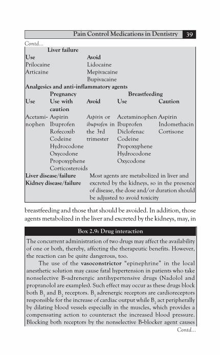
Pain Control Medications in Dentistry 39
breastfeeding and those that should be avoided. In addition, thoseagents metabolized in the liver and excreted by the kidneys, may, in
Liver failureUse AvoidPrilocaine LidocaineArticaine Mepivacaine
BupivacaineAnalgesics and anti-inflammatory agents
Pregnancy BreastfeedingUse Use with Avoid Use Caution
cautionAcetami- Aspirin Aspirin or Acetaminophen Aspirinnophen Ibuprofen ibuprofen in Ibuprofen Indomethacin
Rofecoxib the 3rd Diclofenac CortisoneCodeine trimester CodeineHydrocodone PropoxypheneOxycodone HydrocodonePropoxyphene OxycodoneCorticosteroids
Liver disease/failure Most agents are metabolized in liver andKidney disease/failure excreted by the kidneys, so in the presence
of disease, the dose and/or duration shouldbe adjusted to avoid toxicity
Box 2.9: Drug interaction
The concurrent administration of two drugs may affect the availabilityof one or both, thereby, affecting the therapeutic benefits. However,the reaction can be quite dangerous, too.
The use of the vasoconstrictor “epinephrine” in the localanesthetic solution may cause fatal hypertension in patients who takenonselective B-adrenergic antihypertensive drugs (Nadolol andpropranolol are examples). Such effect may occur as these drugs blockboth B
1 and B
2 receptors. B
1 adrenergic receptors are cardioreceptors
responsible for the increase of cardiac output while B2 act peripherally
by dilating blood vessels especially in the muscles, which provides acompensating action to counteract the increased blood pressure.Blocking both receptors by the nonselective B-blocker agent causes
Contd...
Contd...

Principles of Drug Therapy in Dentistry40
adrenaline to be directed to α-receptors that work by vasoconstrictingperipheral arterioles and hence, high blood pressure.
Cholestyramine (antihyperlipidemia drug) may interfere with theabsorption of acetaminophen, so it should be taken 3 to 4 hoursbefore paracetamol. Paracetamol is primarily metabolized in the liver.The concurrent administration with a hepatic enzyme inducer drugsas tegretol, carbamazepine, rifampin and isoniazid, may reduce theeffects of paracetamol as its metabolism will be increased. Its dose mayneed to be adjusted.
Alcohol use along with any drug metabolized in the liver increasesthe risks of liver damage. The administration of aspirin and live viralvaccine (varicella zoster vaccine) or in the presence of infection maylead to Reye’s syndrome. So, aspirin should be avoided for at least 6weeks. Aspirin also may potentiate the effects of alcohol by increasingthe possibility of gastric bleeding. It was reported that the effect ofaspirin may be reduced if coadministered with ibuprofen, an effectwhich may necessitate delaying ibuprofen 30 minutes after aspirin or8 hours before. Its administration with ketorolac is contraindicated asthis combination is associated with increased tendency towards gastricbleeding and hemorrhage. The effects of warfarin (anticoagulant) andoral hypoglycemic were potentiated when used along with aspirin.Other drugs that may get potentiated if administered with ASA includephenytoin, valproic acid (anticonvulsant), thiopental (ultra short actingbarbiturate) and thyroxin hormone.
Other NSAIDs may interfere with excretion of lithium(antipsychotic/antimanic), methotrexate (for rheumatoid arthritis andpsoriasis) and aminoglycosides leading to their toxicity. They alsoparticipate in increasing the effects of anticoagulant drugs. The effectof antihypertensive drugs appear to be reduced possibly due to theinhibition of prostaglandin which may have a role in regulation ofblood pressure. Celecoxib acts by inhibiting hepatic enzymes and thismay lead to increased level of these drugs metabolized in the liver as β-blockers, antidepressants and antipsychotic. If co administered withaspirin, the risk of gastric ulcer is increased. Fluconazole antifungalmay increase the blood concentration of celecoxib as it may reduce itshepatic elimination.
Opioids should not be administered with drugs that slow brainfunctions (as alcohol, barbiturates, and muscle relaxants) to avoid the
Contd...
Contd...

Pain Control Medications in Dentistry 41
potential risk of CNS and respiratory depression which can bedangerous.
Carbamazepine and phenytoin are enzyme inducers whichincrease the rate of opioid metabolism. In the opposite, quinidineantiarrhythmic agent is enzyme inhibitor and may cause toxicity ofopioid. Opioids may cause severe constipation if given along withantidiarrheal due to the added constipation effects.
Corticosteroids may lead to spread of infection, so should beavoided in episodes of infection and after liveviral immunization. Theeffects of diuretics can be counteracted by cortisole as it causes saltand water retention. Cortisone also counteracts the effect ofhypoglycemic agents as it possesses a hyperglycemic effect. Enzymeinducers also cause increased cortisol elimination.
case of kidney and/or liver failure result in toxicity. These agentsmay be replaced or their dosage modified.
BIBLIOGRAPHY
1. Ashok, Monica, Rashmi, Mahendra. Cyclooxygenase-2 inhibitorsin postoperative pain and chronic pain management. Indian JAnesthesia. 2005;49(3):170-9.
2. Brenadette Jaeger. Non-odontogenic toothache and chronicheadache and neck pains. Endodontics. 5th edn. BC Decker.
2002.3. Cawson, Odell. Cawson’s essentials of oral pathology and
medicine, 7th edn. Churchill Livingstone, 2005.4. Daneil A Haas. An update on analgesics for the management of
acute postoperative dental pain. Journal of the Canadian dentalassociation. 2002; 68 (2).
5. John I Ingle. Differential diagnosis and treatment of dental pain.Endodontics. 5th edn. BC Decker, 2002.
6. K Hrgreaves. Drugs for Pain Management in Dentistry. AustralianDental Journal Medication Supplement, 2005;50:4.
7. Kenneth S Saladin. Anatomy and Physiology, 1st edn. WBC/McGraw-Hill; 1998.
8. MeReC Bulletin. Volume 11. The Use of oral analgesics in primarycare. 2000;11.
Contd...

Principles of Drug Therapy in Dentistry42
9. Mickle Richard. A color handbook of oral medicine. Monson;2004.
10. Norman, Paul. Differential diagnosis of oral and maxillofaciallesions. 5th edn. Elesiver; 2007.
11. Parveen Clark. Clinical Medicine. 3rd edn. ELBS; 1995.12. Paul D Eleazer. Pharmacology for endodontics. Endodontics. 5th
edn. BC Decker; 2002.13. Peterson, Ellis, Hupp, Tucker. Contemporary oral and maxillofacial
surgery. 4th edn. Mosby; 2004.14. Randa, Abeer. Operative Dentistry. MUST. 2008.15. Sukkar, El-Munshid, Ardawi. Concise Human physiology.
Blackwell science; 1998.

Microorganisms, when they gain entry into the body, may be ableto cause infections. Although, the main tool of the dentist is surgery,
INTRODUCTION
Bacterial InfectionsAntibiotics
Mechanism of ActionInhibitors of the Bacterial Cell Wall SynthesisInhibition of Protein SynthesisInhibition of Nucleic Acid Synthesis
Systemic AgentsPenicillinCephalosporinsQuinolonesNitroimidazolesMacrolidesLincomycinsTetracyclinesAminoglycosidesVancomycin
Clinical Use of Systemic AntibioticsManagement of Established InfectionsAdjuvant in the Treatment of Periodontal DiseasesProphylaxis against Surgical Wound Infections (SWI)Prophylaxis against Metastatic InfectionsProphylaxis against Bacterial EndocarditisProphylaxis against Prosthetic Joint Infections
Local and Topical AgentsGeneral ConsiderationsSpecific Considerations
Chapter 3Antimicrobial Agents Used in Dentistry:
Bacterial Infections

Principles of Drug Therapy in Dentistry44
anti-infective agents are sometimes necessary. Because dentists areprescribers, a thorough knowledge about microbiology, pathologyand pharmacology is a must. Indiscriminate drug use may lead tovariable risks ranging from increased burden on the body organs;liver and kidneys, to more serious complications as drug interactionsand allergies. Therefore, a dentist should also know when to providethese agents and what precaution he might need to make. In thischapter, bacterial infections and the most commonly usedantibiotics are of interest. Antifungal and antiviral agents arediscussed in Chapter three and four, respectively.
BACTERIAL INFECTIONS
Most orofacial infections are mixed in nature, being associatedwith aerobic and anaerobic bacteria of both gram-positive andgram-negative species (Box 3.1). Although established infectionsare primarily anaerobic, it appears that anaerobic bacteriaproliferate later as the aerobic species result in reduced oxygenenvironment. This is important as the choice of antibiotic maychange regarding the stage of the infection.
For an infection to get established, bacteria of sufficientvirulence and number should gain entry into the body tissues, a
Box 3.1: Most prominent bacteria in odontogenic infections
AerobicGram (+) ive
Cocci—Streptococci/Staphylococci spp.Rods—Corynebacterium spp.
Gram (–) iveCocci—Neisseria spp.Rods—Haemophilus
AnaerobicGram (+) ive
Cocci—Streptococcus/PeptostreptococcusRods—Lactobacillus/Actinomyces
Gram (–) iveCocci—Veillonella spp.Rods—Provetella/Porphyromonas spp.

Antimicrobial Agents Used in Dentistry 45
process known as invasion. This would mean alteration in thebalance among the normal flora or an introduction of bacteriaforeign to the site of infection. Virulent factors enable bacteria toinduce tissue inflammation and damage by several ways. This includestheir ability to adhere and produce enzymes and toxins. The body,in a way to resist such insults, reacts by an inflammatory process.Inflammation is triggered either by the damaged cells or by thebacteria toxins. Of particular interest are endotoxins, which arethe lipopolysaccharide part of gram-negative cell wall elaboratedwhenever the bacterial cell undergoes division or disintegration(death). Endotoxins cause the release of chemical mediators ofacute inflammation, as IL1, from macrophages and other cells,inducing fever and hypotension.
The role of bacterial enzymes also takes part in theinflammatory process, as well as the final clinical appearance of theinfection. Collagenase producing bacteria, as Streptococcus, areassociated with spreading infections like cellulitis and facial spaceinfections, whereas coagulase-producing Staphylococcus infectionsproduce a localized abscess or infection. Although, this effect is offavorable appearance to the clinician, it is produced in a way toprotect the focus of infection from the immune response. In theselocalized infections, incision and drainage is the best way to allowblood circulation and immune elements to reach the site of infection.
ANTIBIOTICS
Antibiotics are used to augment the body immunity to eradicatethe infection or prevent the condition from becoming worse. Life-threatening conditions, like Ludwig’s angina, may arise from dentalinfections. Bacterial endocarditis is another example of life-threatening infection-related to dental procedures, although therole of the dentist is actually preventive.
The ideal use of antibiotics should be directed to the precisecausative organism with the narrowest spectrum possible. However,most dental infections are treated on presumptive basis with thechoice of empirical antibiotics. Precise determination of the causative

Principles of Drug Therapy in Dentistry46
microorganism is achieved by the procedure of culturing and themost effective antibiotic is determined by the sensitivity tests.However, this seems impractical as most infections get better if notresolved by the time, the results of culture and sensitivity tests show.So, the use of culture and sensitivity tests is reserved for more seriousor rapidly progressing conditions and for those recurring or resistingtreatment. Conditions of compromised host defense also indicatethe use of culture and sensitivity tests.
Antibiotics work on basis of selective toxicity which means thatantibiotics are toxic only to the organisms they were aimed to.However, this appears to be relative as some actions can be exertedon the body. Therefore, antibiotic risks should be kept in mindwhenever prescribed and balanced against the benefits it mayproduce (Box 3.2).
Mechanism of Action
A common classification of antibiotics is based on their mechanismof actions (Table 3.1). Antibiotics are also described according totheir final effect on bacteria as bactericidal (rapidly killing as the β-lactam group) or bacteriostatic (inhibiting like macrolides,
Box 3.2: Major risks of antibiotic use
Allergic reactions ranging from severe anaphylactic reactions to simplerashes
Gastrointestinal irritation like nausea, vomiting, diarrhea andabdominal pain
Hepatotoxicity affecting liver functionsNephrotoxicity affecting renal functionsOtotoxicity due to accumulation in the perilymph and endolymph of
inner earSuperinfections as candidiasis and pseudomembranous colitis; mainly
due to alteration of the normal floraDrug interactions see the interaction boxDrug resistance where the drug is no longer effective on certain
microorganisms

Antimicrobial Agents Used in Dentistry 47
tetracyclines and lincomycins). Whenever possible, bactericidalantibiotics should be used.
Inhibitors of the Bacterial Cell Wall Synthesis
These are bactericidal agent called the β-lactam group. The bacterialcell wall is the organelle responsible for the protection againstenvironmental changes, mainly the osmotic pressure. It is composedof cross-linked peptidoglycans and the cross-linking appears to playa major role in its rigidity. β-lactam antibiotics bind to the penicillinbinding proteins (PBPs) present on the surface of the cell wall. Theprocess of transpeptidization is inhibited and the cross-linkingprocess is stopped. In addition, these antibiotics act by the releaseof autolysin enzymes, by inhibiting their inhibitor enzymes. Autolysinstake part in the remodeling process of the bacterial cell wall, aprocess that loses control when the responsible controlling enzymeis inhibited.
Inhibition of Protein Synthesis
Antibiotics of this group are the macrolides, tetracyclines,aminoglycosides and the lincomycins. All are bacteriostatic; althoughbactericidal in high concentration. Bacterial ribosomes are theorganelles responsible for protein production under the control ofbacterial DNA and RNA. These ribosomes are made of two subunits
Table 3.1: Mechanism of action of most commonly used antibiotics
Mechanism of action Group
Inhibition of cell wall synthesis β-lactam groupPenicillinsCephalosporinsGlycopeptidesMacrolides
Inhibition of protein synthesis LincomycinsTetracyclinesAminoglycosides
Interference with DNA NitroimidazolesFluoroquinolones

Principles of Drug Therapy in Dentistry48
30S and 50S, collectively known as 70S. Some antibiotics act bybinding to the 30S (as tetracycline and aminoglycosides) while othersact on the 50S (like macrolides and lincomycins).
Aminoglycosides block the protein synthesis process by bindingto receptor P12 present on the 30S subunit. Consequently, proteinsynthesis is blocked in addition to misreading of the mRNA and,hence producing non-functional proteins. Some actions also canbe exerted by disruption of polysomes, a strand of ribosomes thatread a specific mRNA to produce proteins. Tetracyclines, on theother hand, act by preventing the attachment of tRNA. Macrolidesand lincomycins act similarly, by blocking the attachment of tRNAto ribosomes. Macrolides bind to receptor 23S rRNA present onthe 50S.
Inhibition of Nucleic Acid Synthesis
Quinolones and nitroimidazoles (Flygel) are two bactericidal agentswhich act by interrupting bacterial DNA. Quinolones act byinhibiting bacterial gyrase, an enzyme responsible for unwindingthe DNA before its replication, thus interfering with bacterialreplication. Metronidazole, on the other hand, combines withbacterial macromolecules, producing DNA dysfunction andstructural defects.
SYSTEMIC AGENTS
The following section is a brief description of the most commonlyused agents in the management of orofacial infections.
Penicillin
Penicillins are the most commonly and safely used antibiotics. Theirnatural origin is from the mold Penicillium notatum. Penicillins actby inhibiting the bacterial cell wall synthesis and are bactericidal inaction. Members of pencillin group are listed in Table 3.2.
Natural penicillins are mainly penicillin G (benzyl P) andpenicillin V (phenoxymethyl P). Penicillin G is active mainly ongram-positive bacteria and some gram-negative ones. Parenteral

Antimicrobial Agents Used in Dentistry 49
administration ensures adequate concentration and thus, this agentis reserved for severe cases and cases where parenteral route isindicated. Penicillin V is more stable in gastric environment thanpenicillin G and, thus, is present in the oral form. Both have similarspectra, with more action on gram-negative bacteria by the PGform.
Amoxicillin, an agent of the aminopenicillin group, haveextended spectrum over the natural penicillins on the gram-negativebacteria, but of lesser effects on the gram-positive ones. It isconsidered one of the safest and most commonly used agents. It iswell-absorbed orally and provides good tissue distribution.β-lactamase inhibitors like sulbactam, tazobactam or clavulanicacid act by increasing the spectrum of penicillins and by resisting
Table 3.2: Members of the penicillin group, route ofadministration and dosage
Members Route Dosage
Penicillin G IV, IM 106 IU /qidPenicillin GAmpicillin O, IV, IM 500 mg/qidAmpicillinFarcocillinAmpicillin- O, IV, IM 375 mg/bidSulbactamUnictamAmoxicillin O, IV, IM 500 mg/qidAmoxicidAmoxilAmoxicillin-clavulanate O, IV, IM 1g/bidAugmentinE-moxcavHibioticCloxacillin- O, IV, IM 500 mg/qidAmpicillinAmpiclox(250+250)

Principles of Drug Therapy in Dentistry50
β-lactamase enzyme. These resistant strains may inactivate β-lactamantibiotics by breaking the β-lactam ring converting them intopenicilloic acid which is the inactive form. Ampicillin is anotheraminopenicillin but with more activity on the gram-positive bacteria.Penicillinase-resistant penicillins, like cloxacillin and dicloxacillin,are resistant to the enzymes produced by resistant strains.
Resistance to penicillin by the following bacteria is documented:Veillonella, prevotella, denticola, S. mitis, S. oralis, S. salivarius andFusobacterium. Resistance is due to the production of β-lactamaseenzymes, absent PBPs or decreased intrabacterial concentration ofthe drug either by decreased permeability or increased efflux.
Penicillins are excreted by the renal system, so care should bepracticed in patients with renal problems.
Common adverse effects of penicillins include GIT troubles(nausea, vomiting, diarrhea), allergy to the β-lactam ring, andsuperinfection (candidiasis or pseudomembranous colitis). Cationictoxicity (with benzyl penicillin) may occur due to the addition ofsodium or potassium, an effect that can be avoided by providing astronger agent with lesser dose and frequency. Other effects includenephrotoxicity and neurotoxicity.
Cephalosporins
Cephalosporins are naturally derived from the Fungi Cephalosporium.They share similar properties with penicillins as they have the sameβ-lacatam ring in their structure and, too, bind to the PBPs.Accordingly, similar spectrum is anticipated, as well as, similaradverse reactions like allergy and resistance.
Cephalosporins are divided into generations according to thespectrum of their activity. First generation act on both gram-positiveand negative bacteria and, hence they are the most useful agents.Second and third generations act more on gram-negative bacteriaand their use in dentistry may be based on culture and sensitivitytests, as in resistant or refractory infections. A fourth generationwas also developed, but its use is mainly restricted to those strains

Antimicrobial Agents Used in Dentistry 51
resistant to the 2nd and 3rd generation. Table 3.3 shows commonagents of cephalosporins and their doses.
Like penicillins, cephalosporins are also excreted by means ofthe renal system and should be provided with care in renal patients.
Common adverse effects are similar to those of the penicillingroup. History of allergy to penicillin indicates possible allergy tocephalosporins and should be avoided accordingly. GIT troublesand superinfections are also possible. Increased bleeding tendencymay be associated with agents that have the tetrazole ring in theirstructure; like cefuroxime. The tetrazole ring acts by inhibiting
Table 3.3: Members of cephalosporins, route ofadministration and dosage
Members Route DosageFirst GenerationCephalexin Oral 250–500 mg/tidKeflexNeocefCefadroxil Oral 500 mg/bidDuricefCurisafeSecond GenerationCefuroxime O, IV, IM 250 mg/bidCefumaxHebiuroximeCefaclor O, IV, IM 250–500 mg/tidServiclorThird GenerationCefixime Oral 200–400 mg/oidXimacefCefotaxime IV, IM 250–500 mg/bidCefotaxClaforanFourth GenerationCefepime IM, IV 500 mg/bidWinCefMaxipime

Principles of Drug Therapy in Dentistry52
vitamin K epoxide reductase enzyme which reduces vitamin K. Thenet result is decreased prothrombine level.
Quinolones
Mainly due to their relative safety and good potency, quinoloneshave gained popularity in clinical practice. The addition of fluorideeven increased their potency. They are bactericidal agents as theyact by inhibition of topoisomerase II (DNA gyrase) and IV enzymes.These enzymes are responsible of changing the DNA shape, but notstructure, during its replication. Inhibiting these enzymes wouldinterfere with DNA synthesis. By means of macrophages and otherinf lammatory cells, f luoroquinolones possess good tissuedistribution and are able to reach bone and sites of inflammation.
Quinolones have good oral absorption, although should notbe taken with antacids, calcium and other ions as this may interferewith its absorption.
Second generation fluoroquinolones (ciprofloxacin andnorfloxacin) act mainly on gram-negative and some gram-positivebacteria, while the agents of the 3rd generation (levofloxacillin)have extended functions on the gram-positive species. The newlydeveloped fourth generation (moxifloxacillin) has even more actionon both gram-negative and gram-positive with some actions onanaerobes. Table 3.4 list common fluoroquinolones and their doses.
Resistance to these agents by pseudomonas and somestaphylococci was reported. These strains may alter their DNAenzymes or cell permeability, rendering these agents ineffective.
Adverse effects to quinolones include GIT troubles (nausea,vomiting and diarrhea), central nervous system symptoms(headache, drowsiness, and insomnia) and connective tissue effects.Quinolones should be avoided in epileptic patients. The effects onarticular cartilage, tendons and other connective tissues were onlyproven on experimental animals, but as a precaution, it should beavoided in children and elderly. It also increases muscle weakness,an effect which contraindicates their use in patients with Myasthenia

Antimicrobial Agents Used in Dentistry 53
gravis. Other adverse reactions include phototoxicity and allergy.Phototoxicity may necessitate the use of sunscreen if exposure tothe sun is mandatory.
Nitroimidazoles
Nitroimidazoles are basically antiprotozoal agents used commonlyto treat amoebic infections. Their effect on anaerobic gram-negativebacteria is the reason behind its popularity. It is the agent of choiceto treat pseudomembranous colitis, a resultant superinfection dueto the use of a broad-spectrum antibiotic. In dentistry, it is commonlyused to treat anaerobic infections caused by black pigmentedbacteria, formerly known as bacteroides, as ANUG and aggressiveperiodontitis. They are also used in colon preparation beforesurgeries as it has anti-inflammatory properties in the large intestine.Nitroimidazoles are effective antidiarrheal agents.
Table 3.4: Members of fluoroquinolones, route ofadministration and dosage
Second GenerationCiprofloxacin Oral 500 mg/bidCiprobayCipromaxCiprofarOfloxacin Oral 200 mg/bidTarivan 200Ofloxin 200Oflicin 200Third GenerationLevofloxacin Oral, IV 500 mg/oidLevoxinLee-floxFourth GenerationMoxifloxacin IM, IV 400 mg/oidAvalox 400Idelox 400Moxacin 400

Principles of Drug Therapy in Dentistry54
Nitroimidazoles act by binding to macromolecules, includingDNA, causing their dysfunction and toxicity. They are bactericidaland have good tissue distribution.
Strains of lactobacillus, A. israelii, P. denticola, E. corrodens andA.a were reported to be resistant to metronidazole. Such resistanceis mainly due to enzyme deactivation, decreased cellularconcentration or even by intrinsic resistance mechanism.
Being metabolized in the liver, metronidazole is affected by theconcurrent use of liver enzyme inducers or inhibitors and precautionsshould be undertaken accordingly (see the interaction box).
Common side effects of these agents include metallic taste (dueto its re-absorption into the enterohepatic circulation), GITtroubles, headache, and peripheral neuropathy. It is associatedwith the characteristic disulfiram reaction or antiabuse effect iftaken with alcohol, so it should not be combined with it. It was alsoreported that nitroimidazoles are potentially mutagenic, so shouldbe avoided in pregnancy. Agents and doses of nitroimidazoles arelisted in Table 3.5.
Macrolides
Literally, a macrolide means a large molecule. Naturally, they arederived from Streptomyces erythreus, and the oldest agent iserythromycin. Erythromycin is considered as an alternative to β-lactam agents when they are contraindicated.
Table 3.5: Members of nitroimidazoles, route ofadministration and dosage
Member Route Dosage
Metronidazole Oral, IV 250–500 mg/tidFlagylAmirzoleMetronalTinidazole Oral 250–500 mg/tidProtozol

Antimicrobial Agents Used in Dentistry 55
They are bacteriostatic, acting by binding to the 50S ribosomalsubunit and, hence, block protein synthesis by the bacterial cells. Inhigh doses, erythromycin is bactericidal. Macrolides have good tissuedistribution and are able to reach the sites of inflammation via theinflammatory cells.
Erythromycin has a similar spectrum to penicillins, being activemainly on gram-positive bacteria. However, erythromycin isinactivated by gastric acid and has poor lipopolysaccharidepenetration. This means that it may have poor effects on gram-negative bacteria. In a way to protect erythromycin from gastricacid, they are either present with enteric coating or in easterifiedform. Newer members like clarithromycin and azithromycin havebeen developed to overcome such disadvantages (Table 3.6).
Clarithromycin is a methylated form of erythromycin, causes lessGIT upset, and enhances patient compliance as it is used twice daily. Itacts on gram-positive as well as gram-negative and anaerobes (P. gingivalisand A.a). It is also excreted in saliva in high concentrations.
Azithromycin adds more compliance as it is used once perday. However, it has lesser activity on gram-positive bacteria whencompared to erythromycin.
Telithromycin, a ketolide, was developed to act on resistantstrains (methylase-producing) and possesses activity like that ofazithromycin.
Table 3.6: Members of macrolides, route of administration and dosage
Member Route Dosage
Erythromycin Oral 250–500 mg/ qidErythrocin 500Erythrin 500Clarithromycin Oral 250–500 mg /bidClaribioticClarithroAzithromycin Oral 250–500 mg /oidAzithrolidZithromax

Principles of Drug Therapy in Dentistry56
Macrolides are metabolized in the liver and excreted in bile.They are liver enzyme inhibitors, with consequent inhibition ofother drug metabolism if taken at the same tame (see the druginteraction box).
Common adverse reactions to macrolides include GITproblems, ototoxicity and cholestatic jaundice, especially with theestolate form. Their use may precipitate acute attacks of porphyria(with sensory and motor neuropathy), so their use is contraindicatedin those patients. Their use is also associated with altered taste, dueto altered salivary composition.
Lincomycins
Naturally produced from Streptomyces lincolnensis, lincomycins’structure may be modified by the addition of chlorine to producethe more commonly used clindamycin. Due to its good distributionin bone and the ability to act on gram-negative bacteria, clindamycinhas gained its popularity for the treatment of odontogenicinfections. Clindamycin gets concentrated in inflammatory cellsand hence, reaches sites of inflammation at high concentrations. Itacts mainly on gram-negative anaerobic bacteria. It is bacteriostaticas it acts on the bacterial ribsosomal 50S subunit, thus, interferingwith protein synthesis. It is metabolized in the liver, so precautionsshould be taken when used in patients with liver disease. Membersand doses are listed in Table 3.7.
One of the common adverse reactions to the use of clindamycinis pseudomembranous colitis, although it might be also caused bythe use of other broad spectrum antibiotics, like cephalosporins.Clostridium difficile is one of the normal flora of the intestine. It is a
Table 3.7: Members of lincomycins, route ofadministration and dosage
Member Route DosageClindamycin Oral, IV, IM 300 mg/tidDalacin-CClindam 300

Antimicrobial Agents Used in Dentistry 57
gram-positive spore forming bacillus which is resistant toclindamycin. It may proliferate when the other members of thebacterial f lora are suppressed by the used antibiotic. Onceestablished, the infection is associated with watery bloody diarrheaalong with necrosis, microabscesses and pseudomembraneformation. The infection is treated by metronidazole. However,severe cases would require the use of vancomycin.
Tetracyclines
Tetracyclines, as the name implies, are cyclic structures composed offour fused rings. They are naturally produced from Streptomyces.
They are bacteriostatic in action, acting by inhibiting proteinsynthesis by binding to the 30S subunit. They are broad spectrum,acting on both gram-positive and negative bacteria. In addition,non-bacterial microorganisms can be affected. A.a and otherperiodontopathogens are susceptible to tetracycline, so it may bechosen to treat aggressive periodontitis or refractory periodontaldiseases. It also possesses non-bacterial properties like anticollagenaseactivity, substantivity and enhancement of reattachment andregeneration of periodontal tissues.
Resistant strains to tetracycline include α-hemolytic Streptococcus(S. mitis) and some anaerobes (P. gingivalis, P. intermedia andBacteroids forsythus). These bacteria are able to avoid intracellularconcentration by diminishing membrane permeability, enhancingdrug efflux, or both. Others are able to produce ribosomalprotecting proteins to destroy or inactivate the agent.
Their use should be avoided with the intake of diary products,antacids or other ions like iron or magnesium, as this may lead tothe formation of non-absorbable chelates. Tetracyclines getconcentrated at liver, kidneys and calcifying tissues, includingneoplasms. They also achieve high salivary concentrations, so localaction can be anticipated. Metabolism is by means of the liver, andthey are excreted in bile where re-absorption into the enterohepaticcirculation may occur. The members of tetracyclines are presentedin Table 3.8.
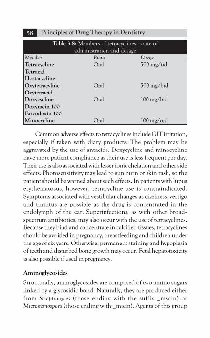
Principles of Drug Therapy in Dentistry58
Common adverse effects to tetracyclines include GIT irritation,especially if taken with diary products. The problem may beaggravated by the use of antacids. Doxycycline and minocyclinehave more patient compliance as their use is less frequent per day.Their use is also associated with lesser ionic chelation and other sideeffects. Photosensitivity may lead to sun burn or skin rash, so thepatient should be warned about such effects. In patients with lupuserythematosus, however, tetracycline use is contraindicated.Symptoms associated with vestibular changes as dizziness, vertigoand tinnitus are possible as the drug is concentrated in theendolymph of the ear. Superinfections, as with other broad-spectrum antibiotics, may also occur with the use of tetracyclines.Because they bind and concentrate in calcified tissues, tetracyclinesshould be avoided in pregnancy, breastfeeding and children underthe age of six years. Otherwise, permanent staining and hypoplasiaof teeth and disturbed bone growth may occur. Fetal hepatotoxicityis also possible if used in pregnancy.
Aminoglycosides
Structurally, aminoglycosides are composed of two amino sugarslinked by a glycosidic bond. Naturally, they are produced eitherfrom Streptomyces (those ending with the suffix _mycin) orMicromonospora (those ending with _micin). Agents of this group
Table 3.8: Members of tetracyclines, route ofadministration and dosage
Member Route DosageTetracycline Oral 500 mg/tidTetracidHostacyclineOxytetracyline Oral 500 mg/bidOxytetracidDoxycycline Oral 100 mg/bidDoxymcin 100Farcodoxin 100Minocycline Oral 100 mg/oid

Antimicrobial Agents Used in Dentistry 59
are listed in Table 3.9. As their use is associated with serious toxicities,they are reserved for severe infections, like those caused by gram-negative aerobic bacilli. So, safer agents should be tried first. Theyalso have a synergistic action if combined with β-lactam antibiotics.In this case, the β-lactam agents serve by disturbing the cell wallallowing more aminoglycosides to enter the bacteria.
Aminoglycosides act on gram-negative aerobic, facultativeanaerobic but not strict anaerobic. Their entry is based on thepresence of oxygen transport system and the lesser oxygenrequirement of the cell, the lesser its concentration intracellularly.They also need what is called membrane pores for entry, a structurepresent only in gram-negative membranes. Therefore, resistantbacteria would be those strict anaerobic, as well as those able toreduce their oxygen uptake. Some strains block the porin channelswhile others produce acetyl transferrase inactivating enzymes. Onceinside the cell, they bind to the 30S protein, thus, inhibiting proteinsynthesis. They also act by interrupting mRNA reading producingdefective, non-functional or even toxic proteins. Although act byinhibiting protein synthesis, aminoglycosides are bactriocidal by aprocess which is still unknown.
Table 3.9: Members of aminoglycosides, route ofadministration and dosage
Member Route DosageStreptomycin IM 1g/oidStreptomycinGentamycin IM, IV 80 mg/tidGaramycin 80Refobacin 80Tobramycin IM, IV 80 mg/bidTobracinNebcinAmikacin IM, IV 100 mg /bidAmikin 100Amikacin 100

Principles of Drug Therapy in Dentistry60
Due to the polycationic structure, these agents have poor oralabsorption, so, given by the parenteral route. Neomycin is highlytoxic to the kidneys and only is present in the topical or oral form(for local treatment of the bowel). Aminoglycosides concentratemainly in the endolymph and perilymph of the ear and the renalcortex, the reason behind their ototoxicity and nephrotoxicity. Inaddition, they are not metabolized in the body; instead, they areexcreted in the active form by the kidneys.
Common adverse reactions to aminoglycosides includeototoxicity. It is the result of destruction of hair cells in the organ ofCorti, which is irreversible. The patient may complain of deafnessand vertigo (loss of balance), the latter is due to the effect on thevestibular apparatus. Reversible, and sometimes, irreversible damageto the kidneys is also possible as these drugs concentrate in the renalcortex. Topically applied agents may be associated with allergicreactions, seen as contact dermatitis.
Vancomycin
Vancomycin, a glycopeptide, is used parentally for severe infectionsas septicemia, severe bone infections, and infections with resistantstrains as MRSA or MRSE. It is also used to treat pseudo-membranous colitis when metronidazole treatment fails. They arealso used for prophylaxis against infections. It is administered byslow IV infusion over 60 to 90 minutes (Table 3.10). No oraladministration is possible, except, for local treatment of the intestinesas in C. difficile infections. It is partially metabolized and excretedby the renal system.
Table 3.10: Members of vancomycin, route ofadministration and dosage
Member Route DosageVancomycin Slow IV infusion 500 mgVancocin 500Vancolon 500

Antimicrobial Agents Used in Dentistry 61
Vancomycin is bactriocidal, acting by the inhibition ofpeptidoglycan polymerization. It also acts by damaging the cellmembrane underneath by interrupting its phospholipids. Its actionis exerted mainly on gram-positive strains including the multidrugresistant Staphylococcus aureus and epidermidis (MRSA and MRSE).Resistance to vancomycin may be the result of altered permeabilityto the drug with or without decreased binding to its receptor.
Common adverse reactions include fever, chills andthrombophlebitis at the venous access site. Rapid IV infusionproduces “red man syndrome”, characterized by flushing and shock.Such effect is produced by the release of histamine and onceoccurred, it can be managed by the administration of antihistamineand corticosteroids. Pretreatment with antihistamine may beprovided as a preventive measure. Other adverse reactions includeototoxicity and nephrotoxicity, especially if co-administered withaminoglycosides.
CLINICAL USE OF SYSTEMIC ANTIBIOTICS
In the following section, the main uses of antibiotics in dentistry willbe discussed. Some conditions requiring antibiotic treatment willbe briefly presented, as regard to the causative microorganism andthe most effective antibiotic to use. Box 3.3 lists the main dentaluses of systemic antibiotics. Table 3.11, provides a summary of thecommon antibiotics along with their spectra.
Management of Established Infections
Although antibiotics only assist the body immunity, they are neededin the management of odontogenic infections. However, not allinfections need antibiotic therapy and the clinician should focus
Box 3.3: Clinical use of antibiotics•Treatment of established infections•Adjuvant in the management of periodontal diseases•Prophylaxis against surgical wound infections (SWI)•Prophylaxis against blood borne “metastatic” infections

Principles of Drug Therapy in Dentistry62
on removal of the cause. Removal of the cause is translated inclinical terms into drainage (by RCT, extraction, or I and D),debridement, cleansing and dressing. These surgical procedures areusually enough as the area of head and neck is rich in blood supply,which is further enhanced by drainage. Usually, the body is able toreverse the condition towards healing without the use of antibiotics.
Table 3.11: Summery of the spectrum of each group of antibiotics
Agent Range of activity
PenicillinsPenicillins G/V Mainly Gm (+)ive with some Gm –iveAmoxicillin Gm –ive but lesser gm (+)ive than
penicillinCloxacillin Active on penicillin—resistant staphCephalosporins1st generation Gm (+)ive and gm –ive2nd, 3rd and More activity to gm –ive. 4th generation4th generation act especially on strains resistant to 2nd
and 3rd generationsMacrolidesErythromycin Gm +ive, like penicillinClarithromycin Gm +ive and gm –iveAzithromycin More activity to gm –ive, less active on
gm +ive than erythromycinTelithromycin Like azithromycin, but more active on
resistant strainsFluoroquinolonesCiprofloxacin Gm –ive and some gm + iveLevofloxacillin Gm –ive with more activity on gm +iveMoxifloxacillin Gm –ive, gm + ive and anaerobesMetronidazole Antiprotozoal with effect on gm –ive
anaerobic bacteriaClinamycin Anaerobic with some gm +ive stainsTetracyclines Gm +ive and gm –iveAminoglycosides Gm –ive/aerobesVancomycin Gm +ive, mainly MRSA/MRSE and C.
difficile

Antimicrobial Agents Used in Dentistry 63
Moreover, if antibiotics are used alone, the irritating focus ofinfection will not be totally removed and relapse will occur. Theiradministration in that case will be like adding risks and burden onthe body. The clinician should be careful whenever he prescribes anantibiotic, regarding its risks and benefits. They should be providedonly when their benefits outweigh risks. If the infection isaccompanied with systemic manifestations as fever >100o F or 38.3to 38.8o C, malaise, lymphadenopathy or trismus, antibiotictreatment is usually indicated. Infection is also associated withelevated blood pressure, high pulse rate (100 bpm), and rapidrespiration (>20/min). Severe conditions should be referred to aspecialist and hospital care may be necessary. These includeconditions with breathing or swallowing difficulties, high grade fever(> 102º F), pulse > 100 bpm, or severe trismus with mouth openingless than 10 mm.
Certain odontogenic infections are known to usually have thesystemic manifestations previously described. So, they most probablyrequire antibiotic treatment along with the surgical part. Box 3.4lists the common odontogenic infections indicating use of antibioticsand Box 3.5 lists those which do not. Generally, an empiricalantibiotic is used on a presumptive basis according to the knowledgeof the causative organism and the antibiotic spectrum. Of course,the best method is to have culture and sensitivity tests to provide thebest matching antibiotic and prevent unnecessary overdose or wrongantibiotic. This procedure is known to take time, and forconvenience, it is performed in persistent infections, recurrentconditions, dangerous or rapidly spreading diseases, and in case ofcompromised host defense. The empirical use of antibiotic is startedand maintained until the results from the lab show.
Acute pericoronitis, inflammation of the pericoronal flap(operculum), is usually associated with a partially erupted tooth.The etiologic cycle begins once inflamed as food entrapment andtrauma from opposing dentition aggravate the condition. Inmoderate to severe conditions the following symptoms are present:fever, malaise lymphadenopathy, and trismus. Advanced conditions

Principles of Drug Therapy in Dentistry64
Box 3.4: Established infections in relation to oral and facial tissuesAcute Periodontal Conditions
Acute pericoronitis (moderate-severe)Necrotizing periodontal conditionsDisseminating periodontal abscessSpecific bacterial infections:
N. gonorrhoeaeS. gingivostomatitis
Acute Periapical ConditionsAcute apical abscessAcute exacerbation of chronic abscess (phoenix)
Major Infections of Bone and FaceCancrum oris “noma”Fascial space infections “cellulitis”OsteomyelitisOsteoradionecrosisActinomycosis
Salivary Gland InfectionsBacterial sialadenitis
Trauma Related ConditionsExtensive lacerationVisibly contaminated woundsDelayed wound managementContaminated open fractures
Other Infections of Oral RelevanceTuberculosisLeprosySyphilisScarlet fever “scarletina”
Box 3.5: Infections not requiring antibiotic useChronic gingivitisMild-moderate pericoronitisNon-disseminating periodontal abscessChronic draining abscessAs a complement in RCTDry socket
Antimicrobial Agents Used in Dentistry 65
are associated with pericoronal abscess which may spread toadjacent facial spaces. The associated microorganisms includeBorrelia vincentii and Fusiformis dentium. The condition is besttreated with amoxicillin with metronidazole and a mouthwash.However, the condition needs debridement with drainage (byanteroposterior incision) in case of abscess in fluctuant stage. Furthertreatment considerations include operculectomy or extraction fordefinite treatment of the case. Acute pericoronitis may beaccompanied with ANUG.
ANUG is usually caused by several factors, most important ofwhich is the underlying systemic condition. Usually, stress,malnutrition, smoking or debilitating diseases are present. Clinically,ANUG is associated with acute inflammation, necrosis andulcerations. The infection is described as a fusospirocheal one withan invasive component, a factor important to be considered duringits management. The usual sings and symptoms include fever, GITtroubles, fetid odor and increased salivation. The condition isinitially treated by debridement (with hydrogen peroxide swap undertopical anesthesia), supragingival scaling and medications. Thefollowing antibiotics have been frequently used: amoxicillin ormacrolides with metronidazole. The condition needs several recallvisits and the underlying local and systemic factors should becorrected. The condition may be mistaken with several otherconditions as streptococcal gingivostomatitis, gonococcal gingivitis,diphtheria, or acute herpetic gingivostomatitis.
Streptococcal gingivostomatitis is a gram-positive coccalinfection caused by S. viridans advancing from inflamed tonsils.The condition is not associated with linear erythema as in ANUG.Instead the gingiva is diffusely inflamed. In addition, neither necrosisnor fetid odors are present. The condition is managed byamoxicillin or erythromycin.
Gonococcal gonorrhea is a gram-negative infection caused byNeisseria gonorrhoeae, and is one of the sexually transmitted diseases.It is associated with sore throat and suppurative inflammation

Principles of Drug Therapy in Dentistry66
with pseudomembrane slough and ulcers. The condition is treatedby amoxicillin, ciprofloxacin or cephalosporins.
Diphtheria, caused by the gram-positive Corynebacteriumdiphtheriae, is characterized by sore throat with tonsillar lesionsand pseudomembrane which is difficult to wipe off. Fever, malaiseand lymph node enlargement may also be present. The conditionmay get complicated by mycocarditis or paralysis. It is treated byantibiotics (penicillins or erythromycin) and antitoxins.
ANUG may progress to involve the investing bone causingNUP. It may involve other facial tissues causing more seriouscondition known as cancrum oris or noma. The disease involves anecrotizing and gangrenous process causing a cone-shaped lesionwith the apex directed superficially. The condition needs antibiotictreatment with amoxicillin or aminoglycosides combined withmetronidazole. Supportive treatment and local debridement areof great importance.
Severe periodontal abscess may be associated with throbbingradiating pain and systemic manifestations. Drainage, eitherintrasulcularly or by external I and D, is necessary. The conditionshould be differentiated from acute apical abscess as the nonvitaltooth will require drainage through RCT.
Acute apical periodontitis (AAP) is moderate to severeinflammation of the apical periodontal ligament and it is associatedwith severe pain. It may result from mechanical, chemical or biologicinsults introduced to that tissue either by improper RCT or by thecaries process. Analgesics, antibiotics and drainage along withocclusal relief are needed. The condition may be converted to acuteapical abscess (AAA), which is also a painful condition. Thecondition is associated with swelling and expansion which is moreapically located when compared to lateral periodontal abscess. Theinvolved tooth is nonvital or with large caries/metallic restorations.It needs drainage, either through RCT or by extraction, antibioticsand analgesics. The chosen agents include amoxicillin or penicillin(but not augmentin, as it was found to be of no more benefit).

Antimicrobial Agents Used in Dentistry 67
Metronidazole may be added to cover the anaerobic species.Clarithromycin (but not erythromycin as it is less effective onanaerobes and gram-negative) or clindamycin can be used assubstitutes. AAA may be superimposed on chronic apical one, acondition known as recrudescent abscess. The condition may becaused by blockage of the spontaneous drainage provided by theformed fistula. Its clinical presentation is similar to AAA andrequires the same treatment. AAA may also spread into tissue spacescausing fascial space infections.
Fascial space infections are spreading in nature withinflammatory edema and microorganisms entrapped in the tissuespaces. Pain, swelling, fever, malaise, and lymph node abnormalityare usual. Usually, penicillin (with cloxacillin for resistant Staph.),or clarithromycin are used. A serious complication as a result ofspreading of facial cellulitis is Ludwig’s angina. It is a bilateralinvolvement of submental, sublingual and submandibular spaceswith board-like brawny swelling. It may be related to infection spreadfrom the 2nd or 3rd molars. Difficulty in breathing, swallowing orboth may necessitate hospital care. Its treatment follows the samegeneral principles of drainage and antibiotics, however. Anotherserious sequel of infection spread in the fascial spaces is the cavernoussinus thrombosis which may lead to blindness or even death. Insuch a case, antibiotics, anticoagulants, and surgical drainage arethe required treatment. Antibiotics should be administered toprevent such dangerous conditions.
Osteomyelitis, inf lammation of bone marrow andendosteum/periosteum compartments, may require prolongedantibiotic treatment, ranging from four to eight weeks for the acutetype, while reaching up to six months in the chronic one. Theresponsible microorganisms include Peptostreptococcus and otheranaerobic gram-negative species. The chosen type of antibiotic isusually clindamycin, owing to its ability to concentrate in bone.Other antibiotics used include ciprofloxacin and cephalosporins.Management is by surgical debridement (to remove infected plate,wire or teeth) sequestrectomy, hyperbaric oxygen therapy, treatment

Principles of Drug Therapy in Dentistry68
of the underlying systemic factors (nutrition, immune suppression,debilitating diseases).
In cases of irradiated jaws, osteoradionecrosis is one of thecomplications that requires months, if not years, in order to heal.Osteoradionecrosis is a hypoxic nonhealing lesion that may occurin bones exposed to high doses of radiotherapy (6500 rad or 65Gy).The lesion itself is not an infection and the use of antibiotics iseither to prevent or treat secondary bacterial infections. Onceestablished, the formed sequestration should be removed, antibioticshould be administered and considerations about hyperbaricoxygen therapy should be made. Such patients may require antibioticadministration as prophylaxis to prevent infections in case ofextraction, implant placement or RCT.
Actinomycosis is a soft tissue infection which tends to localizein the form of pseudotumor in area close to the neck. It is dusky-redin color and has multiple sinus tracts. The causative microorganismsinclude Actinomyces israelii and A. viscosus (anaerobic gram-positive).The infection is quite stubborn requiring long time to heal. Theused antibiotics include: penicillin (IV), amoxicillin, tetracycline orerythromycin. In addition, I and D and excision of the sinus tractsshould be done.
Bacterial sialadenitis is an infection of the major salivaryglands. It is usually mixed in nature with pain, erythema, andsuppuration. Its incidence is associated with poor nutrition,dehydration or chronic illness, all of which can be present in elderly.Staphylococuss aureus is associated with the infection. The infectionneeds antibiotic treatment and the following are commonly used:penicillin, amoxicillin or 1st generation cephalosporin. Cultureand sensitivity tests may be required, however.
Trauma-related infections that need antibiotic treatmentinclude extensive lacerations, contaminated wounds and delayedwound management (> 3 h). Extensive lacerations will also requiremultilayered suturing in addition to the antibiotic regimen.Contaminated wounds would also need hemostasis, cleansing,debridement and closure. The usual antibiotic prescribed is

Antimicrobial Agents Used in Dentistry 69
penicillin. In case of tooth avulsion, antibiotics are provided toguard against any possible infections as the re-implanted tooth isconsidered infected. Other treatment considerations include tetanusboost or vaccine, splinting, recall visits and RCT. Contaminatedopen fractures or delayed management of clean open fractures,too, are assumed to be infected and require antibiotic treatment.Open or severe fractures are at high liability to get infected and,hence, the use of antibiotics is justified.
Scarlet fever is a gram-positive streptococcal infection of thepharynx. Fever, vomiting and lymphadenopathy are usually present.The tongue presents characteristic lesion during the early and latephases of the infection. Early lesion is characterized by prominenterythemic papilla over a white coat producing a “strawberry”appearance. The late lesion is devoid of the white coat, hence,resembles “raspberry”. The lesion is treatable with penicillin,cephalosporin or erythromycin.
Tuberculosis is anaerobic infection with the acid fast bacillusMycobacterium tuberculosis. It is related to the oral cavity as it may bemanifested in the form of ulcers which have characteristic punchedout appearance with everted edges. Scrofula, which is massive cervicallymph node enlargement with discharging sinus, may be seen too.The case would require consultation and treatment by a specialist.
Syphilis, a spirocheatal infection with the gram-negativeTreponema pallidium, manifests itself in the oral cavity as ulcersthrough its three stages. Accordingly, chancre, snail tract ulcers, ordeep punched out ulceration may be present. The condition shouldbe referred to the specialist of interest for treatment.
Leprosy, an infection caused by Mycobacterium leprae, causesoral and cutaneous hypopigmented lesions as well as, loss of sensoryfunctions. This condition, too, should be referred to a specialist.
Adjuvant in the Treatment of Periodontal Diseases
Certain forms of periodontitis may require the use of suitableantibiotics along with the conventional periodontal therapy andoral hygiene measures. Scaling and root planning should be done

Principles of Drug Therapy in Dentistry70
first and the patient cooperation should be assured. Some formsof periodontitis show no response to the provided conventionaltherapy and these are: Refractory periodontitis, aggressiveperiodontitis and periodontitis as a manifestation of systemic disease(Box 3.6). The use of antibiotics, either systemically or locally, aidsthe conventional therapy by providing a means of accessibility todeeper sites.
Refractory periodontitis is defined as continuous loss ofclinical attachment despite instrumentation and good oral hygieneunder healthy endodontic conditions. The condition may beassociated with deep invasions by the causative microorganisms,defective host defense, or both. Culture and sensitivity tests may beneeded to identify the causative organisms and the most matchingantibiotic against them. The following antibiotics may be useful:Augmentin, clindamycin, tetracycline, ciprofloxacin, azithromycinand metronidazole. Sometimes, it might be necessary to combinetwo antibiotics together in order to increase the spectrum and tolower the individual doses. This usually means the addition ofmetronidazole to another agent. Such therapy may eliminate theneed for surgical periodontal therapy.
Aggressive periodontitis have characteristic clinical featuresof rapid bone loss in relatively young individuals. The radiographicappearance of angular arch-like defects localized to the molar/incisor (LAP) area is noticeable. The associated microorganismsinclude A.a and P. gingivalis, which are virulent and invasive. Apossible defective immunity, as well as, cementopathy may be alsopresent. The condition may respond to conventional therapy,modified Widman flap and tetracycline given at the dose of
Box 3.6: Periodontal conditions requiringantibiotic administration
Refractory periodontitis (RP)Aggressive periodontitis (AP)Periodontitis as manifestation of systemic diseaseImplant associated infections (peri-implantitis)

Antimicrobial Agents Used in Dentistry 71
1 g/day in divided dose for two weeks. Recall visits should be madeevery month and good oral hygiene should be optimized. Anotherantibiotic regimen includes the combination of amoxicillin andmetronidazole. The generalized form may be treated as refractoryperiodontitis.
Periodontitis as manifestation of systemic disease is associatedwith lowered immune response which may be associated withhematological, as leukemia and leukopenia, or genetic diseases, asDown syndrome. It is differentiated from aggressive periodontitisin that the medical problem is a major factor in the disease process,while in AP, it is not. The medical evaluation is mandatory. Theconventional therapy may be augmented by the use of antibioticsaccording to the results of culture and sensitivity tests, however.
In implant-associated infections or peri-implantitis, bone lossand inflammation may lead to implant failure. Antibiotics areadministered (for 10 days) as a nonsurgical solution which mayspare the patient from additional surgeries. In this case, it is clearthan the benefits of antibiotics outweigh their risks. Conventionaltherapy and good oral hygiene are mandatory.
Prophylaxis against Surgical Wound Infections (SWI)
Some surgeries, especially major ones, are at high susceptibility toget infected even when strict aseptic principles are adhered to. Inaddition, contamination may be present in some injuries.Antibiotics are provided to prevent the establishment of the infectionwhich delays healing and needs higher doses and longer periods ofantibiotic therapy. Prophylaxis, when indicated, uses shorter timeand lower dose of antibiotics than that used to manage establishedinfections. This means lesser chances of developing bacterialresistance, lesser burden on the body and better healing potential.So, prevention of SWI would help both the patient and the dentist.The procedure must be as aseptic as possible, though. Thoseconditions at low risks of infections should not receive antibioticsas their use is not justified, except in those with defective immunity.

Principles of Drug Therapy in Dentistry72
The conditions at higher risks are described in the following sectionsand are listed in Table 3.12.
Cases requiring antibioticprophylaxis
Class of surgery•Major oral surgery class I,
extraoral surgery when duration> 2 h
•Combined intraoral/extraoralapproach (II surgeries)
•Major oral surgeries involvingthe manipulation of infectedtissues (III and IV surgeries)
Trauma-related cases•Extensive lacerations•Closed fractures if managed by
intraoral approach, or whenthe duration > 2 h
•Open fractures, even when firstseen
Cases not requiring antibioticprophylaxis
•Minor oral surgeries (frene-ctomy, minor alveoplasty,biopsy, small bone lesions)
•Major class I surgery < 2 hduration
•First seen lacerations < 3 h•Small, clean lacerations•Closed fractures managed by
extraoral approach within 2 h
Table 3.12: Surgical wound prophylaxis, the recommendedconditions vs. low risk cases
Cases irrespective of class of surgery or duration•Patient with ASA* > 2•Patient with poor immunity•Preoperative stay > 3 days•Placement of implant•Placement of grafts
* ASA is the classification of American Society of Anesthesiologistsand is indicated as follows: ASA 1 = healthy, no medical problems,ASA2 = mild systemic disease which is well controlled, ASA3 =severe systemic disease or controlled disease of more than one systemwhich is not incapacitating but with some functional limitations,ASA4 = severe systemic disease which is a constant threat to life (oran end-stage disease), ASA5 = morbid, not expected to live for 24 hwithout an operation, ASA6 = declared brain-dead patient.

Antimicrobial Agents Used in Dentistry 73
Regarding the class of surgery (Table 3.13), major oralsurgeries, either intraoral or combined intraoral/extraoral(class II), even if kept clean, will need antibiotic prophylaxis. It isdifficult to attain aseptic field in the intraoral environment whencompared to that of skin. Extraoral surgeries (Class I) do not needantibiotic prophylaxis as they are clean in nature. However, class Isurgeries are prone to infections when the procedure takes morethan two hours. Minor oral surgeries, like extraction, frenectomy,minor alveoplasty, are at lower risk of infections and, hence, noantibiotic is required. Surgeries that involve the manipulation ofcontaminated tissues are associated with major break through inthe aseptic technique. Antibiotics should be provided as the risk ofinfection is high.
Traumatic injuries of the soft tissues may require prophylaxisif they exhibit extensive laceration or when they are visibilitycontaminated, even if first seen. Small, noncontaminated and firstseen lacerations are at lower risks of developing an infection.However, if management is delayed more than three hours, thebacterial population increases to a level that these injuries areconsidered infected. Open fractures of the facial bones also needprophylaxis as they are at high risk of infection. Antibiotics shouldbe provided immediately and maintained till get managed. If the
Table 3.13: Classes of oral and maxillofacial surgeries
Minor surgeries Simple surgeries, e.g. impactions, alveoloplastyClass I Clean surgery (at skin), in clean aseptic field, e.g.
TMJ, parotid gland surgeriesClass II Clean-contaminated, combined intra- and extra-
oral approach, or visibly contaminated, e.g. majortumor surgery, major preprosthetic surgery
Class III Contaminated surgery, acute inflammation, orwith major breakthrough the aseptic technique,e.g. compound fractures
Class IV Dirty, infected (with pus) surgery, e.g. visiblycontaminated wounds

Principles of Drug Therapy in Dentistry74
management is delayed for more than three hours without the useof antibiotics, the wound is considered infected. Closed fractures,on the other hand, require prophylaxis, if to be managed byintraoral approach or when the procedure itself requires morethan two hours. Otherwise, first seen closed fractures managedextra orally and within two hours do not require prophylaxis.
Some conditions may require antibiotic prophylaxis even whenthe condition or the surgery does not indicate so. Patients withdebilitated immunity either by a systemic disease or local factors (asradiotherapy) are at higher risks to develop infections and will needmore time and more aggressive therapy to treat established ones.
Generally speaking, patients with ASA > 2 require antibioticprophylaxis to prevent life-threatening infections (Table 3.12).Implants and bone grafts induce local changes that the host defensemechanism is altered. Both procedures are invasive in nature,expensive and time consuming. Antibiotic prophylaxis is considereda safe and cheap option when compared to the possible infection.
The most commonly involved microorganisms, regardingorofacial surgeries, include aerobic gram-positive Streptococcus,anaerobic gram-positive Peptostreptococcus and rods. It appearsthat the aerobic stains should be primarily covered, while anaerobicones do not need complete coverage. Accordingly, penicillins, butnot amoxicillin or ampicillin, erythromycin and vancomycin are goodchoices. First generation cephalosporins also provide good results,while other generations focus more on the gram-negative species. Incase of extraoral surgeries, cloxacillin is added to penicillin in orderto cover the resistant Staphylococcus aureus strains. Vancomycin isespecially active on those strains, as well as, on Streptococcus.
Oral route of administration is an acceptable and less invasiveone. It is used in surgeries in which local anesthesia was used. Oraladministration should start one to two hours before the surgery.Parenteral route (slow IV injection or continuous infusion) is usedin case of general anesthesia, high-risk patients, and in those unableto take anything by the mouth. In these patients, antibiotics shouldbe started at the time of induction. Although double the therapeuticdose, prophylactic antibiotics are provided in single doses. The

Antimicrobial Agents Used in Dentistry 75
duration at which intraoperative doses are provided is half that ofthe therapeutic one (Table 3.14). No postoperative doses arerequired.
Prophylaxis against Metastatic Infections
Metastatic infections are defined as infections developing at distantor anatomically separate locations of their original site.Microorganisms gain entry through oral tissues to infect the heartor artificial joints. For a metastatic infection to occur, three factorsshould be concurrently present: (1) susceptible tissue (valvulardefect), (2) microbial seeding mechanism (surgery or bleeding); and(3) impaired host defense. The most dental procedures associatedwith significant bacteremia as well as those which can be done withoutrisk of developing an infection are listed in Box 3.7.
Prophylaxis against Bacterial Endocarditis
Bacterial endocarditis is the infection and inflammation of theendocardium or heart valves which are usually defective. The defectmay be produced by turbulent blood flow which causes loss ofendocardium and exposure of the collagen layer which gets covered
Table 3.14: Antibiotics commonly used in prophylaxis of SWI,route, dose and duration. Comparative therapeutic doses and
duration are also present for comparison
Agent Route Therapeutic First Intra-dose prophylactic operative
dose dose
Penicillin G IV 1 mega unit/6 h 2 mega units Every 3 hPenicillin V Oral 500 mg/6 h 1 gramCloxacillin Oral/IV 500/6 h 1 g 2–3 hCephalosporins 500/6 h 1 g 3 h(Cefadroxil)Clindamycin Oral/IV 300 mg/6 h 600 mg 3 hErythromycin Oral 400 mg/12 h 800 mg 6 hVancomycin IV 500 mg/6 h 1 g 3 h
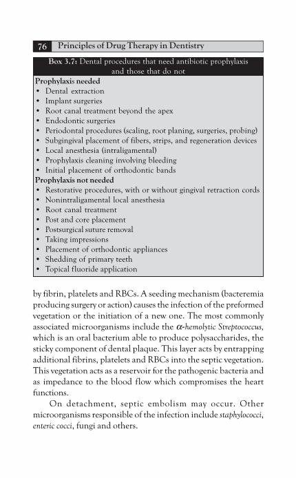
Principles of Drug Therapy in Dentistry76
by fibrin, platelets and RBCs. A seeding mechanism (bacteremiaproducing surgery or action) causes the infection of the preformedvegetation or the initiation of a new one. The most commonlyassociated microorganisms include the α-hemolytic Streptococcus,which is an oral bacterium able to produce polysaccharides, thesticky component of dental plaque. This layer acts by entrappingadditional fibrins, platelets and RBCs into the septic vegetation.This vegetation acts as a reservoir for the pathogenic bacteria andas impedance to the blood flow which compromises the heartfunctions.
On detachment, septic embolism may occur. Othermicroorganisms responsible of the infection include staphylococci,enteric cocci, fungi and others.
Box 3.7: Dental procedures that need antibiotic prophylaxisand those that do not
Prophylaxis needed• Dental extraction• Implant surgeries• Root canal treatment beyond the apex• Endodontic surgeries• Periodontal procedures (scaling, root planing, surgeries, probing)• Subgingival placement of fibers, strips, and regeneration devices• Local anesthesia (intraligamental)• Prophylaxis cleaning involving bleeding• Initial placement of orthodontic bandsProphylaxis not needed• Restorative procedures, with or without gingival retraction cords• Nonintraligamental local anesthesia• Root canal treatment• Post and core placement• Postsurgical suture removal• Taking impressions• Placement of orthodontic appliances• Shedding of primary teeth• Topical fluoride application

Antimicrobial Agents Used in Dentistry 77
Once established, infective endocarditis is manifested by fever,vasculitis, heart murmur and septic embolism. It also can lead toserious results as damage to the heart valves, impeded heart function,heart failure, renal failure or cerebral embolism.
The condition is managed by penicillin/gentamicincombination through IV administration for four to six weeksfollowed by oral antibiotics for two to four weeks. In addition, asurgery may be required to repair the resultant defect.
The best management of such a case is to prevent its occurrence.Patients at high or moderate risks should receive antibioticprophylaxis (Box 3.8), while those at low risk do not. Therecommended regimen is shown in Tables 3.14 and 3.15.
Box 3.8: Patient classification according to degree of risks todevelop bacterial endocarditis
High-riskProsthetic valvesHistory of previous endocarditisCongenital heart disease (cyanotic)•Teratology of Fallot•Transposition of great arteries•Single ventricle defectSurgically constructed pulmonary shuntsModerate riskCongenital defects other than high-riskAcquired valvular dysfunction (rheumatic heart disease)Mitral valve prolapse with valvular regurgitation/thickened leafletsHypertrophic cardiomyopathyLow-risk (antibiotics not indicated)Mitral valve prolapse without valvular regurgitationPrevious rheumatic fever or Kawasaki disease without valvular
dysfunctionPrevious coronary artery bypass surgeryCardiac pacemakers and defibrillatorsIsolated atrial septal defectPhysiologic innocent heart murmurs

Principles of Drug Therapy in Dentistry78
Patients with previous history of infective endocarditis havehigher rates of developing vegetation and infection. In addition,they may have prosthetic valves installed to repair the damageinduced by the previous infection, which by itself increases thepossibility of developing the infection. In addition, the tissues aroundthese prosthetic valves may get inflamed and they may get loose,which is dangerous.
Teratology of Fallot is a cyanotic heart disease associated withfour defects occurring together, and these are: Ventricular septaldefect, pulmonary artery stenosis, overriding aorta and rightventricular hypertrophy. The patient is seen with dyspnea, fatigue,and hypoxic episodes. Finger clubbing and polycythemia are alsoseen.
The patient general growth is retarded. The condition itselfneeds corrective surgery and is of high-risk to develop bacterialendocarditis.
Ventricular septal defect (VSD) alone is of moderate risk. It isthe condition in which the left ventricle communicates with theright one. Because it is stronger, the left ventricle forces blood intothe right ventricle which pumps it into the pulmonary circulation.The pulmonary pressure is increased reaching that of the systemicpressure. Blood turns back through the defect to the left ventricle(reversed shunt). The defect should be surgically corrected. Othercongenital defects are of moderate risks.
Table 3.15: Prophylactic oral antibiotics* against IE,agents and doses
Member DosageAdults Children
Amoxicillin 2 g 50 mg/kgClindamycin 600 mg 20 mg/kgCephalexin/cefadroxil 2 g 50 mg/kgAzithromycin 500 mg 15 mg/kgClarithromycin
* Administration should start one hour before operation

Antimicrobial Agents Used in Dentistry 79
Atrial septal defect, of low-risk group if isolated, is acommunication between both atria which causes blood to passfrom the left atria to the right one which gets dilated. Pulmonarypressure increases and the patient may complain of dyspnea. Atrialarrhythmia, fibrillation and in later stages, right sided heart failuremay occur. If significant, it may need surgical correction before theage of ten years.
Acquired valvular dysfunction may follow Kawasaki’s diseaseor the more common rheumatic heart fever. Rheumatic heartfever is an inflammatory disease which is common among children.It is caused by α-hemolytic Streptococcus reaching the heart fromoropharyngeal infections. Organs other than the heart like: skin,joints and CNS are also affected. The causative bacteria induces anautoimmune reaction in which the produced immunoglobulinsact by damaging the heart (endocardium, myocardium andpericardium are all affected). The damaged valves are associatedwith regurgitation in the blood flow. The patient complains ofpainful joints (migratory polyarthritis), fever, malaise, anorexia,murmurs, spasmodic movements and speech alteration (CNS), anderythema marginatum (pink skin rash with slightly raised edges).The condition is treated with penicillin and the patient undergoesmonthly penicillin prophylaxis till the age of 25 years or up to fiveyears after the initial incidence. In such patients, the choice ofantibiotic prophylaxis for dental procedure should be an agentother than β-lactam group (erythromycin is a good substitute) asbacteria may be resistant to them.
Kawasaki’s disease (Japan) is another acute condition affectingchildren. It is of unknown etiology, but a role of immune reactionis implemented. The condition is associated with fever persisting formore than five days, generalized vasculitis (including the coronaryartery), unilateral lymphadenopathy, mucocutaneous and ocularlesions. Serious cardiac complications as dysrhythmia and aneurysmof the coronary artery may predispose to myocardial infarction.The condition is treated by daily aspirin (80–100 mg/kg/day) andsingle dose of gamma globulin.

Principles of Drug Therapy in Dentistry80
Mitral valve regurgitation is the backflow of blood throughincompetent valves into the left ventricle with atrial dilatation. Itmay be caused by rheumatic fever, aortic valve disease, myocarditis,infective endocarditis, and collagen disorders like Marfan’ssyndrome and Ehlers Danlos syndrome. The patient may complainof palpitation, dyspnea, orthopnea, and fatigue. Anticoagulants,diuretics and ACE inhibitors may be provided. A surgery to repairor replace the mitral valve is needed.
Mitral valve prolapse, another moderate risk condition, iscaused by excessively large leaflets of the valves or enlarged mitralannulus (opening). Long chordae tendineae and defective papillarymuscles may also be the cause. Significant regurgitation may warrantvalvular surgery. However, if insignificant, drug therapy alone (anti-arrhythmia and β-blockers) may suffice and in that case, noantibiotic prophylaxis is needed.
Hypertrophic cardiomyopathy is associated with enlargedthin-walled left ventricle, which exhibits poor functions and lowcardiac output. The valvular movements are reduced and the leftventricular contractions are disorganized. Syncope, dyspnea, angina,arrhythmia and even sudden death may occur. The condition istreated with antiarrhythmic, β-blockers and surgical repair.
Previous coronary artery bypass surgery to managemyocardial infarction, by reconstruction of the coronary arterywith vein graft form other site of the body, is not susceptible toinfection, so, no need for antibiotic prophylaxis.
Cardiac pacemakers, devices implanted in the chest with wirerunning through the superior vena cava to reach the right side ofthe heart, are also of low-risk group.
Prophylaxis against Prosthetic Joint Infections
Implanted prosthetic joints to replace damaged ones may get infectedafter oral procedures or even by established odontogenic infections.In these patients, infections should be managed aggressively toprevent infection and failure of prosthetic joints. Oral procedures

Antimicrobial Agents Used in Dentistry 81
mentioned in Box 3.7 also apply to prosthetic joint infection.Similar antibiotic regimen is also used. However, only certainpatients will require the prophylaxis (Box 3.8).
LOCAL AND TOPICAL AGENTS
To have a direct effect on the infecting microorganisms, it seemslogical to apply the antimicrobial agent directly to the infected site.Natural antimicrobial compounds as immunoglobulins and salivaryenzymes produce similar effects. It was also thought that the greaterthe systemic antibiotic concentration in serum, the more effective itis in the treatment of periodontal diseases. Local application ofantibiotics, especially in the management of periodontal diseases,should maintain constant therapeutic concentration in the face ofsmall periodontal pocket/sulcus area and the continuous washingaction of the gingival crevicular fluids. If these two obstacles aremanaged, it would be possible to avoid the use of systemic antibiotics.In addition, the chance of tissue invasion at recall visits byinstrumentation with or without surgery is reduced.
Application methods are various with different degree ofsuccess. Topical application by direct placement or mouthwashprovides only transient and superficial effects. This method is usedas a preventive measure, as well as, in providing transient therapeuticeffects. If applied in pocket therapy, mouthwashes provide onlyfour percent pocket penetration. The use of irrigators (supragingivalor subgingival) provide deeper pocket penetration. However, theused irrigant will be washed away rapidly by the flow of gingivalfluid. The installation of slow releasing device at the pocket ensuresadequate concentration of the active agent at longer periods. Theused local devices include polymers, dialysis tubing, chips or gelsthat harden on time. After the therapeutic period, these devicesshould be removed. In the following section, the commonly usedagents in the prevention and treatment of oral infections are brieflydiscussed.
Chlorhexidine digluconate (0.12%), is an antiseptic agentwhich is active on gram-negative and gram-positive bacteria, as well

Principles of Drug Therapy in Dentistry82
as, other microorganisms like fungi. The drug acts by membranedisturbance action. It can be used locally to reduce microbialpopulation and interrupt plaque layer (therapeutic). It also impedesplaque formation as it adheres to the dental pellicle and preventsthe adherence of bacteria, a property known as substantivity. Theaddition of f luoride adds additional anticaries as well asantibacterial effects (Table 3.16).
It is better to be used after flossing and brushing to allowchorohexidine binding to the dental pellicle and to avoid itsdeactivation by the anionic compounds present in the toothpaste.In addition, food and smoking should be avoided after using thisagent for the same reasons. As a mouth wash, it should be used inundiluted form for 30 seconds twice daily. It is also considered asone of the best solutions for irrigation systems in concentration upto 2.5 percent. It is also used with acrylic chips or biodegradablegelatin chips for local application.
Common side effects of chlorhexidine include: staining of teethor restorations, allergic reactions, parotid gland swelling, increasedcalculus formation and altered taste sensation.
Table 3.16: Prophylactic parenteral* antibiotics,agents and doses
Member DosageAdults Children
Ampicillin 2 g 50 mg/kgClindamycin 600 mg 20 mg/kgVancomycin† 1 g 20 mg/kgHigh-risk patientsAmpicillin + 2 g 50 mg/kgGentamicin 1.5 mg/kg 1.5 mg/kg**Vancomycin† + 1 g 1.5 mg/kgGentamicin 1.5 mg/kg 1.5 mg/kg**
* Given 30 minutes before surgery.** Child dose should not exceed adult dose.† Given in slow IV infusion at 1 to 2 hours.

Antimicrobial Agents Used in Dentistry 83
Hydrogen peroxide is a pale-blue, weak acid but strong oxidantand antiseptic. It is naturally produced by oxygen metabolism inaerobic bacteria and once formed; it is decomposed by the action ofperoxidase enzymes into oxygen and water. The absence of theseenzymes in anaerobic bacteria makes them unable to live in highoxygen tension environments. It is used as an antiseptic/disinfectant,cleansing agent through its effervescence, and to stop bleeding fromoozing capillaries. In addition, it can be applied directly over necroticlesions caused by ANUG and as a mouthwash. It is used also forother anaerobic infections like pericoronitis and abscess (Table 3.17).
Povidone-iodine compounds are common oropharyngealantiseptic agents which are active on bacteria, spores, fungi andeven viruses. It is used in full concentration for therapeutic uses,and in diluted form as a preventive agent. It is also applied as ascrub before surgeries (Table 3.17). This agent should be avoided inpatients with thyroid diseases, cases of known allergy to the agent orin cases of pregnancy and breastfeeding.
Tetracycline compounds are also available as mouthwash,irrigant solution and cream. Tetracycline is a broad spectrumantibiotic with additional anticollagenase activity. Like chlorhexidine,it also prevents the adherence of bacteria to dental and soft tissuesurfaces (substantivity). It can be used in the treatment of oralulcers (with or without cortisone) and in the management ofperiodontal diseases. Local application is in the form of actisitefibers, which are nonresorbable plastic copolymer 0.5 mm indiameter, with 25 percent tetracycline powder. The releasing deviceis subgingivally placed, secured with an adhesive and left for 7 to 12days. Great results are expected of reduced bleeding on probing,decreased pocket depth and improved clinical attachment level.Other agents, like doxycycline hyclate in biodegradable polymer(Atridox) and minocycline 2 percent gel (Dentomycin), have alsobeen proven to be as effective. In addition, metronidazole benzoate25 percent (gel) is packable in the periodontal pocket after whichthe gel form increases in viscosity to reach a hardened state. Thistoo has been proven to be of benefits.

Principles of Drug Therapy in Dentistry84
Creams, like oxytetracycline or neomycin, are available forRCT dressings between appointments and for root conditioningduring periodontal surgeries. They are also indicated as a dressingover traumatized skin or surgical sites in the facial area.
Table 3.17: Common topical antibiotics and antiseptics
Agent Form Uses
Chlorhexidine Solution Therapeutic useHexitol MouthwashAntiseptol Irrigation
Maintenance of OHChips Local application
Hydrogen peroxide Solution MouthwashLocal applicationRoot canal irrigantAntihemorrhagic
Povidone iodine Solution TherapeuticBetadine mouthwashPovidin Surgical preparation
Neomycin Cream Same as oxytetracycline(with cortisone)
Fusidic acid Cream Topical antisepticFusidine
Tetracycline Solution MouthwashIrrigant
Cream Topical antisepticRCT dressingRoot conditioner
Actisite fiber Local application
Doxycycline hyclate Biodegradable Local applicationAtridox polymer
Minocycline Gel Local applicationDentomycin
Metronidazole Gel Local application
Antimicrobial Agents Used in Dentistry 85
GENERAL CONSIDERATIONS
To gain maximum benefits, antibiotics should be used along with theremoval of the cause (I and D, RCT or extraction). Debridementand drainage reduce bacterial population, amount of their toxinsand tissue barriers. The blood circulation is enhanced and this allowsaccess of both natural body immune elements and the used antibioticsto reach the infection site. Irrigation with normal saline or antisepticsolution flushes bacteria away, provides antiseptic effect and in somecases, provides organic dissolution effects (as in RCT).
Patient’s compliance should be also considered. They shouldbe provided with adequate post surgical instructions and care.Supportive treatment in the form of analgesics and anti-inflammatory drugs, high calorie diet, high fluid intake, and theuse of antiseptic mouthwash should be considered, too.
Antibiotics should be chosen carefully as regards to their costs,frequency of intake and the duration of their use. Compliance isenhanced as the frequency is decreased. To attain high initial serumlevel, a loading dose (doubled initial dose) is used. The patientshould be put on a recall visit in two to three days to allow evaluationof the provided treatment and antibiotics. Usually the condition isimproved. As a rule, antibiotics should continue for two to threedays after initial improvement. However, nonresponsive casesindicate incomplete or improper treatment, resistant strains orwrong antibiotics.
SPECIFIC CONSIDERATIONS
Certain conditions require specific considerations, like children,pregnancy and breastfeeding, and patients with liver or kidneydiseases.
In children, quinolones, metronidazole, clindamycin (infants)and tetracyclines (below the age of 8) are contraindicated.Modification of the form and dose are presented in Table 3.18.The table also lists those drugs that can be used safely or avoided incase of pregnancy and breastfeeding. Breastfeedings should be done

Principles of Drug Therapy in Dentistry86
Table 3.18: Showing specific considerations regarding antibioticsPregnancy and Breastfeeding
Use Use with Caution Avoid
Penicillins Clindamycin TetracyclineErythromycin Fluoroquinolones(not estolate Metronidazole form) ClarithromycinAzithromycin Erythromycin (estolateCephalosporin form)
AminoglycosidesVancomycin
ChildrenContraindicated Safe drugs Modified doseTetracyclines Penicillin V 25–50 mg/kg/day in 4 dividedMetronidazole doses (max 3 g/day)Fluoroquinolones Amoxicillin
Ampicillin 50–100 mg/kg/day in 4 divideddoses (max single dose not morethan 250 mg)
Cephalexin 25–50 mg/kg/day in 4 divideddose (max 3 g/day)
Clindamycin 10–25 mg/kg/day in 3-4 divided(not for doseinfants)Erythromycin 10–25 mg/kg/day in 3-4 divided
dose (max 2 g/day)Kidney disease/failure
Use safely Modify AvoidClindamycin Penicillin AminoglycosidesErythromycin Amoxicillin VancomycinClarithromycin Clavulanic acidDoxycycline Metronidazole
CephalosporinCiprofloxacin
Liver disease/failureUse safely Modify AvoidPenicillin Clarithromycin TetracyclinesAmoxicillin Clindamycin ErythromycinAmpicillin Metronidazole estolateCephalosporin Augmentin AminoglycosidesMinocycline Erythromycin Azithromycin
stearate

Antimicrobial Agents Used in Dentistry 87
Box 3.9: Drug interactionAntibiotics may interact with food and/or drugs if taken together.Azithromycin, ciprofloxacin and tetracycline absorption is affectedby the intake of calcium (tablets or dietary), iron, magnesium, andaluminum. If must be taken, they should be taken at least two hoursafter antibiotics are taken to allow for their absorption. Food mayenhance the absorption of other types of antibiotics like clarithro-mycin. With clindamycin, food decreases the degree of gastric upset.The effect of oral contraception is reduced with the intake ofantibiotics (broad spectrum), as the bacteria responsible for theactivation of the drug is inhibited, causing reduced level of the agent.If the patient is on contraceptive pills, she should be advised to useextra methods of contraception.
Most antibiotics enhance the effects of anticoagulant therapy.However, tetracycline decreases the effect of heparin. The concurrentintake of antibiotics and analgesic may decrease the bioavailability ofantibiotic. However, diclofenac appears to increase the level oftetracycline and ibuprofen increases cephalosporin.
Certain drugs act on the liver microsomal enzyme system either byenhancing or inhibiting it. Enhancement of the hepatic enzymes causeseither rapid elimination of the active drug (due to increased metabolism),or increased rate of drug activation (if the metabolites are the activecomponents). Examples of hepatic enzyme inducers include: phenobarbital(barbiturates), carbamazepine (antiepileptic), phenytoin (antiepileptic) andrifampin (antibiotic, used for treatment of TB). On the other hand,hepatic enzyme inhibitors act on the opposite way. Examples of drugsproducing such effects include: macrolides, ketoconazole (antifungal),ritonavir (antiviral for HIV treatment), and omeprazol (protein pumpinhibitor used in peptic ulcers). Therefore, the level of antibioticsmetabolized in the liver will be either increased with possible toxicity ordecreased below the effective level. Macrolides, clavulanic acid,tetracyclines and clindamycin are metabolized in the liver, so, special
Contd...
before taking antibiotics to reduce their level in breast milk. Certaindrugs (tetracycline or erythromycin estolate) induce liver damageand should be avoided in liver disease. Table 3.18 lists those drugsthat can be used safely, those requiring modification and those thatshould be avoided.

Principles of Drug Therapy in Dentistry88
consideration should be made before the concurrent intake of theseagents along with those drugs affecting the hepatic enzymes.
Tetracyclines reduce the effect of penicillin of these two aretaken together (antagonism). Aminoglycosides or clavulanic acid havesynergic effect if taken with penicillins. Aminoglycosides effects areincreased as the penicillins allow their intrabacterial concentrationby cell wall disturbance. Clavulanic acid acts by guarding the β-lactamring against β-lactamase enzyme. Probencid (uricosuric agent) issometimes used to increase the level of β-lactams as it blocks tubularsecretion of penicillins. Renal toxicity is enhanced if taken withdirutics.
The level of tetracycline is decreased by the concurrent intake ofcimtidine (agent that reduces gastric acid secretions, used for thetreatment of peptic ulcer), kaolin (antidiarrheal) and with ACE inhibitors(angiotensin converting enzyme). Tetracycline causes lithium toxicity.Tetracycline is metabolized in the liver, so concurrent intake of hepaticenzyme inducers reduces its effects. With methoxyflurane (inhalationalanesthetic), the risk of renal damage is increased.
Macrolides are concentrated and metabolized in the liver andthey are hepatic enzyme inhibitors, as well. They may cause toxicity ofthose drugs metabolized in the liver, as: theophylline (bronchodilator),carbamazepine, phenytoin, valproic acid (antiepileptics), midazolam,chlorpropamide (hypoglycemic drug), digoxin (antiarrhythmia andfor heart failure) and warfarin. With lovastatin (cholesterol loweringagent), tetracyclines increase the muscle wasting effects. Cardiacarrhthymia can be produced if prescribed with cisapride (agent thatreduces gastric efflux and increases gastric emptying), terfenadine(antihistamine), or pimozide (antipsychotic). Erythromycin toxicitymay occur if taken with cimetidine or ritonavir.
NSAIDs increase the risks of seizures if taken with ciprofloxacin.Such effect is also possible if theophyllin is co-taken. The effect ofhypoglycemic drugs may be potentiated if taken with quinolones.They may also result in phenytoin toxicity. The level offluoroquinolones may be decreased by the intake of sucralfate (forpeptic ulcers) or calcium. If taken with cyclosporine (immunesuppressant), renal toxicity is enhanced.
Contd...
Contd...

Antimicrobial Agents Used in Dentistry 89
BIBLIOGRAPHY
1. Adrea Mobelli, Maurizio S Tonetti. Topical antimicrobial agents.antimicrobials used in dental practice. 2nd edn. Quintessence;2001.
2. Andre Montazem. Antibiotic prophylaxis in dentistry. Mount SinaiSchool of Med (NY). 65(15–16):388-92.
3. Anil G Ghom. Textbook of Oral Med. Drugs Used in Pregnancy.Jaypee; 2006:891-2.
4. CPG development group. Malaysia Ministry of Health, antibioticprophylaxis against SWI for oral surgical procedures; 2003.
5. Crispan Scully, Roderick A Cawson. Antibacterials (appendix 29-3). Medical Problems in Dentistry. 5th edn. Elsevier; 2006.
6. Crispan Scully, Roderick A Cawson. Aspects of drugs and materialsuse in dentistry; medical problems in dentistry. 5th edn. Elsevier;2006.
7. Crispan Scully, Roderick A Cawson. Liver disease. Medical Problemsin Dentistry. 5th edn. Elsevier; 2006.
8. Crispan Scully, Roderick A Cawson. Women’s health, MedicalProblems in Dentistry. 5th edn. Elsevier; 2006.
9. David L, Sebastian G. Chemotherapeutic agents in treatment ofperiod diseases. Carranza’s Clinical Periodontlogly. 9th edn.Suanders; 2003.
10. Govan, Macfarlane, Callander. Infection, illustrated pathology,4th edn. Churchill Livingstone; 1995.
Metronidazole causes lithium, fluorouracil (anticancer) andphenytoin toxicities. Hypoglycemic effects may be potentiated, too.If taken with alcohol, metronidazole causes an antabuse effect ordisulfiram reaction (flushing of skin, rapid heart beats, confusionand fainting). Its metabolism is in the liver, so care should be taken inthose with liver disease or taking drugs that act on the hepatic enzymes.
Aminoglycosides, nephrotoxic, and such effect is furtherincreased of taken with diuretics as mannitol (osmotic diuretic),frusemide (loop dirutic) or amphotericin (antifungal). Ototoxic effectsmay also be potentiated. Aminoglycosides decrease the effects ofcorticosteroids. They act synergistically with β-lactams and vancomycin.
Contd...

Principles of Drug Therapy in Dentistry90
11. Henein Willi, Usama Tharwat. Master in Therapeutic Drugs(MTD). Nubar Printing House; 2008.
12. Craig J, Lief K, Eugene I. Microbiology of endodontics, Ingle, 5thedn. BC Decker Inc; 2002.
13. Jawetz, Mehnick, Adelberg’s. Antimicrobial Chemotherapy.Medical Microbiology. 20th edn. Appleton and Lange; 1995.
14. Joan Otomo-Corgel. Perio therapy in the female patient. Carranza’sClinical Periodontologly, 9th edn. Suanders; 2003.
15. John H, Louice C, Jayshree D, Philip A. Antibiotic resistance ingeneral dental practice a cause for concern? Journal of antimicrobialchemotherapy (JAC). 2004;53:567-76.
16. Lief K Bakland. Endodontic considerations in dental trauma.Ingle. 5th edn. BC Decker Inc; 2002.
17. Mariano Sanz, David H. Individual drugs, antimicrobials used indental practice. 2nd edn. Quintessence; 2001.
18. Moh Torabinejad, Richard E. Periradicular lesions, Ingle. 5th edn.BC Decker Inc; 2002.
19. Paul D Eleazer. Pharmacology for Endodontics, Endodontics, Ingle.5th edn. BC Decker Inc; 2002.
20. Petterson. Complex odontogenic infection. Contemporary Oraland Maxillofacial Surgery. 4th edn. Mosby; 2003.
21. Petterson. Principles of management and prevention ofodontogenic infections. Contemporary Oral and MaxillofacialSurgery. 4th edn. Mosby; 2003.
22. Cawson RA, Odell EW. Major oral and facial infections, Cawson’sEssentials of Oral Path and Med. 7th edn. Elsevier; 2002.
23. Rafael Povoda Roda. Antibiotic use in dental proactice: a review.Med Oral Patol Cir Bucal. 2007;12:E186-92.
24. Richard A Harvey, Pamela C. Lippincott’s illustrated review ofpharmacology. 4th edn. Lippincott Williams and Wilkins; 2009.
25. Richard J, Nagy G, Michael G. Treatment of refractory periodontitis,aggressive periodontitis, nectrotizing ulcerative periodontitis andperiodontitis as manifestation of systemic disease. Carranza’s ClinicalPeriodontology. 9th edn. Suanders; 2003.
26. SN Kholosa, Amit Kholosa. Drug interactions, medicine for dentalstudents. 1st edn. Delhi: Asiaprintograph; 2001.
27. Thomas F. Supragingival and subgingival irrigation. Carranza’sClinical Periodontology. 9th edn. Suanders; 2003.

Chapter 4Antimicrobial Agents Used in
Dentistry: Antifungals
FUNGAL INFECTIONS
Fungi are eukaryotes; this means that they have cells with higherinternal organization when compared to bacteria. A fungal cell hasa cell membrane, DNA double helix and other organelles likeendoplasmic reticulum and Golgi apparatus. Of special interest isthe cell membrane which is composed of proteins andphospholipids, just similar to the human cells. This similarity isresponsible of the toxic effects of most antifungal agents. However,fungal membranes differ from human one in the type of sterolcontent. Where ergosterol is the main sterol of fungal cell membrane,the human cells have cholesterol. Fungi also possess chitin cell wall,in contrast to bacteria which have peptidoglycan cell wall, adifference which indicates that antibiotics are not usually effectiveon fungi.
Although present as normal flora of the skin and mucousmembranes, fungi are responsible of a number of infections. Suchinfections may be superficial, confined to skin and/or mucousmembranes, or systemic which involves the internal body organslike the lungs. Fungal infections may be caused by either opportunistic
INTRODUCTION
Fungal InfectionsAntifungal Agents
Amphotericin BNystatinAzoles
Clinical Use of Antifungal AgentsGeneral ConsiderationsSpecific Considerations
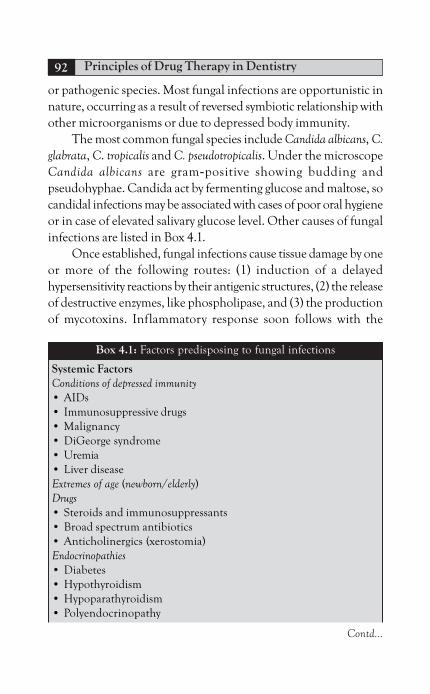
Principles of Drug Therapy in Dentistry92
or pathogenic species. Most fungal infections are opportunistic innature, occurring as a result of reversed symbiotic relationship withother microorganisms or due to depressed body immunity.
The most common fungal species include Candida albicans, C.glabrata, C. tropicalis and C. pseudotropicalis. Under the microscopeCandida albicans are gram-positive showing budding andpseudohyphae. Candida act by fermenting glucose and maltose, socandidal infections may be associated with cases of poor oral hygieneor in case of elevated salivary glucose level. Other causes of fungalinfections are listed in Box 4.1.
Once established, fungal infections cause tissue damage by oneor more of the following routes: (1) induction of a delayedhypersensitivity reactions by their antigenic structures, (2) the releaseof destructive enzymes, like phospholipase, and (3) the productionof mycotoxins. Inflammatory response soon follows with the
Box 4.1: Factors predisposing to fungal infections
Systemic FactorsConditions of depressed immunity• AIDs• Immunosuppressive drugs• Malignancy• DiGeorge syndrome• Uremia• Liver diseaseExtremes of age (newborn/elderly)Drugs• Steroids and immunosuppressants• Broad spectrum antibiotics• Anticholinergics (xerostomia)Endocrinopathies• Diabetes• Hypothyroidism• Hypoparathyroidism• Polyendocrinopathy
Contd...

Antimicrobial Agents Used in Dentistry: Antifungals 93
accumulation of inflammatory cells, edema and induction ofepithelial cells to produce hyperplasia.
For a fungal infection to be managed, the causative local orsystemic factors (Box 4.1) should be identified and eliminated first.A topical or systemic antifungal agent, or both, can be used. In thefollowing is a review on the topical and systemic agents that are usedto treat fungal infections.
ANTIFUNGAL AGENTS
The most commonly used antifungal agents are listed in Box 4.2.
Amphotericin B
This agent is a double bonded macrolide, naturally produced byStreptomyces nodosus. Its effect is dose-related, being fungicidal atsufficient doses.
It acts by forming pores as it binds to ergosterol. The formedchannels cause leakage of fungal contents, mainly potassium, whichcauses fungal cells to deteriorate. The agent may also select human
Blood dyscrasia• Anemia• Neutropenia• Leukemia• LymphomaMalnutrition (deficiency of folic acid, iron, vitamin C or B)Local FactorsRepeated traumaExcessive smokingBad oral hygieneXerostomiaProlonged use of topical antisepticsTopical steroid applicationIncreased salivary acidityMoisture or humidity*Denture use* Applies to cutaneous lesions and angular cheilitis.
Contd...

Principles of Drug Therapy in Dentistry94
cholesterol for binding and this may be the reason behind toxiceffects of the drug. Amphotericin is effective on Candida,Cryptococcus, Aspergillus, and Histoplasma species. Resistant strainslike C. Lusitaniae and Pseudallescheria boydii were reported. Suchresistance may be due to alteration in the target ergosterol contentin the fungal cytoplasmic membrane.
Amphotericin is poorly absorbed from the GIT, so it is usedeither topically or in intravenous form. It is metabolized in the liverand excreted in bile and urine. In liver and/or renal diseases, thedose should be adjusted. In healthy adults, the dose should not bemore than 1.5 mg/kg (Table 4.1).
Common adverse reactions to amphotericin are mainly dueto binding to the sterol cholesterol of the human cells. These can be
Box 4.2: Various antifungal agents
POLYENES• Amphotericin B• NystatinAZOLESImidazoles• Clotrimazole• Miconazole• KetoconazoleTriazoles• Fluconazole• Itraconazole
Table 4.1: Polyene antifungals
Agent Form DoseAmphotericin B IV 0.3 mg/kg/ 4–6 hFungizone O. susp 50–100 mg QID/2 WNystatin DropsMycostatin Pastilles 100,000 u/mlFungistatin Lozenges QIDAntimycot O. susp
Ointment

Antimicrobial Agents Used in Dentistry: Antifungals 95
immediately observed (acute) or seen after some time on long-termuse (chronic). Immediate adverse effects start at one to three hoursafter the drug intake and include fever, chills, nausea, hypotension,headache and thrombophlebitis. Corticosteroids, as hydrocortisone,and paracetamol may be administered to inhibit or minimize sucheffects. Thrombophlebitis may necessitate the use of heparin.
Long-term effects include nephrotoxicity, anemia and neurologicmanifestations like impaired hearing and convulsions. Renal effectsinclude loss of magnesium and potassium due to affected tubularfunctions. Such effects may need adequate hydration in the form ofIV saline before amphotericin administration. Nephrotoxic drugs,like aminoglycosides or cyclosporin, should be avoided. DecreasedRBCs production with the resultant normochromic-normocyticanemia may result. Such effect may be potentiated by the use ofZidovudine antiviral therapy.
Nystatin
This antifungal agent is originally developed from S. noursei. It wasfirst developed in New York State. It is poorly absorbed from theGIT and has toxic effects if provided via the intravenous route.Therefore, its use is limited to topical application (Table 4.2).Available in the form of cream, troches, or oral suspension, nystatinis effective on Candida albicans infections, which are the mostcommon fungal infections. This agent acts in a manner similar toamphotericin B. Adverse effects to nystatin include GIT troubleslike nausea, vomiting and diarrhea which are mainly due to poorabsorption. Attention to sucrose content should be paid as it mayinterfere with blood sugar control in diabetes and may predisposeto dental caries.
Azoles
These are fungistatic agents with wide range of activity. They act bya process that involves blocking of the cytochrome P450 enzyme.This enzyme is required to convert lanosterol to ergosterol. The endresult is depressed ergosterol synthesis and, hence, cell membrane

Principles of Drug Therapy in Dentistry96
disturbance. The human hepatic microsomal enzyme system maybe affected and this contributes to disturbed metabolism of otherdrugs and drug interactions. In addition, the human steroidhormone synthesis may be affected.
Resistance to azoles may be due to altered target enzyme and/or increased pumping of the agent out of the fungal cytoplasm.Imidazoles group includes mainly ketoconazole, clotrimazole andmiconazole. Table 4.2 shows the form, dose and brand names ofthese agents. Triazole agents include fluconazole and itraconazole.They are shown in Table 4.3.
KetoconazoleKetoconazole is a broad spectrum antifungal useful in the treatmentof blastomycosis, coccidioidomycosis and histoplasmosis, andchronic mucocutaneous candidiasis.
Table 4.2: Imidazole antifungal agents
Agent Form Dose
Clotrimazole
Canesten Cream 3–5/day/2 W
Canestan O. susp
Locasten* Cream solution
Miconazole
Miconol Cream TID/2 W
Mykotral Solution
Miconaz H* Gel
Micoban* Gel
Daktarin O Gel
Ketoconazole Tablet Adults
Tocon ,, 200–400 mg/day/for
Kizol ,, 2 W
Fungizol ,, Children
Nizoral ,, 5–10 mg/kg/day for 2 W
* With hydrocortisone 1 percent.

Antimicrobial Agents Used in Dentistry: Antifungals 97
It is available in both the topical and systemic forms. Oraltablets of this agent need acidic environment for proper absorptionthrough the gastric lining. So, conditions of hypochlorhydria orachlorhydria, where there is decreased or absent secretion of HClfrom stomach, are associated with reduced ketoconazole absorptionunless placed in some sort of acidic medium. In addition, drugs ormeals that elevate the stomach pH will act in the same way, so,should be avoided. This would include H2-histamine receptorblockers, antacids and diary products. Ketoconazole is metabolizedin the liver and considered hepatotoxic.
Adverse effects of ketoconazole use include GIT troubles,hepatic enzyme induction and hepatotoxicity, decreased steroidsynthesis with the resultant impotence, decreased libido, menstrualdisturbances and gynecomastia. Drug interactions with ketoconazoleare indicated in the drug interaction (Box 4.4).
MiconazoleMiconazole has similar spectrum and mechanism of action asketoconazole; however, it is used only topically or in the IV form. Itis less effective than amphotericin but with more serious adverseeffects. Its applications are mainly for severe systemic or chronicmucocutaneous candidiasis. It has mild antibacterial effects, aproperty that makes this agent especially useful in case of angularcheilitis. Adverse effects to miconazole include GIT disturbance,rashes, fever and chills.
Table 4.3: Triazole antifungal agents
Agent Form DoseFluconazoleF-zole Capsule 50–150 mg /day/7 days.FluzoneTriconalDiflucan 100 mg Vial 50 ml IV (2 mg/ml)ItraconazoleItrapex Capsule 100 mg/day/2WItranoxItracon

Principles of Drug Therapy in Dentistry98
ClotrimazoleThis agent has several advantages. First, it has the ability to bind tooral tissues and gets released later at slower rate, a property thatprolongs the time of contact between the antifungal agent and thetarget lesion. Second, it has effects on microorganisms other thanfungi, like corynebacteria and protozoa. It also proved to be usefulin the prevention or treatment of noninfection diarrhea. Adverseeffects to the agent include irritation, burning sensation and rashes.
FluconazoleFluconazole is a drug of choice to treat deep and mucocutaneouscandidiasis. It is a wide spectrum antifungal agent and is present inthe oral and IV forms (Table 4.3). In contrast to ketoconazole, theoral form is not affected by alkaline environment or food. Becauseit has lesser effects on the cytochrome P450 enzymes, fluconazolealso has lesser adverse reactions and drug interactions.
Fluconazole is well distributed in the body tissue and is able toreach the CNS. It is poorly metabolized, so should be avoided inkidney disease.
Although of wide range of activity, resistant strains, likeC. galabrata and C. krusei, have been discovered.
Adverse reactions to fluconazole include GIT troubles,headache and drowsiness. Depletion of potassium may also occur,so potassium supplement may be required.
ItraconazoleThis potent, broad spectrum antifungal agent is considered one ofthe first agents against systemic mycosis. Like fluconazole,itraconazole has lesser endocrine adverse effects. In the capsule form,acidic environment and food appear to enhance its absorption.However, the suspension form (used to treat esophageal lesions)may be interrupted by food and antacids.
Itraconazole is metabolized in the liver and care should bepracticed in case of liver disease. It has lesser effects on the renalsystem, so in case of renal disease, no dose adjustment is required.

Antimicrobial Agents Used in Dentistry: Antifungals 99
All azole agents should be avoided in pregnancy (see specialconsideration).
Adverse reactions include GIT problems, rashes, hypertension,headache, edema, and depletion of potassium.
CLINICAL USE OF ANTIFUNGAL AGENTS
Fungal infections, related to oral and perioral tissues, are listed inBox 4.3. The aim of this section is to briefly describe the commonforms of fungal infections and how to treat them. The reader isadvised to refer to textbooks of medicine for further details.
Oral thrush: Pseudomembranous candidiasis is common innew born babies, elderly asnd in conditions of iron deficiency.Steroid or antibiotic use can also be possible causes. HIV should besuspected if no causative factor is clinically identifiable.
Box 4.3: Clinical use of antifungals
Superficial mycosesAcute• Pseudomembranous candidiasis (thrush)• Erythmatous candidiasis• Angular cheilitisChronic• Hyperplastic candidiasis• Atrophic candidiasis• Median rhomboid glossitis• Mucocutaneous candidiasisDeep mycoses• Histoplasmosis• Blastomycosis• Mucormycosis• Aspergillosis• Cryptococcosis• Coccidioidomycosis• Paracoccidioidomycosis• Pneumocystosis

Principles of Drug Therapy in Dentistry100
Clinically, the lesions are in the form of patches or flecks withwhite-yellow covering pseudomembrane that can be easily wipedoff. A red base is left on wiping off such lesions, which can be quitepainful. Most commonly affected sites include soft palate, cheekmucosa, vestibules, gingiva and the tongue.
The condition should be differentiated from other lesions likethose occurring as a result of burns, mechanical trauma or chemicalinjuries as they may be of similar clinical appearance. Careful historyusually reveals the cause. Oral thrush may also resemble mucouspatches of syphilis which appear as discrete, small and painlesslesions. Candidal infection is more diffuse and often painful onscraping. Superficial bacterial lesions, as staphylococcal or gonorrhealinfections, may be differentiated on the basis of laboratoryinvestigation. Candida have the characteristic appearance ofpseudohyphae and spores. Necrotic ulcers caused by systemic diseaseor gangrenous stomatitis may be confused with candidal lesions asboth have common systemic involvement. However, candidiasis isusually more superficial.
Once diagnosed, the underlying predisposing factors shouldbe determined and corrected. Appropriate laboratory investigationsare recommended. Topical antifungals like nystatin, amphotericinor miconazole have been tried but they were found of limited value,possibly due to poor patient compliance and poor contact with thetarget lesion. The use of systemic antifungal agents, like fluconazoleor itraconazole, for two weeks is more effective. Chlorhexidinemouthwash is useful, too. Persistent or recurrent lesions may beassociated with uncorrected predisposing factor. The conditionmay turn into chronic form (CMC). In these infections, HIVprobability is high.
Acute erythmatous candidiasis: This form is often associatedwith steroid or antibiotic use. Systemic use of these agents produceslocalized lesions, whereas topical application is associated with morediffuse involvement.
To correct such factors, consultation regarding antibioticsshould be made. The effects of topical steroid can be minimized by

Antimicrobial Agents Used in Dentistry: Antifungals 101
gargling with water after each application. Topical agents may beused to treat such conditions. Systemic agents are useful for patientswith impaired immunity.
Angular cheilitis: Inflamed, erythmatous and ulcerated anglesof the mouth is known as angular cheilitis. The main cause is humiditywhich favors fungal proliferation. Such humidity is created by salivawhich may be leaking as a result of incompetent labial seal or reducedfacial vertical dimension in association with dentures. Lip lickinghabit may also be the cause. Angular cheilitis may be associatedwith intraoral lesions, commonly with denture sore mouth. Thecondition may also be caused by deficiency of vitamin B.
The diagnosis of the condition is straightforward. However, itmay be associated with Staphylococcus aureus infection. Suchpathogen may be inoculated from the nose and it is associated withmore redness and possibly bleeding. To confirm, a separate smearfrom each angle/nares is performed.
Oral lesions, when present, should be treated as they act as areservoir of fungi. Angular cheilitis is treated with miconazole creamapplied to the angles of the mouth. Fortunately, miconazole is alsoeffective on S. aureus, so, it can be used in case of mixed infection. Ifthe lesion is purely bacterial in origin, it should be treated withfusidic acid cream applied every six hours.
Chronic hyperplastic candidiasis: This white lesion is similarto leukoplakia in that it cannot be wiped off and, may havepremalignant potential. The lesion can be a mixture of a leukoplakiaalong with fungal infection. It commonly affects the cheek mucosaor the dorsum of the tongue. Epithelial alteration may be secondaryto the fungal infection; however, it is possible that fungal infectionis secondary to epithelial alteration. Excessive smoking is a usualfinding, and the patient may have suffered from previous episodesof denture stomatitis and/or angular cheilitis. A biopsy is indicatedin this case to exclude malignancy and to confirm the diagnosis.The microscopic appearance may reveal fungal hyphae, hyperplasticepithelium with or without dysplasia along with chronicinflammatory cells.

Principles of Drug Therapy in Dentistry102
The fungal component is treated by the use of a systemicantifungal; like itraconazole or fluconazole for about three weeks.This is more effective than the use of polyene agents which requirelonger period and more patient compliance. Persistent epithelialalteration indicates excision and histologic examination.
Chronic atrophic candidiasis: Chronic erythmatous form ofcandidal infection is associated with the improper use of dentalappliances, especially removable dentures. Dentures are eithercontinuously worn or the patient is not careful about oral hygiene.Continuous wearing of dentures provides areas isolated from salivaand oral environment, which favors fungal proliferation. The fittingsurface of the used appliance is loaded with layers of fungal plaque,which may deeply infiltrate its bulk. The associated lesion iserythmatous with clear demarcations that clearly indicate theconfiguration of the used appliance. Patients complaints are usuallyminimal, however, when reported they are in the form of discomfortor burning sensations. White flecks or angular cheilitis may be alsoassociated with such lesions. The condition may be confused withcontact allergy, where all tissues in contact are irritated and notstrictly demarcated by denture boundaries. In addition, allergy maybe confirmed by epicutaneous tests.
The condition is managed by correction of the underlyingfactors. The used appliance is cleaned with sodium hypochloritesolution overnight for three weeks. Those with metal parts are bettercleaned and soaked in chlorhexidine solution to avoid corrosion.The appliance should be removed from the mouth for eight hoursper day to provide tissue rest. The fungal infection is treated by theuse of topical agent (amphotericin, nystatin, or miconazole) appliedboth to the oral lesion and the fitting surface of the associatedappliance for four weeks.
Median rhomboid glossitis: It was considered that MRG is ofdevelopmental etiology with the partial persistence of tuberculumimpar. However, its recurrent nature and absence in children alongwith its resolution with the use of antifungal therapy supports itsrelation to fungal infection. It appears as a rhomboidal depapillated

Antimicrobial Agents Used in Dentistry: Antifungals 103
lesion that lies just anterior to the circumvallate papilla. Anothersimilar lesion may be seen in the opposing palatal tissue, possiblydue to toxic response to the lesion on the tongue, a conditionknown as “kissing disease”. The lesion is usually asymptomatic. Smearor culture may be made to confirm the diagnosis, although, theclinical appearance is characteristic. The differential diagnosis mayinclude squamous cell carcinoma, erythroplakia, and geographictongue.
Topical antifungal agents for prolonged periods (up to 3months) may be the treatment of choice. Better results are expectedfrom the systemic application in shorter duration. The underlyingsystemic condition should be examined to role out any associateddiseases.
Chronic mucocutaneous candidiasis: This form of candidalinfection represents a form of deep infection that involves deeperparts of skin and mucous membranes. It is recognized to be resistantto treatment and it is difficult to manage. Several causes have beenimplemented, and accordingly, several types exist. In Type I, whichis known as the familial or the limited type, the lesions are mild butappear since birth, and are known to be persistent. Sidropenia(iron deficiency) may be another finding. Iron supplements alongwith systemic antifungal agents are the required treatment of thisform.
Type II or the diffuse type is characterized by the disfiguringgranulomatous lesions which diffusely involve skin and mucousmembranes. Immune defects may be an associated finding. The riskof developing secondary bacterial infections, like superficial andpulmonary infections, is high. The condition needs systemicantifungal (itraconazole) along with an antibiotic to guard againstbacterial infections.
Type III, or the endocrinopathy candidiasis syndrome, is mildbut exhibits a persistent candidial infection which is followed bymultiple glandular dysfunctions later in life (10–15 years). It isbelieved that such dysfunction is caused by specific autoantibodies

Principles of Drug Therapy in Dentistry104
directed to several glandular tissues. The commonly affected glandsinclude adrenal glands (Addison’s disease), parathyroids(hypoparathryroidism), thymus (DiGeorge syndrome). Thecondition requires medical management of the multiple glandulardysfunctions and a systemic antifungal.
Candidiasis thyoma syndrome is the 4th type of this groupand is known as the “late onset mucocutaneous candidiasis”. Atumor of the thymus gland is present and it should be surgicallyremoved.
Systemic mycosis: This means that the fungal infection hasreached deeper sites in the body tissues and organs. It may affecthealthy individuals, where it is asymptomatic and self-limiting. Inthose with poor immunity, however, the disease is progressive,disseminated and possibly fatal. The infection may involve the lungs,maxillary sinuses and other organs. It is associated with agranulomatous lesion with abscess formation. Orally, the lesionmay appear as a nodular tumor-like mass or chronic oral ulcerwith bizarre configuration. History usually reveals defective immunityand/or travel to an endemic area. Smear, culture, X-ray on thechest and MRI are further useful investigations. Systemic antifungalis the treatment of choice.
GENERAL CONSIDERATIONS
All fungal infections should be treated with suspension of underlyingsystemic or local factors. As a rule, when no obvious underlyingfactor can be determined, AIDS should be suspected. It is theidentification and correction of these factors which provides thedefinite treatment. The antifungal part of the management providesthe way towards elimination of the incident infection, but notpreventing it from recurrence, and sometimes, fails to eradicate theinfection due to the persistence of the underlying factors.
Superficial lesions, especially in healthy individuals, are managedby topical agents for at least of two weeks after clinical resolution. Itappears, however, that compliance to topical agents is low as it haslonger duration, more frequency and needs greater attention.

Antimicrobial Agents Used in Dentistry: Antifungals 105
To prevent recurrence, deep or systemic mycoses require theuse of systemic antifungals for at least one to two weeks after clinicalresolution. In chronic forms, it may need up to four weeks ofantifungal therapy to completely eradicate the infection. Systemicantifungal can be used in case of recurrent or persistent infections,as well as, in case of compromised immunity.
Chlorhexidine mouthwash is a valuable adjunct to antifungaltherapy as it is fungicidal, as well as, interferes with fungal adherenceto oral tissues.
SPECIFIC CONSIDERATIONS
Specific considerations regarding pregnancy and breastfeeding, liverand kidney diseases are presented in Table 4.4. Possible druginteractions are indicated in the drug interaction Box at the end ofthis chapter.
Azoles should not be used in case of pregnancy, liver or kidneydisease, cardiac diseases (as they may precipitate cardiac failure).
Table 4.4: Showing specific considerations regarding antifungal use
Pregnancy and BreastfeedingUse Use with caution AvoidNystatin Clotrimazole (in 2nd All azoles (teratogenic
or 3rd trimesters) should not be usedin children below theage of 2 years)
Liver/renal disease Use AvoidNystatin Azoles*
*Heptatotoxic agents.
Box 4.4: Drug interactionMost interactions occur with the systemically applied agents. Theeffects of amphotericin B are decreased if combined with ketoconazole,as the former acts on the ergosterol part of fungal membrane, whereasthe latter acts by inhibition of membrane synthesis.
The azoles are P450 enzyme inhibitors; therefore, their use maycause toxicity of those metabolized in the liver. This includes
Contd...

Principles of Drug Therapy in Dentistry106
cyclosporin, phenytoin, tolbutamide or warfarin. Rifampicin is hepaticenzyme inducer and it causes rapid elimination of ketoconazole and,hence, less effects.
The same could occur when any enzyme inducer used along withan azole agent.
Ketoconazole absorption is decreased by the use of H2 inhibitors,
antiacids or alkaline food stuff. Antimuscarinics like scopolamine,hyoscine or atropine may also interfere with absorption of azoles.
Grapefruit juice may hamper the absorption of itraconazole.Azoles may cause cardiac dysrhythmia if taken with terfenadine,
astemizole or cisapride. They also act by inhibiting the metabolism oforal anticoagulant, statins (cholesterol lowering agent) or quinidine.The use of statins is also associated with increased myopathy. The oralcontraceptive effect may be reduced with the use of azole agents. So,extra means of birth control measures should be advised.
The azoles should not be coadministered with those nephrotoxicagents so as not to increase the burden on the kidneys.
They may also precipitate acute attacks of porphyria (porphyrindisorder) in susceptible patients.
BIBLIOGRAPHY
1. Anil G Ghom. Textbook of Oral Med. Drugs Used in Pregnancy.Jaypee; 2006.891-2.
2. Crispan Scully, Roderick A Cawson. Liver disease. Medical Problemsin Dentistry. 5th edn. Elsevier; 2006.
3. Crispan Scully, Roderick A. Cawson. aspects of drugs and materialsuse in dentistry. medical problems in dentistry. 5th edn. Elsevier;2006.
4. Crispan Scully, Roderick A. Cawson. women’s health. MedicalProblems in Dentistry. 5th edn. Elsevier; 2006.
5. Crispan Scully, Roderick A. Cawson. Antibacterials (appendix29-3). Medical Problems in Dentistry. 5th edn. Elsevier; 2006.
6. Henein Willi, Usama Tharwat. systemic and topical antifungals.Master in Therapuetic Drugs (MTD). Nubar Printing House; 2008.
7. Jacob F, MD, AB. Topical and systemic antifungal, antimicrobialsused in dental practice. 2nd edn. Quintessence; 2001.
Contd...

Antimicrobial Agents Used in Dentistry: Antifungals 107
8. Jeffrey C. Medical Mycology. Medical Microbiology. 20th edn.Appleton and Lange; 1995.
9. Michael AO, Richard CK. Erythemia/white lesions. A Color HandBook of Oral Med. Manson Publishing Ltd; 2004.
10. Norman K, Wood and Paul W Goaz. Solitary red lesions.Differential Diagnosis of Oral and Maxillofacial lesions. 5th edn.Elsevier.
11. RA Cawson, EW Odell, Cadidosis. Cawson’s Essentials of OralPath and Med. 7th edn. Elsevier; 2002.
12. Richard A Harvey, Pamela C. Antifungals Lippincott’s illustratedreview of pharmacology. 4th edn. Lippincott Williams and Wilkins;2009.
13. SN Kholosa and Amit Kholosa. Drug interactions. medicine fordental students. 1st edn. Delhi: Asia printograph; 2001.

Chapter 5Antimicrobial Agents Used in Dentistry:
Antiviral Agents
VIRAL INFECTIONS
Viruses are obligate intracellular parasites, which live and dependon other living cells for their replication. Viruses are simple intheir structure being composed mainly of three compartments;namely a nucleic material, protective protein coating and certainenzymes essential to start their replication.
The protective protein coat, also known as the nucleocapsid,protects the viral genetic material and plays a role in viralattachment and penetration into the host cells, possibly throughthe glycoprotein spikes present on its surface. Viruses attach tocertain cellular receptors and this may explain the cellularselectivity of certain viruses (Table 5.1). Entry may occur eitherby fusion of the virus with the host membrane or by the processof endocytosis.
Regarding the nucleic acid content, viruses contain eitherDNA or RNA but not both. In addition, these materials can beeither single stranded (SS) or double stranded (DS). Inside the
INTRODUCTION
Viral InfectionsIndividual Drugs
Nucleotide AnalogsPyrophosphate AnalogsRecombinant Proteins
Clinical Use of Antiviral DrugsGeneral ConsiderationsSpecific Considerations

Antimicrobial Agents Used in Dentistry 109
Table 5.1: List of viruses with examples of pathological lesionsthey produce
Virus Target tissue Associated infection/disease
HERPES VIRUSESHHV1 (HSV1) Oral epithelial cells PHGS
Neural tissues Recurrent herpeticlesions
HHV2 (HSV2) Genital epithelial cells Genital herpesNeural tissues
HHV3 (HZV) Same as HHV1 Chickenpox (Varicella)Shingles (zoster)
HHV4 (EBV) Glandular tissue Glandular feverRespiratory tract Hairy leukoplakiaLymphocytes Burkitt’s lymphoma
HHV5 (CMV) Oral epithelium Glandular feverLymphocytes
HHV6 T-lymphocytes Glandular feverPersistentlymphadenopathyChronic fatiguesyndrome
HHV7 T-lymphocytes As a cofactor withHHV6
HHV8 Kaposi’s sarcomaBody cavity-basedlymphoma
ENTEROVIRUSESCoxsackie viruses A Epithelial cells HFMD
HerpanginaCoxsackie viruses B HerpanginaPARAMYXOVIRUSESMeasles (rubeola) Respiratory tract MeaslesMeasles (rubella) Any other tissue German measlesMumps MumpsHUMMAN Epithelial tissue Squamous cellPAPILLOMA papillomaVIRUS (HPV) Condyloma
CartsRETROVIRUSES CD4 on T cells AIDs(HIV)
HHV = Human Herpes Virus, HSV = Herpes Simplex Virus, HZV = HerpesZoster Virus, EBV = Epstein Barr Virus, CMV = Cytomegalovirus, PHGS =Primary Herpetic Gingivostomatitis, HFMD = Hand Foot and Mouth Disease.

Principles of Drug Therapy in Dentistry110
host cell, the genome gets uncoated and the process of genetranscription and synthesis of nucleic material and protein coatingis initiated.
The enzymes present are just to direct the host cellularribosomal system to their demands. Polymerase enzyme is themain viral enzyme and it initiates the polymerization reactionfor the synthesis of viral nucleic material.
Virally infected cells get exhausted and their functions aswell as their structures are deteriorated. Cellular damage or lysismay be due to the action of cytotoxic cells which recognize viralpolypeptides on the infected cell surface. The production of auto-antibodies by β-cells may affect unrelated cells and produce lesionsinconsistent with the site of infection. The production ofhyperplastic or neoplastic lesions may be due to the cellularresponse to their new function as machinery for viral replication.
It should be noted that viral infections are associated withspread and replication of the virus before their manifestationsappear, an important point to consider in the treatment of viralinfections. Once the manifestations appear, the use of antiviralagents is of limited value. This signifies the prodromal signs andsymptoms as the use of antiviral agents is the best at this stage ofthe viral infection.
INDIVIDUAL DRUGS
Box 5.1 lists the most commonly used antiviral drugs in the dentalpractice. Antiviral agents mainly act as analogs to essential viralmolecules to stop their replication.
Nucleotide Analogs
A nucleic acid is a polymer of many nucleotides. Nucleotidesthemselves are made up of three components and these are: Amonosaccharide, a nitrogenous base and a phosphate group. Thenitrogenous bases are either single ringed (pyrimidines) or doubleringed (purines). Pyrimidines are uracil, cytosine or thymidine,while the purine bases are either adenine or guanine.

Antimicrobial Agents Used in Dentistry 111
The antiviral analogue acts by substituting the naturalnitrogenous bases with nonfunctional synthetic analog resultingin termination of the polymerization reaction and the productionof nonfunctional genetic strand. This would stop viral replication.
Acyclovir. This is a purine base analog which uses guanosine.It gets activated by the enzyme thymidine kinase which is codedfor, by the viral genes. In this way, acyclovir exhibits highconcentration in virally-infected cells. Thymidine kinase catalyzesphosphorylation reaction which converts acyclovir into acyclovirmonophosphate. The drug is further phosphorylated by the hostenzymes into di- and triphosphated forms. The final formcompetes with guanine for the viral DNA polymerase. Onceattached to the polymerizing DNA, the agent soon stops thereaction. The agent may cause the polymerase enzyme to loss itsfunction too.
Acyclovir is most effective on those viruses which encodefor their own thymidine kinase like HSV and HZV. Other viruseslike CMV or EBV are less susceptible. Resistance to acyclovir hasbeen attributed to the lack of thymidine kinase or polymeraseenzyme.
The drug is partially metabolized and gets eliminated bymeans of the renal system. This notifies care when used in patient
Box 5.1: Antiviral agents most commonly used in dental practice
Nucleotide analogs• Acyclovir• Valacyclovir• Penciclovir• Famciclovir• Ganciclovir• CidofovirPyrophosphate analogs• Foscarnet sodiumRecombinant proteins• Interferon-α
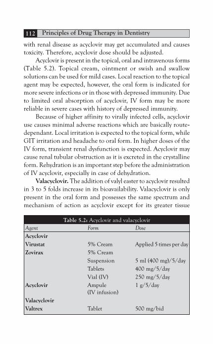
Principles of Drug Therapy in Dentistry112
with renal disease as acyclovir may get accumulated and causestoxicity. Therefore, acyclovir dose should be adjusted.
Acyclovir is present in the topical, oral and intravenous forms(Table 5.2). Topical cream, ointment or swish and swallowsolutions can be used for mild cases. Local reaction to the topicalagent may be expected, however, the oral form is indicated formore severe infections or in those with depressed immunity. Dueto limited oral absorption of acyclovir, IV form may be morereliable in severe cases with history of depressed immunity.
Because of higher affinity to virally infected cells, acycloviruse causes minimal adverse reactions which are basically route-dependant. Local irritation is expected to the topical form, whileGIT irritation and headache to oral form. In higher doses of theIV form, transient renal dysfunction is expected. Acyclovir maycause renal tubular obstruction as it is excreted in the crystallineform. Rehydration is an important step before the administrationof IV acyclovir, especially in case of dehydration.
Valacyclovir. The addition of valyl easter to acyclovir resultedin 3 to 5 folds increase in its bioavailability. Valacyclovir is onlypresent in the oral form and possesses the same spectrum andmechanism of action as acyclovir except for its greater tissue
Table 5.2: Acyclovir and valacyclovirAgent Form DoseAcyclovirVirustat 5% Cream Applied 5 times per dayZovirax 5% Cream
Suspension 5 ml (400 mg)/5/dayTablets 400 mg/5/dayVial (IV) 250 mg/5/day
Acyclovir Ampule 1 g/5/day(IV infusion)
ValacyclovirValtrex Tablet 500 mg/bid

Antimicrobial Agents Used in Dentistry 113
concentration (Table 5.2). The compound is broken apart intothe active form in the GIT and liver. Therefore, GIT side effectsare not uncommon.
Penciclovir. This antiviral agent is an acrylic guanosinenucleotide analog which undergoes similar mono-phosphorylation, as acyclovir, by the enzyme thymidine kinase.Penciclovir is present only in the topical form and it has about20 to 30 times longer duration of action than acyclovir (Table5.3). It also can produce higher intracellular concentration. Thiswould reduce the frequency of application which enhancespatients’ compliance. It has the same spectrum as acyclovir. Theadverse reactions to this agent are minimal.
Famciclovir. This agent is the prodrug of penciclovir, madeto allow its use via the oral form. It is a deoxyguanosine analoguewhich by means of the GIT and liver undergoes deacetylationprocess, converting it into the active form. Famciclovir exhibitsexcellent GIT absorption and has 3 to 5 times higherbioavailability than acyclovir (Table 5.3). This agent is suitablefor HZV infections, although effective for the HSV, too. Adversereactions to the drug are minimal except for nausea and headache.
Ganciclovir. Ganciclovir is a similar analogue to acyclovirbut it is a good substrate for the enzyme phosphotrasferase whichis present predominantly in CMV. It is a potent antiviral agentwith activity about 8 to 20 times that of acyclovir. Due to itsserious side effects, its use is preserved for serious viral infectionscaused by CMV, as CMV retinitis in HIV patients. Its use mayalso be justified in for the prevention of CMV infections in organtransplantation or HIV patients.
Table 5.3: Penciclovir and famciclovirAgent Form DosePenciclovir 1% Cream every 2 hoursFamciclovirFamvir Tablet 250 mg 2–3 times/dayPropencivir Tablet 500 mg 2–3 times/day

Principles of Drug Therapy in Dentistry114
Ganciclovir is present in three forms; intravitreal, oral andintravenous. For the treatment of CMV retinitis, intravitrealinjection of ganciclovir every six months is the treatment ofchoice. In case of serious CMV infections, ganciclovir is used inthe IV form. Oral form is used as a maintenance therapy (Table5.4).
Adverse reactions include bone marrow suppression withthe resultant anemia, neutropenia and thrombocytopenia. It isalso toxic to the kidneys and the nervous system. Other effectsinclude nausea, diarrhea and flatulence. The drug is teratogenic,embryotoxic and carcinogenic in experimental animals, so shouldbe avoided in pregnancy.
Cidofovir. This single-ringed cytosine analog acts only onDNA viruses by inhibiting their polymerase enzymes. Because ofits effects on the renal system, it is only approved to be used incase of severe CMV infections, especially in those with HIV. Toreduce renal toxicity, probenecid and saline solution should beadministered. Cidofovir is present in topical gel, intravenous andintravitreal forms. Its use should be avoided in case of renaldisease.
Pyrophosphate Analogs
Foscarnet SodiumThis is a pyrophosphate analog which acts on the DNApolymerase with the resultant blockade of the DNA/RNA chainand by the formation of nonfunctional complexes. Foscarnet
Table 5.4: Doses/forms of ganciclovirAgent Form DoseGanciclovir IV 10 mg/kg/day
Or 500 mg/6 hoursCymeven Tablet 250–500 mg/bidValacyte Tablet 450 mg /bid

Antimicrobial Agents Used in Dentistry 115
sodium is not a nitrogenous base, so, its effect is not dependanton the presence of an activating enzyme. This feature makes ituseful in case of acyclovir-resistant viruses and CMV infections.However, resistance to foscarnet has been described and it isattributed to gene mutations which result in altered target. It ispresent in IV and oral forms. The associated adverse reactionsare common despite its low affinity for host polymerase enzymes.Renal failure, electrolyte imbalance, nausea, vomiting, anemiaand seizures are expected.
Recombinant Proteins
Interferon is a glycoprotein naturally produced in response toviral infections and bacterial endotoxins. In response to viralinfections, interferon acts by inducing cellular enzymes, namelyendonucleases, which act by degrading viral mRNA and, thus,preventing viral replication. Interferon also acts by increasing thenumber of natural killers and the expression of viral antigens.The agent is present in several forms (Table 5.5). IV/IM formsare generally indicated for chronic hepatitis B and C. Intralesionalform can be used to treat venereal warts caused by HPV, whilethe submucous one is used to treat other oral virally-related lesionslike Kaposi’s sarcoma and oral hairy leukoplakia. Interferon isnot present in the oral form as it is unstable in the gastricenvironment. Common adverse reactions include flu-likesymptoms, hypotension, myelosuppression, autoimmunedisorders (like thyroiditis), neurotoxicity and psychiatric disorders.
Table 5.5: Doses and forms of interferonAgent Form DoseINF-ααααα2B IMSC 3 million IUIntron-ARoferon-AEgyferon
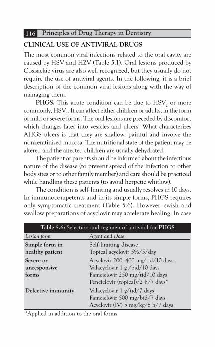
Principles of Drug Therapy in Dentistry116
CLINICAL USE OF ANTIVIRAL DRUGS
The most common viral infections related to the oral cavity arecaused by HSV and HZV (Table 5.1). Oral lesions produced byCoxsackie virus are also well recognized, but they usually do notrequire the use of antiviral agents. In the following, it is a briefdescription of the common viral lesions along with the way ofmanaging them.
PHGS. This acute condition can be due to HSV2 or more
commonly, HSV1. It can affect either children or adults, in the form
of mild or severe forms. The oral lesions are preceded by discomfortwhich changes later into vesicles and ulcers. What characterizesAHGS ulcers is that they are shallow, painful and involve thenonkeratinized mucosa. The nutritional state of the patient may bealtered and the affected children are usually dehydrated.
The patient or parents should be informed about the infectiousnature of the disease (to prevent spread of the infection to otherbody sites or to other family member) and care should be practicedwhile handling these patients (to avoid herpetic whitlow).
The condition is self-limiting and usually resolves in 10 days.In immunocompetents and in its simple forms, PHGS requiresonly symptomatic treatment (Table 5.6). However, swish andswallow preparations of acyclovir may accelerate healing. In case
Table 5.6: Selection and regimen of antiviral for PHGS
Lesion form Agent and Dose
Simple form in Self-limiting diseasehealthy patient Topical acyclovir 5%/5/day
Severe or Acyclovir 200–400 mg/tid/10 daysunresponsive Valacyclovir 1 g /bid/10 daysforms Famciclovir 250 mg/tid/10 days
Penciclovir (topical)/2 h/7 days*
Defective immunity Valacyclovir 1 g/tid/7 daysFamciclovir 500 mg/bid/7 daysAcyclovir (IV) 5 mg/kg/8 h/7 days
*Applied in addition to the oral forms.

Antimicrobial Agents Used in Dentistry 117
of severe, nonresponsive lesions, or in those with high susceptibilityto severe complications (like those with defective immunity or thosewith eczema or skin burns), systemic antiviral is needed.
Recurrent herpetic lesions. HSV remains dormant, or instate of chronic infection, in the sensory neural ganglia. Thedormant viruses remain protected from the immune factors thereand once activated (by stress, sunlight, trauma, etc.), they flowdown the axonal pathways to infect the tissues they supply.Recurrent lesions are usually mild and less responsive to antiviraltherapy.
Herpes labialis or “cold sores” is a lesion of multiple blistersat the border of the lip which ruptures, scabs and heals withoutscarring. The condition can be prevented or altered by theapplication of antiviral cream at the onset of the prodromalsymptoms. However, immediate lesions are not preceded by anysymptoms and appear all of a sudden. These lesions are associatedwith viral dormancy in the epithelial cells rather than in the neuraltissue. In case of immune suppression, antiviral cream may not beenough and a systemic antiviral agent may be required (Table 5.7).Systemic antiviral therapy may be used as a prophylaxis in case ofsurgeries involving the ganglia or facial peel procedures to avoidoutbreaks of the virus. Chronic suppressive therapy to prevent viralinfections in those with more than six recurrences annually or incases associated with erythema multiforme is also indicated.
Table 5.7: Selection and regimen of antiviral for recurrentherpetic lesions
Lesion form Agent and dose
Simple form in Self-limiting diseasehealthy patient Topical acyclovir 5%/5/day
Topical penciclovir 1%Defective immunity Acyclovir 400 mg/5 times till healingChronic suppression Acyclovir 400 mg/bid/1y
Valacyclovir 1 g/OD/1yFamciclovir 250 mg/bid/1y

Principles of Drug Therapy in Dentistry118
Recurrent intraoral herpetic lesions involve mainly sites oftrauma like the attached gingiva and the palatal mucosa. Recurrentlesions involve only one surface, in contrast to primary one, and isoften less severe. In its early stages, acyclovir, valacyclovir, famcicloviror foscarnet may help in its prevention or modification.
The diagnosis of herpes simplex lesions is based on clinicalappearance and is often straightforward. The diagnosis may needto be confirmed sometimes and the use of Tzanck test may showmultinucleated epithelial giant cells which are virally-infected.Other tests include viral culture and immunofluorescence, butthese need to be carried out in the hospital environment.
Varicella. Chickenpox, a childhood disease caused by HZV,gains its access through the mucous membranes of the upperrespiratory tract. It gets distributed by blood (viremia) to othertissues. The lesions start by the production of macules, papulesand later the clear drop vesicles which soon rupture and scab.The infection is self-limiting in healthy individuals, but may belife-threatening in patients with defective immunity (risk offulminating varicella pneumonia). Passive immunization may beindicated for those who are pregnant to prevent effects on thedeveloping fetus (Table 5.8).
Shingles. Like HSV, HZV remains in chronic infection stateat the dorsal root ganglia, commonly of the L1, L2, C3, T5, andsensory branches of the trigeminal nerve. Depressed immunity,trauma and elderly all act by triggering its activation. Areassupplied by the affected ganglia are seen with prodromal pain,being burning, or in some cases neuralgic in nature. Vesicles laterdevelop and follow the same course as the primary infection exceptfor that secondary bacterial infections are common which mayresult in scarring. This condition may be followed by anotherpainful condition known as post-herpetic neuralgia (PHN) whichis more prevalent in elderly. Systemic antiviral therapy is indicatedin case of severe involvement, in those with depressed immunityand in elderly (Table 5.8). The associated pain may be difficultto manage with the usual analgesics and the following were tried:

Antimicrobial Agents Used in Dentistry 119
adjuvant analgesics (as tricyclic antidepressants, carbamazepine),physical stimulation (like massaging or applying firm pressure),or thermal application (in the form of local heat or cold spray).Prednisone may help in pain relief and healing enhancement.
Glandular fever. Also known as infectious mononucleosis,glandular fever is caused mainly by EBV. However, other viruseslike CMV or HHV6 may be involved. The disease is characterizedby rashes and jaundice in the febrile type, exudates on the fauceswith sore throat and edema in the anginose type and lymph nodeenlargement with splenomegaly in the glandular type. The diseaseis associated with the production of autoantibodies withagglutination effects on sheep or horse RBCs (Paul Bunnell test).The disease is self-limiting and requires no treatment other thansupportive measures. Pharyngeal edema may necessitate the useof steroids to guard against air way obstruction. It should be notedthat administration of amoxicillin may produce a nonallergic skinrashes on the extremities. No antiviral is required as they showno effects on the symptoms or the virally-infected cells.
Table 5.8: Selection and regimen ofantiviral for HZV infections
Associated lesions Agent and doseVaricellaHealthy Self-limiting, only supportive measures and
infection precautions
Depressed Acyclovir (IV) 15–30 mg/kg/day for 7 daysimmunity/severe cases
Shingles
Healthy Topical acyclovir 5% and antiseptic mouthwash
Depressed Acyclovir 800 mg/5t/10 daysimmunity/ Valacyclovir 1 g/tid/10 dayssevere cases Famciclovir 500 mg/tid/10 days
Elderly Same as severe cases plus prednisone 40–60 mg/day/3 weeks†
† Prophylactic against PHN, decreased gradually.

Principles of Drug Therapy in Dentistry120
Caused by Coxsackie viruses, herpangina and hand-foot-mouthdisease are more common in children. Because their treatmentneeds only supportive measure and needs no antiviral therapy,they should be differentiated from other lesions like AHGS orHZV infections.
Mumps is an acute inflammation of major salivary glands,most commonly being the parotid. It should be differentiated frombacterial sialadenitis, buccal space infection and lymphadenitis.Serum test for S and V antibodies is positive. The condition,however, does not need antiviral therapy and only symptomatictreatment is indicated. In children, aspirin should be avoided toprevent fulmination of the disease (Reye’s syndrome).
GENERAL CONSIDERATIONS
Most viral infections are self-limiting, but some may require theuse of antiviral therapy. Antiviral drugs may help in decreasingpain and the time needed for healing. Because viral replicationoccurs before the manifestations start to appear, it would bedifficult to prevent viral infections. Early use of antiviral agentswould provide better results than when used late. It should beremembered that the antiviral therapy does not affect the viral inits dormant state and periods of recurrence are expected. Infants,elderly and immunocompromised should be treated moreaggressively.
The patient’s nutritional state should be taken into consider-ation. The patient is instructed to increase fluid intake and tohave high calorie diet. Pain should be managed to allow for restand adequate nutrition. Secondary bacterial infections shouldbe prevented by the use of antiseptic mouthwash. The patientshould be informed of the infectious nature of the lesions toavoid infection spread to other sites like the conjunctiva or otherfamily members.

Antimicrobial Agents Used in Dentistry 121
SPECIFIC CONSIDERATIONS
Regarding pregnancy, most antiviral agents are harmful andshould be avoided. However, topical forms are associated withlesser effects and hence considered safer. In sever cases, it mightbe justified to use systemic agents, like acyclovir, however.
The urinary system is involved in the process of antiviral elimi-nation. Therefore, it is expected that patients with renal disordersmight suffer toxicity if an antiviral is used in the normal doses.Consultation regarding the dose and choice of antiviral agentsshould be made in case of renal disease. Table 5.9 summarizes thespecific considerations regarding antiviral use.
Table 5.9: Showing specific considerationsregarding antiviral use
PregnancyUse AvoidAcyclovir cream Acyclovir*Penciclovir cream Valacyclovir
GanciclovirFamciclovirBreastfeeding
UseAcyclovir Ganciclovir†Renal diseaseDose adjustment‡ Avoid (Nephrotoxic)Acyclovir Foscarnet sodiumValacyclovir Cidofovir
* Can be used in serious cases where its benefits are justified over itseffects.
† Rifampicin can be used as a replacement.‡ Drugs accumulate in the body with possible toxicity.

Principles of Drug Therapy in Dentistry122
BIBLIOGRAPHY
1. Amir H Ajar, Peter J Chauvin. AHGS in adults: A review of 13cases. Journal of Canadian Dental Association. 2002;68(4).
2. Anil G Ghom. Textbook of Oral Med. Antiviral Drugs. JaypeeBrothers Medical Publishers; 2006.
3. Crispan Scully, Roderick A Cawson. Viral infections. 5th edn.Elsevier; 2006.
4. G Wayne Raborn, Michael GA. Grace, recurrent herpes simplexlabialis: Selected Thrapeutic Options. Canaidan DentalAssociation. 2003;96(8):498-503.
5. Henein Willi, Usama Tharwat. Systemic and topical antifungals.Master in Therapuetic Drugs (MTD). Nubar Printing House; 2008.
6. Jacob F, MD, AB. Topical and systemic antifungal and antiviral.antimicrobials used in dental practice. 2nd edn. Quintessence;2001.
7. Jawetz, Mehnick and Adelberg’s. Prevention and treatment of viralinfections. Medical Microbiology. 20th edn. Appleton and Lange;1995.
Box 5.2: Drug interaction
Acyclovir appears to reduce the level of valproic acid or phenytoin.On the other hand, it may be associated with theophylline toxicity.Theophylline toxicity has also been associated with concurrent use ofinterferon, recombinant protein which inhibits the hepatic microsomalsystem. Other drugs metabolized in the liver are also at risk of toxicity.As with penicillin, the use of probenecid also elevates the level ofacyclovir or valacyclovir.
Because of their nephrotoxic effects, acyclovir, valacyclovir,ganciclovir or cidofovir should not be used with nephrotoxic agents.
Zidovudine, agent used in the management of AIDs, is knownfor its nephrotoxicity and bone marrow suppression. It may result inlethargy when used with acyclovir, and severe bone marrow depressionalong with blood dyscrasias if used with ganciclovir.
Interferon is known for its inhibitory effects on the hepaticenzymes. Accordingly, drugs metabolized in the liver may accumulatein the body and cause toxicity. In addition, interferon used withzidovudine potentiates the myelosuppression effects.

Antimicrobial Agents Used in Dentistry 123
8. Michael AO, Richard CK. Blisters. A Color Hand Book of OralMed. Manson Publishing Ltd; 2004;42-8.
9. MJ McCullough and NW Savage. Oral viral infections and thetherapeutic use of antiviral agents in dentistry. Australian DentalJournal Medication Supplement. 2005;50:4.
10. RA Cawson, EW Odell. Diseases of oral mucosa and mucosalinfections. Cawson’s Essentials of Oral Path and Med. 7th edn.Elsevier; 2002;178-83.
11. Richard A, Harvey, Pamela C. Antiviral drug. Lippincott’sillustrated review of pharmacology. 4th edn. Lippincott Williamsand Wilkins; 2009;442-4.

An emergency is an urgent situation with possible life-threateningoutcome if no immediate medical care is provided. It reflects theinability of the body to maintain its internal environment constant(loss of homeostasis). This is why an external help is needed.
It is well known that emergency rates would be expected to behigher in elderly and immunocompromised individuals, as thesepatients respond unfavorably to stress. Long and extensive dentalprocedures are also potential factors.
PREVENTION
Even if an emergency situation is managed successfully, the 100percent success remains its prevention. Although it occurs all of asudden, an emergency can be expected. It appears that most medical
Chapter 6Emergency Drugs Used in Dentistry
INTRODUCTION
PreventionPreparednessEmergency Drugs
OxygenEpinephrineNitroglycerin
Primary DrugsAnticonvulsantsCorticosteroidsAntihistaminesBronchodilatorsAnalgesicsAntihypoglycemic AgentsRespiratory Stimulants
Management of Emergencies

Emergency Drugs Used in Dentistry 125
emergencies occurring in the dental clinic are related to stress.Stressful situation can be caused by inadequate pain control, fear,long or extensive procedures, as seen in endodontic procedures ororal surgeries. Therefore, stress reduction by pharmacological ornonpharmacological procedures is the first line defense againstemergencies. Sedation is one of the options available to reduceanxiety and stress and should be used in those who appearunmanageable by the gentle handling approach. The risk ofrespiratory depression and sedative overdose are possible emergenciesassociated with the use of sedatives and the dental team should beable to manage such situations.
Although every patient is a suspect of at least a simpleemergency, some are at great risks. This would include those withCVD like AP; those on chronic drug therapy and of course thosewith positive history of allergy. A medical consultation, regarding amedically complex patient, is recommended even if the dentist issure about his precautions. Referral to a physician is surely prudentin case of suspension of undetectable diseases.
PREPAREDNESS
Preparedness literally means the condition of full readiness, andthis is especially meant for military actions. If an emergency attacksin the dental clinic, it should be faced with an ordered and a wellorganized team (army) with a leader who shows no signs of surpriseor lack of confidence. The team should have enough knowledgeand equipment to manage an emergency successfully. There shouldbe a plan which was discussed and practiced before. Every memberof the team should be assigned a specific task to perform at everyemergency situation (Box 6.1).
A thorough knowledge and practice in BLS for every memberof the dental team is a must. CPR ensures adequate blood perfusionto the brain and this saves time till more advanced medical helparrives. Current certification, periodic refreshing and recertificationare prerequisites for clinical work. The dentist or the leader, however,should have a certification in advanced cardiac life support (ACLS)

Principles of Drug Therapy in Dentistry126
as well. The oral and maxillofacial surgeon should be equippedwith a certification in advanced trauma life support and he usuallyleads the dental team.
The dental clinic should be prepared with the primaryemergency drugs. In order not to do harm, the use of drugs,indications and contraindications should be revised thoroughly bythe leader. Knowledge about the underlying pathophysiology of anemergency enables the clinician to use the proper drug. Emergencydrugs should be labeled and preferably prestocked for easier accessand more convenient use. The use of these drugs often requiresspecial equipment and these should be ready as well (Table 6. 1).
The team should be able to perform various procedures likemonitoring of the vital signs (blood pressure and pulse),venipuncture, parenteral injections and endotracheal intubation.
For advanced management, ambulance help with hospitalemergency care may be required. Therefore, a valid phone numberof an emergency department of the nearest hospital should bereadily displayed.
EMERGENCY DRUGS
For efficient emergency management, it might be needed to applythe use of certain emergency drugs (Box 6.2). For maximum benefits,therapeutic uses and contraindications should be constantly revised.Otherwise, more harm can be produced than benefits.
Box 6.1: Tasks assigned to the members of the dental teamin case of an emergency
Leader Recognize and directly manage the condition
Assistant Assist the leader
Nurse • Prepare emergency drugs• Receive the ambulance down hall
Receptionist • Make all phone calls to bring advanced support• Watch and indicate the time elapsed since drug
administration and for vital signs rechecking• Record the details of the event

Emergency Drugs Used in Dentistry 127
An emergency kit prepared with the suitable emergency drugsshould be available. It is important that the kit does not containtoo much items to avoid confusion of the user. Ideally, the basicitems necessary for basic or initial management should be included.For more convenience, the agents should be in the preloaded form.
Oxygen
The basic requirement of every cell in the body in order to produceenergy is oxygen. For oxygen to reach the body cells, three systems
Table 6.1: Necessary equipment for administration of emergencydrugs in the dental clinic
Use Equipment Description
Oxygen Mouth barriers • Allows for safe mouth toadministration mouth ventilation
Pocket mask • Provides mouth to maskventilation with 50% oxygen
• Useful for single rescuerBag-valve mask • Uses atmospheric or pure
oxygen with positive pressurefrom the compressible bag
• Useful in two- rescuerAirway Suction units • Aspirates excess fluid, help inmanagement foreign body retrieval
• Preferable to be with separatepower source
Oropharyngeal Provide patent unimpeded pathairway for air to reach the lungsNasopharyngealairway
Injectable Needles Gauge 18–21drugs Syringes 5 or 10 ml
Tourniquet • Helps in locating veins for IVaccess
• May also help in reduction ofthe venous return to the heartand in bleeding management
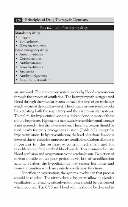
Principles of Drug Therapy in Dentistry128
are involved. The respiratory system works by blood oxygenationthrough the process of ventilation. The heart pumps this oxygenatedblood through the vascular system to reach the level of gas exchangewhich occurs at the capillary level. The central nervous system worksby regulating both the respiratory and the cardiovascular systems.Therefore, for hypoxemia to occur, a defect of one or more of theseshould be present. Hypoxemia may cause irreversible neural damageif not reversed in less than four minutes. Therefore, oxygen should beused nearly for every emergency situation (Table 6.2), except forhyperventilation. In hyperventilation, the level of carbon dioxide islowered due to excessive unnecessary ventilation. Carbon dioxide isimportant for the respiratory control mechanism and forvasodilatation of the cerebral blood vessels. This ensures adequateblood perfusion and oxygenation to the cerebral tissue. Depletion ofcarbon dioxide causes poor perfusion via loss of vasodilatationactivity. Further, the hypothalamus may secrete hormones andneurotransmitters which may interfere with heart functions.
For efficient oxygenation, the systems involved in that processshould be checked. The airway should be patent allowing alveolarventilation. Life-saving cricothyroidotomy should be performedwhen required. The CVS and blood volume should be checked to
Box 6.2: List of emergency drugs
Mandatory drugs• Oxygen• Epinephrine• Glycerin trinitrateBasic emergency drugs• Anticonvulsants• Corticosteroids• Antihistamines• Bronchodilators• Analgesics• Antihypoglycemics• Respiratory stimulant

Emergency Drugs Used in Dentistry 129
ensure adequate oxygen transport from the alveolar sacs to thecellular cytoplasm. Otherwise, oxygen supply will not produce theexpected results.
Epinephrine
Epinephrine is the second most commonly used emergency drug. Itis normally produced as a hormone by the adrenal medulla and asa neurotransmitter by the sympathetic system. It binds to theadrenergic receptors with the net result of preparing the body towithstand and survive extreme situations and stress. Adrenalineincreases the cardiac output, by enhancing its contractility, via theβ
1-adrenergic cardiac receptors. Peripheral vasoconstriction, via
the α1-adrenergic receptors, serves by elevating the blood pressure
for better brain and heart perfusion. In these two organs, however,blood vessels dilate under the effect of adrenaline bound to β2-receptors. This ensures sufficient blood perfusion for betterperformance.
Other useful effects of adrenaline include relaxation ofbronchial smooth muscle, stabilization of mast cell granules, and
Table 6.2: Mandatory drugs, doses and routes of administration
Oxygen Dose varies according to the technique ofadministration:• Mouth to mouth (17%)• Face mask (40–60%)• Bag-valve mask (75–95%)
Epinephrine* • IM into the tongue or the floor of the0.5–1 mg (1:1000) mouth. The deltoid muscle can be used
• IV, but better to be reserved for cardiacarrest or severe conditions
• SC, but has slow action
Nitroglycerin 0.5 mg sublingual tabletAngisid
Nitrolingual 0.4 mg spray
* Short duration, subsequent doses may be needed.

Principles of Drug Therapy in Dentistry130
hyperglycemic effects. In case of hypoglycemia, epinephrine may beused to elevate blood glucose level. It acts by stimulating the alfacells of the pancreas to secrete glucagon. Other hyperglycemic effectsare produced by enhanced hepatic glycogenolysis and inhibitedinsulin secretion.
Adrenaline is useful in many emergency situations. This includesacute allergic reactions, asthmatic attacks, cardiac arrest and evenin hypoglycemia.
Epinephrine, however, should not be used in patients withhistory of hypertension (risk of CVA or cardiac arrhythmia). Thenumber of adrenaline receptors is increased in case of high levels ofthe thyroxin hormone (hyperthyroidism) producing amplifiedeffects of adrenaline. If needed, hyperthyroid patients should begiven minimal doses. Table 6.2 shows the doses and routes ofadministration of epinephrine.
Adrenaline is metabolized by the catechol O-methyl transferaseenzyme (COMT) which is present in the GIT and the postsynapticclefts. Therefore, the oral form is expected to be rapidly inactivated.Intraneural metabolism is achieved by the monoamine oxidaseenzyme (MOA). That is why adrenaline is short acting and the vitalsigns should be rechecked every 10 minutes when used in anemergency situation as further administration may be needed.
Nitroglycerin
As the name implies, nitroglycerin is a compound of nitric or nitrousacid easter of glycerol. Nitroglycerin is volatile in nature and is ableto produce vasodilatation which enhances blood flow andoxygenation. It also acts by relaxing the venous system which helpsin reducing the preload on the myocardium. This also would reducethe amount of oxygen requirement. All these effects madenitroglycerin a valuable remedy in the management of ischemiccardiac attacks and in case of high blood pressure.
Nitroglycerin is used sublingually to provide relief from anginalattach, usually within 1 to 3 minutes. It can be supplied in the formof tablet or spray (Table 6.2).

Emergency Drugs Used in Dentistry 131
PRIMARY DRUGS
Anticonvulsants
Convulsions or seizures are conditions of sudden, but temporary,loss of consciousness accompanied with abnormal body movements.Epileptic attacks are caused by abnormally increased electric activityof a zone of neural cells in the cerebral cortex. These cells exhibitabnormal firing potential which is reached before normal cells.
Usually, fits are self-terminated, and the interference withanticonvulsant may be needed only in prolonged cases (statusepilepticus) or in conditions interfering with respiration. Forefficient control, the cause of the attack should be determined.Seizures can be idiopathic or produced secondarily tohyperventilation, hypoglycemia, syncope or thyroid storm.
Benzodiazepines (diazepam and lorazepam) are the agentsused to end seizures (Table 6.3). They act on the GABAA receptorswhich would interfere with the cerebral neurotransmission. Themost significant side effect of benzodiazepines is respiratorydepression and prolonged sedation. Flumazenil, a benzodiazepineantagonist, may be required to reverse these side effects. It works bycompetitive inhibition and has more affinity to the GABA receptorsthan the benzodiazepines. However, the duration of action is shortand frequent administration may be needed.
Care should be taken in patients’ dependant on benzodiazepinetherapy or those on tricyclic antidepressant medication as they areprone to seizures when flumazenil is provided.
Table 6.3: Anticonvulsants and sedative antagonistsAgent Route DoseDiazepamValium IV, IM 5–10 mlValpamLorazepam* IV, IM 4 mgFlumazenil† IV only 100 mg/mlAnexate
* Short acting benzodiazepine.† Benzodiazepine antagonist.

Principles of Drug Therapy in Dentistry132
Corticosteroids (Table 6.4)
Physiologically, corticosteroids play an important role in the body.Cortisol from the adrenal cortex ensures normal performancedespite stressful situations, like pain and disease. Cortisol receptorsare found at every cell of the body. When secreted, cortisol acts byincreasing glucose level for consumption and elevates the bloodpressure for better organ perfusion and function. Patients withadrenal insufficiency or those dependants on long-term steroidtherapy are at risk of cortisol shortage in case of stress or urgentsituations. Instead of coping with stress, these patients may collapse.
Another function of cortisol is its potent anti-inflammatory/antiallergic effect which makes it the absolute option for acuteinflammation and allergic conditions. It acts by dramaticallydecreasing the peripheral WBCs. It also interferes with thedegranulation process of histamine-producing cells. In addition,PG, leukoterines and other inflammatory mediators are all blockedas it inhibits the phospholipas A enzyme, an enzyme responsible forthe synthesis of arachidonic acid from membrane phospholipids.
Antihistamines
Histamine is a local inflammatory mediator which increases vascularpermeability and causes edema. It is produced by degranulation ofbasophils or mast cells. These cells are sensitized by IgE which waspreviously produced in response to a similar antigenic stimulation.Once released, histamine acts on histamine receptors which arefound at great numbers in the lungs, GIT and skin. Binding to H
1
Table 6.4: Corticosteroids
Agent Dose Route
Hydrocortisone 100–200 mg IV, IMSolu cortef (100 mg/ml)Dexamethasone† 4 mg/ml IV, IMOradexon
† Long acting corticosteroid.

Emergency Drugs Used in Dentistry 133
receptors, histamine mediates vascular permeability, smooth musclecontraction and mucus secretions. H
2 receptors, on the other hand,
are present in the stomach and their activation facilitates gastricacid secretion.
Antihistamines bind competitively to histamine receptors butthey don’t inhibit histamine production. Their actions may taketime, so, in some cases adrenaline may be used for more rapidrelieve of allergy. Antihistamines are useful in mild or delayed allergicreactions (Table 6.5). Severe allergies would necessitate the useadrenaline and corticosteroids. Cortisone acts as an anti-allergicagent by blocking all the inflammatory mediators.
Antihistamines should not be used for acute asthmatic attacksas they may aggravate the condition by their drying effects.
Bronchodilators
β2-agonists, like albuterol, are useful in managing conditions of
bronchospasm and acute allergies. As stated for epinephrine,β
2-receptors are responsible for bronchodilation. These selective agents
replaced theophylline which was associated with undesirable effectsand drug interactions. They are present in spray form (Table 6.6).
Isoproterenol, however, is not selective and it acts on both β1
and β2. Therefore, more side effects are to be expected on the heart.
Such effect can be useful, like adrenaline, in case of cardiac emergencies.
Analgesics
Analgesia may be required in certain painful medical conditionslike MI. Due to severe pain and anxiety, the oxygen demand and
Table 6.5: Antihistamines*
Agent % mg/ml Dose (mg)
Diphenylhydramine 10 20–50Chlorpheniramine 10 10–20BeadrylPheniramine 22.5 45Avil
* All can be provided by IV or IM routes.

Principles of Drug Therapy in Dentistry134
work load are increased, which may aggravate the ischemic heartattack. By rapid pain relief and anxiety control, the load over theheart will be reduced, as well as, its oxygen requirements.
Strong opioids like morphine sulfate or meperidine(Table 6.7), act on the opioid receptors with the result of intensepain relief. Care should be practiced, however, in case of braindamage or asthmatic patients as these analgesics induce respiratorydepression and cause histamine release. Morphine sulfate has auseful effect other than being a strong analgesic as it acts by relievingpulmonary edema.
Naloxone narcotic antagonist may be needed to reverseunwanted effects like respiratory depression or unwanted sedation(Table 6.7). It works by displacing opioid from their receptors.Naloxone has about ten times more affinity to opioid receptorsthan morphine. Reversed sedation is expected in 30 seconds.
Nitrous oxide is also a potent analgesic which works also bybronchodilation and enhanced perfusion to the brain. However,muscle relaxation may affect the rate of respiration, an effect whichdeserves special attention.
ASA, although not used for the purpose of analgesia, is ofbenefit in case of MI. It secondarily affects platelet aggregation viablocking the thromboxane A
2 enzyme. This antiplatelet effect reduces
the chances of death in patients having an IM as the risk of thrombusformation is lowered.
Antihypoglycemic AgentsHypoglycemia may be caused by starvation or by relatively higherinsulin dose in diabetics. Stress, exercise or insufficient food intakemay disturb the balance between blood glucose level and insulin. Asa result, fainting, convulsion and in severe cases, brain damage and
Table 6.6: BronchodilatorsAgent Dose and routeAlbuterol Ventolin 0.1 mg/spray with two sprays consideredSalbutamol sufficientVental

Emergency Drugs Used in Dentistry 135
Table 6.7: Analgesics necessary for management of emergencies
Agent Dose Route
Morphine sulfate 10 mg Oral(10 mg/mL) 5 mg IM
2 mg IVMeperidine (50 mg/ml) 50 mg/ml SCPethedineNaloxone* (0.4/ml) 0.4-2 mg IV, SCNarcanNitrous oxide gas 35% with Inhalation
65% oxygenASA† Aspocid 75–325 mg Oral
* Narcotic antagonist.† Non-enteric coated, chew and swallow.
death may the results. Brain cells depend on glucose for theirmetabolism and there is no glucose storage mechanism in theirstructure. In this case, antihypoglycemics may be life-saving.
The conscious patient can be supplied with sugar tablets orsoft drink (Table 6.8). Dextrose IV in 50 percent concentration isindicated in case of hypoglycemia in unconscious patients. Glucagoncan be used instead of dextrose when the latter is not present orwhen it is difficult to obtain venous access. Glucagon is an alfa cellpancreatic hormone which acts by elevating the blood glucose levelby enhancing hepatic glycogenolysis and gluconeogenesis.Epinephrine is another hormone which elevates blood glucoselevel. It, too, acts by enhancement of glycogenolysis, induction ofglucagon secretion and inhibition of insulin production.
Table 6.8: Antihypoglycemic agents
Agent Form and dose
Sugar Glucose tablets 3 g with vitamin C 150 mgChocolate bar or soft drinks
Dextrose 50%, 50 ml IVGlucagon 1 mg IM or SCEpinephrine (1:1000) 0.5 ml SC

Principles of Drug Therapy in Dentistry136
Respiratory Stimulants
Aromatic ammonia is used as a respiratory stimulant and toawaken patients who are in syncope, due to its irritating propertiesto the mucous membrane (Table 6.9). Thus, the respiratory andthe vasomotor centers in the medulla are stimulated. Bothrespiratory rate and blood pressure are elevated which help inregaining consciousness. For the fainted patients, aromatic ammoniamay be the only required drug. In asthmatic patients, aromaticammonia may precipitate an asthmatic attack, so its use is notrecommended.
MANAGEMENT OF EMERGENCIES
In the following section, a brief description of some common medicalemergencies that may occur in the dental clinic is provided in theform of Tables 6.10 to 6.18. The purpose of this section is tointroduce these emergencies, delineate the clinical appearance, causeand pathophysiology and the proper management.
Table 6.9: Respiratory stimulant
Agent Dose and route
Aromatic ammonia 0.3–0.4 ml, inhalation
Table 6.10: Syncope (fainting, vasovagal attack)
Definition and • Fainting or losing consciousnessclinical features • Characterized by weakness, pallor, sweating, and
loss of consciousness• If severe or prolonged, it might progress into a
seizure
Pathophysiology • Related to stress and anxiety or extreme fear.Hypoglycemia can be another factor
• This causes overstimulation of the vagal systemwhich decreases the cardiac output and bloodpressure resulting in cerebral hypoxia
Contd...

Emergency Drugs Used in Dentistry 137
Management • Lie the patient down into a flat position withlegs elevated
• Manage airway and administer oxygen• Monitor vital signs• Loosen clothes to enhance breathing and apply
cold on the forehead. Cooling causes vasocons-triction which enhances blood return
• The condition usually resolves in 2 minutes asthe sympathetic system is activated
• Aromatic ammonia may be used for prolongedfainting or with slow breathing
• If the patient does not wake up, consider othercauses of fainting and prepare for hospitaltransfer
Table 6.11: Acute asthmatic attack
Definition Paroxysmal allergic condition associated withattacks of dyspnea
Clinical features • Dyspnea, expiratory wheezing sound andcyanosis
• Anxiety and apprehension aggravate thecondition and loss of consciousness may occur
Pathophysiology Allergic reaction to drugs (penicillin for example)or foreign material (like latex) causes broncho-spasm, increased mucus production as well asedema of the mucous membrane. The conditioncan be stress-related
Management • Reassurance to minimize stress• Place in erect position to enhance breathing• Give up to 4 metered doses of bronchodilator• Supply with oxygen to prevent hypoxia• Status asthmaticus (prolonged attack) may need
the following:– IV sulbutamol (250 microgram) or 0.3 ml
epinephrine SC– IV steroids– Transfer to hospital
Contd...

Principles of Drug Therapy in Dentistry138
Table 6.12: Hyperventilation
Definition and • Abnormally increased respiratory rate, butwith decreased depth of respiration
• Characterized by dyspnea with shallow butrapid breaths
• Fainting, perioral paresthesia, tremors, tetany(carpopedal spasm) and stomach pain withvomiting
• Cardiac manifestations may occur andtachycardia, palpitation and chest pain maybe seen
• The patient may lose consciousness
Pathophysiology • Anxiety, pain or fear causes tachypnea withincreased carbon dioxide elimination. Thiswould create metabolic imbalance (respiratoryalkalosis)
• The firing potential of muscle fibers and nervecells is lowered and the tissues become hyper-excitable and possibly self-excited
Management • Oxygen is contraindicated• Try to decrease the respiratory rate by
reassurance and anxiety reduction• Increase the carbon dioxide content by re-
breathing the exhaled air through paper bagor clubbed hands
• If needed, provide nitrous oxide or midazolam2–4 mg IM to control anxiety
Table 6.13: Epilepsy
Definition Brain disorder associated with excessive electricalactivity in a certain focus in the cerebral cortex
Clinical features Characterized by sudden loss of consciousnesswith body rigidity, respiratory apnea and cyanosisat the tonic phase. The clonic phase ischaracterized by involuntary jerky movements
clinical features
Contd...

Emergency Drugs Used in Dentistry 139
Pathophysiology Stimulus like pain, stress, hypoxia* or infectionmay trigger the zone of abnormal cells to produceexcessive electrical discharge
Management • Protect the patient from injury and place inrecovery position. This would allow forsecretions to sweep out and prevent the inter-ference of the tongue
• Give oxygen and monitor the vital signs. Fitsincrease oxygen demand and compromiserespiration, as well
• If not recovered in 10 min (status epilepticus),give diazepam 10 mg IV or midazolam 5 mg IMand call for medical emergency
* Careful consideration when giving benzodiazepines, oxygensupplement is a wise decision.
Table 6.14: Acute anginal attackDefinition Acute chest pain due to narrowed coronary arteryClinical features • Moderate to severe retrosternal compressing
pain which is sometimes reported as “anelephant standing on the chest”
• Pain may radiate to the left shoulder or arm orleft side of the lower jaw
• Pulse is regular, and if not it might be an MI• It differs from indigestion in that the patient
does not feel hungry and not on antacid therapyPathophysiology Increased oxygen demand due to stress or anxiety
with atheroma narrowing the coronary arteryexposes the myocardium to ischemic attack andthis causes pain
Management • Reassure, enhance ventilation and decreasevenous return by erect position
• Sublingual nitroglycerin tablet (0.5 mg) or spray• Give oxygen• Check vital signs, if pulse is irregular treat as MI• If not resolved in 5 min, give another tablet of
glycerin trinitrite• After 3 doses, if pain continues, manage as MI
Contd...

Principles of Drug Therapy in Dentistry140
Table 6.15: Myocardial infarction
Definition and • Similar to AP except for the irregularity inclinical features pulse and reduced blood pressure. However,
the associated pain may be more severe andnot resolved by the use of nitroglycerin
• If not managed properly, the patient may loseconsciousness or goes into cardiac arrest*
Pathophysiology • The presence of atheroma in the coronaryarteries may be big enough to cut off the bloodsupply to the myocardium
• Ischemic attack is precipitated by stress oranxiety with increase heart oxygen requirement.Cellular necrosis or infarction results
Management • If MI is not suspected, the condition ismanaged initially as AP
• If pain is not resolved after 3 nitroglycerintablets, or when MI is suspected due toirregular pulse or low BP then give:
– Analgesia: morphine sulfate 10 mg ormeperidien 50 mg IM. Nitrous oxideanalgesia may be used (50/50)
– ASA 300 mg to prevent thrombusformation
• If cardiac arrest ensues, start CPR and monitorvital signs till advanced help arrives
* Silent form of MI may occur in diabetic patients where no chestpain is felt. However, shortness of breaths, sense of impeding doomeand general feeling of weakness may be the reported symptoms. MIis a common cause of cardiac arrest.
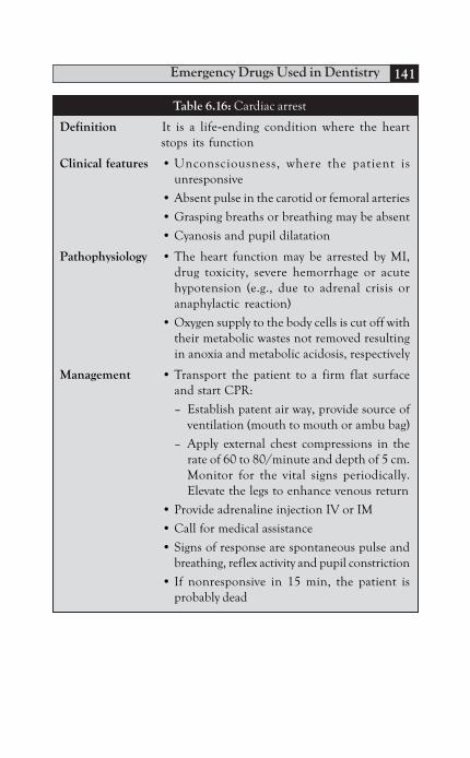
Emergency Drugs Used in Dentistry 141
Table 6.16: Cardiac arrest
Definition It is a life-ending condition where the heartstops its function
Clinical features • Unconsciousness, where the patient isunresponsive
• Absent pulse in the carotid or femoral arteries
• Grasping breaths or breathing may be absent
• Cyanosis and pupil dilatation
Pathophysiology • The heart function may be arrested by MI,drug toxicity, severe hemorrhage or acutehypotension (e.g., due to adrenal crisis oranaphylactic reaction)
• Oxygen supply to the body cells is cut off withtheir metabolic wastes not removed resultingin anoxia and metabolic acidosis, respectively
Management • Transport the patient to a firm flat surfaceand start CPR:
– Establish patent air way, provide source ofventilation (mouth to mouth or ambu bag)
– Apply external chest compressions in therate of 60 to 80/minute and depth of 5 cm.Monitor for the vital signs periodically.Elevate the legs to enhance venous return
• Provide adrenaline injection IV or IM
• Call for medical assistance
• Signs of response are spontaneous pulse andbreathing, reflex activity and pupil constriction
• If nonresponsive in 15 min, the patient isprobably dead

Principles of Drug Therapy in Dentistry142
Table 6.17: Hypoglycemia
Definition A condition of reduced blood glucose level usuallydue to imbalance between the hypoglycemic drugand calorie intake
Clinical features Slurred speech, blurred vision, altered behavior,sweating, weakness tachycardia, anxiety, loss ofconsciousness and convulsions
Pathophysiology • Normal insulin injection with missed meal,stress, anxiety or infection all increase the energydemand and, thus, exhaust the glucose available
• Continued low glucose level leads to neuralnecrosis
Management • Place in flat position• Conscious (recognizable early signs): provide oral
glucose• Unconscious patient:
– Glucose IV (50 ml/50% dextrose) over 2 to 3minutes,Or
– Glucagon 1 ml IM,Or
– Epinephrine* 0.5 ml (1:1000) SC– Monitor vital signs and provide oxygen
Table 6.18: Anaphylactic reaction
Definition Literally it means “lack of protection”, a conditionof severe immediate hypersensitivity reaction
Clinical features • Cutaneous: Including erythemia, urticaria andangioedema
• Respiratory: Dyspnea and wheezing soundsfollowed by cyanosis
• GIT and urinary: Vomiting and urinaryincontinence
• Circulatory: Hypotension, tachycardia, cardiacdysrythmia or even cardiac arrest
Pathophysiology • Allergen (usually in the IV form) combines withthe already formed IgE* creating antigen-
Contd...

Emergency Drugs Used in Dentistry 143
antibody complexes which, in turn, cause degr-anulation of mast cells. Histamine and otherinflammatory mediators are librated in massiveamounts
• This leads to vasodilatation, increased vascularpermeability and hypotension
• Bronchospasm and laryngoedema cause dyspneaor even apnea
Management • Enhance venous return by placing in supineposition
• 0.5 mg epinephrine (1:1000) IV or IM (it acts byincreasing cardiac output, blood pressure,bronchodilation and stabilization of mast cells).Check the effects of epinephrine every 5minutes. Give other injections if needed
• Provide basic life support (CPR):
– Airway (A): Establish patent airway and ifneeded cricothyroidotomy or tracheotomymay be performed
– Breathing (B): Provide oxygen
– Circulation (C): Chest compressions in caseof cardiac arrest
• 100 to 200 mg hydrocortisone IV (to maintainblood pressure and for absolute anti-allergicaction)
• 50 mg diphenylhydramine or 10 mg chlor-pheniramine (antihistamine) IV or IM
• Monitor vital signs periodically till advancedmedical support arrives
* IgE is found at higher concentrations at the adenoids, tonsils,bronchi, GIT and urinary bladder.
Contd...

Principles of Drug Therapy in Dentistry144
BIBLIOGRAPHY
1. Archibald D, Reginald H. Preparing for medical emergencies inthe dental office. J Can Dent Assoc. 1999;65:248-6.
2. Australian Dental Association Inc. Guidelines for good practiceon emergency in dental practice.
3. Kate Mellville. Oxygen resuscitation in emergency damages brainfunction. Science A GoGo. 2007.
4. Moris S Clark and Benjamin E Wall. 20-year follow-up survey ofmedical emergency education in US dental school. J of dentaleducation. 2006;70:12.
5. Morton B Rosenberg. Drugs for medical emergencies.Pharmacology and therapeutic for dentistry. 4th edn. JA ENTIDA Frank J Mosby; 1998.
6. Nigel D, Jason L. Medical Emergencies in Dentistry. OxfordUniversity Press; 2006.
7. Padmaja Uday Kumar. Textbook of pharmacology for dental andallied health sciences. 2nd edn. Jaypee Brothers Medical Publishers;2006.
8. Patarinski D. Indications and contraindications of oxygen therapyin respiratory insufficiency. Pub Med Vutr Boles. 1967;15(4):44-50.
9. PJ Champan. Medical emergencies in dental practice and choiceof drugs and equipment: a survey of Australian dentists. Australiandental J. 1997;42:2.
10. Richard A, Pamela C. Lippincott’s illustrated review of pharmacology.4th edn. Lippincott Williams and Wilkins; 2009.

A
Acetaminophen 29Acquired valvular dysfunction 79Actinomyces israelii 68Actinomycosis 68Acute
apical periodontitis 66erythematous candidiasis 100inflammation of major
salivary glands 120pericoronitis 63
Acyclovir 111Adjuvant analgesics 35Aggressive periodontitis 70Albuterol 133Aminoglycosides 58Amoxicillin 49, 65Amphotericin B 93Ampicillin 50Analgesics 133Anemia 114Angular cheilitis 101Antibiotics 45Anticonvulsants 131Antidepressants 36Antihistamines 132Anxiety 21Arachidonic acid 17Aromatic ammonia 136Aspirin 29Atrial septal defect 79Atypical facial pain 36Azithromycin 55Azoles 95
Index
B
Bacterialinfections 44sialadenitis 68, 120
Bacteroids forsythus 57Benzodiazepines 131Bone marrow suppression 114Borrelia vincentii and Fusiformis
dentium 65Bradykinin 17Bronchodilators 133
C
Cancrum oris 66Cardiac pacemakers 80Cephalosporins 50Chickenpox 118Chlorhexidine 37
digluconate 81Chronic
atrophic candidiasis 102hyperplastic candidiasis 101infection 117mucocutaneous candidiasis
103Cidofovir 114Ciprofloxacin 52Clarithromycin 55Class of surgery 73Clinical use of
antifungal agents 99antiviral drugs 115systemic antibiotics 61
Principles of Drug Therapy in Dentistry146
Closed fractures 74Clostridium difficile 56Clotrimazole 98Cold sores 117Contaminated wounds 68Corynebacterium diphtheriae 66Coxsackie viruses 119
D
Dextrose IV 135Diarrhea 114Diazepam 131Diphtheria 66Drugs for pain relief 28
E
Electronic anesthesia 23Enteral route 3Epinephrine 129, 135Epithelial cells 117Ergotamine 36Erythromycin 55, 65Extensive lacerations 68, 73
F
Famciclovir 113Fascial space infections 67Fluconazole 98Flumazenil 131Foscarnet sodium 114Fulminating varicella pneumonia
118Fungal infections 91
G
Ganciclovir 113, 114General anesthesia 24Glandular fever 119
Gonococcal gonorrhea 65Guanosine 111
H
Hand-foot-mouth disease 119Herpangina 119Herpes labialis 117Histamine 17Hydrogen peroxide 83Hypertrophic cardiomyopathy 80
I
Ibuprofen 31Implant-associated infections 71Implants and bone grafts 74Inhalational route 6Inhibition of
nucleic acid synthesis 48protein synthesis 47
Inhibitors of bacterial cell wallsynthesis 47
Intramuscular injection 6Intraoperative pain 20Intravenous
routes 4sedation 24
Intravitreal injection ofganciclovir 114
Isoproterenol 133Itraconazole 98
K
Kawasaki's disease 79Ketoconazole 96
L
Laughing gas 24Leprosy 69

Index 147
Levofloxacillin 52Lincomycins 56Lorazepam 131Lymphadenitis 120
M
Macrolides 54with metronidazole 65
Management ofemergencies 136established infections 61
Median rhomboid glossitis 102Meperidine 134Metronidazole 65Miconazole 97Mitral valve
prolapse 80regurgitation 80
Morphine sulfate 134Mouthwash 65Moxifloxacillin 52Mumps 120
N
Naloxone 134Nausea 114Neisseria gonorrhoeae 65Nervous system 114Neuralgic pain 36Neurophysiology of pain 17Neutropenia 114Nitroglycerin 130Nitroimidazoles 53Nitrous oxide 24, 134Nonsteroidal anti-inflammatory
drugs 29Norfloxacin 52Nucleotide analogs 110Nystatin 95
O
Open fractures 73Opioids 32Oral thrush 99Osteomyelitis 67Osteoradionecrosis 68Oxygen 127
P
Pain control medications indentistry 16
Paracetamol 29Parenteral route 4Paul Bunnell test 119Penciclovir 113Penicillin
binding proteins 47G 48V 48
Periodontal abscess 66Polymerase enzyme 111, 114Postoperative pain management
25Potassium ions 17Povidone-iodine compounds 83Previous
coronary artery bypass surgery80
history of infectiveendocarditis 78
Prophylaxis againstbacterial endocarditis 75metastatic infections 75prosthetic joint infections 80surgical wound infections 71
Prostaglandin 17Prosthetic valves 78Pseudallescheria boydii 94Pseudomembranous colitis 56

Principles of Drug Therapy in Dentistry148
Q
Quinolones 52
R
Recurrent herpetic lesions 117Refractory periodontitis 70Renal system 114Respiratory stimulants 136Reye's syndrome 120Rheumatic heart fever 79
S
Scarlet fever 69Secondary bacterial infections 37Sensory neural ganglia 117Severe CMV infections 114Shingles 118Staphylococcus aureus 74Steroidal anti-inflammatory drugs
33Streptococcal gingivostomatitis
65Streptomyces
erythreus 54lincolnensis 56nodosus 93
Subcutaneous injections 6
Syphilis 69Systemic mycosis 104
T
Telithromycin 55Teratology of Fallot 78Tetracyclines 57, 83Thrombocytopenia 114Thymidine kinase 111TMJ dysfunction syndrome 36Tooth avulsion 69Traumatic injuries 73Treatment of periodontal diseases
69Tuberculosis 69
U
Upper respiratory tract 118Urinary system 121
V
Valacyclovir 112Vancomycin 60Varicella 118Ventricular septal defect 78Viral infections 108Viremia 118