Principles and applications of optogenetics in ... · PRIMER Principles and applications of...
13
PRIMER Principles and applications of optogenetics in developmental biology Daniel Krueger 1 , Emiliano Izquierdo 1 , Ranjith Viswanathan 1,2 , Jonas Hartmann 1 , Cristina Pallares Cartes 1 and Stefano De Renzis 1, * ABSTRACT The development of multicellular organisms is controlled by highly dynamic molecular and cellular processes organized in spatially restricted patterns. Recent advances in optogenetics are allowing protein function to be controlled with the precision of a pulse of laser light in vivo, providing a powerful new tool to perturb developmental processes at a wide range of spatiotemporal scales. In this Primer, we describe the most commonly used optogenetic tools, their application in developmental biology and in the nascent field of synthetic morphogenesis. KEY WORDS: Embryonic development, Optogenetics, Signaling, Synthetic biology, Tissue morphogenesis Introduction Optogenetics is a technique that, as its names implies, combines genetics and optics to control protein function with light – a principle initially developed by neuroscientists with the aim of controlling neuronal activity with cellular and millisecond-temporal precision (Boyden et al., 2005; Zemelman et al., 2002). It was actually Francis Crick who first suggested that light could help with understanding the complexity of the brain by allowing the activation or inhibition of individual neurons: ‘The ideal signal would be light, probably at an infrared wavelength to allow the light to penetrate far enough. This seems rather farfetched but it is conceivable that molecular biologists could engineer a particular cell type to be sensitive to light in this way’ (Crick, 1999). It took only a few years for the development of the first optogenetic applications for controlling neuronal activity and behavior of a living animal (Banghart et al., 2004; Bi et al., 2006; Boyden et al., 2005; Lima and Miesenböck, 2005; Zemelman et al., 2002). What Francis Crick did not anticipate, however, is that the technology he had envisioned could also be applied to untangle the complexity of organismal development. Similar to the function of neuronal networks, development of multicellular organisms requires cells to interact in a dynamic manner and to coordinate their behavior through the action of chemical signals. Spatiotemporal regulation is thus a key feature of developmental processes, and optogenetics provides a powerful tool kit for precise subcellular- to tissue-scale perturbations with sub-minute temporal accuracy. By controlling the power and frequency of the light input, optogenetics allows tunable control over protein activity, which can be instrumental to uncover system-level properties that would not be otherwise discoverable using complete loss-of-function perturbations (see Box 1). In this Primer, we first provide a general description of the most commonly used tools in optogenetics for cell and developmental biology and then discuss how some of these tools have been employed to address developmental biology questions in model organisms. Concepts and approaches in optogenetics for cell and developmental biology The first optogenetic methods employed rhodopsin-like photosensitive ion channels to stimulate neuronal activity with light (reviewed by Yizhar et al., 2011). Opening of channel pores leads to an influx of ions into the cell, which causes a change in the electric potential across the membrane, and, depending on the channel type, excitation or silencing of neuronal activity. Although optogenetic methods regulating the membrane potential are very useful in neurobiology, their applications in developmental biology are limited as most developmental processes do not rely on changes in membrane potential. The development of a second generation of optogenetic modules based on photoreceptor protein domains that undergo light-induced dimerization/oligomerization or unfolding upon light activation (photo-uncaging) has provided the means to control a wide range of cell and developmental processes (Table 1). The majority of these light-sensitive protein domains derive from plants or cyanobacteria and function in a bio-orthogonal manner when used in animals. When appropriately coupled to a protein of interest, they allow regulation of the protein’s intracellular localization, clustering state, interaction with binding partners, or (in the case of enzymes) catalytic activity, using light of defined wavelengths (Fig. 1). Protein localization is typically controlled using heterodimerization systems consisting of a subcellularly localized anchor that interacts in a light-dependent manner with a cognate photosensitive domain tagged to a protein of interest (Fig. 1A). Relocalization of a target protein to a specific site in the cell can positively regulate its function by enabling it to interact with downstream binding partners and effectors. Alternatively, protein function can be inhibited by sequestering it away from its site of action. The regulation of protein clustering is based on photosensitive protein domains that oligomerize upon activation by light (Fig. 1B). Depending on the target protein, clustering can either positively or negatively regulate protein function, for example by increasing the local concentration of signaling molecules or inhibiting functionality by steric hindrance, respectively. Protein sequestration allows the inactivation of a photo-tagged target protein by capturing it within multimeric protein complexes (Fig. 1C) (Lee et al., 2014). A target protein can also be bioengineered to contain a 1 European Molecular Biology Laboratory (EMBL), Developmental Biology Unit Meyerhofstrasse 1, 69117 Heidelberg, Germany. 2 Heidelberg University, Faculty of Biosciences, Heidelberg, 69117, Germany. *Author for correspondence ([email protected]) S.D.R., 0000-0003-4764-2070 This is an Open Access article distributed under the terms of the Creative Commons Attribution License (https://creativecommons.org/licenses/by/4.0), which permits unrestricted use, distribution and reproduction in any medium provided that the original work is properly attributed. 1 © 2019. Published by The Company of Biologists Ltd | Development (2019) 146, dev175067. doi:10.1242/dev.175067 DEVELOPMENT
Transcript of Principles and applications of optogenetics in ... · PRIMER Principles and applications of...
PRIMER
Principles and applications of optogenetics in developmentalbiologyDaniel Krueger1, Emiliano Izquierdo1, Ranjith Viswanathan1,2, Jonas Hartmann1, Cristina Pallares Cartes1 andStefano De Renzis1,*
ABSTRACTThe development of multicellular organisms is controlled by highlydynamic molecular and cellular processes organized in spatiallyrestricted patterns. Recent advances in optogenetics are allowingprotein function to be controlled with the precision of a pulse of laserlight in vivo, providing a powerful new tool to perturb developmentalprocesses at awide range of spatiotemporal scales. In this Primer, wedescribe the most commonly used optogenetic tools, their applicationin developmental biology and in the nascent field of syntheticmorphogenesis.
KEY WORDS: Embryonic development, Optogenetics, Signaling,Synthetic biology, Tissue morphogenesis
IntroductionOptogenetics is a technique that, as its names implies, combinesgenetics and optics to control protein function with light – aprinciple initially developed by neuroscientists with the aim ofcontrolling neuronal activity with cellular and millisecond-temporalprecision (Boyden et al., 2005; Zemelman et al., 2002). It wasactually Francis Crick who first suggested that light could help withunderstanding the complexity of the brain by allowing the activationor inhibition of individual neurons: ‘The ideal signal would be light,probably at an infrared wavelength to allow the light to penetrate farenough. This seems rather farfetched but it is conceivable thatmolecular biologists could engineer a particular cell type to besensitive to light in this way’ (Crick, 1999). It took only a few yearsfor the development of the first optogenetic applications forcontrolling neuronal activity and behavior of a living animal(Banghart et al., 2004; Bi et al., 2006; Boyden et al., 2005; Lima andMiesenböck, 2005; Zemelman et al., 2002).What Francis Crick did not anticipate, however, is that the
technology he had envisioned could also be applied to untangle thecomplexity of organismal development. Similar to the functionof neuronal networks, development of multicellular organismsrequires cells to interact in a dynamic manner and to coordinate theirbehavior through the action of chemical signals. Spatiotemporalregulation is thus a key feature of developmental processes, andoptogenetics provides a powerful tool kit for precise subcellular- totissue-scale perturbations with sub-minute temporal accuracy.
By controlling the power and frequency of the light input,optogenetics allows tunable control over protein activity, whichcan be instrumental to uncover system-level properties that wouldnot be otherwise discoverable using complete loss-of-functionperturbations (see Box 1).
In this Primer, we first provide a general description of the mostcommonly used tools in optogenetics for cell and developmentalbiology and then discuss how some of these tools have beenemployed to address developmental biology questions in modelorganisms.
Concepts and approaches in optogenetics for cell anddevelopmental biologyThe first optogenetic methods employed rhodopsin-likephotosensitive ion channels to stimulate neuronal activity withlight (reviewed by Yizhar et al., 2011). Opening of channel poresleads to an influx of ions into the cell, which causes a change in theelectric potential across the membrane, and, depending on thechannel type, excitation or silencing of neuronal activity. Althoughoptogenetic methods regulating the membrane potential are veryuseful in neurobiology, their applications in developmental biologyare limited as most developmental processes do not rely on changesin membrane potential. The development of a second generation ofoptogenetic modules based on photoreceptor protein domains thatundergo light-induced dimerization/oligomerization or unfoldingupon light activation (photo-uncaging) has provided the means tocontrol a wide range of cell and developmental processes (Table 1).The majority of these light-sensitive protein domains derive fromplants or cyanobacteria and function in a bio-orthogonal mannerwhen used in animals. When appropriately coupled to a protein ofinterest, they allow regulation of the protein’s intracellularlocalization, clustering state, interaction with binding partners, or(in the case of enzymes) catalytic activity, using light of definedwavelengths (Fig. 1). Protein localization is typically controlledusing heterodimerization systems consisting of a subcellularlylocalized anchor that interacts in a light-dependent manner with acognate photosensitive domain tagged to a protein of interest(Fig. 1A). Relocalization of a target protein to a specific site in thecell can positively regulate its function by enabling it to interactwith downstream binding partners and effectors. Alternatively,protein function can be inhibited by sequestering it away from itssite of action. The regulation of protein clustering is based onphotosensitive protein domains that oligomerize upon activation bylight (Fig. 1B). Depending on the target protein, clustering can eitherpositively or negatively regulate protein function, for example byincreasing the local concentration of signaling molecules orinhibiting functionality by steric hindrance, respectively. Proteinsequestration allows the inactivation of a photo-tagged target proteinby capturing it within multimeric protein complexes (Fig. 1C) (Leeet al., 2014). A target protein can also be bioengineered to contain a
1European Molecular Biology Laboratory (EMBL), Developmental Biology UnitMeyerhofstrasse 1, 69117 Heidelberg, Germany. 2Heidelberg University, Faculty ofBiosciences, Heidelberg, 69117, Germany.
*Author for correspondence ([email protected])
S.D.R., 0000-0003-4764-2070
This is an Open Access article distributed under the terms of the Creative Commons AttributionLicense (https://creativecommons.org/licenses/by/4.0), which permits unrestricted use,distribution and reproduction in any medium provided that the original work is properly attributed.
1
© 2019. Published by The Company of Biologists Ltd | Development (2019) 146, dev175067. doi:10.1242/dev.175067
DEVELO
PM
ENT
photosensitive protein domain that unfolds upon light activationcausing the exposure of a hidden signaling motif or relieving aprotein from allosteric auto-inhibition. This strategy of photo-uncaging allows, for example, direct control over the enzymaticactivity of a target protein by light (Fig. 1D). Below, we describesome of the more widely used photoreceptor protein domains andtheir use in dimerization/clustering and photo-uncaging applications.For a comprehensive overview on available photoreceptors foroptogenetic applications, see the OptoBase database (https://www.optobase.org/).
Light-oxygen-voltage (LOV) domainsLOV domain-containing proteins are a large family found inplants, fungi, algae and bacteria that comprises more than6000 predicted sequences (Pudasaini et al., 2015) with functionaland topological diversity (Glantz et al., 2016). The LOV coredomain is composed of a conserved Per-Arnt-Sim (PAS) domain,which is ∼110 amino acids long and forms a five-strandedantiparallel β-sheet fold and α-helical connector elements thatbind to a flavin chromophore (present in all organisms) as aphotoreactive co-factor. Upon blue-light illumination, a covalentbond forms between a cysteine in the PAS domain and flavin
(adduct formation), which leads to a conformational change andunfolding of one of the α-helices (e.g. LOV2 Jα) (Crosson andMoffat, 2001; Zayner et al., 2012) (Fig. 2A). This unfolding can beused to expose an engineered recognition motif controllingintracellular protein trafficking, or protein-protein interaction(Fig. 2B,C).
In addition, LOV domains have been engineered to induceprotein dimerization (Guntas et al., 2015; Kawano et al., 2015;Strickland et al., 2012; Wang et al., 2012). Different LOV domainspossess distinct activation kinetics, sensitivity and relaxation time(reversion to the dark state) (Pudasaini et al., 2015). Theseparameters should be carefully considered when selecting a LOVdomain for optogenetic applications (Table 1). Below, we introducesome of the most commonly used modules for in vivo applications,which we also discuss later within the context of development.
AsLOV2 and derivativesAsLOV2 is derived from the LOV2 domain of Avena sativaphototropin 1 and has been successfully used for several purposes.For example, in the PA-Rac system, the AsLOV2 domain was used tophoto-cage the small GTPase Rac (Wang et al., 2010; Wu et al.,2009). In addition, the LINuS (light-inducible nuclear localization
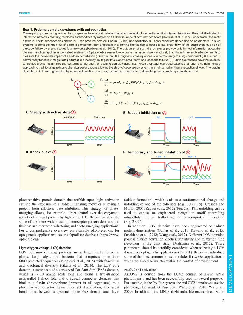
Box 1. Probing complex systems with optogeneticsDeveloping systems are governed by complex molecular and cellular interaction networks laden with non-linearity and feedback. Even relatively simpleinteraction networks featuring feedback and non-linearity may exhibit a diverse range of complex behaviors (Isomura et al., 2017). For example, the motifshown in A with dependencies shown in B can produce both equilibrium (C, left) and oscillatory (C, right) behaviors depending on parameters. In suchsystems, a complete knockout of a single component may propagate in a domino-like fashion to cause a total breakdown of the entire system, a sort ofcascade failure by analogy to artificial networks (Buldyrev et al., 2010). The outcomes of such drastic events provide only limited information about thedynamic functioning of the unperturbed system (D). Optogenetics serves to overcome this issue in twoways. First, it facilitates time-resolved experiments tomeasure the immediate impact of a sudden perturbation (E) rather than the long-term consequences of a permanently missing component (D). Second, itallows finely tuned low-magnitude perturbations that may not trigger total system breakdown and ‘cascade failures’ (F). Both approaches have the potentialto provide crucial insight into the system’s wiring and the resulting complex dynamics. Precise optogenetic perturbations thus offer a complementaryapproach to traditional genetic and chemical perturbations allowing the study of developing systems in a holistic, rather than a reductionist, way. The graphsillustrated in C-F were generated by numerical solution of ordinary differential equations (B) describing the example system shown in A.
AA B
C
D Knock out of
C Steady with active state E Sudden inhibition of
Acti
vity
Time
Equilibrium Light
F Temporary and tuned inhibition ofA
A
A
Oscillation
Time
Time Time
Time Time
Time Time
Acti
vity
Acti
vity
Acti
vity
Light
Light Light
B
A
2
PRIMER Development (2019) 146, dev175067. doi:10.1242/dev.175067
DEVELO
PM
ENT

Table1.
Phy
sico
-che
mical
prop
ertie
sof
themos
tcom
mon
lyus
edop
toge
netic
mod
ules
inde
velopm
entalb
iology
Mod
ule
Com
pone
nt(s)
Excita
tion
peak
Rev
ersibility
Rev
ersion
inda
rkCo-factor
Size(kDa)
Molec
ular
func
tion
Adv
antage
sDisad
vantag
esSelec
tedin
vivo
application(s)
Cryptoc
hrom
e(Ken
nedy
etal.,
2010
)
CRY2/CIBN
450nm
Stoch
astic
∼5min
FAD
CRY2:
57kD
a;CIBN:2
0kD
aHeterod
imerization;
clus
terin
gEas
yto
implem
ent;
CRY2alon
eca
nform
oligom
eric
clus
ters
Inco
mpa
tible
with
GFP*;
largetagsize
Cellcon
trac
tility,D
roso
phila
(Den
ekean
dDiT
alia,2
018;
Gug
lielm
ieta
l.,20
15;
Izqu
ierdoet
al.,20
18;
Krueg
eret
al.,20
18,2
019)
Differen
tiatio
n,Droso
phila
(Hua
nget
al.,20
17;
McD
aniele
tal.,
2019
);ce
llsign
aling,
Droso
phila
(Kau
ret
al.,20
17)
Cellsigna
ling,
Xen
opus
(Kris
hnam
urthyet
al.,20
16).
Phy
toch
rome
(Lev
skay
aet
al.,
2009
)
PHYB/PIF6
660nm
Ligh
t-indu
ced:
750nm
∼20
hPhy
to-
chromob
ilin/
phyc
o-cyan
obilin
(exo
geno
us)
PHYB:∼
100kD
a;PIF6:
11.5
kDa
Heterod
imerization
Can
besp
ecifica
llysw
itche
doffw
ithlight
(700
nm);
compa
tible
with
GFPflu
ores
cent
repo
rters
Nee
dsan
exog
enou
sco
-factor;req
uires
optim
izationfor
implem
entatio
n(protein
leve
ls,e
tc.);large
tag
size
Cellp
olarity,z
ebrafish(Buc
kley
etal.,20
16)
iLID (G
untaset
al.,
2015
)
AsL
OV2/Ssp
B45
0nm
Stoch
astic
Tun
able
FMN
AsL
OV2:
16kD
a;Ssp
B:1
3kD
aHeterod
imerization
Tun
able
kine
tics;
smalltag
size
;eas
yto
implem
ent
Inco
mpa
tible
with
GFP*
Cellsigna
ling,
Droso
phila
(Joh
nson
andToe
ttche
r,20
19;J
ohns
onet
al.,20
17)
TULIP
(Stricklan
det
al.,
2012
)
AsL
OV2/eP
DZ
450nm
Stoch
astic
Tun
able
FMN
AsL
OV2:
16kD
a;eP
DZ:2
1kD
aHeterod
imerization
Tun
able
kine
tics;
easy
toim
plem
ent
Inco
mpa
tible
with
GFP*
Celld
ivision,
C.e
lega
ns(Fielm
ichet
al.,20
18)
Organ
elle
trafficking
,C.
eleg
ans(H
arterin
ket
al.,
2016
)Differen
tiatio
n,se
aurch
in(U
chidaan
dYajim
a,20
18)
Vivid
(VVD)
(Wan
get
al.,
2012
)
VVD
450nm
Stoch
astic
Tun
able
FAD
20kD
aHom
odim
erization
Hom
odim
erform
ation;
tuna
blekine
tics
Inco
mpa
tible
with
GFP*
Cellsigna
ling,
cellcu
lture
(Iso
muraet
al.,20
17)
Mag
nets
(Kaw
anoet
al.,
2015
)
pMag
/nMag
450nm
Stoch
astic
Tun
able
FAD
16kD
aHeterod
imerization
Widerang
eof
tuna
ble
kine
tics(sec
onds
toho
urs)
Inco
mpa
tible
with
GFP*
Gen
eex
pres
sion
,mou
se(Jun
get
al.,20
19)
TAEL
(Motta-M
ena
etal.,20
14)
EL2
2245
0nm
Stoch
astic
∼1min
FMN
23kD
aExo
geno
usge
neex
pres
sion
Hom
odim
erform
ation;
optim
ized
for
exog
enou
sge
neex
pres
sion
inze
brafish
Inco
mpa
tible
with
GFP*
Gen
eex
pres
sion
,zeb
rafish
(Rea
deet
al.,20
17)
LITE (Kon
erman
net
al.,20
13)
TALE
-CRY2/
CIB1-VP64
450nm
Stoch
astic
∼5min
FAD
TALE
-CRY2:
162kD
a;CIB1-VP64
:50
kDa
End
ogen
ousge
neex
pres
sion
Optim
ized
for
mod
ulationof
endo
geno
usge
neex
pres
sion
Inco
mpa
tible
with
GFP*
Gen
eex
pres
sion
,mou
se(Kon
erman
net
al.,20
13)
LINuS
/LANS
(Niope
ket
al.,
2014
;Yum
erefen
diet
al.,20
15)
AsL
OV2
450nm
Stoch
astic
∼5min
FMN
18kD
aProtein
shuttling
Ena
bles
light-in
duce
dnu
clea
rim
port
Nee
dsop
timization‡;
inco
mpa
tible
with
GFP*
Gen
eex
pres
sion
,C.e
lega
ns(Yum
erefen
diet
al.,20
15)
The
photos
ensitiveco
mpo
nent
oftheresp
ectiveop
toge
netic
mod
uleisun
derline
din
the‘Com
pone
nt(s)’ca
tego
ry.
FAD,flavinad
eninedinu
cleo
tide;
FMN,flavinmon
onuc
leotide.
*Owingto
spec
tral
overlap.
‡Optim
izationrequ
iresad
ditio
nor
remov
alof
endo
geno
ussh
uttling
sign
als,
such
asNESor
NLS
.
3
PRIMER Development (2019) 146, dev175067. doi:10.1242/dev.175067
DEVELO
PM
ENT
signals)/LANS (light-activated nuclear shuttle) and LEXY (light-inducible nuclear export system) systems were implemented tocontrol nuclear import and export, respectively (Niopek et al., 2014,2016; Yumerefendi et al., 2015). Finally, protein heterodimerizationcan be achieved using the TULIP (tunable, light-controlledinteracting protein tags) (Strickland et al., 2012) and iLID(improved light-induced dimer) (Guntas et al., 2015) systems(Fig. 2D,E).
EL222, VVD and derivativesEL222 is a naturally occurring transcription factor fromErythrobacter litoralis containing a LOV domain that uponblue-light illumination dimerizes and binds to specific regulatoryelements of target genes. EL222 was adapted in the TA4-EL222(TAEL) system to control gene expression in cell culture and duringorganismal development. The fungal photoreceptor Vivid (VVD)from Neurospora crassa and VVD-derived magnets (Kawanoet al., 2015; Wang et al., 2012) can be used to control proteinhomodimerization and heterodimerization, respectively (Fig. 2G,H;Table 1). Similar to EL222, VVD and derivatives were employed totemporarily control gene expression in cell culture systems (Isomuraet al., 2017; Kim and Song, 2016; Nihongaki et al., 2017) as well asin the mouse brain (Jung et al., 2019).
Cryptochrome 2 (CRY2)CRY2 is another blue-light photoreceptor class (unrelated to LOVdomains) from Arabidopsis thaliana belonging to the cryptochromeprotein family, also present in most animals, which binds flavinadenine dinucleotide as a co-factor (Kennedy et al., 2010). Uponblue-light illumination, CRY2 undergoes photoisomerization andbinds to the N-terminal domain of CRYPTOCHROME-INTERACTING BASIC-HELIX-LOOP-HELIX 1 (CIB1), usuallyreferred to as CIBN (Kennedy et al., 2010). By anchoring CIBN tothe plasmamembrane, or to any other intracellular compartment, it ispossible to control the localization of a target protein fused to CRY2to that location upon photoactivation. The dissociation time of theCRY2-CIBN complex is ∼5 min, making this system suitablefor most in vivo applications (Table 1). When CRY2 is expressedalone (without CIBN), it tends to form homo-oligomers uponphotoactivation, often causing inhibition of tagged protein function(Taslimi et al., 2014). CRY2 can also be efficiently activated usingtwo-photon excitation (950 nm), which enables locally restrictedillumination patterns with cellular and subcellular precision deepinside tissues in living organisms (Guglielmi et al., 2015; Kruegeret al., 2018). Visible blue light (405–488 nm) has the disadvantagethat an undefined volume is activated when deeper focal planes areexcited, thus limiting the precision with which subcellularoptogenetic activation can be achieved (see below).
Phytochrome B (PHYB)PHYB belongs to the phytochrome family of photoreceptors found inplants and bacteria, and has the unique feature of being sensitive tored and far-red light. Phytochromes were first identified inArabidopsis thaliana in five different isoforms (PHYA-PHYE)that, upon photoactivation with red light (∼650 nm), formheterodimers with phytochrome interacting factors, such as PIF3and PIF6 (Mathews and Sharrock, 1997; Rockwell et al., 2006). Thedissociation of the dimer in the dark is very slow (∼20 h), but it can beinstantaneously triggered by far-red (∼740 nm) illumination, whichmakes this system ideally suited for applications requiring fast on/offcontrol of protein activity (Table 1). In addition, simultaneousillumination with red and far-red light in partially overlapping spatial
patterns allows very precise control of protein activity withsubcellular precision, which has been clearly demonstrated inmammalian cell culture (Levskaya et al., 2009) and in developingzebrafish embryos (Buckley et al., 2016). A major limitation of thisoptogenetic system is the requirement of a chromophore, such asphytochromobilin or phycocyanobilin, which is not present in animalcells and needs to be provided exogenously.
A summary of the optogenetic modules discussed in this Primerand their technical specifications is provided in Table 1. Besidesdifferences in biochemical activity (excitation wavelength, co-factorbinding, reversion in the dark, etc.), their molecular size should bealso considered, especially when trying to tag small proteins. Forexample, PHYB is almost four times bigger than GFP, whereas iLIDis only half the size (Table 1).
Optogenetics as a new precision perturbation tool indevelopmental biologyDecoding signaling dynamics during developmentSpatiotemporal regulation of signaling pathways is key for thegeneration of diverse responses during the development ofmulticellular organisms. Owing to limitations in the toolsavailable to manipulate signals at the relevant spatiotemporalscale, it has been so far challenging to study the impact of signaldynamics in vivo. What kind of information is encoded in a dynamicsignaling system during development? Do cells measure absolutesignal levels or changes in concentration over time? Does thefrequency of a signaling stimulus matter?
Some of these questions were elegantly addressed by usingoptogenetic tools to interrogate how cells respond to theconcentration and duration of Erk mitogen-activated protein kinasesignaling during Drosophila embryonic development (Johnson andToettcher, 2019; Johnson et al., 2017). Erk signaling is required forcells to adopt distinct fates at different positions in the earlyDrosophila embryo. Discriminative upstream factors activate Erksignaling to form head structures at the anterior pole, gut endodermat the posterior pole, and neurogenic cells at the lateral side.Although genetic approaches allowed a clear demonstration for the
Dark
Light
Localization SequestrationClustering Photo-uncagingBA C D
Photosensor (dark)
Inactive target protein
Photosensor (excited)
Active target protein
Interaction partner
LOV2-based photosensor
Light
Key
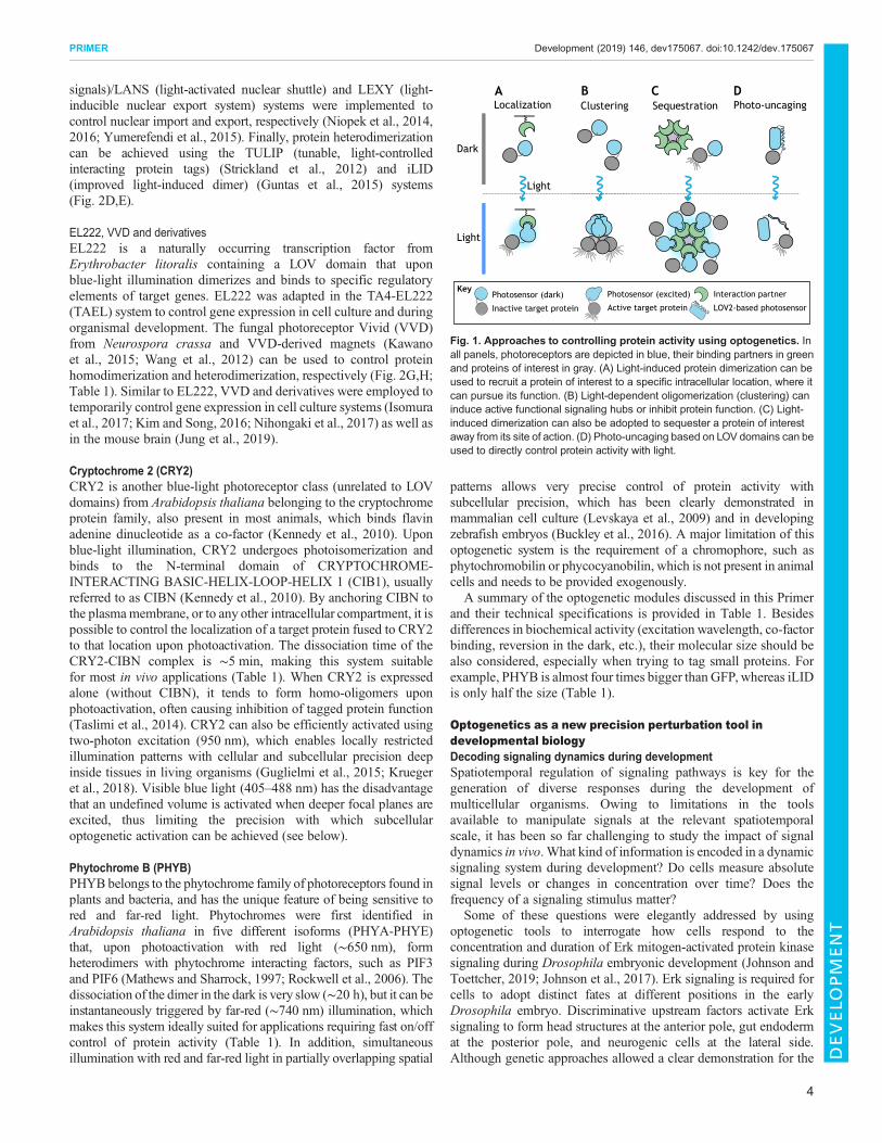
Fig. 1. Approaches to controlling protein activity using optogenetics. Inall panels, photoreceptors are depicted in blue, their binding partners in greenand proteins of interest in gray. (A) Light-induced protein dimerization can beused to recruit a protein of interest to a specific intracellular location, where itcan pursue its function. (B) Light-dependent oligomerization (clustering) caninduce active functional signaling hubs or inhibit protein function. (C) Light-induced dimerization can also be adopted to sequester a protein of interestaway from its site of action. (D) Photo-uncaging based on LOV domains can beused to directly control protein activity with light.
4
PRIMER Development (2019) 146, dev175067. doi:10.1242/dev.175067
DEVELO
PM
ENT
requirement of Erk signaling in cell fate specification, they did notallow understanding of how Erk signaling input is differentiallyconferred to achieve cell fate control. Johnson and co-workersimplemented an optogenetic method based on the iLID proteinheterodimerization system to precisely regulate Erk signaling inspace and time by controlling the activity of the Ras exchange factorSos, which upon membrane recruitment starts an endogenoussignaling cascade that culminates in Erk activation (Fig. 3A,B).Using this method, they demonstrated that cell fate switches in theembryo are triggered by the cumulative dosage of Erk signaling,rather than the duration or amplitude of signaling pulses (Johnsonand Toettcher, 2019). These results contradict the ‘transient versussustained’ model proposed in cell culture, which hypothesizes thatcells interpret quantitative differences in signaling dynamics, such asthe duration of signaling inputs, as determinants for cell fatespecification (Dowdle et al., 2014; Ebisuya et al., 2005; Marshall,1995; Nakakuki et al., 2010). Instead, in vivo the picture emergingfrom optogenetic control of Erk signaling is that cell fate control isencoded in the total amount of Erk activity integrated over time(Johnson and Toettcher, 2019; Johnson et al., 2017). Similarly,Krishnamurthy et al. implemented the CRY2/CIBN system tophotoactivate Raf1, a kinase acting upstream of Erk (Krishnamurthyet al., 2016). Optogenetic activation of Raf1 in Xenopus embryosafter germ layer specification (a developmental stage during whichapplications of traditional genetics is technically challenging)induced ectopic tail-like structures in the head region, suggestingthat Raf1 activation is sufficient to cause transformation of theembryonic tissue (Krishnamurthy et al., 2016).
During development, signaling pathways (such as Erk) arecontrolled by morphogen molecules, which are distributed inspatial gradients and provide positional information for tissuepatterning and cell differentiation (Wolpert, 1969). The first-identified morphogen, Bicoid (Bcd), patterns cells along theanterior-posterior axis of the early Drosophila embryo (Drieveret al., 1989). However, the exact time period during which cellsintegrate positional information has remained elusive. Huang et al.employed optogenetics to temporally control Bcd activity and explorehow the dynamics of the Bcd morphogen gradient is interpretedduring early development (Huang et al., 2017). By expressingCRY2-tagged Bcd, they rescued bcd−/− mutant embryonic development inthe dark, indicating that the protein functions normally. Whenexposed to blue light, however, embryos failed to develop head andthorax structures, resembling the bcd mutant phenotype. Byrestricting Bcd activity to different time windows, they found thattargets induced by high Bcd concentration (i.e. targets with low-affinity binding sites) require longer temporal exposure to Bcd,compared with targets induced by low Bcd concentration (i.e. targetswith high-affinity binding sites). Thus, optogenetics helped to revealdynamic aspects of morphogen-sensing mechanisms, namely thatcell fates depending on high Bcd concentration also require a longerperiod of Bcd exposure (Huang et al., 2017).
Comparable optogenetic approaches inhibiting target proteinactivity in specific tissues were used to demonstrate the temporalrequirement of specific factors at distinct stages of development.For example, McDaniel and co-workers show that the pioneerfactor Zelda, a master regulator of zygotic genome activation
Blue light
SspB
Jα
SsrA ePDZ
Jα
LOVpep
EL222Ncap
Ncap
− +
Opposingcharges
Dar
kLi
ght
blue light
JαPAS
LOV
Dark
Unfolding
LOV
Light
Uncaged target protein
LOV
Light
Exposed motif
Light
− +
A B C
D iLID A.sativa LOV2
E TULIP A. sativa LOV2
F TAEL E. litoralis LOV
G Vivid (VVD) N. crassa LOV
H Magnets VVD-derived
LOV
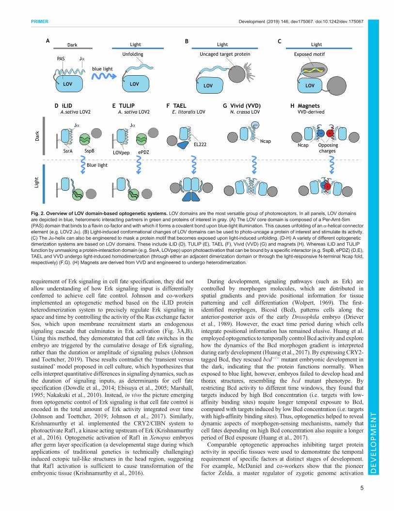
Fig. 2. Overview of LOV domain-based optogenetic systems. LOV domains are the most versatile group of photoreceptors. In all panels, LOV domainsare depicted in blue, heteromeric interacting partners in green and proteins of interest in gray. (A) The LOV core domain is composed of a Per-Arnt-Sim(PAS) domain that binds to a flavin co-factor and with which it forms a covalent bond upon blue-light illumination. This causes unfolding of an α-helical connectorelement (e.g. LOV2 Jα). (B) Light-induced conformational changes of LOV domains can be used to photo-uncage a protein of interest and stimulate its activity.(C) The Jα-helix can also be engineered to mask a protein motif that becomes exposed upon light-induced unfolding. (D-H) A variety of different optogeneticdimerization systems are based on LOV domains. These include iLID (D), TULIP (E), TAEL (F), Vivid (VVD) (G) and magnets (H). Whereas iLID and TULIPfunction by unmasking a protein-interaction domain (e.g. SsrA, LOVpep) upon photoactivation that can be bound by a specific interactor (e.g. SspB, ePDZ) (D,E),TAEL and VVD undergo light-induced homodimerization (through either an adjacent dimerization domain or through the light-responsive N-terminal Ncap fold,respectively) (F,G). (H) Magnets are derived from VVD and engineered to undergo heterodimerization.
5
PRIMER Development (2019) 146, dev175067. doi:10.1242/dev.175067
DEVELO
PM
ENT

EL222
∫dtBlue light
SspBSsrA
InactiveRac AsLOV2
Active Rac
InactiveRac
Actomyosin remodeling
ActiveRac
Protein shuttling
Nucleus
HiddenNLS
Revealedmotif
Hiddenmotif
Nuclearimport
C TAEL D Gene expression
A iLID
G Photoactivatable Rac1
H Cell motility
E LINuS/LANS
GDP
Sos
Ras
Cell signalingon
GTP
Raf
MekErk
DNA-bindingdomain
Trans-activationdomain
Transcription
GenePromoter
Plasmamembrane
Protrudingmembrane
AsLOV2
F Gene expression
Dark Light
B Tissue patterning
Dark Light
Dark Light
Dark Light
t
tt
Fig. 3. See next page for legend.
6
PRIMER Development (2019) 146, dev175067. doi:10.1242/dev.175067
DEVELO
PM
ENT
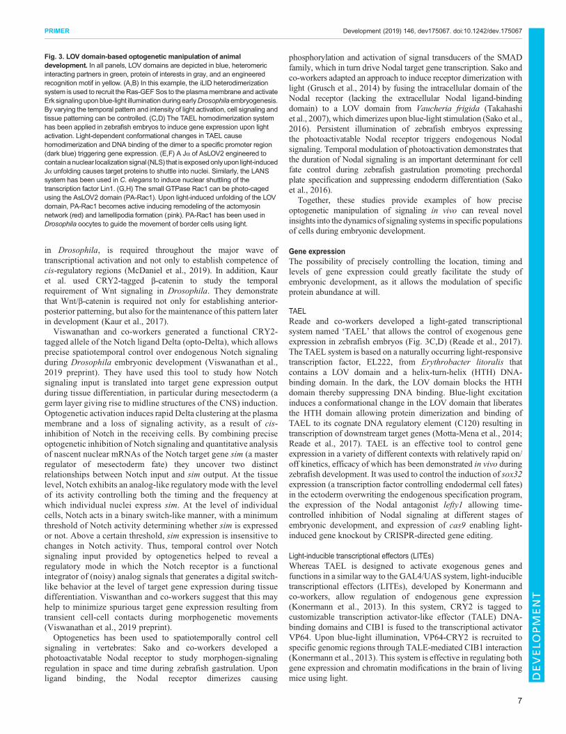
in Drosophila, is required throughout the major wave oftranscriptional activation and not only to establish competence ofcis-regulatory regions (McDaniel et al., 2019). In addition, Kauret al. used CRY2-tagged β-catenin to study the temporalrequirement of Wnt signaling in Drosophila. They demonstratethat Wnt/β-catenin is required not only for establishing anterior-posterior patterning, but also for the maintenance of this pattern laterin development (Kaur et al., 2017).Viswanathan and co-workers generated a functional CRY2-
tagged allele of the Notch ligand Delta (opto-Delta), which allowsprecise spatiotemporal control over endogenous Notch signalingduring Drosophila embryonic development (Viswanathan et al.,2019 preprint). They have used this tool to study how Notchsignaling input is translated into target gene expression outputduring tissue differentiation, in particular during mesectoderm (agerm layer giving rise to midline structures of the CNS) induction.Optogenetic activation induces rapid Delta clustering at the plasmamembrane and a loss of signaling activity, as a result of cis-inhibition of Notch in the receiving cells. By combining preciseoptogenetic inhibition of Notch signaling and quantitative analysisof nascent nuclear mRNAs of the Notch target gene sim (a masterregulator of mesectoderm fate) they uncover two distinctrelationships between Notch input and sim output. At the tissuelevel, Notch exhibits an analog-like regulatory mode with the levelof its activity controlling both the timing and the frequency atwhich individual nuclei express sim. At the level of individualcells, Notch acts in a binary switch-like manner, with a minimumthreshold of Notch activity determining whether sim is expressedor not. Above a certain threshold, sim expression is insensitive tochanges in Notch activity. Thus, temporal control over Notchsignaling input provided by optogenetics helped to reveal aregulatory mode in which the Notch receptor is a functionalintegrator of (noisy) analog signals that generates a digital switch-like behavior at the level of target gene expression during tissuedifferentiation. Viswanthan and co-workers suggest that this mayhelp to minimize spurious target gene expression resulting fromtransient cell-cell contacts during morphogenetic movements(Viswanathan et al., 2019 preprint).Optogenetics has been used to spatiotemporally control cell
signaling in vertebrates: Sako and co-workers developed aphotoactivatable Nodal receptor to study morphogen-signalingregulation in space and time during zebrafish gastrulation. Uponligand binding, the Nodal receptor dimerizes causing
phosphorylation and activation of signal transducers of the SMADfamily, which in turn drive Nodal target gene transcription. Sako andco-workers adapted an approach to induce receptor dimerization withlight (Grusch et al., 2014) by fusing the intracellular domain of theNodal receptor (lacking the extracellular Nodal ligand-bindingdomain) to a LOV domain from Vaucheria frigida (Takahashiet al., 2007), which dimerizes upon blue-light stimulation (Sako et al.,2016). Persistent illumination of zebrafish embryos expressingthe photoactivatable Nodal receptor triggers endogenous Nodalsignaling. Temporal modulation of photoactivation demonstrates thatthe duration of Nodal signaling is an important determinant for cellfate control during zebrafish gastrulation promoting prechordalplate specification and suppressing endoderm differentiation (Sakoet al., 2016).
Together, these studies provide examples of how preciseoptogenetic manipulation of signaling in vivo can reveal novelinsights into the dynamics of signaling systems in specific populationsof cells during embryonic development.
Gene expressionThe possibility of precisely controlling the location, timing andlevels of gene expression could greatly facilitate the study ofembryonic development, as it allows the modulation of specificprotein abundance at will.
TAELReade and co-workers developed a light-gated transcriptionalsystem named ‘TAEL’ that allows the control of exogenous geneexpression in zebrafish embryos (Fig. 3C,D) (Reade et al., 2017).The TAEL system is based on a naturally occurring light-responsivetranscription factor, EL222, from Erythrobacter litoralis thatcontains a LOV domain and a helix-turn-helix (HTH) DNA-binding domain. In the dark, the LOV domain blocks the HTHdomain thereby suppressing DNA binding. Blue-light excitationinduces a conformational change in the LOV domain that liberatesthe HTH domain allowing protein dimerization and binding ofTAEL to its cognate DNA regulatory element (C120) resulting intranscription of downstream target genes (Motta-Mena et al., 2014;Reade et al., 2017). TAEL is an effective tool to control geneexpression in a variety of different contexts with relatively rapid on/off kinetics, efficacy of which has been demonstrated in vivo duringzebrafish development. It was used to control the induction of sox32expression (a transcription factor controlling endodermal cell fates)in the ectoderm overwriting the endogenous specification program,the expression of the Nodal antagonist lefty1 allowing time-controlled inhibition of Nodal signaling at different stages ofembryonic development, and expression of cas9 enabling light-induced gene knockout by CRISPR-directed gene editing.
Light-inducible transcriptional effectors (LITEs)Whereas TAEL is designed to activate exogenous genes andfunctions in a similar way to the GAL4/UAS system, light-inducibletranscriptional effectors (LITEs), developed by Konermann andco-workers, allow regulation of endogenous gene expression(Konermann et al., 2013). In this system, CRY2 is tagged tocustomizable transcription activator-like effector (TALE) DNA-binding domains and CIB1 is fused to the transcriptional activatorVP64. Upon blue-light illumination, VP64-CRY2 is recruited tospecific genomic regions through TALE-mediated CIB1 interaction(Konermann et al., 2013). This system is effective in regulating bothgene expression and chromatin modifications in the brain of livingmice using light.
Fig. 3. LOV domain-based optogenetic manipulation of animaldevelopment. In all panels, LOV domains are depicted in blue, heteromericinteracting partners in green, protein of interests in gray, and an engineeredrecognition motif in yellow. (A,B) In this example, the iLID heterodimerizationsystem is used to recruit the Ras-GEFSos to the plasmamembrane and activateErk signaling upon blue-light illumination during earlyDrosophila embryogenesis.By varying the temporal pattern and intensity of light activation, cell signaling andtissue patterning can be controlled. (C,D) The TAEL homodimerization systemhas been applied in zebrafish embryos to induce gene expression upon lightactivation. Light-dependent conformational changes in TAEL causehomodimerization and DNA binding of the dimer to a specific promoter region(dark blue) triggering gene expression. (E,F) A Jα of AsLOV2 engineered tocontainanuclear localization signal (NLS) that isexposedonlyupon light-inducedJα unfolding causes target proteins to shuttle into nuclei. Similarly, the LANSsystem has been used in C. elegans to induce nuclear shuttling of thetranscription factor Lin1. (G,H) The small GTPase Rac1 can be photo-cagedusing the AsLOV2 domain (PA-Rac1). Upon light-induced unfolding of the LOVdomain, PA-Rac1 becomes active inducing remodeling of the actomyosinnetwork (red) and lamellipodia formation (pink). PA-Rac1 has been used inDrosophila oocytes to guide the movement of border cells using light.
7
PRIMER Development (2019) 146, dev175067. doi:10.1242/dev.175067
DEVELO
PM
ENT

LINuS/LANSAnother promising approach to control gene expression is to directthe localization of transcriptional regulators (activators, repressorsor epigenetic factors) from the cytoplasm to the nucleus and viceversa. Light-dependent nuclear/cytoplasmic shuttling systems weredeveloped to trigger nuclear translocation in the case of LINuS(Niopek et al., 2014) and LANS (Yumerefendi et al., 2015)(Fig. 3E,F) or to induce nuclear export as in the case of LEXY(Niopek et al., 2016). All these shuttling tools are based onengineered LOV domains that contain cryptic signaling motifs inthe LOV domain’s α-helix (e.g. AsLOV2 Jα) that are hidden in thedark and become exposed upon light stimulation allowinginteraction with specific regulators of nuclear import/export,which facilitate target protein shuttling. Apart from traffickingsignals, motifs such as post-translational modification sequences ordegrons, which regulate protein degradation rates, could also beengineered into LOV domains to produce additional tools to controlcell and developmental biology processes.
Collective cell migrationThe feasibility of implementing optogenetics to modulatemorphogenesis of multicellular organisms was first demonstrated inDrosophila and provided a clear demonstration of the power ofoptogenetics to control single cell behavior in vivo (Fig. 3G,H) (Wanget al., 2010). Wang and co-workers investigated the mechanismscontrolling collective cell migration by focusing on border cellmigration during Drosophila oogenesis. Border cells are aninterconnected group of six to eight cells that moves a totaldistance of ∼175 µm guided by a complex signaling environmentwithin the Drosophila ovary. Wang and co-workers implemented aphotoactivatable analog of the small GTPase Rac (PA-Rac1), whichwas developed to control migration in cell culture (Wu et al., 2009).Rac1 is a pivotal regulator of the actin cytoskeleton controllingcell adhesion, migration and polarity. PA-Rac1 is a fusion proteinbetween Rac1 and the AsLOV2 domain, which in the darkprevents Rac from interacting with its downstream effectors bysteric inhibition of the effector-binding site. Light illuminationinduces a conformational change that liberates Rac1 from thisinhibitory state (photo-uncaging) and triggers Rac1 activity. PA-Rac1 provides a tool for the polarized remodeling of the cytoskeletonwith full temporal control and subcellular precision. Photoactivationof Rac1 in single cells during border cell migration is sufficient toguide collective cell movements indicating that cells sense directionas a group according to relative levels of Rac activity (Wang et al.,2010). Further studies have also demonstrated the utility of thismethod to study cell migration in zebrafish embryos (Walterset al., 2010).During embryonic development, cells receive signaling inputs to
gain migratory competence (permissive signaling) and to guidetheir movements along specific routes (instructive signaling)(Reig et al., 2014). Non-canonical Wnt signaling, for example, isrequired for coordinated cell migration during metazoandevelopment (De Calisto et al., 2005). To understand better hownon-canonical Wnt signaling affects directed cell migration duringzebrafish gastrulation, Capek and co-workers engineered a light-sensitive version of the non-canonical Wnt receptor Frizzled 7 (Fz7)by substituting the intracellular domains of the photoreceptorrhodopsin with the corresponding domains of Fz7 (Capek et al.,2019). Using this new tool, they demonstrated that uniformphotoactivation rescues mesenchymal cell migration duringgastrulation of otherwise Fz7 mutant zebrafish embryos. Thisresult argues that, in addition to its instructive role in controlling
cell polarization in epithelial tissues, non-canonical Wnt signalingacts permissively in directing zebrafish mesenchymal migration,without the requirement of localized subcellular activation ofFz7 signaling.
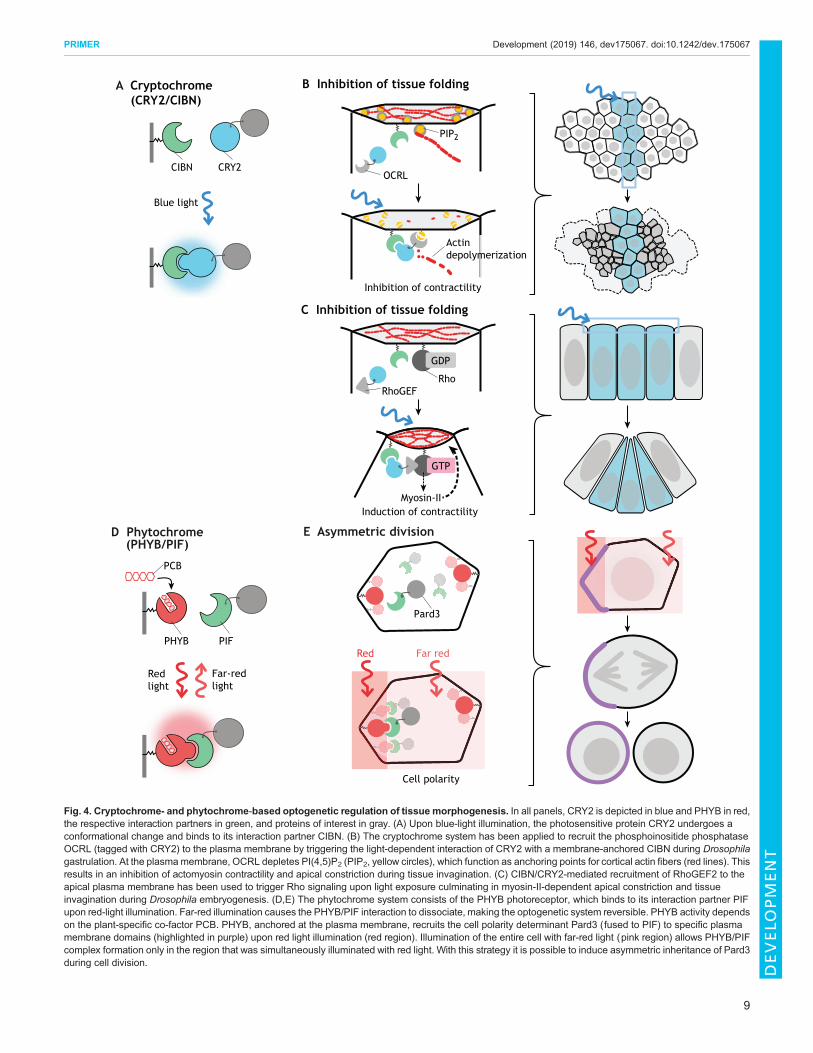
Tissue morphogenesisMorphogenesis of tissues requires coordination among cellpopulations, which leads to the emergence of group properties thatare rarely observed in isolated cells, such as symmetry breaking,pattern formation, shape remodeling and regeneration (Xavierda Silveira dos Santos and Liberali, 2019). The extent to whichchanges in the behavior of single cells influences their neighbors andcontrols large-scale tissue remodeling has been difficult to study usingconventional genetic approaches, owing to limited ability to targetindividual cells at will. Guglielmi and co-workers set out to examinethe dynamics of tissuemorphogenesis duringDrosophila gastrulation,when apical constriction of cells at the ventral midline initiatesinvagination and formation of the ventral furrow.Apical constriction isinduced by contractions of actomyosin filaments anchored at theplasma membrane via actin-binding proteins, localization of whichdepends on membrane phospholipids and in particular on thephosphatidylinositol phosphate PI(4,5)P2 (Bezanilla et al., 2015).Using the CRY2/CIBN system to achieve local control over PI(4,5)P2levels at the plasmamembrane, Guglielmi and co-workers showed thatlocal inhibition of apical constriction is sufficient to cause a globalarrest of tissue invagination (Guglielmi and De Renzis, 2017;Guglielmi et al., 2015). By varying the spatial pattern of inhibition,they further demonstrated that the coordinated contractile behaviorresponds to local tissue geometrical constraints (Fig. 4A,B). Together,the results demonstrate that apical constriction is necessary not only toinitiate but also to sustain tissue folding and that the geometry of theventral furrow tissue impacts the way individual cells constrict. Theseexperiments highlight how optogenetics can be used to dissect theinterplay between cell-cell interaction, force transmission and tissuegeometry during complex morphogenetic processes.
A similar CRY2/CIBN-based system was recently employed byDeneke and co-workers to dissect the connection between cell cycledynamics and cortical actomyosin contractility during earlyDrosophila embryogenesis. They adopted an optogenetic system tostimulate Rho signaling and apical constriction (Fig. 4C,D; seebelow) (Izquierdo et al., 2018) to increase the contractility of corticalactomyosin during early syncytial nuclear divisions (Deneke et al.,2019). They used this system to distinguish the role of cortical versuscytoplasmic actin contractility in nuclear positioning, a question thatcould not be addressed using actin-depolymerizing drugs orconventional genetic approaches that would result in a generalimpairment of actin dynamics. Using optogenetics they showed thatprecise spatiotemporal activation of cortical contractility leads to thegeneration of cytoplasmic flows, which in turn control nuclearpositioning and mitotic synchrony. These results argue that corticaland not cytoplasmic contractility drives uniform nuclear positioningand elucidate a self-organized mechanism that links cell cycleoscillators and embryo mechanics.
Subcellular optogeneticsThe spatial resolution of optogenetics is limited by theresolution and precision of the applied optical illuminationdevice as well as the diffusion of the optogenetic components.In simple 2D systems, such as cultured cells, subcellularphotoactivation can be achieved by optimizing the light power,frequency and duration of illumination (Benedetti et al., 2018;Meshik et al., 2019). However, many research questions in
8
PRIMER Development (2019) 146, dev175067. doi:10.1242/dev.175067
DEVELO
PM
ENT
D Phytochrome (PHYB/PIF)
PHYB PIF
PCB
Redlight
Far-redlight
Pard3
Cell polarity
Red Far red
E Asymmetric division
CIBN CRY2
Blue light
RhoGEFRho
GDP
GTP
Induction of contractilityMyosin-II
OCRL
B Inhibition of tissue foldingA Cryptochrome (CRY2/CIBN)
PIP2
Actindepolymerization
Inhibition of contractility
C Inhibition of tissue folding
Fig. 4. Cryptochrome- and phytochrome-based optogenetic regulation of tissue morphogenesis. In all panels, CRY2 is depicted in blue and PHYB in red,the respective interaction partners in green, and proteins of interest in gray. (A) Upon blue-light illumination, the photosensitive protein CRY2 undergoes aconformational change and binds to its interaction partner CIBN. (B) The cryptochrome system has been applied to recruit the phosphoinositide phosphataseOCRL (tagged with CRY2) to the plasma membrane by triggering the light-dependent interaction of CRY2 with a membrane-anchored CIBN during Drosophilagastrulation. At the plasmamembrane, OCRL depletes PI(4,5)P2 (PIP2, yellow circles), which function as anchoring points for cortical actin fibers (red lines). Thisresults in an inhibition of actomyosin contractility and apical constriction during tissue invagination. (C) CIBN/CRY2-mediated recruitment of RhoGEF2 to theapical plasma membrane has been used to trigger Rho signaling upon light exposure culminating in myosin-II-dependent apical constriction and tissueinvagination during Drosophila embryogenesis. (D,E) The phytochrome system consists of the PHYB photoreceptor, which binds to its interaction partner PIFupon red-light illumination. Far-red illumination causes the PHYB/PIF interaction to dissociate, making the optogenetic system reversible. PHYB activity dependson the plant-specific co-factor PCB. PHYB, anchored at the plasma membrane, recruits the cell polarity determinant Pard3 (fused to PIF) to specific plasmamembrane domains (highlighted in purple) upon red light illumination (red region). Illumination of the entire cell with far-red light (pink region) allows PHYB/PIFcomplex formation only in the region that was simultaneously illuminated with red light. With this strategy it is possible to induce asymmetric inheritance of Pard3during cell division.
9
PRIMER Development (2019) 146, dev175067. doi:10.1242/dev.175067
DEVELO
PM
ENT

developmental biology require spatially confined perturbationswith subcellular precision within the depth of a tissue with acomplex shape. Below, we discuss strategies to achieve subcellularoptogenetic control in deep focal volumes and in tissues withcurved morphology.Taking advantage of the reversible properties of the phytochrome
system, Buckley and co-workers successfully demonstrated theability to control cell polarity by rapidly and reversibly recruitingpolarity proteins to specific subcellular regions in the depth of aliving zebrafish embryos (Fig. 4D,E and Fig. 5). The establishmentand maintenance of epithelial apicobasal polarity is a tightlyregulated process, which is of key importance during organismaldevelopment. Its misregulation causes loss of epithelial integrity,increased cell motility and neoplastic transformation. In theirexperiments, Buckley and co-workers controlled the localization ofthe apical polarity protein Pard3 with subcellular precision in theembryo’s enveloping layer epithelium during neural tube formation(Buckley et al., 2016). To achieve this, PHYB was anchored at theplasma membrane through a CAAX (prenylation) anchor and Pard3was tagged with PIF6. Localized illumination in a region of theplasma membrane using red light of 650 nm caused recruitment ofPard3 to that location, whereas simultaneous global illumination inwith far-red light at 750 nm caused dissociation of the complexelsewhere. As red light-induced PHYB/PIF6 dimer formation isapproximately seven times faster than far-red light-induced dimerdissociation, Pard3 can be recruited to subregions of the plasmamembrane. Importantly, this method allows the manipulation of cellpolarity at will in vivo, which could be instrumental for futureresearch studying the interplay between cell polarity and tissuemorphogenesis. However, as already mentioned above, thephytochrome system requires the addition of a chromophore,which can pose some technical challenges, especially in organismssuch as the Drosophila embryo that are not permeable toexogenously applied molecules.To overcome such limitations, Krueger and co-workers
developed an approach based on the use of a subcellular-localized CIBN anchor and two-photon illumination, whichallows localized photoactivation patterns in tissues with complexmorphology, such as folded epithelia in the gastrulating embryo(Fig. 5). Most studies investigating ventral furrow formation duringDrosophila gastrulation focused on apical constriction and theupregulation of the molecular motor myosin-II at the apicalsurface, but it was unclear whether additional regulation of myosin-II at the basal surface is also required. Computer simulationspredicted a requirement of basal relaxation for completing tissueinvagination (Polyakov et al., 2014); however, owing to the lack ofgenetic mutations interfering specifically with the basal pool ofmyosin-II, it was impossible to test these models experimentally.Kruger and co-workers used their subcellular optogenetic system toprecisely manipulate Rho signaling and myosin-II activity at thebasal surface of the invaginating cells in Drosophila (Kruegeret al., 2018). Indeed, they could specifically counteract the loss ofbasal myosin-II during ventral furrow invagination anddemonstrate that maintaining myosin-II levels at the basal surfaceinhibits apical constriction, cell shape changes and tissueinvagination (Krueger et al., 2018). Importantly, their method notonly allows for spatial precision, but also permits quantitativecontrol of myosin-II levels.
Using optogenetics to reconstruct morphogenesisThe ability to manipulate signaling systems and cell behaviorwith spatiotemporal precision provides the potential to study
organismal development not only by interfering with the normalseries of events driving morphogenesis (e.g. defining the necessaryconditions), but also to guide it and reconstruct it (e.g. definingthe sufficiency conditions). The modular nature of morphogenesisimplies that it should be possible to single out individual modules,determine the minimum set of requirements that are sufficientto drive morphological remodeling, and eventually reconstructmorphogenesis (synthetic morphogenesis; Fig. 6). Recently,Izquierdo and co-workers used optogenetics to reconstitute tissueinvagination during Drosophila embryonic development in tissuesthat otherwise would not undergo internalization (Izquierdo et al.,2018). Using two-photon stimulation of RhoGEF2 tagged withCRY2 and a CIBN plasma membrane anchor, Rho signaling can betriggered to activate myosin-II and thus local cell contractility(Fig. 4C,D). Precise spatial and temporal activation of Rhosignaling at the apical surface of epithelial cells on the dorsal sideof the embryo is sufficient to trigger apical constriction and tissuefolding independently of any pre-determined condition ordifferentiation program associated with endogenous invaginationprocesses. The resulting optogenetics-guided furrows can betriggered at any position along the dorsal-ventral or anterior-posterior embryo axes in response to the spatial pattern and level of
Restrictedillumination
Two-photonmicroscopy
Local photoactivation
Subcellular
Localizedoptogenetic
anchor
Localizedoptogenetic anchor
UniformanchorDeactivatable
optogenetic module(phytochrome)
Local activation,global deactivation
Activation
Deactivation
Light
Fig. 5. Subcellular optogenetics. Three different approaches have been sofar employed to achieve subcellular photoactivation (activated photoreceptorcolored in blue, membrane-anchored components in green). Left: Using thePHYB photoreceptor anchored uniformly at the plasma membrane, it ispossible to locally photoactivate a subcellular region. Red light-induced PHYB/PIF6 dimer formation is approximately seven times faster than far-red light-induced dissociation. Stimulation of a subregion of interest using red light (redregion) with simultaneous deactivation of thewhole cell using far-red light (pinkregion) results in locally restricted photoactivation. Middle: Two-photonexcitation using near-infrared light enables locally restricted light delivery (blueblurred line) and photoactivation deep inside living tissues by temporally andspatially restricting the laser light to a focal volume in the femtoliter range.Right: Subcellular optogenetic activation can also be achieved in tissues ofcomplex morphology by engineering an optogenetic anchor in such a way thatit localizes only to the site of the cell where optogenetic activation is desired(green cell outline). Components of the cell polarity machinery are idealcandidates for designing optogenetic anchors, as recently demonstrated bythe use of PatJ to manipulate myosin-II activity specific at the cell base duringDrosophila gastrulation. Using this approach, even whole-cell photoactivationresults in a locally confined activation of the optogenetic system. See text formore details.
10
PRIMER Development (2019) 146, dev175067. doi:10.1242/dev.175067
DEVELO
PM
ENT

optogenetic activation. In addition, rectangular patterns ofphotoactivation cause cells to constrict anisotropically, whereassquared patterns cause isotropic constriction, which demonstratesthe impact of tissue geometry on individual cell behavior. By tuningthe strength of Rho signaling activation, different contractilebehaviors can be induced: discontinuous optogenetic activationresults in pulsatile apical constrictions, whereas sustained activationinduces continuous apical constriction and invagination (Izquierdoet al., 2018). These results demonstrate how optogenetics can beused to reconstruct morphogenesis and study input–outputrelationships, by coupling signaling systems and tissue shapechanges during embryonic development.
Concluding remarksIn this Primer, we have illustrated how optogenetic techniques can beused inmodel organisms to address developmental biology questions,emphasizing the unique advantages of precise spatiotemporalperturbations. As with any other technique, optogenetics is not freeof limitations. In particular, expression levels and dark-state activity ofnew optogenetic probes (i.e. the extent to which an optogeneticmodule is active prior to photoactivation) need to be carefullyassessed. When considering stimulation of signaling pathways, it isadvisable to combine such perturbations with correspondingdownstream biosensors to ensure that pathway activity is within thephysiological range. Additionally, the diffusion of photoactivated
?
B Feedback controlA Cell behaviors
Fig. 6. Reconstructing morphogenesis using synthetic biology approaches. (A) Morphogenesis relies on a common set of mechanisms (modules)involving changes in cell behaviors that occur at specific time points and locations, and that give rise to highly complex forms and patterns. (B) By enabling thedelivery of precise spatiotemporally controlled inputs, optogenetics allow individual modules to be triggered at will and determine theminimum set of requirementssufficient to drive morphological remodeling. Computerized feedback control could be used to automatically tune optogenetic inputs in real time accordingto the desiredmorphogenetic outputs (synthetic morphogenesis). Such an experimental set-up has been recently developed to achieve robust perfect adaptation(RPA) (Aoki et al., 2019) of gene expression in single Saccharomyces cerevisiae yeast cells (Rullan et al., 2018). This combination of optogenetic and controltheory concepts should allow us to eventually reconstruct complex morphogenetic processes and build synthetic embryos. The embryos depicted in thisfigure represent the corral Monoxenia darwinii during gastrulation as drawn by Ernst Haeckel (Haeckel, 1891).
11
PRIMER Development (2019) 146, dev175067. doi:10.1242/dev.175067
DEVELO
PM
ENT
optogenetic modules can cause complication especially for studyingextracellular morphogen signaling or long-range transport withincells. This could be overcome by using the phytochrome system,which allows activation in the desired region and simultaneous de-activation elsewhere. Although optogenetics is only a technique, theresults discussed in this article do suggest some common new themesemerging from the use of this methodology to study the complexquestion of how multicellular organisms develop. Optogeneticsallows us to establish very direct cause-effect relationshipsbetween gene activities and developmental phenotypes and todecode the engineering principles controlling cell fate decisions andmorphogenesis. The possibility of modulating signaling pathways atwill with cellular precision in vivomeans that we have now the abilityto reverse engineer and guide organismal development to the extentthat we should be able in the near future to build synthetic embryos(Fig. 6). This will allow us to both test theories of morphogenesis andalso facilitate the design of tissues with potential applications inregenerative medicine.
AcknowledgementsWe thank all speakers and participants of the 2019 EMBO course on non-neuronaloptogenetics for insightful comments and discussion.
Competing interestsThe authors declare no competing or financial interests.
FundingThis work was supported by European Molecular Biology Laboratory internalfunding (S.D.R.).
ReferencesAoki, S. K., Lillacci, G., Gupta, A., Baumschlager, A., Schweingruber, D. andKhammash, M. (2019). A universal biomolecular integral feedback controller forrobust perfect adaptation. Nature 570, 533-537. doi:10.1038/s41586-019-1321-1
Banghart, M., Borges, K., Isacoff, E., Trauner, D. andKramer, R. H. (2004). Light-activated ion channels for remote control of neuronal firing. Nat. Neurosci. 7,1381-1386. doi:10.1038/nn1356
Benedetti, L., Barentine, A. E. S., Messa, M.,Wheeler, H., Bewersdorf, J. andDeCamilli, P. (2018). Light-activated protein interaction with high spatial subcellularconfinement. Proc. Natl. Acad. Sci. USA 115, E2238-E2245. doi:10.1073/pnas.1713845115
Bezanilla, M., Gladfelter, A. S., Kovar, D. R. and Lee, W.-L. (2015). Cytoskeletaldynamics: a view from the membrane. J. Cell Biol. 209, 329-337. doi:10.1083/jcb.201502062
Bi, A., Cui, J., Ma, Y.-P., Olshevskaya, E., Pu, M., Dizhoor, A. M. and Pan, Z.-H.(2006). Ectopic expression of a microbial-type rhodopsin restores visualresponses in mice with photoreceptor degeneration. Neuron 50, 23-33. doi:10.1016/j.neuron.2006.02.026
Boyden, E. S., Zhang, F., Bamberg, E., Nagel, G. and Deisseroth, K. (2005).Millisecond-timescale, genetically targeted optical control of neural activity. Nat.Neurosci. 8, 1263-1268. doi:10.1038/nn1525
Buckley, C. E., Moore, R. E., Reade, A., Goldberg, A. R., Weiner, O. D. andClarke, J. D. W. (2016). Reversible optogenetic control of subcellular proteinlocalization in a live vertebrate embryo. Dev. Cell 36, 117-126. doi:10.1016/j.devcel.2015.12.011
Buldyrev, S. V., Parshani, R., Paul, G., Stanley, H. E. and Havlin, S. (2010).Catastrophic cascade of failures in interdependent networks. Nature 464,1025-1028. doi:10.1038/nature08932
Capek, D., Smutny, M., Tichy, A.-M., Morri, M., Janovjak, H. and Heisenberg, C.-P. (2019). Light-activated Frizzled7 reveals a permissive role of non-canonicalWNT signaling in mesendoderm cell migration. eLife 8, e42093. doi:10.7554/eLife.42093
Crick, F. (1999). The impact of molecular biology on neuroscience. Philos.Trans. R. Soc. Lond. B Biol. Sci. 354, 2021-2025. doi:10.1098/rstb.1999.0541
Crosson, S. andMoffat, K. (2001). Structure of a flavin-binding plant photoreceptordomain: Insights into light-mediated signal transduction. Proc. Natl. Acad. Sci.USA 98, 2995-3000. doi:10.1073/pnas.051520298
De Calisto, J. D., Araya, C., Marchant, L., Riaz, C. F. and Mayor, R. (2005).Essential role of non-canonical Wnt signalling in neural crest migration.Development 132, 2587-2597. doi:10.1242/dev.01857
Deneke, V. E. and Di Talia, S. (2018). Chemical waves in cell and developmentalbiology. J. Cell Biol. 217, 1193-1204. doi:10.1083/jcb.201701158
Deneke, V. E., Puliafito, A., Krueger, D., Narla, A. V., De Simone, A., Primo, L.,Vergassola, M., De Renzis, S. and Di Talia, S. (2019). Self-organized nuclearpositioning synchronizes the cell cycle in Drosophila embryos. Cell 177,925-941.e17. doi:10.1016/j.cell.2019.03.007
Dowdle, W. E., Nyfeler, B., Nagel, J., Elling, R. A., Liu, S., Triantafellow, E.,Menon, S., Wang, Z., Honda, A., Pardee, G. et al. (2014). SelectiveVPS34 inhibitor blocks autophagy and uncovers a role for NCOA4 in ferritindegradation and iron homeostasis in vivo. Nat. Cell Biol. 16, 1069-1079. doi:10.1038/ncb3053
Driever, W., Thoma, G. and Nusslein-Volhard, C. (1989). Determination of spatialdomains of zygotic gene expression in the Drosophila embryo by the affinity ofbinding sites for the bicoid morphogen. Nature 340, 363-367. doi:10.1038/340363a0
Ebisuya, M., Kondoh, K. and Nishida, E. (2005). The duration, magnitude andcompartmentalization of ERK MAP kinase activity: mechanisms for providingsignaling specificity. J. Cell. Sci. 118, 2997-3002. doi:10.1242/jcs.02505
Fielmich, L.-E., Schmidt, R., Dickinson, D. J., Goldstein, B., Akhmanova, A. andvan den Heuvel, S. (2018). Optogenetic dissection of mitotic spindle positioningin vivo. eLife 7, e38198. doi:10.7554/eLife.38198
Glantz, S. T., Carpenter, E. J., Melkonian, M., Gardner, K. H., Boyden, E. S.,Wong, G. K.-S. and Chow, B. Y. (2016). Functional and topological diversity ofLOV domain photoreceptors. Proc. Natl. Acad. Sci. USA 113, E1442-E1451.doi:10.1073/pnas.1509428113
Grusch, M., Schelch, K., Riedler, R., Reichhart, E., Differ, C., Berger, W., Ingles-Prieto, Á. and Janovjak, H. (2014). Spatio-temporally precise activation ofengineered receptor tyrosine kinases by light. EMBO J. 33, 1713-1726. doi:10.15252/embj.201387695
Guglielmi, G. and De Renzis, S. (2017). Optogenetic inhibition of apicalconstriction during Drosophila embryonic development. Methods Cell Biol. 139,167-186. doi:10.1016/bs.mcb.2016.10.007
Guglielmi, G., Barry, J. D., Huber, W. and De Renzis, S. (2015). An optogeneticmethod to modulate cell contractility during tissue morphogenesis. Dev. Cell 35,646-660. doi:10.1016/j.devcel.2015.10.020
Guntas, G., Hallett, R. A., Zimmerman, S. P., Williams, T., Yumerefendi, H.,Bear, J. E. and Kuhlman, B. (2015). Engineering an improved light-induceddimer (iLID) for controlling the localization and activity of signaling proteins. Proc.Natl. Acad. Sci. USA 112, 112-117. doi:10.1073/pnas.1417910112
Haeckel, E. H. P. A. (1891). Anthropogenie, oder, Entwickelungsgeschichte desMenschen: Keimes- und Stammesgeschichte. Leipzig: W. Engelmann.
Harterink, M., van Bergeijk, P., Allier, C., de Haan, B., van den Heuvel, S.,Hoogenraad, C. C. and Kapitein, L. C. (2016). Light-controlled intracellulartransport in Caenorhabditis elegans. Curr. Biol. 26, R153-R154. doi:10.1016/j.cub.2015.12.016
Huang, A., Amourda, C., Zhang, S., Tolwinski, N. S. and Saunders, T. E. (2017).Decoding temporal interpretation of the morphogen Bicoid in the early Drosophilaembryo. eLife 6, e26258. doi:10.7554/eLife.26258
Isomura, A., Ogushi, F., Kori, H. and Kageyama, R. (2017). Optogeneticperturbation and bioluminescence imaging to analyze cell-to-cell transfer ofoscillatory information. Genes Dev. 31, 524-535. doi:10.1101/gad.294546.116
Izquierdo, E., Quinkler, T. and De Renzis, S. (2018). Guided morphogenesisthrough optogenetic activation of Rho signalling during early Drosophilaembryogenesis. Nat. Commun. 9, 2366. doi:10.1038/s41467-018-04754-z
Johnson, H. E. and Toettcher, J. E. (2019). Signaling dynamics control cell fate inthe early Drosophila embryo. Dev. Cell 48, 361-370.e3. doi:10.1016/j.devcel.2019.01.009
Johnson, H. E., Goyal, Y., Pannucci, N. L., Schupbach, T., Shvartsman, S. Y.and Toettcher, J. E. (2017). The spatiotemporal limits of developmental Erksignaling. Dev. Cell 40, 185-192. doi:10.1016/j.devcel.2016.12.002
Jung, H., Kim, S.-W., Kim, M., Hong, J., Yu, D., Kim, J. H., Lee, Y., Kim, S., Woo,D., Shin, H.-S. et al. (2019). Noninvasive optical activation of Flp recombinase forgenetic manipulation in deep mouse brain regions.Nat. Commun. 10, 314. doi:10.1038/s41467-018-08282-8
Kaur, P., Saunders, T. E. and Tolwinski, N. S. (2017). Coupling optogenetics andlight-sheet microscopy, a method to study Wnt signaling during embryogenesis.Sci. Rep. 7, 16636. doi:10.1038/s41598-017-16879-0
Kawano, F., Suzuki, H., Furuya, A. and Sato, M. (2015). Engineered pairs ofdistinct photoswitches for optogenetic control of cellular proteins. Nat. Commun.6, 6256. doi:10.1038/ncomms7256
Kennedy, M. J., Hughes, R. M., Peteya, L. A., Schwartz, J. W., Ehlers, M. D. andTucker, C. L. (2010). Rapid blue-light-mediated induction of protein interactions inliving cells. Nat. Methods 7, 973-975. doi:10.1038/nmeth.1524
Kim, K.-T. and Song, M.-R. (2016). Light-induced Notch activity controlsneurogenic and gliogenic potential of neural progenitors. Biochem. Biophys.Res. Commun. 479, 820-826. doi:10.1016/j.bbrc.2016.09.124
Konermann, S., Brigham, M. D., Trevino, A. E., Hsu, P. D., Heidenreich, M., LeCong, M., Platt, R. J., Scott, D. A., Church, G. M. and Zhang, F. (2013). Opticalcontrol of mammalian endogenous transcription and epigenetic states. Nature500, 472-476. doi:10.1038/nature12466
Krishnamurthy, V. V., Khamo, J. S., Mei, W., Turgeon, A. J., Ashraf, H. M.,Mondal, P., Patel, D. B., Risner, N., Cho, E. E., Yang, J. et al. (2016). Reversible
12
PRIMER Development (2019) 146, dev175067. doi:10.1242/dev.175067
DEVELO
PM
ENT
optogenetic control of kinase activity during differentiation and embryonicdevelopment. Development 143, 4085-4094. doi:10.1242/dev.140889
Krueger, D., Tardivo, P., Nguyen, C. and De Renzis, S. (2018). Downregulation ofbasal myosin-II is required for cell shape changes and tissue invagination. EMBOJ. 37, e100170. doi:10.15252/embj.2018100170
Krueger, D., Quinkler, T., Mortensen, S. A., Sachse, C. and DeRenzis, S. (2019).Cross-linker-mediated regulation of actin network organization controls tissuemorphogenesis. 218, 2743-2761. J. Cell Biol. doi:10.1083/jcb.201811127
Lee, S., Park, H., Kyung, T., Kim, N. Y., Kim, S., Kim, J. and Heo, W. D. (2014).Reversible protein inactivation by optogenetic trapping in cells. Nat. Methods 11,633-636. doi:10.1038/nmeth.2940
Levskaya, A., Weiner, O. D., Lim, W. A. and Voigt, C. A. (2009). Spatiotemporalcontrol of cell signalling using a light-switchable protein interaction. Nature 461,997-1001. doi:10.1038/nature08446
Lima, S. Q. and Miesenbock, G. (2005). Remote control of behavior throughgenetically targeted photostimulation of neurons. Cell 121, 141-152. doi:10.1016/j.cell.2005.02.004
Marshall, C. J. (1995). Specificity of receptor tyrosine kinase signaling: transientversus sustained extracellular signal-regulated kinase activation. Cell 80,179-185. doi:10.1016/0092-8674(95)90401-8
Mathews, S. and Sharrock, R. A. (1997). Phytochrome gene diversity. Plant CellEnviron. 20, 666-671. doi:10.1046/j.1365-3040.1997.d01-117.x
McDaniel, S. L., Gibson, T. J., Schulz, K. N., Fernandez Garcia, M., Nevil, M.,Jain, S. U., Lewis, P. W., Zaret, K. S. and Harrison, M. M. (2019). Continuedactivity of the pioneer factor zelda is required to drive zygotic genome activation.Mol. Cell 74, 185-195.e4. doi:10.1016/j.molcel.2019.01.014
Meshik, X., O’Neill, P. R. and Gautam, N. (2019). Physical plasma membraneperturbation using subcellular optogenetics drives integrin-activated cellmigration. ACS Synth. Biol. 8, 498-510. doi:10.1021/acssynbio.8b00356
Motta-Mena, L. B., Reade, A., Mallory, M. J., Glantz, S., Weiner, O. D., Lynch,K. W. and Gardner, K. H. (2014). An optogenetic gene expression system withrapid activation and deactivation kinetics. Nat. Chem. Biol. 10, 196-202. doi:10.1038/nchembio.1430
Nakakuki, T., Birtwistle, M. R., Saeki, Y., Yumoto, N., Ide, K., Nagashima, T.,Brusch, L., Ogunnaike, B. A., Okada-Hatakeyama, M. and Kholodenko, B. N.(2010). Ligand-specific c-Fos expression emerges from the spatiotemporalcontrol of ErbB network dynamics. Cell 141, 884-896. doi:10.1016/j.cell.2010.03.054
Nihongaki, Y., Furuhata, Y., Otabe, T., Hasegawa, S., Yoshimoto, K. and Sato,M. (2017). CRISPR–Cas9-based photoactivatable transcription systems toinduce neuronal differentiation. Nat. Methods 14, 963-966. doi:10.1038/nmeth.4430
Niopek, D., Benzinger, D., Roensch, J., Draebing, T., Wehler, P., Eils, R. and DiVentura, B. (2014). Engineering light-inducible nuclear localization signals forprecise spatiotemporal control of protein dynamics in living cells.Nat. Commun. 5,4404. doi:10.1038/ncomms5404
Niopek, D., Wehler, P., Roensch, J., Eils, R. and Di Ventura, B. (2016).Optogenetic control of nuclear protein export. Nat. Commun. 7, 10624. doi:10.1038/ncomms10624
Polyakov, O., He, B., Swan, M., Shaevitz, J. W., Kaschube, M. and Wieschaus,E. (2014). Passive mechanical forces control cell-shape change duringDrosophila ventral furrow formation. Biophys. J. 107, 998-1010. doi:10.1016/j.bpj.2014.07.013
Pudasaini, A., El-Arab, K. K. and Zoltowski, B. D. (2015). LOV-based optogeneticdevices: light-driven modules to impart photoregulated control of cellularsignaling. Front. Mol. Biosci. 2, 18. doi:10.3389/fmolb.2015.00018
Reade, A., Motta-Mena, L. B., Gardner, K. H., Stainier, D. Y., Weiner, O. D. andWoo, S. (2017). TAEL: a zebrafish-optimized optogenetic gene expressionsystem with fine spatial and temporal control. Development 144, 345-355. doi:10.1242/dev.139238
Reig, G., Pulgar, E. and Concha, M. L. (2014). Cell migration: from tissue culture toembryos. Development 141, 1999-2013. doi:10.1242/dev.101451
Rockwell, N. C., Su, Y.-S. and Lagarias, J. C. (2006). Phytochrome structure andsignaling mechanisms. Annu. Rev. Plant Biol. 57, 837-858. doi:10.1146/annurev.arplant.56.032604.144208
Rullan M., Benzinger D., Schmidt G. W., Milias-Argeitis A., Khammash M.(2018). An optogenetic platform for real-time, single-cell interrogation of stochastictranscriptional regulation. Mol. Cell 70, 745–756.e6. doi:10.1016/j.molcel.2018.04.012
Sako, K., Pradhan, S. J., Barone, V., Ingles-Prieto, Á., Muller, P., Ruprecht, V.,Capek, D., Galande, S., Janovjak, H. and Heisenberg, C.-P. (2016).Optogenetic control of nodal signaling reveals a temporal pattern of nodalsignaling regulating cell fate specification during gastrulation. Cell Rep. 16,866-877. doi:10.1016/j.celrep.2016.06.036
Strickland, D., Lin, Y., Wagner, E., Hope, C. M., Zayner, J., Antoniou, C.,Sosnick, T. R., Weiss, E. L. and Glotzer, M. (2012). TULIPs: tunable, light-controlled interacting protein tags for cell biology. Nat. Methods 9, 379-384.doi:10.1038/nmeth.1904
Takahashi, F., Yamagata, D., Ishikawa, M., Fukamatsu, Y., Ogura, Y., Kasahara,M., Kiyosue, T., Kikuyama, M., Wada, M. and Kataoka, H. (2007).AUREOCHROME, a photoreceptor required for photomorphogenesis instramenopiles. Proc. Natl. Acad. Sci. USA 104, 19625-19630. doi:10.1073/pnas.0707692104
Taslimi, A., Vrana, J. D., Chen, D., Borinskaya, S., Mayer, B. J., Kennedy, M. J.and Tucker, C. L. (2014). An optimized optogenetic clustering tool for probingprotein interaction and function. Nat. Commun. 5, 4925. doi:10.1038/ncomms5925
Uchida, A. and Yajima, M. (2018). An optogenetic approach to control proteinlocalization during embryogenesis of the sea urchin.Dev. Biol. 441, 19-30. doi:10.1016/j.ydbio.2018.06.015
Viswanathan, R., Necakov, A., Trylinski, M., Harish, R. K., Krueger, D.,Esposito, E., Schweisguth, F., Neveu, P. and Renzis, S. D. (2019).Optogenetic inhibition of Delta reveals digital Notch signaling output duringtissue differentiation. EMBO Rep. doi:10.15252/embr.201947999
Walters, K. B., Green, J. M., Surfus, J. C., Yoo, S. K. andHuttenlocher, A. (2010).Live imaging of neutrophil motility in a zebrafish model of WHIM syndrome. Blood116, 2803-2811. doi:10.1182/blood-2010-03-276972
Wang, X., Chen, X. and Yang, Y. (2012). Spatiotemporal control of gene expressionby a light-switchable transgene system. Nat. Methods 9, 266-269. doi:10.1038/nmeth.1892
Wang, X., He, L., Wu, Y. I., Hahn, K. M. and Montell, D. J. (2010). Light-mediatedactivation reveals a key role for Rac in collective guidance of cell movement invivo. Nat. Cell Biol. 12, 591-597. doi:10.1038/ncb2061
Wolpert, L. (1969). Positional information and the spatial pattern of cellulardifferentiation. J. Theor. Biol. 25, 1-47. doi:10.1016/S0022-5193(69)80016-0
Wu, Y. I., Frey, D., Lungu, O. I., Jaehrig, A., Schlichting, I., Kuhlman, B. andHahn, K. M. (2009). A genetically encoded photoactivatable Rac controls themotility of living cells. Nature 461, 104-108. doi:10.1038/nature08241
Xavier da Silveira dos Santos, A. and Liberali, P. (2019). From single cells totissue self-organization. FEBS J. 286, 1495-1513. doi:10.1111/febs.14694
Yizhar, O., Fenno, L. E., Davidson, T. J., Mogri, M. and Deisseroth, K. (2011).Optogenetics in neural systems. Neuron 71, 9-34. doi:10.1016/j.neuron.2011.06.004
Yumerefendi, H., Dickinson, D. J., Wang, H., Zimmerman, S. P., Bear, J. E.,Goldstein, B., Hahn, K. and Kuhlman, B. (2015). Control of protein activity andcell fate specification via light-mediated nuclear translocation. PLoS ONE 10,e0128443. doi:10.1371/journal.pone.0128443
Zayner, J. P., Antoniou, C. and Sosnick, T. R. (2012). The amino-terminal helixmodulates light-activated conformational changes in AsLOV2. J. Mol. Biol. 419,61-74. doi:10.1016/j.jmb.2012.02.037
Zemelman, B. V., Lee, G. A., Ng, M. and Miesenbock, G. (2002). Selectivephotostimulation of genetically chARGed neurons. Neuron 33, 15-22. doi:10.1016/S0896-6273(01)00574-8
13
PRIMER Development (2019) 146, dev175067. doi:10.1242/dev.175067
DEVELO
PM
ENT