Sclerosing cholangitis andbiliary calculi -primary secondary?

Clinic of Gastroenterology Department of Medicine
Helsinki University Hospital and Helsinki University Helsinki, Finland
PRIMARY SCLEROSING CHOLANGITIS FROM CHILDHOOD TO ADULT AGE:
RISK FACTORS, MONITORING AND OUTCOME
ANDREA TENCA
ACADEMIC DISSERTATION
To be presented, with the permission of the Faculty of Medicine of the University of Helsinki, for public examination in Auditorium XII, University main building,
on August 11th 2017, at 12 noon.
Helsinki 2017

Supervised by Professor Martti Färkkilä, MD, PhD Clinic of Gastroenterology, Department of Medicine Helsinki University Hospital and Helsinki University Helsinki, Finland
Docent Kaija-Leena Kolho, MD, PhD Department of Paediatric Gastroenterology Children’s Hospital and Helsinki University Helsinki, Finland
Reviewed by Docent Juha Saarnio, MD, PhD Department of Surgery Oulu University Hospital Oulu, Finland
Docent Markku Heikkinen, MD, PhD Clinic of Gastroenterology Kuopio University Hospital Kuopio, Finland
To be discussed with Professor Cyriel Ponsioen Clinic of Gastroenterology Academic Medical Centre Amsterdam, the Netherlands
ISBN 978-951-51-3499-8 (paperback) ISBN 978-951-51-3500-1 (PDF) Unigrafia Helsinki 2017

“Considerate la vostra semenza: fatti non foste a viver come bruti,
ma per seguir virtute e canoscenza.”
Dante Alighieri The Divine Comedy
The Hell Canto XXVI

4
TABLE OF CONTENTS LIST OF THE ORIGINAL PUBLICATIONS ......................................................................... 6 LIST OF ABBREVIATIONS .................................................................................................. 7 ABSTRACT........................................................................................................................... 9 1. REVIEW OF THE LITERATURE ............................................................................... 12 1.1 Background............................................................................................................. 12 1.2 Definition and epidemiology ................................................................................... 12 1.2.1 Primary sclerosing cholangitis and differential diagnosis ...................................... 12 1.2.2 Incidence and prevalence ...................................................................................... 16 1.2.3 Association with inflammatory bowel disease........................................................ 17 1.3 Aetiopathogenesis of primary sclerosing cholangitis ............................................. 18 1.3.1 Genetic background ............................................................................................... 18 1.3.2 Environmental risk factors ...................................................................................... 19 1.3.3 Gut microbiota ........................................................................................................ 22 1.3.4 Other factors: immunological mechanism and bile toxicity .................................... 23 1.4 Clinical onset, natural history and outcome of primary sclerosing cholangitis ...... 23 1.4.1 Children .................................................................................................................. 23 1.4.2 Adults ...................................................................................................................... 28 1.4.3 Malignancy in primary sclerosing cholangitis ......................................................... 31 1.4.3.1 Cholangiocarcinoma ........................................................................................... 31 1.4.3.2 Colorectal carcinoma .......................................................................................... 32 1.4.3.3 Gallbladder carcinoma ....................................................................................... 33 1.5 Diagnostic techniques in primary sclerosing cholangitis ....................................... 33 1.5.1 Liver histology and the role of biopsy ..................................................................... 33 1.5.2 Score for autoimmune hepatitis ............................................................................. 34 1.5.3 The role of endoscopic retrograde cholangiography ............................................. 35 1.5.3.1 Diagnosis of primary sclerosing cholangitis ....................................................... 35 1.5.3.2 Treatment of complications ................................................................................ 35 1.5.3.3 Surveillance and follow-up of malignancy .......................................................... 36 1.5.4 The role of magnetic resonance imaging ............................................................... 37 1.6 Treatment of PSC ................................................................................................... 41 1.6.1 Bile acids ................................................................................................................ 41 1.6.2 Antibiotics ............................................................................................................... 42 1.6.3 Other medical treatment ......................................................................................... 42 1.6.4 Liver transplantation ............................................................................................... 42 1.7 Surrogate markers for prognosis in PSC ............................................................... 43 2. AIMS ........................................................................................................................... 44 3. MATERIALS AND METHODS ................................................................................... 46 3.1 Study design, population, setting, timing ............................................................... 46 3.2 Case ascertainment................................................................................................ 49 3.3 ERC and brush cytology ......................................................................................... 49 3.4 Collection of the data .............................................................................................. 52 3.5 Statistical analysis .................................................................................................. 55 3.6 Ethical consideration .............................................................................................. 55 4. RESULTS ................................................................................................................... 56 4.1 Environmental risk factors in paediatric-onset PSC (study I) ................................ 56 4.2 Clinical course and prognosis of paediatric-onset PSC (study II) ......................... 58 4.3 MRI-MRCP and ERC in PSC disease activity and severity evaluation (study III) 61

5
4.4 ERC and brush cytology: screening for biliary dysplasia and risk factors for neoplasia (study IV). ........................................................................................................... 63 5. DISCUSSION ............................................................................................................. 65 5.1 Considerations on study design, population, timing and data collection ............... 65 5.2 Considerations on case ascertainment and ERC .................................................. 66 5.3 The questionnaire ................................................................................................... 66 5.4 Environmental risk factors in paediatric-onset PSC .............................................. 67 5.5 Outcome of paediatric-onset PSC.......................................................................... 68 5.6 PSC activity and severity: MRI-MRCP compared with ERC ................................. 70 5.7 ERC and brush cytology: screening and risk factors for cholangiocarcinoma ...... 71 6 CONCLUSIONS AND FURTHER STUDIES ............................................................. 72 REFERENCES ................................................................................................................... 74 ACKNOWLEDGMENTS ..................................................................................................... 91 ORIGINAL PUBLICATIONS ............................................................................................... 93

6
LIST OF THE ORIGINAL PUBLICATIONS
This thesis is based on the following publications: Study I Tenca A, Färkkilä M, Jalanko H, Vapalahti K, Arola J, Jaakkola T, Penagini R, Vapalahti O, Kolho KL. Environmental Risk Factors of Pediatric-Onset Primary Sclerosing Cholangitis and Autoimmune Hepatitis. J Pediatr Gastroenterol Nutr. 2016 Mar;62(3):437-42. Study II Tenca A, Färkkilä M, Arola J, Jaakkola T, Penagini R, Kolho KL. Clinical Course and Prognosis of Pediatric-Onset Primary Sclerosing Cholangitis. United European Gastroenterol J. 2016 Aug;4(4): 562-9 Study III Tenca A, Mustonen H, Lind K, Lantto E, Kolho KL, Boyd S, Arola J, Jokelainen K, Färkkilä M. The Role of MRI-MRCP in the Evaluation of Disease Activity and Severity in Primary Sclerosing Cholangitis Compared to ERC. Submitted. Study IV Boyd S*, Tenca A*, Jokelainen K, Mustonen H, Krogerus L, Arola J, Färkkilä MA. Screening Primary Sclerosing Cholangitis and Biliary Dysplasia with Endoscopic Retrograde Cholangiography and Brush Cytology: Risk Factors for Biliary Neoplasia. Endoscopy. 2016 May;48(5):432-9 The publications are referred to in the text by their roman numerals. The preliminary data study II was used to write my PhD dissertation in Gastroenterology. The thesis is entitled ‘Paediatric onset primary sclerosing cholangitis: clinical course and outcome’ and was discussed 28th January 2015 at the University of Milano, Italy. *Equal contribution.

7
LIST OF ABBREVIATIONS
AIH: autoimmune hepatitis AILD: autoimmune liver disease ALP: alkaline phosphatase ALT: alanine aminotransferase ANA: anti-nuclear antibody ANCA: anti-neutrophil cytoplasmic antibody ASMA: anti-smooth muscle antibody AST: aspartate aminotransferase CC: cholangiocarcinoma CCL25: chemokine C ligand 25 CD: Crohn’s disease 95% CI: 95% confidence interval CRC: colonrectal cancer EHBD: extrahepatic bile ducts ERC: endoscopic retrograde cholangiography FISH: fluorescent in situ hybridisation FUT2: fucosyltransferase 2 GGT: gamma-glutamyl transpeptidase HR: hazard ratio HUS: Helsinki University Hospital IAC: IgG4-associated cholangitis IAIHG: international autoimmune hepatitis group IBD: inflammatory bowel disease IC: indeterminate colitis ICD-10: 10th revision of the International Statistical Classification of Diseases and Related Health Problems IgG: immunoglobulin G IHBD: intrahepatic bile ducts IQR: interquartile range LPS: lipopolysaccharide LT: liver transplantation LTA: lipoteichoic acid MAdCAM-1: mucosal vascular addressin cell adhesion molecule 1 MRCP: magnetic resonance cholangio-pancreatography MRI: magnetic resonance imaging NF-kB: nuclear factor kappa-light-chain-enhancer of activated B cells
8
NOD: nucleotide-binding-oligomerisation domain OR: Odds ratio PAMPs: pathogen-associated molecular patterns PSC-AIH: overlap syndrome PSC: primary sclerosing cholangitis QUADAS: quality assessment of diagnostic accuracy SE: standard error SSC: secondary sclerosing cholangitis TLRs: toll-like receptor UC: ulcerative colitis UDCA: ursodeoxycholic acid VAP1: vascular adhesion protein 1

9
ABSTRACT
BACKGROUND AND AIM Primary sclerosing cholangitis (PSC) is a chronic cholestatic liver disease of unknown aetiology that involves either intrahepatic or extrahepatic bile ducts (IHBD and EHBD, respectively), or both. PSC occurs in both children and in adults. This thesis includes two studies conducted in a paediatric PSC population and two studies conducted in an adult PSC population. The common denominator was endoscopic retrograde cholangiography (ERC) with brush cytology that was performed in all patients for the diagnosis and follow-up of the disease. The aims were to: i) identify the possible environmental risk factors (Study I) and report the long-term outcome (Study II) of paediatric-onset PSC, ii) compare ERC and magnetic resonance imaging with cholangiopancreatography (MRI-MRCP) in the evaluation of disease activity and severity of patients with PSC (Study III) and evaluate the role of ERC with brush cytology as screening for cholangiocarcinoma (CC) in patients with PSC (Study IV). MATERIAL AND METHODS Study design. Population-based, case-control questionnaire study (Study I), observational retrospective cohort study (Study II, III and IV). Catchment area. PSC was diagnosed, followed-up (or both) in Helsinki University Hospital (HUH). Population, timing and source. The following patients were traced. Study I: 71 patients with a new diagnosis of paediatric-onset (age < 16 years) PSC, PSC-AIH and AIH (namely together autoimmune liver diseases or AILD) between 1985-2011. Two control groups were used: 1) 91 IBD patients matched for gender and age, collected from the IBD Population Registry at HUH and 2) 716 healthy subjects matched for gender, age and also place of birth at the time of AILD diagnosis, collected from the Population Registry Centre. A questionnaire of 22 items strongly connected with Finnish environment was administered. Study II: 41 patients with a new diagnosis of paediatric-onset PSC between 1993-2011. Study III: 48 patients with PSC who underwent ERC and MRI-MRCP within + 3 months for the diagnosis or the follow-up of the disease. Study IV: 261 patients with a new diagnosis of PSC (age > 18 years) between 1 January 2006 and 31 October 2011. Case ascertainment. PSC diagnosis was based on the following criteria: i) typical cholangiographic features of the disease, ii) elevation of ALP or GGT (or both) and iii) negative AMA. Patients with secondary sclerosing cholangitis were excluded. Patients fulfilling criteria of both PSC and AIH were considered as having PSC-AIH. All cholangiographic images were scored according to the modified Amsterdam PSC score. ERC. In all patients, ERC was performed in a standardised fashion for the diagnosis and follow-up of PSC. Collection of data. All demographic, clinical, biochemical, histologic/cytologic, radiologic (MRI-MRCP score and biliary enhancement), endoscopic (ERC score) and outcome data were retrospectively collected and reviewed as appropriate. Statistical analysis. Data are presented as numbers, rates, mean with standard deviation or median with range or interquartile range. Fisher’s exact test, Kruskal-Wallis test, Mann-Whitney test or the

10
Wilcox test and linear-by-linear association were used when appropriate. Odds ratio (OR) or hazard ratios (HR) with their 95% confidence intervals (95% CI) are reported. Kaplan-Mayer survival analysis was performed. Univariate and multivariate analyses were performed. Statistically significant differences were considered when p < 0.05. RESULTS Study I: The final response rate to the questionnaire was 51/71 (72%) in AILD cases, 59/91 (65%) in IBD controls and 292/716 (41%) in healthy controls. In multivariate analysis, children ‘living with a cat in a block of flats’ had a higher risk (OR 3.6; 95% CI: 1.2-10.8) of having AILD than healthy controls, but not IBD controls. No other risk factors (i.e., number of siblings, place of birth, place of living, alimentary behaviour, contact with other animals, associated disorders etc.) were found. Study II: 33/41 children were included in the final analysis. IBD association was in found 25/33 (76%). Cirrhosis was present at time of diagnosis in 3/33 (9%). At the end of follow-up (9 years, range 2-20 years) all children were alive and no malignancy occurred. 4/25 (12%) patients with associated IBD underwent colectomy. 12/33 (36%) had progression of intrahepatic disease. 29/33 (88%) were not transplanted; 26/29 (78%) were not cirrhotic and 3/29 (10%) were cirrhotic. 4/33 (12%) were transplanted after a median of 7.5 years; no PSC recurrence in the graft occurred. Study III: Agreement between ERC and MRCP in the evaluation of PSC disease severity with modified Amsterdam PSC score was only moderate for both IHBD (weighted-k: 0.437; 95% CI: 0.211-0.644) and EHBD (weighted-k: 0.512; 95% CI: 0.303-0.720); the difference was statistically significant only for EHBD (McNemar-Bowker test p = 0.041). MRCP and ERC scores for IHBD were associated with alkaline phosphatase (p = 0.016 and p = 0.018, respectively) and CA19-9 level (p < 0.001 and p = 0.030, respectively); MRCP score for EHBD was also associated with CA19-9 level (p = 0.021). Finally, peribiliary enhancement detected on MRI correlated with cytology findings for both IHBD (Spearman’s rho = 0.322, SE: 0.095, p = 0.022) and EHBD (Spearman’s rho = 0.319, SE: 0.113, p = 0.025, respectively), but not with any other invasive or non-invasive markers of disease activity and progression in PSC. Study IV: Most of the patients were asymptomatic (211/261; 80.8%) and had only mild changes on cholangiography (149/261; 57.1%) at time of first ERC. Follow-up was completed in 249/261 (95%). CC developed in 7 patients and biliary dysplasia in 8 patients; brush cytology was suspicious or malignant in 8 patients at time of PSC diagnosis. Advanced EHBD cholangiographic changes (HR: 1.7; 95% CI: 1.2-2.3), suspicious or malignant brush cytology (HR: 13.5; 95% CI: 4.1-44.9), alanine aminotransferase (HR: 14.2; 95% CI: 1.9-106.4) and CEA (HR: 14.3; 95% CI: 2.0-101.2) were associated with increased risk of biliary neoplasia. CONCLUSIONS An unidentified environmental risk factor (i.e., microbial) especially associated with cats may increase the risk of PSC in children. However, the clinical course and outcome of paediatric-onset PSC seems to be good until adulthood with a high survival rate, with no occurrence of malignancy and LT required in only a minority of patients. Agreement

11
between MRCP and ERC in severity evaluation seems to be only moderate, especially for EHBD. Irrespective of technique (i.e., MRCP or ERC), the severity of cholangiographic biliary changes is associated with some non-invasive surrogate markers of PSC activity and severity. However, as MRI peribiliary enhancement correlates only with biliary cytology, its use in PSC follow-up seems to be low. In this respect, ERC with brush cytology is a good screening tool for detection of biliary dysplasia or neoplasia (or both) in patients with PSC, irrespective of their symptoms or presence of mild disease on cholangiography. Advanced extrahepatic disease and alanine aminotransferase elevation may predict the occurrence of CC.

12
1. REVIEW OF THE LITERATURE
1.1 Background Primary sclerosing cholangitis (PSC) is probably the most challenging liver disorder for a gastroenterologist. The disease spectrum is intriguingly wide and heterogeneous. PSC is regarded worldwide as a rare cholestatic liver disorder, strongly associated with inflammatory bowel disease (IBD) and occurring in adults and even more rarely in children. In the latter, PSC shows a unique phenotype characterised by an enhanced inflammatory response as seen in autoimmune hepatitis (AIH); this is referred to as ‘autoimmune sclerosing cholangitis’ or more commonly as ‘overlap syndrome’. It is still unclear whether PSC and autoimmune sclerosing cholangitis are two distinct entities or two sides of the same disease.
While the aetiopathogenesis of PSC is unknown, a complex interaction between genetic background and unidentified environmental risk factors has been suggested. The natural history and outcome of the disease is still partially known; this is mostly in children from which only a few series have been published. No effective medical treatment is available and the disease leads to liver transplantation (LT), mostly due to cirrhosis. Still, PSC is a pre-neoplastic condition. The chronic liver and bowel inflammation leads to a higher risk of malignancies, namely CC and colorectal cancer (CRC). However, the best method to screen and follow patients with PSC is still unknown. Screening methods are of interest especially in paediatric-onset disease, since children have a long-term disease course. In this respect, follow-up based on imaging (e.g., magnetic resonance imaging), endoscopic retrograde cholangiography (ERC) and cytology is currently used systematically in Helsinki University Hospital (HUH).
This thesis has included both paediatric and adult PSC patients. The common denominator is performance of ERC with brush cytology in all patients. A review of available literature regarding PSC is presented, followed by a discussion of the results from each of the four studies. 1.2 Definition and epidemiology 1.2.1 Primary sclerosing cholangitis and differential diagnosis PSC is a chronic cholestatic liver disease of unknown aetiology characterised by inflammation and progressive fibrosis of the intrahepatic and extrahepatic bile ducts (IHBD and EHBD, respectively). Currently, a diagnosis of PSC is made in a patient with cholestasis and typical alterations of PSC on cholangiography when all other causes of secondary sclerosing cholangitis (SCC) are excluded; liver biopsy is not routinely recommended (Liver 2009, Chapman, Fevery et al. 2010).
PSC is associated with IBD in up to 80% of the patients (Hirschfield, Karlsen et al. 2013, Lazaridis and LaRusso 2016).

13
The disease progressively develops into cirrhosis, end-stage liver disease and death or LT (Liver 2009, Chapman, Fevery et al. 2010). Patients with PSC also have a higher risk of developing malignancy, especially CC and CRC (Hirschfield, Karlsen et al. 2013, Lazaridis and LaRusso 2016).
Small-duct PSC is a disease variant characterised by the same histological and cholestatic features of PSC but with a normal cholangiographic picture. This is due to the fact that inflammation and fibrosis involve only the smallest IHBD; liver biopsy is mandatory in this case (Liver 2009, Chapman, Fevery et al. 2010). One study (Björnsson, Olsson et al. 2008) that included a large number of patients with small-duct and large-duct PSC from three different centres and with long-term follow-up (median 13 years for small-duct PSC vs. median 10 years for large-duct PSC) (Angulo, Maor-Kendler et al. 2002, Björnsson, Boberg et al. 2002, Broomé, Glaumann et al. 2002) reported five main differences between the two forms of the disease. These differences are the following: 1) the diseases did not differ according to gender, age, bilirubin level and presence of IBD at the time of diagnosis; 2) about a quarter of the patients (27.9%) with small-duct PSC progressed to large-duct PSC in a median time of 7 years; 3) no patients with small-duct PSC developed CC during the follow-up compared to 12% in the large-duct group; 4) the prognosis of patients with small-duct PSC (i.e., death and LT as endpoints) was better than those with large-duct PSC; 5) small-duct PSC may also recur in the allograft. The main characteristics of small-duct PSC are shown in Table 1.
Children and young adults may present diagnostic features of PSC and AIH in up to 50% of the cases. This peculiar form is often referred as PSC-AIH overlap syndrome or ‘autoimmune sclerosing cholangitis’ (Gregorio, Portmann et al. 2001). Also in this case, diagnosis is based on laboratory tests (i.e., elevation of transaminases and positivity of autoimmune profile) and liver biopsy (i.e., histologic features of AIH) (Liver 2009, Chapman, Fevery et al. 2010).
As mentioned above, all causes of SCC should be excluded. SSC refers to a group of diseases in which sclerosing cholangitis is linked to a known aetiology (Abdalian and Heathcote 2006) (Table 2.). IgG4-associated cholangitis (IAC) is a peculiar form of SCC that has been described in a number of case reports or case series (Bartholomew, Cain et al. 1963, Björnsson, Chari et al. 2007). The main characteristics of the disease are summarised in Table 1. Approximately 10% of patients with PSC have increased serum IgG4 without having IAC, and these patients were reported to have a poorer outcome than those with normal IgG4 level (Mendes, Jorgensen et al. 2006).

14
Tab
le 1
. The
pri
mar
y di
ffer
ing
feat
ures
am
ong
larg
e-du
ct P
SC, P
SC-A
IH, s
mal
l-duc
t PSC
and
IAC
.
Larg
e-du
ct P
SC
PSC
-AIH
Sm
all-d
uct P
SC
IAC
Gen
der
Mal
e pr
eval
ence
M
ale=
Fem
ale
Mal
e pr
eval
ence
M
ale
prev
alen
ce
Age
A
bout
35
year
s M
ore
com
mon
in c
hild
ren
Abo
ut 3
5 ye
ars
48-7
1 ye
ars
Clin
ical
pre
sent
atio
n N
on-s
peci
fic s
ympt
oms
Non
-spe
cific
sym
ptom
s N
on-s
peci
fic s
ympt
oms
Obs
truct
ive
jaun
dice
A
ssoc
iatio
n w
ith IB
D
Pre
sent
P
rese
nt
Pre
sent
A
bsen
t La
bora
tory
test
s 1.
B
iliru
bin
2.
Alk
alin
e ph
osph
atas
e 3.
Tr
ansa
min
ases
4.
Ig
G4
in s
erum
5.
C
A19-
9
Nor
mal
/ele
vate
d E
leva
ted
Ele
vate
d N
orm
al/s
light
ly e
leva
ted
Usu
ally
nor
mal
Usu
ally
ele
vate
d E
leva
ted
Mar
kedl
y el
evat
ed
Nor
mal
/slig
htly
ele
vate
d U
sual
ly n
orm
al
Nor
mal
/ele
vate
d E
leva
ted
Ele
vate
d N
orm
al/s
light
ly e
leva
ted
Usu
ally
nor
mal
Mar
kedl
y el
evat
ed
Mar
kedl
y el
evat
ed
Nor
mal
/slig
htly
ele
vate
d M
arke
dly
elev
ated
M
arke
dly
elev
ated
C
hola
ngio
grap
hy
1.
Intr
ahep
atic
bile
duc
ts
2.
Extr
ahep
atic
bile
duc
ts
3.
Bot
h 4.
Pa
ncre
as
Invo
lved
R
are
Invo
lved
N
orm
al
Invo
lved
R
are
Invo
lved
N
orm
al
Nor
mal
N
orm
al
Nor
mal
N
orm
al
Nor
mal
/invo
lved
A
lway
s in
volv
ed
- Mas
s/M
PD
enl
arge
men
t H
isto
logy
1.
Lo
calis
atio
n in
the
duct
2.
N
eutr
ophi
ls
3.
Lym
pho-
plas
ma
cells
4.
Eo
sino
phils
5.
Fi
bros
is
6.
Obl
itera
tive
phle
bitis
7.
Ig
G4-
plas
ma
cells
8.
In
terf
ace
hepa
titis
Per
iphe
ry/c
entre
P
rese
nt
Pre
sent
P
rese
nt
Oni
on-s
kinn
ing
Abs
ent
Usu
ally
abs
ent
Rar
e
Per
iphe
ry/c
entre
P
rese
nt
Pre
sent
P
rese
nt
Oni
on-s
kinn
ing
Abs
ent
Abs
ent
Com
mon
Per
iphe
ry/c
entre
P
rese
nt
Pre
sent
P
rese
nt
Oni
on-s
kinn
ing
Abs
ent
Abs
ent
Com
mon
Per
iphe
ry o
f the
duc
t A
bsen
t P
rese
nt
Pre
sent
S
torif
orm
P
rese
nt (a
rtery
nor
mal
) >
30 H
PF
(stil
l unc
lear
) A
bsen
t Th
erap
y U
rsod
eoxy
chol
ic a
cid
Ste
roid
Im
mun
osup
pres
sion
U
rsod
eoxy
chol
ic a
cid
Ste
roid
Im
mun
osup
pres
sion
Pr
ogno
sis
1.
LT
2.
Cho
lang
ioca
rcin
oma
Com
mon
P
rese
nt
Com
mon
P
roba
bly
rare
in c
hild
ren
Unc
omm
on
Rar
e
Not
repo
rted
Not
repo
rted
PSC
: pri
mar
y sc
lero
sing
cho
lang
itis,
AIH
: aut
oim
mun
e he
patit
is, I
AC: I
gG4-
asso
ciat
ed c
hola
ngiti
s, M
PD: m
ain
panc
reat
ic d
uct.

15
Tab
le 2
. Cau
ses o
f sec
onda
ry sc
lero
sing
cho
lang
itis
Cau
se
Path
ogen
esis
A
bdom
inal
trau
ma
Dire
ct d
amag
e B
iliary
dis
orde
rs:
Cho
ledo
colit
hias
is
Cho
lang
ioca
rcin
oma
Rec
urre
nt p
yoge
nic
chol
angi
tis
Iatro
geni
c bi
liary
stri
ctur
e
Chr
onic
stri
ctur
e M
imic
s pr
imar
y sc
lero
sing
cho
lang
itis
e.g.
, par
asiti
c in
fect
ion
in A
sia
e.g.
, afte
r cho
lecy
stec
tom
y
Sys
tem
ic d
isor
ders
: Ig
G4-
asso
ciat
ed c
hola
ngiti
s E
osin
ophi
lic c
hola
ngiti
s S
arco
idos
is
Am
yloi
dosi
s H
istio
cyto
sis
X
Mas
t-cel
l cho
lang
iopa
thy
Gra
ft-ve
rsus
-hos
t dis
ease
All
syst
emic
dis
orde
rs in
volv
ing
the
bilia
ry tr
ee
Isch
emic
cho
lang
itis
e.g.
, afte
r hep
atic
arte
ry th
rom
bosi
s af
ter l
iver
tran
spla
ntat
ion
Por
tal h
yper
tens
ion
e.g.
, cirr
hosi
s A
IDS
-rel
ated
cho
lang
iopa
thy
Cry
ptos
porid
ium
par
vum

16
1.2.2 Incidence and prevalence The epidemiology of PSC in children is underreported. Kaplan et al. (Kaplan, Laupland et al. 2007) conducted a population-based study including 49 PSC patients, 3 of whom were children. The incidence rate of PSC in children was 0.23/100,000 person-years. More recently, Deneau et al. (Deneau, Jensen et al. 2013) conducted a population-based study in the state of Utah, including 607 children with IBD, 29 with PSC, 12 with PSC-AIH and 44 with AIH diagnosed between 1986-2011. 75.9% of the PSC and 50.0% of the PSC-AIH patients were male. The mean age at diagnosis was 13 years (range 5.3-18 years) for PSC and 11.3 years (range 3.1-17.6 years) for PSC-AIH. The incidence of PSC and PSC-AIH was 0.2 and 1.5 per 100,000 children/year, respectively; the prevalence of PSC and PSC-AIH was 0.6 and 0.4 per 100,000 children, respectively.
Recently, a large population-based study on PSC in adults reported incidence and prevalence rates of 0.5/100,000 and 6.0/100,000 inhabitants, respectively, which is markedly lower compared to a previous study (Boonstra, Weersma et al. 2013). In this respect, the epidemiology of PSC in adults was described in one systematic review from Europe (Boonstra, Beuers et al. 2012) and in one systematic review and meta-analysis from USA (Molodecky, Kareemi et al. 2011).
The European systematic review included 11 studies (6 from Europe, 4 from North America and one from Asia) (Escorsell, Parés et al. 1994, Byron and Minuk 1996, Berdal, Ebbesen et al. 1998, Boberg, Aadland et al. 1998, Ang, Fock et al. 2002, Hurlburt, McMahon et al. 2002, Bambha, Kim et al. 2003, Kingham, Kochar et al. 2004, Kaplan, Laupland et al. 2007, Card, Solaymani-Dodaran et al. 2008, Lindkvist, Benito de Valle et al. 2010) on the epidemiology of PSC from 1976-2005; only four studies (Boberg, Aadland et al. 1998, Hurlburt, McMahon et al. 2002, Bambha, Kim et al. 2003, Kaplan, Laupland et al. 2007) fulfilled all quality criteria of inclusion (i.e., definition of studied population, case-finding method and case-ascertainment criteria). Based on this systematic review, the disease more frequently affected males (average 65%; 95% CI: 55-71%). Norway reported the highest worldwide incidence rate (1.31 per 100,000 inhabitants per year) (Boberg, Aadland et al. 1998). Between 1985-2005 in North America, an incidence rate ranging from 0 (in Alaska) to 0.92 (in Canada) per 100,000 inhabitants per year was reported (Hurlburt, McMahon et al. 2002, Bambha, Kim et al. 2003, Kaplan, Laupland et al. 2007). An increasing temporal trend in PSC incidence was also seen.
The American systematic review and meta-analysis included eight studies (6 from Europe and 2 from North America) (Escorsell, Parés et al. 1994, Berdal, Ebbesen et al. 1998, Boberg, Aadland et al. 1998, Bambha, Kim et al. 2003, Kingham, Kochar et al. 2004, Kaplan, Laupland et al. 2007, Card, Solaymani-Dodaran et al. 2008, Lindkvist, Benito de Valle et al. 2010) on the epidemiology of PSC from 1976-2005, of which only six were fully population-based. Based on this systematic review and meta-analysis, the incidence rate ratio of male versus female was 1.70 (95% CI: 13.4-2.07) and the pooled

17
median age was 41 years (range 35-47 years). The overall incidence rate was 0.77 (95% CI: 0.45-1.09) per 100,000 person-years at risk (1.00; 95% CI: 0.82-1.17 per 100,000 person-years at risk after sensitivity analysis); in this case an increasing temporal trend in PSC incidence was also reported.
1.2.3 Association with inflammatory bowel disease In both adults and children, PSC is strongly associated with IBD, namely ulcerative colitis (UC), Crohn’s disease (CD) and indeterminate colitis (IC).
In a population-based study, the prevalence of PSC in children with IBD was 9.9% for UC and 0.6% for CD. PSC-AIH occurred in 2.3% of children with UC and in 0.9% of those with CD (Deneau, Jensen et al. 2013).
According with the literature, the prevalence of IBD in adults with PSC ranges from 0-100% (19-82% for UC, 0-39% for CD and 0-13% for IC) (Chapman, Arborgh et al. 1980, Aadland, Schrumpf et al. 1987, Okada, Mizuno et al. 1996, Ang, Fock et al. 2002, Kaplan, Laupland et al. 2007). These discrepancies may be due to study design, selected populations and diagnostic criteria. In the two systematic reviews mentioned above, the prevalence of IBD was 70% (range 67-73%) and 68% (range 58-77%), respectively. The prevalence of PSC in adults with IBD ranges from 0.8-4.6% (Wewer, Gluud et al. 1991, Mendes, Levy et al. 2007).
A population-based study has recently shown that the prevalence of PSC biliary changes detected by magnetic resonance imaging (MRI) in patients with IBD is three-fold higher than expected based on symptoms. These patients are asymptomatic with normal liver tests but show more clinically severe IBD (Lunder, Hov et al. 2016).
IBD in patients with PSC presents a unique phenotype. The disease is pancolonic, with rectal sparing and backwash ileitis; the inflammation and activity are usually mild (Lundqvist and Broomé 1997, Loftus, Harewood et al. 2005). This finding has also been supported by genetic studies (Ellinghaus, Jostins et al. 2016). Interestingly, in a small paediatric study, the PSC-IBD phenotype did not differ from the IBD phenotype (Lascurain, Jensen et al. 2016). Although patients with PSC-IBD have a milder course of colitis than patients with only IBD, they seem to require colectomy more often for refractory colitis (Sinakos, Samuel et al. 2013, Boonstra, de Vries et al. 2016, Liu, Wang et al. 2016). Pouchitis is more common in PSC-IBD following colectomy and pouch-anal anastomosis (Penna, Dozois et al. 1996). Still, patients with PSC-IBD seem to have a higher rate of CRC compared to patients with only IBD (see also below) (Boonstra, de Vries et al. 2016).
Several studies have compared the characteristics of patients with only PSC and PSC-IBD, yielding conflicting results. Patients with PSC-IBD seem to be more often male and slightly younger than those with only PSC (Boonstra, van Erpecum et al. 2012, Sinakos, Samuel et al. 2013, Liu, Wang et al. 2016). IBD is diagnosed usually before or at the same time of PSC (Boonstra, van Erpecum et al. 2012, Sinakos, Samuel et al. 2013). PSC-IBD

18
have both intrahepatic and extrahepatic involvement and a higher rate of cirrhosis (Liu, Wang et al. 2016). Patients with PSC-IBD seem to have a higher mortality and LT rate than patients with only PSC, which is due to a higher occurrence of malignancy (Ngu, Gearry et al. 2011, Liu, Wang et al. 2016). Patients with PSC-CD are more often female, have more small-duct PSC and may have a better prognosis than those with PSC-UC or IBD only (Fevery, Van Steenbergen et al. 2016). However, IBD seems to have no impact on long-term prognosis (liver-related outcome, i.e., LT and mortality) of patients with PSC-IBD (Navaneethan, Venkatesh et al. 2012, Boonstra, Weersma et al. 2013). The severity of PSC seems to influence the clinical course and outcome of the associated IBD (Marelli, Xirouchakis et al. 2011, Navaneethan, Venkatesh et al. 2012). Marelli et al. reported the outcome of two groups of patients with PSC-UC without LT and PSC-UC who underwent LT. Intriguingly, PSC-UC patients with LT had milder intestinal disease (i.e., activity, relapse, use of steroids, azathioprine, or both, need for surgery) and less colon malignancy (i.e., high-grade dysplasia and carcinoma) than PSC patients without LT (Marelli, Xirouchakis et al. 2011). Procto-colectomy has no beneficial effect on LT survival (Loftus, Aguilar et al. 1998), but is probably protective for PSC recurrence after LT when performed before or close to LT.
The risk of CRC in patients with PSC and PSC-IBD is discussed further below. At present, the European and American Guidelines for PSC recommend colonoscopy in
every patient with PSC without a previous diagnosis of IBD (Liver 2009, Chapman, Fevery et al. 2010).
1.3 Aetiopathogenesis of primary sclerosing cholangitis The aetiopathogenesis of PSC is still unknown. The higher disease incidence in North America and Northern Europe suggests an interaction between genetic background and unidentified environmental factors (Bartholomew, Cain et al. 1963, Hirschfield, Karlsen et al. 2013). 1.3.1 Genetic background Initial evidence of genetic susceptibility was derived from a population-based questionnaire study in Norway showing that first-degree relatives of patients with PSC have an approximate 100-fold increased risk of developing the disease compared with the general population; the risk was also 9- to 39-fold higher among siblings (Bergquist, Lindberg et al. 2005). Validated genome-wide association analyses have shown many different risk loci associated with PSC (Karlsen, Franke et al. 2010, Melum, Franke et al. 2011, Liu, Hov et al. 2013, Ellinghaus, Jostins et al. 2016). The HLA locus on chromosome 6p21 (HLA-B and DRB1*) has the strongest association (Karlsen, Franke et al. 2010, Melum, Franke et al. 2011). The HLA locus probably also plays an important role in children with other autoimmune liver and biliary diseases (Junge, Tiedau et al. 2016, Ylinen, Salmela et al. 2016). Other loci outside the HLA complex, such as those

19
involved in T-cell activation and immunological tolerance (Bcl-2 gene on chromosome 2q13), in bile homeostasis or in other inflammatory conditions (i.e., associations between the macrophage stimulating 1 gene on chromosome 3p21 with UC and CD, the GPC6 gene on chromosome 13q31 with multiple sclerosis and a locus on chromosome 2q35 with UC), have been proposed (Karlsen, Franke et al. 2010, Melum, Franke et al. 2011). A large genome-wide association analysis showed that half of the genes involved in PSC have a weak association with those involved in IBD but a stronger association with other autoimmune diseases (i.e., type 1 diabetes, coeliac disease, rheumatoid arthritis, sarcoidosis and psoriasis) (Liu, Hov et al. 2013). Recently, a large multicentre genome-wide association analysis study including five different autoimmune disorders (i.e., ankylosing spondylitis, UC, CD, PSC, psoriasis) found that the PSC-IBD phenotype is probably genetically distinct from the IBD phenotype, as suggested by biological pleiotropy rather than heterogeneity-like effects of comorbidities among the autoimmune diseases (Ellinghaus, Jostins et al. 2016).
Improved knowledge of the genes and their proteins might in the future lead to identification of new diagnostic and therapeutic targets. 1.3.2 Environmental risk factors The main studies investigating the possible environmental risk factors associated with PSC are summarised in Table 3. Initial evidence of a protective role of smoking was suggested from four non-population-based studies, which also showed that appendectomy was not associated with PSC as demonstrated with UC; the protective role of smoking seems to be unrelated to UC influence (Loftus, Sandborn et al. 1996, van Erpecum, Smits et al. 1996, Mitchell, Thyssen et al. 2002, Florin, Pandeya et al. 2004). One study also suggested a protective effect of tonsillectomy (Mitchell, Thyssen et al. 2002). More recently, a case-control study from Norway confirmed the protective role of smoking and suggested the potential protective role of coffee consumption and hormonal contraception in females (Andersen, Tengesdal et al. 2013). Finally, a multicentre case-control population-based study from the Netherlands confirmed the strong protective role of smoking in PSC, independent of the presence of IBD as well as the lack of association with appendectomy (Boonstra, de Vries et al. 2016). No studies on possible risk factors in a paediatric-onset PSC have been published.
Improved knowledge of environmental risk factors in PSC might in the future lead to effective preventive measures.

20
Tab
le 3
. The
mai
n st
udie
s inv
estig
atin
g po
ssib
le e
nvir
onm
enta
l ris
k fa
ctor
s in
PSC
. St
udy
Des
ign
of th
e st
udy
Res
ults
B
oons
tra K
et a
l. 20
16*
Mul
ticen
tre: 4
4 ho
spita
ls in
Net
herla
nds
Tim
e: 2
008-
2011
C
ases
: 34
3/69
7 (7
6%) f
rom
4 in
depe
nden
t hos
pita
l dat
abas
e§
Con
trols
(mat
ched
for s
ex, a
ge a
nd a
rea)
: 23
2/25
4 he
alth
y co
ntro
ls (9
2%) f
rom
§
370/
404
IBD
con
trols
(92%
) fro
m §
P
opul
atio
n-ba
sed:
50%
of D
utch
pop
ulat
ion
area
Form
er s
mok
er: p
rote
ctiv
e fa
ctor
-
PS
C v
s. h
ealth
y co
ntro
ls O
R: 0
.52
(0.3
5-0.
75)
- P
SC
-UC
vs.
UC
OR
: 0.2
1 (0
.12-
0.34
) -
PS
C-C
D v
s. C
D O
R: 0
.17
(0.0
8-0.
39)
App
ende
ctom
y: n
o as
soci
atio
n -
PS
C v
s. h
ealth
y co
ntro
ls O
R: 1
.17
(0.7
0-1.
98)
- P
SC
-UC
vs.
UC
OR
: 2.5
1 (1
.04-
6.07
)
And
erse
n et
al.
2013
S
ingl
e ce
ntre
: ter
tiary
refe
rral c
entre
in N
orw
ay
Tim
e: p
atie
nts
in th
e re
gist
ry ti
ll 20
11
Cas
es:
240/
336
(73%
) fro
m P
SC
regi
stry
C
ontro
ls (m
atch
ed fo
r sex
and
age
):
245
from
bon
e m
arro
w d
onor
regi
stry
Dai
ly c
offe
e dr
inke
rs: i
nflu
enci
ng fa
ctor
-
PS
C v
s. h
ealth
y co
ntro
ls O
R: 0
.52
(0.3
2-0.
82)
Cur
rent
or f
orm
er s
mok
ers:
pro
tect
ive
fact
ors
- P
SC
vs.
hea
lthy
cont
rols
OR
: 0.3
3; 0
.22-
0.50
U
se o
f hor
mon
al c
ontra
cept
ion:
influ
enci
ng fa
ctor
-
PS
C v
s. h
ealth
y co
ntro
ls 5
1% v
s. 8
5% (p
<0.0
01)
Mitc
hel e
t al.
2002
Sin
gle
cent
re: t
ertia
ry re
ferra
l cen
tre in
UK
Ti
me:
199
7-19
99
Cas
es:
170
PS
C fr
om IB
D c
linic
s, P
SC
dat
abas
e, n
atio
nal
PS
C p
atie
nt g
roup
C
ontro
ls (m
atch
ed fo
r sex
, age
and
are
a):
170
IBD
from
IBD
clin
ics
17
0 pa
tient
s fro
m g
ener
al p
ract
ition
er (p
rogr
essi
ve
call)
Cur
rent
+ fo
rmer
sm
oker
: pro
tect
ive
fact
or
- P
SC
vs.
con
trols
(non
-sm
oker
as
refe
renc
e) O
R: 0
.33
(0.2
1-0.
52);
asso
ciat
ion
inde
pend
ent o
f IB
D
App
ende
ctom
y: n
o as
soci
atio
n -
PS
C v
s. c
ontro
ls O
R: 1
.11
(0.5
7-2.
2)
Tons
illect
omy:
pro
tect
ive
fact
or
- P
SC
vs.
con
trols
OR
: 0.5
7 (0
.34-
0.96
)
Loftu
s et
al.
1996
Sin
gle
cent
re: M
inne
sota
Ti
me:
198
4-19
88
Cas
es: 1
84 P
SC
inpa
tient
s an
d ou
tpat
ient
s C
ontro
ls (m
atch
ed fo
r sex
, age
and
are
a):
184
inpa
tient
s an
d ou
tpat
ient
s (n
o IB
D, n
o P
SC
)
Cur
rent
sm
oker
: pro
tect
ive
fact
or
- P
SC
vs.
con
trols
(non
-sm
oker
as
refe
renc
e) O
R: 0
.13
(0.0
6-0.
30)
- P
SC
no
IBD
vs.
con
trols
OR
: 0.2
3 (0
.05-
1.09
) -
PS
C IB
D v
s. c
ontro
ls O
R: 0
.11
(0.0
4-0.
30)

21
Con
tinue
V
an E
rpec
um e
t al,
1996
Mul
ticen
tre: 2
hos
pita
ls in
Net
herla
nds
Cas
es:
59 P
SC
pat
ient
s C
ontro
ls:
130
UC
19
7 pa
tient
s fro
m o
rthop
aedi
c or
neu
rolo
gica
l clin
ics
Cur
rent
sm
oker
: pro
tect
ive
fact
or
- P
SC
vs.
con
trols
OR
: 0.3
7 (0
.18-
0.76
) A
ppen
dect
omy:
no
asso
ciat
ion
- P
SC
vs.
con
trols
OR
: 1.4
4 (0
.67-
3.12
)
PSC
: pri
mar
y sc
lero
sing
cho
lang
itis,
IBD
: inf
lam
mat
ory
bowe
l dis
ease
, UC
: ulc
erat
ive
colit
is, C
D: Crohn’s
dis
ease
, OR:
odd
s rat
io.
*Thi
s st
udie
d po
pula
tion
was
pa
rt of
th
e po
pula
tion
anal
ysed
in
(B
oons
tra,
Wee
rsm
a et
al
. 20
13)

22
1.3.3 Gut microbiota The gut microbiota has recently gained a lot of interest in the pathogenesis of different diseases (i.e., neurologic, psychiatric, cardiovascular, respiratory, gastrointestinal, autoimmune, metabolic and oncologic). In human beings, the gut microbiota is composed of a trillion microbial cells, living and interacting in the gastrointestinal tract. Endogenous and exogenous factors influence the gut microbiota, such as host genetic features, mode of delivery, host immune response, diet, use of antibiotics or drugs, infections, obesity and allergy. Still, the gut microbiota performs many different functions; it matures and educates the host immune response, provides protection against pathogen overgrowth, regulates intestinal endocrine function (i.e., hormones), neurologic signalling (i.e., neurotransmitters) and bone density (i.e., vitamins), provides a source of energy biogenesis, metabolises bile salts, metabolises drugs and eliminates exogenous toxins (Lynch and Pedersen 2016).
The association between PSC and IBD has suggested that the gut microbiota may be involved in the aetiopathogenesis of PSC. This hypothesis is supported by elegant studies, conducted both in animal models (Haruta, Kikuchi et al. 2010) and in vitro (Mueller, Beutler et al. 2011). The first actors are biliary epithelial cells lining the IHBD and EHBD that are constantly exposed to various pathogen-associated molecular patterns (PAMPs) (i.e., lipopolysaccharide from gram-negative bacteria and lipoteichoic acid from gram-positive bacteria). These PAMPs may ascend from the intestinal lumen via the biliary tract or enter the liver via portal venous circulation (i.e., ‘leaky gut hypothesis’) (Haruta, Kikuchi et al. 2010). A gut-vascular barrier protecting the liver and spleen from bacterial dissemination has been recently postulated (Spadoni, Zagato et al. 2015). Biliary epithelial cells express some PAMPs receptors, such as toll-like receptors (TLRs) and nucleotide-binding-oligomerisation domain (NOD). These receptors orchestrate an immediate innate immune response by triggering important pro-inflammatory target genes (i.e., cytokines and chemokines), ultimately inducing hepatobiliary inflammation and fibrosis. In healthy subjects, biliary epithelial cells show an endotoxin tolerance (both a homo- and a heterotolerance) with respect to PAMPs. In PSC patients, however, an aberrant or hyperactive (or both) chronic immunologic response is present (e.g., an increased expression of TLRs and NODs has been described in biliary epithelial cells of patients with PSC) (Mueller, Beutler et al. 2011). It is currently unclear if this inflammatory response is due to an increased circulation of PAMPs, an abnormal presence of PAMPs (dysbiosis) or an aberrant or hyperreactive innate immune response (Tabibian, Talwalkar et al. 2013).
Finally, the genetic background of the host may influence the composition of the gut microbiota (Folseraas, Melum et al. 2012). PSC patients with a genetic variant of fucosyl transferase 2 (FUT2), an enzyme regulating surface expression of AB0 blood group antigens, presented a different gut microbial composition characterised by increased

23
Firmicutes and decreased Proctobacteria, which in turn might influence an inflammatory response (Rausch, Rehman et al. 2011). 1.3.4 Other factors: immunological mechanism and bile toxicity An interesting theory is hepatic homing. The colon normally expresses two endothelial adhesion molecules, namely MAdCAM-1 and CCL25 (chemokine), which are absent from other vascular beds. Gut dendritic cells activate mucosal lymphocytes that express receptors for MAdCAM-1 ( 4 7) and CCL25 (CCR9) (Grant, Lalor et al. 2001, Eksteen, Grant et al. 2004, Eksteen, Miles et al. 2006). In patients with PSC, an unidentified risk factor (e.g., bacteria) induces inflammation via production of tumour necrosis factor (also produced by an inflamed colon) and VAP1, which in turn results in aberrant hepatic expression of MAdCAM-1 and CCL25 with recruitment of mucosal T cells to the liver. VAP1 can also activate NF-kB. Taken together, this process results in a hepatic homing of effector cells (Aspinall, Curbishley et al. 2010, Liaskou, Karikoski et al. 2011).
Another interesting theory concerns bile acid biology. An alteration in the production of hydrogencarbonate secretion from the apical surface of the cholangiocytes (‘umbrella’) might result in loss of homeostasis with cellular damage by bile acid (Hohenester, Wenniger et al. 2012, Jonker, Liddle et al. 2012). 1.4 Clinical onset, natural history and outcome of primary
sclerosing cholangitis 1.4.1 Children The clinical onset of PSC in children was described in eight studies, summarised in Table 4 (Wilschanski, Chait et al. 1995, Gregorio, Portmann et al. 2001, Feldstein, Perrault et al. 2003, Miloh, Arnon et al. 2009, Deneau, Jensen et al. 2013, Rojas, Bodicharla et al. 2014, Rodrigues, Liu et al. 2016, Valentino, Wiggins et al. 2016).
In 2001, PSC-AIH was diagnosed in up to 50% of the children referred for autoimmune liver disease in a tertiary referral centre in the United Kingdom (Gregorio, Portmann et al. 2001). AIH is usually diagnosed before or at the same time as PSC (Miloh, Arnon et al. 2009), but a single case report described an adolescent of 17 years with AIH diagnosed after the diagnosis of PSC (Mueller, Bianchi et al. 2008). Whether PSC and PSC-AIH are two distinct entities or a result of progression from PSC to AIH (or vice-versa) is still unclear. Small-duct PSC may also occur in children (Miloh, Arnon et al. 2009).
PSC-AIH is associated with IBD in over 70% of the patients as seen in PSC; liver disease can occur before, during or after the diagnosis of the intestinal disease (Feldstein, Perrault et al. 2003, Valentino, Wiggins et al. 2016). Other autoimmune diseases (i.e., diabetes, psoriasis, coeliac disease) are frequent (Deneau, Jensen et al. 2013). Jaundice and hepatosplenomegaly are the most common signs and symptoms at diagnosis of PSC-

24
AIH, whereas non-specific symptoms (i.e., fatigue, abdominal pain, growth impairment) are frequent in pure PSC (Feldstein, Perrault et al. 2003, Miloh, Arnon et al. 2009).
In children with PSC-AIH, the lab test profile resembles that of children with type I AIH (Gregorio, Portmann et al. 2001, Deneau, Jensen et al. 2013). Transaminases (aspartate aminotransferase, AST and alanine aminotransferase, ALT) are usually elevated. Immunoglobulin G (IgG) and gammaglobulins are often also above the upper limit of normal. Positive anti-nuclear antibody (ANA) and anti-smooth muscle antibody (ASMA) are the hallmarks of PSC-AIH as well as AIH. This biochemical pattern suggests an intense autoimmune response that is also reflected by the histological picture (i.e., interface hepatitis). Gamma-glutamyl transpeptidase (GGT) and alkaline phosphatase (ALP) are elevated in both pure PSC and PSC-AIH, but ALP could be falsely within the normal range because of the effect of bone maturation (Feldstein, Perrault et al. 2003, Miloh, Arnon et al. 2009). Anti-neutrophil cytoplasmic antibodies (ANCA) are present in up to 80% of the subjects, but they are not specific for PSC or PSC-AIH, as ANCA is also present in other diseases (i.e., AIH and IBD) (Deneau, Jensen et al. 2013).
At the time of diagnosis, the disease usually affects both IHBD and EHBD or only IHBD; isolated involvement of EHBD is rare (Wilschanski, Chait et al. 1995, Gregorio, Portmann et al. 2001, Feldstein, Perrault et al. 2003, Miloh, Arnon et al. 2009).
Over 50% of the children with PSC or PSC-AIH already present severe fibrosis or cirrhosis on liver histology at diagnosis; a minority have symptoms and signs of portal hypertension (Wilschanski, Chait et al. 1995, Gregorio, Portmann et al. 2001, Feldstein, Perrault et al. 2003, Miloh, Arnon et al. 2009, Rojas, Bodicharla et al. 2014).
The main studies describing the natural history and outcome of PSC in children are summarised in Table 5 (Wilschanski, Chait et al. 1995, Gregorio, Portmann et al. 2001, Feldstein, Perrault et al. 2003, Miloh, Arnon et al. 2009, Deneau, Jensen et al. 2013, Rojas, Bodicharla et al. 2014, Rodrigues, Liu et al. 2016, Valentino, Wiggins et al. 2016).
PSC is associated with a high risk of malignancy, mostly hepatobiliary and CRC (Boberg and Lind 2011, Razumilava, Gores et al. 2011). Malignancy seems to be rare in children (Table 5). Deneau et al. reported two cases of CC in adolescents after 6 and 4 years from PSC diagnosis; both patients died (Deneau, Jensen et al. 2013). In other studies, no cases of malignancy (i.e., hepatobiliary and CRC) were reported after a follow-up ranging from 3.8-6.6 years (Table 5). The need for LT ranged from 8-21% in the different series after a mean time from diagnosis of about 7 years (Table 5). The reported PSC-related death ranged from 0-8% in the different studies (Table 5).

25
Tab
le 4
. Lis
t of t
he m
ain
stud
ies i
nves
tigat
ing
dem
ogra
phy,
clin
ical
man
ifest
atio
n, la
b te
sts a
nd li
ver
hist
olog
y of
PSC
in c
hild
ren.
Stud
y St
udy
desi
gn
Num
ber
of
patie
nts
Mal
e %
M
edia
n
age,
ra
nge
year
s
IBD
%
Sy
mpt
oms
%
Abn
orm
al
liver
test
s %
Sero
logy
%
C
hola
ngio
grap
hy
%
His
tolo
gy
%
Val
entin
o et
al.
2016
Lo
ngitu
dina
l re
trosp
ectiv
e co
hort
PS
C:8
9 A
SC
:31
63
13, 1
.1-
22
14.7
, 1.9
-20
85 31
Non
-spe
cific
:34
Non
-spe
cific
:37
80
83
n.r.
Intra
: 27
Ext
ra: 6
In
tra-E
xtra
: 37
Intra
: 35
Ext
ra: 0
In
tra-E
xtra
: 31
F3-F
4: 4
1 F3
-F4:
42
Rod
regu
es
et a
l. 20
16
Long
itudi
nal
retro
spec
tive
coho
rt
AS
C:2
8 48
10
.7, 8
-11
n.
r. n.
r. A
ST/
ALT
A
LP
GG
T
AN
A:6
4 A
SM
A:7
1 Ig
G>
n.r.
AIH
: 27
F4: 7
7
Roj
as e
t al
. 201
4 Lo
ngitu
dina
l re
trosp
ectiv
e co
hort
PS
C:1
2 A
SC
:11
(92%
)
55
13+2
.2
(mea
n)
90
Fatig
ue:6
3
AS
T/A
LT
GG
T
AN
A: 2
5 A
SM
A: 2
9 A
NC
A: 0
Ig
G: 4
2
n.r.
AIH
: 82
F3-F
4: 6
4
Den
eau
et
al. 2
013
Ret
rosp
ectiv
e po
pula
tion-
base
d
PS
C:2
9
A
SC
:12
76 50
13, 5
-18
(mea
n)
11, 3
-18
(mea
n)
97 75
n.r
n.r.
AS
T/A
LT
ALP
/GG
T A
ST/
ALT
A
LP/G
GT
AN
A: 3
6 A
SM
A: 1
4 A
NC
A: 8
0 A
NA
: 78
AS
MA
: 33
AN
CA
: 80
n.r
n.r.
n.r
n.r

26
Con
tinue
M
iloh
et
al.
2009
Lo
ngitu
dina
l re
trosp
ectiv
e co
hort
47
AS
C:1
2
(25%
) S
DP
SC
:16
(34%
)
62 75 69
12,
2 -20
13
, 4 -
18
10,
2-18
59 50 50
Non
-spe
cific
:81
n.r
n.r
ALT
/AS
T
ALP
: 81
GG
T: 1
00
ALT
G
GT
n.
r.
AN
CA
: 21
IgG
/AN
A/S
MA
: 100
Ig
G/A
NA
/SM
A: 5
6
Intra
: 14
Ext
ra: 1
0 In
tra-E
xtra
:40
n.r.
n.r.
AIH
: 25
F3-F
4: 6
5 A
IH: 1
00
F3-F
4: 5
6 F3
-F4:
44
Feld
stei
n
et a
l. 20
03
Long
itudi
nal
retro
spec
tive
coho
rt
52
AS
C:1
4/40
(3
5%)
65
n.r.
14.7
, 1.
5 -20
n.
r.
81
n.r.
Non
-spe
cific
:71
n.r.
AS
T: 9
2 A
LT: 9
3 A
LP: 7
5 G
GT:
94
Bilir
ubin
: 14
Sim
ilar
AN
CA
: 72
AN
A: 4
3 A
SM
A: 2
8 Ig
G: 7
0 Ig
G
AN
A/S
MA
gl
obul
in
Intra
: 42
Ext
ra: 2
In
tra-E
xtra
:56
n.r.
PS
C: 1
00
AIH
: 27
F3-F
4: 5
4 P
SC
: 86
AIH
: 86
G
rego
rio
et a
l. 20
01
Long
itudi
nal
pros
pect
ive
coho
rt
PS
C:9
A
SC
:27
67 45
6.6,
2-
15.5
11
.8,
2.3 -
16
33 44
Non
e: 8
9 Th
e m
ajor
ity
jaun
dice
and
he
pato
-sp
leno
meg
aly
ALP
/GG
T
AS
T
ALP
/GG
T:74
A
ST:
78
AN
CA
: 44
AN
CA
: 74
AN
A/S
MA
: 96
Not
repo
rted
Intra
: 33
Intra
-Ext
ra:6
7
PS
C: 5
5 A
IH: 1
1 F4
: 22
PS
C: 4
2 A
IH: 4
2 F3
-F4:
65
Wils
chan
ski
et a
l. 19
95
Ret
rosp
ectiv
e
cros
s-se
ctio
nal
32
AS
C:9
? 71
13
, 0.
5-18
53
Th
e m
ajor
ity
non
spec
ific
ALP
: 53
AN
CA
: 40
AN
A/A
SM
A: 7
2 In
tra: 1
9 In
tra-E
xtra
:81
F3-F
4: 5
3
PSC
: pri
mar
y sc
lero
sing
cho
lang
itis,
ASC
: aut
oim
mun
e sc
lero
sing
cho
lang
itis,
SDPS
C: s
mal
l-duc
t PSC
, AIH
: aut
oim
mun
e he
patit
is, I
BD: i
nfla
mm
ator
y bo
wel d
isea
se,
UC
: ul
cera
tive
colit
is, A
ST:
aspa
rtat
e am
inot
rans
fera
se,
ALT:
ala
nine
am
inot
rans
fera
se,
IgG
: im
mun
oglo
bulin
G, A
NA:
ant
inuc
lear
ant
i-bod
y, A
SMA:
ant
i-sm
ooth
m
uscl
e an
tibod
y, G
GT:
gam
ma-
glut
amyl
tran
spep
tidas
e, A
LP: a
lkal
ine
phos
phat
ase,
AN
CA:
ant
i-neu
trop
hil c
ytop
lasm
ic a
ntib
ody,
F: f
ibro
sis,
n.r.:
not
repo
rted
.

27
Tab
le 5
. Lis
t of t
he m
ain
stud
ies i
nves
tigat
ing
the
outc
ome
of P
SC in
chi
ldre
n.
Stud
y St
udy
desi
gn
Num
ber o
f pa
tient
s
Med
ian
follo
w-u
p,
rang
e
year
s
% C
C
Med
ian
time
from
PSC
di
agno
sis
% C
RC
M
edia
n tim
e fr
om IB
D
diag
nosi
s
% L
T M
edia
n tim
e fr
om P
SC
diag
nosi
s
% P
SC-d
eath
M
edia
n tim
e fr
om P
SC
diag
nosi
s V
alen
tino
et a
l. 20
16
Long
itudi
nal
retro
spec
tive
coho
rt
PS
C: 8
9
AS
C: 3
1
3.7,
IQ
R 1
.5-6
.9
0 - 0 -
0 - 0 -
6 n.r. 3 n.r.
Ove
rall
1.7
n.r.
Rod
regu
es e
t al.
2016
Lo
ngitu
dina
l re
trosp
ectiv
e co
hort
AS
C: 2
8 4,
2.7
-7.2
0 - 0 -
3.5
n.r.
3.5
n.r.
Roj
as e
t al.
2014
Lo
ngitu
dina
l re
trosp
ectiv
e co
hort
PS
C: 1
2 A
SC
: 11
(92%
) n.
r. 0 -
0 - 18
n.
r. 0 -
Den
eau
et a
l. 20
13
Ret
rosp
ectiv
e po
pula
tion -
base
d P
SC
: 29
A
SC
: 12
5.6,
0.4
-14
(mea
n)
6.
4, 0
.6- 1
3.3
(mea
n)
6.9
6 an
d 4.
2 ye
ars
0 -
0 - 0 -
17
n.r. 8 n.r.
3 n.r. 8 n.r.
Milo
h et
al.
2009
Lo
ngitu
dina
l re
trosp
ectiv
e co
hort
47
78, 6
-228
m
onth
s 0 -
0 - 19
7
year
s, 4
-19
(m
ean)
2 10
yea
rs a
fter
LT
Feld
stei
n et
al.
2003
Lo
ngitu
dina
l re
trosp
ectiv
e co
hort
52
6.6,
0.2
-16.
7 (m
ean)
0 -
0 - 21
6.
6 ye
ars
+ 3.
6 (m
ean)
8 0.
3,0.
9,1.
6,5.
3 ye
ars
afte
r LT
Gre
gorio
et a
l. 20
01
Long
itudi
nal
pros
pect
ive
coho
rt
PS
C: 9
AS
C: 2
7
6, 5
-15
6,
2-1
6
0 - 0 -
0 - 0 -
0 - 15
n.r.
0 - 0 - W
ilsch
ansk
i et a
l. 19
95
Ret
rosp
ectiv
e
cros
s-se
ctio
nal
32
AS
C: 9
?
3.8,
0-1
5 (m
ean)
0 -
0 - 31
% (+
liste
d)
n.r.
3 n.r.
PSC
: pri
mar
y sc
lero
sing
cho
lang
itis,
ASC
: aut
oim
mun
e sc
lero
sing
chol
angi
tis, C
C: c
hola
ngio
carc
inom
a, C
RC: c
olon
rect
al c
ance
r, LT
: liv
er tr
ansp
lant
atio
n, n
.r.: n
ot
repo
rted
.
28
1.4.2 Adults The majority of data on the clinical onset, course and outcome of PSC in adults is derived from retrospective series (Wiesner, Grambsch et al. 1989, Broomé, Olsson et al. 1996, Ponsioen, Vrouenraets et al. 2002, Tischendorf, Hecker et al. 2007, Boonstra, de Vries et al. 2016).
PSC is associated with IBD in up to 80% of the patients (Broomé, Olsson et al. 1996); UC is the most common (Broomé, Olsson et al. 1996, Ponsioen, Vrouenraets et al. 2002, Tischendorf, Hecker et al. 2007). About 50% of the patients are symptomatic, with jaundice and abdominal pain localised on the upper-right abdominal quadrant as the most common symptoms. Hepatosplenomegaly is usually present (Tischendorf, Hecker et al. 2007). Patients with symptoms at the time of PSC diagnosis have a poorer survival than those who were asymptomatic (Wiesner, Grambsch et al. 1989, Broomé, Olsson et al. 1996). Advanced age at time of PSC diagnosis and elevated bilirubin levels were also associated with poor survival (Tischendorf, Hecker et al. 2007, Tanaka, Takamori et al. 2008).
In adults, Boonstra et al. reported a PSC-AIH rate of 4% and a small-duct PSC rate of 9% (Boonstra, de Vries et al. 2016). PSC-AIH seems to be less common in adults than in children (Boberg, Fausa et al. 1996, Floreani, Rizzotto et al. 2005).
ALP is elevated in over 90% of the patients at the time of diagnosis (Broomé, Olsson et al. 1996). ALT, AST or both are also usually elevated. However, bilirubin may be normal in up to 60% of the cases (Broomé, Olsson et al. 1996, Tischendorf, Hecker et al. 2007). P-ANCA are positive in over 60% of the patients (Tischendorf, Hecker et al. 2007). About 10% of the patients with PSC have elevated IgG4; these patients seem to have a poorer prognosis than those with normal IgG4 levels (Mendes, Jorgensen et al. 2006).
At the time of diagnosis, the disease affects usually both IHBD and EHBD or only IHBD (about 95%); isolated involvement of EHBD is rare (about 5%) (Ponsioen, Vrouenraets et al. 2002, Tischendorf, Hecker et al. 2007).
Over 50% of adults with PSC present already with severe fibrosis or cirrhosis on liver histology at diagnosis; a minority have symptoms and signs of portal hypertension (Broomé, Olsson et al. 1996, Tischendorf, Hecker et al. 2007).
The main studies describing the natural history and outcome of PSC in adults are summarised in Table 6 (Wiesner, Grambsch et al. 1989, Broomé, Olsson et al. 1996, Bergquist, Ekbom et al. 2002, Ponsioen, Vrouenraets et al. 2002, Tischendorf, Hecker et al. 2007, Tanaka, Takamori et al. 2008, Claessen, Vleggaar et al. 2009, Boonstra, de Vries et al. 2016). The majority are retrospective studies, but Boonstra et al. recently published a large population-based study on the natural history and outcome of PSC (Boonstra, Weersma et al. 2013).
CC occurred in 7% of the PSC patients after a median period of one year (range 0-7 years) and the related mortality rate was 80%. The median time between PSC diagnosis

29
and CC was 6 years. 12% of the cases were diagnosed with PSC and CC at initial presentation, 15% within the first year, 37% between 1-10 years and 37% 10 or more years after PSC diagnosis. Almost all patients had large-duct PSC. The risk of CC was 398-fold higher in PSC patients than in the general population, with a cumulative risk after 10, 20 and 30 years of 6%, 14% and 20%, respectively.
CRC occurred in 3% of the PSC patients and the related mortality rate was 50%. All the patients had large-duct PSC. About 95% of the patients had IBD; the median time between IBD diagnosis and CRC was 15 years. The risk of CRC was 9-fold higher in PSC-UC patients than in the general population and 10-fold higher than in the UC control group. The cumulative risk of CRC after 10, 20 and 30 years after IBD diagnosis was 1%, 6% and 13%, respectively. Interestingly, 16% of the patients receiving regular surveillance colonoscopy died of CRC.
Finally, the authors also reported a LT rate of 16% after a median time of 8.1 years. PSC-related death was 16% after a median survival time of 33.6 years; small-duct PSC had a better survival until PSC-related death or LT, but overlap with AIH did not affect transplant-free survival. PSC patients had a four-fold increased risk of mortality compared to the general population. The causes being in order of prevalence were CC, liver failure, LT-related complications and CRC.

30
Tab
le 6
. Lis
t of t
he m
ain
stud
ies i
nves
tigat
ing
the
outc
ome
of P
SC in
adu
lts.
Stud
y St
udy
desi
gn
Num
ber o
f pa
tient
s
Follo
w-u
p (m
edia
n,
rang
e)
CC
%
Med
ian
time,
ra
nge
from
PS
C d
iagn
osis
CR
C%
M
edia
n tim
e,
rang
e fr
om IB
D
diag
nosi
s
LT%
M
edia
n tim
e,
rang
e fr
om
PSC
dia
gnos
is
PSC
-rel
ated
de
ath%
M
edia
n tim
e,
rang
e fr
om
PSC
dia
gnos
is
Boo
nstra
et a
l. 20
13
Ret
rosp
ectiv
e po
pula
tion -
base
d 59
0 92
, 0-4
70
mon
ths
7 6,
0-3
6 ye
ars
3 15
, 0-3
5 ye
ars
16
8.1,
0.3
-31.
3 ye
ars
16
33.6
yea
rs
Tisc
hend
orf e
t al.
2013
§ Lo
ngitu
dina
l re
trosp
ectiv
e co
hort
272
76, 1
-280
m
onth
s 13
.2
n.r.
0.36
n.
r. 39
.6
82, 3
-261
m
onth
s
22.2
30
6 m
onth
s
Cla
esse
n et
al.
2009
Lo
ngitu
dina
l re
trosp
ectiv
e co
hort
211
9, 0
.3-2
5 ye
ars
39
2.5,
0-9
.8 y
ears
41
12
.6, 0
-34.
5 ye
ars
23
14 y
ears
21
Tana
ka e
t al.
2008
§ N
atio
nal s
urve
y 39
1 5.
3, 0
.1-2
0.8
year
s 3.
6 2.
6+3.
5 ye
ars
1 n.r.
9.7
n.r.
13
n.r.
Pon
sioe
n et
al.
2002
§ Lo
ngitu
dina
l re
trosp
ectiv
e co
hort
161
76, 1
-300
m
onth
s 10
27
mon
ths
0 - 8.
7 95
mon
ths
16.1
* 18
yea
rs
Ber
gqui
st A
. 20
02
Long
itudi
nal
retro
spec
tive
coho
rt
604
5.7,
0-2
7.8
year
s 13
.3
incl
udin
g
gall -
blad
der a
nd
hepa
toce
llula
r
n.r.
15
n.r.
28
n.r.
Bro
ome
et a
l. 19
96§
Long
itudi
nal
retro
spec
tive
coho
rt
305
63, 1
-211
m
onth
s 8 n.r.
0 - 11
62
, 1-1
37
mon
ths
14
51, 1
-154
m
onth
s W
iesn
er e
t al.
1989
§ Lo
ngitu
dina
l re
trosp
ectiv
e co
hort
174
6, 2
.7-1
5.5
year
s (m
ean)
6 n.r.
0 - 10
n.
r. 34
11
.9 y
ears
PSC
: pri
mar
y sc
lero
sing
cho
lang
itis,
CC
: cho
lang
ioca
rcin
oma,
CRC
: col
orec
tal c
ance
r, LT
: liv
er tr
ansp
lant
atio
n, n
.r.: n
ot re
port
ed.
* co
mbi
ned
with
LT
as e
ndpo
int
§ st
udy
that
al
so
eval
uate
d pr
ogno
stic
fa
ctor
s, pr
ogno
stic
m
odel
s, or
bo
th.
31
1.4.3 Malignancy in primary sclerosing cholangitis 1.4.3.1 Cholangiocarcinoma Patients with PSC have a higher risk of developing CC during their lifetime (Boonstra, Weersma et al. 2013). In a large cohort study of 604 Swedish patients with PSC, the overall mortality was 28%, of which 44% was due to cancer (Bergquist, Ekbom et al. 2002). The standard incidence ratio for hepatobiliary carcinoma (CC, hepatocellular carcinoma and gallbladder carcinoma) was 161; 37% of the hepatobiliary malignancies were diagnosed within 1 year after the diagnosis of PSC. Carcinoma was discovered during LT in a minority of patients. Interestingly, a 14-fold risk of pancreatic cancer was reported and the risk was not associated to chronic pancreatitis. Burak et al. found that 6.8% of the PSC patients developed CC during a median follow-up period of 11.5 years (incidence 0.6% per year), resulting in a risk ratio of 1.560 (Burak, Angulo et al. 2004). Claessen et al. reported that CC developed in 39% of patients after a median time of 2.5 years from PSC diagnosis; the estimated risk after 10 and 20 years was 9% (Claessen, Vleggaar et al. 2009).
Several studies evaluated the risk factors that are possibly associated with development of CC. These studies yielded very different results, probably due to the lack of rigorous validation (Tyson and El-Serag 2011). In brief, older age at PSC diagnosis, history of variceal bleeding, a long duration of IBD, alcohol consumption, smoking and the genetic polymorphism NKGD2 were reported (Bergquist, Glaumann et al. 1998, Chalasani, Baluyut et al. 2000, Bergquist, Ekbom et al. 2002, Boberg, Bergquist et al. 2002, Burak, Angulo et al. 2004, Shaib, El-Serag et al. 2005, Melum, Buch et al. 2008). Interestingly, PSC duration was not a risk factor for CC (Burak, Angulo et al. 2004).
The European Guidelines for PSC currently do not recommend any screening for CC in PSC patients, while the American Guidelines for PSC recommend screening for CC in patients with a decline in performance status, liver tests or both. (Liver 2009, Chapman, Fevery et al. 2010). The best surveillance approach for CC screening in PSC patients remains unclear. Charatcharoenwitthaya et al. proposed a yearly surveillance based on CA19-9 and ultrasound (Charatcharoenwitthaya, Enders et al. 2008). The best cut-off for CA19-9 was 20 U/mL with a median sensitivity and specificity of 78% (range 0.58-0.90) and 69% (range 0.60-0.73), respectively. Increasing the cut-off up to 200 U/mL led to a dramatic drop in sensitivity (up to 13%) in favour of increased specificity (up to 100%). Among all the imaging techniques evaluated in this study (i.e., ultrasound, computed tomography, magnetic resonance), ultrasound showed the best accuracy (median 90%, range 0.86-0.94). When CA19-9 (cut-off of 20 U/mL) and ultrasound were combined, the best accuracy (median 65%, range 0.58-0.71, when only one of two was positive; median 93%, range 0.88-0.95, when both were positive) was obtained. However, the authors classified and tested findings of CC detected in all imaging techniques as definite (i.e., mass lesion), probable (i.e., parenchymal infiltration), possible (i.e., bile duct stricture

32
with dilatation) and overall CC findings; moving from a possible to definite finding resulted in a slight decrease of sensitivity and an important increase in specificity for all the imaging techniques. For this reason, a patient with a clear liver mass on ultrasound should undergo computed tomography or MRI with or without magnetic resonance cholangiopancreatography (MRI-MRCP) to plan subsequent treatment. Patients with an elevation of CA19-9 or a positive ultrasound should be referred for ERC (see below). Patients with both CA19-9 elevation and positive ultrasound should be referred to MRI-MRCP (see below). However, CA19-9 interpretation should be done cautiously. Sinakos et al. showed that 37% of the PSC patients with an elevation of CA19-9 did not have any evidence of CC (but had evidence of cholangitis, cholestasis or both); 41% of these patients had a persistent elevation after a mean of 4 years (Sinakos, Saenger et al. 2011). Recently, a common genetic variant of FUT2 was also shown to affect the level of CA19-9 in patients with PSC (Wannhoff, Hov et al. 2013, Wannhoff, Folseraas et al. 2016). 1.4.3.2 Colorectal carcinoma Patients with PSC also have a higher risk of developing CRC (Boonstra, Weersma et al. 2013). In a meta-analysis, patients with PSC-IBD had a higher risk (OR: 4.8, 95% CI: 3.6-6.4) of developing CRC than patients with only IBD (Soetikno, Lin et al. 2002). In the large cohort study of 604 Swedish patients with PSC mentioned above, the standard incidence ratio for CRC was 10.3; all malignancies were observed in patients with IBD (Bergquist, Ekbom et al. 2002). Claessen et al. reported that CRC developed in 41% of PSC patients after a median time of 2.5 years from PSC diagnosis. All patients had associated IBD; CRC was diagnosed after a median time of 12.6 years from IBD diagnosis. Interestingly, 68% of CRC were situated in the right colon. The estimated risk of CRC after 10 and 20 years was 14% and 31%, respectively. Still, colon dysplasia developed in 9% of PSC patients, of which 63% underwent colectomy. The estimated risk of dysplasia after 10 and 20 years was 15% and 30%, respectively, for PSC-IBD and 2.3% and 21%, respectively, for PSC only (Claessen, Vleggaar et al. 2009). In another study, patients with PSC-IBD developed dysplasia or CRC relatively soon after the diagnosis of liver or colon disease; this finding supports screening patients with PSC-IBD with colonoscopy once a year (Thackeray, Charatcharoenwitthaya et al. 2011). IBD severity did not enhance the risk of malignancy (Thackeray, Charatcharoenwitthaya et al. 2011).
Patients with PSC only and PSC-CD may have a lower risk of colon dysplasia, CRC and surgery than patients with PSC-UC (Navaneethan, Venkatesh et al. 2016). PSC-CD patients do not seem to have an increased risk of CRC compared to patients with CD alone (Navaneethan, Rai et al. 2016), but this conclusion is still being debated (Lindström, Lapidus et al. 2011).
PSC patients with dominant stenosis and IBD have a short LT-free survival time (i.e., LT and death) compared to those without IBD; these patients have an greater risk of biliary and colon neoplasia than those without IBD (Rudolph, Gotthardt et al. 2010).

33
CRC may develop in all parts of the colon (Thackeray, Charatcharoenwitthaya et al. 2011), though in many studies CRC was reported more frequently in the proximal colon (Claessen, Lutgens et al. 2009, Claessen, Vleggaar et al. 2009, Navaneethan, Rai et al. 2016).
The European and American Guidelines for PSC currently recommend colonoscopy with biopsy every 1-2 years in patients with PSC-IBD for detection of dysplasia and CRC (Liver 2009, Chapman, Fevery et al. 2010)
1.4.3.3 Gallbladder carcinoma Patients with PSC may have a gallbladder mass in about 6-14% of the cases; half of the masses are adenocarcinoma (Buckles, Lindor et al. 2002, Said, Glaumann et al. 2008). Adenocarcinoma develops through a dysplasia-carcinoma sequence (Buckles, Lindor et al. 2002). A lesion size of 1.2 cm has a high sensitivity (100%) and a high negative predictive value (100%) for the detection of gallbladder cancer (Eaton, Thackeray et al. 2012). No risk factors have been identified for gallbladder carcinoma in patients with PSC (Buckles, Lindor et al. 2002). Other common findings are gallstones and cholecystitis (both are found in about 25% of the patients). Gallstones are more common in PSC patients with extrahepatic involvement, are frequently asymptomatic and are not prevented by UDCA (Said, Glaumann et al. 2008). Acute and chronic cholecystitis are frequently associated with gallstones (Said, Glaumann et al. 2008). Cholecystectomy may be associated with a high rate of complications (40%), especially in patients with high Child-Pugh (Eaton, Thackeray et al. 2012). Interestingly, patients with PSC have an enlarged gallbladder and incomplete gallbladder empting, although the reason for this finding is still unclear (van de Meeberg, Portincasa et al. 1996).
The European and American Guidelines for PSC currently recommend annual abdominal ultrasonography for detection of gallbladder abnormalities. In case of a gallbladder mass lesion, cholecystectomy should be considered (Liver 2009, Chapman, Fevery et al. 2010).
1.5 Diagnostic techniques in primary sclerosing cholangitis 1.5.1 Liver histology and the role of biopsy The role of liver biopsy in PSC diagnosis is limited. During early disease there is an infiltration of neutrophils, lymphocytes and plasma cells around bile ducts in the portal tracts (Portmann and Zen 2012). As described above, cholangiocytes play a fundamental role in the innate and adaptive immunity, producing cytokines and chemokines that contribute to inflammation and fibrosis (Syal, Fausther et al. 2012). Inflammation induces fibrosis that is initially localised around bile ducts (onion-skinning fibrosis), which then extends to other portal tracts (i.e., periportal fibrosis) ultimately leading to cirrhosis. Persistent inflammation is also responsible for the progression from normal epithelium to dysplasia and neoplasia.

34
Burak et al. evaluated the impact of liver biopsy on clinical management of 138 patients diagnosed with PSC by cholangiography. Interestingly, liver biopsy provided new information in only 1.3% of patients with a cholangiography-based PSC diagnosis, onion-skinning fibrosis was seen in only 13.8% and about 0.9% had biopsy-related complications (Burak, Angulo et al. 2003).
The European and American Guidelines for PSC currently do not recommend the routine use of liver biopsy in patients with typical PSC bile duct changes on cholangiography (Liver 2009, Chapman, Fevery et al. 2010). Liver biopsy should be performed for the following: 1) evaluation of grading and staging of the disease, 2) suspected PSC-AIH and 3) suspected small-duct PSC. 1.5.2 Score for autoimmune hepatitis PSC-AIH is diagnosed in the presence of characteristics of both PSC and AIH. In this respect, the International Autoimmune Hepatitis Group (IAIHG) score is usually applied to children and adults with suspected overlap syndrome.
The first score was developed by the IAIHG in 1992 to differentiate between adults with AIH (i.e., probable AIH or definite AIH) and PSC (Johnson and McFarlane 1993). This score showed a very high sensitivity (97-100%) but a very low specificity (44-87%); this meant that the test had a low ability to differentiate between patients with PSC and those with AIH. A modified score was developed by the IAIHG in 1999 (Alvarez, Berg et al. 1999), which included the following parameters: sex, ALP:AST ratio, serum globulins or IgG levels, ANA, ASMA or LKM-1 (or combinations thereof), AMA, markers of viral hepatitis, drug history, average alcohol intake, liver histology and other autoimmune diseases. Optional additional parameters included: seropositivity for other autoantibodies, HLA-DR3 or HLA-DR4 and response to therapy. This score better discriminates between PSC, AIH and PSC-AIH in adults (Kaya, Angulo et al. 2000). However, this score also incorporates parameters that are difficult to apply in clinical practice in children (i.e., alcohol intake and ALP), thus affecting the final score. Ebbeson et al. demonstrated in a small retrospective observational study that removing alcohol intake as parameter and replacement of ALP with GGT may improve the specificity of the score in children (Ebbeson and Schreiber 2004).
However, this score is challenging to calculate, as it includes 13 categories with 29 possible scores for each category. A simplified score for AIH was developed by the IAIHG group in 2008, which includes four parameters: autoimmune parameters, IgG level, histology and absence of viral hepatitis (Hennes, Zeniya et al. 2008). This score showed a very high sensitivity and specificity for both probable and definite AIH. The simplified score was also validated in children (Mileti, Rosenthal et al. 2012) and is particularly useful because of its simplicity. Due to risk of complications, however, liver biopsy is not routinely performed in children. Further studies investigating other non-
35
invasive criteria for the diagnosis of AIH and differentiation from other autoimmune liver diseases are warranted. 1.5.3 The role of endoscopic retrograde cholangiography 1.5.3.1 Diagnosis of primary sclerosing cholangitis ERC has been traditionally considered the gold standard for the diagnosis and follow-up of PSC. Common findings include multifocal strictures involving diffusely both intrahepatic and extrahepatic bile ducts. These strictures are usually short or segmented, alternating between normal or slightly dilated segments to produce a so-called ‘beaded’ appearance; saccular dilations might be present (MacCarty, LaRusso et al. 1983). Intrahepatic and extrahepatic biliary lesions are described by the Amsterdam PSC score (box in Figure 2) (Rajaram, Ponsioen et al. 2001). This score was elaborated in 2001 to describe the severity of the bile duct changes on cholangiography (Ponsioen, Vrouenraets et al. 2002). In an independent series of patients with PSC, this score was later shown to also be a reliable prognostic model (Ponsioen, Reitsma et al. 2010).
However, ERC may have severe complications like pancreatitis, cholangitis, bleeding and perforation (Loperfido, Angelini et al. 1998, Masci, Toti et al. 2001, Christensen, Matzen et al. 2004, Andriulli, Loperfido et al. 2007, Cotton, Garrow et al. 2009, Tenca, Pugliese et al. 2011). The overall ERC complication rate in patients with PSC seems to be similar to those in patients without PSC (Etzel, Eng et al. 2008, Bangarulingam, Gossard et al. 2009). Post-ERC pancreatitis is probably slightly higher than usual (7%); the reported risk factors were gender (i.e., female), accidental passage of guide wire into the main pancreatic duct and pre-cut, biliary or pancreatic sphincterotomy (Ismail, Kylänpää et al. 2012). The risk of cholangitis is probably higher (4%) and is not reduced by antibiotics (Etzel, Eng et al. 2008, Bangarulingam, Gossard et al. 2009).
The European and American guidelines currently do not recommend ERC as a first diagnostic step in patients with PSC. MRI-MRCP is recommended unless specific indications are present (see below). 1.5.3.2 Treatment of complications Due to the risk of complications, ERC has become a purely therapeutic procedure (i.e., treatment of strictures and biliary stones removal) (Gluck, Cantone et al. 2008).
In particular, the most challenging diagnosis and treatment remains the dominant stricture in PSC (Aljiffry, Renfrew et al. 2011). ERC should always be performed in PSC patients with a suspected or confirmed symptomatic dominant stricture; extrahepatic localisation should always be a warning for CC (Liver 2009, Chapman, Fevery et al. 2010). PSC patients with dominant stricture have a poorer outcome than those without (Rudolph, Gotthardt et al. 2009). Bacterial overgrowth in bile may be responsible for disease progression (Pohl, Ring et al. 2006). A dominant stricture is defined as follows: i) a stricture of the main bile duct measuring less than 1.5 mm in diameter and ii) a stricture

36
within 2 cm of the bifurcation of the common hepatic duct less than 1.0 mm in diameter (Ponsioen, Lam et al. 1999, Stiehl, Rudolph et al. 2002). A large prospective cohort study of 125 Scandinavian PSC patients showed that dominant stricture developed in 56/125 (45%) patients. Dominant stricture was localised in the common biliary duct (23%), right-hepatic duct (19%), left-hepatic duct (17%) and in multiple ducts (12%) after a mean follow-up of 113 months. Interestingly, dominant stricture can be asymptomatic and liver enzymes can be normal (Björnsson and Olsson 2004).
Endoscopic or percutaneous balloon dilatation of dominant strictures has been reported to be safe, improving the symptoms and lab tests of the patients when present. Narrowing can recur, leading to retreatment (May, Bender et al. 1985, Gluck, Cantone et al. 2008). In a large prospective study, 52/106 (49%) patients developed a dominant stricture that was treated with balloon dilatation (24 French in common biliary duct and 18-24 French in common hepatic duct) at 4-week intervals (all patients were also taking UDCA); a mean of 4.5 dilatations over 5 years were performed with an improvement in LT-free survival (endpoints LT and death) (Stiehl, Rudolph et al. 2002).
The use of a plastic stent after balloon dilatation in dominant stricture is still controversial. In a retrospective study of 32 patients with a symptomatic dominant stricture, a short-term (mean 11 days) plastic stent (10 French) was placed with or without prior dilatation, obtaining a stable improvement of symptoms, lab tests (or both) without any increase in risk of complications (Ponsioen, Lam et al. 1999). However, in another retrospective study that compared a group of patients with dominant stricture who underwent balloon dilatation and stenting to a group of patients with dominant stricture who underwent balloon dilatation only, no benefit of stent placement was observed (Kaya, Petersen et al. 2001).
The European and American Guidelines for PSC currently recommend the use of balloon dilatation for symptomatic dominant stricture and the use of short-term biliary stenting when appropriate (Liver 2009, Chapman, Fevery et al. 2010). 1.5.3.3 Surveillance and follow-up of malignancy ERC has shown the same diagnostic accuracy as MRI-MRCP for detection of CC in patients with PSC, with a sensitivity of 91% (range 0.73-0.98) vs. 89% (range 0.57-0.98), a specificity of 66% (range 0.59-0.72) vs. 75% (range 0.67-0.83), a positive predictive value of 23% (range 0.16-0.34) vs. 23% (range 0.12-1), a negative predictive value of 99% (range 0.95-1) vs. 99% (range 0.94-1) and an accuracy of 69% (range 0.62-0.74) vs. 76% (range 0.68-0.83) (Charatcharoenwitthaya, Enders et al. 2008).
However, ERC also allows collection and interpretation of cytology samples from bile ducts, which are classified as: positive for malignancy, suspicious for malignancy, atypical, negative for malignancy or inadequate cellularity for interpretation. The rationale for collecting and analysing brush cytology was derived from the observations that biliary carcinoma passes through a multi-step process: metaplasia, low-grade dysplasia, high-

37
grade dysplasia and carcinoma (Lewis, Talwalkar et al. 2010). However, cytology evaluation is probably burdened by a very low sensitivity, which is also highly dependent on the definition of ‘positive finding’ (Boberg, Jebsen et al. 2006, Charatcharoenwitthaya, Enders et al. 2008). In this respect, Trikundanathan et al. performed a meta-analysis of cytology performance for CC diagnosis in PSC (Trikudanathan, Navaneethan et al. 2014). The meta-analysis included 11 studies, all fulfilling the Quality Assessment of Diagnostic Accuracy Studies (QUADAS) (Ponsioen, Vrouenraets et al. 1999, Lindberg, Arnelo et al. 2002, Siqueira, Schoen et al. 2002, Lal, Okonkwo et al. 2004, Furmanczyk, Grieco et al. 2005, Boberg, Jebsen et al. 2006, Moff, Clark et al. 2006, Moreno Luna, Kipp et al. 2006, Charatcharoenwitthaya, Enders et al. 2008, Levy, Baron et al. 2008, Halme, Arola et al. 2012). The pool specificity was high (97%, 95% CI: 95-98%) with a very low sensitivity (43%, 95% CI: 35-52%) for the diagnosis of CC in PSC. The positive and negative predictive values were 78% (95% CI: 64-87%) and 87% (95% CI: 85-89%), respectively. The positive- and negative-likelihood ratios were 8.9 (95% CI: 4.0-19.0) and 0.6 (95% CI: 0.4-0.8), respectively. The area under the curve was 0.83, consistent with moderate diagnostic accuracy. The meta-analysis showed a moderate statistical heterogeneity (30.5%). The use of fluorescence in situ hybridisation (FISH) may improve the accuracy of cytology for CC detection (Charatcharoenwitthaya, Enders et al. 2008). FISH is based on fluorescently-labelled DNA probes directed against aneusomy of malignant cells (i.e., abnormal loss or gain of chromosomes or chromosomal loci). The role of FISH as a screening test for detection of CC is still unclear (Bangarulingam, Bjornsson et al. 2010, Barr Fritcher, Kipp et al. 2011, Barr Fritcher, Voss et al. 2015).
The European and American guidelines currently do not recommend the use of ERC with brush cytology and FISH as a screening method for CC in patients with PSC (Liver 2009, Chapman, Fevery et al. 2010). 1.5.4 The role of magnetic resonance imaging MRI-MRCP is now considered as an accurate, safe and cost-effective alternative to ERC for the diagnosis of PSC, but its role as a screening test is still debated (Liver 2009, Chapman, Fevery et al. 2010). Due to its non-invasiveness, MRI-MRCP plays an important role in children (Mieli-Vergani and Vergani 2010).
Recently, Ruiz et al. formulated a score based on MRI with MRCP findings (i.e., dilatation of IHBD, parenchymal enhancement heterogeneity, dysmorphy, portal hypertension) that is able to predict the radiologic progression of PSC (Ruiz, Lemoinne et al. 2014).
To avoid artefacts, MRCP should be performed before any endoscopic treatment (i.e., stent placement). The patient must be fasting for at least 4 hours prior to MRCP and pineapple juice can be administrated as a helpful paramagnetic fluid (Arrivé, Coudray et al. 2007). The planes are usually obtained in an orthogonal coronal fashion to better visualise peripheral ducts. T2-weighted images with 3D reconstruction, when available,

38
are the best choice. MRI with contrast medium should follow MRCP. Two types of contrast medium can be administrated, an extracellular contrast and a hepatocellular contrast. T1- and T2-weighted images are used in different planes (Arrivé, Ruiz et al. 2013).
While MRCP is the best way to visualise the biliary tree, no pathognomonic alterations of PSC have been identified. Strictures, short or segmental dilatations, saccular dilatations and pruning involving IHBD or both IHBD and EHBD are usually described; isolated involvement of EHBD should raise suspicion of CC (Arrivé, Ruiz et al. 2013). Complete segmental or lobular atrophy can be detected in the liver as a consequence of chronic bile obstruction. MRI with extracellular contrast medium is indicated for the characterisation of solid and vascularised lesions (i.e., CC) (Chung, Kim et al. 2009, Rimola, Forner et al. 2009, Chong, Kim et al. 2012, Ringe, Ringe et al. 2012, Yu, Huang et al. 2014). MRI with hepatocellular contrast medium is indicated for the assessment of the parenchymal and periductal inflammation, although the meaning of these findings is currently unclear.
MRI-MRCP can also be useful for detection of biliary stones (T1-weighted images), cirrhosis and portal hypertension (Ruiz, Lemoinne et al. 2014), biliary complications after LT before endoscopic procedures or for the recurrence of PSC after LT (Ravikumar, Tsochatzis et al. 2015, Hildebrand, Pannicke et al. 2016).
The main studies conducted on the role of MRI-MRCP in PSC diagnosis are summarised in Table 7 (Angulo, Pearce et al. 2000, Fulcher, Turner et al. 2000, Ferrara, Valeri et al. 2002, Textor, Flacke et al. 2002, Weber, Krupski et al. 2003, Berstad, Aabakken et al. 2006, Moff, Kamel et al. 2006, Rossi, Sciveres et al. 2013).
Dave et al. performed a meta-analysis of performance of MRCP for PSC diagnosis (Dave, Elmunzer et al. 2010). The meta-analysis included six studies, all fulfilling at least nine of the 14 criteria in the QUADAS. High sensitivity (0.86, 95% CI: 0.80-0.90) and very high specificity (0.94, 95% CI: 0.86-0.98) of MRCP for the diagnosis of PSC were observed. The positive- and negative-likelihood ratio were 15.3 (95% CI: 6.2-38.1) and 0.15 (95% CI: 0.11-0.21), respectively. The area under the curve was 0.91, consistent with high diagnostic accuracy. The meta-analysis showed a high statistical heterogeneity (78%, i.e., high) that significantly decreased (36%, i.e., moderate) after exclusion of one study with a threshold for a positive result favouring sensitivity (Weber, Krupski et al. 2003); the final results did not change substantially. The meta-analysis concluded that MRCP should be used as a first diagnostic step for PSC diagnosis; ERC should be limited for cases in which clinical findings are inconsistent with PSC, cases with underlying cirrhosis, cases with moderately high pre-test probability, cases with negative MRCP or cases performed in centres with limited MRI-MRCP expertise. Although the six studies fulfilled the QUADAS criteria, some limitations were also present: study design (i.e., case-control in some studies), the small number of patients included, the ambiguous gold standard used, the heterogeneity in MRI-MRCP sequence acquisition and finally the absent descriptions of cholangiographic disease severity.

39
Still, ERC may be better than MRI-MRCP for the visualisation of early changes in the small peripheral IHBD (Weber, Kuhlencordt et al. 2008) or for the visualisation of EHBD (Moff, Kamel et al. 2006). Further studies on the cost analysis in ERC and MRI-MRCP are also needed. Finally, studies conducted in paediatric populations are small and show inconsistent results (Ferrara, Valeri et al. 2002, Chavhan, Babyn et al. 2008, Rossi, Sciveres et al. 2013).

40
Tab
le 7
. Lis
t of t
he m
ain
stud
ies i
nves
tigat
ing
the
role
of M
RI-
MR
CP
for
the
diag
nosi
s of P
SC.
Stud
y St
udy
desi
gn
Num
ber o
f pa
tient
s G
old
stan
dard
R
evie
w o
f im
ages
Ti
me
M
RC
P-ER
C
PSC
sev
erity
C
oncl
usio
ns
Ber
stad
et a
l. 20
06§
Ret
rosp
ectiv
e co
hort
66
PS
C, 3
9
Com
posi
te
MR
CP
and
E
RC
2
revi
ewer
s
48 h
ours
N
ot d
escr
ibed
P
PV
94
NP
V 7
8
Text
or e
t al.
2002
§ R
etro
spec
tive
coho
rt 15
0 P
SC
, 34
C
ompo
site
M
RC
P 2
re
view
ers
ER
C 2
re
view
ers
1-14
day
s N
ot d
escr
ibed
P
PV
97
PN
V 9
6
Ferr
ara
et a
l. 20
02§
Pro
spec
tive
coho
rt
21 c
hild
ren
ER
C +
live
r bi
opsy
M
RC
P a
nd
ER
C
2 re
view
ers
1-3
wee
ks
Des
crib
ed
PP
V 1
00 N
PV
63
Fulc
her e
t al.
2000
§ P
rosp
ectiv
e
case
-con
trol
PS
C, 3
4
con
trols
, 68
C
ompo
site
(E
RC
not
in a
ll)
MR
CP
and
E
RC
2
revi
ewer
s
4 w
eeks
N
ot d
escr
ibed
P
PV
94
NP
V 9
4
Ang
ulo
et a
l. 20
00§
Pro
spec
tive
coho
rt
74
Com
posi
te
MR
CP
and
E
RC
2
revi
ewer
s
24 h
ours
N
ot d
escr
ibed
P
PV
95
NP
V 9
2
Ros
si e
t al.
2013
R
etro
spec
tive
ca
se-c
ontro
l P
SC
, 7 c
hild
ren
co
ntro
ls, 1
7 ch
ildre
n
Com
posi
te
MR
CP
1
revi
ewer
E
RC
1 re
view
er
6 m
onth
s N
ot d
escr
ibed
H
igh
diag
nost
ic
accu
racy
Mof
f et a
l. 20
06
Ret
rosp
ectiv
e
case
-con
trol
PS
C, 3
6
cont
rols
, 51
C
ompo
site
M
RC
P 2
re
view
ers
ER
C 2
re
view
ers
6 m
onth
s D
escr
ibed
H
igh
diag
nost
ic
accu
racy
P
oor a
gree
men
t in
dis
ease
se
verit
y O
nly
ER
C g
ood
agre
emen
t for
E
HB
D
MRI
: mag
netic
reso
nanc
e im
agin
g, M
RCP:
mag
netic
reso
nanc
e ch
olan
giop
ancr
eato
grap
hy, E
RC: e
ndos
copi
c re
trog
rade
cho
lang
iogr
aphy
, PSC
: pri
mar
y sc
lero
sing
ch
olan
gitis
, PPV
: pos
itive
pre
dict
ive
valu
e, N
PV: n
egat
ive
pred
ictiv
e va
lue,
EH
BD: e
xtra
hepa
tic b
ile d
ucts
. §
Stud
y in
clud
ed in
the
met
a-an
alys
is b
y (D
ave,
Elm
unze
r et a
l. 20
10)

41
1.6 Treatment of PSC 1.6.1 Bile acids The use of bile acids in the treatment of PSC is still debated. In 2011, the Cochrane Group published a systematic review including eight randomised clinical trials (Beuers, Spengler et al. 1992, Stiehl, Walker et al. 1994, De Maria, Colantoni et al. 1996, Lindor 1997, Mitchell, Bansi et al. 2001, Olsson, Boberg et al. 2005, Lindor, Kowdley et al. 2009, Poropat, Giljaca et al. 2011) that reported allocation of patients with PSC to bile acid (UDCA low dose <13 mg/kg/day and high dose >13 mg/kg/day) versus placebo or no treatment. Interestingly, none of the trials were of high methodological quality (i.e., having adequate generation of the allocation sequence, allocation concealment, double blinding, incomplete data and follow-up) and only five reported sample size calculation and only two used intent-to-treat analyses. According to this systematic review, UDCA did not reduce the risk of any primary outcomes, such as death or LT or decompensated cirrhosis (i.e., varices, ascites and encephalopathy) versus placebo. Regarding secondary outcomes, UDCA did not reduce clinical symptoms versus placebo, but improved liver tests (i.e., bilirubin, ALP, GGT, AST). No differences between short- and long-term treatment as well as high and low dose were observed. The review concluded that there is insufficient evidence to either support or refute UDCA clinical effects in patients with PSC (Poropat, Giljaca et al. 2011). One randomised clinical trial showed that high-dose UDCA (17-23 mg/kg/day) did not reduce the risk of LT or death as well as of CC in patients with PSC (Olsson, Boberg et al. 2005). One randomised, double-blind, placebo-controlled clinical trial in 150 PSC patients taking high-dose UDCA (28-30 mg/kg) was terminated after 6 years because the primary outcomes (i.e., LT, death, cirrhosis, oesophageal varices and CC) occurred significantly more frequently in the UDCA-treated group than placebo. Patients taking high-dose UDCA had more adverse events than placebo (Lindor, Kowdley et al. 2009). Interestingly, the increased risk of adverse events with high-dose UDCA treatment compared to placebo was more frequent in PSC patients with early histological stage or normal bilirubin level (Imam, Sinakos et al. 2011). A recent meta-analysis showed that UDCA did not decrease the risk of CC in patients with PSC (Triantos, Koukias et al. 2011). Finally, UDCA did not decrease the risk of CRC in patients with PSC-IBD in one study (Lindström, Boberg et al. 2012), but rather increased the risk (Eaton, Silveira et al. 2011). Several trials on new therapeutic molecules are on-going (Hirschfield, Karlsen et al. 2013, Lazaridis and LaRusso 2016).
The European Guidelines currently do not provide any recommendation for the general use of UDCA in patients with PSC, whereas the American Guidelines recommend against the use of UDCA in patients with PSC.

42
1.6.2 Antibiotics Given that the gut microbiota has been proposed as possible contributor of pathogenesis in PSC, a number of studies on antibiotics in PSC have been published; three were prospective trials. Färkkilä et al. conducted a multicentre, randomised, double-blind, placebo-controlled trial allocating 80 patients with PSC to UDCA + metronidazole or UDCA + placebo for 36 months. Patients in the UDCA + metronidazole group had a significant improvement in ALP, Mayo PSC score and histologic stage and grade (Färkkilä, Karvonen et al. 2004). Silveira et al. conducted an open-label pilot study in 16 patients with PSC with administration of minocycline. An improvement in ALP, AST and Mayo PSC score was seen, although the stated intent-to-treat analysis was ambiguous, over 50% of the patients reported side effects and about 25% discontinued the study (Silveira, Torok et al. 2009). Tabibian et al. conducted a phase II, randomised, double-blind, pilot study of vancomycin (low and high dose) and metronidazole (low and high dose) for 12 weeks. Patients in the vancomycin group had significantly decreased ALP (primary endpoint) with fewer side effects than the metronidazole group (Tabibian, Weeding et al. 2013). Interestingly, vancomycin seemed to be useful also in children with PSC (Davies, Cox et al. 2008). 1.6.3 Other medical treatment Steroids and immunosuppressive therapy improve laboratory tests and histologic findings linked to AIH in both children and adults with PSC-AIH (Gregorio, Portmann et al. 2001, Floreani, Rizzotto et al. 2005). However, the real impact on patient prognosis is debated, as these drugs do not modify the natural history of PSC (Boberg, Fausa et al. 1996, Feldstein, Perrault et al. 2003).
Several trials on new drugs acting on different pathogenic pathways of PSC are on-going, but these drugs are not discussed in this thesis (Lazaridis and LaRusso 2016).
Symptoms associated with the disease should be treated as appropriate. Pruritus is treated with cholestyramine and, if unsuccessful, with rifampicin, sertraline or naltrexone. Osteopenia and osteoporosis should be treated with calcium, vitamin D and bisphosphonate (Hirschfield, Karlsen et al. 2013). 1.6.4 Liver transplantation PSC accounts for 9% of LT in Europe (Patkowski, Skalski et al. 2010). In children, LT was reported for 17% of pure PSC and 8% of PSC-AIH (Deneau, Jensen et al. 2013). In adults, LT was reported in 16% after a median time of 8 years (Boonstra, Weersma et al. 2013). Indications and allocation do not differ from those with other forms of chronic liver disease; unique indications are represented by intractable itching, recurrent cholangitis non-responsive to antibiotics and biliary dysplasia (Boberg, Jebsen et al. 2006). Choledochojejunostomy was proposed as the method of choice for biliary reconstruction in LT (Welsh and Wigmore 2004). In children, PSC recurrence in the graft seems rare

43
(Feldstein, Perrault et al. 2003). In adults, PSC can recur in the graft in about 14% of the patients, increasing the risk of graft failure, the need for re-transplantation and death. The age and the presence of UC after LT were reported as independent risk factors for PSC recurrence (Ravikumar, Tsochatzis et al. 2015). PSC patients may develop de novo IBD after LT and the risk of CRC continues to be higher after LT (Singh, Loftus et al. 2013). 1.7 Surrogate markers for prognosis in PSC Clinical trial design of new treatments that may improve the prognosis of PSC patients is hampered by two facts, as described above: 1) PSC is a rare disease with a low prevalence in the general population and 2) endpoints such as CC, LT or death occur a median of 12-21 years from diagnosis, which is an excessively lengthy time period in clinical trial studies (Ponsioen, Chapman et al. 2016). Due to these issues, robust surrogate endpoints of PSC prognosis are urgently needed (Ponsioen, Chapman et al. 2016). ALP reduction was used as a primary endpoint in many clinical trials. In a recent retrospective large PSC cohort, ALP level was associated with robust endpoints (i.e., LT, death, or both) and was able to discriminate between patients with poor or good prognosis (de Vries, Wang et al. 2016). A recent interesting study showed that transient elastography was useful for detection and exclusion of higher fibrosis; the transient elastography value correlated well with spleen size and both transient elastography and spleen size could predict strong outcomes in patients with PSC (Ehlken, Wroblewski et al. 2016). Magnetic resonance elastography may also be useful in predicting cirrhosis in PSC patients (Eaton, Dzyubak et al. 2016). A recent study showed that liver biopsy and the three scores used for the classification of PSC histology (i.e., Ishak, Ludwig, Nakanuma) were strongly associated with transplant-free survival in PSC patients (de Vries, Verheij et al. 2015). Bilirubin levels are included in many different prognostic scores, such as Child-Pugh-Turcott, the PSC Mayo Risk Score and the Model for End-Stage Liver Disease Score. In different studies, high bilirubin levels were associated with poor outcome in PSC patients (Haseeb, Siddiqui et al. 2016). Finally, the role of MRI-MRCP as a surrogate prognostic marker in PSC has already been discussed in a previous chapter.

44
2. AIMS
This thesis included two different populations of PSC patients: one group with paediatric-onset PSC and one with adult-onset PSC. The primary endpoint of this thesis was to investigate the possible associated environmental risk factors and the long-term clinical outcome of a cohort of patients with a paediatric-onset PSC. Since the follow-up of PSC patients is usually long-term (e.g., especially in the case of paediatric-onset disease), the secondary endpoint was aimed at investigating the role of MRI-MRCP (a non-invasive technique) compared to ERC with brush cytology (an invasive technique) in the evaluation of PSC disease activity and severity. The role of ERC with brush cytology as a screening method for neoplasia was then evaluated. The specific aims of the singles studies were:
Study I: To investigate the environmental risk factors associated with paediatric-onset PSC (children).
Study II: To report the clinical course and outcome of paediatric-onset PSC (children).
Study III: To compare the role of ERC and MRI-MRCP in the evaluation of disease severity and activity in PSC patients (children and adults).
Study IV: To investigate the role of ERC with brush cytology as a screening method for neoplasia in PSC patients (adults).
The aims are summarised in Figure 1.
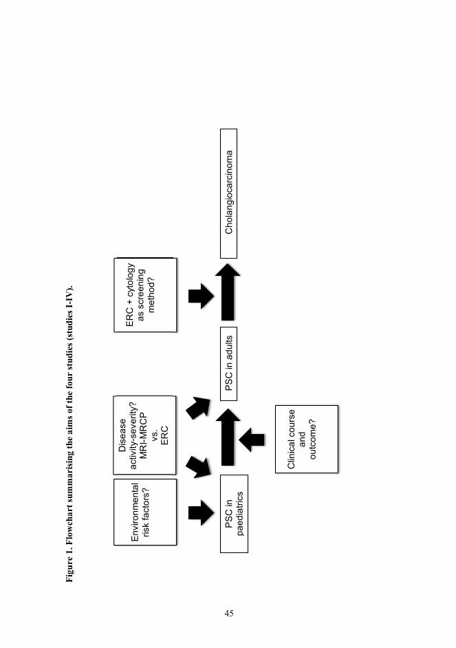
45
Figu
re 1
. Flo
wch
art s
umm
aris
ing
the
aim
s of t
he fo
ur st
udie
s (st
udie
s I-I
V).
PS
C in
pa
edia
trics
P
SC
in a
dults
Env
ironm
enta
l ris
k fa
ctor
s?
Clin
ical
cou
rse
and
ou
tcom
e?
Dis
ease
ac
tivity
-sev
erity
? M
RI-M
RC
P
vs.
ER
C
Cho
lang
ioca
rcin
oma
ER
C +
cyt
olog
y as
scr
eeni
ng
met
hod?

46
3. MATERIALS AND METHODS
3.1 Study design, population, setting, timing All PSC subjects included in the four studies were diagnosed, followed, or both in Helsinki University Hospital (HUH), which is a Tertiary Referral Centre in Finland (population approximately 5.5 million inhabitants) serving a defined population of about 1.5 million inhabitants (including the cities of Helsinki, Vantaa and Espoo). About 30% of this population is paediatric. Most of the patients with suspected PSC in Finland are referred to HUH for disease diagnosis and follow-up. The main characteristics of the studies are summarised in Table 8. Study I. This study was designed as a population-based case-control questionnaire study.
The cases were selected among patients with PSC, AIH and PSC-AIH. Accordingly, all patients (n=71) with a new diagnosis of paediatric-onset (age <16 years) PSC, PSC-AIH and AIH (together autoimmune liver diseases or AILD) between 1985-2011 were traced from an electronic database in Children’s Hospital using the 10th revision of the International Statistical Classification of Diseases and Related Health Problems (ICD-10) codes (i.e., AIH K73 and cholangitis K83).
Two control groups were used. The first group included patients matched for gender and age (n=91), collected from the IBD Population Registry at HUH, as PSC is usually associated with IBD. The second group included healthy subjects matched for gender, age and also place of birth at the time of AILD diagnosis (n=716), collected from the Population Registry Centre. Study II. This study was designed as an observational retrospective cohort study.
All patients with a new diagnosis of paediatric-onset PSC (n=41) between 1993-2011 were traced from an electronic database in Children’s Hospital using ICD-10 codes (i.e., AIH K73 and cholangitis K83).
Patient clinical course and outcome (i.e., LT, malignancy, death) were evaluated between the first consultation for suspected PSC and the last consultation for follow-up of the disease (by the end of December 2013). Study III. This study was designed as an observational retrospective cohort study.
All patients with PSC (n=48) who underwent ERC and MRI-MRCP +3 months for disease diagnosis or follow-up were traced from the PSC Register of HUH. In the PSC register, all demographic, clinical, laboratory and cholangiography data of over 700 patients with a new PSC diagnosis or in disease follow-up have been prospectively included since 2010.
Patient outcome (i.e., LT, malignancy, death) was evaluated by the end of October 2016. Study IV. This study was a designed as an observational retrospective cohort study.

47
All patients (n=261) with a new diagnosis of PSC (age >18 years) between 1 January 2006 and 31 October 2011 were traced from the Electronic Database of Pathology and the PSC register of HUH. Patient outcome was evaluated by the end of October 2013 or LT or CC occurrence.

48
Tab
le 8
. Stu
dy d
esig
n, p
opul
atio
n, se
ttin
g an
d tim
ing
of th
e fo
ur st
udie
s (st
udie
s I-I
V).
Stud
y St
udy
desi
gn
Popu
latio
n So
urce
Ti
me
I P
opul
atio
n-ba
sed
ca
se-c
ontro
l qu
estio
nnai
re
Cas
es n
=71
51 re
spon
dent
s C
ontro
ls:
IBD
n=9
1 59
resp
onde
nts
Hea
lthy
n=71
6
292
resp
onde
nts
Ele
ctro
nic
Dat
abas
e C
hild
ren’
s H
ospi
tal I
CD
-10
code
IB
D R
egis
ter H
UH
P
opul
atio
n R
egis
try C
entre
AIL
D d
iagn
osis
198
5-20
11
II O
bser
vatio
nal r
etro
spec
tive
coho
rt
n=41
E
lect
roni
c D
atab
ase
Chi
ldre
n’s
Hos
pita
l IC
D-1
0 co
de
PS
C d
iagn
osis
199
3-20
11
Out
com
e by
Dec
embe
r 201
3
III
Obs
erva
tiona
l ret
rosp
ectiv
e co
hort
n=48
P
SC
Reg
istry
HU
H
PS
C d
iagn
osis
/follo
w-u
p si
nce
2010
O
utco
me
by O
ctob
er 2
016
IV
Obs
erva
tiona
l ret
rosp
ectiv
e co
hort
n=26
1 E
lect
roni
c D
atab
ase
Pat
holo
gy H
UH
P
SC
Reg
istry
HU
H
PS
C d
iagn
osis
200
6-20
11
Out
com
e by
Oct
ober
201
3
AILD
: aut
oim
mun
e liv
er d
isea
se; P
SC: p
rim
ary
scle
rosi
ng c
hola
ngiti
s, AI
H: a
utoi
mm
une
hepa
titis
, IBD
: inf
lam
mat
ory
bowe
l dis
ease
, HU
H: H
elsi
nki U
nive
rsity
Cen
tral
Hos
pita
l

49
3.2 Case ascertainment PSC diagnosis was based on the following criteria: i) typical features of the disease (i.e., strictures and dilation involving IHBD, EHBD or both) visible on cholangiography images (i.e., ERC and MRI-MRCP, the latter if available), ii) elevation of ALP, GGT or both and iii) negative AMA. Additional criteria were IBD association and liver biopsy suggestive or typical of the disease (e.g., bile duct injury, periductal inflammation or fibrosis, ductopenia, ductural proliferation, cholestasis, portal oedema or fibrosis). Patients with SSC (Table 2) and IAC were excluded (Liver 2009, Chapman, Fevery et al. 2010) (Studies I-IV).
ERC images (and also MRCP images in Study III) were scored for the severity of biliary changes by the Amsterdam PSC score (Rajaram, Ponsioen et al. 2001), modified at HUH for intrahepatic bile duct changes to better define early disease (Box in Figure 2). A global modified Amsterdam PSC score was calculated differently, depending on the primary endpoint of the study. In Studies I and II the modified Amsterdam PSC score for intrahepatic and extrahepatic bile ducts was reported. In Study III, the score was grouped in 1-2 and 3-4 for both intrahepatic and extrahepatic bile ducts as ‘mild’ and ‘severe’ changes, respectively. Finally, in Study IV the scores for intrahepatic bile ducts (right + left) and for extrahepatic bile ducts (common hepatic bile duct + main bile duct) were calculated separately to obtain a score from 0 to 8.
AIH diagnosis was based on the simplified score for AIH diagnosis (Study I) (Mileti, Rosenthal et al. 2012) or the modified diagnostic score for AIH proposed by the IAIHG (Study II) (Alvarez, Berg et al. 1999).
PSC-AIH diagnosis was confirmed when the patient fulfilled both the diagnostic criteria for PSC and AIH (Gregorio, Portmann et al. 2001) (Studies I-IV). 3.3 ERC and brush cytology In all studies the patients underwent ERC with brush cytology. ERC is still considered the gold standard for biliary tree visualisation in patients with suspected (diagnosis) or defined (follow-up) PSC in HUH. Still, brush cytology is routinely performed during ERC in all PSC patients, regardless of biliary change severity; an additional sample for DNA flow cytometry analysis is usually collected only in patients with advanced biliary changes or if a previous cytology was suspicious or indicated aneuploidy.
The procedures were usually performed by the same experienced endoscopists in a standardised fashion. The patient was in prone position and the procedure was performed with the assistance of an anaesthesiologist. After successful cannulation with a papillotomy knife (Jagtome RX; Boston Scientific, Miami, Florida, USA ) and a 0.035-in, 450 cm guide wire (Jagwire; Boston Scientific ), a short biliary sphincterotomy was performed. A balloon catheter was subsequently inserted into the common hepatic bile duct and contrast was injected to visualise first the intrahepatic bile ducts and then the
50
extrahepatic bile ducts, taking all images from the four different planes. Brush cytology was finally collected mainly together from both intra and extrahepatic bile ducts. Dilatation and stenting were performed when appropriate. Patients were monitored after the procedure for 24 hours and eventual complications were treated. According to our internal protocol, the timing of patient follow-up depended on the severity of the biliary changes detected by cholangiography and the presence of biliary dysplasia; in this respect, the patient was referred to LT in case of repeatedly confirmed severe dysplasia, aneuploidy by brush cytology or both (Figure 2).

51
Figure 2. Flowchart showing diagnostic and follow-up protocol of patients with PSC in Helsinki University Hospital.
- Cholestasis - Negative AMA - IBD
MRI-MRCP: suspected PSC
ERC + brush cytology
Modified Amsterdam PSC score Intrahepatic bile ducts: 0 No visible abnormalities 1 Ductular irregularities 2 Multiple caliber change; minimal dilatation 3 Multiple strictures; saccular dilatations, decreased arborisation 4 Only central branches filled despite adequate filling pressure; severe pruning Extrahepatic bile ducts: 0 No visible abnormalities 1 Slight irregularities of duct contour, no stricture 2 Segmental strictures 3 Strictures of almost entire length of duct 4 Extremely irregular margins; diverticulum-like outpouchings
Score <3
Brush cytology
Benign i
ERC and US 3-5 years
Score >4
Brush cytology + DNA flow cytometry
Benign Benign Aneuploid
Suspicious Aneuploid
ERC and CT/MRI
1-2 years
ERC and CT/MRI
6-12 months
ERC and CT/MRI
3-6 months
Repeatedly confirmed
No CC
Liver transplantation Liver surgery

52
3.4 Collection of the data In Studies I-IV, patient records were reviewed and demography, clinical, laboratory, radiology, histology/cytology and outcome data were collected. The main characteristics of the studies are summarised in Table 9.
Study I. In this study, patient medical records were reviewed by two paediatricians for demography, clinical and laboratory data. One pathologist, blinded to patient follow-up, reviewed all the liver histology samples for PSC, AIH features or both. Two endoscopists reviewed and re-scored all cholangiography images (ERC) using the modified Amsterdam PSC score. A questionnaire, previously validated for allergy evaluation in a paediatric cohort of Finnish patients (Suoniemi, Björkstén et al. 1981), was administered to the subjects and their families; if the subject could not remember, the parents were asked to provide the information. The questionnaire was based on 22 items, evaluating possible risk factors of the Finnish environment that were present during the lifetime before AILD diagnosis. These items included number and age of siblings, place of residence, housing, travelling abroad, previous and current smoking status in family, pets or domestic animals at home, contact with pets or domestic animals, chronic diseases, history of parasite infestation, consumption of water or milk products and allergies. The questionnaire was mailed in September 2012 and a reminder was sent in November 2012 to those who did not reply; subjects who did not reply by April 2013 were defined as non-respondents.
Study II. In this study, patient medical records were reviewed by one gastroenterologist and one paediatrician for demography, clinical (i.e., symptoms and signs, presence of IBD or other autoimmune diseases) and laboratory (i.e., bilirubin, ALT, AST, GGT, ALP, IgM, IgG, ANA, ASMA, AMA, anti-LKM1, pANCA) data. In this study, the treatment (i.e., UDCA, corticosteroids, immunosuppressive therapy) and the outcome (i.e., LT, malignancy, death or combinations thereof) were also registered. One pathologist reviewed all liver histology samples for PSC or AIH features (or both); the pathologist was blinded to patient follow-up. Two endoscopists reviewed and re-scored all cholangiography images (ERC) using the modified Amsterdam PSC score.
Study III. In this study the following demography, clinical and laboratory data were collected: gender, age, presence of IBD, overlap with AIH, other associated autoimmune liver diseases, ALP, ALT, CEA and CA19-9.
ERC images were not reviewed and the original modified Amsterdam PSC score was used.
MRI-MRCP images were reviewed in consensus by two radiologists, both blinded to patient follow-up. The radiologists re-scored the MRCP images using the modified Amsterdam PSC score for disease severity and MRI images for peribiliary enhancement thickness as follows: <2 mm, 2-6 mm and >6 mm.
Two pathologists reviewed in consensus cytology samples for biliary neutrophils (0=absent, 1=mild, 2=high), intraepithelial lymphocytes (0=absent, 1=present) and
53
cytology classification after Papanicolau staining (1=normal epithelium, 2= benign atypia, 3=mild suspicion of neoplasia or low-grade dysplasia and 4=high suspicion of neoplasia or high-grade dysplasia, 5=malignant). These data were used as markers of disease activity and severity.
Study IV. In this study, the following demography, clinical and laboratory data were collected: detailed symptoms, associated diseases (i.e., IBD, overlap with AIH), lab tests (i.e., haemoglobin, platelets, albumin, thromboplastin time, INR, bilirubin, ALP, GGT, AST, ALT, IgG4, CA19-9, CEA), surgery (i.e., intestinal dysplasia, colectomy) and malignancy (i.e., CC, CRC) were reviewed. Three pathologists, all blinded to patient follow-up, reviewed in consensus all brush cytology samples as follows: benign, suspicious or malignant. DNA flow cytometry results were also collected. Two pathologists reviewed all histology samples for CC or dysplasia in the explanted liver. Two endoscopists reviewed and re-scored all cholangiography images (ERC) using the modified Amsterdam PSC score.

54
Tab
le 9
. Cha
ract
eris
tics o
f dat
a co
llect
ion
in S
tudi
es I-
IV.
Stud
y ER
C im
ages
revi
ew
MR
I-MR
CP
revi
ew
mAP
SC s
core
H
isto
logy
revi
ew
Cyt
olog
y re
view
I 2
endo
scop
ists
(con
sens
us)
Not
blin
ded
to d
iagn
osis
N
ot p
erfo
rmed
Ye
s 1
path
olog
ist
Not
blin
ded
to d
iagn
osis
B
linde
d to
follo
w-u
p
Not
per
form
ed
II 2
endo
scop
ists
(con
sens
us)
Not
blin
ded
to d
iagn
osis
N
ot p
erfo
rmed
Ye
s 1
path
olog
ist
Not
blin
ded
to d
iagn
osis
B
linde
d to
follo
w-u
p
Not
per
form
ed
III
Not
per
form
ed
2 ra
diol
ogis
ts (c
onse
nsus
) N
ot b
linde
d to
dia
gnos
is
Blin
ded
to E
RC
resu
lt B
linde
d to
follo
w-u
p
Yes
1-2
vs. 3
-4
Not
per
form
ed
2 pa
thol
ogis
ts (c
onse
nsus
) N
ot b
linde
d to
dia
gnos
is
Blin
ded
to fo
llow
-up
IV
2 en
dosc
opis
ts (c
onse
nsus
) N
ot b
linde
d to
dia
gnos
is
Not
per
form
ed
Yes
0-
8 2
path
olog
ists
N
ot b
linde
d to
dia
gnos
is
Blin
ded
to fo
llow
-up
3 pa
thol
ogis
ts (c
onse
nsus
) N
ot b
linde
d to
dia
gnos
is
Blin
ded
to fo
llow
-up
mAP
SC s
core
: m
odifi
ed A
mst
erda
m p
rim
ary
scle
rosi
ng c
hola
ngiti
s sc
ore,
ERC
: en
dosc
opic
ret
rogr
ade
chol
angi
ogra
phy,
MRI
: m
agne
tic r
eson
ance
imag
ing,
MRC
P:
mag
netic
reso
nanc
e ch
olan
giop
ancr
eato
grap
hy.

55
3.5 Statistical analysis Figures are presented as number and rate when categorical (all studies) and as median with range (Study I and II) or interquartile range (Study III) when continuous.
Categorical variables were compared with Fisher’s exact test (Studies I-III). Continuous variables were compared with Kruskal-Wallis test (Study I), Mann-Whitney test or the Wilcox test (Studies II and III) and linear-by-linear association (Study III) as appropriate. The exact Cochran-Armitage trend test or the exact Jonckheere-Terpstra test was applied to determine differences between ordered groups (Study IV). Statistically significant differences were considered when p<0.05.
Study I was designed as a case-controlled population-based questionnaire study. Data were collected matched (sex, age and area) and analysed both matched and non-matched. Finally, the results were shown for the non-matched data because of the reduction in the number of cases (n=48) and healthy controls (n=93) in the non-matched analysis. The sex, age and distribution between the two subsets did not differ statistically when tested with Kruskal-Wallis test (p=0.6070, p=0.5462 and Chi-square test p=0.7457). In matched analysis, the OR and their 95% CI were calculated by logistic regression (Proc Logistic SAS version 9.3, SAS Institute, Cary, NC ) in univariate and multivariate analysis. In univariate analysis, OR and 95% CI were separately calculated for all independent variables using IBD control and healthy control groups. A potential association between variables in univariate analysis was tested with Fisher’s exact test (p<0.05), performing a cross-tabulation with IBD control and healthy control groups both separately and together (all controls). In multivariate analysis, statistically significant variables with their interaction were tested. The questionnaire contained 22 items in open and closed questions. As mentioned above, when the respondents could not remember the answer, their parents were asked to reply for them. Missing values were changed to a ‘no’ answer if another pet was reported or otherwise removed from the analysis.
In Study III the modified Amsterdam PSC score was grouped as follows: 0=no changes, 1-2=mild changes and 3-4=severe changes separately for IHBD and EHBD. The agreement between PSC and MRCP modified Amsterdam PSC scores were tested with weighted kappa-statistic with quadrate weights. The McNemar-Bowker test was used to test differences in pair ordinal variables. Peribiliary enhancement was grouped in <2 mm and >2 mm to balance the groups. The Spearman’s rho was calculated for ordinal variables (i.e., correlation).
Kaplan-Meier survival analysis was performed in Study IV. Univariate and multivariate analyses were performed with Cox proportional hazards regression analysis of the dichotomous endpoint variable of neoplasia (benign or biliary/CC). 3.6 Ethical consideration
All the study protocols were approved by the HUH Ethics committee

56
4. RESULTS
4.1 Environmental risk factors in paediatric-onset PSC (study I) 51/71 AILD cases (72%), 59/91 IBD controls (65%) and 292/716 healthy controls (41%) responded to the questionnaire. Among AILD cases, no statistically significant differences between respondents and non-respondents regarding age, sex and type of diagnosis were observed (p>0.05). Among IBD controls, females responded more often to the questionnaire (p=0.034).
When the variables were tested by univariate analysis, ‘having either a cat or a dog’ resulted in a significant OR of 3.4 (95% CI: 1.5-7.8) when IBD controls were used and an OR of 2.5 (95% CI: 1.2-5.0) when healthy controls were used. When the variables ‘having a cat’ and ‘having a dog’ were tested separately, they still resulted in a higher OR. The ‘having a cat’ ORs were 2.6 (95% CI: 1.0-6.5) and 2.2 (95% CI: 1.1-4.6) when the IBD controls and healthy controls were used, respectively. The ‘having a dog’ ORs were 2.5 (95% CI: 1.1-5.8) and 2.0 (95% CI: 1.0-4.0) when the IBD and healthy controls were used, respectively); these results were statistically less significant.
The variables were then cross-tabulated to test the possible associations affecting the risk factor ‘having a cat or a dog’, using IBD controls and healthy controls separately and together (all controls). AILD cases occurred more often with positive responses to ‘having a cat’ or ‘having either a cat or a dog’ when living in a block of flats.
When the variables ‘type of housing’ and ‘having a cat or a dog’ were tested in distinct strata by multivariate analysis, in the stratum of block of flats having a cat resulted in a significant OR (3.6; 95% CI: 1.2-10.8) when healthy controls were used.
A summary of the results from Study I is shown in Figure 3.

57
Figure 3. Summary of Study I results.
Healthy controls 716 IBD controls 91 AILD cases 71
Questionnaire
IBD controls Respondents 59/91 (65%)
AILD cases Respondents 51/71 (72%)
Healthy controls Respondents
292/716 (41%)
Univariate analysis
AILD cases vs. IBD controls ‘having a cat or a dog’
OR 3.4; 95% CI: 1.5-7.8 and
‘having a dog’ OR 2.5; 95% CI: 1.1-5.8
AILD cases vs. healthy controls ‘having a cat or a dog’
OR 2.5; 95% CI: 1.2-5.0 and
‘having a cat’ OR 2.2; 95% CI: 1.1-4.6
Fisher’s Exact Test for cross-tabulated strata
AILD cases vs. all controls ‘having a cat or a dog’
and ‘block of flats’ 63% vs. 38%
p=0.0464
AILD cases vs. all controls ‘having a cat’
and ‘block of flats’ 42% vs. 17%
p=0.0254
Multivariate analysis
AILD cases vs. healthy controls ‘having a cat’
in ‘a block of flats’
OR: 3.6, 95% CI: 1.2-10.8

58
4.2 Clinical course and prognosis of paediatric-onset PSC
(study II) 33 patients with a paediatric-onset PSC (median age at diagnosis 16 years, range 5-19 years, 21 males) were included; eight patients (6 with a misclassified diagnosis, 1 PSC patient lost to follow-up and 1 PSC-AIH with missing data) were excluded. PSC diagnosis was confirmed in all patients by ERC images. Overlap with AIH was re-evaluated in 25 patients (in 8 patients the modified score for AIH could not be calculated because of missing liver biopsy): pure PSC was confirmed in 10 and PSC-AIH in 15 patients, respectively (AIH was initially overlooked in 7/25 patients, 28%).
As expected, at the time of diagnosis the disease was more frequent in males (21/33; 64%) than in females. Most of the patients (31/33, 94%) presented asymptomatic elevation of liver or cholestasis enzymes or non-specific symptoms of liver disease, however, 3/33 (9%) had cirrhosis and two of them already had related complications. As expected, IBD was associated with PSC in 25/33 (76%) patients, with UC being the most common (23/33, 70%). PSC was evident at the same time as IBD in nine patients (36%), after IBD in 14 patients (60%) and before IBD in two patients (4%). Twenty-two patients (88%) had pancolitis, two had right-sided colitis and one had left-sided colitis. Associated autoimmune disorders were detected in 5/33 (15%) patients.
At the time of diagnosis, serum liver enzymes (i.e., AST, ALT, GGT, ALP) did not differ between PSC and PSC-AIH (p>0.05) (n=25, patients with reassessment of overlap). Serum IgG, IgM and ANA/ASMA positivity were more elevated, more frequent or both in PSC-AIH patients (p=0.002, p=0.03, p<0.001, respectively); ANCA did not differ between PSC and PSC-AIH (p>0.05).
At the time of diagnosis, PSC histological alterations were detected in 11/25 (44%) patients. Interface hepatitis, plasma cell infiltrate and rosetting of liver cells were present in 15/15 (100%), 14/15 (93%) and 1/15 (7%) of the PSC-AIH patients, respectively. Inflammation (grading) was detected in the majority of patients with PSC-AIH (13/15, 80%), but was a rare and mild finding in those with PSC only (2/10, 20%). Overall, fibrosis (staging) 3 was seen in 4/25 patients (16%, 1 PSC and 3 PSC-AIH) and 4 in 2/25 patients (8%, all PSC-AIH). All patients had PSC changes on ERC images, with isolated intrahepatic involvement in 21/33 (64%) and intrahepatic with extrahepatic involvement in 12/33 (36%); no patients had modified Amsterdam score 4.
The median time to outcome was 9 years (range 2-20 years). All patients were alive and no malignancy was diagnosed during the follow-up period. The modified Amsterdam PSC score showed a significant progression of intrahepatic disease in 12 (36%) patients (p=0.001) but not for extrahepatic disease (p=0.85). 29/33 (88%) patients did not receive LT; 26 of these patients were not cirrhotic (1 PSC-AIH patient was under evaluation for LT due to PSC progression) and three were cirrhotic (1 PSC-AIH list for LT, 1 PSC and 1

59
PSC-AIH). LT was performed in 4/33 (33%) patients after a median of 7.5 years from the diagnosis of liver disease (3 PSC-AIH and 1 PSC).
A summary of the results from Study II is shown in Figure 4.

60
Figure 4. Summary of the results of study II
41 patients traced 8 patients dropped out
33 patients PSC or PSC-AIH
33 patients PSC confirmed by
ERC 25/33 (76%) patients
modified score for AIH
10 patients PSC 15 patients PSC-AIH (7 overlooked)
Baseline at diagnosis (n=33): Male: 21/33 (64%) Median age: 16 years; 5-19 years > LFTs or unspecific symptoms:
31/33 (94%) IBD: 25/33 (76%): 70% UC; 9/25
(36%) same time, 14/25 (56%) IBD first, 2/25 (8%) PSC first
Autoimmune disorders: 5/33 (15%) Lab tests (n=25): 10 PSC vs 15 PSC-AIH ALT, AST, ALP, GGT, ANCA p=n.s
IgG p=0.002 IgM p=0.03 ANA/ASMA p<0.001
Liver histology (n=25): PSC alterations: 11/25 (44%) Inflammation (grading any stage)
13/15 (80%) PSC-AIH 2/10 (20%) PSC
Stage 3: 4/25 (16%, 1 PSC and 3 PSC-AIH)
Stage 4: 2/25 (8%, all PSC-AIH)
ERC (n=33): Only intrahepatic 21/33 (64%) Intra and extrahepatic 12/33 (36%)
Follow-up: 9 years; 2-20 years All patients alive No malignancy 4/25 IBD (12%) cholectomy
29/33 (88%) no LT
4/33 (12%) LT median 7.5 years
1 PSC/AIH: jaundice + cirrhosis 2 PSC/AIH: dysplasia and aneuploidy; cirrhosis in one (at diagnosis) 1 PSC: > CA19-9 and CA125
3 (812%
No cirrhosis 26/29 (78%)
Cirrhosis 3/29 (10%); 2 at diagnosis
cirrh
61
4.3 MRI-MRCP and ERC in PSC disease activity and severity evaluation (study III)
48 patients with PSC and ERC performed within + 3 months since MRI-MRCP were traced. 31 patients were male (65%). Median age (interquartile range, IQR) at PSC diagnosis was 30.5 (21.0-41.5) and at ERC was 35.7 (28.0-44.2). As expected, 36/48 (75%) patients had IBD, mostly UC (25/36, 69%); PSC-AIH was present in 5/48 (10%) patients. Associated autoimmune diseases were present in 11/48 (23%) patients.
Overall, 57 ERC and MRI-MRCP were performed in 48 patients. Of the latter, 52/57 (91%) were MRI, because contrast medium was not administered in five patients, and 55/57 (96%) were MRCP, because 3D reconstruction was not available for two patients.
The agreement in scoring disease severity was moderate for both IHBD (weighted-kappa: 0.437; 95% CI: 0.211-0.644) and EHBD (weighted-kappa: 0.512; 95% CI: 0.303-0.720); the difference was statistically significant only for EHBD with a McNemar-Bowker test (p=0.041). However, the overall rate of agreement between ERC and MRCP in detecting any changes was 98% and 78% for IHBD and EHBD, respectively.
The association between modified Amsterdam PSC score evaluated by ERC and markers of disease activity and severity was tested (n=57; n=55 for cytology because two samples were of poor quality). An association between IHBD score and ALP (p=0.018) and CA19-9 level (p=0.030) was found. The association between modified Amsterdam PSC score evaluated by MRCP and markers of disease activity and severity was also tested (n=55). An association between IHBD score and ALP (p= 0.016) and CA19-9 level (p<0.001) was found, but also between EHBD score and CA19-9 level (p=0.021).
The correlation between MRI peribiliary enhancement and markers of disease activity and severity was tested (n=52). A positive correlation between peribiliary enhancement in IHBD and EHBD and cytology class was found (Spearman’s rho=0.322; standard error, SE: 0.095, p=0.022 and Spearman’s rho=0.319; SE: 0.113, p=0.025, respectively).
The patients were followed-up after ERC for a median time (+ IQR) of 3.5 (3.0-4.3) years. 11 (23%) patients received LT (5 patients for end-stage liver disease and 6 patients for suspicion of neoplasia). One patient (2%) died from CC. ERC was repeated after 3 months from the index ERC at least once for dysplasia confirmation in all of the patients. High-grade dysplasia or CC was detected in the explanted liver in five patients; low-grade dysplasia was seen in one patient. One out of six patients died from CC after LT, and one developed colon cancer with metastasis and is still alive.
A summary of the results from Study III is shown in Figure 5.

62
Figure 5. Summary of results from study III.
Cytology class
IHBD EHBD 0 <2mm >2mm 0 <2mm >2mm
1 n=4
2 50%
2 50%
0 0%
1 2 50%
1 25%
1 25%
2 n=39
10 26%
8 20%
21 54%
2 6 15%
11 28%
22 56%
3 n=5
0 0%
2 40%
3 60%
3 0 0%
1 20%
4 80%
4 n=2
0 0%
0 0%
2 100%
4 0 0%
0 0%
2 100%
p=0.023* p=0.031*
48 PSC patients
Baseline characteristics: Male: 31/48 (65%) Median age (+ IQR) at PSC diagnosis: 30.5 (21.0-41.5) Median age (+ IQR) at ERC: 35.7 (28.0-44.2) PSC-AIH: 5/48 (10%) IBD: 36/48 (75%). UC: 25/36 (69%) Other autoimmune diseases: 11/48 (23%)
57 ERC 55 MRCP no 3D in 2
52 MRI no contrast in 5
+ 3 months + 3 months
Agreement modified Amsterdam PSC score Overall IHBD 54/55 (98%) Overall EHBD 43/55 (78%) Weighted-k IHBD: 0.437 (0.211-0.644) Weighted-k EHBD: 0.512 (0.303-0.720)* *McNemar-Bowker test p=0.041
MRCP score IHBD: 1-2 and ALP: 150;74-266 3-4 and ALP: 228;139-454
p=0.016* 1-2 and CA19-9: 6;0-8 3-4 and CA19-9: 11;8-21
p<0.001* MRCP score EHBD:
0 and CA19-9: 3;0-6 1-2 and CA19-9: 8;6-15 3-4 and CA19-9: 10; 7-57
p=0.021* *significant
Modified Amsterdam PSC score
eem
MRC ERC score IHBD: 1-2 and ALP: 150;74-228 3-4 and ALP: 252;141-407
p=0.018* 1-2 and CA19-9: 7;4-11 3-4 and CA19-9: 10;6-33
p=0.030*
Peribiliary enhancement
IHBD Absent: 12/52 (23%) <2 mm: 14/52 (27%) >2 mm: 26/52 (50%)
EHBD Absent: 8/52 (15%) <2 mm: 13/52 (25%) >2 mm: 31/52 (60%)
BD

63
4.4 ERC and brush cytology: screening for biliary dysplasia and risk factors for neoplasia (study IV).
261 patients diagnosed during the study period were included. 125 patients were male (47.9%); the median age at the time of PSC diagnosis for males was 34.5 years. The median age of the whole group was 40.8 years. IBD was associated with PSC in 180/261 (69%) patients, with UC in 119/180 (66%) patients. In this group, intestinal dysplasia and CRC developed in 13/180 (7%) and 4/180 (2%) patients, respectively; colectomy was performed in 30 (17%) patients. Overlap syndrome was observed in 21/216 (8%) patients. Overall, the modified Amsterdam PSC score was 2-3 in 149/261 (57%), 4-9 in 87/261 (33%) and 10-16 in 25/261 (10%) patients, respectively. Most of the patients were asymptomatic (80.8%), but symptoms were not present more often in patients with an advanced modified Amsterdam PSC score (>3) compared to patients with early disease (2-3) (Cochran-Armitage trend test p=0.303). Symptoms were not associated with suspicious brush cytology (Fisher’s exact test p=0.054), but all patients with CC at diagnosis were symptomatic. Among all laboratory tests, bilirubin, ALP, GGT, AST and ALT were significantly higher in patients with a more advanced modified Amsterdam PSC score (p< 0.001). Brush cytology was benign in 243/261 (93%) and suspicious in 16/261 (6%) patients, respectively; and 2/261 (0.8%) patients had CC. The final endpoint was reached by 249/261 (95%) patients after a median follow-up of 4.7 years. According to modified Amsterdam PSC score, 139/249 (56%) patients had mild score (2-3) and all of them had benign findings in brush cytology. An advanced score (4-16) was seen in 110/249 (44%) patients, and brush cytology was benign in 94/110 (84%), suspicious in 14/110 (13%) and malignant in 2/110 (1.8%) patients. Finally, 7/261 (2.7%) patients developed CC after a mean time from PSC diagnosis of 396 days; two cases were diagnosed at the time of PSC diagnosis. 9/261 (3.4%) patients underwent LT: six suspicious, five aneuploidy and four with both; all were confirmed histologically. At multivariate analysis, four variables were independently associated with biliary neoplasia: advanced modified Amsterdam PSC score for EHBD (Hazard ratio, HR: 1.7; 95% CI: 1.2-2.3), suspicious/malignant brush cytology (HR: 13.5; 95% CI: 4.1-44.9), ALT (HR: 14.2; 95% CI: 1.9-106.4) and CEA (HR: 14.3; 95% CI: 2.0-101.2).
A summary of the results from Study IV is reported in Figure 6.

64
Figure 6. Summary of results from study IV.
261 PSC patients
149 score 2-3 Benign cytology 243 Suspicious 16 Malignant 0
87 score 4-9 Benign cytology 72 Suspicious 13 Malignant 2
25 score 10-16 Benign cytology 23 Suspicious 2 Malignant 0
249 PSC patients Endpoint
12 PSC patients No endpoint
139 score 2-3
Benign cytology 139
110 score 4-16
Suspicious 14 Benign cytology 94 Malignant 2 ogy 9 Ma
Endpoint Benign cytology 139
Endpoint Benign cytology 85
Dysplasia 5 CC 2
Gallbladder CA 2
Endpoint Benign cytology 8
Dysplasia 3 CC 3
Endpoint CC 2
Dead
Asymptomatic 80.8% Advanced disease
42.9%
65
5. DISCUSSION
5.1 Considerations on study design, population, timing and data collection
All four studies were designed as observational retrospective studies. Although this design might lead to bias, the rarity of PSC in the general population (Molodecky, Kareemi et al. 2011, Boonstra, Beuers et al. 2012) (especially in children) (Deneau, Jensen et al. 2013), the slow progression of inflammation and fibrosis (with a median LT-free survival ranging from 13 to 21 years) and a median time period to death or LT of 12-21 years (Ponsioen, Chapman et al. 2016) hampers the planning of observational prospective studies in PSC. Study I was the second case-controlled population-based questionnaire study on possible environmental risk factors associated with PSC (Boonstra, de Vries et al. 2016) and the first conducted in a paediatric population that also included AIH. Seven out of eight studies on long-term outcome of patients with paediatric-onset PSC (two published after Study II) are retrospective observational studies (Wilschanski, Chait et al. 1995, Feldstein, Perrault et al. 2003, Miloh, Arnon et al. 2009, Deneau, Jensen et al. 2013, Rojas, Bodicharla et al. 2014, Rodrigues, Liu et al. 2016, Valentino, Wiggins et al. 2016). Study III was the first to address the role of MRI-MRCP in the evaluation of PSC disease activity and severity.
The number of patients included in Studies II and III (n=41 and=44, respectively) was low but is comparable to other studies conducted on the same topic (Gregorio, Portmann et al. 2001, Dave, Elmunzer et al. 2010). Study I had 71 cases that included patients with PSC, AIH and PSC-AIH. The decision to include patients with AIH was based on the frequent overlap between PSC and AIH in paediatric populations and a desire to enlarge the number of cases included. The 91 IBD controls and 716 healthy controls were collected matched for sex, age and geographic area with a case:control ratio of 1:1 and 1:10, respectively. The matching should decrease the interference of confounders. However, the reduction in the number of cases and of controls made the multivariate analysis of matched data less stable than the multivariate analysis of unmatched data (although not statistically different); ultimately, the OR with 95% CI of unmatched data was reported.
As mentioned above, almost all patients with suspected PSC in Finland are referred to HUH. After PSC diagnosis, all patients continue follow-up in this hospital. This procedure should reduce the impact of selection (especially referral bias) and information bias. Since 2010, all data from patients with a new diagnosis of PSC or in follow-up have been prospectively collected into the electronic PSC Database Registry. The source of patient collection was the PSC Database Registry for Study III and partially for Study IV, thus reducing the risk of detection bias, misclassification bias or both. The source of patient collection was the Electronic Database of Children’s Hospital (HUH) using ICD-10 for

66
Studies I and II, as most of the children had been diagnosed before 2010. The identification of cases by ICD-10 might lead to a detection or misclassification bias (or both); however, the centralisation of PSC patients to HUH and the revision of all patient ERC images and partially of liver biopsies should have reduced this risk. Finally, the controls in Study I were selected from IBD Population Registry and Population Registry Centre, reducing the risk of any bias. 5.2 Considerations on case ascertainment and ERC PSC diagnostic criteria were based on current literature (Liver 2009, Chapman, Fevery et al. 2010).
As mentioned above, all cholangiographic images were obtained by ERC in HUH; MRI-MRCP was additionally considered. All PSC changes (i.e., strictures, dilatations and pruning) were scored using the Amsterdam PSC score (Ponsioen, Vrouenraets et al. 2002, Ponsioen, Reitsma et al. 2010), modified in HUH for intrahepatic bile duct changes (Figure 2). This modification consists of dividing the score I into two categories (Figure 2) to better categorise early intrahepatic biliary changes. A possible drawback might be that this modified score is not currently validated. However, in our clinical experience, early intrahepatic biliary changes are often misdiagnosed on MRI-MRCP images due to its low specificity (Dave, Elmunzer et al. 2010). A validation study of this modified Amsterdam PSC score is warranted in the future.
Unexplained persistent or intermittent elevation of ALP, GGT or both was also regarded as criteria. ALP is usually elevated in the majority of PSC patients (Broomé, Olsson et al. 1996). However, since ALP levels vary in children according to their age (due to bone maturation), GGT was also evaluated in the paediatric population (Miloh, Arnon et al. 2009).
The presence of IBD and liver histology was regarded as additional criteria. IBD is not associated with PSC or PSC-AIH in all patients (Tables 4 and 5). Liver histology may have a high rate of false negative results (Burak, Angulo et al. 2003) (Table 4) and is not routinely performed in children without suspected PSC-AIH or small-duct PSC. In this respect, PSC-AIH diagnosis was confirmed retrospectively for all patients with liver histology by applying the simplified AIH score or the available IAIHG score (Study I and II, respectively). These scores were also validated in children (Ebbeson and Schreiber 2004, Mileti, Rosenthal et al. 2012). In Study II, both scores were calculated and no differences were observed. 5.3 The questionnaire A questionnaire was administered to cases and controls in Study I.
A possible drawback is that the questionnaire was not specifically validated for this study. However, the questionnaire was designed for a prior study on the risk factors
67
associated with paediatric-onset hypersensitivity in a group of adolescents living in Finland (Suoniemi, Björkstén et al. 1981).
The questionnaire included only closed questions regarding a possible association with environmental risk factors strongly related to the Finnish environment. Closed questions were used to facilitate responses among respondents and their parents. The missing answers were changed to ‘no answer’ if another pet was reported and they were removed in the final analysis. To avoid recall bias, the parent was asked to provide the answer if the subject could not remember the information.
The questionnaire was first mailed in September 2012 and a reminder was sent in November 2012 to improve patient adherence to the study. However, the final response rate was moderate in cases (72%) and IBD controls (65%) and low in healthy controls (41%). This probably occurred because healthy controls were less motivated to respond to the questionnaire. Still, the response rate could have been improved by a direct interview; however, this is difficult in Finland as many patients live far away from Helsinki. Finally, a similar response rate in cases and controls was previously reported in other population-based studies on paediatric-onset IBD (Turunen, Ashorn et al. 2009, Jakobsen, Paerregaard et al. 2013). 5.4 Environmental risk factors in paediatric-onset PSC The aetiopathogenesis of PSC and PSC-AIH is still unknown; a complex association among genetic, environmental, immunologic and other potential factors has been postulated (Hirschfield, Karlsen et al. 2013, Lazaridis and LaRusso 2016). So far, only five studies investigating possible environmental risk factors in PSC in adults have been published (Table 3). Two of these studies were multicentre and all were based on a questionnaire. One study was population-based (Table 3).
The main finding of this study was that children with a cat and living in a block of flats had a strong risk of developing an AILD compared with healthy controls (OR: 3.6, 95% CI: 1.2-10.8). The lack of difference between cases and IBD controls might be due to the small sample size of the two groups or to risk independent of IBD.
One possibility may be that children living with a cat in a block of flats may have closer contact with some unidentified antigen (e.g., microbe or toxin) found in feline urine or faeces. This hypothesis is also supported by two observations: children living in a bigger house (i.e., low contact) and children having a dog did not have a higher risk. In the latter case, these children probably take their pet outside regularly.
Only one out of five studies investigating possible environmental risk factors in adults with PSC (Table 3) evaluated the association between PSC and contact with farm animals. This study did not find any statistically significant association, although the number of PSC patients with a previous contact was higher compared to controls (Andersen, Tengesdal et al. 2013). Conversely, several studies investigated the association between IBD and pets in adults, yielding conflicting results (Amre, Lambrette et al. 2006,

68
Bernstein, Rawsthorne et al. 2006, Radon, Windstetter et al. 2007, Gearry, Richardson et al. 2010). Amre et al. found that having a household pet increased the risk of CD almost two-fold compared to controls and, interestingly, living in a less crowded environment was a protective factor. However, two studies showed that having a pet was a protective factor for IBD (Bernstein, Rawsthorne et al. 2006, Radon, Windstetter et al. 2007), while one study did not find any association between IBD and pet contact (Gearry, Richardson et al. 2010). These authors speculated on the so-called ‘hygiene hypothesis’, which suggests that an early exposure to some antigens (e.g., microbial) may balance between pro- and anti-inflammatory responses, inducing tolerance. No association between other autoimmune diseases (i.e., juvenile idiopathic arthritis and type 1 diabetes) was seen (Radon, Windstetter et al. 2005, Radon, Windstetter et al. 2010).
While an association between AIH and many different viruses (e.g., hepatitis A, hepatitis C, herpes) was found (Heneghan, Yeoman et al. 2013), the association between PSC and bacterial or viral exposure has only been postulated (Mehal, Hattersley et al. 1992, Nilsson, Taneera et al. 2000, Chen and LaRusso 2002, Ponsioen, Defoer et al. 2002) and no pathogens were zoonotic. However, the ‘gut microbiota hypothesis’ was recently suggested to play an important role in the pathogenesis of PSC (Tabibian, Talwalkar et al. 2013) as well as in many others diseases (Lynch and Pedersen 2016). Gram-positive and gram-negative bacteria induce inflammatory and fibrotic biliary changes similar to those seen in PSC both in animal studies (Haruta, Kikuchi et al. 2010) and in vitro (Mueller, Beutler et al. 2011). In this respect, further studies investigating the association between PSC or PSC-AIH and microbes of felids (e.g., Toxoplasma) or canids (e.g., Toxocara) would be interesting. 5.5 Outcome of paediatric-onset PSC The natural history and outcome of paediatric-onset PSC is still underreported. So far, only eight studies investigating the clinical course and follow-up of PSC in children have been published (Tables 4 and 5) and only one was performed in Europe (Gregorio, Portmann et al. 2001). The majority of these studies were designed as retrospective studies; only one was prospective longitudinal study and one was population-based (Table 4). Interestingly, two of these studies were published after Study II (Rodrigues, Liu et al. 2016, Valentino, Wiggins et al. 2016).
The first main finding of this study was that PSC-AIH was detected in about 60% of the patients in whom liver biopsy was available for the review; the overlap with AIH had been overlooked in seven patients at the time of diagnosis (i.e., after diagnosis of pure PSC). Rojas et al. and Gregorio et al. found an overlap with AIH in about 90% and 50% of the cases, respectively (Gregorio, Portmann et al. 2001, Rojas, Bodicharla et al. 2014). Otherwise, the rate of PSC-AIH in this study was higher than previously reported (Table 4). Interestingly, the overlap between PSC and AIH had been overlooked in about 30% of the original liver biopsies. There are two possible explanations. First, the knowledge on

69
PSC and PSC-AIH has improved over time and the pathologist who reviewed the liver biopsies in the current study was experienced in autoimmune hepatobiliary diseases. Second, PSC and PSC-AIH may be clinically difficult to distinguish. Although transaminases are elevated in PSC-AIH, transaminase levels did not differ between PSC and PSC-AIH in this series, as previously reported (Gregorio, Portmann et al. 2001, Miloh, Arnon et al. 2009). Conversely, the IgG levels, immunoglobulins and positive ANA/SMA were statistically higher in PSC-AIH group, suggesting the importance of performing liver biopsy in children with suspected overlap syndrome to identify typical features of AIH (i.e., interface hepatitis) (Table 4). However, the impact of AIH overlap on PSC clinical course and outcome is unknown, but probably of minor importance. Children with PSC-AIH have been treated with immunosuppressive therapy (i.e., steroids or immunomodulators) with an improvement of laboratory and histological findings (Gregorio, Portmann et al. 2001) but without any improvement of survival (Feldstein, Perrault et al. 2003, Miloh, Arnon et al. 2009). However, further studies on the impact of the therapy on PSC-AIH are needed.
Regarding clinical manifestation at time of diagnosis, PSC occurred mostly in males at a median age of 16 years. The most common presentation was asymptomatic elevation of liver tests or non-specific symptoms of liver disease. These findings were consistent with previous studies (Table 4). PSC was associated with IBD in 70% of the cases, with UC being the most common. The rate of IBD in PSC children ranged from 33-97% among the different studies. PSC and IBD were diagnosed at the same time or IBD was detected before the diagnosis of PSC in almost all the patients; in the Feldstein et al. study IBD became apparent years after the diagnosis of PSC in 15% of the cases (Feldstein, Perrault et al. 2003). The majority of the patients had pancolitis with mild inflammation, which has already been described as a unique PSC-IBD phenotype (Loftus, Harewood et al. 2005). However, four patients (12%) underwent colectomy during the follow-up for severe colitis. Lascurain et al. reported a 5-year probability of colonic surgery of 16.4% in PSC-IBD children (Lascurain, Jensen et al. 2016). A minority of the patients received immunosuppression (i.e., steroids, immunomodulators or both). However, it is not possible to determine if therapy was administered for the liver disease or the intestinal disease, due to the retrospective study design and the failure to identify AIH in some patients in the original diagnosis. The real impact of the therapy on liver disease and associated IBD needs to be elucidated in future studies.
Regarding histology at the time of diagnosis, typical alterations of PSC were detected in only 44% of the children. Gregorio et al. reported PSC changes in 55% and in 48% of children with PSC and PSC-AIH, respectively (Gregorio, Portmann et al. 2001). Feldstein et al. reported PSC changes in all the children’s liver biopsies (Feldstein, Perrault et al. 2003). Due to the high false negative rate, liver biopsy is not routinely recommended for PSC diagnosis. Interface hepatitis was present in all patients with PSC-AIH and in a minority of those with pure PSC, as also reported by Feldstein et al (Feldstein, Perrault et al. 2003). Interestingly, severe fibrosis or cirrhosis were found in 18% of the children at

70
diagnosis (10% in pure PSC and 33% in PSC-AIH), which is lower compared to the rate reported by Miloh and Feldstein (severe fibrosis or cirrhosis in about 56% and 54% of the children at PSC-AIH onset, respectively) (Feldstein, Perrault et al. 2003, Miloh, Arnon et al. 2009). However, at the end of follow-up two cirrhotic children were stable, but one underwent LT; new cirrhosis developed in two children.
At diagnosis, all patients had an intrahepatic involvement; isolated extrahepatic disease was not seen in any patients, as previously reported (Gregorio, Portmann et al. 2001, Feldstein, Perrault et al. 2003, Miloh, Arnon et al. 2009). Disease progression was detected in one third of the children with IHBD at the end of follow-up. The Amsterdam PSC score also has prognostic value (Ponsioen, Vrouenraets et al. 2002). One PSC/AIH patient was under evaluation for LT and one received LT.
This study reported the longest follow-up among all paediatric series (median 9 years, range 2-20 years). All patients were alive and nobody had malignancy. Feldstein at al. reported a decreased survival in children with PSC compared to the general paediatric population (Feldstein, Perrault et al. 2003). Deneau et al. found a CC rate of 6.9% in his series (Deneau, Jensen et al. 2013). One possible explanation is that the follow-up protocol performed in HUH (Figure 2) may be useful in selecting patients for optimal timing for LT. In this respect, two patients underwent LT for repeated confirmed biliary dysplasia and aneuploidy in brush cytology. The LT rate was also lower compared to all the other series (Table 5). Despite being conducted in an adult population, Study IV strongly supports this possibility. The study showed that ERC with brush cytology performed as a screening method for neoplasia, regardless of the presence of symptoms, dominant stricture or both, might identify those patients at increased risk of developing malignancy. A similar study conducted in a larger paediatric population is warranted to extend this result from adults to children. 5.6 PSC activity and severity: MRI-MRCP compared with ERC The first interesting finding was that the agreement between ERC and MRI-MRCP in detecting any PSC changes was very high in IHBD evaluation and good in EHBD evaluation. One study reported that ERC may be more accurate than MRI-MRCP in the diagnosis of early changes of PSC (Weber, Kuhlencordt et al. 2008). However, the agreement between ERC and MRI-MRCP in the severity evaluation of PSC changes by modified Amsterdam PSC score was only moderate for both IHBD and EHBD; the difference was statistically significant only for EHBD. A case-control study found that the inter-observer agreement was poor for both techniques when assessing disease severity (i.e., independently two gastroenterologists for ERC and two radiologists for MRI-MRCP) (Moff, Kamel et al. 2006). The impact of disagreement on patient outcome needs to be evaluated in further studies.
Interestingly, the severity of cholangiographic changes assessed with both techniques (ERC and MRCP) was associated with the level of serum ALP and CA19-9 but not with

71
other markers (i.e., ALT, CEA, biliary neutrophils, intraepithelial lymphocytes and cytology). ALP was recently proposed as a promising surrogate marker of PSC progression, discriminating between PSC patients with poor and good prognosis (de Vries, Wang et al. 2016). CA19-9 with a cut-off of 20 U/mL has been proposed as a marker of CC (Charatcharoenwitthaya, Enders et al. 2008), but its use is limited by low sensitivity (Charatcharoenwitthaya, Enders et al. 2008) and specificity (Sinakos, Saenger et al. 2011). Adjusting the cut-off value according to patient genetic profile (i.e., FUT2, FUT3 or both), may have interesting additional value (Wannhoff, Hov et al. 2013). Finally, ALP, CA19-9 and cholangiographic score may be combined together as a prognostic score in future studies.
Finally, peribiliary enhancement assessed with MRI did not correlate with serologic markers of disease activity and severity (i.e., ALT, ALP and CA19-9) or with inflammation detected in cytology (i.e., biliary neutrophils and intraepithelial inflammation), but with cytologic classification of bile ducts. Peribiliary enhancement is seen in T1-weighted sequences on MRI of PSC patients (Arrivé, Ruiz et al. 2013). The significance of this observation is unknown, but could reflect the grade of biliary inflammation (Arrivé, Ruiz et al. 2013). In fact, PSC is characterised histologically by an inflammatory infiltration, which is localised mostly around the bile ducts (Portmann and Zen 2012). This observation may explain why peribiliary enhancement did not correlate with inflammation detected in cytology (i.e., biliary neutrophils and intraepithelial inflammation). Chronic inflammation may trigger the transition from normal epithelium to CC (Lewis, Talwalkar et al. 2010). However, the correlation between thickness grade of peribiliary enhancement and brush cytology classification needs to be confirmed in further studies. Peribiliary enhancement thickness was classified into <2 mm, 2-6 mm and >6 mm (Ruiz, Lemoinne et al. 2014). Ultimately, the <2 mm and >2 mm groups were used due to the absence of cases in the >6 mm group. Brush cytology may have a low sensitivity for detection of biliary neoplasia (Trikudanathan, Navaneethan et al. 2014). However, Boyd et al. have recently shown that patients with PSC and advanced bile duct disease, elevation of liver function tests (e.g., ALT and ALP), elevation of tumour markers (e.g., CA19-9) and inflammation or suspicious cytology are most likely to develop CC (Boyd, Mustonen et al. 2016). In this respect, peribiliary enhancement did not correlate with any of these serum liver tests. 5.7 ERC and brush cytology: screening and risk factors for
cholangiocarcinoma In contrast with Studies I-III and the literature (Tischendorf, Hecker et al. 2007, Boonstra, Weersma et al. 2013), the male:female ratio was about 1:1. Interestingly, females more often had mild disease compared to males and this may probably contribute to PSC underdiagnosis in women. Similar to Studies I-III and the literature (Tischendorf, Hecker et al. 2007), over 60% of the patients with PSC in this cohort had cholestasis (i.e.,

72
elevation of ALP, GGT or both). Transaminases may be also elevated, but bilirubin is normal in the majority of the patients.
CC may develop in about 7% of patients with PSC and is usually discovered within one year after PSC diagnosis (Boonstra, Weersma et al. 2013). However, PSC patients with mild ERC changes on cholangiography had benign brush cytology and follow-up in this study. Severe ERC changes, dysplasia or malignancy was only found in PSC patients. Study III supports this finding, as cholangiographic score was associated with cytology classification, irrespective of ERC or MRI-MRCP. Patients with PSC and mild changes probably had a negligible risk of developing biliary dysplasia. Brush cytology has a high specificity (97%) but a low sensitivity (43%) for diagnosing CC in PSC patients (Trikudanathan, Navaneethan et al. 2014). However, in the current study only two patients that developed CC had normal brush cytology in the first ERC, having suspicious brush cytology during follow-up. All other patients who developed CC had suspicious cytology at least once. The low sensitivity may have many explanations, such as the skill of the endoscopist in performing brushing, the brush catheter used and the experience of pathologists in processing and reading slides. In the current study, both endoscopists and pathologists were experienced and focused in management of patients with PSC, which may contribute to the final good performance of brushing.
The use of tumour markers CEA and CA19-9 as a screening test for CC diagnosis in PSC patients is still debated (Charatcharoenwitthaya, Enders et al. 2008). Interestingly, in the current study, when patients with CC diagnosed within one year from PSC diagnosis were included in the analysis, CEA and CA19-9 were associated with CC.
Patients with repeated confirmed suspicion of malignancy in brush cytology are referred to LT evaluation at HUH. ERC with systematic brush cytology may detect patients at high risk of developing CC over time; malignancy may be prevented with LT. Study II also suggests this possibility in the paediatric population. In the current study, all patients with suspicious brush cytology had confirmed biliary dysplasia in the explanted liver, although two patients with benign brush cytology during follow-up (suspicious brush without malignancy) developed CC and died from the disease. Further efforts to improve brush cytology sensitivity should be made in the future.
6 CONCLUSIONS AND FURTHER STUDIES
The main findings of the current thesis are: 1. An unidentified cat-linked environmental risk factor (e.g., microbe) is probably
associated with paediatric-onset autoimmune liver diseases (i.e., PSC, AIH and PSC-AIH). Further studies in the future are warranted. Identification of a clear risk factor may lead to future preventive strategies.
2. The outcome of paediatric-onset PSC seems to be good until early adulthood. Further studies on the impact of ERC with brush cytology compared to MRI-MRCP in follow-

73
up and outcome of children with PSC and the natural history, course and outcome of PSC-IBD children are warranted.
3. Although the agreement between ERC and MRCP in scoring PSC bile duct changes is only moderate, the severity of PSC on cholangiography is associated with some surrogate markers of disease activity and severity in PSC (i.e., ALP and CA19-9). Peribiliary enhancement detected on MRI correlates with cytology findings, but not with other invasive and non-invasive surrogate markers of PSC disease activity and severity.
4. Early ERC with systematic brush cytology seems to play a pivotal role in identifying PSC patients with a high risk of developing CC. Further studies should validate this finding in a different cohort.

74
REFERENCES
Aadland, E., E. Schrumpf, O. Fausa, K. Elgjo, A. Heilo, T. Aakhus and E. Gjone (1987). "Primary sclerosing cholangitis: a long-term follow-up study." Scand J Gastroenterol 22(6): 655-664.
Abdalian, R. and E. J. Heathcote (2006). "Sclerosing cholangitis: a focus on secondary causes." Hepatology 44(5): 1063-1074.
Aljiffry, M., P. D. Renfrew, M. J. Walsh, M. Laryea and M. Molinari (2011). "Analytical review of diagnosis and treatment strategies for dominant bile duct strictures in patients with primary sclerosing cholangitis." HPB (Oxford) 13(2): 79-90.
Alvarez, F., P. A. Berg, F. B. Bianchi, L. Bianchi, A. K. Burroughs, E. L. Cancado, R. W. Chapman, W. G. Cooksley, A. J. Czaja, V. J. Desmet, P. T. Donaldson, A. L. Eddleston, L. Fainboim, J. Heathcote, J. C. Homberg, J. H. Hoofnagle, S. Kakumu, E. L. Krawitt, I. R. Mackay, R. N. MacSween, W. C. Maddrey, M. P. Manns, I. G. McFarlane, K. H. Meyer zum Büschenfelde and M. Zeniya (1999). "International Autoimmune Hepatitis Group Report: review of criteria for diagnosis of autoimmune hepatitis." J Hepatol 31(5): 929-938.
Amre, D. K., P. Lambrette, L. Law, A. Krupoves, V. Chotard, F. Costea, G. Grimard, D. Israel, D. Mack and E. G. Seidman (2006). "Investigating the hygiene hypothesis as a risk factor in pediatric onset Crohn's disease: a case-control study." Am J Gastroenterol 101(5): 1005-1011.
Andersen, I. M., G. Tengesdal, B. A. Lie, K. M. Boberg, T. H. Karlsen and J. R. Hov (2013). "Effects of Coffee Consumption, Smoking, and Hormones on Risk for Primary Sclerosing Cholangitis." Clin Gastroenterol Hepatol.
Andriulli, A., S. Loperfido, G. Napolitano, G. Niro, M. R. Valvano, F. Spirito, A. Pilotto and R. Forlano (2007). "Incidence rates of post-ERCP complications: a systematic survey of prospective studies." Am J Gastroenterol 102(8): 1781-1788.
Ang, T. L., K. M. Fock, T. M. Ng, E. K. Teo, T. S. Chua and J. Y. Tan (2002). "Clinical profile of primary sclerosing cholangitis in Singapore." J Gastroenterol Hepatol 17(8): 908-913.
Angulo, P., Y. Maor-Kendler and K. D. Lindor (2002). "Small-duct primary sclerosing cholangitis: a long-term follow-up study." Hepatology 35(6): 1494-1500.
Angulo, P., D. H. Pearce, C. D. Johnson, J. J. Henry, N. F. LaRusso, B. T. Petersen and K. D. Lindor (2000). "Magnetic resonance cholangiography in patients with biliary disease: its role in primary sclerosing cholangitis." J Hepatol 33(4): 520-527.
Arrivé, L., C. Coudray, L. Azizi, M. Lewin, C. Hoeffel, L. Monnier-Cholley, C. Lacombe, S. Vautier, J. Poupon and J. M. Tubiana (2007). "[Pineapple juice as a negative oral contrast agent in magnetic resonance cholangiopancreatography]." J Radiol 88(11 Pt 1): 1689-1694.
Arrivé, L., A. Ruiz, S. El Mouhadi, L. Azizi, L. Monnier-Cholley and Y. Menu (2013). "MRI of cholangitis: traps and tips." Diagn Interv Imaging 94(7-8): 757-770.
Aspinall, A. I., S. M. Curbishley, P. F. Lalor, C. J. Weston, M. Blahova, E. Liaskou, R. M. Adams, A. P. Holt and D. H. Adams (2010). "CX(3)CR1 and vascular adhesion protein-1-dependent recruitment of CD16(+) monocytes across human liver sinusoidal endothelium." Hepatology 51(6): 2030-2039.
Bambha, K., W. R. Kim, J. Talwalkar, H. Torgerson, J. T. Benson, T. M. Therneau, E. V. Loftus, B. P. Yawn, E. R. Dickson and L. J. Melton (2003). "Incidence, clinical

75
spectrum, and outcomes of primary sclerosing cholangitis in a United States community." Gastroenterology 125(5): 1364-1369.
Bangarulingam, S. Y., E. Bjornsson, F. Enders, E. G. Barr Fritcher, G. Gores, K. C. Halling and K. D. Lindor (2010). "Long-term outcomes of positive fluorescence in situ hybridization tests in primary sclerosing cholangitis." Hepatology 51(1): 174-180.
Bangarulingam, S. Y., A. A. Gossard, B. T. Petersen, B. J. Ott and K. D. Lindor (2009). "Complications of endoscopic retrograde cholangiopancreatography in primary sclerosing cholangitis." Am J Gastroenterol 104(4): 855-860.
Barr Fritcher, E. G., B. R. Kipp, J. S. Voss, A. C. Clayton, K. D. Lindor, K. C. Halling and G. J. Gores (2011). "Primary sclerosing cholangitis patients with serial polysomy fluorescence in situ hybridization results are at increased risk of cholangiocarcinoma." Am J Gastroenterol 106(11): 2023-2028.
Barr Fritcher, E. G., J. S. Voss, S. M. Brankley, M. B. Campion, S. M. Jenkins, M. E. Keeney, M. R. Henry, S. M. Kerr, R. Chaiteerakij, E. V. Pestova, A. C. Clayton, J. Zhang, L. R. Roberts, G. J. Gores, K. C. Halling and B. R. Kipp (2015). "An Optimized Set of Fluorescence In Situ Hybridization Probes for Detection of Pancreatobiliary Tract Cancer in Cytology Brush Samples." Gastroenterology 149(7): 1813-1824.e1811.
Bartholomew, L. G., J. C. Cain, L. B. WOOLNER, D. C. UTZ and D. O. FERRIS (1963). "Sclerosing cholangitis: its possible association with Riedel's struma and fibrous retroperitonitis. Report of two cases." N Engl J Med 269: 8-12.
Berdal, J. E., J. Ebbesen and A. Rydning (1998). "[Incidence and prevalence of autoimmune liver diseases]." Tidsskr Nor Laegeforen 118(29): 4517-4519.
Bergquist, A., A. Ekbom, R. Olsson, D. Kornfeldt, L. Lööf, A. Danielsson, R. Hultcrantz, S. Lindgren, H. Prytz, H. Sandberg-Gertzén, S. Almer, F. Granath and U. Broomé (2002). "Hepatic and extrahepatic malignancies in primary sclerosing cholangitis." J Hepatol 36(3): 321-327.
Bergquist, A., H. Glaumann, B. Persson and U. Broomé (1998). "Risk factors and clinical presentation of hepatobiliary carcinoma in patients with primary sclerosing cholangitis: a case-control study." Hepatology 27(2): 311-316.
Bergquist, A., G. Lindberg, S. Saarinen and U. Broomé (2005). "Increased prevalence of primary sclerosing cholangitis among first-degree relatives." J Hepatol 42(2): 252-256.
Bernstein, C. N., P. Rawsthorne, M. Cheang and J. F. Blanchard (2006). "A population-based case control study of potential risk factors for IBD." Am J Gastroenterol 101(5): 993-1002.
Berstad, A. E., L. Aabakken, H. J. Smith, S. Aasen, K. M. Boberg and E. Schrumpf (2006). "Diagnostic accuracy of magnetic resonance and endoscopic retrograde cholangiography in primary sclerosing cholangitis." Clin Gastroenterol Hepatol 4(4): 514-520.
Beuers, U., U. Spengler, W. Kruis, U. Aydemir, B. Wiebecke, W. Heldwein, M. Weinzierl, G. R. Pape, T. Sauerbruch and G. Paumgartner (1992). "Ursodeoxycholic acid for treatment of primary sclerosing cholangitis: a placebo-controlled trial." Hepatology 16(3): 707-714.
Björnsson, E., K. M. Boberg, S. Cullen, K. Fleming, O. P. Clausen, O. Fausa, E. Schrumpf and R. W. Chapman (2002). "Patients with small duct primary sclerosing cholangitis have a favourable long term prognosis." Gut 51(5): 731-735.
Björnsson, E., S. T. Chari, T. C. Smyrk and K. Lindor (2007). "Immunoglobulin G4 associated cholangitis: description of an emerging clinical entity based on review of the literature." Hepatology 45(6): 1547-1554.

76
Björnsson, E. and R. Olsson (2004). "Dominant strictures in patients with primary sclerosing cholangitis-revisited." Am J Gastroenterol 99(11): 2281.
Björnsson, E., R. Olsson, A. Bergquist, S. Lindgren, B. Braden, R. W. Chapman, K. M. Boberg and P. Angulo (2008). "The natural history of small-duct primary sclerosing cholangitis." Gastroenterology 134(4): 975-980.
Boberg, K. M., E. Aadland, J. Jahnsen, N. Raknerud, M. Stiris and H. Bell (1998). "Incidence and prevalence of primary biliary cirrhosis, primary sclerosing cholangitis, and autoimmune hepatitis in a Norwegian population." Scand J Gastroenterol 33(1): 99-103.
Boberg, K. M., A. Bergquist, S. Mitchell, A. Pares, F. Rosina, U. Broomé, R. Chapman, O. Fausa, T. Egeland, G. Rocca and E. Schrumpf (2002). "Cholangiocarcinoma in primary sclerosing cholangitis: risk factors and clinical presentation." Scand J Gastroenterol 37(10): 1205-1211.
Boberg, K. M., O. Fausa, T. Haaland, E. Holter, O. J. Mellbye, A. Spurkland and E. Schrumpf (1996). "Features of autoimmune hepatitis in primary sclerosing cholangitis: an evaluation of 114 primary sclerosing cholangitis patients according to a scoring system for the diagnosis of autoimmune hepatitis." Hepatology 23(6): 1369-1376.
Boberg, K. M., P. Jebsen, O. P. Clausen, A. Foss, L. Aabakken and E. Schrumpf (2006). "Diagnostic benefit of biliary brush cytology in cholangiocarcinoma in primary sclerosing cholangitis." J Hepatol 45(4): 568-574.
Boberg, K. M. and G. E. Lind (2011). "Primary sclerosing cholangitis and malignancy." Best Pract Res Clin Gastroenterol 25(6): 753-764.
Boonstra, K., U. Beuers and C. Y. Ponsioen (2012). "Epidemiology of primary sclerosing cholangitis and primary biliary cirrhosis: a systematic review." J Hepatol 56(5): 1181-1188.
Boonstra, K., E. M. de Vries, N. van Geloven, K. J. van Erpecum, M. Spanier, A. C. Poen, C. M. van Nieuwkerk, B. J. Witteman, H. A. Tuynman, A. H. Naber, P. J. Kingma, U. Beuers, C. Y. Ponsioen and E. P. P. S. Group (2016). "Risk factors for primary sclerosing cholangitis." Liver Int 36(1): 84-91.
Boonstra, K., K. J. van Erpecum, K. M. van Nieuwkerk, J. P. Drenth, A. C. Poen, B. J. Witteman, H. A. Tuynman, U. Beuers and C. Y. Ponsioen (2012). "Primary sclerosing cholangitis is associated with a distinct phenotype of inflammatory bowel disease." Inflamm Bowel Dis 18(12): 2270-2276.
Boonstra, K., R. K. Weersma, K. J. van Erpecum, E. A. Rauws, B. W. Spanier, A. C. Poen, K. M. van Nieuwkerk, J. P. Drenth, B. J. Witteman, H. A. Tuynman, A. H. Naber, P. J. Kingma, H. R. van Buuren, B. van Hoek, F. P. Vleggaar, N. van Geloven, U. Beuers, C. Y. Ponsioen and E. S. Group (2013). "Population-based epidemiology, malignancy risk, and outcome of primary sclerosing cholangitis." Hepatology 58(6): 2045-2055.
Boyd, S., H. Mustonen, A. Tenca, K. Jokelainen, J. Arola and M. A. Färkkilä (2016). "Surveillance of primary sclerosing cholangitis with ERC and brush cytology: risk factors for cholangiocarcinoma." Scand J Gastroenterol: 1-8.
Broomé, U., H. Glaumann, E. Lindstöm, L. Lööf, S. Almer, H. Prytz, H. Sandberg-Gertzén, S. Lindgren, F. T. Fork, G. Järnerot and R. Olsson (2002). "Natural history and outcome in 32 Swedish patients with small duct primary sclerosing cholangitis (PSC)." J Hepatol 36(5): 586-589.
Broomé, U., R. Olsson, L. Lööf, G. Bodemar, R. Hultcrantz, A. Danielsson, H. Prytz, H. Sandberg-Gertzén, S. Wallerstedt and G. Lindberg (1996). "Natural history and
77
prognostic factors in 305 Swedish patients with primary sclerosing cholangitis." Gut 38(4): 610-615.
Buckles, D. C., K. D. Lindor, N. F. Larusso, L. M. Petrovic and G. J. Gores (2002). "In primary sclerosing cholangitis, gallbladder polyps are frequently malignant." Am J Gastroenterol 97(5): 1138-1142.
Burak, K., P. Angulo, T. M. Pasha, K. Egan, J. Petz and K. D. Lindor (2004). "Incidence and risk factors for cholangiocarcinoma in primary sclerosing cholangitis." Am J Gastroenterol 99(3): 523-526.
Burak, K. W., P. Angulo and K. D. Lindor (2003). "Is there a role for liver biopsy in primary sclerosing cholangitis?" Am J Gastroenterol 98(5): 1155-1158.
Byron, D. and G. Y. Minuk (1996). "Clinical hepatology: profile of an urban, hospital-based practice." Hepatology 24(4): 813-815.
Card, T. R., M. Solaymani-Dodaran and J. West (2008). "Incidence and mortality of primary sclerosing cholangitis in the UK: a population-based cohort study." J Hepatol 48(6): 939-944.
Chalasani, N., A. Baluyut, A. Ismail, A. Zaman, G. Sood, R. Ghalib, T. M. McCashland, K. R. Reddy, X. Zervos, M. A. Anbari and H. Hoen (2000). "Cholangiocarcinoma in patients with primary sclerosing cholangitis: a multicenter case-control study." Hepatology 31(1): 7-11.
Chapman, R., J. Fevery, A. Kalloo, D. M. Nagorney, K. M. Boberg, B. Shneider, G. J. Gores and A. A. f. t. S. o. L. Diseases (2010). "Diagnosis and management of primary sclerosing cholangitis." Hepatology 51(2): 660-678.
Chapman, R. W., B. A. Arborgh, J. M. Rhodes, J. A. Summerfield, R. Dick, P. J. Scheuer and S. Sherlock (1980). "Primary sclerosing cholangitis: a review of its clinical features, cholangiography, and hepatic histology." Gut 21(10): 870-877.
Charatcharoenwitthaya, P., F. B. Enders, K. C. Halling and K. D. Lindor (2008). "Utility of serum tumor markers, imaging, and biliary cytology for detecting cholangiocarcinoma in primary sclerosing cholangitis." Hepatology 48(4): 1106-1117.
Chavhan, G. B., P. S. Babyn, D. Manson and L. Vidarsson (2008). "Pediatric MR cholangiopancreatography: principles, technique, and clinical applications." Radiographics 28(7): 1951-1962.
Chen, X. M. and N. F. LaRusso (2002). "Cryptosporidiosis and the pathogenesis of AIDS-cholangiopathy." Semin Liver Dis 22(3): 277-289.
Chong, Y. S., Y. K. Kim, M. W. Lee, S. H. Kim, W. J. Lee, H. C. Rhim and S. J. Lee (2012). "Differentiating mass-forming intrahepatic cholangiocarcinoma from atypical hepatocellular carcinoma using gadoxetic acid-enhanced MRI." Clin Radiol 67(8): 766-773.
Christensen, M., P. Matzen, S. Schulze and J. Rosenberg (2004). "Complications of ERCP: a prospective study." Gastrointest Endosc 60(5): 721-731.
Chung, Y. E., M. J. Kim, Y. N. Park, J. Y. Choi, J. Y. Pyo, Y. C. Kim, H. J. Cho, K. A. Kim and S. Y. Choi (2009). "Varying appearances of cholangiocarcinoma: radiologic-pathologic correlation." Radiographics 29(3): 683-700.
Claessen, M. M., M. W. Lutgens, H. R. van Buuren, B. Oldenburg, P. C. Stokkers, C. J. van der Woude, D. W. Hommes, D. J. de Jong, G. Dijkstra, A. A. van Bodegraven, P. D. Siersema and F. P. Vleggaar (2009). "More right-sided IBD-associated colorectal cancer in patients with primary sclerosing cholangitis." Inflamm Bowel Dis 15(9): 1331-1336.
78
Claessen, M. M., F. P. Vleggaar, K. M. Tytgat, P. D. Siersema and H. R. van Buuren (2009). "High lifetime risk of cancer in primary sclerosing cholangitis." J Hepatol 50(1): 158-164.
Cotton, P. B., D. A. Garrow, J. Gallagher and J. Romagnuolo (2009). "Risk factors for complications after ERCP: a multivariate analysis of 11,497 procedures over 12 years." Gastrointest Endosc 70(1): 80-88.
Dave, M., B. J. Elmunzer, B. A. Dwamena and P. D. Higgins (2010). "Primary sclerosing cholangitis: meta-analysis of diagnostic performance of MR cholangiopancreatography." Radiology 256(2): 387-396.
Davies, Y. K., K. M. Cox, B. A. Abdullah, A. Safta, A. B. Terry and K. L. Cox (2008). "Long-term treatment of primary sclerosing cholangitis in children with oral vancomycin: an immunomodulating antibiotic." J Pediatr Gastroenterol Nutr 47(1): 61-67.
De Maria, N., A. Colantoni, E. Rosenbloom and D. H. Van Thiel (1996). "Ursodeoxycholic acid does not improve the clinical course of primary sclerosing cholangitis over a 2-year period." Hepatogastroenterology 43(12): 1472-1479.
de Vries, E. M., J. Verheij, S. G. Hubscher, M. M. Leeflang, K. Boonstra, U. Beuers and C. Y. Ponsioen (2015). "Applicability and prognostic value of histologic scoring systems in primary sclerosing cholangitis." J Hepatol 63(5): 1212-1219.
de Vries, E. M., J. Wang, M. M. Leeflang, K. Boonstra, R. K. Weersma, U. Beuers, R. B. Geskus and C. Y. Ponsioen (2016). "Alkaline Phosphatase at Diagnosis of Primary Sclerosing Cholangitis and One Year Later: Evaluation of Prognostic Value." Liver Int.
Deneau, M., M. K. Jensen, J. Holmen, M. S. Williams, L. S. Book and S. L. Guthery (2013). "Primary sclerosing cholangitis, autoimmune hepatitis, and overlap in Utah children: epidemiology and natural history." Hepatology 58(4): 1392-1400.
Eaton, J. E., B. Dzyubak, S. K. Venkatesh, T. C. Smyrk, G. J. Gores, R. L. Ehman, N. F. LaRusso, A. A. Gossard and K. N. Lazaridis (2016). "Performance of magnetic resonance elastography in primary sclerosing cholangitis." J Gastroenterol Hepatol 31(6): 1184-1190.
Eaton, J. E., M. G. Silveira, D. S. Pardi, E. Sinakos, K. V. Kowdley, V. A. Luketic, M. E. Harrison, T. McCashland, A. S. Befeler, D. Harnois, R. Jorgensen, J. Petz and K. D. Lindor (2011). "High-dose ursodeoxycholic acid is associated with the development of colorectal neoplasia in patients with ulcerative colitis and primary sclerosing cholangitis." Am J Gastroenterol 106(9): 1638-1645.
Eaton, J. E., E. W. Thackeray and K. D. Lindor (2012). "Likelihood of malignancy in gallbladder polyps and outcomes following cholecystectomy in primary sclerosing cholangitis." Am J Gastroenterol 107(3): 431-439.
Ebbeson, R. L. and R. A. Schreiber (2004). "Diagnosing autoimmune hepatitis in children: is the International Autoimmune Hepatitis Group scoring system useful?" Clin Gastroenterol Hepatol 2(10): 935-940.
Ehlken, H., R. Wroblewski, C. Corpechot, L. Arrivé, T. Rieger, J. Hartl, S. Lezius, P. Hübener, K. Schulze, R. Zenouzi, M. Sebode, M. Peiseler, U. W. Denzer, A. Quaas, C. Weiler-Normann, A. W. Lohse, O. Chazouilleres and C. Schramm (2016). "Validation of Transient Elastography and Comparison with Spleen Length Measurement for Staging of Fibrosis and Clinical Prognosis in Primary Sclerosing Cholangitis." PLoS One 11(10): e0164224.
Eksteen, B., A. J. Grant, A. Miles, S. M. Curbishley, P. F. Lalor, S. G. Hübscher, M. Briskin, M. Salmon and D. H. Adams (2004). "Hepatic endothelial CCL25 mediates

79
the recruitment of CCR9+ gut-homing lymphocytes to the liver in primary sclerosing cholangitis." J Exp Med 200(11): 1511-1517.
Eksteen, B., A. Miles, S. M. Curbishley, C. Tselepis, A. J. Grant, L. S. Walker and D. H. Adams (2006). "Epithelial inflammation is associated with CCL28 production and the recruitment of regulatory T cells expressing CCR10." J Immunol 177(1): 593-603.
Ellinghaus, D., L. Jostins, S. L. Spain, A. Cortes, J. Bethune, B. Han, Y. R. Park, S. Raychaudhuri, J. G. Pouget, M. Hübenthal, T. Folseraas, Y. Wang, T. Esko, A. Metspalu, H. J. Westra, L. Franke, T. H. Pers, R. K. Weersma, V. Collij, M. D'Amato, J. Halfvarson, A. B. Jensen, W. Lieb, F. Degenhardt, A. J. Forstner, A. Hofmann, S. Schreiber, U. Mrowietz, B. D. Juran, K. N. Lazaridis, S. Brunak, A. M. Dale, R. C. Trembath, S. Weidinger, M. Weichenthal, E. Ellinghaus, J. T. Elder, J. N. Barker, O. A. Andreassen, D. P. McGovern, T. H. Karlsen, J. C. Barrett, M. Parkes, M. A. Brown, A. Franke, I. I. G. C. (IIBDGC), I. G. o. A. S. C. (IGAS), I. P. S. G. (IPSCSG), G. A. o. P. C. (GAPC) and P. A. G. E. (PAGE) (2016). "Analysis of five chronic inflammatory diseases identifies 27 new associations and highlights disease-specific patterns at shared loci." Nat Genet 48(5): 510-518.
Escorsell, A., A. Parés, J. Rodés, J. A. Solís-Herruzo, M. Miras and E. de la Morena (1994). "Epidemiology of primary sclerosing cholangitis in Spain. Spanish Association for the Study of the Liver." J Hepatol 21(5): 787-791.
Etzel, J. P., S. C. Eng, C. W. Ko, S. D. Lee, M. D. Saunders, B. Y. Tung, M. B. Kimmey and K. V. Kowdley (2008). "Complications after ERCP in patients with primary sclerosing cholangitis." Gastrointest Endosc 67(4): 643-648.
Färkkilä, M., A. L. Karvonen, H. Nurmi, H. Nuutinen, M. Taavitsainen, P. Pikkarainen and P. Kärkkäinen (2004). "Metronidazole and ursodeoxycholic acid for primary sclerosing cholangitis: a randomized placebo-controlled trial." Hepatology 40(6): 1379-1386.
Feldstein, A. E., J. Perrault, M. El-Youssif, K. D. Lindor, D. K. Freese and P. Angulo (2003). "Primary sclerosing cholangitis in children: a long-term follow-up study." Hepatology 38(1): 210-217.
Ferrara, C., G. Valeri, L. Salvolini and A. Giovagnoni (2002). "Magnetic resonance cholangiopancreatography in primary sclerosing cholangitis in children." Pediatr Radiol 32(6): 413-417.
Fevery, J., W. Van Steenbergen, J. Van Pelt, W. Laleman, I. Hoffman, K. Geboes, S. Vermeire and F. Nevens (2016). "Patients with large-duct primary sclerosing cholangitis and Crohn's disease have a better outcome than those with ulcerative colitis, or without IBD." Aliment Pharmacol Ther 43(5): 612-620.
Floreani, A., E. R. Rizzotto, F. Ferrara, I. Carderi, D. Caroli, L. Blasone and V. Baldo (2005). "Clinical course and outcome of autoimmune hepatitis/primary sclerosing cholangitis overlap syndrome." Am J Gastroenterol 100(7): 1516-1522.
Florin, T. H., N. Pandeya and G. L. Radford-Smith (2004). "Epidemiology of appendicectomy in primary sclerosing cholangitis and ulcerative colitis: its influence on the clinical behaviour of these diseases." Gut 53(7): 973-979.
Folseraas, T., E. Melum, P. Rausch, B. D. Juran, E. Ellinghaus, A. Shiryaev, J. K. Laerdahl, D. Ellinghaus, C. Schramm, T. J. Weismüller, D. N. Gotthardt, J. R. Hov, O. P. Clausen, R. K. Weersma, M. Janse, K. M. Boberg, E. Björnsson, H. U. Marschall, I. Cleynen, P. Rosenstiel, K. Holm, A. Teufel, C. Rust, C. Gieger, H. E. Wichmann, A. Bergquist, E. Ryu, C. Y. Ponsioen, H. Runz, M. Sterneck, S. Vermeire, U. Beuers, C. Wijmenga, E. Schrumpf, M. P. Manns, K. N. Lazaridis, S. Schreiber, J. F. Baines, A. Franke and T. H. Karlsen (2012). "Extended analysis of a genome-wide association

80
study in primary sclerosing cholangitis detects multiple novel risk loci." J Hepatol 57(2): 366-375.
Fulcher, A. S., M. A. Turner, K. J. Franklin, M. L. Shiffman, R. K. Sterling, V. A. Luketic and A. J. Sanyal (2000). "Primary sclerosing cholangitis: evaluation with MR cholangiography-a case-control study." Radiology 215(1): 71-80.
Furmanczyk, P. S., V. S. Grieco and S. N. Agoff (2005). "Biliary brush cytology and the detection of cholangiocarcinoma in primary sclerosing cholangitis: evaluation of specific cytomorphologic features and CA19-9 levels." Am J Clin Pathol 124(3): 355-360.
Gearry, R. B., A. K. Richardson, C. M. Frampton, A. J. Dodgshun and M. L. Barclay (2010). "Population-based cases control study of inflammatory bowel disease risk factors." J Gastroenterol Hepatol 25(2): 325-333.
Gluck, M., N. R. Cantone, J. J. Brandabur, D. J. Patterson, J. E. Bredfeldt and R. A. Kozarek (2008). "A twenty-year experience with endoscopic therapy for symptomatic primary sclerosing cholangitis." J Clin Gastroenterol 42(9): 1032-1039.
Grant, A. J., P. F. Lalor, S. G. Hübscher, M. Briskin and D. H. Adams (2001). "MAdCAM-1 expressed in chronic inflammatory liver disease supports mucosal lymphocyte adhesion to hepatic endothelium (MAdCAM-1 in chronic inflammatory liver disease)." Hepatology 33(5): 1065-1072.
Gregorio, G. V., B. Portmann, J. Karani, P. Harrison, P. T. Donaldson, D. Vergani and G. Mieli-Vergani (2001). "Autoimmune hepatitis/sclerosing cholangitis overlap syndrome in childhood: a 16-year prospective study." Hepatology 33(3): 544-553.
Halme, L., J. Arola, K. Numminen, L. Krogerus, H. Mäkisalo and M. Färkkilä (2012). "Biliary dysplasia in patients with primary sclerosing cholangitis: additional value of DNA ploidity." Liver Int 32(5): 783-789.
Haruta, I., K. Kikuchi, E. Hashimoto, M. Nakamura, H. Miyakawa, K. Hirota, N. Shibata, H. Kato, Y. Arimura, Y. Kato, T. Uchiyama, H. Nagamune, M. Kobayashi, Y. Miyake, K. Shiratori and J. Yagi (2010). "Long-term bacterial exposure can trigger nonsuppurative destructive cholangitis associated with multifocal epithelial inflammation." Lab Invest 90(4): 577-588.
Haseeb, A., A. Siddiqui, L. J. Taylor, K. Cox and D. G. Adler (2016). "Elevated Serum Bilirubin Level Correlates With the Development of Cholangiocarcinoma, Subsequent Liver Transplantation, and Death in Patients With Primary Sclerosing Cholangitis." J Clin Gastroenterol 50(5): 431-435.
Heneghan, M. A., A. D. Yeoman, S. Verma, A. D. Smith and M. S. Longhi (2013). "Autoimmune hepatitis." Lancet 382(9902): 1433-1444.
Hennes, E. M., M. Zeniya, A. J. Czaja, A. Parés, G. N. Dalekos, E. L. Krawitt, P. L. Bittencourt, G. Porta, K. M. Boberg, H. Hofer, F. B. Bianchi, M. Shibata, C. Schramm, B. Eisenmann de Torres, P. R. Galle, I. McFarlane, H. P. Dienes, A. W. Lohse and I. A. H. Group (2008). "Simplified criteria for the diagnosis of autoimmune hepatitis." Hepatology 48(1): 169-176.
Hildebrand, T., N. Pannicke, A. Dechene, D. N. Gotthardt, G. Kirchner, F. P. Reiter, M. Sterneck, K. Herzer, H. Lenzen, C. Rupp, H. Barg-Hock, P. de Leuw, A. Teufel, V. Zimmer, F. Lammert, C. Sarrazin, U. Spengler, C. Rust, M. P. Manns, C. P. Strassburg, C. Schramm, T. J. Weismüller and G. P. S. Group (2016). "Biliary strictures and recurrence after liver transplantation for primary sclerosing cholangitis: A retrospective multicenter analysis." Liver Transpl 22(1): 42-52.
Hirschfield, G. M., T. H. Karlsen, K. D. Lindor and D. H. Adams (2013). "Primary sclerosing cholangitis." Lancet 382(9904): 1587-1599.

81
Hohenester, S., L. M. Wenniger, C. C. Paulusma, S. J. van Vliet, D. M. Jefferson, R. P. Elferink and U. Beuers (2012). "A biliary HCO3- umbrella constitutes a protective mechanism against bile acid-induced injury in human cholangiocytes." Hepatology 55(1): 173-183.
Hurlburt, K. J., B. J. McMahon, H. Deubner, B. Hsu-Trawinski, J. L. Williams and K. V. Kowdley (2002). "Prevalence of autoimmune liver disease in Alaska Natives." Am J Gastroenterol 97(9): 2402-2407.
Imam, M. H., E. Sinakos, A. A. Gossard, K. V. Kowdley, V. A. Luketic, M. Edwyn Harrison, T. McCashland, A. S. Befeler, D. Harnois, R. Jorgensen, J. Petz, J. Keach, A. C. DeCook, F. Enders and K. D. Lindor (2011). "High-dose ursodeoxycholic acid increases risk of adverse outcomes in patients with early stage primary sclerosing cholangitis." Aliment Pharmacol Ther 34(10): 1185-1192.
Ismail, S., L. Kylänpää, H. Mustonen, J. Halttunen, O. Lindström, K. Jokelainen, M. Udd and M. Färkkilä (2012). "Risk factors for complications of ERCP in primary sclerosing cholangitis." Endoscopy 44(12): 1133-1138.
Jakobsen, C., A. Paerregaard, P. Munkholm and V. Wewer (2013). "Environmental factors and risk of developing paediatric inflammatory bowel disease -- a population based study 2007-2009." J Crohns Colitis 7(1): 79-88.
Johnson, P. J. and I. G. McFarlane (1993). "Meeting report: International Autoimmune Hepatitis Group." Hepatology 18(4): 998-1005.
Jonker, J. W., C. Liddle and M. Downes (2012). "FXR and PXR: potential therapeutic targets in cholestasis." J Steroid Biochem Mol Biol 130(3-5): 147-158.
Junge, N., M. Tiedau, M. Verboom, M. Hallensleben, R. Blasczyk, J. Schlue, I. Goldschmidt, E. D. Pfister and U. Baumann (2016). "Human leucocyte antigens and pediatric autoimmune liver disease: diagnosis and prognosis." Eur J Pediatr 175(4): 527-537.
Kaplan, G. G., K. B. Laupland, D. Butzner, S. J. Urbanski and S. S. Lee (2007). "The burden of large and small duct primary sclerosing cholangitis in adults and children: a population-based analysis." Am J Gastroenterol 102(5): 1042-1049.
Karlsen, T. H., A. Franke, E. Melum, A. Kaser, J. R. Hov, T. Balschun, B. A. Lie, A. Bergquist, C. Schramm, T. J. Weismüller, D. Gotthardt, C. Rust, E. E. Philipp, T. Fritz, L. Henckaerts, R. K. Weersma, P. Stokkers, C. Y. Ponsioen, C. Wijmenga, M. Sterneck, M. Nothnagel, J. Hampe, A. Teufel, H. Runz, P. Rosenstiel, A. Stiehl, S. Vermeire, U. Beuers, M. P. Manns, E. Schrumpf, K. M. Boberg and S. Schreiber (2010). "Genome-wide association analysis in primary sclerosing cholangitis." Gastroenterology 138(3): 1102-1111.
Kaya, M., P. Angulo and K. D. Lindor (2000). "Overlap of autoimmune hepatitis and primary sclerosing cholangitis: an evaluation of a modified scoring system." J Hepatol 33(4): 537-542.
Kaya, M., B. T. Petersen, P. Angulo, T. H. Baron, J. C. Andrews, C. J. Gostout and K. D. Lindor (2001). "Balloon dilation compared to stenting of dominant strictures in primary sclerosing cholangitis." Am J Gastroenterol 96(4): 1059-1066.
Kingham, J. G., N. Kochar and M. B. Gravenor (2004). "Incidence, clinical patterns, and outcomes of primary sclerosing cholangitis in South Wales, United Kingdom." Gastroenterology 126(7): 1929-1930.
Lal, A., A. Okonkwo, S. Schindler, D. De Frias and R. Nayar (2004). "Role of biliary brush cytology in primary sclerosing cholangitis." Acta Cytol 48(1): 9-12.

82
Lascurain, L., M. K. Jensen, S. L. Guthery, J. Holmen and M. Deneau (2016). "Inflammatory Bowel Disease Phenotype in Pediatric Primary Sclerosing Cholangitis." Inflamm Bowel Dis 22(1): 146-150.
Lazaridis, K. N. and N. F. LaRusso (2016). "Primary Sclerosing Cholangitis." N Engl J Med 375(12): 1161-1170.
Levy, M. J., T. H. Baron, A. C. Clayton, F. B. Enders, C. J. Gostout, K. C. Halling, B. R. Kipp, B. T. Petersen, L. R. Roberts, A. Rumalla, T. J. Sebo, M. D. Topazian, M. J. Wiersema and G. J. Gores (2008). "Prospective evaluation of advanced molecular markers and imaging techniques in patients with indeterminate bile duct strictures." Am J Gastroenterol 103(5): 1263-1273.
Lewis, J. T., J. A. Talwalkar, C. B. Rosen, T. C. Smyrk and S. C. Abraham (2010). "Precancerous bile duct pathology in end-stage primary sclerosing cholangitis, with and without cholangiocarcinoma." Am J Surg Pathol 34(1): 27-34.
Liaskou, E., M. Karikoski, G. M. Reynolds, P. F. Lalor, C. J. Weston, N. Pullen, M. Salmi, S. Jalkanen and D. H. Adams (2011). "Regulation of mucosal addressin cell adhesion molecule 1 expression in human and mice by vascular adhesion protein 1 amine oxidase activity." Hepatology 53(2): 661-672.
Lindberg, B., U. Arnelo, A. Bergquist, A. Thörne, A. Hjerpe, S. Granqvist, L. O. Hansson, B. Tribukait, B. Persson and U. Broomé (2002). "Diagnosis of biliary strictures in conjunction with endoscopic retrograde cholangiopancreaticography, with special reference to patients with primary sclerosing cholangitis." Endoscopy 34(11): 909-916.
Lindkvist, B., M. Benito de Valle, B. Gullberg and E. Björnsson (2010). "Incidence and prevalence of primary sclerosing cholangitis in a defined adult population in Sweden." Hepatology 52(2): 571-577.
Lindor, K. D. (1997). "Ursodiol for primary sclerosing cholangitis. Mayo Primary Sclerosing Cholangitis-Ursodeoxycholic Acid Study Group." N Engl J Med 336(10): 691-695.
Lindor, K. D., K. V. Kowdley, V. A. Luketic, M. E. Harrison, T. McCashland, A. S. Befeler, D. Harnois, R. Jorgensen, J. Petz, J. Keach, J. Mooney, C. Sargeant, J. Braaten, T. Bernard, D. King, E. Miceli, J. Schmoll, T. Hoskin, P. Thapa and F. Enders (2009). "High-dose ursodeoxycholic acid for the treatment of primary sclerosing cholangitis." Hepatology 50(3): 808-814.
Lindström, L., K. M. Boberg, O. Wikman, I. Friis-Liby, R. Hultcrantz, H. Prytz, H. Sandberg-Gertzén, P. Sangfelt, A. Rydning, G. Folvik, M. Gangsøy-Kristiansen, A. Danielsson and A. Bergquist (2012). "High dose ursodeoxycholic acid in primary sclerosing cholangitis does not prevent colorectal neoplasia." Aliment Pharmacol Ther 35(4): 451-457.
Lindström, L., A. Lapidus, A. Ost and A. Bergquist (2011). "Increased risk of colorectal cancer and dysplasia in patients with Crohn's colitis and primary sclerosing cholangitis." Dis Colon Rectum 54(11): 1392-1397.
Liu, J. Z., J. R. Hov, T. Folseraas, E. Ellinghaus, S. M. Rushbrook, N. T. Doncheva, O. A. Andreassen, R. K. Weersma, T. J. Weismüller, B. Eksteen, P. Invernizzi, G. M. Hirschfield, D. N. Gotthardt, A. Pares, D. Ellinghaus, T. Shah, B. D. Juran, P. Milkiewicz, C. Rust, C. Schramm, T. Müller, B. Srivastava, G. Dalekos, M. M. Nöthen, S. Herms, J. Winkelmann, M. Mitrovic, F. Braun, C. Y. Ponsioen, P. J. Croucher, M. Sterneck, A. Teufel, A. L. Mason, J. Saarela, V. Leppa, R. Dorfman, D. Alvaro, A. Floreani, S. Onengut-Gumuscu, S. S. Rich, W. K. Thompson, A. J. Schork, S. Næss, I. Thomsen, G. Mayr, I. R. König, K. Hveem, I. Cleynen, J. Gutierrez-Achury, I. Ricaño-Ponce, D. van Heel, E. Björnsson, R. N. Sandford, P. R. Durie, E.

83
Melum, M. H. Vatn, M. S. Silverberg, R. H. Duerr, L. Padyukov, S. Brand, M. Sans, V. Annese, J. P. Achkar, K. M. Boberg, H. U. Marschall, O. Chazouillères, C. L. Bowlus, C. Wijmenga, E. Schrumpf, S. Vermeire, M. Albrecht, J. D. Rioux, G. Alexander, A. Bergquist, J. Cho, S. Schreiber, M. P. Manns, M. Färkkilä, A. M. Dale, R. W. Chapman, K. N. Lazaridis, A. Franke, C. A. Anderson, T. H. Karlsen, U.-P. Consortium, I. I. G. Consortium and I. P. S. Group (2013). "Dense genotyping of immune-related disease regions identifies nine new risk loci for primary sclerosing cholangitis." Nat Genet 45(6): 670-675.
Liu, K., R. Wang, V. Kariyawasam, M. Wells, S. I. Strasser, G. McCaughan, C. Corte and R. W. Leong (2016). "Epidemiology and outcomes of primary sclerosing cholangitis with and without inflammatory bowel disease in an Australian cohort." Liver Int.
Liver, E. A. f. t. S. o. t. (2009). "EASL Clinical Practice Guidelines: management of cholestatic liver diseases." J Hepatol 51(2): 237-267.
Loftus, E. V., H. I. Aguilar, W. J. Sandborn, W. J. Tremaine, R. A. Krom, A. R. Zinsmeister, I. W. Graziadei and R. H. Wiesner (1998). "Risk of colorectal neoplasia in patients with primary sclerosing cholangitis and ulcerative colitis following orthotopic liver transplantation." Hepatology 27(3): 685-690.
Loftus, E. V., G. C. Harewood, C. G. Loftus, W. J. Tremaine, W. S. Harmsen, A. R. Zinsmeister, D. A. Jewell and W. J. Sandborn (2005). "PSC-IBD: a unique form of inflammatory bowel disease associated with primary sclerosing cholangitis." Gut 54(1): 91-96.
Loftus, E. V., W. J. Sandborn, W. J. Tremaine, D. W. Mahoney, A. R. Zinsmeister, K. P. Offord and L. J. Melton (1996). "Primary sclerosing cholangitis is associated with nonsmoking: a case-control study." Gastroenterology 110(5): 1496-1502.
Loperfido, S., G. Angelini, G. Benedetti, F. Chilovi, F. Costan, F. De Berardinis, M. De Bernardin, A. Ederle, P. Fina and A. Fratton (1998). "Major early complications from diagnostic and therapeutic ERCP: a prospective multicenter study." Gastrointest Endosc 48(1): 1-10.
Lunder, A. K., J. R. Hov, A. Borthne, J. Gleditsch, G. Johannesen, K. Tveit, E. Viktil, M. Henriksen, Ø. Hovde, G. Huppertz-Hauss, O. Høie, M. L. Høivik, I. Monstad, I. C. Solberg, J. Jahnsen, T. H. Karlsen, B. Moum, M. Vatn and A. Negård (2016). "Prevalence of Sclerosing Cholangitis Detected by Magnetic Resonance Cholangiography in Patients With Long-term Inflammatory Bowel Disease." Gastroenterology 151(4): 660-669.e664.
Lundqvist, K. and U. Broomé (1997). "Differences in colonic disease activity in patients with ulcerative colitis with and without primary sclerosing cholangitis: a case control study." Dis Colon Rectum 40(4): 451-456.
Lynch, S. V. and O. Pedersen (2016). "The Human Intestinal Microbiome in Health and Disease." N Engl J Med 375(24): 2369-2379.
MacCarty, R. L., N. F. LaRusso, R. H. Wiesner and J. Ludwig (1983). "Primary sclerosing cholangitis: findings on cholangiography and pancreatography." Radiology 149(1): 39-44.
Marelli, L., E. Xirouchakis, G. Kalambokis, E. Cholongitas, M. I. Hamilton and A. K. Burroughs (2011). "Does the severity of primary sclerosing cholangitis influence the clinical course of associated ulcerative colitis?" Gut 60(9): 1224-1228.
Masci, E., G. Toti, A. Mariani, S. Curioni, A. Lomazzi, M. Dinelli, G. Minoli, C. Crosta, U. Comin, A. Fertitta, A. Prada, G. R. Passoni and P. A. Testoni (2001). "Complications of diagnostic and therapeutic ERCP: a prospective multicenter study." Am J Gastroenterol 96(2): 417-423.

84
May, G. R., C. E. Bender, N. F. LaRusso and R. H. Wiesner (1985). "Nonoperative dilatation of dominant strictures in primary sclerosing cholangitis." AJR Am J Roentgenol 145(5): 1061-1064.
Mehal, W. Z., A. T. Hattersley, R. W. Chapman and K. A. Fleming (1992). "A survey of cytomegalovirus (CMV) DNA in primary sclerosing cholangitis (PSC) liver tissues using a sensitive polymerase chain reaction (PCR) based assay." J Hepatol 15(3): 396-399.
Melum, E., S. Buch, C. Schafmayer, H. Kalthoff, J. Tepel, S. Schreiber, T. H. Karlsen and J. Hampe (2008). "Investigation of cholangiocarcinoma associated NKG2D polymorphisms in colorectal carcinoma." Int J Cancer 123(1): 241-242.
Melum, E., A. Franke, C. Schramm, T. J. Weismüller, D. N. Gotthardt, F. A. Offner, B. D. Juran, J. K. Laerdahl, V. Labi, E. Björnsson, R. K. Weersma, L. Henckaerts, A. Teufel, C. Rust, E. Ellinghaus, T. Balschun, K. M. Boberg, D. Ellinghaus, A. Bergquist, P. Sauer, E. Ryu, J. R. Hov, J. Wedemeyer, B. Lindkvist, M. Wittig, R. J. Porte, K. Holm, C. Gieger, H. E. Wichmann, P. Stokkers, C. Y. Ponsioen, H. Runz, A. Stiehl, C. Wijmenga, M. Sterneck, S. Vermeire, U. Beuers, A. Villunger, E. Schrumpf, K. N. Lazaridis, M. P. Manns, S. Schreiber and T. H. Karlsen (2011). "Genome-wide association analysis in primary sclerosing cholangitis identifies two non-HLA susceptibility loci." Nat Genet 43(1): 17-19.
Mendes, F. D., R. Jorgensen, J. Keach, J. A. Katzmann, T. Smyrk, J. Donlinger, S. Chari and K. D. Lindor (2006). "Elevated serum IgG4 concentration in patients with primary sclerosing cholangitis." Am J Gastroenterol 101(9): 2070-2075.
Mendes, F. D., C. Levy, F. B. Enders, E. V. Loftus, P. Angulo and K. D. Lindor (2007). "Abnormal hepatic biochemistries in patients with inflammatory bowel disease." Am J Gastroenterol 102(2): 344-350.
Mieli-Vergani, G. and D. Vergani (2010). "Unique features of primary sclerosing cholangitis in children." Curr Opin Gastroenterol 26(3): 265-268.
Mileti, E., P. Rosenthal and M. G. Peters (2012). "Validation and modification of simplified diagnostic criteria for autoimmune hepatitis in children." Clin Gastroenterol Hepatol 10(4): 417-421.e411-412.
Miloh, T., R. Arnon, B. Shneider, F. Suchy and N. Kerkar (2009). "A retrospective single-center review of primary sclerosing cholangitis in children." Clin Gastroenterol Hepatol 7(2): 239-245.
Mitchell, S. A., D. S. Bansi, N. Hunt, K. Von Bergmann, K. A. Fleming and R. W. Chapman (2001). "A preliminary trial of high-dose ursodeoxycholic acid in primary sclerosing cholangitis." Gastroenterology 121(4): 900-907.
Mitchell, S. A., M. Thyssen, T. R. Orchard, D. P. Jewell, K. A. Fleming and R. W. Chapman (2002). "Cigarette smoking, appendectomy, and tonsillectomy as risk factors for the development of primary sclerosing cholangitis: a case control study." Gut 51(4): 567-573.
Moff, S. L., D. P. Clark, A. Maitra, A. Pandey and P. J. Thuluvath (2006). "Utility of bile duct brushings for the early detection of cholangiocarcinoma in patients with primary sclerosing cholangitis." J Clin Gastroenterol 40(4): 336-341.
Moff, S. L., I. R. Kamel, J. Eustace, L. P. Lawler, S. Kantsevoy, A. N. Kalloo and P. J. Thuluvath (2006). "Diagnosis of primary sclerosing cholangitis: a blinded comparative study using magnetic resonance cholangiography and endoscopic retrograde cholangiography." Gastrointest Endosc 64(2): 219-223.

85
Molodecky, N. A., H. Kareemi, R. Parab, H. W. Barkema, H. Quan, R. P. Myers and G. G. Kaplan (2011). "Incidence of primary sclerosing cholangitis: a systematic review and meta-analysis." Hepatology 53(5): 1590-1599.
Moreno Luna, L. E., B. Kipp, K. C. Halling, T. J. Sebo, W. K. Kremers, L. R. Roberts, E. G. Barr Fritcher, M. J. Levy and G. J. Gores (2006). "Advanced cytologic techniques for the detection of malignant pancreatobiliary strictures." Gastroenterology 131(4): 1064-1072.
Mueller, T., C. Beutler, A. H. Picó, O. Shibolet, D. S. Pratt, A. Pascher, P. Neuhaus, B. Wiedenmann, T. Berg and D. K. Podolsky (2011). "Enhanced innate immune responsiveness and intolerance to intestinal endotoxins in human biliary epithelial cells contributes to chronic cholangitis." Liver Int 31(10): 1574-1588.
Mueller, T., L. Bianchi and M. Menges (2008). "Autoimmune hepatitis 2 years after the diagnosis of primary sclerosing cholangitis: an unusual overlap syndrome in a 17-year-old adolescent." Eur J Gastroenterol Hepatol 20(3): 232-236.
Navaneethan, U., T. Rai, P. G. Venkatesh and R. P. Kiran (2016). "Primary sclerosing cholangitis and the risk of colon neoplasia in patients with Crohn's colitis." Gastroenterol Rep (Oxf) 4(3): 226-231.
Navaneethan, U., P. G. Venkatesh, R. Jegadeesan, V. Lourdusamy, J. P. Hammel, R. P. Kiran and B. Shen (2016). "Comparison of outcomes for patients with primary sclerosing cholangitis associated with ulcerative colitis and Crohn's disease." Gastroenterol Rep (Oxf) 4(1): 43-49.
Navaneethan, U., P. G. Venkatesh, B. A. Lashner, B. Shen and R. P. Kiran (2012). "The impact of ulcerative colitis on the long-term outcome of patients with primary sclerosing cholangitis." Aliment Pharmacol Ther 35(9): 1045-1053.
Navaneethan, U., P. G. Venkatesh, S. Mukewar, B. A. Lashner, F. H. Remzi, A. J. McCullough, R. P. Kiran, B. Shen and J. J. Fung (2012). "Progressive primary sclerosing cholangitis requiring liver transplantation is associated with reduced need for colectomy in patients with ulcerative colitis." Clin Gastroenterol Hepatol 10(5): 540-546.
Ngu, J. H., R. B. Gearry, A. J. Wright and C. A. Stedman (2011). "Inflammatory bowel disease is associated with poor outcomes of patients with primary sclerosing cholangitis." Clin Gastroenterol Hepatol 9(12): 1092-1097; quiz e1135.
Nilsson, H. O., J. Taneera, M. Castedal, E. Glatz, R. Olsson and T. Wadström (2000). "Identification of Helicobacter pylori and other Helicobacter species by PCR, hybridization, and partial DNA sequencing in human liver samples from patients with primary sclerosing cholangitis or primary biliary cirrhosis." J Clin Microbiol 38(3): 1072-1076.
Okada, H., M. Mizuno, K. Yamamoto and T. Tsuji (1996). "Primary sclerosing cholangitis in Japanese patients: association with inflammatory bowel disease." Acta Med Okayama 50(5): 227-235.
Olsson, R., K. M. Boberg, O. S. de Muckadell, S. Lindgren, R. Hultcrantz, G. Folvik, H. Bell, M. Gangsøy-Kristiansen, J. Matre, A. Rydning, O. Wikman, A. Danielsson, H. Sandberg-Gertzén, K. A. Ung, A. Eriksson, L. Lööf, H. Prytz, H. U. Marschall and U. Broomé (2005). "High-dose ursodeoxycholic acid in primary sclerosing cholangitis: a 5-year multicenter, randomized, controlled study." Gastroenterology 129(5): 1464-1472.
Patkowski, W., M. Skalski, K. Zieniewicz, P. Nyckowski, P. Smoter and M. Krawczyk (2010). "Orthotopic liver transplantation for cholestatic diseases." Hepatogastroenterology 57(99-100): 605-610.

86
Penna, C., R. Dozois, W. Tremaine, W. Sandborn, N. LaRusso, C. Schleck and D. Ilstrup (1996). "Pouchitis after ileal pouch-anal anastomosis for ulcerative colitis occurs with increased frequency in patients with associated primary sclerosing cholangitis." Gut 38(2): 234-239.
Pohl, J., A. Ring, W. Stremmel and A. Stiehl (2006). "The role of dominant stenoses in bacterial infections of bile ducts in primary sclerosing cholangitis." Eur J Gastroenterol Hepatol 18(1): 69-74.
Ponsioen, C. Y., R. W. Chapman, O. Chazouillères, G. M. Hirschfield, T. H. Karlsen, A. W. Lohse, M. Pinzani, E. Schrumpf, M. Trauner and G. J. Gores (2016). "Surrogate endpoints for clinical trials in primary sclerosing cholangitis: Review and results from an International PSC Study Group consensus process." Hepatology 63(4): 1357-1367.
Ponsioen, C. Y., J. Defoer, F. J. Ten Kate, G. J. Weverling, G. N. Tytgat, Y. Pannekoek and P. M. Wertheim-Dillen (2002). "A survey of infectious agents as risk factors for primary sclerosing cholangitis: are Chlamydia species involved?" Eur J Gastroenterol Hepatol 14(6): 641-648.
Ponsioen, C. Y., K. Lam, A. W. van Milligen de Wit, K. Huibregtse and G. N. Tytgat (1999). "Four years experience with short term stenting in primary sclerosing cholangitis." Am J Gastroenterol 94(9): 2403-2407.
Ponsioen, C. Y., J. B. Reitsma, K. M. Boberg, L. Aabakken, E. A. Rauws and E. Schrumpf (2010). "Validation of a cholangiographic prognostic model in primary sclerosing cholangitis." Endoscopy 42(9): 742-747.
Ponsioen, C. Y., S. M. Vrouenraets, W. Prawirodirdjo, R. Rajaram, E. A. Rauws, C. J. Mulder, J. B. Reitsma, S. H. Heisterkamp and G. N. Tytgat (2002). "Natural history of primary sclerosing cholangitis and prognostic value of cholangiography in a Dutch population." Gut 51(4): 562-566.
Ponsioen, C. Y., S. M. Vrouenraets, A. W. van Milligen de Wit, P. Sturm, M. Tascilar, G. J. Offerhaus, M. Prins, K. Huibregtse and G. N. Tytgat (1999). "Value of brush cytology for dominant strictures in primary sclerosing cholangitis." Endoscopy 31(4): 305-309.
Poropat, G., V. Giljaca, D. Stimac and C. Gluud (2011). "Bile acids for primary sclerosing cholangitis." Cochrane Database Syst Rev(1): CD003626.
Portmann, B. and Y. Zen (2012). "Inflammatory disease of the bile ducts-cholangiopathies: liver biopsy challenge and clinicopathological correlation." Histopathology 60(2): 236-248.
Radon, K., D. Windstetter, A. L. Poluda, B. Mueller, E. von Mutius, S. Koletzko and C. A. u. K. z. T. C. A. D. a. A. C. S. Group (2007). "Contact with farm animals in early life and juvenile inflammatory bowel disease: a case-control study." Pediatrics 120(2): 354-361.
Radon, K., D. Windstetter, D. Poluda, R. Häfner, S. Thomas, H. Michels and E. von Mutius (2010). "Exposure to animals and risk of oligoarticular juvenile idiopathic arthritis: a multicenter case-control study." BMC Musculoskelet Disord 11: 73.
Radon, K., D. Windstetter, S. Solfrank, E. von Mutius, D. Nowak, H. P. Schwarz and C. A. D. a. C. t. A. C. S. Group (2005). "Exposure to farming environments in early life and type 1 diabetes: a case-control study." Diabetes 54(11): 3212-3216.
Rajaram, R., C. Y. Ponsioen, C. B. Majoie, J. W. Reeders and J. S. Lameris (2001). "Evaluation of a modified cholangiographic classification system for primary sclerosing cholangitis." Abdom Imaging 26(1): 43-47.
Rausch, P., A. Rehman, S. Künzel, R. Häsler, S. J. Ott, S. Schreiber, P. Rosenstiel, A. Franke and J. F. Baines (2011). "Colonic mucosa-associated microbiota is influenced
87
by an interaction of Crohn disease and FUT2 (Secretor) genotype." Proc Natl Acad Sci U S A 108(47): 19030-19035.
Ravikumar, R., E. Tsochatzis, S. Jose, M. Allison, A. Athale, F. Creamer, B. Gunson, V. Iyer, M. Madanur, D. Manas, A. Monaco, D. Mirza, N. Owen, K. Roberts, G. Sen, P. Srinivasan, S. Wigmore, G. Fusai, B. Fernando and A. Burroughs (2015). "Risk factors for recurrent primary sclerosing cholangitis after liver transplantation." J Hepatol 63(5): 1139-1146.
Razumilava, N., G. J. Gores and K. D. Lindor (2011). "Cancer surveillance in patients with primary sclerosing cholangitis." Hepatology 54(5): 1842-1852.
Rimola, J., A. Forner, M. Reig, R. Vilana, C. R. de Lope, C. Ayuso and J. Bruix (2009). "Cholangiocarcinoma in cirrhosis: absence of contrast washout in delayed phases by magnetic resonance imaging avoids misdiagnosis of hepatocellular carcinoma." Hepatology 50(3): 791-798.
Ringe, K. I., B. P. Ringe, H. Bektas, J. P. Opherk, A. Reichelt, J. Lotz, F. Wacker and B. C. Meyer (2012). "Characterization and staging of central bile duct stenosis--evaluation of the hepatocyte specific contrast agent gadoxetate disodium." Eur J Radiol 81(11): 3028-3034.
Rodrigues, A. T., P. M. Liu, E. D. Fagundes, T. C. Queiroz, P. de Souza Haueisen Barbosa, S. L. Silva, A. C. Simões E Silva, D. M. Miranda, A. R. Ferreira and L. R. Alberti (2016). "Clinical Characteristics and Prognosis in Children and Adolescents With Autoimmune Hepatitis and Overlap Syndrome." J Pediatr Gastroenterol Nutr 63(1): 76-81.
Rojas, C. P., R. Bodicharla, G. Campuzano-Zuluaga, L. Hernandez and M. M. Rodriguez (2014). "Autoimmune hepatitis and primary sclerosing cholangitis in children and adolescents." Fetal Pediatr Pathol 33(4): 202-209.
Rossi, G., M. Sciveres, L. Maruzzelli, G. Curcio, S. Riva, M. Traina, F. Tuzzolino, A. Luca, B. Gridelli and G. Maggiore (2013). "Diagnosis of sclerosing cholangitis in children: blinded, comparative study of magnetic resonance versus endoscopic cholangiography." Clin Res Hepatol Gastroenterol 37(6): 596-601.
Rudolph, G., D. Gotthardt, P. Kloeters-Plachky, D. Rost, H. Kulaksiz and A. Stiehl (2010). "In PSC with dominant bile duct stenosis, IBD is associated with an increase of carcinomas and reduced survival." J Hepatol 53(2): 313-317.
Rudolph, G., D. Gotthardt, P. Klöters-Plachky, H. Kulaksiz, D. Rost and A. Stiehl (2009). "Influence of dominant bile duct stenoses and biliary infections on outcome in primary sclerosing cholangitis." J Hepatol 51(1): 149-155.
Ruiz, A., S. Lemoinne, F. Carrat, C. Corpechot, O. Chazouillères and L. Arrivé (2014). "Radiologic course of primary sclerosing cholangitis: assessment by three-dimensional magnetic resonance cholangiography and predictive features of progression." Hepatology 59(1): 242-250.
Said, K., H. Glaumann and A. Bergquist (2008). "Gallbladder disease in patients with primary sclerosing cholangitis." J Hepatol 48(4): 598-605.
Shaib, Y. H., H. B. El-Serag, J. A. Davila, R. Morgan and K. A. McGlynn (2005). "Risk factors of intrahepatic cholangiocarcinoma in the United States: a case-control study." Gastroenterology 128(3): 620-626.
Silveira, M. G., N. J. Torok, A. A. Gossard, J. C. Keach, R. A. Jorgensen, J. L. Petz and K. D. Lindor (2009). "Minocycline in the treatment of patients with primary sclerosing cholangitis: results of a pilot study." Am J Gastroenterol 104(1): 83-88.

88
Sinakos, E., A. K. Saenger, J. Keach, W. R. Kim and K. D. Lindor (2011). "Many patients with primary sclerosing cholangitis and increased serum levels of carbohydrate antigen 19-9 do not have cholangiocarcinoma." Clin Gastroenterol Hepatol 9(5): 434-439.e431.
Sinakos, E., S. Samuel, F. Enders, E. V. Loftus, W. J. Sandborn and K. D. Lindor (2013). "Inflammatory bowel disease in primary sclerosing cholangitis: a robust yet changing relationship." Inflamm Bowel Dis 19(5): 1004-1009.
Singh, S., E. V. Loftus and J. A. Talwalkar (2013). "Inflammatory bowel disease after liver transplantation for primary sclerosing cholangitis." Am J Gastroenterol 108(9): 1417-1425.
Siqueira, E., R. E. Schoen, W. Silverman, J. Martin, M. Rabinovitz, J. L. Weissfeld, K. Abu-Elmaagd, J. R. Madariaga, A. Slivka and J. Martini (2002). "Detecting cholangiocarcinoma in patients with primary sclerosing cholangitis." Gastrointest Endosc 56(1): 40-47.
Soetikno, R. M., O. S. Lin, P. A. Heidenreich, H. S. Young and M. O. Blackstone (2002). "Increased risk of colorectal neoplasia in patients with primary sclerosing cholangitis and ulcerative colitis: a meta-analysis." Gastrointest Endosc 56(1): 48-54.
Spadoni, I., E. Zagato, A. Bertocchi, R. Paolinelli, E. Hot, A. Di Sabatino, F. Caprioli, L. Bottiglieri, A. Oldani, G. Viale, G. Penna, E. Dejana and M. Rescigno (2015). "A gut-vascular barrier controls the systemic dissemination of bacteria." Science 350(6262): 830-834.
Stiehl, A., G. Rudolph, P. Klöters-Plachky, P. Sauer and S. Walker (2002). "Development of dominant bile duct stenoses in patients with primary sclerosing cholangitis treated with ursodeoxycholic acid: outcome after endoscopic treatment." J Hepatol 36(2): 151-156.
Stiehl, A., S. Walker, L. Stiehl, G. Rudolph, W. J. Hofmann and L. Theilmann (1994). "Effect of ursodeoxycholic acid on liver and bile duct disease in primary sclerosing cholangitis. A 3-year pilot study with a placebo-controlled study period." J Hepatol 20(1): 57-64.
Suoniemi, I., F. Björkstén and T. Haahtela (1981). "Dependence of immediate hypersensitivity in the adolescent period on factors encountered in infancy." Allergy 36(4): 263-268.
Syal, G., M. Fausther and J. A. Dranoff (2012). "Advances in cholangiocyte immunobiology." Am J Physiol Gastrointest Liver Physiol 303(10): G1077-1086.
Tabibian, J. H., J. A. Talwalkar and K. D. Lindor (2013). "Role of the microbiota and antibiotics in primary sclerosing cholangitis." Biomed Res Int 2013: 389537.
Tabibian, J. H., E. Weeding, R. A. Jorgensen, J. L. Petz, J. C. Keach, J. A. Talwalkar and K. D. Lindor (2013). "Randomised clinical trial: vancomycin or metronidazole in patients with primary sclerosing cholangitis - a pilot study." Aliment Pharmacol Ther 37(6): 604-612.
Tanaka, A., Y. Takamori, G. Toda, S. Ohnishi and H. Takikawa (2008). "Outcome and prognostic factors of 391 Japanese patients with primary sclerosing cholangitis." Liver Int 28(7): 983-989.
Tenca, A., D. Pugliese, D. Consonni, P. Cantù, G. Rossi, M. Francesca Donato, D. Conte and R. Penagini (2011). "Bleeding after sphincterotomy in liver transplanted patients with biliary complications." Eur J Gastroenterol Hepatol 23(9): 778-781.
Textor, H. J., S. Flacke, D. Pauleit, E. Keller, M. Neubrand, B. Terjung, J. Gieseke, C. Scheurlen, T. Sauerbruch and H. H. Schild (2002). "Three-dimensional magnetic resonance cholangiopancreatography with respiratory triggering in the diagnosis of

89
primary sclerosing cholangitis: comparison with endoscopic retrograde cholangiography." Endoscopy 34(12): 984-990.
Thackeray, E. W., P. Charatcharoenwitthaya, D. Elfaki, E. Sinakos and K. D. Lindor (2011). "Colon neoplasms develop early in the course of inflammatory bowel disease and primary sclerosing cholangitis." Clin Gastroenterol Hepatol 9(1): 52-56.
Tischendorf, J. J., H. Hecker, M. Krüger, M. P. Manns and P. N. Meier (2007). "Characterization, outcome, and prognosis in 273 patients with primary sclerosing cholangitis: A single center study." Am J Gastroenterol 102(1): 107-114.
Triantos, C. K., N. M. Koukias, V. N. Nikolopoulou and A. K. Burroughs (2011). "Meta-analysis: ursodeoxycholic acid for primary sclerosing cholangitis." Aliment Pharmacol Ther 34(8): 901-910.
Trikudanathan, G., U. Navaneethan, B. Njei, J. J. Vargo and M. A. Parsi (2014). "Diagnostic yield of bile duct brushings for cholangiocarcinoma in primary sclerosing cholangitis: a systematic review and meta-analysis." Gastrointest Endosc 79(5): 783-789.
Turunen, P., M. Ashorn, A. Auvinen, S. Iltanen, H. Huhtala and K. L. Kolho (2009). "Long-term health outcomes in pediatric inflammatory bowel disease: a population-based study." Inflamm Bowel Dis 15(1): 56-62.
Tyson, G. L. and H. B. El-Serag (2011). "Risk factors for cholangiocarcinoma." Hepatology 54(1): 173-184.
Valentino, P. L., S. Wiggins, S. Harney, R. Raza, C. K. Lee and M. M. Jonas (2016). "The Natural History of Primary Sclerosing Cholangitis in Children: A Large Single-Center Longitudinal Cohort Study." J Pediatr Gastroenterol Nutr 63(6): 603-609.
van de Meeberg, P. C., P. Portincasa, F. H. Wolfhagen, K. J. van Erpecum and G. P. VanBerge-Henegouwen (1996). "Increased gall bladder volume in primary sclerosing cholangitis." Gut 39(4): 594-599.
van Erpecum, K. J., S. J. Smits, P. C. van de Meeberg, F. H. Linn, F. H. Wolfhagen, G. P. vanBerge-Henegouwen and A. Algra (1996). "Risk of primary sclerosing cholangitis is associated with nonsmoking behavior." Gastroenterology 110(5): 1503-1506.
Wannhoff, A., T. Folseraas, M. Brune, C. Rupp, K. Friedrich, J. Knierim, K. H. Weiss, P. Sauer, C. Flechtenmacher, P. Schirmacher, W. Stremmel, J. R. Hov and D. N. Gotthardt (2016). "A common genetic variant of fucosyltransferase 2 correlates with serum carcinoembryonic antigen levels and affects cancer screening in patients with primary sclerosing cholangitis." United European Gastroenterol J 4(1): 84-91.
Wannhoff, A., J. R. Hov, T. Folseraas, C. Rupp, K. Friedrich, J. A. Anmarkrud, K. H. Weiss, P. Sauer, P. Schirmacher, K. M. Boberg, W. Stremmel, T. H. Karlsen and D. N. Gotthardt (2013). "FUT2 and FUT3 genotype determines CA19-9 cut-off values for detection of cholangiocarcinoma in patients with primary sclerosing cholangitis." J Hepatol 59(6): 1278-1284.
Weber, C., G. Krupski, J. Lorenzen, R. Grotelüschen, U. Seitz, X. Rogiers and G. Adam (2003). "[MRCP in primary sclerosing cholangitis]." Rofo 175(2): 203-210.
Weber, C., R. Kuhlencordt, R. Grotelueschen, U. Wedegaertner, T. L. Ang, G. Adam, N. Soehendra and U. Seitz (2008). "Magnetic resonance cholangiopancreatography in the diagnosis of primary sclerosing cholangitis." Endoscopy 40(9): 739-745.
Welsh, F. K. and S. J. Wigmore (2004). "Roux-en-Y Choledochojejunostomy is the method of choice for biliary reconstruction in liver transplantation for primary sclerosing cholangitis." Transplantation 77(4): 602-604.
90
Wewer, V., C. Gluud, P. Schlichting, F. Burcharth and V. Binder (1991). "Prevalence of hepatobiliary dysfunction in a regional group of patients with chronic inflammatory bowel disease." Scand J Gastroenterol 26(1): 97-102.
Wiesner, R. H., P. M. Grambsch, E. R. Dickson, J. Ludwig, R. L. MacCarty, E. B. Hunter, T. R. Fleming, L. D. Fisher, S. J. Beaver and N. F. LaRusso (1989). "Primary sclerosing cholangitis: natural history, prognostic factors and survival analysis." Hepatology 10(4): 430-436.
Wilschanski, M., P. Chait, J. A. Wade, L. Davis, M. Corey, P. St Louis, A. M. Griffiths, L. M. Blendis, S. P. Moroz and L. Scully (1995). "Primary sclerosing cholangitis in 32 children: clinical, laboratory, and radiographic features, with survival analysis." Hepatology 22(5): 1415-1422.
Ylinen, E., L. Salmela, J. Peräsaari, T. Jaatinen, A. Tenca, O. Vapalahti, M. Färkkilä, H. Jalanko and K. L. Kolho (2016). "Human leucocyte antigens B*08, DRB1*03 and DRB1*13 are significantly associated with autoimmune liver and biliary diseases in Finnish children." Acta Paediatr.
Yu, X. R., W. Y. Huang, B. Y. Zhang, H. Q. Li and D. Y. Geng (2014). "Differentiation of infiltrative cholangiocarcinoma from benign common bile duct stricture using three-dimensional dynamic contrast-enhanced MRI with MRCP." Clin Radiol 69(6): 567-573.

91
ACKNOWLEDGMENTS
I wish to express my deep gratitude to: Professor Martti Färkkilä for introducing me to the study of primary sclerosing cholangitis and to the management of patients affected by this disorder. His experience, his vast knowledge in the field of gastroenterology and his accuracy in clinical work will always guide me in my professional life. Professor Kaija-Leena Kolho for introducing me to the study of paediatric patients affected by liver disorders. Her scientific and practical support in approaching every single study and article has been of primary help in realising this work. Her friendship has helped me in times of discouragement. The official reviewers Docent Juha Saarnio and Docent Markku Heikkinen for their precious comments and advice. Tytti Jaakkola for helping me in data collection in paediatric populations. Docents Harri Mustonen, Katariina Vapalahti and Professor Olli Vapalahti for their patience in answering to my countless questions and doubts about statistics! I have really realised how much it is important for clinicians and statisticians to speak ‘the same language’. Sonja Boyd for reviewing all cytology and histology samples, which was pivotal work in all the four projects. Moreover, Sonja helped me in any ‘bureaucratic step’ of submission and printing this thesis at Helsinki University, with friendship and patience…thank you Sonja. Docent Johanna Arola for reviewing all histology samples. Eila Lantto and Kati Lindt for reviewing all MRI images and positive confrontation on imaging challenges in PSC. Docent Kalle Jokelainen for friendship and for his concrete and daily help in clinical work, which encouraged me in everyday work. Study nurses Virpi Pelkkonen, Pirkko Tuukkala and Anne Nikkonen for their invaluable help in patient recruitment. All the other co-authors for their support in making this thesis possible. Dr. Ho Derek for the language revision.

92
Research support from The Paediatric Research Foundation, The Sigrid Juselius Foundation and The Helsinki University Hospital Research Fund. All my Finnish colleagues for accepting me and helping me every day in my clinical work. All my Italian professors (Dario Conte and Roberto Penagini) and colleagues who have taught me a lot in clinical work and in research. For the many and unforgettable years spent together. My family (my mother Francesca, my father Maurizio and my sister Cristina) simply for their love, which I still feel present, real and authentic despite the distance. Special thanks also to my family in Estonia (Tatjana, Aleksandr, Alex, Jelena, my little nephew Mark) who have accepted me like a son and a brother in their life. Julia for her love, which has supported me during these years. Last but not least to all the patients, who are ‘the heart’ of our work. Behind every single number in an article there is a man or a woman with their own history, their own family and friends and their own feelings and emotions, which they share with us. Following career, the everyday routine and the stress linked to our job may cool down the special relationship between patient and doctor. So, I will try to keep always the patient as ‘the North Star’ guiding my professional work for the rest of my life.

93
ORIGINAL PUBLICATIONS