The challenges in primary sclerosing cholangitis ... · The challenges in primary sclerosing...
20
Review The challenges in primary sclerosing cholangitis – Aetiopathogenesis, autoimmunity, management and malignancy q Tobias J. Weismu ¨ ller, Jochen Wedemeyer, Stefan Kubicka, Christian P. Strassburg, Michael P. Manns * Department of Gastroenterology, Hepatology and Endocrinology, Hannover Medical School, Carl Neuberg Str. 1, 30655 Hannover, Germany Primary sclerosing cholangitis (PSC) is a chronic cholestatic liver disease, characterized by progressive inflammation and fibrosis of the bile ducts, resulting in biliary cirrhosis and is associated with a high risk of cholangiocarcinoma. The majority of patients are young, male and have coexisting inflammatory bowel disease. PSC is found with a prevalence of 10/ 100,000 in Northern European populations. The pathophysiology of PSC is a complex multistep process including immunological mech- anisms, immunogenetic susceptibility and disorders of the biliary epithelia. The diagnosis is primarily based on endoscopic chol- angiography although magnetic resonance imaging is increasingly used; biochemistry and immunoserology as well as histology play only a minor role. Due to the high risk of developing cholangiocarcinoma and also other tumours of the GI tract, surveillance strategies are essential, however they have yet to be established and evaluated. Biochemical parameters, clinical risk factors, endoscopic procedures and imaging techniques contribute to the early identification of patients at risk. Since medical therapy of PSC with ursodeoxycholic acid does not improve survival, to date, liver transplantation is the only option with a cure potential; if transplantation is accurately timed, transplanted PSC patients have an excellent rate of survival. However if cholangiocarci- noma is detected, a curative treatment is not possible in the majority of cases. The present review critically summarizes the current knowledge on the aetiopathogenesis of PSC and gives an overview of the diagnostic approaches, surveillance strategies and therapeutic options. Primary sclerosing cholangitis is a disease of unknown aetiology and without any further curative treatment options apart from liver transplantation. Therefore it may be regarded as the greatest challenge in hepatology today. Ó 2008 European Association for the Study of the Liver. Published by Elsevier B.V. All rights reserved. Keywords: PSC; Primary sclerosing cholangitis; Aetiology; Pathogenesis; Immunology; Surveillance; Diagnosis; Therapy; Transplantation; Malignancy; Cholangiocarcinoma 0168-8278/$34.00 Ó 2008 European Association for the Study of the Liver. Published by Elsevier B.V. All rights reserved. doi:10.1016/j.jhep.2008.01.020 q The Authors T.J. Weismu ¨ ller, J. Wedemeyer, C.P. Strassburg and S. Kubicka state that they have nothing to declare regarding conflict of interest and funding with respect to this manuscript. M.P. Manns is an investigator/consultant/speaker at Novartis, Roche, Schering-Plough, Gilead Sciences Inc., Tibotec, Vertex, GlaxoSmithKline, Boehringer Ingelheim, Bristol-Myers Squibb, Idenix and Merck. M.P. Manns received meeting support and lecture fees from the Falk Foundation, Freiburg, Germany. M.P. Manns received funding from Deutsche Forschungsgemeinschaft (KF0119, SFB 621, SFB738), DFG Cluster of Excellence ‘‘Rebirth” and Transplantation centre. * Corresponding author. Tel.: +49 511 532 3306; fax: +49 511 532 4896. E-mail address: [email protected] (M.P. Manns). Abbreviations: PSC, primary sclerosing cholangitis; IBD, inflammatory bowel disease; UC, ulcerative colitis; MHC, major histocompatibility complex; HLA, human leukocyte antigen; MIC, MHC class I chain-like; CR5D32, 32-bp deletion of the chemokine receptor 5; ICAM-1, intercellular adhesion molecule-1; CFTR, cystic fibrosis transmembrane conductance regulator; ANA, antinuclear antibodies; p-ANCA, perinuclear-staining, antineutrophil cytoplasmic antibodies; AIH, autoimmune hepatitis; PBC, primary biliary cirrhosis; p-ANNA, peripheral antineutrophil nuclear antibodies; TNF-a, tumour necrosis factor-a; VAP-1, vascular adhesion protein-1; MadCAM-1, mucosal addressin cell adhesion molecule-1; BEC, biliary epithelial cells; MDR, multidrug resistance protein; UDCA, ursodeoxycholic acid; IL, interleukin; TLR, toll-like receptors; IFNc, interferon c; CC, cholangiocarinoma; iNOS, inducible nitric oxide synthase; ERC, endoscopic retrograde cholangiography; ERCP, endoscopic retrograde cholangiopancreatography; MRC, magnetic resonance cholangiography; MRCP, magnetic resonance cholangiopancreatography; AMA, antimitochondrial antibodies; sdPSC, small-duct PSC; IgG, immune globulin G; MELD, model for end-stage liver disease; CEA, carcinoembryonic antigen; CA19-9, carbohydrate antigen 19-9; MRI, magnetic resonance imaging; CT, computed tomography; FDG-PET, positron emission tomography with 18 F-fluorodeoxyglucose; US, ultrasound; IDUS, intraductal US; IGF-I, insulin-like growth factor I; CRC, colorectal cancer; OLT, orthotopic liver transplantation; PDT, photodynamic therapy. www.elsevier.com/locate/jhep Journal of Hepatology 48 (2008) S38–S57
Transcript of The challenges in primary sclerosing cholangitis ... · The challenges in primary sclerosing...

www.elsevier.com/locate/jhep
Journal of Hepatology 48 (2008) S38–S57
Review
The challenges in primary sclerosing cholangitis – Aetiopathogenesis,autoimmunity, management and malignancyq
Tobias J. Weismuller, Jochen Wedemeyer, Stefan Kubicka,Christian P. Strassburg, Michael P. Manns*
Department of Gastroenterology, Hepatology and Endocrinology, Hannover Medical School, Carl Neuberg Str. 1, 30655 Hannover, Germany
Primary sclerosing cholangitis (PSC) is a chronic cholestatic liver disease, characterized by progressive inflammation and
fibrosis of the bile ducts, resulting in biliary cirrhosis and is associated with a high risk of cholangiocarcinoma. The majority
of patients are young, male and have coexisting inflammatory bowel disease. PSC is found with a prevalence of 10/100,000
in Northern European populations. The pathophysiology of PSC is a complex multistep process including immunological mech-
anisms, immunogenetic susceptibility and disorders of the biliary epithelia. The diagnosis is primarily based on endoscopic chol-
angiography although magnetic resonance imaging is increasingly used; biochemistry and immunoserology as well as histologyplay only a minor role. Due to the high risk of developing cholangiocarcinoma and also other tumours of the GI tract, surveillance
strategies are essential, however they have yet to be established and evaluated. Biochemical parameters, clinical risk factors,
endoscopic procedures and imaging techniques contribute to the early identification of patients at risk. Since medical therapy
of PSC with ursodeoxycholic acid does not improve survival, to date, liver transplantation is the only option with a cure potential;
if transplantation is accurately timed, transplanted PSC patients have an excellent rate of survival. However if cholangiocarci-
noma is detected, a curative treatment is not possible in the majority of cases. The present review critically summarizes the
current knowledge on the aetiopathogenesis of PSC and gives an overview of the diagnostic approaches, surveillance strategies
and therapeutic options. Primary sclerosing cholangitis is a disease of unknown aetiology and without any further curativetreatment options apart from liver transplantation. Therefore it may be regarded as the greatest challenge in hepatology today.
� 2008 European Association for the Study of the Liver. Published by Elsevier B.V. All rights reserved.
Keywords: PSC; Primary sclerosing cholangitis; Aetiology; Pathogenesis; Immunology; Surveillance; Diagnosis; Therapy;
Transplantation; Malignancy; Cholangiocarcinoma
0168-8278/$34.00 � 2008 European Association for the Study of the Liver. Published by Elsevier B.V. All rights reserved.
doi:10.1016/j.jhep.2008.01.020
q The Authors T.J. Weismuller, J. Wedemeyer, C.P. Strassburg and S. Kubicka state that they have nothing to declare regarding conflict of interestand funding with respect to this manuscript. M.P. Manns is an investigator/consultant/speaker at Novartis, Roche, Schering-Plough, Gilead SciencesInc., Tibotec, Vertex, GlaxoSmithKline, Boehringer Ingelheim, Bristol-Myers Squibb, Idenix and Merck. M.P. Manns received meeting support andlecture fees from the Falk Foundation, Freiburg, Germany. M.P. Manns received funding from Deutsche Forschungsgemeinschaft (KF0119, SFB621, SFB738), DFG Cluster of Excellence ‘‘Rebirth” and Transplantation centre.
* Corresponding author. Tel.: +49 511 532 3306; fax: +49 511 532 4896.E-mail address: [email protected] (M.P. Manns).Abbreviations: PSC, primary sclerosing cholangitis; IBD, inflammatory bowel disease; UC, ulcerative colitis; MHC, major histocompatibility complex;
HLA, human leukocyte antigen; MIC, MHC class I chain-like; CR5D32, 32-bp deletion of the chemokine receptor 5; ICAM-1, intercellular adhesionmolecule-1; CFTR, cystic fibrosis transmembrane conductance regulator; ANA, antinuclear antibodies; p-ANCA, perinuclear-staining, antineutrophilcytoplasmic antibodies; AIH, autoimmune hepatitis; PBC, primary biliary cirrhosis; p-ANNA, peripheral antineutrophil nuclear antibodies; TNF-a,tumour necrosis factor-a; VAP-1, vascular adhesion protein-1; MadCAM-1, mucosal addressin cell adhesion molecule-1; BEC, biliary epithelial cells;MDR, multidrug resistance protein; UDCA, ursodeoxycholic acid; IL, interleukin; TLR, toll-like receptors; IFNc, interferon c; CC, cholangiocarinoma;iNOS, inducible nitric oxide synthase; ERC, endoscopic retrograde cholangiography; ERCP, endoscopic retrograde cholangiopancreatography; MRC,magnetic resonance cholangiography; MRCP, magnetic resonance cholangiopancreatography; AMA, antimitochondrial antibodies; sdPSC, small-ductPSC; IgG, immune globulin G; MELD, model for end-stage liver disease; CEA, carcinoembryonic antigen; CA19-9, carbohydrate antigen 19-9; MRI,magnetic resonance imaging; CT, computed tomography; FDG-PET, positron emission tomography with 18F-fluorodeoxyglucose; US, ultrasound; IDUS,intraductal US; IGF-I, insulin-like growth factor I; CRC, colorectal cancer; OLT, orthotopic liver transplantation; PDT, photodynamic therapy.

T.J. Weismuller et al. / Journal of Hepatology 48 (2008) S38–S57 S39
1. Introduction
Primary sclerosing cholangitis (PSC) is a chronic andprogressive cholestatic liver disease, which is character-ized by inflammation and fibrosis of mainly the largebile ducts leading to biliary cirrhosis in a high percent-age of patients. It is associated with inflammatory boweldisease (IBD) in the majority of cases and is associatedwith a high risk of hepatobiliary as well as extrahepaticmalignancies.
In 1929, J.A. Bargen described a case of a biliary cir-rhosis in a patient with ulcerative colitis (UC); this wasthe first description of this disease in English [1] and sub-sequently he observed further cases of hepatic lesions inUC. In a series of 93 patients with UC, Kimmelstielet al. reported in 1950 an increased frequency of liverdamage [2], including bile casts and interlobular hepati-tis. The term ‘‘Primary sclerosing cholangitis” was firstcoined in the early 1960s, when the diagnosis of this dis-ease was based primarily on findings at laparotomy; butit was not until retrograde cholangiography by fiberduodenoscope introduced in 1970, that this diseasebecame easier to diagnose and later also easier to treat.During the last three decades significant progress wasmade regarding the diagnostic and therapeutic methods,but still we are facing important challenges: the aetio-pathogenesis of PSC remains poorly understood, themedical treatment of PSC is insufficient and the earlydetection of malignant complications to allow timelytherapy, namely, liver transplantation is difficult.
Data on the epidemiology of PSC are rare and origi-nate from a few major tertiary referral centres in North-ern Europe or North America. In addition, the studiesavailable mostly have methodological limitations [3].Furthermore, PSC obviously does not occur with thesame frequency worldwide. In Northern Europe, Canadaor Minnesota incidence rates between 0.9 and 1.3/100,000/year and prevalence between 8.5 and 13.6/100,000 have been reported [4–7], while in southern Eur-ope, Asia or Alaska this disease is seen much less fre-quently [8–11]. Between 55% and 71% of the patientsare male and the mean age at diagnosis is around 40 years;a concomitant IBD can be found in 62–73% of patientsand conversely 3–4% of patients with IBD have alsoPSC [3,12]. Ulcerative colitis (UC) is most common, butan association with Crohn’s disease has been describedin 1–14% of all PSC patients. However, in Japan [10]and Singapore [9] patients appear to be older at diagnosisand an associated IBD is less frequent. As numerous stud-ies demonstrated, PSC is a disease of non-smokers, sincecurrent smokers have a decreased risk with an odds-ratioof 0.13–0.17 for the development of PSC [13,14].
The clinical course of PSC is characterized by recur-rent episodes of cholangitis, during which the diseaseslowly progresses. While patients are initially oftenasymptomatic, they suffer over the years from jaundice,
pruritus, fever and finally all the symptoms of end-stageliver disease can appear. Nevertheless in some patientsthe disease can also rapidly progress when after a periodof stability septic biliary complications occur. The maincauses of death are cholangiocarcinoma and liver fail-ure. The mean time from diagnosis to death or livertransplantation ranges from 9.6 to 12 years and cholan-giocarcinoma develops in 8–13.2% [15–17].
2. Aetiology and pathogenesis
While the cause of PSC still remains unknown, thereare currently numerous approaches evolving that help usto understand the multiple mechanisms involved in aeti-opathogenesis [18]. Based on an adequate immunoge-netic background, immunopathogenetic mechanismsoccur, which cause inflammatory changes of the bileducts possibly triggered or intensified by infectiouspathogens [19,20]. The following review describes sev-eral aspects of the aetiopathogenesis of PSC, i.e. PSCas a genetic disease, as an autoimmune disease, as aninflammatory disease triggered by infectious agentsand as a cholangiopathy (Table 1).
2.1. PSC as a genetic disease
First-degree relatives of PSC patients have a PSCprevalence of 0.7% and siblings have an even higherprevalence of 1.5% [21]; this approximately 100-foldincreased risk of PSC between genetically related indi-viduals illustrates the importance of a genetic predispo-sition for the development of the disease. However, PSCis a complex non-mendelian disorder and the suscepti-bility to the disease is probably based on a combinationof certain alleles of the major histocompatibility com-plex (MHC) and other non-MHC gene-polymorphisms.
The MHC encodes among others the human leuko-cyte antigen (HLA) class I and HLA class II molecules,which are involved in T-cell response, as well as theMHC class I chain-like (MIC)a-molecules, which playa role for the innate immune response, especially asligands for natural killer cells [18,19,22]. MHC-haplo-types with an increased risk of PSC include several riskalleles like MICA � 008, DRB1 � 0301, DRB1 � 1301 orDRB1 � 1501; the strongest association was found forthe MICA � 008-homozygosity with an odds-ratio of5.01. Other haplotypes like DRB1 � 0701, DRB1 � 0401and MICA � 002 are found in lower frequency inpatients with PSC compared with controls and hencethey are designated as protective haplotypes with anreduced risk of PSC development [23–25].
Genes outside the MHC-region also contribute to thesusceptibility to PSC or influence the disease progression;most of them are involved in immune regulation. Unfor-tunately, most of the studies describing an influence of

Table 1
Summary of the current pathogenetic concepts possibly involved in the aetiology of primary sclerosing cholangitis
Pathogenetic concept Pros Cons
PSC as genetic disease – Increased prevalence of PSC among first-degreerelatives [21]
– Association with HLA-haplotypes is only weakand not mandatory
– Association with certain MHC- and non-MHC-alleles[23–31]
– Studies on non-HLA polymorphisms are notreproducible or contradictory
PSC as an autoimmunedisease
– Increased incidence of co-existing autoimmunediseases [20]
– No response on immunosuppressive treatment
– Presence of multiple autoantibodies [33] – Male predominance– Antibodies are not specific and do not correlate
with clinical parametersPSC as inflammatory reactionon infectious agents
– Co-expression of VAP-1 and MadCAM-1 in thegut and the liver of patients with PSC and IBDallows an enterohepatic lymphocyte circulation[41,42,44,45]
– In PSC patients without IBD enterohepaticlymphocyte circulation is not a conclusive concept
– In a rat model small intestinal bacterial overgrowth leadto biliary strictures and portal inflammation [48]
– No evidence of sign. bacteraemia in UC [46]
– Helicobacter species can be found in 24–75% of PSClivers [50,52]
– No evidence of small intestinal bacterialovergrowth or disturbed intestinal permeabilityin PSC patients [49]
– Helicobacter species are not found more often inlivers of PSC patients than in non-cholestaticliver diseases [51]
PSC as a cholangiopathy – Knockout of the Mdr2 gene which encodes a canalicularphospholipid transporter in mice, results in a sclerosingcholangitis [61,62]
– In human PSC patients a significant variationof the corresponding MDR3-gene could notbe found [63]
– Sera of PSC patients contain autoantibodies against ashared peptide in biliary and colon epithelium [65]
– Biliary epithelial cells that are activated byserum-autoantibodies produce cytokines and triggerinflammation [66,67]
S40 T.J. Weismuller et al. / Journal of Hepatology 48 (2008) S38–S57
genetic alterations on PSC development are not repro-ducible. For instance a 32-bp deletion of the chemokinereceptor 5 (CR5D32), which is frequently found in North-ern European countries, results in a reduced receptorexpression on T-cells; a recent study on a Belgian popula-tion showed a significant lower frequency of this muta-tion compared with healthy control subjects suggestinga protective effect [26]. However, this is in contrast to aprevious study from Australia, which found a higher fre-quency of CR5D32 in PSC patients [27]. E469E-homozy-gosity of the intercellular adhesion molecule (ICAM)-1was associated with protection against PSC in one study[28], but again this finding was not reproducible in a big-ger subsequent study [29]. Another polymorphism withan increased PSC-susceptibility is the G to A substitutionat position 308 in the TNF-a promoter [30].
The findings regarding a role of the cystic fibrosistransmembrane conductance regulator (CFTR) are alsocontradictory; while one study demonstrated anincreased prevalence of CFTR abnormalities in PSCpatients, these results were not confirmed by others[31,32].
2.2. PSC as an autoimmune disease
The described strong association of PSC-susceptibil-ity and progression with certain HLA-haplotypes as well
as with other immune-regulating gene-polymorphisms(e.g. ICAM-1, CR5D32) underlines the fundamental roleof immunogenetic mechanisms in the pathogenesis ofPSC. The hypothesis of PSC as an autoimmune diseaseis supported by the high frequency of inflammatorybowel disease in PSC patients, the increased incidenceof other coexisting autoimmune diseases [20] and thepresence of multiple autoantibodies [33]. Nevertheless,because of its male predominance, its non-response onimmunosuppressive treatment and the missing evidenceof an PSC-specific autoantigen, PSC must be regardedwith caution as an autoimmune disease [18,19].
In PSC multiple non-specific autoantibodies, whichare rather an epiphenomena to chronic inflammation,can be found; these include antinuclear antibodies(ANA) in 7–77%, anticardiolipin antibodies in 4–66%,anti-smooth-muscle antibodies in 13–20%, anti-thyroidperoxidase (TPO) antibodies in 16% and rheumatoidfactor in 15% [19,33]. Atypical perinuclear-staining,antineutrophil cytoplasmic antibodies (p-ANCA) canbe found in 60–93% of patients with PSC but also inpatients with AIH, PBC or UC [34–36]. Terjung et al.identified a 50-kDa nuclear envelope protein as targetantigen in 92% of atypical p-ANCA and proposed themore accurate term ‘‘peripheral antineutrophil nuclearantibodies” (p-ANNA) [37]; nevertheless it is still ques-tionable, whether this target protein or p-ANNAs are
T.J. Weismuller et al. / Journal of Hepatology 48 (2008) S38–S57 S41
involved in pathogenesis. The same group attributed asignificant diagnostic role to ANCAs as the only anti-bodies in PSC; however, there is no clear correlationof ANCA-serum-titers and clinical parameters, so theyare not helpful in clinical management.
Further hints of an involvement of humoral immu-nity are the early observations of elevated circulatingimmune complexes [38] as well as the complement acti-vation with elevated C3d and C4d in PSC comparedwith obstructive cholestasis [39].
The finding of a T-cell predominant portal infiltrateindicates the role of cellular immunity in PSC [19]. Indeedthe function of liver derived T lymphocytes of PSCpatients seems to be considerably altered by a TNF-adependent mechanism with impaired cytokine produc-tion and reduced proliferative responses to mitogens [40].
In consideration of the strong link between PSC andIBD, the hypothesis of an enterohepatic circulation oflymphocytes generated in the gut occurred, which persistas long-lived memory cells and upon activation triggerhepatic inflammation; this concept explains why PSCsometimes develops even many years after proctocolec-tomy [41,42]. The recirculation could be facilitated bythe co-expression of vascular adhesion protein (VAP)-1 [43] and mucosal addressin cell adhesion molecule(MadCAM)-1 [44] in both organs in patients with PSCand IBD, while under normal conditions their expres-sion is restricted to the gut (MadCAM-1) resp. the liver(VAP-1). Livers of patients with PSC showed strongexpression of CCL25, a chemokine normally expressedonly in the gut and thymus; it allows CCR9+ T-cells,which are generated during colonic inflammation, toinfiltrate the liver by adhesion to MadCAM-1 [45].
2.3. PSC as an inflammatory reaction to infectious agents
Some authors see PSC as an immune-mediatedinflammatory disease rather than as an autoimmune dis-ease. This would be consistent with a role of bacterial orviral antigens, which enter the portal circulation throughthe mucosa in IBD and trigger as molecular mimics animmune reaction leading to PSC. However, in a studyon eight patients with UC no significant bacteraemiawas verified in specimens of mesenteric and peripheralvenous blood obtained during surgery for uncontrolleddisease [46]; moreover in histological studies portal phle-bitis in PSC was usually mild and did not differ frompatients with UC without PSC [47]. Small intestinal bac-terial overgrowth lead to biliary strictures and portalinflammation in a rat model [48], but it does not seemto contribute to the pathogenesis of PSC in humans;in a study on 22 PSC patients only one showed signifi-cant small intestinal bacterial overgrowth and the intes-tinal permeability was normal in all patients [49].
Current data concerning a possible role of helicobac-ter species in the pathogenesis of PSC are controversial.
In a first study using PCR techniques to identify helico-bacter in liver biopsies, 9 of 12 (75%) samples from PSCpatients and 11 of 12 samples from PBC patients werepositive, while all the normal livers and 92% of non-cholestatic cirrhotic livers were negative; furthermorePSC patients with UC were more likely to be positive[50]. Nonetheless, a subsequent study could detect heli-cobacter species only in 5 of 13 PSC livers as well asin 10 of 29 non-cholestatic livers and therefore dis-claimed an influence of helicobacter on PSC [51]. Arecent study also found helicobacter positive PCR inonly 24% of PSC livers and 9.7% of non-biliary liver dis-eases [52].
Some authors suspected viruses like Cytomegalovirusor Reovirus type 3 played a role in PSC pathogenesis,however, more comprehensive studies did not supportthis hypothesis [53,54].
Numerous studies analyzed bile obtained duringERCP and found enteric bacteria [55,56] or even fungalinfections with Candida [57]; yet these findings weremore frequent in patients with previous ERCP and/orwith dominant stenoses so that bile duct infectionsseemed to be more relevant for PSC progression ratherthan for aetiopathogenesis and primary manifestationof the disease.
Altogether, infectious agents probably do not directlycause PSC but could activate an immune reaction by theabove mentioned enterohepatic lymphocyte circulationor could accelerate disease progression when leadingto a biliary infection.
2.4. PSC as a cholangiopathy
PSC is a disease mainly of the large bile ducts andbelongs to the cholangiopathies, a group of varioushereditary or acquired diseases of the biliary tree withcholestasis as a common symptom and with cholangio-cytes as primary target cells of the disease process[58,59].
The complex osmotic secretory process of bile forma-tion depends on a number of membrane transport sys-tems including ion transporters and organic-solutetransporters. These transporters are differentiallyexpressed on the sinusoidal and the canalicular mem-brane of hepatocytes and on biliary epithelial cells(BEC) [60]. Knockout of the Mdr2 gene, which encodesa canalicular transporter for phospholipids in mice,causes absence of phospholipids in bile and leads to asclerosing cholangitis with hepatobiliary changes thatresemble PSC in humans. Interestingly enough, feedingof ursodeoxycholic acid (UDCA) in these mice loweredalkaline phosphatase levels but increased alanine amino-transferase levels and led to bile infarcts, while 24-nor-UDCA improved liver tests and liver histology [61,62].Although in human PSC patients significant variationsof the corresponding MDR3, the human equivalent of

S42 T.J. Weismuller et al. / Journal of Hepatology 48 (2008) S38–S57
the mouse Mdr2 gene, have not been found [63], thismouse model directs attention to a possible role of hepa-tobiliary transporters and changes of bile compositionand may help to understand some pathogenetic andtherapeutic principals in PSC.
Das et al. looked into the issue of how BEC becomethe target of an immune reaction in patients with PSCand UC. They developed murine monoclonal antibodiesagainst a colonic protein that reacts with IgG fromcolon specimens solely of UC patients. This antibodyreacted additionally with mucosal or epidermal epithe-lial cells of the gall bladder, the bile duct, the hepaticducts and the skin, but not with other organs such assynovia, eye tissue or the small intestine [64]. In contrastto patients with PBC, other liver diseases or normal con-trols, approximately two-thirds of the sera of patientswith PSC contained autoantibodies against this epitope[65], and therefore, it might be a candidate as a targetprotein for immune reaction on biliary epithelia inpatients with UC.
The group from the Karolinska Institute in Swedenfound antibodies against isolated BEC in the sera of63% of PSC patients which induced BEC to producehigh levels of interleukin (IL) 6 and increased the expres-sion of the adhesion molecule CD44 [66]. A recent studyfrom the same group demonstrated that the stimulationof BEC with these antibodies induced the expression ofToll-like receptors (TLR), extracellular signal-regulatedkinase and transcription factors. When further stimu-lated with lipopolysaccharide the TLR expressing BECproduced high levels of cytokines such as IL8, interferonc (IFNc) and tumour necrosis factor a (TNF-a) [67]. Asprevious studies from Spirli et al. showed, each combi-nation of IL6, IL1, TNF-a or IFNc stimulated biliaryepithelia to generate NO and thus inhibited cAMP-dependent fluid secretion of isolated bile duct units[68,69]. In summary, these findings clarify how anti-body-activated BEC further stimulated by bacterialproducts, trigger chronic inflammation which leads toductular cholestasis.
2.5. Aetiopathogenesis of malignancies in PSC
Chronic inflammatory diseases such as PSC are fre-quently linked with an enhanced risk for cancer. A verylarge study compared the risk of extra- and intrahepaticmalignancies during a median follow-up period of 5.7years (0–27.8) in 604 PSC patients with that of the gen-eral Swedish population [15]. The frequency of hepatob-iliary cancer in PSC patients was 13.3% and 37% ofthese malignancies were diagnosed less than one yearafter PSC was diagnosed. It has been estimated thatthe incidence of cholangiocarinoma (CC) in PSCpatients is 1–1.5% per year. In addition to the 161-foldincreased risk for hepatobiliary cancer, PSC patientsshowed a 10-fold risk for colorectal cancer and a 14-fold
risk for pancreatic cancer compared to the generalpopulation.
CC can arise at any stage of PSC and is the leadingcause of death in these patients. It develops either as amass lesion in the liver or as a ductal carcinoma in thebiliary tree with a ratio of 1:4 to 1:7 in different studies[70]. In patients with PSC, cholangiocarcinoma occursapproximately in 15% in the liver, 20% in the distal com-mon biliary duct, and in 65% in the hilar region. Therisk factors for cholangiocarcinogenesis in PSC patientsare poorly defined. Smoking and alcohol consumptionhave been suggested as risk factors while duration ofPSC or inflammatory bowel disease appears not to beassociated with an increased risk for CC in PSCpatients.
The pathogenesis and molecular mechanisms of CCdevelopment in PSC are poorly understood. In experi-mental models, bile has been shown to induce oxidativestress and to up-regulate genes involved in carcinogene-sis [71]. In PSC patients, the cholangiocytes are exposedto cytokines of inflammatory pathways such as interleu-kin-6 which prolongs survival of malignant cholangio-cytes [72]. In addition, several investigations havesuggested that inflammatory cytokines cause induciblenitric oxide synthase (iNOS) expression in cholangio-cytes with peroxynitrite formation, oxidative DNAdamage and inhibition of DNA repair. Subsequentaccumulation of mutations in tumour suppressor genesand oncogenes as well as development of genetic insta-bility finally leads to dysplasia of biliary epithelia andCC. Little is known about a characteristic pattern orhierarchy of different molecular alterations in PSC-asso-ciated CC. There are few data regarding molecular alter-ations in k-ras, an important oncogene, and p53 one ofthe most important tumour suppressor genes. Onco-genic k-ras mutations are present in approximately30% of the PSC-related CC, and overexpression of p53or p53 mutations has been observed in 30–80% of thesetumours [73,74]. p53 mutations seem to occur only inmalignant tissue, while k-ras mutations are alsoobserved in dysplasia of bile duct epithelia in PSCpatients, which suggest that, in contrast to p53 muta-tions, k-ras mutation is an early event in PSC-relatedcholangiocarcinogenesis. The p16INK4a is a major reg-ulator of the cell cycle and also frequently altered in dif-ferent tumour types. Several studies showed thatp16INK4a inactivation by chromosome 9p21 loss,mutations within the coding gene and mutations ormethylation of the p16INK4a promoter is common inPSC-associated cholangiocarcinoma [75,76].
On the whole, PSC appears to be caused by a com-plex interaction between deregulated immune mecha-nisms in genetically predisposed persons andenvironmental factors such as infectious agents. How-ever, this multitude of possible pathogenetic processesshould allow the question, as to whether PSC is perhaps

T.J. Weismuller et al. / Journal of Hepatology 48 (2008) S38–S57 S43
just a common clinical end-stage syndrome of a numberof similar diseases with different aetiologies. Tumourdevelopment of the liver, CC but also HCC, and extra-hepatic tissues such as pancreas and colon in PSC still isa black box. However, bringing light into this complexarea may give significant clues to this overall poorlyunderstood and mysterious disease.
3. Diagnosis of PSC
The clinical symptoms described in PSC as well as thelaboratory tests are not specific but result in furtherdiagnostic steps such as cholangiography, magnetic res-onance imaging or liver biopsy. The definite diagnosis ofPSC cannot be confirmed until secondary causes of cho-langitis are ruled out.
3.1. Clinical presentation
At the time of diagnosis, a considerable proportion ofthe patients (21–44%) is asymptomatic [6,17,77] and isidentified just through incidental or selective (in caseof IBD) testing of the liver enzymes. One recent studyshowed that in contrast to the period 1984–1998,patients in the following 6 years (1998–2004) were moreoften asymptomatic and older when first diagnosed ordiagnosed in earlier pre-cirrhotic stages and presentedless frequently with associated IBD [78].
Symptomatic patients present frequently withabdominal pain (33–37%), jaundice (27–30%) or pruri-tus (20–40%) and with fever (11–35%). Other commonfindings in clinical examinations and abdominal ultra-sound are hepatomegaly (44–55%) or splenomegaly(29–30%). Only 2–4% patients have ascites and 2.6–6%have a history of variceal bleeding prior to diagnosis[16,17,78,79]. The general prevalence of esophageal var-ices diagnosed by upper endoscopy varies from 7% to36% [17,80]. Fatigue was described as a common symp-tom in PSC. However, a sophisticated study addressingthis issue demonstrated that fatigue did not differ in PSCpatients and patients with IBD alone, and that its prev-alence was even lower than in age- and sex-matched sub-jects from the general population [81].
Cholangiocarcinoma is found in 3.3% of PSCpatients within the first 3 months after diagnosis, andin 5% within the first year. These patients tend to bemore symptomatic on initial diagnosis; therefore, thefirst clinical evaluation is of particular importance[15,17,82].
Apart from the well-known association with IBDmore than 20% of PSC patients display at least oneadditional extraintestinal autoimmune feature: most fre-quently, insulin dependent diabetes mellitus was foundin 10.1%, thyroid disorders in 8.4% and psoriasis in4.2% [20].
Another frequent finding in more advanced stages ofthe disease is an osteopenic bone disease with 50% of thepatients having a bone mineral density below the frac-ture threshold [83].
3.2. Biochemistry and immunoserology
PSC patients typically present with a cholestatic bio-chemical profile with 3–10 times of the upper limitincreased levels of the serum alkaline phosphatase(AP). However, this finding is neither specific nor man-datory [79,84]. Slight increases of the serum aminotrans-ferase values are also frequently found. Serum bilirubinlevels are usually normal at the beginning, but with pro-gression of the disease they increase with fluctuationsdue to choledocholithiasis or dominant stenoses. Sincehepatic synthesis function is initially unimpaired,parameters like albumin, cholinesterase or prothrombintime are normal at early stages.
As mentioned above, multiple autoantibodies can bedetected in PSC [33] however, as Terjung et al. [36] sta-ted, only p-ANCA might play some diagnostic role andthere is no correlation with clinical parameters or theclinical spectrum of the disease.
3.3. Endoscopy
The most important diagnostic tool in the establish-ment of PSC are the cholangiographic features of thebiliary tract. So far, endoscopic retrograde cholangiog-raphy (ERC) remains the current gold standard forimaging of the biliary tract in patients with PSC. Theintra- and/or extrahepatic bile ducts show localized ormultifocal strictures and intervening segments of normalor dilated ducts. The cholangiographic appearance ofPSC includes a broad spectrum of features. While somepatients may have primarily extrahepatic bile duct alter-ations, others might represent with normal common bileduct but significant intrahepatic changes. Li-Yeng andGoldberg proposed a classification of biliary tract alter-ations in PSC. This classification has been slightly mod-ified by Majoie et al. and amended by Rajaram et al.(Table 2) [85,86]. The classification separately gradesextra- and intrahepatic affections of the bile ducts. Alter-ations range from strictures with minimal dilatations tobasically complete loss of peripheral ducts. Examples ofradiographic PSC-associated alterations are shown inFig. 1. Endoscopically detectable alterations of the bileducts and biliary tree are limited to patients with largebile duct PSC. Patients with small bile duct PSC presentwith similar biochemical and histological features asPSC, but with a normal cholangiogram [87]. Therefore,a normal cholangiogram in a patient with cholestasiscannot rule out PSC and requires additional diagnostictests, such as a liver biopsy. Vice versa, other cholestaticliver diseases present with ERC features similar to PSC

Table 2
Cholangiographic classification system for primary sclerosing cholangitis (modified from [112,146])
Intrahepatic
Type 0 No abnormalitiesType I Multiple strictures with normal caliber of the bile ducts or minimal dilatationsType II Multiple short, bandlike strictures, saccular dilatations, decreased arborisationType III Despite adequate filling pressure only central branches filled; severe pruning, one or more outpouchings
Extrahepatic
Type 0 No abnormalitiesType I Irregularities of extrahepatic duct contour, without distant narrowingType II Segmental stenosis of extrahepatic duct, with smooth or irregular marginType III Irregular stenosis and beading of almost entire length of the common ductType IV Extremely irregular margin of the extrahepatic duct, diverticulumlike outpoutchings
S44 T.J. Weismuller et al. / Journal of Hepatology 48 (2008) S38–S57
[88]. Especially ischemic lesions and secondary biliarycholangitis may present with similar bile duct lesion inthe cholangiogram. Over recent years, various groupsreported an ischemic-like cholangiopathy with second-ary sclerosing cholangitis and biliary cast formation inpatients who had survived prolonged intensive caretreatment (Fig. 2) [89–91]. Therefore, in addition toERC the patient’s history, laboratory tests and histologyhave to be taken into account before the diagnosis ofPSC can be established. The risk of post ERCP pancre-atitis is not increased in PSC patients if compared tonon-PSC patients [92,93]. While various studies foundthe incidence of ERC-induced cholangitis to be approx-imately 1% in unselected patient cohorts, van Milligenet al. reported a 10% incidence of cholangitis in PSCpatients [94–97]. The increased risk of cholangitis sup-ports the concept of antibiotic prophylaxis and additionof antibiotics to the contrast agent. Stiehl et al. forexample, reported an ERC-related cholangitis in only3.3% in their cohort of PSC patients, however, they con-sequently administered peri-interventional i.v. antibiot-ics and added antibiotics to the contrast agent [93].Unfortunately, prospective and comparative data havenot been reported so far concerning this specific issue.
3.4. Magnetic resonance cholangiography (MRC)
Considering the inevitable risks of ERC for seriouscomplications such as cholangitis, pancreatitis, perfora-
Fig. 1. Typical examples of PSC cholangiograms. (A) Type I intrahepatic a
alterations.
tion or bleeding, alternative imaging procedures fordiagnosing PSC have become more desirable. Hence,in recent years, magnetic resonance cholangiography(MRC) as a non-invasive technique has increasinglybeen used in the diagnosis of PSC.
A number of studies comparing both proceduresfound that MRC showed a sensitivity of 80–91%, a spec-ificity of 85–99% and an accuracy between 83% and 93%[98–103]. These results were only a little inferior to thoseobtained with ERC-techniques with a sensitivity 89–96%, specificity 8–100% and accuracy 85–97% [100,102].
Despite similar accuracy, the findings leading to thediagnosis of PSC differ for both modalities. ERC depictsmore bile duct stenoses and pruning while MRC findsmore skip dilatations together with bile duct occlusions[103] (Fig. 3).
MRC has the advantage of visualizing bile ductsproximal to a complete bile duct obstruction and of pro-viding additional diagnostic information on the liverparenchyma [100]. With the exception of some contrain-dications such as claustrophobia or the existence ofmetallic implants, diagnostic quality images can beobtained by MRC in nearly all patients, particularly inthose individuals with biliary-enteric anastomosis orgastric bypass as well.
However, because of inferior spatial resolution, severestenoses may appear as complete occlusions in MRC andmild wall irregularities can be easily overestimated [103].Furthermore, in cirrhotic patients and if PSC is limited
lterations, (B) Type II intraheptic alterations, (C) Type III intraheptic

Fig. 2. Secondary biliary sclerosis can mimick cholangiographic features of PSC. (A) The cholangiogram of a patient with ischemic-like cholangiopathy
and biliary cast formation after prolonged anamnestic polytrauma with sepsis and mechanical ventilation. (B) A biliary cast that had been removed from
the hepatic duct in this patient.
T.J. Weismuller et al. / Journal of Hepatology 48 (2008) S38–S57 S45
to peripheral ducts, the disease appears to be more diffi-cult to detect with MRC [99]. Naturally, one considerablelimitation of MRC is the fact that further diagnostic (i.e.
Fig. 3. ERC with the corresponding MRC of two patients with PSC. Patient A
high-grade stenosis at the cystic duct junction. Patient B features a long-segmen
be profoundly narrowed in ERC while MRC accentuates the dilated bile ducts
brush cytology) or therapeutic (i.e. dilatation) interven-tions are not possible, but very necessary in the majorityof cases according to one study [100].
presents with multifocal strictures of the intrahepatic bile ducts and with a
t filiform stenosis of the common bile duct; the intrahepatic ducts seem to
in intervening segments.

S46 T.J. Weismuller et al. / Journal of Hepatology 48 (2008) S38–S57
Altogether, MRC seems to be a good initial approachin the diagnosis of PSC in asymptomatic patients with-out cholestasis or with only moderate cholestasis. Itshould be considered for follow-up studies or when acomplete visualization of the biliary tract is necessary.However, ERC remains the gold standard at least forinitial diagnosis and PSC management including exclu-sion of malignancy of stenotic bile ducts; in particularwhen diagnostic or therapeutic interventions areexpected, then ERC is the procedure of choice[100,101,103].
3.5. Histology
The main histopathological findings in PSC are lessspecific and include portal fibrosis (60–80%) and portallymphocyte infiltration (69%), even cirrhosis is presentin 9–33%. The more specific periductal fibrosis withthe typical ‘‘onion skin”-pattern resulting in ductopeniaas well as bile duct proliferation is found in 8–55% andcholestasis can be observed in 7–50% [47,104].
Ludwig et al. classified histological features in PSC infour stages [47]: At stage I, changes such as cholangitisor portal hepatitis are confined to the portal tracts, atstage II fibrosis or hepatitis are also periportally found,septal fibrosis and/or bridging necrosis indicate stageIII, while biliary cirrhosis defines stage IV.
However, because of the merely non-specific changespercutaneous liver biopsy is rarely diagnostic in PSCand is used primarily to exclude other coexisting diseasesor to support the diagnosis in doubtful cases. A retro-spective study revealed that liver biopsy in patients witha known PSC (assured by ERC) added new informationand affected clinical management in only 1.3% [105].Moreover, serial liver biopsies demonstrated a highdegree of sampling variability [104], since PSC is a focaldisease, so the usefulness of liver biopsy for staging, assome authors proposed, has to be questioned. There-fore, histology is no longer included in current survivalmodels for PSC [17,106].
3.6. Differential diagnosis and variant syndromes
Other cholestatic liver diseases that feature similarcholangiographic findings as PSC have already beenmentioned. In case of an AMA-negative cholestaticdisease with a normal cholangiogram, liver biopsy isrecommended as the next diagnostic step, to rule out,among others, an AMA-negative PBC, an autoimmunecholangitis and sarcoidosis or cholestatic hepatitis[107]. If the histology is compatible with PSC andIBD is present, small-duct PSC (sdPSC) can beassumed.
Ludwig et al. [47] coined the phrase small-duct PSCinstead of the obsolete term ‘‘pericholangitis” as desig-nation for a chronic hepatitis associated with IBD, a
normal cholangiogram and with biochemical and histo-logical features compatible with PSC. The question ofwhether IBD presence is mandatory for the diagnosishas not yet been definitively resolved. According to cur-rent studies, only 3–17% of patients with sdPSC die orundergo liver transplantation compared with 42–47%in the group of patients with large-duct involvement.In addition, up to the present time cholangiocarcinoma(CC) has not been reported in patients with sdPSC [108–111]. Approximately 12–16% of patients develop fea-tures of large-duct PSC during follow-up. UDCA ther-apy appears to improve liver biochemistry, howeverdoes not delay disease progression [111].
An overlap syndrome of PSC and autoimmune hepa-titis (AIH) is presumed in patients with PSC, who alsofulfil the diagnostic criteria for AIH. Based on therevised AIH scoring system [112], overlap was foundin 1.4–8% of PSC patients [17,113,114]. PaediatricAIH patients showed an overlap with primary sclerosingcholangitis in 49% [115]. Patients with overlap syndromeexhibited higher serum levels of aminotransferases, IgG,total globulins and higher titers of autoantibodies. Thesepatients were younger at presentation, association withIBD was less common [116] and their median histologi-cal score was higher than in patients with PSC alone[114]. Immunosuppressive therapy appears to be benefi-cial with a significant reduction of aminotransferasesand the transplant-free survival appears to be higherthan in PSC alone [113,116].
Patients with IgG4-related autoimmune pancreatitis(sclerosing pancreatitis) often feature sclerosing cholan-gitis as an extrapancreatic manifestation. This type ofsclerosing cholangitis resembles PSC in the cholangio-gram but responds well to steroid therapy. It is charac-terized histologically by a marked lymphoplasmacyticinfiltration with IgG4-positive plasma cells andCD4+/CD25+ regulatory T-cells [117–119]. On theother hand, a recent study from the Mayo clinic foundelevated IgG4-levels in 9% of PSC patients [120]. Thesepatients had in addition significantly higher levels oftotal bilirubin and of alkaline phosphatase, a lower fre-quency of IBD and a shorter time to liver transplanta-tion. There were no differences in age, gender or inhistory of pancreatitis. The authors speculated that thisspecial subset of PSC patients may behave similar topatients with autoimmune pancreatitis and that treat-ment of these patients with corticosteroids should beconsidered.
4. Surveillance
One of the major challenges for clinicians, withregard to the limited long-term prognosis of PSC, isan effective surveillance strategy in the medical care ofthese patients in order to select the optimum time for

T.J. Weismuller et al. / Journal of Hepatology 48 (2008) S38–S57 S47
liver transplantation and to identify biliary tract malig-nancies as early as possible. Thus a number of studiesattempted to figure out clinical, biochemical, endoscopicor imaging parameters which may help physicians toidentify the patients with reduced long-term prognosisdue to hepatic decompensation or cholangiocarcinoma(CC). However, all the surveillance strategies describedare neither prospective nor are they based on patientsof differing ethnicity enrolled by multiple centres fromvarious parts of the world.
4.1. Biochemical and clinical parameters for surveillance
Due to the fact that the prognostic scores for cir-rhotic diseases such as the model for end-stage liver dis-ease (MELD) or the Child–Pugh score do not assesssurvival in PSC well, to date a couple of prognosticscores which estimate survival of PSC patients havebeen developed, e.g. the well known Mayo survivalmodel [106]. Age and bilirubin were identified as inde-pendent predictive parameters in all of these scores[16,17,106,121], whereas the histological stage was onlyincluded in older scores. Low albumin proved to be ofprognostic relevance in the two current scores [17,106],while aspartate aminotransferase could be identified asan independent prognostic parameter only from theMayo group. Other clinical parameters which evolvedas independent prognostic factors in one or two of thescores were splenomegaly, hepatomegaly or varicealbleeding. Another recently published study identifiedan aspartate to alanine aminotransferase ratio P1 aspredictor of liver-related death with an almost 4-foldhigher risk [122].
Multiple studies looked for risk factors or predictiveparameters for CC; however the fact that most of theresults of these studies were not reproducible, and insome cases even contradictory, demonstrates the enor-mous difficulty of predicting hepatobiliary malignancyin PSC. Parameters found to be predictive only in singlestudies which could not be verified in other studies were:history of variceal bleeding and history of proctocolec-tomy (mostly due to refractory UC) [123], smoking[82,124], alcohol consumption [125], higher bilirubinlevels on admission, previous colorectal cancer, and noUDCA treatment [126]. Three studies identified a longerduration of IBD as a risk factor [124,126,127] andseveral studies identified a recent diagnosis of PSC asa predictor of malignancy [124,126–128]. At the timeof cancer diagnosis, patients with hepatobiliary carci-noma present more often with abdominal pain accord-ing to three studies [82,124,127]. Hence a patient witha recently diagnosed PSC with severe symptoms and along history of IBD should be examined very carefullyin order to rule out the possibility of CC.
The detection of serum tumour markers such as car-cinoembryonic antigen (CEA) or carbohydrate antigen
19-9 (CA19-9) was regarded initially as a helpful diag-nostic tool in diagnosing CC in PSC. Preliminary retro-spective studies found sufficient accuracy for acombined score of CA19-9 and CEA [129] respectivelysignificantly higher levels of CA19-9 in PSC patientswith CC [82]. Also, three current retrospective studieswhich used higher cut-off values between 100 U/mland 200 U/ml [124,125,130] found significant correla-tions with CC. However, the results of two prospectivestudies in which serial tests of tumour markers wereperformed were disappointing in predicting CC[125,131]. The main disadvantage of tumour markersis their unspecific increase in case of acute cholangitisor dominant stenoses [132]. Furthermore, onlyadvanced cases seem to be detectable by CA19-9 whichmakes it inappropriate for surveillance [130]. Serumtrypsinogen-2 was recently found to be superior toCA19-9 in differentiating patients with PSC and CCfrom patients with PSC alone [133]; further studiesare needed to support these promising results.
4.2. Imaging techniques for surveillance
Imaging techniques too are of restricted value inthe early detection of CC in PSC due to the fact thatthey are unable to detect CC at a stage which allowsfor curative resection or liver transplantation. Indeed,in a retrospective study on 30 PSC patients with bil-iary tract carcinoma, CT and MRI were able todetect CC in about 85% [134], however this non-blinded retrospective study had several limitationsand did not provide information on the patients’ out-come. In another study on 48 patients with PSC andCC, the diagnosis of CC was suspected in only 63%by CT and in 46% by ultrasound (US) [127] andthe majority of cases were too far advanced for cura-tive treatment.
Dynamic positron emission tomography with 18F-flu-orodeoxyglucose (FDG-PET) was found to be useful forscreening of CC in PSC patients, since in a study on 24patients it detected (in contrast to CT) correctly 3 of 4CC/high-grade-dysplasia and was false positive in only1 of 20 patients [135]. However, other studies did notconfirm these results [70], so that the value of FDG-PET as a surveillance method in PSC should be evalu-ated in prospective studies.
The sensitivity of ultrasound in detecting CC in PSCis low; nonetheless, it might be useful to assess cirrhoticcomplications of advanced disease. Furthermore, USallows to detect gallbladder polyps, which are in about50% of cases malignant in PSC [136] which would indi-cate a cholecystectomy. Furthermore, due to the factthat the risk of pancreatic carcinoma appears to beincreased more than 10-fold in PSC [15], ultrasoundmight be useful for screening in this high-riskpopulation.

S48 T.J. Weismuller et al. / Journal of Hepatology 48 (2008) S38–S57
4.3. Endoscopic methods for surveillance
Unfortunately, ERC alone has a low sensitivity andspecificity to discriminate between benign and malignantbile duct stenoses [82,137]. Therefore, routine surveil-lance ERCs have not been shown to be of significantbenefit for the patients. Increase in cholestasis however,should trigger endoscopic examinations [124]. In addi-tion to radiographic delineation of the biliary tree,ERC can be complemented by brush cytology, forcepsbiopsy, intraductal ultrasound and direct visualizationof the biliary tree using ‘through the scope‘ cholangios-copy (Fig. 4). Depending on the various studies, thepublished sensitivity of ERC guided biliary brush cytol-ogy can be very low [138–140]. However, brush cytologyhas a high specificity for CC [138–140]. The low sensitiv-ity requires repeat examinations, if cytology is negativewhile the cholangiogram is suspect of CC. ERC can beperformed to advance forceps in the bile duct andobtaining histological samples. Advancing of rigidbiopsy forceps in the common bile duct can induce per-forations in particular in the hand of the unskilled phy-sician. Unfortunately, to date no wire guided biopsyforceps has been introduced, so that performing abiopsy in the hepatic ducts remains difficult. However,the combination of forceps biopsy and brush cytologyincreases sensitivity [141]. Cholangioscopy enablesdirect visualization of the biliary epithelium. In a recentstudy from our department, Tischendorff et al. demon-
Fig. 4. Cholangiogram of a PSC patient. The arrow indicates a polypoid
mass in the hilar region. Forceps biopsy revealed CC.
strated that cholangioscopy is superior to ERC alonein discriminating between malignant and benign stric-tures (Fig. 5) [142]. With the development of a 4-waydeflecting cholangioscope and cholangioscopy guidedbiopsies further improvement in the diagnosis of earlyCC may be achieved [143].
Intraductal ultrasound may be another importanttechnical addition for differential diagnosis of dominantstrictures. In addition to cholangioscopy, Tischendorffet al. studied the role of intraductal ultrasound to dis-criminate between benign and malignant strictures. Incomparison to solely ERC, intraductal ultrasound(IDUS) significantly increased sensitivity from 62.5%to 87.5% and specificity from 53.1% to 90.6% [144]. Inaddition to the above mentioned methods, ERC canbe used to aspirate bile fluid. Kubicka and colleaguesmeasured mutations in the K-ras oncogene in epitheliaderived from bile fluid of PSC patients [145]. While k-ras mutations can be detected in the bile of PSC patientswithout CC, however, patients that were positive for k-ras mutations were at a significantly higher risk todevelop CC. Biliary insulin-like growth factor I (IGF-I) is significantly increased in patients with CC andcan distinguish between carcinoma and benign strictures[146]. Whether these promising data can be applied forPSC patients, still needs to be investigated.
Approximately 60–80% of patients with PSC sufferfrom IBD [17,147]. UC constitutes the biggest groupwith nearly 80% [17,147]. Chronic intestinal inflamma-tion increases the risk of colonic neoplasms. A subse-quent increase in colorectal cancer incidence has beenreported in association with ulcerative colitis. A cancerincidence of approximately 9% and 30% after 20 and30 years of disease has been reported [148]. Several stud-ies have indicated that patients with UC and coexistingPSC may be at an even higher risk for the developmentof CRC [149–154]. Broome et al. reported an almost 5-fold increase in the absolute cumulative risks of develop-ing colorectal cancer or dysplasia for UC patients withPSC after 20 years of colitis [154]. If possible, surveil-lance colonoscopy should be performed during remis-sion in order to allow differentiation between reactivechanges from dysplasia. Chromoendoscopy and zoomcolonoscopy might be helpful to unmask intraepithelialneoplasia and guide biopsy [155,156].
5. Treatment
5.1. Medical therapy
Present day data and clinical experience do not sug-gest that PSC represents a disease which is curable bymedical therapy [157]. A cure would include theimprovement or normalization of abnormal cholestaticbiochemical features but more importantly the improve-

Fig. 5. Cholangiogram (A) and cholangioscopic appearance (B) of a benign stricture in a patient with PSC.
T.J. Weismuller et al. / Journal of Hepatology 48 (2008) S38–S57 S49
ment of sclerosing changes to the intra- and extrahepaticbiliary tree, which ultimately lead to biliary cirrhosis, toepisodes of cholangitis, and which carry the risk of cho-langiocellular carcinoma. The only available drug thatcombines a favourable toxicity profile and can lead toa reduction of cholestatic serum parameters is currentlyursodeoxycholic acid (UDCA). Predictive scores whichhave been developed to assess the progress of PSC inview of the clinical experiences of high interindividualvariability and unpredictable acceleration episodesalmost always contain serum bilirubin as a parameter[16,77,106,121,158,159]. Between 1998 and 2000 foursuch scores have been reported that employ bilirubinin addition to age, histology, variceal bleeding, hepato-megaly, inflammatory bowel disease, albumin, AST,and haemoglobin [16,77,106,121]. From this perspective,an improvement of the parameter bilirubin, common tothese four scores, would be a plausible indicator of animproved prognosis. However, a number of controver-sies surround the use of UDCA. In two studies byMitchell et al. and Harnois et al. published in 2001, animprovement was documented using 20 mg/kg bodyweight, and 25–30 mg/kg body weight, respectively[160,161]. Both use UDCA doses which are considerablyhigher than those common in the therapy of primary bil-iary cirrhosis (PBC) (15 mg/kg body weight). Fromthese data a higher dose appeared to be more beneficialin PSC. However, a study analyzing UDCA in bile as afunction of oral UDCA dose found that doses exceeding25 mg/kg body weight are not likely to be useful sincethe maximum transport of UDCA into the bile levelledoff at this dose with no further increase [162]. After theseand other initial reports, a meta-analysis was publishedin 2002 [162] which concluded that UDCA therapyimproved biochemical parameters but that the overallbeneficial effect in patients with PSC, in particular sur-
vival benefit, was uncertain. In 2005 a large study wasreported that appeared to confirm this view. Olssonet al. studied 219 PSC patients in a placebo-controlledtrial [163]. Treatment was carried out with 17–23 mg/kg body weight of UDCA and a trend towards a bettersurvival and less need for transplantation was seenwhich did not reach statistical significance. A differencein the incidence of CC was not observed. However, sta-tistical analyses reported in this study concluded that346 patients would have been required to reach statisti-cal significance. Based on the body of the literatureavailable, a positive effect of UDCA at present cannotbe excluded and clearly larger placebo-controlled studiesare required. This will only be possible in multi-centreapproaches.
An additional effect of UDCA has been seen in tworeports which observed a decrease of the dysplasia incolon polyps associated with UDCA doses as low as10–15 mg/kg body weight [164,165]. Although thisrequires confirmation in larger studies, the associationof PSC with ulcerative colitis in 75% of affected individ-uals would make this an interesting ancillary effect ofUDCA therapy.
The issue of immunosuppression in PSC is controver-sial and the majority of centres and publications do notrecommend the routine administration of corticoste-roids and other immunosuppressants [157,166]. In PSCone of the most feared and unpredictable complicatingfactors is bacterial cholangitis and cholangiosepsis.Immunosuppression would be expected to aggravatethis complication. In rare instances such as overlappingfeatures of PSC and autoimmune hepatitis (AIH),immunosuppression may be of benefit but this requiresrigorous documentation of AIH which includes biop-sies, autoimmune serology and suggestive biochemistry[167,168].
S50 T.J. Weismuller et al. / Journal of Hepatology 48 (2008) S38–S57
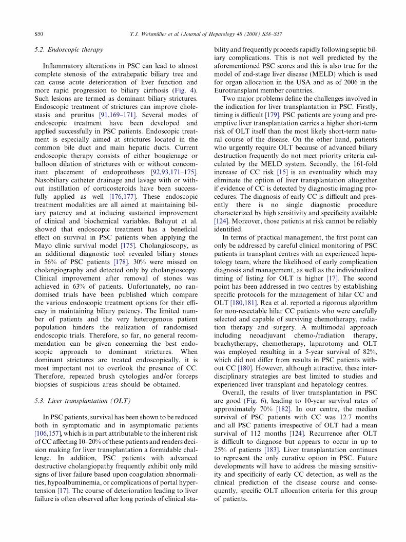
5.2. Endoscopic therapy
Inflammatory alterations in PSC can lead to almostcomplete stenosis of the extrahepatic biliary tree andcan cause acute deterioration of liver function andmore rapid progression to biliary cirrhosis (Fig. 4).Such lesions are termed as dominant biliary strictures.Endoscopic treatment of strictures can improve chole-stasis and pruritus [91,169–171]. Several modes ofendoscopic treatment have been developed andapplied successfully in PSC patients. Endoscopic treat-ment is especially aimed at strictures located in thecommon bile duct and main hepatic ducts. Currentendoscopic therapy consists of either bougienage orballoon dilation of strictures with or without concom-itant placement of endoprotheses [92,93,171–175].Nasobiliary catheter drainage and lavage with or with-out instillation of corticosteroids have been success-fully applied as well [176,177]. These endoscopictreatment modalities are all aimed at maintaining bil-iary patency and at inducing sustained improvementof clinical and biochemical variables. Baluyut et al.showed that endoscopic treatment has a beneficialeffect on survival in PSC patients when applying theMayo clinic survival model [175]. Cholangioscopy, asan additional diagnostic tool revealed biliary stonesin 56% of PSC patients [178]. 30% were missed oncholangiography and detected only by cholangioscopy.Clinical improvement after removal of stones wasachieved in 63% of patients. Unfortunately, no ran-domised trials have been published which comparethe various endoscopic treatment options for their effi-cacy in maintaining biliary patency. The limited num-ber of patients and the very heterogenous patientpopulation hinders the realization of randomisedendoscopic trials. Therefore, so far, no general recom-mendation can be given concerning the best endo-scopic approach to dominant strictures. Whendominant strictures are treated endoscopically, it ismost important not to overlook the presence of CC.Therefore, repeated brush cytologies and/or forcepsbiopsies of suspicious areas should be obtained.
5.3. Liver transplantation (OLT)
In PSC patients, survival has been shown to be reducedboth in symptomatic and in asymptomatic patients[106,157], which is in part attributable to the inherent riskof CC affecting 10–20% of these patients and renders deci-sion making for liver transplantation a formidable chal-lenge. In addition, PSC patients with advanceddestructive cholangiopathy frequently exhibit only mildsigns of liver failure based upon coagulation abnormali-ties, hypoalbuminemia, or complications of portal hyper-tension [17]. The course of deterioration leading to liverfailure is often observed after long periods of clinical sta-
bility and frequently proceeds rapidly following septic bil-iary complications. This is not well predicted by theaforementioned PSC scores and this is also true for themodel of end-stage liver disease (MELD) which is usedfor organ allocation in the USA and as of 2006 in theEurotransplant member countries.
Two major problems define the challenges involved inthe indication for liver transplantation in PSC. Firstly,timing is difficult [179]. PSC patients are young and pre-emptive liver transplantation carries a higher short-termrisk of OLT itself than the most likely short-term natu-ral course of the disease. On the other hand, patientswho urgently require OLT because of advanced biliarydestruction frequently do not meet priority criteria cal-culated by the MELD system. Secondly, the 161-foldincrease of CC risk [15] is an eventuality which mayeliminate the option of liver transplantation altogetherif evidence of CC is detected by diagnostic imaging pro-cedures. The diagnosis of early CC is difficult and pres-ently there is no single diagnostic procedurecharacterized by high sensitivity and specificity available[124]. Moreover, those patients at risk cannot be reliablyidentified.
In terms of practical management, the first point canonly be addressed by careful clinical monitoring of PSCpatients in transplant centres with an experienced hepa-tology team, where the likelihood of early complicationdiagnosis and management, as well as the individualizedtiming of listing for OLT is higher [17]. The secondpoint has been addressed in two centres by establishingspecific protocols for the management of hilar CC andOLT [180,181]. Rea et al. reported a rigorous algorithmfor non-resectable hilar CC patients who were carefullyselected and capable of surviving chemotherapy, radia-tion therapy and surgery. A multimodal approachincluding neoadjuvant chemo-/radiation therapy,brachytherapy, chemotherapy, laparotomy and OLTwas employed resulting in a 5-year survival of 82%,which did not differ from results in PSC patients with-out CC [180]. However, although attractive, these inter-disciplinary strategies are best limited to studies andexperienced liver transplant and hepatology centres.
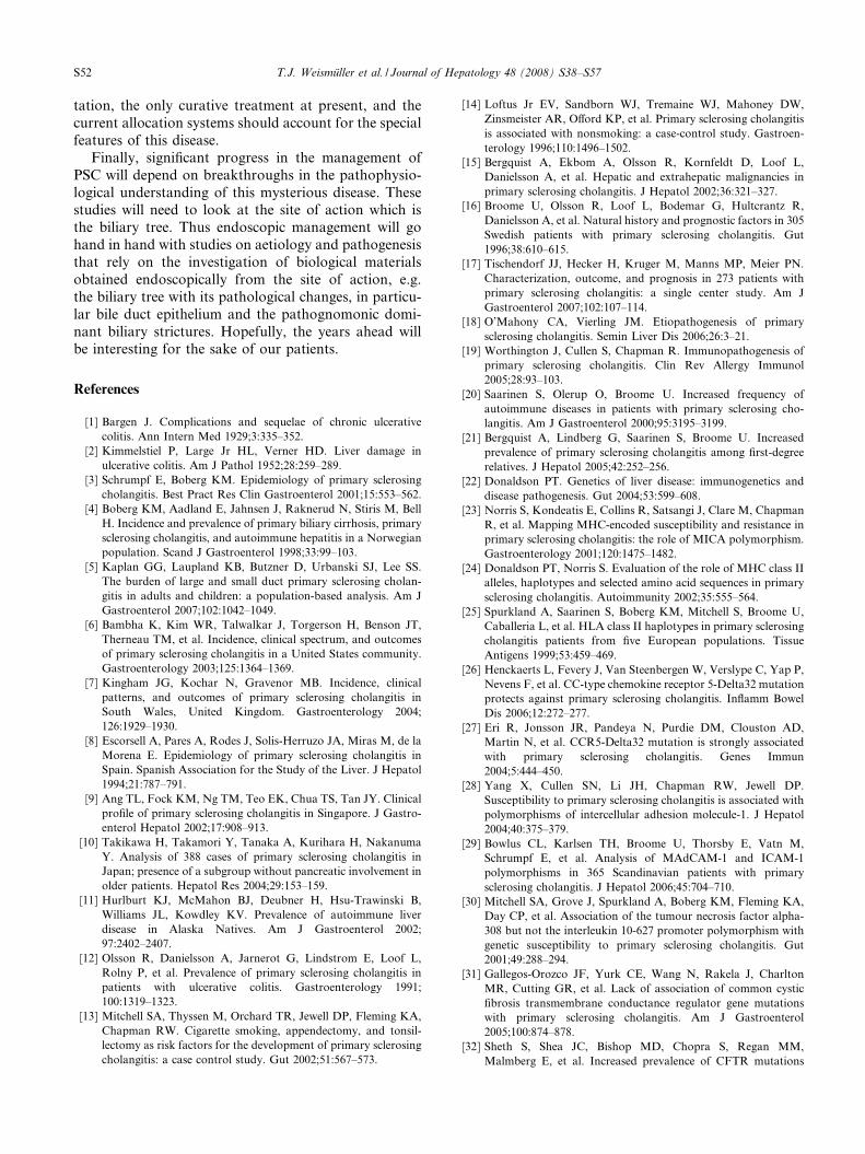
Overall, the results of liver transplantation in PSCare good (Fig. 6), leading to 10-year survival rates ofapproximately 70% [182]. In our centre, the mediansurvival of PSC patients with CC was 12.7 monthsand all PSC patients irrespective of OLT had a meansurvival of 112 months [124]. Recurrence after OLTis difficult to diagnose but appears to occur in up to25% of patients [183]. Liver transplantation continuesto represent the only curative option in PSC. Futuredevelopments will have to address the missing sensitiv-ity and specificity of early CC detection, as well as theclinical prediction of the disease course and conse-quently, specific OLT allocation criteria for this groupof patients.

Fig. 6. Kaplan–Meier analysis of cumulative survival after liver trans-
plantation from 01/2003 to 08/2007 at Hannover Medical School
comparing 55 patients with PSC (incl. five patients with CC) and 318
patients with other chronic liver diseases (hepatitis B and C, alcoholic
liver disease, hepatocellular carcinoma and others). The log-rank-test
shows a significant better survival of patients with PSC (p < 0.01).
T.J. Weismuller et al. / Journal of Hepatology 48 (2008) S38–S57 S51
5.4. Treatment of cholangiocarcinoma in PSC
Surgical resection or liver transplantation is the onlycurative option for patients with PSC-associated CC.However, the prognosis of patients with CC is poor,even after surgical resection. A large population-basedstudy recently showed that survival after surgery forextrahepatic CC has dramatically improved since 1973.However, patients with intrahepatic CC have achievedan improvement in survival largely confined to morerecent years. This may be explained by innovative devel-opments in imaging technology, improvements inpatient selection and advances in surgical techniques[184]. In the therapy of hilar CC, the most favourableresults with 5-year survival rates of 61% are achievedby no-touch-technique, en-bloc-resection and widetumour-free margins [185].
Compared to patients with non-malignant diseases,patients with PSC-associated CC have a worse progno-sis after liver transplantation. Due to the shortage ofdonor organs, liver transplantation for patients withCC has therefore been abandoned by most liver centres.The above mentioned non-randomised pilot study withneoadjuvant radiochemotherapy reported excellent 5-year survival rates of 80%, at least in a subset of patientswith PSC-associated CC. In one study liver transplanta-tion with neoadjuvant chemoradiation achieved an evenbetter survival with less recurrence than conventionalresection [180]. There are presently no randomised con-trolled studies using this neoadjuvant strategy and,therefore, the value of neoadjuvant radiochemotherapyand liver transplantation for patients with PSC-associ-
ated CC is still unclear. Obvious limitations of this strat-egy are a substantial drop out rate during theneoadjuvant therapy due to tumour progression andtreatment-related complications, such as vascular com-plications after liver transplantation [186].
Palliative therapy of PSC-associated CC includesendoscopic management of biliary stenosis (dilatation,stenting), treatment of bacterial cholangitis and systemicchemotherapy. A randomised study with photodynamictherapy of CC resulted in prolongation of survival (493days versus 98 days; p < 0.0001), improved biliary drain-age and better quality of life for the patients comparedwith endoscopic stenting alone [187]. Although a ran-domised study investigating the combination of chemo-therapy and PDT is still not available, there is evidenceof synergistic antitumoural activity of local PDT andsystemic chemotherapy in cell culture experiments andearly clinical studies [188].
There is only a limited number of studies regardingthe systemic treatment options for biliary cancers. Todate, the best response rates have been achieved withcombination chemotherapies containing platinum ana-logues and gemcitabine. In the absence of larger clinicalphase III trials, a standard chemotherapy for biliarycancers does not exist today [189].
6. Conclusion
Primary sclerosing cholangitis represents in manyways one of the most intriguing challenges of currenthepatology. Its aetiology is still mysterious though anumber of promising experimental approaches enlightenthe different aspects of pathophysiology. PSC is not aclassical autoimmune disease but the increasing under-standing of underlying immunological mechanismsstimulates further investigations; the interactionbetween biliary epithelial cells and cellular and humoralimmunity and the search for triggers of a deregulatedimmunity is of particular interest in this context. Atpresent, there is no effective pharmacotherapy available,thus new insights into pathophysiological mechanismswill hopefully lead to new therapeutic innovations.Advances of endoscopic techniques promise additionalpossibilities in diagnosis and treatment. Even thoughthe clinical course of the disease is rather variable, themean survival of PSC patients is markedly shortened.Cholangiocarcinoma is a frequent outcome of PSC witha very poor prognosis. Therefore clinical management isexceptionally demanding. Advances in new imagingtechniques expand the diagnostic options and allownew views on the disease. Considering the high risk ofmalignancy effective and evidence-based surveillancestrategies based on a combination of endoscopic, imag-ing and biochemical techniques are urgently required.These will be beneficial for the timing of liver transplan-
S52 T.J. Weismuller et al. / Journal of Hepatology 48 (2008) S38–S57
tation, the only curative treatment at present, and thecurrent allocation systems should account for the specialfeatures of this disease.
Finally, significant progress in the management ofPSC will depend on breakthroughs in the pathophysio-logical understanding of this mysterious disease. Thesestudies will need to look at the site of action which isthe biliary tree. Thus endoscopic management will gohand in hand with studies on aetiology and pathogenesisthat rely on the investigation of biological materialsobtained endoscopically from the site of action, e.g.the biliary tree with its pathological changes, in particu-lar bile duct epithelium and the pathognomonic domi-nant biliary strictures. Hopefully, the years ahead willbe interesting for the sake of our patients.
References
[1] Bargen J. Complications and sequelae of chronic ulcerativecolitis. Ann Intern Med 1929;3:335–352.
[2] Kimmelstiel P, Large Jr HL, Verner HD. Liver damage inulcerative colitis. Am J Pathol 1952;28:259–289.
[3] Schrumpf E, Boberg KM. Epidemiology of primary sclerosingcholangitis. Best Pract Res Clin Gastroenterol 2001;15:553–562.
[4] Boberg KM, Aadland E, Jahnsen J, Raknerud N, Stiris M, BellH. Incidence and prevalence of primary biliary cirrhosis, primarysclerosing cholangitis, and autoimmune hepatitis in a Norwegianpopulation. Scand J Gastroenterol 1998;33:99–103.
[5] Kaplan GG, Laupland KB, Butzner D, Urbanski SJ, Lee SS.The burden of large and small duct primary sclerosing cholan-gitis in adults and children: a population-based analysis. Am JGastroenterol 2007;102:1042–1049.
[6] Bambha K, Kim WR, Talwalkar J, Torgerson H, Benson JT,Therneau TM, et al. Incidence, clinical spectrum, and outcomesof primary sclerosing cholangitis in a United States community.Gastroenterology 2003;125:1364–1369.
[7] Kingham JG, Kochar N, Gravenor MB. Incidence, clinicalpatterns, and outcomes of primary sclerosing cholangitis inSouth Wales, United Kingdom. Gastroenterology 2004;126:1929–1930.
[8] Escorsell A, Pares A, Rodes J, Solis-Herruzo JA, Miras M, de laMorena E. Epidemiology of primary sclerosing cholangitis inSpain. Spanish Association for the Study of the Liver. J Hepatol1994;21:787–791.
[9] Ang TL, Fock KM, Ng TM, Teo EK, Chua TS, Tan JY. Clinicalprofile of primary sclerosing cholangitis in Singapore. J Gastro-enterol Hepatol 2002;17:908–913.
[10] Takikawa H, Takamori Y, Tanaka A, Kurihara H, NakanumaY. Analysis of 388 cases of primary sclerosing cholangitis inJapan; presence of a subgroup without pancreatic involvement inolder patients. Hepatol Res 2004;29:153–159.
[11] Hurlburt KJ, McMahon BJ, Deubner H, Hsu-Trawinski B,Williams JL, Kowdley KV. Prevalence of autoimmune liverdisease in Alaska Natives. Am J Gastroenterol 2002;97:2402–2407.
[12] Olsson R, Danielsson A, Jarnerot G, Lindstrom E, Loof L,Rolny P, et al. Prevalence of primary sclerosing cholangitis inpatients with ulcerative colitis. Gastroenterology 1991;100:1319–1323.
[13] Mitchell SA, Thyssen M, Orchard TR, Jewell DP, Fleming KA,Chapman RW. Cigarette smoking, appendectomy, and tonsil-lectomy as risk factors for the development of primary sclerosingcholangitis: a case control study. Gut 2002;51:567–573.
[14] Loftus Jr EV, Sandborn WJ, Tremaine WJ, Mahoney DW,Zinsmeister AR, Offord KP, et al. Primary sclerosing cholangitisis associated with nonsmoking: a case-control study. Gastroen-terology 1996;110:1496–1502.
[15] Bergquist A, Ekbom A, Olsson R, Kornfeldt D, Loof L,Danielsson A, et al. Hepatic and extrahepatic malignancies inprimary sclerosing cholangitis. J Hepatol 2002;36:321–327.
[16] Broome U, Olsson R, Loof L, Bodemar G, Hultcrantz R,Danielsson A, et al. Natural history and prognostic factors in 305Swedish patients with primary sclerosing cholangitis. Gut1996;38:610–615.
[17] Tischendorf JJ, Hecker H, Kruger M, Manns MP, Meier PN.Characterization, outcome, and prognosis in 273 patients withprimary sclerosing cholangitis: a single center study. Am JGastroenterol 2007;102:107–114.
[18] O’Mahony CA, Vierling JM. Etiopathogenesis of primarysclerosing cholangitis. Semin Liver Dis 2006;26:3–21.
[19] Worthington J, Cullen S, Chapman R. Immunopathogenesis ofprimary sclerosing cholangitis. Clin Rev Allergy Immunol2005;28:93–103.
[20] Saarinen S, Olerup O, Broome U. Increased frequency ofautoimmune diseases in patients with primary sclerosing cho-langitis. Am J Gastroenterol 2000;95:3195–3199.
[21] Bergquist A, Lindberg G, Saarinen S, Broome U. Increasedprevalence of primary sclerosing cholangitis among first-degreerelatives. J Hepatol 2005;42:252–256.
[22] Donaldson PT. Genetics of liver disease: immunogenetics anddisease pathogenesis. Gut 2004;53:599–608.
[23] Norris S, Kondeatis E, Collins R, Satsangi J, Clare M, ChapmanR, et al. Mapping MHC-encoded susceptibility and resistance inprimary sclerosing cholangitis: the role of MICA polymorphism.Gastroenterology 2001;120:1475–1482.
[24] Donaldson PT, Norris S. Evaluation of the role of MHC class IIalleles, haplotypes and selected amino acid sequences in primarysclerosing cholangitis. Autoimmunity 2002;35:555–564.
[25] Spurkland A, Saarinen S, Boberg KM, Mitchell S, Broome U,Caballeria L, et al. HLA class II haplotypes in primary sclerosingcholangitis patients from five European populations. TissueAntigens 1999;53:459–469.
[26] Henckaerts L, Fevery J, Van Steenbergen W, Verslype C, Yap P,Nevens F, et al. CC-type chemokine receptor 5-Delta32 mutationprotects against primary sclerosing cholangitis. Inflamm BowelDis 2006;12:272–277.
[27] Eri R, Jonsson JR, Pandeya N, Purdie DM, Clouston AD,Martin N, et al. CCR5-Delta32 mutation is strongly associatedwith primary sclerosing cholangitis. Genes Immun2004;5:444–450.
[28] Yang X, Cullen SN, Li JH, Chapman RW, Jewell DP.Susceptibility to primary sclerosing cholangitis is associated withpolymorphisms of intercellular adhesion molecule-1. J Hepatol2004;40:375–379.
[29] Bowlus CL, Karlsen TH, Broome U, Thorsby E, Vatn M,Schrumpf E, et al. Analysis of MAdCAM-1 and ICAM-1polymorphisms in 365 Scandinavian patients with primarysclerosing cholangitis. J Hepatol 2006;45:704–710.
[30] Mitchell SA, Grove J, Spurkland A, Boberg KM, Fleming KA,Day CP, et al. Association of the tumour necrosis factor alpha-308 but not the interleukin 10-627 promoter polymorphism withgenetic susceptibility to primary sclerosing cholangitis. Gut2001;49:288–294.
[31] Gallegos-Orozco JF, Yurk CE, Wang N, Rakela J, CharltonMR, Cutting GR, et al. Lack of association of common cysticfibrosis transmembrane conductance regulator gene mutationswith primary sclerosing cholangitis. Am J Gastroenterol2005;100:874–878.
[32] Sheth S, Shea JC, Bishop MD, Chopra S, Regan MM,Malmberg E, et al. Increased prevalence of CFTR mutations
T.J. Weismuller et al. / Journal of Hepatology 48 (2008) S38–S57 S53
and variants and decreased chloride secretion in primarysclerosing cholangitis. Hum Genet 2003;113:286–292.
[33] Angulo P, Peter JB, Gershwin ME, DeSotel CK, Shoenfeld Y,Ahmed AE, et al. Serum autoantibodies in patients with primarysclerosing cholangitis. J Hepatol 2000;32:182–187.
[34] Mulder AH, Horst G, Haagsma EB, Limburg PC, KleibeukerJH, Kallenberg CG. Prevalence and characterization of neutro-phil cytoplasmic antibodies in autoimmune liver diseases. Hepa-tology 1993;17:411–417.
[35] Bansi DS, Fleming KA, Chapman RW. Importance of antine-utrophil cytoplasmic antibodies in primary sclerosing cholangitisand ulcerative colitis: prevalence, titre, and IgG subclass. Gut1996;38:384–389.
[36] Terjung B, Spengler U. Role of auto-antibodies for the diagnosisof chronic cholestatic liver diseases. Clin Rev Allergy Immunol2005;28:115–133.
[37] Terjung B, Spengler U, Sauerbruch T, Worman HJ. ‘‘Atypical p-ANCA” in IBD and hepatobiliary disorders react with a 50-kilodalton nuclear envelope protein of neutrophils and myeloidcell lines. Gastroenterology 2000;119:310–322.
[38] Bodenheimer Jr HC, LaRusso NF, Thayer Jr WR, Charland C,Staples PJ, Ludwig J. Elevated circulating immune complexes inprimary sclerosing cholangitis. Hepatology 1983;3:150–154.
[39] Senaldi G, Donaldson PT, Magrin S, Farrant JM, Alexander GJ,Vergani D, et al. Activation of the complement system inprimary sclerosing cholangitis. Gastroenterology 1989;97:1430–1434.
[40] Bo X, Broome U, Remberger M, Sumitran-Holgersson S.Tumour necrosis factor alpha impairs function of liver derivedT lymphocytes and natural killer cells in patients with primarysclerosing cholangitis. Gut 2001;49:131–141.
[41] Grant AJ, Lalor PF, Salmi M, Jalkanen S, Adams DH. Homingof mucosal lymphocytes to the liver in the pathogenesis ofhepatic complications of inflammatory bowel disease. Lancet2002;359:150–157.
[42] Eksteen B, Miles AE, Grant AJ, Adams DH. Lymphocytehoming in the pathogenesis of extra-intestinal manifestations ofinflammatory bowel disease. Clin Med 2004;4:173–180.
[43] Salmi M, Jalkanen S. Endothelial ligands and homing ofmucosal leukocytes in extraintestinal manifestations of IBD.Inflamm Bowel Dis 1998;4:149–156.
[44] Grant AJ, Lalor PF, Hubscher SG, Briskin M, Adams DH.MAdCAM-1 expressed in chronic inflammatory liver diseasesupports mucosal lymphocyte adhesion to hepatic endothelium(MAdCAM-1 in chronic inflammatory liver disease). Hepatology2001;33:1065–1072.
[45] Eksteen B, Grant AJ, Miles A, Curbishley SM, Lalor PF,Hubscher SG, et al. Hepatic endothelial CCL25 mediates therecruitment of CCR9+ gut-homing lymphocytes to the liver inprimary sclerosing cholangitis. J Exp Med 2004;200:1511–1517.
[46] Palmer KR, Duerden BI, Holdsworth CD. Bacteriological andendotoxin studies in cases of ulcerative colitis submitted tosurgery. Gut 1980;21:851–854.
[47] Ludwig J, Barham SS, LaRusso NF, Elveback LR, Wiesner RH,McCall JT. Morphologic features of chronic hepatitis associatedwith primary sclerosing cholangitis and chronic ulcerative colitis.Hepatology 1981;1:632–640.
[48] Lichtman SN, Sartor RB, Keku J, Schwab JH. Hepaticinflammation in rats with experimental small intestinal bacterialovergrowth. Gastroenterology 1990;98:414–423.
[49] Bjornsson E, Cederborg A, Akvist A, Simren M, Stotzer PO,Bjarnason I. Intestinal permeability and bacterial growth of thesmall bowel in patients with primary sclerosing cholangitis.Scand J Gastroenterol 2005;40:1090–1094.
[50] Nilsson HO, Taneera J, Castedal M, Glatz E, Olsson R,Wadstrom T. Identification of Helicobacter pylori and otherHelicobacter species by PCR, hybridization, and partial DNA
sequencing in human liver samples from patients with primarysclerosing cholangitis or primary biliary cirrhosis. J Clin Micro-biol 2000;38:1072–1076.
[51] Boomkens SY, de Rave S, Pot RG, Egberink HF, Penning LC,Rothuizen J, et al. The role of Helicobacter spp. in thepathogenesis of primary biliary cirrhosis and primary sclerosingcholangitis. FEMS Immunol Med Microbiol 2005;44:221–225.
[52] Krasinskas AM, Yao Y, Randhawa P, Dore MP, Sepulveda AR.Helicobacter pylori may play a contributory role in the patho-genesis of primary sclerosing cholangitis. Dig Dis Sci 2007;52:2265–2270.
[53] Mehal WZ, Hattersley AT, Chapman RW, Fleming KA. Asurvey of cytomegalovirus (CMV) DNA in primary sclerosingcholangitis (PSC) liver tissues using a sensitive polymerase chainreaction (PCR) based assay. J Hepatol 1992;15:396–399.
[54] Minuk GY, Rascanin N, Paul RW, Lee PW, Buchan K, KellyJK. Reovirus type 3 infection in patients with primary biliarycirrhosis and primary sclerosing cholangitis. J Hepatol 1987;5:8–13.
[55] Pohl J, Ring A, Stremmel W, Stiehl A. The role of dominantstenoses in bacterial infections of bile ducts in primary sclerosingcholangitis. Eur J Gastroenterol Hepatol 2006;18:69–74.
[56] Bjornsson ES, Kilander AF, Olsson RG. Bile duct bacterialisolates in primary sclerosing cholangitis and certain other formsof cholestasis – a study of bile cultures from ERCP. Hepato-gastroenterology 2000;47:1504–1508.
[57] Kulaksiz H, Rudolph G, Kloeters-Plachky P, Sauer P, Geiss H,Stiehl A. Biliary Candida infections in primary sclerosingcholangitis. J Hepatol 2006;45:711–716.
[58] Tietz PS, Larusso NF. Cholangiocyte biology. Curr OpinGastroenterol 2006;22:279–287.
[59] Lazaridis KN, Strazzabosco M, Larusso NF. The cholangiop-athies: disorders of biliary epithelia. Gastroenterology 2004;127:1565–1577.
[60] Trauner M, Meier PJ, Boyer JL. Molecular pathogenesis ofcholestasis. N Engl J Med 1998;339:1217–1227.
[61] Fickert P, Wagner M, Marschall HU, Fuchsbichler A, ZollnerG, Tsybrovskyy O, et al. 24-norUrsodeoxycholic acid is superiorto ursodeoxycholic acid in the treatment of sclerosing cholangitisin Mdr2 (Abcb4) knockout mice. Gastroenterology 2006;130:465–481.
[62] Fickert P, Zollner G, Fuchsbichler A, Stumptner C, WeigleinAH, Lammert F, et al. Ursodeoxycholic acid aggravates bileinfarcts in bile duct-ligated and Mdr2 knockout mice viadisruption of cholangioles. Gastroenterology 2002;123:1238–1251.
[63] Pauli-Magnus C, Kerb R, Fattinger K, Lang T, Anwald B,Kullak-Ublick GA, et al. BSEP and MDR3 haplotype structurein healthy Caucasians, primary biliary cirrhosis and primarysclerosing cholangitis. Hepatology 2004;39:779–791.
[64] Das KM, Vecchi M, Sakamaki S. A shared and uniqueepitope(s) on human colon, skin, and biliary epitheliumdetected by a monoclonal antibody. Gastroenterology 1990;98:464–469.
[65] Mandal A, Dasgupta A, Jeffers L, Squillante L, Hyder S, ReddyR, et al. Autoantibodies in sclerosing cholangitis against a sharedpeptide in biliary and colon epithelium. Gastroenterology1994;106:185–192.
[66] Xu B, Broome U, Ericzon BG, Sumitran-Holgersson S. Highfrequency of autoantibodies in patients with primary sclerosingcholangitis that bind biliary epithelial cells and induce expres-sion of CD44 and production of interleukin 6. Gut2002;51:120–127.
[67] Karrar A, Broome U, Sodergren T, Jaksch M, Bergquist A,Bjornstedt M, et al. Biliary epithelial cell antibodies link adaptiveand innate immune responses in primary sclerosing cholangitis.Gastroenterology 2007;132:1504–1514.

S54 T.J. Weismuller et al. / Journal of Hepatology 48 (2008) S38–S57
[68] Spirli C, Nathanson MH, Fiorotto R, Duner E, Denson LA,Sanz JM, et al. Proinflammatory cytokines inhibit secretion in ratbile duct epithelium. Gastroenterology 2001;121:156–169.
[69] Spirli C, Fabris L, Duner E, Fiorotto R, Ballardini G, RoskamsT, et al. Cytokine-stimulated nitric oxide production inhibitsadenylyl cyclase and cAMP-dependent secretion in cholangio-cytes. Gastroenterology 2003;124:737–753.
[70] Fevery J, Verslype C, Lai G, Aerts R, Van Steenbergen W.Incidence, diagnosis, and therapy of cholangiocarcinoma inpatients with primary sclerosing cholangitis. Dig Dis Sci2007;52:3123–3135.
[71] Komichi D, Tazuma S, Nishioka T, Hyogo H, Chayama K.Glycochenodeoxycholate plays a carcinogenic role in immortal-ized mouse cholangiocytes via oxidative DNA damage. FreeRadic Biol Med 2005;39:1418–1427.
[72] Meng F, Yamagiwa Y, Ueno Y, Patel T. Over-expression ofinterleukin-6 enhances cell survival and transformed cell growthin human malignant cholangiocytes. J Hepatol2006;44:1055–1065.
[73] Ahrendt SA, Rashid A, Chow JT, Eisenberger CF, Pitt HA,Sidransky D. p53 overexpression and K-ras gene mutations inprimary sclerosing cholangitis-associated biliary tract cancer. JHepatobiliary Pancreat Surg 2000;7:426–431.
[74] Boberg KM, Schrumpf E, Bergquist A, Broome U, Pares A,Remotti H, et al. Cholangiocarcinoma in primary sclerosingcholangitis: K-ras mutations and Tp53 dysfunction are impli-cated in the neoplastic development. J Hepatol 2000;32:374–380.
[75] Taniai M, Higuchi H, Burgart LJ, Gores GJ. p16INK4apromoter mutations are frequent in primary sclerosing cholan-gitis (PSC) and PSC-associated cholangiocarcinoma. Gastroen-terology 2002;123:1090–1098.
[76] Ahrendt SA, Eisenberger CF, Yip L, Rashid A, Chow JT, PittHA, et al. Chromosome 9p21 loss and p16 inactivation inprimary sclerosing cholangitis-associated cholangiocarcinoma. JSurg Res 1999;84:88–93.
[77] Wiesner RH, Grambsch PM, Dickson ER, Ludwig J, MacCartyRL, Hunter EB, et al. Primary sclerosing cholangitis: naturalhistory, prognostic factors and survival analysis. Hepatology1989;10:430–436.
[78] Bergquist A, Said K, Broome U. Changes over a 20-year periodin the clinical presentation of primary sclerosing cholangitis inSweden. Scand J Gastroenterol 2007;42:88–93.
[79] Talwalkar JA, Lindor KD. Primary sclerosing cholangitis.Inflamm Bowel Dis 2005;11:62–72.
[80] Zein CO, Lindor KD, Angulo P. Prevalence and predictors ofesophageal varices in patients with primary sclerosing cholangi-tis. Hepatology 2004;39:204–210.
[81] Bjornsson E, Simren M, Olsson R, Chapman RW. Fatigue inpatients with primary sclerosing cholangitis. Scand J Gastroen-terol 2004;39:961–968.
[82] Bergquist A, Glaumann H, Persson B, Broome U. Risk factorsand clinical presentation of hepatobiliary carcinoma in patientswith primary sclerosing cholangitis: a case-control study. Hepa-tology 1998;27:311–316.
[83] Hay JE, Lindor KD, Wiesner RH, Dickson ER, Krom RA,LaRusso NF. The metabolic bone disease of primary sclerosingcholangitis. Hepatology 1991;14:257–261.
[84] Lee YM, Kaplan MM. Primary sclerosing cholangitis. N Engl JMed 1995;332:924–933.
[85] Majoie CB, Reeders JW, Sanders JB, Huibregtse K, Jansen PL.Primary sclerosing cholangitis: a modified classification ofcholangiographic findings. AJR Am J Roentgenol1991;157:495–497.
[86] Rajaram R, Ponsioen CY, Majoie CB, Reeders JW, Lameris JS.Evaluation of a modified cholangiographic classification systemfor primary sclerosing cholangitis. Abdom Imaging 2001;26:43–47.
[87] Wee A, Ludwig J. Pericholangitis in chronic ulcerative colitis:primary sclerosing cholangitis of the small bile ducts? Ann InternMed 1985;102:581–587.
[88] Trauner M, Boyer JL. Cholestatic syndromes. Curr OpinGastroenterol 2004;20:220–230.
[89] Engler S, Elsing C, Flechtenmacher C, Theilmann L, StremmelW, Stiehl A. Progressive sclerosing cholangitis after septic shock:a new variant of vanishing bile duct disorders. Gut2003;52:688–693.
[90] Gelbmann CM, Rummele P, Wimmer M, Hofstadter F, Gohl-mann B, Endlicher E, et al. Ischemic-like cholangiopathy withsecondary sclerosing cholangitis in critically ill patients. Am JGastroenterol 2007;102:1221–1229.
[91] Byrne MF, Chong HI, O’Donovan D, Sheehan KM, LeaderMB, Kay E, et al. Idiopathic cholangiopathy in a biliary castsyndrome necessitating liver transplantation following headtrauma. Eur J Gastroenterol Hepatol 2003;15:415–417.
[92] van Milligen de Wit AW, van Bracht J, Rauws EA, Jones EA,Tytgat GN, Huibregtse K. Endoscopic stent therapy for dom-inant extrahepatic bile duct strictures in primary sclerosingcholangitis. Gastrointest Endosc 1996;44:293–299.
[93] Stiehl A, Rudolph G, Kloeters-Plachky P, Sauer P, Walker S.Development of dominant bile duct stenoses in patients withprimary sclerosing cholangitis treated with ursodeoxycholic acid:outcome after endoscopic treatment. J Hepatol 2002;36:151–156.
[94] van Milligen de Wit AW, Rauws EA, van Bracht J, Mulder CJ,Jones EA, Tytgat GN, et al. Lack of complications followingshort-term stent therapy for extrahepatic bile duct strictures inprimary sclerosing cholangitis. Gastrointest Endosc1997;46:344–347.
[95] Freeman ML, Nelson DB, Sherman S, Haber GB, Herman ME,Dorsher PJ, et al. Complications of endoscopic biliary sphinc-terotomy. N Engl J Med 1996;335:909–918.
[96] Loperfido S, Angelini G, Benedetti G, Chilovi F, Costan F, DeBerardinis F, et al. Major early complications from diagnosticand therapeutic ERCP: a prospective multicenter study. Gastro-intest Endosc 1998;48:1–10.
[97] Masci E, Toti G, Mariani A, Curioni S, Lomazzi A, Dinelli M,et al. Complications of diagnostic and therapeutic ERCP: aprospective multicenter study. Am J Gastroenterol2001;96:417–423.
[98] Ernst O, Asselah T, Sergent G, Calvo M, Talbodec N, Paris JC,et al. MR cholangiography in primary sclerosing cholangitis.AJR Am J Roentgenol 1998;171:1027–1030.
[99] Fulcher AS, Turner MA, Franklin KJ, Shiffman ML, SterlingRK, Luketic VA, et al. Primary sclerosing cholangitis: evaluationwith MR cholangiography – a case-control study. Radiology2000;215:71–80.
[100] Angulo P, Pearce DH, Johnson CD, Henry JJ, LaRusso NF,Petersen BT, et al. Magnetic resonance cholangiography inpatients with biliary disease: its role in primary sclerosingcholangitis. J Hepatol 2000;33:520–527.
[101] Moff SL, Kamel IR, Eustace J, Lawler LP, Kantsevoy S, KallooAN, et al. Diagnosis of primary sclerosing cholangitis: a blindedcomparative study using magnetic resonance cholangiographyand endoscopic retrograde cholangiography. GastrointestEndosc 2006;64:219–223.
[102] Berstad AE, Aabakken L, Smith HJ, Aasen S, Boberg KM,Schrumpf E. Diagnostic accuracy of magnetic resonance andendoscopic retrograde cholangiography in primary sclerosingcholangitis. Clin Gastroenterol Hepatol 2006;4:514–520.
[103] Textor HJ, Flacke S, Pauleit D, Keller E, Neubrand M,Terjung B, et al. Three-dimensional magnetic resonancecholangiopancreatography with respiratory triggering in thediagnosis of primary sclerosing cholangitis: comparison withendoscopic retrograde cholangiography. Endoscopy 2002;34:984–990.

T.J. Weismuller et al. / Journal of Hepatology 48 (2008) S38–S57 S55
[104] Olsson R, Hagerstrand I, Broome U, Danielsson A, JarnerotG, Loof L, et al. Sampling variability of percutaneous liverbiopsy in primary sclerosing cholangitis. J Clin Pathol1995;48:933–935.
[105] Burak KW, Angulo P, Lindor KD. Is there a role for liver biopsyin primary sclerosing cholangitis? Am J Gastroenterol2003;98:1155–1158.
[106] Kim WR, Therneau TM, Wiesner RH, Poterucha JJ, Benson JT,Malinchoc M, et al. A revised natural history model for primarysclerosing cholangitis. Mayo Clin Proc 2000;75:688–694.
[107] Kim WR, Ludwig J, Lindor KD. Variant forms of cholestaticdiseases involving small bile ducts in adults. Am J Gastroenterol2000;95:1130–1138.
[108] Bjornsson E, Boberg KM, Cullen S, Fleming K, Clausen OP,Fausa O, et al. Patients with small duct primary sclerosingcholangitis have a favourable long term prognosis. Gut2002;51:731–735.
[109] Broome U, Glaumann H, Lindstom E, Loof L, Almer S, PrytzH, et al. Natural history and outcome in 32 Swedish patients withsmall duct primary sclerosing cholangitis (PSC). J Hepatol2002;36:586–589.
[110] Angulo P, Maor-Kendler Y, Lindor KD. Small-duct primarysclerosing cholangitis: a long-term follow-up study. Hepatology2002;35:1494–1500.
[111] Charatcharoenwitthaya P, Angulo P, Enders FB, Lindor KD.Impact of inflammatory bowel disease and ursodeoxycholic acidtherapy on small-duct primary sclerosing cholangitis. Hepatol-ogy 2008;47:133–142.
[112] Alvarez F, Berg PA, Bianchi FB, Bianchi L, Burroughs AK,Cancado EL, et al. International Autoimmune Hepatitis GroupReport: review of criteria for diagnosis of autoimmune hepatitis.J Hepatol 1999;31:929–938.
[113] van Buuren HR, van Hoogstraten HJE, Terkivatan T, SchalmSW, Vleggaar FP. High prevalence of autoimmune hepatitisamong patients with primary sclerosing cholangitis. J Hepatol2000;33:543–548.
[114] Kaya M, Angulo P, Lindor KD. Overlap of autoimmunehepatitis and primary sclerosing cholangitis: an evaluation of amodified scoring system. J Hepatol 2000;33:537–542.
[115] Gregorio GV, Portmann B, Karani J, Harrison P, DonaldsonPT, Vergani D, et al. Autoimmune hepatitis/sclerosing cholan-gitis overlap syndrome in childhood: a 16-year prospective study.Hepatology 2001;33:544–553.
[116] Floreani A, Rizzotto ER, Ferrara F, Carderi I, Caroli D,Blasone L, et al. Clinical course and outcome of autoimmunehepatitis/primary sclerosing cholangitis overlap syndrome. Am JGastroenterol 2005;100:1516–1522.
[117] Kawa S, Hamano H, Umemura T, Kiyosawa K, Uehara T.Sclerosing cholangitis associated with autoimmune pancreatitis.Hepatol Res 2007;37:S487–S495.
[118] Nakanuma Y, Zen Y. Pathology and immunopathology ofimmunoglobulin G4-related sclerosing cholangitis: the latestaddition to the sclerosing cholangitis family. Hepatol Res2007;37:S478–S486.
[119] Zen Y, Harada K, Sasaki M, Sato Y, Tsuneyama K, Haratake J,et al. IgG4-related sclerosing cholangitis with and withouthepatic inflammatory pseudotumor, and sclerosing pancreatitis-associated sclerosing cholangitis: do they belong to a spectrum ofsclerosing pancreatitis? Am J Surg Pathol 2004;28:1193–1203.
[120] Mendes FD, Jorgensen R, Keach J, Katzmann JA, Smyrk T,Donlinger J, et al. Elevated serum IgG4 concentration in patientswith primary sclerosing cholangitis. Am J Gastroenterol2006;101:2070–2075.
[121] Dickson ER, Murtaugh PA, Wiesner RH, Grambsch PM,Fleming TR, Ludwig J, et al. Primary sclerosing cholangitis:refinement and validation of survival models. Gastroenterology1992;103:1893–1901.
[122] Nyblom H, Nordlinder H, Olsson R. High aspartate to alanineaminotransferase ratio is an indicator of cirrhosis and pooroutcome in patients with primary sclerosing cholangitis. Liver Int2007;27:694–699.
[123] Burak K, Angulo P, Pasha TM, Egan K, Petz J, LindorKD. Incidence and risk factors for cholangiocarcinoma inprimary sclerosing cholangitis. Am J Gastroenterol2004;99:523–526.
[124] Tischendorf JJ, Meier PN, Strassburg CP, Klempnauer J, HeckerH, Manns MP, et al. Characterization and clinical course ofhepatobiliary carcinoma in patients with primary sclerosingcholangitis. Scand J Gastroenterol 2006;41:1227–1234.
[125] Chalasani N, Baluyut A, Ismail A, Zaman A, Sood G, Ghalib R,et al. Cholangiocarcinoma in patients with primary sclerosingcholangitis: a multicenter case-control study. Hepatology2000;31:7–11.
[126] Brandsaeter B, Isoniemi H, Broome U, Olausson M, BackmanL, Hansen B, et al. Liver transplantation for primary sclerosingcholangitis; predictors and consequences of hepatobiliary malig-nancy. J Hepatol 2004;40:815–822.
[127] Boberg KM, Bergquist A, Mitchell S, Pares A, Rosina F,Broome U, et al. Cholangiocarcinoma in primary sclerosingcholangitis: risk factors and clinical presentation. Scand JGastroenterol 2002;37:1205–1211.
[128] Leidenius M, Hockersted K, Broome U, Ericzon BG, Friman S,Olausson M, et al. Hepatobiliary carcinoma in primary scleros-ing cholangitis: a case control study. J Hepatol 2001;34:792–798.
[129] Ramage JK, Donaghy A, Farrant JM, Iorns R, Williams R.Serum tumor markers for the diagnosis of cholangiocarcinomain primary sclerosing cholangitis. Gastroenterology1995;108:865–869.
[130] Levy C, Lymp J, Angulo P, Gores GJ, Larusso N, Lindor KD.The value of serum CA 19-9 in predicting cholangiocarcinomasin patients with primary sclerosing cholangitis. Dig Dis Sci2005;50:1734–1740.
[131] Bjornsson E, Kilander A, Olsson R. CA 19-9 and CEA areunreliable markers for cholangiocarcinoma in patients withprimary sclerosing cholangitis. Liver 1999;19:501–508.
[132] Petersen-Benz C, Stiehl A. Impact of dominant stenoses on theserum level of the tumor marker CA19-9 in patients with primarysclerosing cholangitis. Z Gastroenterol 2005;43:587–590.
[133] Lempinen M, Isoniemi H, Makisalo H, Nordin A, Halme L,Arola J, et al. Enhanced detection of cholangiocarcinoma withserum trypsinogen-2 in patients with severe bile duct strictures. JHepatol 2007;47:677–683.
[134] Campbell WL, Ferris JV, Holbert BL, Thaete FL, Baron RL.Biliary tract carcinoma complicating primary sclerosing cholan-gitis: evaluation with CT, cholangiography, US, and MRimaging. Radiology 1998;207:41–50.
[135] Prytz H, Keiding S, Bjornsson E, Broome U, Almer S, CastedalM, et al. Dynamic FDG-PET is useful for detection ofcholangiocarcinoma in patients with PSC listed for liver trans-plantation. Hepatology 2006;44:1572–1580.
[136] Buckles DC, Lindor KD, Larusso NF, Petrovic LM, Gores GJ.In primary sclerosing cholangitis, gallbladder polyps are fre-quently malignant. Am J Gastroenterol 2002;97:1138–1142.
[137] Bergquist A, Broome U. Hepatobiliary and extra-hepatic malig-nancies in primary sclerosing cholangitis. Best Pract Res ClinGastroenterol 2001;15:643–656.
[138] Boberg KM, Jebsen P, Clausen OP, Foss A, Aabakken L,Schrumpf E. Diagnostic benefit of biliary brush cytology incholangiocarcinoma in primary sclerosing cholangitis. J Hepatol2006;45:568–574.
[139] Ponsioen CY, Vrouenraets SM, van Milligen de Wit AW, SturmP, Tascilar M, Offerhaus GJ, et al. Value of brush cytology fordominant strictures in primary sclerosing cholangitis. Endoscopy1999;31:305–309.

S56 T.J. Weismuller et al. / Journal of Hepatology 48 (2008) S38–S57
[140] Lindberg B, Arnelo U, Bergquist A, Thorne A, Hjerpe A,Granqvist S, et al. Diagnosis of biliary strictures in conjunctionwith endoscopic retrograde cholangiopancreaticography, withspecial reference to patients with primary sclerosing cholangitis.Endoscopy 2002;34:909–916.
[141] Ponchon T, Gagnon P, Berger F, Labadie M, Liaras A,Chavaillon A, et al. Value of endobiliary brush cytology andbiopsies for the diagnosis of malignant bile duct stenosis:results of a prospective study. Gastrointest Endosc1995;42:565–572.
[142] Tischendorf JJ, Kruger M, Trautwein C, Duckstein N, SchneiderA, Manns MP, et al. Cholangioscopic characterization ofdominant bile duct stenoses in patients with primary sclerosingcholangitis. Endoscopy 2006;38:665–669.
[143] Chen YK, Pleskow DK. SpyGlass single-operator peroralcholangiopancreatoscopy system for the diagnosis and therapyof bile-duct disorders: a clinical feasibility study (with video).Gastrointest Endosc 2007;65:832–841.
[144] Tischendorf JJ, Meier PN, Schneider A, Manns MP, Kruger M.Transpapillary intraductal ultrasound in the evaluation ofdominant bile duct stenoses in patients with primary sclerosingcholangitis. Scand J Gastroenterol 2007;42:1011–1017.
[145] Kubicka S, Kuhnel F, Flemming P, Hain B, Kezmic N, RudolphKL, et al. K-ras mutations in the bile of patients with primarysclerosing cholangitis. Gut 2001;48:403–408.
[146] Alvaro D, Macarri G, Mancino MG, Marzioni M, Bragazzi M,Onori P, et al. Serum and biliary insulin-like growth factor I andvascular endothelial growth factor in determining the cause ofobstructive cholestasis. Ann Intern Med 2007;147:451–459.
[147] Ponsioen CY, Vrouenraets SM, Prawirodirdjo W, Rajaram R,Rauws EA, Mulder CJ, et al. Natural history of primarysclerosing cholangitis and prognostic value of cholangiographyin a Dutch population. Gut 2002;51:562–566.
[148] Ekbom A, Helmick C, Zack M, Adami HO. Ulcerative colitisand colorectal cancer. A population-based study. N Engl J Med1990;323:1228–1233.
[149] Aadland E, Schrumpf E, Fausa O, Elgjo K, Heilo A, Aakhus T,et al. Primary sclerosing cholangitis: a long-term follow-up study.Scand J Gastroenterol 1987;22:655–664.
[150] Brentnall TA, Haggitt RC, Rabinovitch PS, Kimmey MB,Bronner MP, Levine DS, et al. Risk and natural history ofcolonic neoplasia in patients with primary sclerosing cholangitisand ulcerative colitis. Gastroenterology 1996;110:331–338.
[151] Broome U, Lindberg G, Lofberg R. Primary sclerosing cholan-gitis in ulcerative colitis – a risk factor for the development ofdysplasia and DNA aneuploidy? Gastroenterology 1992;102:1877–1880.
[152] Kornfeld D, Ekbom A, Ihre T. Is there an excess risk forcolorectal cancer in patients with ulcerative colitis and concom-itant primary sclerosing cholangitis? A population based study.Gut 1997;41:522–525.
[153] Shetty K, Rybicki L, Brzezinski A, Carey WD, Lashner BA. Therisk for cancer or dysplasia in ulcerative colitis patients withprimary sclerosing cholangitis. Am J Gastroenterol 1999;94:1643–1649.
[154] Broome U, Lofberg R, Veress B, Eriksson LS. Primary scleros-ing cholangitis and ulcerative colitis: evidence for increasedneoplastic potential. Hepatology 1995;22:1404–1408.
[155] Kiesslich R, Fritsch J, Holtmann M, Koehler HH, Stolte M,Kanzler S, et al. Methylene blue-aided chromoendoscopy for thedetection of intraepithelial neoplasia and colon cancer inulcerative colitis. Gastroenterology 2003;124:880–888.
[156] Hurlstone DP, McAlindon ME, Sanders DS, Koegh R, LoboAJ, Cross SS. Further validation of high-magnification chromo-scopic-colonoscopy for the detection of intraepithelial neoplasiaand colon cancer in ulcerative colitis. Gastroenterology2004;126:376–378.
[157] LaRusso NF, Shneider BL, Black D, Gores GJ, James SP, DooE, et al. Primary sclerosing cholangitis: summary of a workshop.Hepatology 2006;44:746–764.
[158] Okolicsanyi L, Fabris L, Viaggi S, Carulli N, Podda M, Ricci G.Primary sclerosing cholangitis: clinical presentation, naturalhistory and prognostic variables: an Italian multicentre study.The Italian PSC Study Group. Eur J Gastroenterol Hepatol1996;8:685–691.
[159] Farrant JM, Hayllar KM, Wilkinson ML, Karani J, PortmannBC, Westaby D, et al. Natural history and prognostic variablesin primary sclerosing cholangitis. Gastroenterology1991;100:1710–1717.
[160] Mitchell SA, Bansi DS, Hunt N, Von Bergmann K, Fleming KA,Chapman RW. A preliminary trial of high-dose ursodeoxycholicacid in primary sclerosing cholangitis. Gastroenterology2001;121:900–907.
[161] Harnois DM, Angulo P, Jorgensen RA, Larusso NF, LindorKD. High-dose ursodeoxycholic acid as a therapy for patientswith primary sclerosing cholangitis. Am J Gastroenterol2001;96:1558–1562.
[162] Rost D, Rudolph G, Kloeters-Plachky P, Stiehl A. Effect of high-dose ursodeoxycholic acid on its biliary enrichment in primarysclerosing cholangitis. Hepatology 2004;40:693–698.
[163] Olsson R, Boberg KM, de Muckadell OS, Lindgren S,Hultcrantz R, Folvik G, et al. High-dose ursodeoxycholicacid in primary sclerosing cholangitis: a 5-year multicenter,randomized, controlled study. Gastroenterology 2005;129:1464–1472.
[164] Pardi DS, Loftus Jr EV, Kremers WK, Keach J, Lindor KD.Ursodeoxycholic acid as a chemopreventive agent in patientswith ulcerative colitis and primary sclerosing cholangitis. Gas-troenterology 2003;124:889–893.
[165] Tung BY, Emond MJ, Haggitt RC, Bronner MP, Kimmey MB,Kowdley KV, et al. Ursodiol use is associated with lowerprevalence of colonic neoplasia in patients with ulcerative colitisand primary sclerosing cholangitis. Ann Intern Med2001;134:89–95.
[166] van Hoogstraten HJ, Vleggaar FP, Boland GJ, van SteenbergenW, Griffioen P, Hop WC, et al. Budesonide or prednisone incombination with ursodeoxycholic acid in primary sclerosingcholangitis: a randomized double-blind pilot study. Belgian-Dutch PSC Study Group. Am J Gastroenterol 2000;95:2015–2022.
[167] Boberg KM, Fausa O, Haaland T, Holter E, Mellbye OJ,Spurkland A, et al. Features of autoimmune hepatitis in primarysclerosing cholangitis: an evaluation of 114 primary sclerosingcholangitis patients according to a scoring system for thediagnosis of autoimmune hepatitis. Hepatology 1996;23:1369–1376.
[168] Beuers U, Rust C. Overlap syndromes. Semin Liver Dis2005;25:311–320.
[169] Stiehl A. Primary sclerosing cholangitis: the role of endoscopictherapy. Semin Liver Dis 2006;26:62–68.
[170] Stiehl A, Rost D. Endoscopic treatment of dominant stenoses inpatients with primary sclerosing cholangitis. Clin Rev AllergyImmunol 2005;28:159–165.
[171] Bjornsson E, Lindqvist-Ottosson J, Asztely M, Olsson R.Dominant strictures in patients with primary sclerosing cholan-gitis. Am J Gastroenterol 2004;99:502–508.
[172] Johnson GK, Geenen JE, Venu RP, Schmalz MJ, Hogan WJ.Endoscopic treatment of biliary tract strictures in sclerosingcholangitis: a larger series and recommendations for treatment.Gastrointest Endosc 1991;37:38–43.
[173] Lombard M, Farrant M, Karani J, Westaby D, Williams R.Improving biliary-enteric drainage in primary sclerosing cholan-gitis: experience with endoscopic methods. Gut 1991;32:1364–1368.

T.J. Weismuller et al. / Journal of Hepatology 48 (2008) S38–S57 S57
[174] Linder S, Soderlund C. Endoscopic therapy in primary sclerosingcholangitis: outcome of treatment and risk of cancer. Hepato-gastroenterology 2001;48:387–392.
[175] Baluyut AR, Sherman S, Lehman GA, Hoen H, Chalasani N.Impact of endoscopic therapy on the survival of patients withprimary sclerosing cholangitis. Gastrointest Endosc2001;53:308–312.
[176] Wagner S, Gebel M, Meier P, Trautwein C, Bleck J, Nashan B,et al. Endoscopic management of biliary tract strictures inprimary sclerosing cholangitis. Endoscopy 1996;28:546–551.
[177] Allison MC, Burroughs AK, Noone P, Summerfield JA. Biliarylavage with corticosteroids in primary sclerosing cholangitis. Aclinical, cholangiographic and bacteriological study. J Hepatol1986;3:118–122.
[178] Awadallah NS, Chen YK, Piraka C, Antillon MR, ShahRJ. Is there a role for cholangioscopy in patients withprimary sclerosing cholangitis? Am J Gastroenterol 2006;101:284–291.
[179] Wiesner RH, Porayko MK, Dickson ER, Gores GJ, LaRussoNF, Hay JE, et al. Selection and timing of liver transplantationin primary biliary cirrhosis and primary sclerosing cholangitis.Hepatology 1992;16:1290–1299.
[180] Rea DJ, Heimbach JK, Rosen CB, Haddock MG, Alberts SR,Kremers WK, et al. Liver transplantation with neoadjuvantchemoradiation is more effective than resection for hilar cholan-giocarcinoma. Ann Surg 2005;242:451–458.
[181] Sudan D, DeRoover A, Chinnakotla S, Fox I, Shaw Jr B,McCashland T, et al. Radiochemotherapy and transplantationallow long-term survival for nonresectable hilar cholangiocarci-noma. Am J Transplant 2002;2:774–779.
[182] Graziadei IW, Wiesner RH, Marotta PJ, Porayko MK, Hay JE,Charlton MR, et al. Long-term results of patients undergoingliver transplantation for primary sclerosing cholangitis. Hepa-tology 1999;30:1121–1127.
[183] Graziadei IW, Wiesner RH, Batts KP, Marotta PJ, LaRusso NF,Porayko MK, et al. Recurrence of primary sclerosing cholangitisfollowing liver transplantation. Hepatology 1999;29:1050–1056.
[184] Nathan H, Pawlik TM, Wolfgang CL, Choti MA, Cameron JL,Schulick RD. Trends in survival after surgery for cholangiocar-cinoma: a 30-year population-based SEER database analysis. JGastrointest Surg 2007;11:1488–1496.
[185] Jonas S, Benckert C, Thelen A, Lopez-Hanninen E, Rosch T,Neuhaus P. Radical surgery for hilar cholangiocarcinoma. Eur JSurg Oncol 2007:23.
[186] Mantel HT, Rosen CB, Heimbach JK, Nyberg SL, Ishitani MB,Andrews JC, et al. Vascular complications after orthotopic livertransplantation after neoadjuvant therapy for hilar cholangio-carcinoma. Liver Transpl 2007;13:1372–1381.
[187] Ortner ME, Caca K, Berr F, Liebetruth J, Mansmann U, HusterD, et al. Successful photodynamic therapy for nonresectablecholangiocarcinoma: a randomized prospective study. Gastro-enterology 2003;125:1355–1363.
[188] Crescenzi E, Chiaviello A, Canti G, Reddi E, Veneziani BM,Palumbo G. Low doses of cisplatin or gemcitabine plusphotofrin/photodynamic therapy: disjointed cell cycle phase-related activity accounts for synergistic outcome in metastaticnon-small cell lung cancer cells (H1299). Mol Cancer Ther2006;5:776–785.
[189] Malek NP, Greten T, Kubicka S. Systemic treatment of liver andbiliary tumors. Internist (Berl) 2007;48:46–49.