Primary Mediastinal large B-Cell
12
Note: Consider Clinical Trials as treatment options for eligible patients. Primary Mediastinal Large B-Cell Lymphoma Page 1 of 12 ESSENTIAL: ● Hematopathology review of all slides with at least one paraffin block or 15 unstained slides representative of the tumor. Rebiopsy if consult material is nondiagnostic. ● Adequate morphology and immunophenotyping to establish diagnosis 1 ○ Paraffin Panel: CD3, CD20 and/or another pan-B-cell marker (CD19, PAX-5, CD79a) or ○ Flow cytometry immunophenotypic studies: CD45 (LCA), CD3, CD5, CD10, CD19, CD20, CD22, kappa and lambda light chains ● Additional immunohistochemical studies to determine subgroup: CD5, CD10, CD15, CD23, CD54, CD79a, CD95, BCL-2, BCL-6, MUM-1, MIB1 (Ki67), TRAF1 and nuclear REL OF USE IN CERTAIN CIRCUMSTANCES: ● EBER in situ hybridization, LMP-1, HHV-8, CD138, CD30, TdT and ALK1 ● FISH studies to detect gene rearrangements; involving: MYC, BCL-2 and/or BCL-6 ● Molecular studies to detect clonality of the IgH gene STRONGLY RECOMMENDED: ● FNA or core biopsy for tissue array/banking by protocol Department of Clinical Effectiveness V3 Approved by the Executive Committee of the Medical Staff on 12/12/2017 See Page 2, Induction Therapy INITIAL EVALUATION PATHOLOGIC DIAGNOSIS 1 Typical immunophenotype: diffuse positivity for CD20 or another pan B-cell marker 2 See Appendix A: International Prognostic Index (IPI) on Page 5 3 For young patients receiving limited anthracycline, this can be omitted 4 See Physical Activity, Nutrition, and Tobacco Cessation Algorithms; ongoing reassessment of lifestyle risks should be a part of routine clinical practice ESSENTIAL: ● Physical exam: attention to node-bearing areas, including Waldeyer's ring, and to size of liver and spleen ● ECOG performance status ● B symptoms (fever, sweats, weight loss) ● CBC with differential and platelets, LDH, BUN, creatinine, albumin, AST, ALT, total bilirubin, alkaline phosphatase, serum calcium, uric acid ● Beta - 2 microglobulin ● Screening for HIV 1 and 2, hepatitis B and C (HBcAb, HBaAg, HCVAb) ● Chest x-ray, PA and LAT ● CT neck, chest, abdomen and pelvis ● Unilateral or bilateral bone marrow biopsy with or without aspirate ● Calculation of IPI 2 ● Muga scan 3 or echocardiogram ● PET/CT ● Discuss fertility issues and sperm banking for patients of child bearing potential ● Lifestyle risk assessment 4 OF USE IN SELECTED CASES: ● CT or MRI of head ● Pregnancy test ● Consider lumbar puncture and intrathecal chemotherapy if paranasal sinus, testicular, epidural, greater than or equal to 2 extranodal sites, or if IPI 2 score greater than or equal to 3 ● Consider thoracentesis if clinically indicated This practice algorithm has been specifically developed for MD Anderson using a multidisciplinary approach and taking into consideration circumstances particular to MD Anderson, including the following: MD Anderson’s specific patient population; MD Anderson’s services and structure; and MD Anderson’s clinical information. Moreover, this algorithm is not intended to replace the independent medical or professional judgment of physicians or other health care providers. This algorithm should not be used to treat pregnant women.
-
Upload
nguyendiep -
Category
Documents
-
view
215 -
download
0
Transcript of Primary Mediastinal large B-Cell

Note: Consider Clinical Trials as treatment options for eligible patients.
Primary Mediastinal Large B-Cell Lymphoma Page 1 of 12
ESSENTIAL:
● Hematopathology review of all slides with at least one paraffin
block or 15 unstained slides representative of the tumor. Rebiopsy if
consult material is nondiagnostic.
● Adequate morphology and immunophenotyping to establish
diagnosis1
○ Paraffin Panel: CD3, CD20 and/or another pan-B-cell marker
(CD19, PAX-5, CD79a) or
○ Flow cytometry immunophenotypic studies: CD45 (LCA), CD3,
CD5, CD10, CD19, CD20, CD22, kappa and lambda light chains
● Additional immunohistochemical studies to determine subgroup:
CD5, CD10, CD15, CD23, CD54, CD79a, CD95, BCL-2, BCL-6,
MUM-1, MIB1 (Ki67), TRAF1 and nuclear REL
OF USE IN CERTAIN CIRCUMSTANCES:
● EBER in situ hybridization, LMP-1, HHV-8, CD138, CD30, TdT
and ALK1
● FISH studies to detect gene rearrangements; involving: MYC, BCL-2
and/or BCL-6
● Molecular studies to detect clonality of the IgH gene
STRONGLY RECOMMENDED:
● FNA or core biopsy for tissue array/banking by protocol
Department of Clinical Effectiveness V3
Approved by the Executive Committee of the Medical Staff on 12/12/2017
5
See Page 2, Induction Therapy
INITIAL EVALUATIONPATHOLOGIC DIAGNOSIS
1 Typical immunophenotype: diffuse positivity for CD20 or another pan B-cell marker
2 See Appendix A: International Prognostic Index (IPI) on Page 5
3 For young patients receiving limited anthracycline, this can be omitted
4 See Physical Activity, Nutrition, and Tobacco Cessation Algorithms; ongoing reassessment of lifestyle risks should be a part of routine clinical practice
ESSENTIAL:
● Physical exam: attention to node-bearing areas, including Waldeyer's ring,
and to size of liver and spleen
● ECOG performance status
● B symptoms (fever, sweats, weight loss)
● CBC with differential and platelets, LDH, BUN, creatinine, albumin, AST,
ALT, total bilirubin, alkaline phosphatase, serum calcium, uric acid
● Beta - 2 microglobulin
● Screening for HIV 1 and 2, hepatitis B and C (HBcAb, HBaAg, HCVAb)
● Chest x-ray, PA and LAT
● CT neck, chest, abdomen and pelvis
● Unilateral or bilateral bone marrow biopsy with or without aspirate
● Calculation of IPI2
● Muga scan3 or echocardiogram
● PET/CT
● Discuss fertility issues and sperm banking for patients of child bearing
potential
● Lifestyle risk assessment4
OF USE IN SELECTED CASES:
● CT or MRI of head
● Pregnancy test
● Consider lumbar puncture and intrathecal chemotherapy if paranasal sinus,
testicular, epidural, greater than or equal to 2 extranodal sites, or if IPI2
score greater than or equal to 3
● Consider thoracentesis if clinically indicated
This practice algorithm has been specifically developed for MD Anderson using a multidisciplinary approach and taking into consideration circumstances particular to MD Anderson, including the following: MD Anderson’s specific patient population; MD Anderson’s services and structure; and MD Anderson’s clinical information. Moreover, this algorithm is not intended to replace the independent medical or professional judgment of physicians or other health care providers . This algorithm should not be used to treat pregnant women.
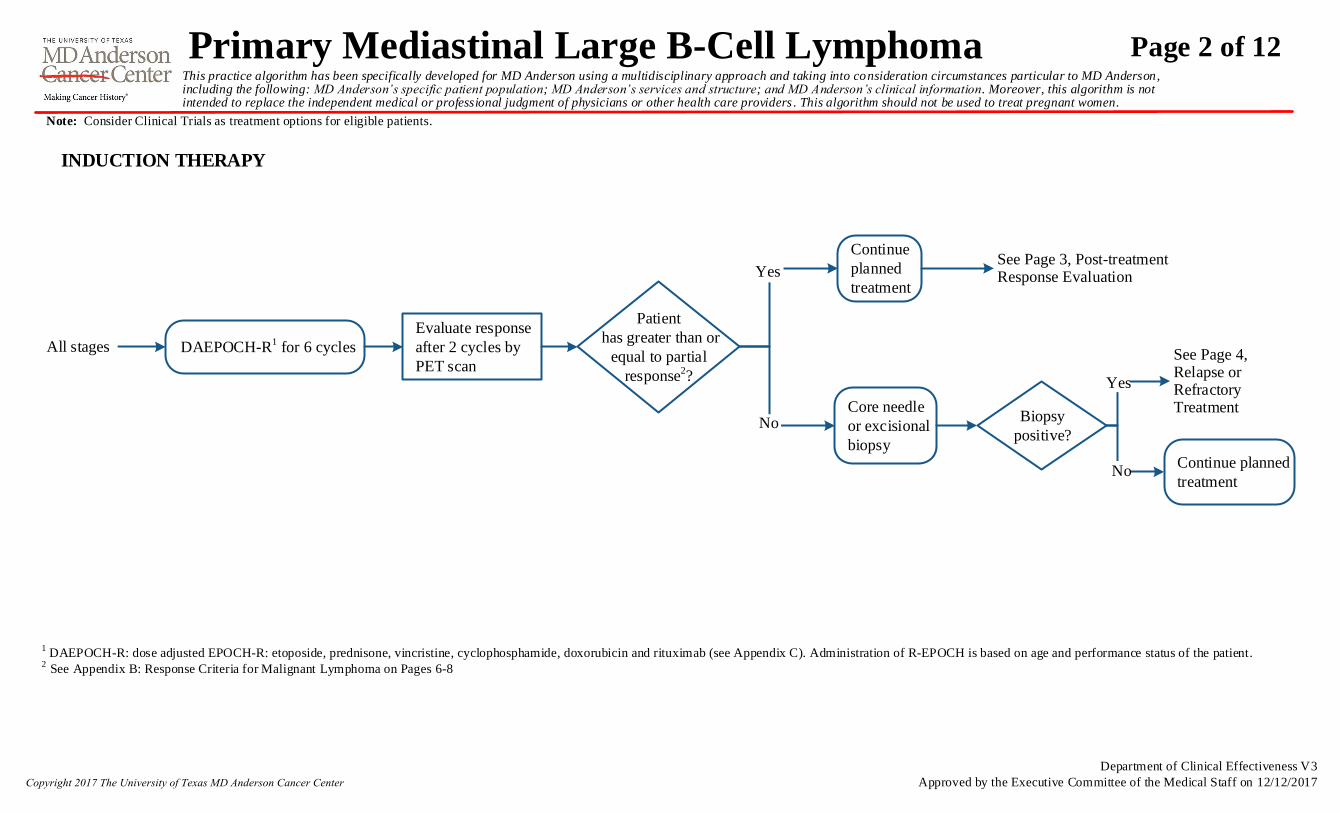
Note: Consider Clinical Trials as treatment options for eligible patients.
All stages
Evaluate response
after 2 cycles by
PET scan
Continue
planned
treatment
INDUCTION THERAPY
Patient
has greater than or
equal to partial
response2?
See Page 4, Relapse or Refractory Treatment
DAEPOCH-R1 for 6 cycles
Yes
No
See Page 3, Post-treatment Response Evaluation
1 DAEPOCH-R: dose adjusted EPOCH-R: etoposide, prednisone, vincristine, cyclophosphamide, doxorubicin and rituximab (see Appendix C). Administration of R-EPOCH is based on age and performance status of the patient.
2 See Appendix B: Response Criteria for Malignant Lymphoma on Pages 6-8
Core needle
or excisional
biopsy
Biopsy
positive?
Yes
NoContinue planned
treatment
Primary Mediastinal Large B-Cell Lymphoma Page 2 of 12This practice algorithm has been specifically developed for MD Anderson using a multidisciplinary approach and taking into consideration circumstances particular to MD Anderson, including the following: MD Anderson’s specific patient population; MD Anderson’s services and structure; and MD Anderson’s clinical information. Moreover, this algorithm is not intended to replace the independent medical or professional judgment of physicians or other health care providers . This algorithm should not be used to treat pregnant women.
Department of Clinical Effectiveness V3
Approved by the Executive Committee of the Medical Staff on 12/12/2017

Note: Consider Clinical Trials as treatment options for eligible patients.
Complete treatment
● Repeat PET scans in 6-8 weeks
until normalization or stabilization
● Consider biopsy of residual mass
only if high clinical suspicion or
close imaging follow-up not
feasible
● Consider Radiation Oncology
evaluation
Yes
No
RESULTS
POST-TREATMENT RESPONSE EVALUATION
Complete response1
Deauville 1-3
PET
normalized?
History and physical with CT and
labs as clinically indicated:
● Every 3 months for 1 year, then
● Every 4 months for 2 years, then
● Every 6 months for 2 years, then
● Annually
Complete treatment
No response orprogressive disease1
Deauville 5
Partial response1 Deauville 4 PET – equivocal3
1 See Appendix B: Response Criteria for Malignant Lymphoma on Pages 6-8
2 Bulky disease: mass 7.5 cm or greater on CT imaging
3 PET equivocal: maximum standardized uptake value (SUV) greater than mediastinal blood pool in the residual mediastinal mass
Biopsy Biopsy
positive?
Yes
No
See Page 4, Relapse
Biopsy
positive?
Yes
No
See Page 4, Relapse
Multidisciplinary conference and/or consider
Radiation Oncology evaluation if:
● Biopsy or residual mass negative though
high clinical/radiographic suspicion
● Biopsy of residual mass is not possible due
to location/patient refusal with high
clinical/radiographic suspicion
Multidisciplinary conference and/or
follow-up evaluation if previous
bulky mass2 or large residual non-
fluorodeoxyglucose (FDG) avid mass
Multidisciplinary conference and/or
consider Radiation Oncology
evaluation if previous bulky mass2 or
large residual non-FDG avid mass
Primary Mediastinal Large B-Cell Lymphoma Page 3 of 12This practice algorithm has been specifically developed for MD Anderson using a multidisciplinary approach and taking into consideration circumstances particular to MD Anderson, including the following: MD Anderson’s specific patient population; MD Anderson’s services and structure; and MD Anderson’s clinical information. Moreover, this algorithm is not intended to replace the independent medical or professional judgment of physicians or other health care providers . This algorithm should not be used to treat pregnant women.
Department of Clinical Effectiveness V3
Approved by the Executive Committee of the Medical Staff on 12/12/2017

Note: Consider Clinical Trials as treatment options for eligible patients.
CONSOLIDATION /
ADDITIONAL THERAPY
RELAPSE or
REFRACTORY #1
Relapse or refractory
ICE = ifosfamide, carboplatin, etoposide
ESHAP = etoposide, methylprednisolone, high-dose cytarabine, cisplatin
MINE = mesna, ifosfamide, mitoxantrone, etoposide
DHAP = dexamethasone, cytarabine, cisplatin
1 See Appendix B: Response Criteria for Malignant Lymphoma on Page 6
2 Clinical trials or individual regimens: patients who progress after three successive regimens are unlikely to derive additional benefit from currently utilized combination chemotherapy regimens, except for patients with disease-free interval
● Clinical trial
● New non-cross-resistant
regimen, chemo-
immunotherapy
(e.g., rituximab with
one of the following
regimens: ICE, ESHAP,
MINE, DHAP)
● Consider radiation
therapy for consolidation
with involved site approach
Patient candidate for high dose therapy
High dose therapy plus autologous
or allogeneic stem cell transplant
(in the context of a clinical trial)
ADDITIONAL
THERAPY
Individual approach including:
clinical trial2, palliative chemotherapy
or palliative radiation therapy
Patient not a candidate for high dose therapy
Patient is not a candidate for high dose therapy
Patient
has greater than or
equal to partial
response1?
Yes
No
Primary Mediastinal Large B-Cell Lymphoma Page 4 of 12This practice algorithm has been specifically developed for MD Anderson using a multidisciplinary approach and taking into consideration circumstances particular to MD Anderson, including the following: MD Anderson’s specific patient population; MD Anderson’s services and structure; and MD Anderson’s clinical information. Moreover, this algorithm is not intended to replace the independent medical or professional judgment of physicians or other health care providers . This algorithm should not be used to treat pregnant women.
Department of Clinical Effectiveness V3
Approved by the Executive Committee of the Medical Staff on 12/12/2017

APPENDIX A: International Prognostic Index (IPI) Age-Adjusted International Prognostic Index
Pre-Treatment Characteristics, ALL PATIENTS:
● Age greater than 60 years
● Serum LDH greater than one times upper limit of normal
● ECOG performance status 2-4
● Stage III or IV
● Extranodal involvement greater than 1 site
International Index, ALL PATIENTS:
Number of characteristics
● Low 0 or 1
● Low intermediate 2
● High intermediate 3
● High 4 or 5
Pre-Treatment Characteristics, ALL PATIENTS LESS
THAN OR EQUAL TO 60 YEARS:
● Serum LDH greater than one times upper limit of normal
● ECOG performance status 2-4
● Extranodal involvement greater than 1 site
International Index, ALL PATIENTS LESS THAN OR
EQUAL TO 60 YEARS:
Number of characteristics
● Low 0
● Low intermediate 1
● High intermediate 2
● High 3
Primary Mediastinal Large B-Cell Lymphoma Page 5 of 12This practice algorithm has been specifically developed for MD Anderson using a multidisciplinary approach and taking into consideration circumstances particular to MD Anderson, including the following: MD Anderson’s specific patient population; MD Anderson’s services and structure; and MD Anderson’s clinical information. Moreover, this algorithm is not intended to replace the independent medical or professional judgment of physicians or other health care providers . This algorithm should not be used to treat pregnant women.
Department of Clinical Effectiveness V3
Approved by the Executive Committee of the Medical Staff on 12/12/2017

Response and Site
Cheson, B. D., Fisher, R. I., Barrington, S. F., Cavalli, F., Schwartz, L. H., Zucca, E., & Lister, T. A. (2014). Recommendations for initial evaluation, staging, and response assessment of Hodgkin and non-Hodgkin lymphoma:
the Lugano classification. Journal of clinical oncology, 32(27), 3059-3067.
PET-CT-Based Response
Score 1, 2, or 3 with or without a residual on 5PS
It is recognized that in Waldeyer’s ring or extranodal sites with high physiologic
uptake or with activation within spleen or marrow (e.g., with chemotherapy or
myeloid colony-stimulating factors), uptake may be greater than normal
mediastinum and/or liver. In this circumstance, complete metabolic response
may inferred if uptake at sites of initial involvement is no greater than
surrounding normal tissue even if the tissue has high physiologic uptake
CT-Based Response
Target nodes/nodal masses must regress to less than or equal to 1.5 cm in LDi
No extralymphatic sites of disease
Spleen must be regressed by greater than 50% in length beyond normal
APPENDIX B: Revised Criteria for Response Assessment
No evidence of FDG-avid disease in marrow Normal by morphology
Score 4 or 5 with reduced uptake compared with baseline and residual
mass(es) of any size
At interim, these findings suggest responding disease
At end of treatment, these findings indicate residual disease
Lymph nodes and
extralymphatic sites
No Uptake
Deauville 1-3
Bone marrow
Complete Complete metabolic response Complete radiologic response (all of the following)
Nonmeasured lesion
Organ enlargement
New lesion
Not applicable
Not applicable
None
Absent
Regress to normal
None
Lymph nodes and
extralymphatic sites
Deauville 5
Partial
Nonmeasured lesion
Organ enlargement
Deauville 4
New lesion
Partial metabolic response Partial remission (all of the following)
Not applicable
Not applicable
None
Greater than or equal to 50% decrease in sum of product diameter (SPD) of
up to 6 target measurable nodes and extranodal sites
When a lesion is too small to measure on CT, assign 5 mm x 5 mm as the
default value
When no longer visible, 0 x 0 mm. For a node greater than 5 mm x 5 mm, but
smaller than normal, use actual measurement for calculation
Absent/normal, regressed, but no increase
None
Continued on next page
Primary Mediastinal Large B-Cell Lymphoma Page 6 of 12This practice algorithm has been specifically developed for MD Anderson using a multidisciplinary approach and taking into consideration circumstances particular to MD Anderson, including the following: MD Anderson’s specific patient population; MD Anderson’s services and structure; and MD Anderson’s clinical information. Moreover, this algorithm is not intended to replace the independent medical or professional judgment of physicians or other health care providers . This algorithm should not be used to treat pregnant women.
Department of Clinical Effectiveness V3
Approved by the Executive Committee of the Medical Staff on 12/12/2017
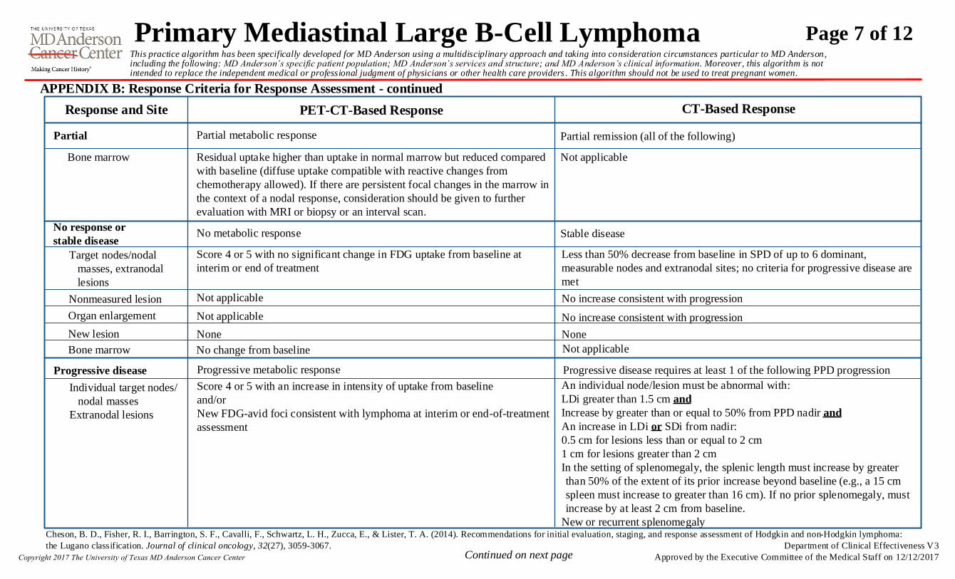
APPENDIX B: Response Criteria for Response Assessment - continued
Response and Site PET-CT-Based Response
Residual uptake higher than uptake in normal marrow but reduced compared
with baseline (diffuse uptake compatible with reactive changes from
chemotherapy allowed). If there are persistent focal changes in the marrow in
the context of a nodal response, consideration should be given to further
evaluation with MRI or biopsy or an interval scan.
CT-Based Response
Not applicable
No increase consistent with progression
Score 4 or 5 with no significant change in FDG uptake from baseline at
interim or end of treatment
Bone marrow
Partial Partial metabolic response Partial remission (all of the following)
Target nodes/nodal
masses, extranodal
lesions
No response or
stable disease
Nonmeasured lesion
Organ enlargement
New lesion
No metabolic response Stable disease
Not applicable
Not applicable
None
Less than 50% decrease from baseline in SPD of up to 6 dominant,
measurable nodes and extranodal sites; no criteria for progressive disease are
met
No increase consistent with progression
None
Bone marrow No change from baseline Not applicable
Score 4 or 5 with an increase in intensity of uptake from baseline
and/or
New FDG-avid foci consistent with lymphoma at interim or end-of-treatment
assessment
Individual target nodes/
nodal masses
Extranodal lesions
Progressive disease Progressive metabolic response Progressive disease requires at least 1 of the following PPD progression
An individual node/lesion must be abnormal with:
LDi greater than 1.5 cm and
Increase by greater than or equal to 50% from PPD nadir and
An increase in LDi or SDi from nadir:
0.5 cm for lesions less than or equal to 2 cm
1 cm for lesions greater than 2 cm
In the setting of splenomegaly, the splenic length must increase by greater
than 50% of the extent of its prior increase beyond baseline (e.g., a 15 cm
spleen must increase to greater than 16 cm). If no prior splenomegaly, must
increase by at least 2 cm from baseline.
New or recurrent splenomegaly
Continued on next page
Cheson, B. D., Fisher, R. I., Barrington, S. F., Cavalli, F., Schwartz, L. H., Zucca, E., & Lister, T. A. (2014). Recommendations for initial evaluation, staging, and response assessment of Hodgkin and non-Hodgkin lymphoma:
the Lugano classification. Journal of clinical oncology, 32(27), 3059-3067.
Primary Mediastinal Large B-Cell Lymphoma Page 7 of 12This practice algorithm has been specifically developed for MD Anderson using a multidisciplinary approach and taking into consideration circumstances particular to MD Anderson, including the following: MD Anderson’s specific patient population; MD Anderson’s services and structure; and MD Anderson’s clinical information. Moreover, this algorithm is not intended to replace the independent medical or professional judgment of physicians or other health care providers . This algorithm should not be used to treat pregnant women.
Department of Clinical Effectiveness V3
Approved by the Executive Committee of the Medical Staff on 12/12/2017
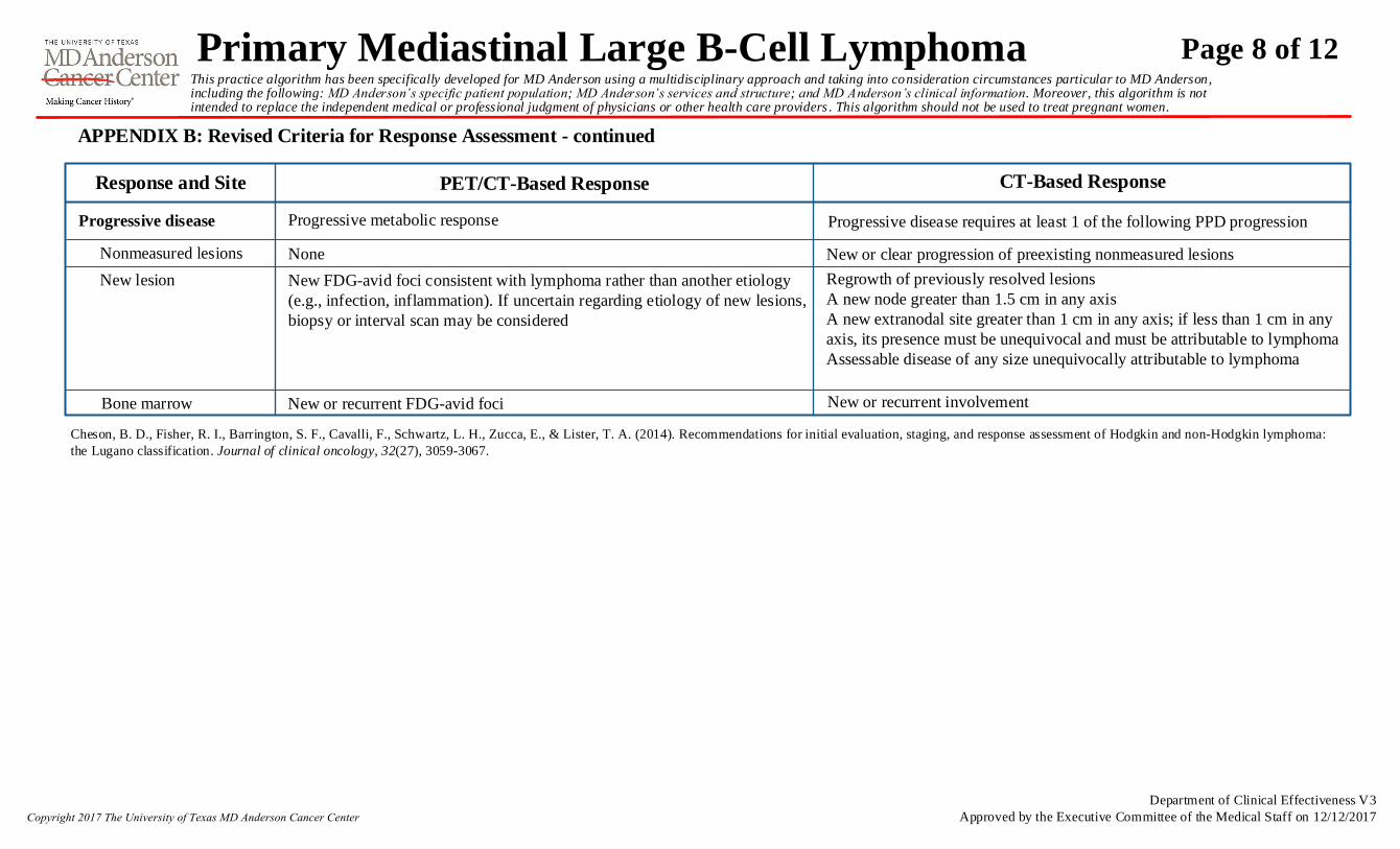
APPENDIX B: Revised Criteria for Response Assessment - continued
Response and Site PET/CT-Based Response CT-Based Response
New or clear progression of preexisting nonmeasured lesions
Progressive disease Progressive metabolic response Progressive disease requires at least 1 of the following PPD progression
Nonmeasured lesions
New lesion
None
New FDG-avid foci consistent with lymphoma rather than another etiology
(e.g., infection, inflammation). If uncertain regarding etiology of new lesions,
biopsy or interval scan may be considered
Regrowth of previously resolved lesions
A new node greater than 1.5 cm in any axis
A new extranodal site greater than 1 cm in any axis; if less than 1 cm in any
axis, its presence must be unequivocal and must be attributable to lymphoma
Assessable disease of any size unequivocally attributable to lymphoma
Bone marrow New or recurrent FDG-avid foci New or recurrent involvement
Cheson, B. D., Fisher, R. I., Barrington, S. F., Cavalli, F., Schwartz, L. H., Zucca, E., & Lister, T. A. (2014). Recommendations for initial evaluation, staging, and response assessment of Hodgkin and non-Hodgkin lymphoma:
the Lugano classification. Journal of clinical oncology, 32(27), 3059-3067.
Primary Mediastinal Large B-Cell Lymphoma Page 8 of 12This practice algorithm has been specifically developed for MD Anderson using a multidisciplinary approach and taking into consideration circumstances particular to MD Anderson, including the following: MD Anderson’s specific patient population; MD Anderson’s services and structure; and MD Anderson’s clinical information. Moreover, this algorithm is not intended to replace the independent medical or professional judgment of physicians or other health care providers . This algorithm should not be used to treat pregnant women.
Department of Clinical Effectiveness V3
Approved by the Executive Committee of the Medical Staff on 12/12/2017
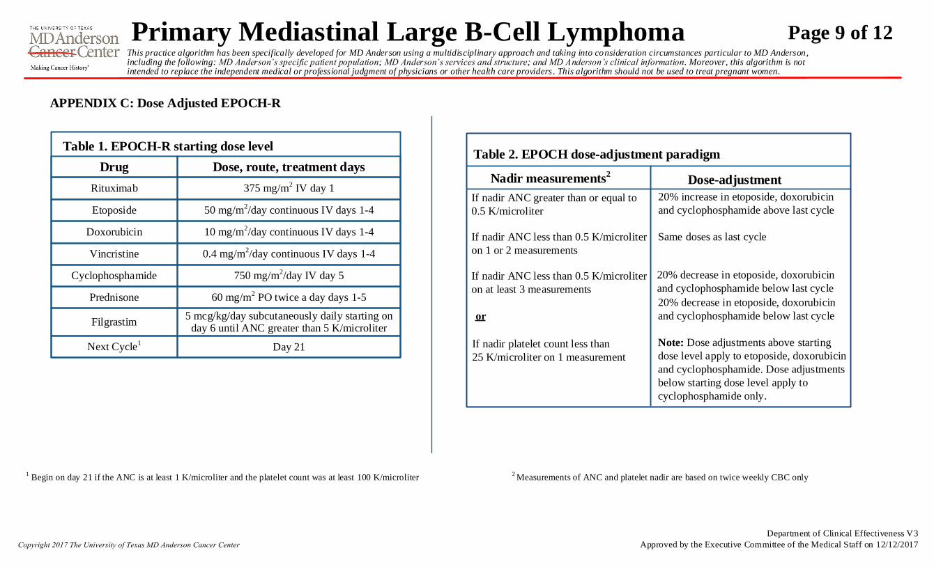
1 Begin on day 21 if the ANC is at least 1 K/microliter and the platelet count was at least 100 K/microliter
2 Measurements of ANC and platelet nadir are based on twice weekly CBC only
APPENDIX C: Dose Adjusted EPOCH-R
Table 2. EPOCH dose-adjustment paradigm
Nadir measurements2Dose-adjustment
If nadir ANC greater than or equal to
0.5 K/microliter
If nadir ANC less than 0.5 K/microliter
on 1 or 2 measurements
If nadir ANC less than 0.5 K/microliter
on at least 3 measurements
If nadir platelet count less than
25 K/microliter on 1 measurement
or
20% increase in etoposide, doxorubicin
and cyclophosphamide above last cycle
20% decrease in etoposide, doxorubicin
and cyclophosphamide below last cycle
20% decrease in etoposide, doxorubicin
and cyclophosphamide below last cycle
Note: Dose adjustments above starting
dose level apply to etoposide, doxorubicin
and cyclophosphamide. Dose adjustments
below starting dose level apply to
cyclophosphamide only.
Same doses as last cycle
Drug Dose, route, treatment days
Table 1. EPOCH-R starting dose level
Rituximab 375 mg/m2 IV day 1
Etoposide 50 mg/m2/day continuous IV days 1-4
Doxorubicin 10 mg/m2/day continuous IV days 1-4
Vincristine 0.4 mg/m2/day continuous IV days 1-4
Cyclophosphamide 750 mg/m2/day IV day 5
Prednisone 60 mg/m2 PO twice a day days 1-5
Filgrastim5 mcg/kg/day subcutaneously daily starting on
day 6 until ANC greater than 5 K/microliter
Next Cycle1Day 21
Primary Mediastinal Large B-Cell Lymphoma Page 9 of 12This practice algorithm has been specifically developed for MD Anderson using a multidisciplinary approach and taking into consideration circumstances particular to MD Anderson, including the following: MD Anderson’s specific patient population; MD Anderson’s services and structure; and MD Anderson’s clinical information. Moreover, this algorithm is not intended to replace the independent medical or professional judgment of physicians or other health care providers . This algorithm should not be used to treat pregnant women.
Department of Clinical Effectiveness V3
Approved by the Executive Committee of the Medical Staff on 12/12/2017

SUGGESTED READINGS
Barrington, S. F., & Mikhaeel, N. G. (2014). When should FDG‐PET be used in the modern management of lymphoma?. British journal of haematology, 164(3), 315-328.
Barrington, S. F., Mikhaeel, N. G., Kostakoglu, L., Meignan, M., Hutchings, M., Müeller, S. P., ... & Hoekstra, O. S. (2014). Role of imaging in the staging and response assessment of
lymphoma: consensus of the International Conference on Malignant Lymphomas Imaging Working Group. Journal of clinical oncology, 32(27), 3048-3058
Cheson, B. D., Fisher, R. I., Barrington, S. F., Cavalli, F., Schwartz, L. H., Zucca, E., & Lister, T. A. (2014). Recommendations for initial evaluation, staging, and response assessment of
Hodgkin and non-Hodgkin lymphoma: the Lugano classification. Journal of clinical oncology, 32(27), 3059-3067.
Corazzelli, G., Capobianco, G., Arcamone, M., Ballerini, P. F., Iannitto, E., Russo, F., ... & Pinto, A. (2009). Long-term results of gemcitabine plus oxaliplatin with and without
rituximab as salvage treatment for transplant-ineligible patients with refractory/relapsing B-cell lymphoma. Cancer chemotherapy and pharmacology, 64(5), 907-916.
Crump, M., Baetz, T., Couban, S., Belch, A., Marcellus, D., Howson‐Jan, K., ... & Paul, N. (2004). Gemcitabine, dexamethasone, and cisplatin in patients with recurrent or refractory
aggressive histology B‐cell non‐Hodgkin lymphoma. Cancer, 101(8), 1835-1842.
Dunleavy, K., Pittaluga, S., Maeda, L. S., Advani, R., Chen, C. C., Hessler, J., ... & Staudt, L. M. (2013). Dose-adjusted EPOCH-rituximab therapy in primary mediastinal B-cell
lymphoma. New England Journal of Medicine, 368(15), 1408-1416.
Gisselbrecht, C., Glass, B., Mounier, N., Gill, D., Linch, D., Trneny, M., ... & Schmitz, N. (2009). R-ICE versus R-DHAP in relapsed patients with CD20 diffuse large B-cell lymphoma
(DLBCL) followed by autologous stem cell transplantation: CORAL study. Journal of Clinical Oncology, 27(15S), 8509-8509.\
Gutierrez, M., Chabner, B. A., Pearson, D., Steinberg, S. M., Jaffe, E. S., Cheson, B. D., ... & Wilson, W. H. (2000). Role of a doxorubicin-containing regimen in relapsed and resistant
lymphomas: an 8-year follow-up study of EPOCH. Journal of Clinical Oncology, 18(21), 3633-3642.
Illidge, T., Specht, L., Yahalom, J., Aleman, B., Berthelsen, A. K., Constine, L., ... & Wirth, A. (2014). Modern radiation therapy for nodal non-hodgkin lymphoma—target definition
and dose guidelines from the international lymphoma radiation oncology group. International Journal of Radiation Oncology* Biology* Physics, 89(1), 49-58.
López, A., Gutiérrez, A., Palacios, A., Blancas, I., Navarrete, M., Morey, M., ... & Rodríguez, J. (2008). GEMOX‐R regimen is a highly effective salvage regimen in patients with
refractory/relapsing diffuse large‐cell lymphoma: a phase II study. European journal of haematology, 80(2), 127-132.
Martelli, M., Ceriani, L., Zucca, E., Zinzani, P. L., Ferreri, A. J., Vitolo, U., ... & Balzarotti, M. (2014). [18F] fluorodeoxyglucose positron emission tomography predicts survival after
chemoimmunotherapy for primary mediastinal large B-cell lymphoma: results of the International Extranodal Lymphoma Study Group IELSG-26 Study. Journal of Clinical
Oncology, 32(17), 1769-1775. Continued on next page
Primary Mediastinal Large B-Cell Lymphoma Page 10 of 12This practice algorithm has been specifically developed for MD Anderson using a multidisciplinary approach and taking into consideration circumstances particular to MD Anderson, including the following: MD Anderson’s specific patient population; MD Anderson’s services and structure; and MD Anderson’s clinical information. Moreover, this algorithm is not intended to replace the independent medical or professional judgment of physicians or other health care providers . This algorithm should not be used to treat pregnant women.
Department of Clinical Effectiveness V3
Approved by the Executive Committee of the Medical Staff on 12/12/2017

SUGGESTED READINGS - continued
Martín, A., Conde, E., Arnan, M., Canales, M. A., Deben, G., Sancho, J. M., ... & Nistal, S. (2008). R-ESHAP as salvage therapy for patients with relapsed or refractory diffuse large B-cell
lymphoma: the influence of prior exposure to rituximab on outcome. A GEL/TAMO study. Haematologica, 93(12), 1829-1836.
Meignan, M., Gallamini, A., Itti, E., Barrington, S., Haioun, C., & Polliack, A. (2012). Report on the third international workshop on interim positron emission tomography in lymphoma
held in Menton, France, 26–27 September 2011 and Menton 2011 consensus. Leukemia & lymphoma, 53(10), 1876-1881.
Shipp, M. A., Harrington, D. P., Anderson, J. R., Armitage, J. O., Bonadonna, G., Brittinger, G., ... & Cowan, R. A. (1993). A predictive model for aggressive non-Hodgkin's lymphoma.
New England Journal of Medicine, 329(14), 987-994.
Wilson, W. H., Grossbard, M. L., Pittaluga, S., Cole, D., Pearson, D., Drbohlav, N., ... & Raffeld, M. (2002). Dose-adjusted EPOCH chemotherapy for untreated large B-cell lymphomas: a
pharmacodynamic approach with high efficacy. Blood, 99(8), 2685-2693.
Zelenetz, A. D., Hamlin, P., Kewalramani, T., Yahalom, J., Nimer, S., & Moskowitz, C. H. (2003). Ifosfamide, carboplatin, etoposide (ICE)-based second-line chemotherapy for the
management of relapsed and refractory aggressive non-Hodgkin’s lymphoma. Annals of Oncology, 14(suppl_1), i5-i10.
Primary Mediastinal Large B-Cell Lymphoma Page 11 of 12This practice algorithm has been specifically developed for MD Anderson using a multidisciplinary approach and taking into consideration circumstances particular to MD Anderson, including the following: MD Anderson’s specific patient population; MD Anderson’s services and structure; and MD Anderson’s clinical information. Moreover, this algorithm is not intended to replace the independent medical or professional judgment of physicians or other health care providers . This algorithm should not be used to treat pregnant women.
Department of Clinical Effectiveness V3
Approved by the Executive Committee of the Medical Staff on 12/12/2017

Bouthaina S. Dabaja, MD (Radiation Oncology)
Michelle A. Fanale, MD (Lymphoma/Myeloma)
Luis E. Fayad, MD (Lymphoma/Myeloma)
Nathan Fowler, MD (Lymphoma/Myeloma)
Wendy Garcia, BS♦
Fredrick B. Hagemeister, MD (Lymphoma/Myeloma)
Firoze Jameel, MSN, RN, OCN♦
Sarah A. Milgrom, MD (Radiation Oncology)
Sattva S. Neelapu, MD (Lymphoma/Myeloma)
Yasuhiro Oki, MD (Lymphoma/Myeloma)
Robert Orlowski, MD, PhD (Lymphoma/Myeloma)
Chelsea C. Pinnix, MD, PhD (Radiation Oncology)Ŧ
Alma Rodriguez, MD (Lymphoma/Myeloma)
Felipe Samaniego, MD, MPH (Lymphoma/Myeloma)
Sheeba K. Thomas, MD (Lymphoma/Myeloma)
Michael Wang, MD (Lymphoma/Myeloma)
Donna M. Weber, MD (Lymphoma/Myeloma)
Jason R. Westin, MD (Lymphoma/Myeloma)Ŧ
This practice algorithm is based on majority expert opinion of the Lymphoma Center Faculty at the University of Texas MD Anderson Cancer Center. It was developed using a multidisciplinary
approach that included input from the following:
DEVELOPMENT CREDITS
ŦCore Development Team
♦ Clinical Effectiveness Development Team
Primary Mediastinal Large B-Cell Lymphoma Page 12 of 12This practice algorithm has been specifically developed for MD Anderson using a multidisciplinary approach and taking into consideration circumstances particular to MD Anderson, including the following: MD Anderson’s specific patient population; MD Anderson’s services and structure; and MD Anderson’s clinical information. Moreover, this algorithm is not intended to replace the independent medical or professional judgment of physicians or other health care providers . This algorithm should not be used to treat pregnant women.
Department of Clinical Effectiveness V3
Approved by the Executive Committee of the Medical Staff on 12/12/2017